PROFESSIONAL SKILLS 1 3RD YEAR SEMESTER 6 RADIOGRAPHY. THE URINARY SYSTEM Uz. Fatema shmus aldeen Tel
|
|
|
- Noreen Kennedy
- 5 years ago
- Views:
Transcription
1 PROFESSIONAL SKILLS 1 3RD YEAR SEMESTER 6 RADIOGRAPHY THE URINARY SYSTEM Uz. Fatema shmus aldeen Tel
2 Professional skills-2 THE URINARY SYSTEM
3 The urinary system (review anatomy and physiology) The urinary system consist of :- 1\ two kidneys. 2\ two ureters. 3\ urinary bladder. 4\ urethra.


4 The kidneys perform the essential function of removing waste products from the blood and regulating the water fluid levels. The diagram below shows the basic structure of the kidney.
5 ureter is a small tube, about 25 cm long carries urine from the renal pelvis to the urinary bladder where it is stored and from which is periodically voided being discharged through the urethra,which is the final passageway for the flow of urine. The anatomy of the kidney, ureter, bladder is best displayed by using diagnostic U\S and IVU.
6 Urinary Disorders Common indications Trauma Spontaneous renal hemorrhage Obstructing ureteral Hematuria congenital abnormalities such as absent kidney. Cyst & abcess loss of renal function (uremia) Neoplasm (kidney, ureter, bladder, urethra 1-Plain KUB This will reveal opaque renal calculi. It may also be used to check, abdominal preparation and to determine position. 2- I.V.U Most sensitive examination to determine physiological function 3. Computed tomography- Most sensitive single examination for calculi and renal neoplasms. 4. Ultrasound Relatively efficient for detecting renal masses or ureteral obstruction Useful when there is a need to avoid ionizingradiation, such as in examining pregnant women and children 6
7 Ultrasound is less sensitive than CT for the detection of renal masses. but it is Ultrasound-Imaging procedure of choice for renal failure
8
9 1-Procedure of I.V.U Pt preparation :- -Allergy Check Fasting for 6-8 hours before the procedure. WHY?? This ensures that your intestines are empty of faces. -laxatives taken to clear the intestines.
: A.P.")

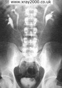
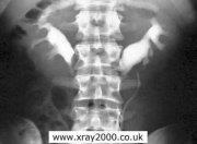
10 sequences of the I.V.U Procedure 1- plain film (KUB): A.P. supine To adjusted the patient preparation, the position,t he exposure factors and if there any radio opaque stone.
11 2-5 min film (nephrogram ) : A.P. of the renal areas shows outline of the kidneys. 5min. Nephrogram


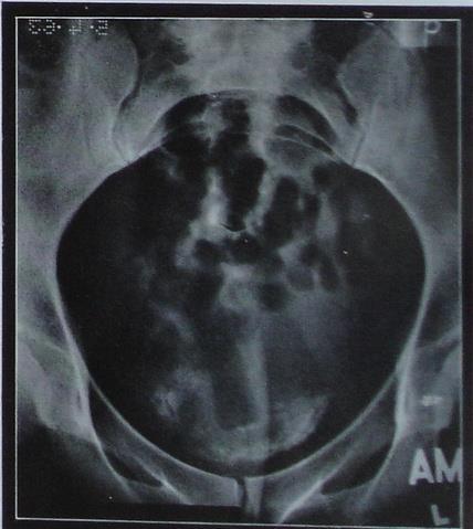
12 3-15 min film :Compression applied to distend the pelvicalyceal systems to demonstrate any filling defects 15min.
: *Structure best")


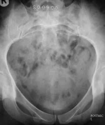
13 4-25 min (release ): *Structure best shown :All urinary system Kidneys, ureters and urinary bladder. 25min(release )

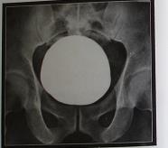
14 5- full bladder.
15 6- after maturation.
16 2- Ascending urethrogram ( retrograde urethrography) Is a radiological contrast examination of the male urethra. Indication :- 1- urethral stricture. 2- trauma. 3- perforation. 4- false passage. Contra indication :- acute urethritis. Contrast media :- media. diluted water soluble contrast


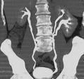
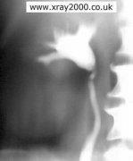
17 Full length of male urethra
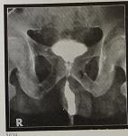
18 3-Cystogram radiological contrast examination of urinary bladder. Indication :- 1- trauma. 2-fistula. 3- diverticulum. Contra indication :- acute urethritis. Procedure :- fill the bladder by 3 ways : 1- by using catheter through urethra. 2- direct suprapupic puncture. 3- after IVU ( full bladder). Projections : AP, Lat and Oblique of the bladder.
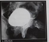
19 Cystogram
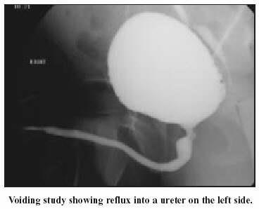
20 4-Micturation cystourethrogram Is radiological contrast examination of bladder and urethra during micturation. Indication :- 1- vasico ureteric reflux. 2- PUV( posterior urethral valve ). 3-urethral stricture. 4-control of micturition. Contra indication :- acute urethritis.
21

22 Full length voiding film showing reflux into the right kidney

23 Urethral stricture :
24 5-Selective renal angiography Is radiological contrast examination of the renal artery. Indication :- renal artery obstruction due to: 1\ mass. 2\ thrombus 3\ lymph nodes.

25 RT renal artery Abdominal aorta
26 6-CT Urography is a specialized radiological examination designed to evaluate the urinary tract (kidneys, ureters and bladder) using computed tomography (CT). evaluate urinary tract with the added advantage of visualizing all of the other intaabdominal and pelvic structures as well, These examinations are best performed using Spiral CT scanners.
27 What are the indications for this study? detect kidney stones. evaluate patients with blood in their urine (hematuria). contrast injection ever necessary to evaluate kidneys stones. CT without contrast media is increasingly used to detect smaller stones with less calcium not visualised otherwise.
 representing a papillary urothelial tumor of the")
28 CTu of the abdomen with contrast reformatted in the coronal projection shows a filling defect in the left lateral wall of the urinary bladder (red arrow) representing a papillary urothelial tumor of the bladder
29 7-MRU Magnetic resonance (MR) urography comprises an evolving group of techniques with the potential for allowing optimal noninvasive evaluation of many abnormalities of the urinary tract.
30 The most common MR urographic techniques can be divided into two categories: A-static-fluid MR urography B-excretory MR urography. Static MRU is also used for the visualization of urinary tract disorders in women during pregnancy. Excretory MR urography is performed during the excretory phase of enhancement after the intravenous administration of thus, the patient must have sufficient renal function
31
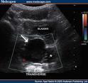
32 8- ultrasonography is an excellent imaging modality as it is noninvasive, reliable and affordable. Usage: investigate the kidney, bladder, and prostate gland. It can also be combined with voiding providing an indication of the residual volume. This gives an indirect measure of bladder function.
33 Ultrasound can detect stones above about 3-4 mm in the kidneys or pelvis, but is, for practical purposes, insensitive regarding to stones more downstream. On the other hand, ultrasound can, in most cases, rule out any significant obstruction resulting in distension of the collecting system.
34 Suspected left ureteric calculus on ultrasound.
35 9-NUCLEAR MEDICINE This involves low amounts of radiation and provides information regarding renal perfusion, function and the contribution each kidney is making to total function.
36
37 CT MRI Examples of different upnormal cases By different modalities US Nuclear Medicien

38 Ectopic kidney Doublex kidney

39 Absent kidney Lead to other kidney enlarge in size called compensatory hypertrophy

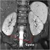
40 Horse shoe kidney

41 Uretral and bladder diverticulum

42 Multiple bladder diverticula

43 Bladder transitional cell carcinoma. Bladder image shows a filling defect

44 Ca bladder

45 Urographic image demonstrates the typical "cobra head" configuration of an orthotopic ureterocele in the bladder (arrow).

46 Hydro nephrosis

47 Renal calculi
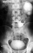
.")
48 Preliminary( plain ) radiograph shows an 8-mm calculus in the ureter (arrow). (same previous patient

49 Calculus. This case demonstrates how a calculus can be obscured by contrast material.

50 Renal cyst
 at the margins of an unenhanced")
51 Simple cyst. Nephrotomogram shows build-up of normal parenchymal tissue (cortical "beaking") at the margins of an unenhanced mass in the lower pole of the left kidney (arrows). The lesion was confirmed to be a simple cyst at US.


52 CTU image shows ileal conduit (IC) after cystectomy for bladder cancer.
 excretory MR urography.")
53 20-year-old man with ureteropelvic junction narrowing (arrow). Coronal maximum intensity projection (MIP) excretory MR urography. The renal pelvis shows typical dilatation and convex inferior border.
 excretory MR urograph shows filling defect (arrow) in left distal")
54 Figure 5. A 55-year-old man with left-sided ureteral stone. Coronal maximum intensity projection (MIP) excretory MR urograph shows filling defect (arrow) in left distal ureter that is causing mild pyeloureterectasis..
 image from excretory MR urography shows the postoperative urinary tract anatomy.")
55 56-year-old woman with ileal loop urinary diversion. Maximum intensity projection (MIP) image from excretory MR urography shows the postoperative urinary tract anatomy. Both sides are dilated because of stenosis (arrowheads) close to the ureteroenteric implantation site.

56 . Ultrasound images reveal an echogenic focus in the dilated left upper ureter. Mild dilatation of the upper ureter also noted.

57 Bladder cyst
58 Renal function test by nuclear medicine image
R adio logical investigations of urinary system
 R adio logical investigations of urinary system There are 4 main radiological Ix: 1 IVU: Intravenous urography. 2- U/S 3-CT scan 4-Radioisotope scan. Others (not frequently used): MRI, arteriography, antegrade
Excretory urography (EU) or IVP US CT & radionuclide imaging
 or IVP US CT & radionuclide imaging") Excretory urography (EU) or IVP US CT & radionuclide imaging MRI arteriography studies requiring catherization or direct puncture of collecting system EU & to a lesser extent CT provide both functional
Excretory urography (EU) or IVP US CT & radionuclide imaging MRI arteriography studies requiring catherization or direct puncture of collecting system EU & to a lesser extent CT provide both functional
Pediatric Ure-Radiology*
 Pediatric Ure-Radiology* HERMAN GROSSMAN, M.D. Professor of Radiology and Pediatrics, Duke University Medical Center, Durham, North Carolina "Routine" radiologic studies do not, often enough, concentrate
Genitourinary. Common Clinical Scenarios Protocoling Module. Patty Ojeda & Mariam Shehata
 The following training module was developed as a quality improvement project to serve as an educational tool for junior radiology residents. The following diagnostic radiology protocoling modules were
Lec-8 جراحة بولية د.نعمان
 4th stage Lec-8 جراحة بولية د.نعمان 11/10/2015 بسم هللا الرحمن الرحيم Ureteric, Vesical, & urethral stones Ureteric Calculus Epidemiology like renal stones Etiology like renal stones Risk factors like
URINARY TRACT IMAGING - BASIC PRINCIPLES
 URINARY TRACT IMAGING - BASIC PRINCIPLES Clinical Radiology Every physician needs a basic understanding of diagnostic imaging to understand how to order the appropriate studies and to understand the resulting
Contents. Review anatomy of the urinary tract Imaging modalities
 Contents Review anatomy of the urinary tract Imaging modalities The Urinary Tract Kidneys ตาแหน งไต (position) อย ใน retroperitoneum ระด บ T12-L3 โดยไต ขวาจะม ระด บตากว าไตซ ายเล กน อย ร ปร าง (shape)
Hydronephrosis. Nephrosis. Refers to the kidney
 What is hydronephrosis? Hydro Nephrosis Refers to water or fluid Refers to the kidney A build-up of fluid (urine) in the kidney is the medical term for a build-up of urine in the kidney. As the urine builds
Uroradiology For Medical Students
 Uroradiology For Medical Students Lesson 4: Cystography & Urethrography - Part 2 American Urological Association Review Cystography is useful in evaluating the bladder, the urethra and the competence of
Outline. Introduction to imaging modalities of the urinary system. Case base learning of common diseases in urinary tract
 Outline Introduction to imaging modalities of the urinary system Case base learning of common diseases in urinary tract Outline Introduction to imaging modalities of the urinary system Case base learning
Outline. Introduction to imaging modalities of the urinary system. Case base learning of common diseases in urinary tract
 Outline Introduction to imaging modalities of the urinary system Case base learning of common diseases in urinary tract Diagnostic Investigations in Urinary System PLAIN KUB EXCRETORY UROGRAPHY RETROGRADE
Case MDCT 3D reconstructed features of posterior urethral valve
 Case 12688 MDCT 3D reconstructed features of posterior urethral valve Hidayatullah Hamidi Third year Resident of Radiology French medical institute for children Radiology Department; Kabul, Afghanistan;
THE operation of reimplantation of the ureter into the bladder has undergone
 REIMPLANTATION OF THE URETER INTO THE BLADDER J. G. WARDEN, M.D., and C. C. HIGGINS, M.D. Department of Urology THE operation of reimplantation of the ureter into the bladder has undergone a stormy course
Acute renal colic Radiological investigation in patients with renal colic
 Acute renal colic Radiological investigation in patients with renal colic Mikael Hellström Professor Department of Radiology Sahlgrenska University Hospital Göteborg University 0.9-1.8/1.000 inhabitants
RENAL SCINTIGRAPHY IN THE 21 st CENTURY
 RENAL SCINTIGRAPHY IN THE 21 st CENTURY 99m Tc- MAG 3 with zero time injection of Furosemide (MAG 3 -F 0 ) : A Fast and Easy Protocol, One for All Indications Clinical Experience Congenital Disorders PROTOCOL
Proceedings of the 34th World Small Animal Veterinary Congress WSAVA 2009
 www.ivis.org Proceedings of the 34th World Small Animal Veterinary Congress WSAVA 2009 São Paulo, Brazil - 2009 Next WSAVA Congress : Reprinted in IVIS with the permission of the Congress Organizers IMAGING
IMAGING OF THE UROGENITAL TRACT
 IMAGING OF THE UROGENITAL TRACT 1 A) URINARY TRACT There are many methods of imaging the urinary tract but plain abdominal X-ray and ultrasound scan are usually done first in most cases, especially in
IVU ((INTRAVENOUSUROGRAM))
)") IVU ((INTRAVENOUSUROGRAM)) Anatomy The urinary system consists of the following : 2 kidneys, 2 ureters,1 bladder, 1 urethra Renal pelvis Minor calyx Major calyx Proximal ureter Pelvi-uretric junction
IVU ((INTRAVENOUSUROGRAM)) Anatomy The urinary system consists of the following : 2 kidneys, 2 ureters,1 bladder, 1 urethra Renal pelvis Minor calyx Major calyx Proximal ureter Pelvi-uretric junction
Find Medical Solutions to Your Problems HYDRONEPHROSIS. (Distension of Renal Calyces & Pelvis)
") HYDRONEPHROSIS (Distension of Renal Calyces & Pelvis) Hydronephrosis is the distension of the renal calyces and pelvis due to accumulation of the urine as a result of the obstruction to the outflow of
HYDRONEPHROSIS (Distension of Renal Calyces & Pelvis) Hydronephrosis is the distension of the renal calyces and pelvis due to accumulation of the urine as a result of the obstruction to the outflow of
IVU ((INTRAVENOUSUROGRAM))
)") IVU ((INTRAVENOUSUROGRAM)) Anatomy The urinary system consists of : 2 kidneys, 2 ureters,1 bladder, 1 urethra Renal pelvis Minor calyx Major calyx Renal parenchyma Proximal ureter Pelvi-uretric junction
IVU ((INTRAVENOUSUROGRAM)) Anatomy The urinary system consists of : 2 kidneys, 2 ureters,1 bladder, 1 urethra Renal pelvis Minor calyx Major calyx Renal parenchyma Proximal ureter Pelvi-uretric junction
ASSESSING THE PLAIN ABDOMINAL RADIOGRAPH M A A M E F O S U A A M P O F O
 ASSESSING THE PLAIN ABDOMINAL RADIOGRAPH M A A M E F O S U A A M P O F O Introduction The abdomen (less formally called the belly, stomach, is that part of the body between the thorax (chest) and pelvis,
Radiological Assessment of the Kidney in Patients with Hematuria
 March 2005 Radiological Assessment of the Kidney in Patients with Hematuria Jeremy L. McKay, Harvard Medical School Year III Hematuria Signs and Symptoms Microscopic or gross hematuria Abdominal pain Fever
Obstetrics Content Outline Obstetrics - Fetal Abnormalities
 Obstetrics Content Outline Obstetrics - Fetal Abnormalities Effective February 2007 10 16% renal agenesis complete absence of the kidneys occurs when ureteric buds fail to develop Or degenerate before
Spectrum of Micturating Cystourethrogram Revisited: A Pictorial Assay
 603 International Journal of Collaborative Research on Internal Medicine & Public Health Spectrum of Micturating Cystourethrogram Revisited: A Pictorial Assay Abhinav Jain 1, Vivek Setia 1, Shweta Agnihotri
URINARY SYSTEM I. Kidneys II. Nephron Unit and Urine Formation
 URINARY SYSTEM I. Kidneys A. Location and Structure 1. Retroperitoneal 2. Between T12 and L3 3. Rt. kidney slightly lower 4. Two bean shaped organs 5. Adrenal gland 6. Internal construction a. Renal cortex
4 th Year Urology Core Objectives Keith Rourke (Revised June 1, 2007)
") 4 th Year Urology Core Objectives Keith Rourke (Revised June 1, 2007) I. Genitourinary Trauma: 1. Goal: The student will be able to demonstrate a basic clinical approach to the management & diagnosis of
4 th Year Urology Core Objectives Keith Rourke (Revised June 1, 2007) I. Genitourinary Trauma: 1. Goal: The student will be able to demonstrate a basic clinical approach to the management & diagnosis of
Fetal Renal Malformations: The Role of Ultrasound in Diagnosis & Management
 Fetal Renal Malformations: The Role of Ultrasound in Diagnosis & Management 12 weeks Alfred Abuhamad, M.D. Eastern Virginia Medical School 13 weeks 2nd trimester Medullary pyramids Renal Sinus Cortex 2nd
URINARY SYSTEM. MEDICAL TERMINOLOGY Chapter Six HIT #141. Anatomy
 URINARY SYSTEM MEDICAL TERMINOLOGY Chapter Six HIT #141 Anatomy Kidneys = bean-shaped organs, located on each side of the spinal column, removal of waste from the blood. Nephron = microscopic located in
Hydronephrosis. What is hydronephrosis?
 What is hydronephrosis? Hydronephrosis Hydronephrosis describes the situation where the urine collecting system of the kidney is dilated. This may be a normal variant or it may be due to an underlying
Radiographic Procedures III (RAD 228)
") Radiographic Procedures III (RAD 228) Urinary System RADIOGRAPHIC EXAMINATIONS Urinary System Antegrade Exam IVU Functional test Hypertensive evaluation as per protocol Retrograde Exams Retrograde Urography
Radiographic Procedures III (RAD 228) Urinary System RADIOGRAPHIC EXAMINATIONS Urinary System Antegrade Exam IVU Functional test Hypertensive evaluation as per protocol Retrograde Exams Retrograde Urography
Obstructive Uropathy. PATHOPHYSIOLOGIC CHANGES UUO vs BUO. Arry Rodjani Urology Department Ciptomangunkusumo Hospital Jakarta
 Obstructive Uropathy PATHOPHYSIOLOGIC CHANGES UUO vs BUO Arry Rodjani Urology Department Ciptomangunkusumo Hospital Jakarta INTRODUCTION Obstructive uropathy refers to the functional or anatomic obstruction
Imaging the Urogenital System
 maging the Urogenital System Tony Pease, DVM, MS, DACVR Assistant Professor of Radiology North Carolina State University Reading Thrall Chapters 42-46 Prostate Gland Not visible radiographically in normal
Uroradiology Tutorial For Medical Students
 Uroradiology Tutorial For Medical Students Lesson 3: Cystography & Urethrography Part 1 American Urological Association Introduction Conventional radiography of the urinary tract includes several diagnostic
Five Views of Transitional Cell Carcinoma: One Man s Journey
 September 2006 Five Views of Transitional Cell Carcinoma: One Man s Journey Amsalu Dabela, Harvard Medical School III Outline Overview: Renal Anatomy Our Patient s Story Diagnostic Imaging Studies Appearance
Bladder Case # 1. Principal Diagnosis: Bladder Tumor, Suspect Transitional Cell Carcinoma. Secondary Diagnoses: 1. Hypertension. 2. Hyperlipidemia.
 DISCHARGE SUMMARY Bladder Case # 1 Date: 04/22/2010 Principal Diagnosis: Bladder Tumor, Suspect Transitional Cell Carcinoma. Secondary Diagnoses: 1. Hypertension. 2. Hyperlipidemia. Hospital Course: Mr.
Request Card Task ANSWERS
 Request Card Task ANSWERS Medical Student Workbook Author: Dr Sam Leach, SpR Case 1 What differential diagnoses are most likely? Which investigation is most appropriate? Case 1 The most likely diagnosis
Imaging the Urinary Tract
 Imaging the Urinary Tract Laura Armbrust, DVM, DACVR Gregory F. Grauer, DVM, MS, DACVIM Kansas State University Radiographic and ultrasound imaging in addition to history, physical examination, and clinicopathologic
CONGENITAL ANTERIOR URETHRAL DIVERTICULUM
 CONGENITAL ANTERIOR URETHRAL DIVERTICULUM W Y Cheong, H K Cheng, K P Tan SYNOPSIS We report the first documented case in Singapore of a congenital saccular anterior urethral diverticulum causing bladder
Renal Trauma: Management Options
 Renal Trauma: Management Options Immediate surgical repair Nephrectomy Conservative management Alonso RC et al. Kidney in Danger: CT Findings of Blunt and Penetrating Renal Trauma. RadioGraphics 2009;
Glossary of Terms Primary Urethral Cancer
 Patient Information English Glossary of Terms Primary Urethral Cancer Advanced cancer A tumour that grows into deeper layers of tissue, adjacent organs, or surrounding muscles. Anaesthesia (general, spinal,
URINARY DIVERSIONS. Susan Hilton, MD and Nicholas Papanicolaou, MD Co-Chiefs, CT Section Hospital of the University of Pennsylvania
 URINARY DIVERSIONS Susan Hilton, MD and Nicholas Papanicolaou, MD Co-Chiefs, CT Section Hospital of the University of Pennsylvania Neither of us has any financial relationships with commercial interests
Audit of split-bolus CT urography for the investigation of haematuria over a 12 month period at two district general hospitals
 Audit of split-bolus CT urography for the investigation of haematuria over a 12 month period at two district general hospitals Poster No.: C-1349 Congress: ECR 2010 Type: Educational Exhibit Topic: Genitourinary
How I Do It - Evaluation of the Urethra
 How I Do It - Evaluation of the Urethra Parvati Ramchandani, MD Professor, Radiology and Surgery University of Pennsylvania Medical Center Philadelphia, PA, USA Disclosure of Commercial Interest Neither
3/16/2015 VCUG. T2-weighted MRI of lower pelvis
 1 Reference: Grayson DE, Abbot RM, Levy AD, Sherman PM (2002) Emphysematous infections of the abdomen and pelvis: a pictorial review. RadioGraphics 22: 543-561. 2 VCUG T2-weighted MRI of lower pelvis Reference:
Acute flank pain in children: Imaging considerations
 Acute flank pain in children: Imaging considerations Carlos J. Sivit MD Rainbow Babies and Children s Hospital Case Western Reserve School of Medicine Flank pain Results from distention of ureter or renal
Information for Patients
 Information for Patients Congenital Malformation in the Urinary Tract: Ureteral Duplication, Ureterocele, and Ectopic Ureter English Table of contents Ureteral Duplication... 3 Symptoms and Diagnosis...
Obstructive Nephropathy
 Obstructive Nephropathy Liza A. Lucero RN, FNP-C, MSN Renal Medicine Associates Conflicts No conflict of interests Obstructive Nephropathy Objectives Definition of Obstructive Nephropathy Causes Clinical
Urinary system Ultrasound (Renal & Urinary bladder)
") Urinary system Ultrasound (Renal & Urinary bladder) Edited & Presented by ; Hussien A.B ALI DINAR. Msc.Phd ISRRT Associate Member Lecturer (National university) Reporting Sonographer (PHC) Objective By
Urinary system Ultrasound (Renal & Urinary bladder) Edited & Presented by ; Hussien A.B ALI DINAR. Msc.Phd ISRRT Associate Member Lecturer (National university) Reporting Sonographer (PHC) Objective By
Radical cystectomy and urinary diversion: Normal anatomy and complications
 Radical cystectomy and urinary diversion: Normal anatomy and complications Poster No.: C-0648 Congress: ECR 2014 Type: Scientific Exhibit Authors: J. M. Marin, N. alegre, P. Perez Martin, A. Velarde Pedraza
Role of imaging in evaluation of genitourinary i trauma Spectrum of GU injuries Relevance of imaging findings in determining management Focus on MDCT
 Genitourinary Tract Injuries 6 th Nordic Course Scott D. Steenburg, MD Assistant Professor University of Maryland Department of Radiology Division of Trauma and Emergency Radiology R Adams Cowley Shock
Renal Artery Stenosis With Severe Hypertension: A Case Report
 CASE REPORT Renal Artery Stenosis With Severe Hypertension: A Case Report Suwaid MA ABSTRACT Background: Renal artery stenosis (RAS) is found in 77% of hypertensive patients and is responsible for 1-2%
THE UROLOGY GROUP
 THE UROLOGY GROUP www.urologygroupvirginia.com 1860 Town Center Drive Suite 150/160 Reston, VA 20190 703-480-0220 19415 Deerfield Avenue Suite 112 Leesburg, VA 20176 703-724-1195 224-D Cornwall Street,
Nonurographic evaluation of renal calculous disease 1
 Contributions Nonurographic evaluation of renal calculous disease 1 Gregory P. Borkowski, M.D. Craig R. George, M.D. Peter B. O'Donovan, M.D. While excretory urography has been useful in the evaluation
Index. Note: Page numbers of article titles are in boldface type.
 Magn Reson Imaging Clin N Am 12 (2004) 587 591 Index Note: Page numbers of article titles are in boldface type. A Adenoma(s), adrenal, gadolinium-enhanced MR imaging in, 533 534 hyperfunctioning versus
CLINICAL PRESENTATION AND RADIOLOGY QUIZ QUESTION
 Donald L. Renfrew, MD Radiology Associates of the Fox Valley, 333 N. Commercial Street, Suite 100, Neenah, WI 54956 1/22/2011 Radiology Quiz of the Week # 4 Page 1 CLINICAL PRESENTATION AND RADIOLOGY QUIZ
RADIOLOGY (SURGERY) BY MARYAM MALIK Rawalpindi Medical College
 BY MARYAM MALIK Rawalpindi Medical College") RADIOLOGY (SURGERY) BY MARYAM MALIK Rawalpindi Medical College NORMAL BOWEL GAS PATTERN Any part of the bowel may be visible if it contains gas/air within the lumen. Gas/air is of low density and forms
RADIOLOGY (SURGERY) BY MARYAM MALIK Rawalpindi Medical College NORMAL BOWEL GAS PATTERN Any part of the bowel may be visible if it contains gas/air within the lumen. Gas/air is of low density and forms
Interventional management of postoperative ureteric complications after pelvic surgery
 Interventional management of postoperative ureteric complications after pelvic surgery Poster No.: C-0169 Congress: ECR 2015 Type: Scientific Exhibit Authors: R. Tabashy, A. Hamed, S. El-Sebai; Cairo/EG
 INTRAUTERINE DEVICE = IUD INTRAUTERINE DEVICE = IUD CONGENITAL DISORDERS Pyometra = pyometrea is a uterine infection, it is accumulation of purulent material in the uterine cavity. Ultrasound is usually
Prenatal Hydronephrosis
 Prenatal Hydronephrosis What is hydronephrosis? Hydronephrosis is dilation of the kidney, specifically the renal pelvis (place where urine is stored after its production). This can be the result of an
IMAGING OF UPPER UT TCC
 IMAGING OF UPPER UT TCC IS THERE AN EVIDENCE BASED STRATEGY? S A MOUSSA FRCS Ed, FRCR WESTERN GENERAL HOSPITAL EDINBURGH UPPER TRACT TCC 0.7-4% of patients with primary bladder cancer develops UT-TCC.
Chapter 6: Genitourinary and Gastrointestinal Systems 93
 Chapter 6: Genitourinary and Gastrointestinal Systems 93 Chapter 6 Genitourinary and Gastrointestinal Systems Embryology Three sets of excretory organs or kidneys develop in human embryos: Pronephros:
Perineal Sonography in Diagnosis of an Ectopic Ureteric Opening Into the Urethra
 Case Series Perineal Sonography in Diagnosis of an Ectopic Ureteric Opening Into the Urethra S. Boopathy Vijayaraghavan, MD, DMRD Objective. To study the role of perineal sonography in the diagnosis of
BLADDER CANCER: PATIENT INFORMATION
 BLADDER CANCER: PATIENT INFORMATION The bladder is the balloon like organ located in the pelvis that stores and empties urine. Urine is produced by the kidneys, is conducted to the bladder by the ureters,
Excretory Cystograms After Voiding
 July, 1947 17 Excretory Cystograms After Voiding JAMES R. DILLON,* M.D., Sani Franicisco Y ANY pathological conditions of the upper urinary tract and bladder can be diagnosed and differentiated by excretory
Urinary tract obstruction
 Urinary tract obstruction Common causes : stone, blood clot Radiographic findings depend on I. Level of obstruction II. Severity of obstruction : partial or complete III. Timing of obstruction Pathophysiology
Lecture 56 Kidney and Urinary System
 Lecture 56 Kidney and Urinary System The adrenal glands are located on the superomedial aspect of the kidney The right diagram shows a picture of the kidney with the abdominal walls and organs removed
Cowper's syringocele: pathological manifestations and radiological aspects.
 Cowper's syringocele: pathological manifestations and radiological aspects. Poster No.: C-1468 Congress: ECR 2011 Type: Authors: Keywords: DOI: Educational Exhibit E. Guidi, B. Ginanni, N. Armillotta,
MICTURATING CYSTOURETHROGRAPHY- A PICTORIAL ESSAY
 PICTORIAL REVIEW MICTURATING CYSTOURETHROGRAPHY- A PICTORIAL ESSAY Palle Lalitha, 1 M. Ch. Balaji Reddy, 1 K. Jagannath Reddy, 1 Vijaya Kumari 2 1 2 Department of Radiology, Focus Diagnostic Center, Punjagutta,
PICTORIAL ESSAY. Experiences of using a single post-contrast CT scan of the urinary tract after triphasic contrast injection
 Experiences of using a single post-contrast CT scan of the urinary tract after triphasic contrast injection P C Pretorius, FCRad (Diag) SA Drs Visser, Erasmus, Vawda & Partners, Port Elizabeth Corresponding
L. Alexandre Frigini MD; Aaron Thomas, MD; Veronica Lenge de Rosen, MD
 Computed Tomography Urography (CTU) for Evaluation of Asymptomatic microscopic hematuria. Is intravenous contrast administration warranted for all patients? A retrospective evaluation utilizing ACR s Appropriateness
Lab Monitor Images Dissection of the Abdominal Vasculature + Lower Digestive System
 Lab Monitor Images Dissection of the Abdominal Vasculature + Lower Digestive System Stomach & Duodenum Frontal (AP) View Nasogastric tube 2 1 3 4 Stomach Pylorus Duodenum 1 Duodenum 2 Duodenum 3 Duodenum
Indications and effectiveness of the open surgery in vesicoureteral reflux
 Indications and effectiveness of the open surgery in vesicoureteral reflux Suzi DEMIRBAG, MD Department of Pediatric Surgery, Gulhane Military Medical Academy, Ankara, TURKEY Vesicoureteral reflux (VUR)
Abdomen and Pelvis CT (1) By the end of the lecture students should be able to:
 By the end of the lecture students should be able to:") RAD 451 Abdomen and Pelvis CT (1) By the end of the lecture students should be able to: State the common indications for Abdomen and pelvis CT exams Identify possible contra indications for Abdomen and
RAD 451 Abdomen and Pelvis CT (1) By the end of the lecture students should be able to: State the common indications for Abdomen and pelvis CT exams Identify possible contra indications for Abdomen and
8/14/2017. Kidney location & visualization. Brief Review with tips & Case Based Illustrations. Size = x L2. Size =
 Dr. Russell Tucker, DACVR Brief Review with tips & Case Based Illustrations Kidney location & visualization K9 Kidneys: Rt @ T13-L1 Lt @ L2-L4 Kidney visualization K9 Kidneys: Rt @ T13-L1 Lt @ L2-L4 Size
Rad Lab 4 Unknowns: Genitourinary!
 Rad Lab 4 Unknowns: Genitourinary! Peter Clarke MD! Don Di Salvo, MD! Clerkship Directors for Radiology! Harvard Medical School! Brigham and Women s Hospital! Dana Farber Cancer Institute! Case 1: 69 year
Continuing Professional Development
 Volume 15.4.1 This Lecture Serties qualifies for 0.5 Informal CPD Learning Hours Continuing Professional Development Pelvic Calcifications By Dr Kristin Grace (DACBR) WHAT IS THAT??? Calcifications in
AUA Guidelines for Imaging Known or Suspected Ureteral Calculi. Michael Ferrandino, MD Assoc Professor of Urology Duke University Medical Center
 AUA Guidelines for Imaging Known or Suspected Ureteral Calculi Michael Ferrandino, MD Assoc Professor of Urology Duke University Medical Center Imaging for Urolithiasis Justification for the Guidelines
My Patient Has Abdominal Pain PoCUS of the Biliary Tract and the Urinary Tract
 My Patient Has Abdominal Pain PoCUS of the Biliary Tract and the Urinary Tract Objectives PoCUS for Biliary Disease PoCUS for Renal Colic PoCUS for Urinary Retention Biliary Disease A patient presents
INTERNATIONAL SPINAL CORD INJURY DATA SETS URINARY TRACT IMAGING BASIC DATA SET - COMMENTS
 1 INTERNATIONAL SPINAL CORD INJURY DATA SETS URINARY TRACT IMAGING BASIC DATA SET - COMMENTS The working-group consists of: Fin Biering-Sørensen representing the Executive Committee of The International
Rare case of urinary bladder agenesis - Multislice CT abdomen imaging
 Rare case of urinary bladder agenesis - Multislice CT abdomen imaging Venkatraman Indiran 1*, Kabilan Chokkappan 2, Emmanuel Gunaseelan 2 1. Department of Radiodiagnosis, Sree Balaji Medical College and
Urinary 1 Checklist Gross Anatomy of the Urinary System
 Urinary 1 Checklist Gross Anatomy of the Urinary System Urinary system Kidneys Parietal peritoneum Retroperitoneal Renal fascia The urinary system consists of two kidneys, two ureters, the urinary bladder,
Separating and Distorted Nephroliths Signs of Renal Squamous Cell Carcinoma
 Chin J Radiol 2003; 28: 203-208 203 Separating and Distorted Nephroliths Signs of Renal Squamous Cell Carcinoma TZE-YU LEE SHEUNG-FAT KO CHUNG-CHENG HUANG YU-FENG CHENG Department of Radiology, Chang Gung
The number following the procedure code is the TRICARE payment group. KIDNEY
 TRICARE/CHAMPUS POLICY MANUAL 6010.47-M JUNE 25, 1999 S POLICY CHAPTER 13 SECTION 9.1 ADDENDUM 1, SECTION 8 TRICARE-APPROVED AMBULATORY SURGERY S - URINARY SYSTEM The number following the procedure code
Urologic investigations
 Urologic investigations د. Laboratory studies EXAMINATION OF URINE Urinalysis: Urinalysis is one of the most important and useful urologic tests available. Reasons for inadequate urinalyses include: (1)
CLINICAL PRESENTATION AND RADIOLOGY QUIZ QUESTION
 Donald L. Renfrew, MD Radiology Associates of the Fox Valley, 333 N. Commercial Street, Suite 100, Neenah, WI 54956 09/17/2011 Radiology Quiz of the Week # 38 Page 1 CLINICAL PRESENTATION AND RADIOLOGY
Coding Companion for Urology/Nephrology. A comprehensive illustrated guide to coding and reimbursement
 Coding Companion for Urology/Nephrology A comprehensive illustrated guide to coding and reimbursement 2014 Contents Getting Started with Coding Companion...i Integumentary...1 Arteries and Veins...15 Lymph
RADIOLOGY OF THE URINARY TRACT CHAPTER 9 239
 RADIOLOGY OF THE URINARY TRACT CHAPTER 9 239 in length. They lie cephalad to the kidneys, with the right just posterior to the inferior vena cava (IVC) and the left anteromedial to the upper pole of the
Developmental Abnormalities of the Kidneys and GU System
 A5 Developmental Abnormalities of the Kidneys and GU System Erin Parilla, MD Neonatologist Pediatrix Medical Group, Tampa, FL The speaker has signed a disclosure form and indicated she has no significant
Imaging Ejaculatory Disorders and Hematospermia
 ATHENS 4-6 October 2018 European Society of Urogenital Radiology Imaging Ejaculatory Disorders and Hematospermia Parvati Ramchandani, MD Professor, Radiology and Surgery University of Pennsylvania Medical
Evaluation of Imaging Abnormalities of Ureter using MDCT Urography
 DOI: 10.7860/IJARS/2017/23458:2250 Radiology Section Original Article Evaluation of Imaging Abnormalities of Ureter using MDCT Urography Vedaraju Kadaba Shamachar, Vijay Kumar Kenchanahalli Rangaswamy,
Bladder Trauma Data Collection Sheet
 Bladder Trauma Data Collection Sheet If there was no traumatic injury with PENETRATION of the bladder DO NOT proceed Date of injury: / / Time of injury: Date of hospital arrival: / / Time of hospital arrival:
Ureters, Urinary Bladder & Urethra
 Ureters, Urinary Bladder & Urethra Please check our Editing File هذا العمل ال يغني عن المصدر األساسي للمذاكرة Lecture 2 } و م ن ي ت و ك ع ل ا لل ه ف ه و ح س ب ه { Objectives o Describe the course of ureter
Genitourinary Trauma Introduction GU Trauma overlooked
 Genitourinary Trauma Introduction GU Trauma overlooked 10-20% of all injured patients Long term morbidity Impotence Incontinence Life-threatening injuries first Urethral Injury Plan Bladder Injury Kidney
Nongynecological causes of acute and chronicpelvic pain. Amela Sofić UKC Sarajevo Bosnia and Herzegovina
 Nongynecological causes of acute and chronicpelvic pain Amela Sofić UKC Sarajevo Bosnia and Herzegovina One of the most challenging problems in a clinical routine is the pelvic pain It is useful to classify
HMM 4401 Genito-urinary tract diseases
 HMM 4401 Genito-urinary tract diseases Urine production Core elements: Glomerulus, proximal and distal convoluted tube, loop of Henle, collecting tubules, ureters, bladder, sphincter, uretra, and out
UBC Department of Urologic Sciences Lecture Series. Urological Trauma
 UBC Department of Urologic Sciences Lecture Series Urological Trauma Disclaimer: This is a lot of information to cover and we are unlikely to cover it all today These slides are to be utilized for your
MUSCLE-INVASIVE AND METASTATIC BLADDER CANCER
 MUSCLE-INVASIVE AND METASTATIC BLADDER CANCER (Text update March 2008) A. Stenzl (chairman), N.C. Cowan, M. De Santis, G. Jakse, M. Kuczyk, A.S. Merseburger, M.J. Ribal, A. Sherif, J.A. Witjes Introduction
1. Congenital Anomalies of Kidney and Ureter 1
 CONTENTS 1. Congenital Anomalies of Kidney and Ureter 1 1.1 Antenatal Pelviureteric Junction Obstruction 1 1.2 Bilateral Pelviureteric Junction Obstruction 3 1.3 Circumcaval Ureter 6 1.4 Crossed Renal