Introduction and Definitions
|
|
|
- Nora Kelly
- 6 years ago
- Views:
Transcription
1 Bowel obstruction
2 Introduction and Definitions Accounts for 5% of all acute surgical admissions Patients are often extremely ill requiring prompt assessment, resuscitation and intensive monitoring Obstruction A mechanical blockage arising from a structural abnormality that presents a physical barrier to the progression of gut contents. Ileus is a paralytic or functional variety of obstruction Obstruction is: Partial or complete Simple or strangulated
3 investigations
4
 that are not seen on plain abdominal film because they are filled")
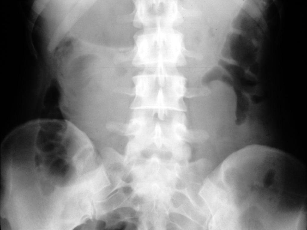
5 LEFT: Plain abdominal film in a patient with an acute abdomen, showing no abnormalities. RIGHT: Subsequent CT shows distended small bowel loops (arrowheads) that are not seen on plain abdominal film because they are filled with fluid only and do not contain intraluminal air.
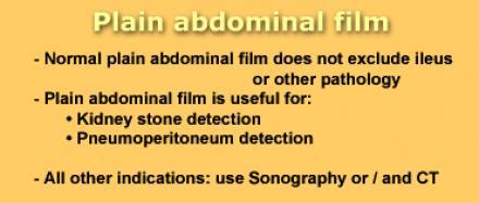
6 Chest X-ray This is an essential examination in any patient with acute abdomen because: 1-It is the best radiograph to show the presence of a small pneumoperitoneum. 2-A number of chest conditions may present as an acute abdominal pain : pneumonia (particularly lower lobe), MI,. 3- Acute abdominal conditions may be complicated by chest pathology: pleural effusion frequently complicate acute pancreatitis. 4-Even when the chest radiograph is normal it acts as a valuable baseline.

7 Small amount
8


9 Causes of bowel obstruction Luminal F. Body Bezoars Gall stone Food Particles A. lumbricoides Mural Neoplasims lipoma polyps leiyomayoma hematoma lymphoma carcimoid carinoma secondary Tumors Crohns TB Stricture Intussusception Congenital Extraluminal Postoperative adhesions Congenital adhesions Hernia Volvulus

10 Radiological Evaluation Normal Scout Always request: Supine, Erect and CXR Gas pattern: Fluid Levels: Gastric, Colonic and 1-2 small bowel Gastric 1-2 small bowel Check gasses in 4 areas: 1. Caecal 2. Hepatobiliary 3. Free gas under diaphragm 4. Rectum Look for calcification Look for soft tissue masses, psoas shadow Look for fecal pattern
11 The distinction between small & large-bowel dilatation Small bowel large bowel 1. vulvulae conniventes present in jejunum absent 2. number of loops many few 3. distribution of loops central peripheral 4. haustra absent present 5. diameter 3-5 cm 5 cm + 6. radius of curvature small large 7. solid feces absent *present haustra may be completely absent from the descending & sigmoid colon.

 Vulvulae coniventae Ileum: may")
12 The Difference between small and large bowel obstruction Large bowel Peripheral ( diameter 8 cm max) Presence of haustration Small Bowel Central ( diameter 5 cm max) Vulvulae coniventae Ileum: may appear tubeless


13 SMALL BOWEL OBSTRUCTION Note dilated small bowel centrally placed with air/fluid levels on upright exam. Ng tube ERECT

14 if fluid filled loops The dilated small bowel loops appears as a sausage, oval or round soft tissue densities that change in position in different views, sometime with small gas bubbles trapped in rows between the vulvulae conniventes on horizontal ray films; this is known as 'string of beads' sign which is virtually diagnostic of small bowel obstruction and does not occur in normal people.

15 CT- SMALL BOWEL OBSTRUCTION DISTAL NORMAL BOWEL PROXIMAL DILATED BOWEL Proximal loops are dilated and distil loops are collapsed indicating an obstruction. Obstruction most likely due to adhesions in a patient with history of abdominal surgery


16 SM. BOWEL BARIUM STUDY HERNIA CT Note hernia in right lower quadrant on both exams accounting for obstruction. Hernia is likely cause if there is no history of prior surgery.

17 Incarcerated Inguinal Hernia

18 COLON POST OP ADYNAMIC ILEUS LARGE AND SMALL BOWEL SM. BOWEL Symmetric dilation of large and small bowel is seen normally as a post operative ileus.


19 POST OP ADYNAMIC ILEUS Colon resection
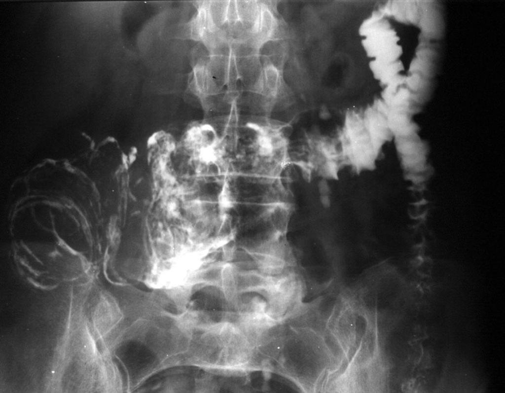
20 Gall stone ileus This is a mechanical obstruction caused by the impaction of one or more gall stones in the intestine, usually in the terminal ileum, but rarely in the duodenum or the colon. The commonest radiological signs to be observed are : 1- A gas shadow within the bile ducts and/ or the gall bladder. 2- Complete or incomplete intestinal obstruction. 3- An abnormal location of an already observed gall stone.

21 Gall stone ileus

22 See also gall-bladder outlined by gas, recent passage of stone.
23 intussusception
24 intussusception
25 Ileo- caecal intussusception

26 COLON OBSTRUCTION Distension extends to distil descending colon.

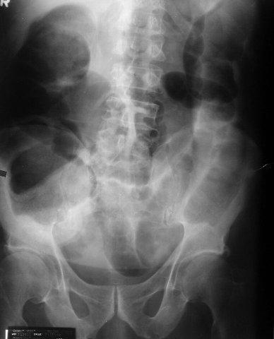
27 Sigmoid Volvulus Colonic Obstruction
28 Sigmoid volvulus This is the classic volvulus, occurring in old, mentally subnormal patients. It is usually chronic with intermittent acute attacks. Radiological signs : inverted U shaped distended loop which is devoid of haustra (ahaustral). Liver or left flank overlap signs. Apex of the volvulus above T10. Air fluid ratio greater than 2:1.


29 COLON SIGMOID VOLVULUS Dilated horse-shoe shaped sigmoid colon due to volvulus. COFFEE BEAN SIGN


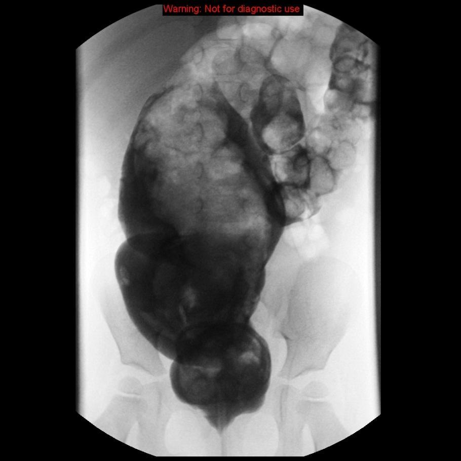
30 Barium fills to point of obstruction and twist of sigmoid colon COLON VOLVULUS BEAK SIGN

31 sigmoid volvulus
32 Cecal volvulus (Right colon volvulus) This account for less than 2% of adult intestinal obstruction ( young age group). The diagnosis of acute cecal volvulus is rarely made on clinical ground alone, and so radiological diagnosis become much more important & it is usually comprises a distended lower abdominal viscus with one or two haustral markings, concomitant small bowel dilatation & a collapsed left half of the colon.

33 Caecal volvulus

34 Caecal volulus
35 Hirschsprungs disease

36
Plain abdomen The standard films are supine & erect AP views (alternative to erect, lateral decubitus film is used in ill patients).
.") Plain abdomen The standard films are supine & erect AP views (alternative to erect, lateral decubitus film is used in ill patients). The stomach can be readily identified by its location, gastric rugae
Plain abdomen The standard films are supine & erect AP views (alternative to erect, lateral decubitus film is used in ill patients). The stomach can be readily identified by its location, gastric rugae
Abdominal radiology 腹部放射線學
 Abdominal radiology 腹部放射線學 台北醫學大學 - 市立萬芳醫院 留偉順 laowilson@hotmail.com The Normal Abdominal Series Chest Supine abdomen Erect abdomen Left lateral decubitus abdomen Learning objectives Understanding normal
Role of radiology and imaging in the daignosis of acute abdominal conditions
 Role of radiology and imaging in the daignosis of acute abdominal conditions Miah MAY Introduction In our day to day practice we have to face many of the acute abdominal conditions. As we know acute abdomen
No Disclosures. Approach to Abdominal Radiographs
 Approach to Abdominal Radiographs Tapas K. Tejura, M.D. Assistant Professor of Clinical Radiology Keck Medical Center of USC tapas.tejura@med.usc.edu No Disclosures 34-year-old male with acute abdominal
Pathology of Intestinal Obstruction. Dr. M. Madhavan, MBBS., MD., MIAC, Professor of Pathology Saveetha Medical College
 Pathology of Intestinal Obstruction Dr. M. Madhavan, MBBS., MD., MIAC, Professor of Pathology Saveetha Medical College Pathology of Intestinal Obstruction Objectives list the causes of intestinal obstruction
UNDERSTANDING X-RAYS: ABDOMINAL IMAGING THE ABDOMEN
 UNDERSTANDING X-RAYS: ABDOMINAL IMAGING THE ABDOMEN Radiology Enterprises radiologyenterprises@gmail.com www.radiologyenterprises.com STOMACH AND SMALL BOWEL STOMACH AND SMALL BOWEL Swallowed air is a
General Data. 王 X 村 78 y/o 男性
 General Data 王 X 村 78 y/o 男性 Chief Complaint Vomiting twice this early morning Fever up to 38.9ºC was noted Present Illness (1) Old CVA with left side weakness for more than 10 years and with bed ridden
U Lecture Objectives. U Nordic Forum Trauma & Emergency Radiology. Bowel obstruction. U Bowel Obstruction: Etiologies
 Nordic Forum Trauma & Emergency Radiology Lecture Objectives Bowel Obstruction To illustrate the spectrum of acute obstruction of the small and the large bowel To explain how these bowel obstructions may
Intestinal Obstruction
 By the Name of ALLAH the Most Gracious the Most Merciful Intestinal Obstruction د. أحمد اسامة حسن Specialist in General Surgery and Laparoscopic Surgery To be read in Bailey & Love s Short Practice of
Nordic Forum - Trauma & Emergency Radiology. Bowel Obstruction: Imaging Update
 Nordic Forum - Trauma & Emergency Radiology Bowel Obstruction: Imaging Update Borut Marincek Institute of Diagnostic Radiology University Hospital Zurich, Switzerland Acute Abdomen Bowel Obstruction Bowel
Many of the disease processes that acutely affect the abdomen produce
 ABC of Emergency Radiology Radiographic signs oftrauma THE ABDOMEN-II D A Nicholson, P A Driscoll Many of the disease processes that acutely affect the abdomen produce radiographic signs, but most of these
Acute Abdomen 急腹症 钱黎俊. Radiology, Renji Hospital. Shanghai Jiaotong University School of Medicine
 Acute Abdomen 急腹症 Radiology, Renji Hospital Shanghai Jiaotong University School of Medicine 钱黎俊 Learning objectives To understand the normal anatomy of the erect abdominal plain film To understand and
ASSESSING THE PLAIN ABDOMINAL RADIOGRAPH M A A M E F O S U A A M P O F O
 ASSESSING THE PLAIN ABDOMINAL RADIOGRAPH M A A M E F O S U A A M P O F O Introduction The abdomen (less formally called the belly, stomach, is that part of the body between the thorax (chest) and pelvis,
이희정. Plain Abdominal Radiography in Infants and Children. Hee Jung Lee, M.D.
 대한소아소화기영양학회지 : 제 14 권제 2 호 2011 DOI: 10.5223/kjpgn.2011.14.2.130 종설 영유아및소아의단순복부 X- 선사진 계명대학교의과대학영상의학교실 이희정 Plain Abdominal Radiography in Infants and Children Hee Jung Lee, M.D. Department of Radiology,
Intestinal Obstruction Clinical Presentation & Causes
 Intestinal Obstruction Clinical Presentation & Causes V Chidambaram-Nathan Consultant Transplant and General Surgeon Sheffield Kidney Institute Northern General Hospital Intestinal Obstruction One of the
CHEST & ABDOMINAL X-RAYS MALIKA IBRAHIM CORE MEDICAL TRAINEE BLACKPOOL VICTORIA HOSPITAL DATA INTERPRETATION COURSE FEB 20, 2017
 CHEST & ABDOMINAL X-RAYS MALIKA IBRAHIM CORE MEDICAL TRAINEE BLACKPOOL VICTORIA HOSPITAL DATA INTERPRETATION COURSE FEB 20, 2017 1. Sample x-rays 2. Basic chest x-ray interpretation skills 3. Chest x-ray
Abdominal Assessment
 Abdominal Assessment Mary Marian, MS,RD,CSO University of AZ, Tucson, AZ Neha Parekh, MS,RD,LD,CNSC Cleveland Clinic, Cleveland, OH Objectives: 1. Outline the steps in performing an abdominal examination.
CLINICAL PRESENTATION AND RADIOLOGY QUIZ QUESTION
 Donald L. Renfrew, MD Radiology Associates of the Fox Valley, 333 N. Commercial Street, Suite 100, Neenah, WI 54956 09/17/2011 Radiology Quiz of the Week # 38 Page 1 CLINICAL PRESENTATION AND RADIOLOGY
X-ray Corner. Imaging of the Small Bowel. Pantongrag-Brown L. Case 1. A 63-year-old man presented with abdominal pain, nausea and vomiting.
 THAI J 42 Imaging of the Small Bowel GASTROENTEROL 2015 X-ray Corner Imaging of the Small Bowel Pantongrag-Brown L Small bowel is the longest tubular organ in the body, about 18-22 feet. It is anchored
Adult bowel obstruction with acute abdomen: spectrum of CT findings
 Adult bowel obstruction with acute abdomen: spectrum of CT findings Poster No.: C-1571 Congress: ECR 2013 Type: Educational Exhibit Authors: L. Turturici, G. Gherarducci, F. Bianchi, R. Pascale, M. Tonerini,
Radiology. Undergraduate Radiology Sample Questions
 Radiology Undergraduate Radiology Sample Questions April 2012 The following examples are offered of questions that might be used to assess undergraduate radiology. There are 3 different styles: An OSCE
RADIOLOGY (SURGERY) BY MARYAM MALIK Rawalpindi Medical College
 BY MARYAM MALIK Rawalpindi Medical College") RADIOLOGY (SURGERY) BY MARYAM MALIK Rawalpindi Medical College NORMAL BOWEL GAS PATTERN Any part of the bowel may be visible if it contains gas/air within the lumen. Gas/air is of low density and forms
RADIOLOGY (SURGERY) BY MARYAM MALIK Rawalpindi Medical College NORMAL BOWEL GAS PATTERN Any part of the bowel may be visible if it contains gas/air within the lumen. Gas/air is of low density and forms
Pediatric Bowel Obstruction
 Pediatric Bowel Obstruction Matt Zerden, Harvard Medical School III Patient 1 16 year old presents with severe, episodic abdominal pain, nausea and vomiting. Questionable abdominal mass in RLQ Previous
DR JAIKISHOR JOTHIRAJ MD POST GRADUATE DEPT OF RADIODIAGNOSIS
 DR JAIKISHOR JOTHIRAJ MD POST GRADUATE DEPT OF RADIODIAGNOSIS YASHODAMMAL 70 YRS OD LADY had C/o diffuse lower abdominal pain 20 days h/o blood in stools 4 days h/o vomiting 2 days h/o burning micturation
TIPS AND PITFALLS IN PLAIN FILM INTERPRETATION
 TIPS AND PITFALLS IN PLAIN FILM INTERPRETATION Dr Philip Touska MBBS, BMedSci(Hons), MRCS, DO-HNS, FRCR Radiology Fellow Guy s & St Thomas Hospitals LEARNING OBJECTIVES Where do we go wrong? Common pitfalls
Clinics in diagnostic imaging (105)
") M e d i c a l E d u c a t i o n Singapore Med J 2005; 46(9) : 483 CME Article Clinics in diagnostic imaging (105) C T Wai, G Lau, C J L Khor Fig. 1 Abdominal radiograph obtained on admission. CASE PRESENTATION
M e d i c a l E d u c a t i o n Singapore Med J 2005; 46(9) : 483 CME Article Clinics in diagnostic imaging (105) C T Wai, G Lau, C J L Khor Fig. 1 Abdominal radiograph obtained on admission. CASE PRESENTATION
Christopher Lau Kings County Hospital SUNY Downstate Medical Center February 24, 2011
 Christopher Lau Kings County Hospital SUNY Downstate Medical Center February 24, 2011 37 year old male presented with 1 day history of abdominal pain Pain was diffuse but worst in the epigastric area No
A novel plain abdominal radiograph sign to diagnose malrotation with volvulus
 A novel plain abdominal radiograph sign to diagnose malrotation with volvulus Nataraja RM 1, Mahomed AA 1* 1. Department of Paediatric Surgery, Royal Alexandra Hospital for Sick Children, Brighton,UK *
Cecal Volvulus: Case Presentation and Review of CT Findings
 August 2011 Cecal Volvulus: Case Presentation and Review of CT Findings Omar Pardesi, Harvard Medical School Year III Our Patient LD: History & Physical HPI: 28 y.o. female presents with diffuse abdominal
Adult Intussusception
 Bahrain Medical Bulletin, Vol. 27, No. 3, September 2005 Adult Intussusception Suhair Alsaad, MBCHB, CABS, FRCSI* Mariam Al-Muftah, MBCHB** Objectives: Adult intussusception is a rare entity. We present
Lab Monitor Images Dissection of the Abdominal Vasculature + Lower Digestive System
 Lab Monitor Images Dissection of the Abdominal Vasculature + Lower Digestive System Stomach & Duodenum Frontal (AP) View Nasogastric tube 2 1 3 4 Stomach Pylorus Duodenum 1 Duodenum 2 Duodenum 3 Duodenum
Gastrointestinal Pathology. August 2007
 Gastrointestinal Pathology August 2007 Case 1 Dysphagia and halitosis Case 1 Dilatation of the oesophagus with a smooth narrowing of its lower end. The large volume of contained fluid indicates delayed
Medical application of transabdominal ultrasound in gastrointestinal diseases
 Medical application of transabdominal ultrasound in gastrointestinal diseases Hsiu-Po Wang Department of Emergency Medicine National Taiwan University Hospital Real-time ultrasound has become a standard
Back to Basics: What Imaging Test should I order? Jeanne G. Hill, M.D. Pediatric Radiology Medical University of South Carolina
 Back to Basics: What Imaging Test should I order? Jeanne G. Hill, M.D. Pediatric Radiology Medical University of South Carolina Disclosure Neither I nor any member of my immediate family has a relevant
Radiology of the abdomen Lecture -1-
 Radiology of the abdomen Lecture -1- Objectives To know radiology modalities used in abdomen imaging mainly GI tract. To know advantages and disadvantages of each modality. To know indications and contraindications
X-ray Corner. Imaging of The Colon. Pantongrag-Brown L
 110 Imaging of The Colon X-ray Corner Imaging of The Colon Pantongrag-Brown L Imaging modalities used in colon include plain radiographs, barium enema, US, CT, PET CT and MRI. Barium enema (BE) is declining
Residents Section Pattern of the Month
 Residents Section Pattern of the Month Krajewski et al. olonic Dilation Residents Section Pattern of the Month Residents inradiology Katherine Krajewski 1 ettina Siewert Ronald L. Eisenberg Krajewski K,
Sonographic Appearances of Common Gut Pathology in Paediatric Patients: Comparison with Plain Abdominal Radiography
 3668 Radiographer Text 1/4/04 2:57 PM Page 11 The Radiographer vol. 51: 11-17 Sonographic Appearances of Common Gut Pathology in Paediatric Patients: Comparison with Plain Abdominal Radiography Lino Piotto
A rare case of intestinal obstruction due to internal hernia. Dr. Jayanth 3 rd year PG Dept. Of General Surgery
 A rare case of intestinal obstruction due to internal hernia Dr. Jayanth 3 rd year PG Dept. Of General Surgery One of the common cause of acute abdomen May lead to high morbidity and mortality if not treated
INVESTIGATIONS OF GASTROINTESTINAL DISEAS
 INVESTIGATIONS OF GASTROINTESTINAL DISEAS Lecture 1 and 2 دز اسماعيل داود فرع الطب كلية طب الموصل Radiological tests of structure (imaging) Plain X-ray: May shows soft tissue outlines like liver, spleen,
Vomiting in children: The good coordination between radiologists and pediatricians is the key to success
 Vomiting in children: The good coordination between radiologists and pediatricians is the key to success C. Santos Montón 1, M. T. Garzon Guiteria 2, A. Hortal Benito-Sendín 1, K. El Karzazi 1, P. Sanchez
What is Your Diagnosis?
 What is Your Diagnosis? Izabela Ragan, Class of 2014 Signalment Species: Canine Breed: English Bulldog Sex: Male castrated Date of birth: 04/14/11 Presenting Complaint Dog was presented for vomiting and
X-rays. Dr Will Dooley
 X-rays Dr Will Dooley Plan Chest X-Rays Abdominal X-Rays Exam approach Presentation skills EMQ EMQ- answers Chest X-Ray - Systematic Approach D R Details RIP Image Quality +/- OBVIOUS ABNORMALITY A B C
Hirschprung s. Meconium plug R/S >1 R/S <1
 NEONATAL ABDOMINAL EMERGENCIES LOW OBSTRUCTION HIGH OBSTRUCTION INTESTINAL OBSTRUCTION High obstruction - proximal to mid-ileumileum Few dilated, air filled bowel loops Complete obstruction diagnosed by
Small-bowel Obstruction - the imaging contribute
 Small-bowel Obstruction - the imaging contribute Poster No.: C-2098 Congress: ECR 2015 Type: Educational Exhibit Authors: S. C. S. Silva, S. Dutra, D. Garrido, D. N. Silva, I. C. S. P. 1 1 2 1 1 1 2 Basto
Computed tomography (CT) imaging review of small bowel obstruction
 imaging review of small bowel obstruction") Computed tomography (CT) imaging review of small bowel obstruction Poster No.: C-1602 Congress: ECR 2010 Type: Educational Exhibit Topic: GI Tract Authors: A. Vousough, D. S. Prasad ; Aberdeen/UK, Leeds/UK
Computed tomography (CT) imaging review of small bowel obstruction Poster No.: C-1602 Congress: ECR 2010 Type: Educational Exhibit Topic: GI Tract Authors: A. Vousough, D. S. Prasad ; Aberdeen/UK, Leeds/UK
Computed tomography (CT) imaging review of small bowel obstruction
 imaging review of small bowel obstruction") Computed tomography (CT) imaging review of small bowel obstruction Poster No.: C-1602 Congress: ECR 2010 Type: Educational Exhibit Topic: GI Tract - Small Bowel Authors: A. Vousough, D. S. Prasad ; Aberdeen/UK,
Computed tomography (CT) imaging review of small bowel obstruction Poster No.: C-1602 Congress: ECR 2010 Type: Educational Exhibit Topic: GI Tract - Small Bowel Authors: A. Vousough, D. S. Prasad ; Aberdeen/UK,
Abdominal Imaging: Luminal organs. Rowland Illing MA BMBCh DM FLS MRCS(Eng) FRCR
 FRCR") Abdominal Imaging: Luminal organs Rowland Illing MA BMBCh DM FLS MRCS(Eng) FRCR Aims Reference text & resources Management of a patient Imaging what and when to use What to ask and how to describe Segments
Abdominal Imaging: Luminal organs Rowland Illing MA BMBCh DM FLS MRCS(Eng) FRCR Aims Reference text & resources Management of a patient Imaging what and when to use What to ask and how to describe Segments
Intestinal obstruction key. What should we look for?
 Intestinal obstruction key. What should we look for? Poster No.: C-1689 Congress: ECR 2013 Type: Educational Exhibit Authors: R. Carreño-Gonzalez, L. Renza, E. Navarro-Sanchis, M. D. 1 2 3 1 4 1 2 SÁNCHEZ
Evidence Process for Abdominal Pain Guideline Research 11/16/2017. Guideline Review using ADAPTE method and AGREE II instrument 11/16/2017
 Evidence Process for Abdominal Pain Guideline Research Guideline Review using ADAPTE method and AGREE II instrument Approximately 139 Potentially relevant guidelines identified in various resources* 59
Neonatal intestinal obstruction: how to make etiological diagnosis?
 Neonatal intestinal obstruction: how to make etiological diagnosis? Poster No.: C-1414 Congress: ECR 2013 Type: Educational Exhibit Authors: W. Mnari, M. Zguidi, A. Zrig, M. Maatouk, B. Hmida, R. Salem,
Neonatal intestinal obstruction: how to make etiological diagnosis?
 Neonatal intestinal obstruction: how to make etiological diagnosis? Poster No.: C-1414 Congress: ECR 2013 Type: Educational Exhibit Authors: W. MNARI, M. Zguidi, A. Zrig, M. MAATOUK, B. Hmida, R. Salem,
Volvulus of the Gastrointestinal Tract: x-ray and CT imaging
 Volvulus of the Gastrointestinal Tract: x-ray and CT imaging Poster No.: C-0076 Congress: ECR 2013 Type: Educational Exhibit Authors: E. Papadaki, S. Paschalidou, S. GIANNOU ; Rethymno, CR/ 1 2 2 3 1 3
A case of cecal volvulus in a cerebral palsy patient: Usefulness of multidetector computed tomography for preoperative diagnosis
 Kawasaki Medical Journal 38(4):205-209,2012 205 A case of cecal volvulus in a cerebral palsy patient: Usefulness of multidetector computed tomography for preoperative diagnosis Yusuke MATSUI 1), Munenori
National Museum of Health and Medicine
 National Museum of Health and Medicine Otis Historical Archives Bower Photograph Collection Date of Records: 1910s-1920s Size: 1 box Finding Aid: by Eric W. Boyle (2012) Biographical Note: Col. Morris
Nasogastric tube. Stomach. Pylorus. Duodenum 1. Duodenum 2. Duodenum 3. Duodenum 4
 Esophagus Barium Swallow Stomach and Duodenum 4 year old Upper GI Nasogastric tube Stomach and Duodenum 4 year old Upper GI Nasogastric tube Stomach Pylorus Duodenum 1 Duodenum 2 Duodenum 3 Duodenum 4
Volvulus characterization in radiology: A review
 Volvulus characterization in radiology: A review Poster No.: C-1677 Congress: ECR 2010 Type: Topic: Educational Exhibit GI Tract Authors: C. Antunes, M. Seco, A. Canelas, C. Ruivo, C. Paulino, F. Cruz,
In children 3 months to 3 years of age intussusception is
 Baird Mallory, MD, 1 and Yale Popowich, MD 2 In children 3 months to 3 years of age intussusception is one of the most common causes of a distal small bowel obstruction. It is often associated with intermittent
Clearing the mind before the "caliber change": Diagnostic algorithm for small bowel obstruction.
 Clearing the mind before the "caliber change": Diagnostic algorithm for small bowel obstruction. Poster No.: C-0255 Congress: ECR 2014 Type: Educational Exhibit Authors: C. Santos Montón, D. Oquillas Izquierdo,
The Value of Urgent Barium Enema and Computed Tomography in Acute Malignant Colonic Obstruction: Is Urgent Barium Enema Still Necessary?
 J Radiol Sci 2012; 37: 105-110 The Value of Urgent Barium Enema and Computed Tomography in Acute Malignant Colonic Obstruction: Is Urgent Barium Enema Still Necessary? Chun-Chao Huang 1,2 Fei-Shih Yang
Strangulating Obstruction of the Bowel: A Reevaluation of Radiographic Criteria
 Strangulating Obstruction of the Bowel: A Reevaluation of Radiographic Criteria DAVID BRYK1 Fifty consecutive cases of strangulating obstruction were compared with 100 consecutive cases of surgically proven
LOOKING FOR AIR IN ALL THE WRONG PLACES Richard M. Gore, MD North Shore University Health System University of Chicago Evanston, IL
 SIGNIFICANCE OF EXTRALUMINAL ABDOMINAL GAS: LOOKING FOR AIR IN ALL THE WRONG PLACES Richard M. Gore, MD North Shore University Health System University of Chicago Evanston, IL SCBT/MR 2012 October 26,
Plain Radiographs in Non-Traumatic Abdominal Pain. Plain Radiographs in Non-Traumatic Abdominal Pain
 Jake Block, MD Associate Professor Associate Vice-Chairman for Clinical Operations Director, Musculoskeletal and Emergency Radiology Department of Radiology and Radiological Sciences Vanderbilt University
Relationship Between Small Bowel Obstruction and Small Bowel Feces Sign: Four Cases Report
 Case Report Elmer Press Relationship Between Small Bowel Obstruction and Small Bowel Feces Sign: Four Cases Report Altintoprak Fatih a, e, Gunduz Yasemin b, Yalkin Omer c, Gundugdu Kemal c, Serbulent Gokhan
Gastrointestinal Tract Imaging. Objectives. Reference. VMB 960 April 6, Stomach Small Intestine Colon. Radiography & Ultrasound
 Gastrointestinal Tract Imaging VMB 960 April 6, 2009 Stomach Small Intestine Colon Objectives Radiography & Ultrasound Contrast Examination of the Small Intestine Reference Chapters 45 47 Pages 750 805
Small Bowel Obstruction
 Residents Section Pattern of the Month Small owel Obstruction Residents Section Pattern of the Month Downloaded from www.ajronline.org by 37.44.193.56 on 12/16/17 from IP address 37.44.193.56. Copyright
Plain Abdominal Radiography & GI series 腹部與腸胃道之放射線學
 Plain Abdominal Radiography & GI series 腹部與腸胃道之放射線學 陳潤秋台北市立聯合醫院 chenranchou@tpech.gov.tw Jen-Ai H. Standing abdomen Jen-Ai H. www.nlm.nih.gov Supine KUB Jen-Ai H. medicalcenter.osu.edu Jen-Ai H. Checklist:
Spleen indications of splenectomy complications OPSI
 Intestinal obstruction Differences between adynamic ileus and mechanical obstruction Aetiology Pathophysiology (Cluster contractions- bowel proximal to the obstruction dilate- wall of obstructed gut is
Pediatric Abdomen Trauma
 Pediatric Abdomen Trauma Susan D. John, MD, FACR Pediatric Trauma Trauma is leading cause of death and disability in children and adolescents Causes and effects vary between age groups Blunt trauma predominates
Gas patterns on plain abdominal radiographs: a pictorial review
 1 Royal Hallamshire Hospital, Sheffield Teaching Hospitals NHS Trust, Sheffield, South Yorkshire, UK 2 Barnsley Hospital NHS Foundation Trust, Barnsley, South Yorkshire, UK Correspondence to Dr R E Musson,
GIT RADIOLOGY. Water-soluble contrast media (e.g. gastrograffin) are the other available agents.which doesn t cause inflammatory peritonitis..
 are the other available agents.which doesn t cause inflammatory peritonitis..") GIT RADIOLOGY Imaging techniques-general principles: Contrast examinations: Barium sulphate is the best contrast for GIT (with good mucosal coating & excellent opacification & being inert); but is contraindicated
GIT RADIOLOGY Imaging techniques-general principles: Contrast examinations: Barium sulphate is the best contrast for GIT (with good mucosal coating & excellent opacification & being inert); but is contraindicated
Pitfalls in the CT diagnosis of appendicitis
 The British Journal of Radiology, 77 (2004), 792 799 DOI: 10.1259/bjr/95663370 E 2004 The British Institute of Radiology Pictorial review Pitfalls in the CT diagnosis of appendicitis 1 C D LEVINE, 2 O
Phillip A. Bilderback, MD, Ryan K. Smith, BA, and W. Scott Helton, MD, FACS
 gastrointestinal tract and abdomen INTESTINAL OBSTRUCTION Phillip A. Bilderback, MD, Ryan K. Smith, BA, and W. Scott Helton, MD, FACS Intestinal obstruction is a common medical problem and accounts for
GASTROINTESTINAL SYSTEM
 GASTROINTESTINAL SYSTEM Topographic Anatomy of the Abdomen Surface Landmarks Xiphoid process T9/T10 Inferior costal margin L2/L3 Iliac Crest L4 level ASIS L5/S1 level Pubic symphysis level of greater trochanter
Abdominal Plain Radiograph in Neonatal Intestinal Obstruction. G Raghavendra Prasad*, Amtul Aziz
 Journal of Neonatal Surgery 2017; 6(1):6 Doi:10.21699/jns.v6i1.483 REVIEW ARTICLE Abdominal Plain Radiograph in Neonatal Intestinal Obstruction G Raghavendra Prasad*, Amtul Aziz Deccan College of Medical
12 Blueprints Q&A Step 2 Surgery
 12 Blueprints Q&A Step 2 Surgery 34. A 40-year-old female has been referred to you for a recent ER and hospital admission, from which she was given a diagnosis of acute diverticulitis. Treatment at that
Mohamed EL-hemaly Gastro- intestinal surgical center, Mansoura University.
 Mohamed EL-hemaly Gastro- intestinal surgical center, Mansoura University. Chronic transmural inflammatory process of the bowel & affects any part of the gastro -intestinal tract from the mouth to the
DIAGNOSTIC VALUE OF PLAIN ABDOMINAL RADIOGRAPH IN NON TRAUMATIC CAUSES OF ACUTE ABDOMEN
 30 Original article DIAGNOSTIC VALUE OF PLAIN ABDOMINAL RADIOGRAPH IN NON TRAUMATIC CAUSES OF ACUTE ABDOMEN Dr Deepak Rajput ( Associate Professor ) Dr Aashaka Patel ( Resident ) Radio diagnosis Department,
Imaging findings in complications of bariatric surgery.
 Imaging findings in complications of bariatric surgery. Poster No.: C-1791 Congress: ECR 2012 Type: Educational Exhibit Authors: A. Fernandez Alfonso, G. Anguita Martinez, D. C. Olivares Morello, C. García
Small Intestine Bezoar: Computed Tomography Appearance
 Chin J Radiol 2001; 26: 197-202 197 ORIGINAL ARTICLE Small Intestine Bezoar: Computed Tomography Appearance KUNG-SHIH YING SHIN-HWA WU TAI-YU CHANG CHUNG-HSEIN LEE Department of Radiology, Cheng Ching
Long Case Set 02. Dr Raviraj Uppoor. Dr Sameer Shamshuddin. Consultant Radiologist Cumberland Infirmary, Carlisle, UK
 Long Case Set 02 www.frcrtutorials.com Dr Raviraj Uppoor MBBS, DMRD, DNB, FRCR Consultant Radiologist Cumberland Infirmary, Carlisle, UK Dr Sameer Shamshuddin MBBS, DMRD, FRCR Consultant Radiologist Royal
Role of imaging in the evaluation of the acute abdomen
 Prof. András Palkó MD, PhD Role of imaging in the evaluation of the acute abdomen Faculty of General Medicine University of Szeged Hungary 1 Definition Sudden onset of severe symptoms requiring emergency
ACUTE ABDOMEN. Dr. M Asadi. Surgical Oncology Research Center MUMS. Assistant Professor of General Surgery
 ACUTE ABDOMEN Dr. M Asadi Assistant Professor of General Surgery Surgical Oncology Research Center MUMS Definition I. The term Acute Abdomen refers to signs & symptoms of abdominal pain and tenderness,
General Surgery Service
 General Surgery Service Patient Care Goals and Objectives Stomach/Duodenum and Bariatric assessed for a) Obesity surgery b) Treatment of i) Adenocarcinoma of the stomach ii) GIST iii) Carcinoid 2) Optimize
Diseases of Stomach & Abomasum. Physical influences Gastric Ulcers Gastritis Parasitic Diseases
 Diseases of Stomach & Abomasum Physical influences Gastric Ulcers Gastritis Parasitic Diseases Neoplasia Physical Influences Acute gastric dilation & volvulus (GDV) Displaced abomasum Chronic gastric dilation
Bowel Obstructions in Older Children
 Residents Section Pattern of the Month Hryhorczuk et al. owel Obstructions in Older Children Residents Section Pattern of the Month Residents inradiology nastasia Hryhorczuk 1 Edward Y. Lee 1,2 Ronald
J of Evolution of Med and Dent Sci/ eissn , pissn / Vol. 3/ Issue 26/June 30, 2014 Page 7326
 DIAGNOSIS AND MANAGEMENT OF SUBACUTE INTESTINAL OBSTRUCTION: A PROSPECTIVE STUDY Amit Ojha 1, Anjani Jalaj 2, Shaleen Tiwari 3, Vikram Mujalde 4, Prasheel 5 HOW TO CITE THIS ARTICLE: Amit Ojha, Anjani
A 34 year old woman with Vomiting and abdominal pain
 A 34 year old woman with Vomiting and abdominal pain The patient was a 34 y/o woman admitted because of epigastric pain developed from 2 months ago. It was a crampy pain without radiation that became better
Chapter 14: Training in Radiology. DDSEP Chapter 1: Question 12
 DDSEP Chapter 1: Question 12 A 52-year-old white male presents for evaluation of sudden onset of abdominal pain and shoulder pain. His past medical history is notable for a history of coronary artery disease,
Case Report A Case of Stercoral Perforation Detected on CT Requiring Proctocolectomy in a Heroin-Dependent Patient
 Case Reports in Surgery Volume 2016, Article ID 2893925, 4 pages http://dx.doi.org/10.1155/2016/2893925 Case Report A Case of Stercoral Perforation Detected on CT Requiring Proctocolectomy in a Heroin-Dependent
The role of abdominal X-rays in the investigation of suspected acute appendicitis
 Journal of Medicine and Medical Sciences Vol. 2(11) pp. 1216-1220, November 2011 Available online@ http://www.interesjournals.org/jmms Copyright 2011 International Research Journals Full Length Research
Gastrointestinal Tract. Anatomy of GI Tract. Anatomy of GI Tract. (Effective February 2007) (1%-5%)
 (1%-5%)") Gastrointestinal Tract (Effective February 2007) (1%-5%) Anatomy of GI Tract Esophagus bulls-eye or target EG junction seen on sagittal scan posterior to left lobe of liver and anterior to aorta Anatomy
Gastrointestinal Tract (Effective February 2007) (1%-5%) Anatomy of GI Tract Esophagus bulls-eye or target EG junction seen on sagittal scan posterior to left lobe of liver and anterior to aorta Anatomy
UNIT 5 MAINTENANCE SYSTEMS Digestive System Test Bank
 UNIT 5 MAINTENANCE SYSTEMS Digestive System Test Bank Objective 5.01 Describe the basic functions of the digestive system. 1. What is the main function of the digestive system? a. Hold and receive food
INTESTINAL OBSTRUCTION ESCAPED SURGERY: MECONIUM PLUG
 7 INTESTINAL OBSTRUCTION ESCAPED SURGERY: MECONIUM PLUG Oluwayemi IO 1 *, Ade-Ojo IP 2, Olofinbiyi BA 2 1. Department of Paediatrics, Ekiti State University Teaching Hospital, Ado-Ekiti, Ekiti State, Nigeria
Proceedings of the American Association of Equine Practitioners - Focus Meeting. Focus on Colic. Indianapolis, IN, USA 2011
 www.ivis.org Proceedings of the American Association of Equine Practitioners - Focus Meeting Focus on Colic Indianapolis, IN, USA 2011 Next Focus Meetings: July 22-24, 2012 - Focus on Hind Limb Lameness
OUTLINE ANATOMY, RADIOGRAPHY,
 16 ABDOMEN R OUTLINE SUMMARY OF PROJECTIONS, 84 ANATOMY, 85 Abdominopelvic cavity, 85 SUMMARY OF ANATOMY, 86 SUMMARY OF PATHOLOGY, 86 EXPOSURE TECHNIQUE CHART, 87 ABBREVIATIONS, 87 RADIOGRAPHY, 88 Abdominal
Bowel obstruction and tumors
 Bowel obstruction and tumors Intestinal Obstruction Obstruction of the GI tract may occur at any level, but the small intestine is most often involved because of its relatively narrow lumen. Causes: Hernias
Deep Enteroscopy Methods to Diagnose Small Bowel IBD
 Deep Enteroscopy Methods to Diagnose Small Bowel IBD Name: Institution: Peter Draganov University of Florida, Gainesville, FL Overview Types of enteroscopy Enteroscopy equipment Enetoscopy do and don'ts
Pitfalls of the Pediatric Chest and Abdomen SPR 2017
 Pitfalls of the Pediatric Chest and Abdomen SPR 2017 Richard I. Markowitz, MD, FACR Children s Hospital of Philadelphia Perelman School of Medicine University of Pennsylvania No Disclosures Cognitive Perceptual
NOTES: The Digestive System (Ch 14, part 2)
") NOTES: The Digestive System (Ch 14, part 2) PANCREAS Structure of the pancreas: The pancreas produces PANCREATIC JUICE that is then secreted into a pancreatic duct. The PANCREATIC DUCT leads to the The
NOTES: The Digestive System (Ch 14, part 2) PANCREAS Structure of the pancreas: The pancreas produces PANCREATIC JUICE that is then secreted into a pancreatic duct. The PANCREATIC DUCT leads to the The