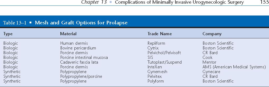
Figure 13 1 Technique of anterior repair with plication of the pubocervical fascia for cystocele repair.
|
|
|
- Job Anderson
- 6 years ago
- Views:
Transcription
1
2
3


4 Figure 13 1 Technique of anterior repair with plication of the pubocervical fascia for cystocele repair. Figure 13 2 Technique of posterior repair with plication of the recto-vaginal fascia for rectocele repair.

5 vaginal vault descensus, 3 no vaginal procedure has the extensive use and historical data comparable with those for the sacrospinous ligament fixation. In this technique, the sacrospinous space is entered through an anterior, midline, or posterior approach and the relevant pelvic anatomy is identi- Figure 13 3 Retropubic view of paravaginal repair with reapproximation of the pubocervical fascia to the arcus tendineus fasciae pelvis (ATFP). Figure 13 4 Site-specific defect repair of rectocele with closure of transverse fascial defect. fied (Fig. 13 5). One or two sutures are taken through the upper edge of the sacrospinous-coccygeal ligament complex and then are attached to the vaginal apex using a full-thickness suture or pulley stitch (Fig. 13 6). Placement of the sacrospinous suture can be performed using a free needle holder, Miya hook, Dechamps ligature carrier, Schutt needle punch device, Endo-stitch (Ethicon, Somerville, New Jersey), or
6 Capio needle driver (Boston Scientific, Natick, Massachusetts). Although the traditional sacrospinous ligament fixation describes unilateral suspension, many gynecologic surgeons favor bilateral sacrospinous suspension to improve cure rates and avoid unilateral deviation of the vaginal apex. Figure 13 5 Anatomy of the sacrospinous space, including the ischial spine, sacrospinous ligament, arcus tendineus fasciae pelvis (ATFP), and coccygeus muscle. (Copyright Cleveland Clinic Foundation.) P u d e n d a l B u n d l e U r e t e r Ligament

7 Figure 13 6 Technique of sacrospinous ligament fixation using pulley stitch to attach the vagina in the sacrospinous ligament for apical support. Uterosacral Ligament Suspension In an effort to provide a more anatomically correct midline suspension point, uterosacral ligament
8 suspension, performed through a vaginal, abdominal, or laparoscopic approach, has gained recent popularity. Similar to the classically described McCall cul-de-plasty, the uterosacral ligament suspension technique begins with adequate identification of the uterosacral ligaments through either a transperitoneal or a retroperitoneal vaginal approach. One or two sutures are then taken through the uterosacral ligament on each side and then attached to the vaginal apex. Care should be taken to identify the ureter before stitch placement, owing to its proximity and the possibility of kinking or obstruction. Tiedown of the suture results in anatomic restoration, with re-creation of the apical supports of the uterosacral ligament to the vaginal apex, as well as in closure of the anterior and posterior fascial planes (Fig. 13 7). Iliococcygeus/Arcus Tendineus Fasciae Pelvis Fixation To address the needs of patients with poor uterosacral ligament strength, and to avoid complications associated with sacrospinous ligament suspension such as buttock pain and pudendal neurovascular injury, iliococcygeus and arcus tendineus fasciae pelvis (ATFP) suspensions have been described. A dissection similar to that for sacrospinous ligament fixation is performed, and the relevant pelvic anatomy including the ischial spine, ATFP, and sacrospinous-coccygeal ligament complex is identified. One or two sutures are taken through either the ileococcygeus muscle or the ATFP and then attached to the vaginal apex using a full-thickness suture or pulley stitch. Unilateral or bilateral suspension can be performed.
9
10 Chapter 13 Complications of Minimally Invasive Urogynecologic Surgery
11

12 Posterior Intravaginal Slingplasty Vault Suspension Posterior Intravaginal Slingplasty Vault Suspension (Tyco-US Surgical, Norwalk, Connecticut) is a minimally invasive vaginal technique for vaginal vault suspension that uses needles and synthetic mesh to re-create apical support at the level of the ischial spine without midline deviation. The IVS Figure 13 7 Vaginal approach to uterosacral ligament suspension with incorporation of the anterior pubocervical fascia (PCF) and posterior recto-vaginal fascia (RVF) in the uterosacral suspension suture (USL). AD, apical defect; B, bladder; R, rectum. device consists of a trocar with a removable and reversible stylet into which a multifilament polypropylene tape can be threaded and brought up through the trocar. The procedure begins with bilateral sacrospinous space dissection and identification of the relevant anatomy, as previously described. Stab incisions in the buttocks are made bilaterally 3 cm lateral and 3 cm inferior to the anus. The trocar with tape attached is introduced through the stab wound, advanced through the ischiorectal fossa, and exited through the coccygeus muscle into the sacrospinous space under direct finger guidance. Alternatively, the needle can be exited lateral to the ischial spine near the insertion of the ATFP. The stylet with tape attached is then pulled out through the trocar. The same procedure is repeated on the other side with an empty stylus, and the free end of the tape is attached and pulled through the contralateral space. The apical portion of the tape is then attached to the vaginal apex, rectovaginal fascia, or graft material. The vaginal mucosa is closed and the traction on the free ends of the tape retracts the apex cephalad, thereby creating apical support. Excess tape is excised. Mesh/Graft Augmentation of Prolapse Repair Mesh or graft augmentation of prolapse repair has most recently been introduced with the hopes of improving long-term cure rates and reducing the risk of recurrence, which has been reported to be as high as 30%. In this technique, traditional colporrhaphy is performed and then a synthetic mesh or donor graft is used to reinforce the repair (Fig. 13 8). As with general surgery mesh use for hernia repairs, the augmented repair is thought to have greater strength and Figure 13 8 Graft augmentation of rectocele repair with attachment of graft to levator muscles laterally and

13 perineal body distally.
14
15
 and placement and attachment of the graft or mesh are nonstandardized.")
16 Figure 13 9 Tension-free vaginal tape (TVT) sling procedure: Instrumentation. Shown are introducer, rigid catheter guide, and TVT device (polypropylene mesh and attached trocars). durability. A variety of graft or mesh materials are available (Table 13 1) and placement and attachment of the graft or mesh are nonstandardized. Direct mesh attachment, needle passage of the mesh, and nonattachment all have been described. Regardless of the technique, complications including mesh erosion and infection, dyspareunia or pelvic pain, and recurrence have been reported, and the gynecologic surgeon should approach this technique with caution in view of the limited data currently available. Minimally Invasive Sling Procedures TRANSVAGINAL RETROPUBIC SLING Introduced in 1991, the tension-free vaginal tape (TVT) technique represents a revolutionary improvement in the sub-urethral sling procedure. Unique characteristics of the TVT technique include midurethral positioning of the sling, use of a polypropylene weave tape, and tension-free application without the need for large incisions or fascial attachment. Instrumentation for this procedure consists of a reusable stainless steel introducer, a reusable rigid catheter guide, and the TVT device, a single-use apparatus composed of a 1 40 cm strip of polypropylene mesh (Prolene, Ethicon Inc., Somerville, New Jersey) covered by a plastic sheath and held between two stainless steel needles (Fig. 13 9). The plastic sheath is designed to (1) cover the synthetic mesh during placement of the sling, thereby reducing the incidence of postoperative infection or graft rejection, and (2) allow easy passage and placement of the tape, which is configured to stay fixed in place once the smooth protective cover is removed. This protective sheath is removed before completion of the procedure. Two small abdominal skin incisions (0.5 to 1.0 cm) are made on each side of the midline just above the pubic symphysis. A small sagittal incision (1.5 cm) is then made in the midline of the anterior vaginal wall approximately 1 cm proximal to the external urethral meatus. The edges of this incision are grasped using tissue clamps, and minimal dissection is used to free the vaginal wall from the urethra and to develop a small paraurethral space bilaterally (Fig ). The rigid catheter guide is then inserted into the Foley catheter, facilitating identification of the urethra and the bladder neck during passage of the suspension needles. To minimize the risk of bladder or urethral perforation, the handle of the guide is deflected to the ipsilateral side just before insertion of the suspension needles. Before placement of the sling, the introducer is attached to one of the stainless steel needles, and the speculum is removed from the vagina. The shaft of the introducer is grasped and the tip of the needle is then inserted into the previously developed paraurethral space. The needle is angulated slightly laterally, and the endopelvic fascia is perforated just behind the inferior surface of the pubic symphysis. On entry into the retropubic space, the needle is guided up to the abdominal incision while maintaining contact with the back of the pubic bone, thereby minimizing the risk of vascular or hollow viscous injury. A second layer of resistance is felt as the needle passes through the muscular and fascial layers of the abdominal wall. Passage of the needle is completed once the needle tip passes through the small abdominal incision on the corresponding side (Fig ). Before complete extraction of the placement needle, unintentional bladder perforation should be ruled out. The rigid catheter guide is removed, and the bladder is emptied using the indwelling catheter. The
17 catheter is then removed, and cystoscopy is performed to confirm integrity of the bladder lumen. If needle penetration of the bladder lumen is noted, the needle-introducer assembly is withdrawn, the bladder is drained, and the needle is reinserted. Once correct placement of the needle has been confirmed, the needle is detached from the introducer and passed completely through the abdominal incision. This technique is then repeated in an
18

19 Figure Tension-free vaginal tape (TVT) sling procedure: Vaginal dissection. Bilateral channels are created for needle insertion and mesh placement. identical fashion on the contralateral side to ensure that the tape lies flat against the suburethral tissue at the level of the midurethra. After passage of the needles has been completed, a clamp or scissors is inserted between the suburethral portion of the tape and the urethra. Gentle traction on the abdominal ends of the tape removes any excess tape material and brings the tape into contact with the instrument (Fig ). Both ends of the tape are then cut at their attachment to the needles. All instruments are removed from the surgical field, and the patient, if awake, is asked to perform a cough stress test to ensure that continence is restored without overcorrection. The tension on the tape is adjusted as appropriate. The plastic sheath of the abdominal ends of the tape is then identified and grasped with a forceps. With an instrument between the urethra and the tape, the plastic sheath is removed, leaving the Prolene tape secured under the midurethra without tension. The vaginal incision is closed. Next, the abdominal ends of the tape are cut just below the surface of the skin, without need for suture fixation. The friction between the tissues and the Prolene mesh keeps the tape in place while maintaining adequate suburethral support. Finally, the abdominal incisions are closed using either subcuticular stitches or surgical tape (Fig ). Several TVT me-too products are currently available from a variety of manufacturers, using a polypropylene weave sling with slight variations in instrumentation and surgical technique (Table 13 2). Results and complications should be similar to those reported for TVT.
 sling procedure: Final adjustment of the TVT sling after placement.")

20 Figure Tension-free vaginal tape (TVT) sling procedure: Passage of TVT needle and sling mesh through the retropubic space. Figure Tension-free vaginal tape (TVT) sling procedure: Final adjustment of the TVT sling after placement. This step can be performed in conjunction with a cough test to avoid overtightening the sling. TRANSABDOMINAL RETROPUBIC SLING After the introduction of the TVT procedure, an abdominal approach to the minimally invasive midurethral sling procedure was introduced by American Medical Systems (AMS). The SPARC (i.e., suprapubic arc)

21 procedure uses Stamey-type Figure Tension-free vaginal tape (TVT) sling procedure: Completed procedure with placement of suburethral hammock.
 to place a TVT-like mesh tape in the midurethra in an effort to be more urologyfriendly while emulating traditional transvaginal needle suspension and traditional suburethral sling procedures.")

22 needles (Fig ) to place a TVT-like mesh tape in the midurethra in an effort to be more urologyfriendly while emulating traditional transvaginal needle suspension and traditional suburethral sling procedures. It rapidly gained popularity as an effective and familiar alternative to the TVT procedure. The procedure begins with a similar dissection to the TVT, with a slightly larger vaginal incision used to allow finger insertion to the endopelvic fascia. The SPARC needles are inserted through each abdominal incision, guided through the retropubic space against the pubic bone, and exited through the vaginal incision under direct finger guidance. An identical procedure is performed on the contralateral side. Cystoscopy is performed to exclude bladder injury. The tape is attached to each needle, and the needles are retracted, thereby placing both arms of the sling. The sling is adjusted and the sheath removed as in the TVT procedure. Figure Suprapubic arc (SPARC) sling procedure: Instrumentation. Shown are retropubic needles and the SPARC device (polypropylene mesh with attachment dilators).
23 As with TVT, several SPARC me-too products are currently available from a variety of manufacturers; all incorporate a polypropylene weave sling with slight variations in instrumentation and surgical technique (Table 13 3). Results and complications should be similar to those reported for SPARC. TRANSOBTURATOR SLING In an effort to simplify the procedure and reduce complications associated with retropubic passage of the minimally invasive midurethral sling needles, a transobturator approach has been advocated. This outsidein procedure initially was described in Europe and recently has been introduced in the United States. For the transobturator sling procedure, a 2-cm midline vaginal incision is made approximately 1 cm proximal to the external urethral meatus. The paraurethral tissue is bluntly dissected laterally underneath the pubic ramus until the medial edge of the obturator foramen is palpable. A small incision is made bilaterally in the skin of the groin area at the medial edge of the obturator foramen identified by palpation. This incision usually is just medial to the growing crease at the level of the clitoris. A helical or Emmet needle is then introduced through the groin incision, passed through the obtura-
24
25
26

27 Figure Transobturator sling procedure: Passage of needle through transobturator space. Bladder injury is prevented with exit of the needle through the vaginal incision under direct finger guidance. tor foramen, and exited through the vaginal incision under direct finger guidance (Fig ). This procedure is repeated on the contralateral side. Cystoscopy is performed to rule out unintentional bladder injury. The mesh is attached in the needles, and the needles are withdrawn, placing both arms of the sling. Sling adjustment and sheath removal are similar to these steps in the TVT procedure. Avoiding passage of the needles through the retropubic space, the transobturator approach theoretically should reduce the risk of bowel, bladder, and major blood vessel injury. In addition, the transobturator technique results in gentle lateral placement, rather than midline retropubic placement, potentially reducing postoperative voiding dysfunction secondary to bladder outlet obstruction (Fig ). Current product offerings for the outside-in approach include Obtape from Mentor (Santa Barbara, California), Monarc from AMS (Minnecpolis, Minnesota), Obtryx from Boston Scientific (Natick, Massachusetts), and Urotextransobturator from CR Bard (Covington, Georgia). Gynecare (Somerville, New Jersey) has introduced a variation of its TVT device, the TVT-Obturator, which proposes an insideout approach to further minimize risk of vascular injury (Table 13 4). INCIDENCE OF INJURIES The incidence of injuries following minimally invasive procedures for correction of prolapse and incontinence is variable and highly dependent on surgical technique, clinician experience, and patient characteristics. In various studies, the incidence of major complications ranged from 0.5% to 12%. Shull and associates reported an 11% risk of perioperative morbidity including hemorrhage requiring transfusion, pelvic nerve injury, deep vein thrombosis, visceral injury, and infection,


28 Figure Transobturator (TOT) sling procedure: Lateral placement of transobturator, rather than midline attachment typical of transvaginal and retropubic approach, may result in less voiding dysfunction SPARC/TVT, Suprapublic arc/tension-free vaginal tape. associated with transvaginal repair of cystocele, 4 as well as a 1% risk of transfusion requirement, 1% risk of ureteral injury or kinking, and a 0.3% perioperative death rate associated with transvaginal uterosacral ligament suspension. 5 In their review of 110 patients undergoing iliococcygeus vaginal vault suspension, Meeks and co-workers reported a 37% postoperative complication rate including postoperative transfusions, one bowel injury, and one bladder injury. 6 Buttock pain associated with placement of suture in the area of the pudendal nerve in the sacrospinous space has been reported to occur in up to 6% of patients, with resolution of the pain with conservative management in a majority. 7 The increased risk of significant bladder and ureteral injury following vaginal surgery, reported to be 2%, has prompted some gynecologic surgeons to recommend routine cystoscopy following major vaginal procedures. 8 Mesh and graft use in prolapse surgery is still in an early stage of development, and long-term data regarding complications are scarce. In a limited series of 91 patients undergoing composite Vicrylpolypropylene mesh augmentation for rectocele repair, Lim and colleagues reported no significant intraoperative complications, except for minor hematoma, with an incidence of 2.2%. Minor vaginal protrusion was noted in 7.8% of patients (7 of 90) at 6 to 12 weeks and in 12.9% (4 of 31) at 6 months and beyond. All patients were managed by trimming the mesh, without need for removal. 9 In a small series of 52 patients undergoing polypropylene mesh augmentation for repair of cystocele or rectocele (or both), Adhoute reported a success rate of 95% or 100%, respectively, on follow-up of 27 months. Vaginal erosion occurred in only 2 patients. 10 Eglin and colleagues have similarly reported a low incidence of mesh erosion or exposure, 5% at 18 months postoperative follow-up evaluation, using a transobturator technique to place the mesh in a subvesical position for cystocele repair. 11 Use of donor graft materials may be associated with reduced complications but also decreased cure rates. In their review of advanced cystocele repair in 33 women using Alloderm (Boston Scientific), a donor skin graft, Clemmons and associates reported a 41% objective failure rate and a 3% subjective failure rate at median 18-month follow-up evaluation. Twenty-one women (64%) were sexually active, and none complained of postoperative dyspareunia. Complications included 1 case of febrile morbidity, 1 cystotomy, and 1 anterior wall breakdown secondary
29 12 to hematoma formation caused by heparin therapy. No other erosions or rejections were seen.
30 Despite the recent introduction of the minimally invasive midurethral sling procedure, the incidence of injury with this technique has been well studied as a result of its rapid adoption and abundant literature. Significant data are available regarding the TVT procedure, with more limited data for the transabdominal retropubic sling and transobturator sling. In a review of their first 350 cases of the TVT procedure, Karram and associates reported a 4.9% incidence of bladder perforation and a 0.9% incidence of intraoperative hemorrhage. Postoperative complications included voiding dysfunction in 17 women (4.9%), requirement for anticholinergic therapy in 42 women (12%), recurrent bladder infections in 38 women (10.9%), mesh erosion or exposure in 3 patients (0.9%), and nerve injury in 3 patients (0.9%). Six women (1.7%) had persistent voiding dysfunction necessitating takedown of the sling. 13 Similar complication rates were noted in a multi-institutional review of findings in 241 patients undergoing the TVT procedure in Canada, with a bladder perforation rate of 5.8% and an intraoperative hemorrhage rate of 2.5%. Postoperative complications included urinary retention in 19.7% of patients, pelvic hematoma in 1.9%, and suprapubic wound infection in 0.4%. Rates for late complications de novo urgency, persistent suprapubic discomfort, and mesh erosion were 15%, 7.5%, and 0.4%, respectively. 14 In the largest study to date, a nationwide analysis of 1455 patients undergoing TVT in Finland reported that the incidence of bladder perforation was 38 per 1000, that of intraoperative blood loss greater than 200 ml was 19 per 1000, of major vessel injury 0.7 per 1000, of nerve injury 0.7 per 1000, of vaginal hematoma 0.7 per 1000, and of urethral lesion 0.7 per The incidence of minor voiding difficulty was 76 per 1000, that of urinary tract infection 41 per 1000, of complete postoperative urinary retention 23 per 1000, of retropubic hematoma 19 per 1000, of wound infection 8 per 1000, and of vaginal defect healing 7 per No case of tape rejection or lifethreatening complication occurred, and the incidence of complications necessitating laparotomy was 3.4 per Significant complications include bowel injury and obstruction, 16,17 bladder injury with vulvar edema, 18 and nerve injury. 19 Data regarding complications associated with the transabdominal retropubic sling are less abundant. In a review of their first 140 SPARC cases, Kobashi and colleagues reported 4 intraoperative transfusions, 1 retropubic hematoma requiring evacuation, and 1 case of small bowel injury. These investigators recommended caution with any technique involving blind passage of retropubic needles. 20 Tseng has reported a clinically significant greater risk of bladder injury after SPARC than after TVT (12.9% versus 0.0%; P =.112), although the difference was not statistically significant. 21 In a multicenter trial of 104 women undergoing SPARC in three centers, the overall complication rate was 44.2% (46 of 104 procedures). The perioperative complication rate was 10.5%, including 11 bladder injuries. A significant difference in the bladder injury rate was observed between women with and those without previous incontinence surgery (respectively, 4 of 11 [36.3%] versus 7 of 93 [7.5%]; P <.001). No hemorrhaging occurred. The early postoperative complication rate was 22.1%. The main complication was voiding disorders (in 11 patients), which necessitated intermittent self-catheterization for less than 15 days (1.3 ± 1.1 days, range 1 to 10 days). The late postoperative complication rate was 11.5%, including de novo urge symptoms in 12 women. 22 Data regarding the recently introduced transobturator tape (TOT) sling procedure are limited, but initial results regarding surgical outcomes and complications are encouraging. In the earliest case reports of the procedure, Delorme reported no intraoperative complications in 32 women undergoing the procedure. One patient had prolonged urinary retention, which subsequently resolved, and de novo urge incontinence developed in two patients. In a randomized trial in 61 patients undergoing either TVT or TOT, bladder injury and postoperative voiding dysfunction were both more common after TVT than after TOT, 9.7% versus 0% and 25.8% versus 13.3%, respectively. 23 Despite direct guidance of the transobturator tape through the vaginal incision, bladder injury has been reported and intraoperative cystoscopy is recommended. 24 PREVENTION, RECOGNITION, AND MANAGEMENT OF INJURIES
31 Intraoperative/Postoperative Bleeding and Hematoma Prevention Intraoperative hemorrhage is a common complication of pelvic surgery and can be minimized with proper dissection technique and development of proper surgical planes. During prolapse surgery, initial submucosal injection of an anesthetic-plusepinephrine solution may reduce blood loss and facilitate entry into the proper plane. Sharp dissection allows entry into the proper plane, at which time blunt dissection can be used to extend the plane of dissection. Care should be taken in using a gauze-overthe-finger technique until a proper plane is identified, because this mode of dissection can result in greater bleeding and tissue damage. During a minimally invasive sling procedure, bleeding can be prevented by injection of an anesthetic-plus-epinephrine solution into the retropubic and transobturator spaces, resulting in hydrodissection and decreased risk of intraoperative bleeding or hematoma. Proper dissection and D
32
33 Chapter 13 Complications of Minimally Invasive Urogynecologic Surgery
34 Figure Retropubic anatomy of major blood vessels and nerves potentially at risk of injury from tension-free

35 vaginal tape (TVT) procedures. D correct insertion and guidance of the needles should reduce risk of excessive hemorrhage. Recognition Recognition of intraoperative hemorrhage often is obvious, but the source of bleeding sometimes can be difficult to determine and locate. During prolapse repair, most hemorrhage occurs with entry into the incorrect surgical plane. Risk factors include advanced prolapse with thickening of the tissue, previous vaginal surgery with resulting scarring, uterine fibroids with increased vascular supply, and menstruation. Caution is indicated with use of these procedures in patients on anticoagulant therapy or with coagulation disorders, who may require preoperative counseling and management. Location of the bleeding vessels most commonly is the paravaginal plexus, which is injured during lateral dissection and easily accessible to hemostatic suture placement. Advanced techniques using small incisions and entry into the sacrospinous space make identification of the bleeding vessels more difficult. With minimally invasive sling procedures, bleeding most often occurs from the paravaginal plexus during retropubic needle placement and from the obturator vessels during transobturator sling procedures. Again, because of the small incisions and dissection associated with these minimally invasive procedures, localization of the bleeding source often is difficult, and conservative management is preferred. The operator must be knowledgeable of the relevant pelvic anatomy to ensure that injury to major blood vessels with passage of the retropubic needles or transobturator needles has not occurred (Fig ). Patients with hematoma may present with abnormal vital signs suggesting anemia and hypovolemia but most commonly complain of pain in the area of the hematoma and bleeding. Retropubic and transobturator hematomas are associated with pain in the area, a palpable mass, and surrounding ecchymosis. A sacrospinous hematoma is associated with vaginal bleeding, palpable mass on rectal examination, and pelvic pain or dyschezia.
36 Management Management of intraoperative hemorrhage and postoperative hematoma follows general principles of surgical hemostasis. The patient must be given colloid and blood products if bleeding is extensive or vital signs are abnormal. The source of bleeding should be located and cauterized or sutured as needed. Hemostasis should be ensured. Unfortunately, owing to the minimally invasive nature of recent prolapse and incontinence procedures, the incisions often are too small to provide adequate visualization and location of the leading source. Extension of the incision seldom improves visualization because of the inaccessible location (retropubic or sacrospinous space) of the potential bleeder. In such instances, tamponade and use of hemostatic agents (Surgicel) are preferred. Use of a 30-mL Foley catheter inserted into the bleeding area and then expanded can lead to effective tamponade. The catheter can be removed intraoperatively or sutured in place and removed in the postoperative period. In many cases, vaginal packing after the procedure is therapeutic. For cases of suspected retropubic venous bleeding, several hours of bladder distention with backfilling and clamping of an indwelling Foley catheter can be helpful. In patients with persistent postoperative bleeding indicated by vaginal bleeding or unstable vital signs or decreasing hematocrit, surgical reexploration or interventional radiology with embolization therapy is required. Hematomas often can be managed conservatively with observation and symptomatic treatment including bedrest, pain medications, anti-inflammatory drugs, and local heat. In patients being managed conservatively, careful follow-up should be instituted for early recognition of abscess or mesh or graft erosion, if applicable. Large or expanding hematomas may require more aggressive treatment including radiologically guided drainage or surgical evacuation depending on clinical presentation. Urinary Tract or Rectal Injury Prevention Prevention of viscous injury to the lower urinary tract or rectum requires good surgical technique and knowledge of the relevant anatomy. In patients with high risk for potential bowel injury, preoperative mechanical bowel preparation should be considered. Surgical dissection should be maintained in the proper plane, especially for patients at high risk for injury, including those with abnormal anatomy secondary to advanced prolapse, history of previous pelvic surgery, and attenuation of tissues secondary to vaginal atrophy. Special care should be taken in patients with enterocele, because the thinned vaginal mucosa comes into direct contact with the peritoneum and underlying bowel. Again, submucosal injection of an anesthetic-plus-epinephrine solution can reduce the chance of injury with initial incision. Correct technique of dissection and needle placement will prevent bowel or bladder injury. Care should be taken during dissection for prolapse repair and subsequent needle placement. Rectal injury has been reported with significant regularity during dissection into the sacrospinous space, especially in patients with adhesions secondary to previous surgery. Sharp dissection is recommended in such cases. Patients with previous retropubic surgery and alterations in the bladder anatomy are at higher risk for bladder perforation during retropubic minimally invasive sling procedures (Fig ). The risk of bowel injury can be reduced by placing the patient in Trendelenburg position.

37 Recognition The most significant aspect of management of urinary tract and bowel injuries is recognition. Meticulous and methodical dissection should alert the experienced surgeon for any unintentional injury to these hollow-viscus organs. Expulsion of urine and or stool is an obvious sign, as is unexplained blood in the bladder or rectum. To rule out bladder, ureteral, or urethral injury, cystoscopy with a 70-degree lens after intravous injection of 5 ml of indigo carmine is recommended. Examination of the entire bladder lumen, including the dome, lateral sidewalls, and trigone, is required. The ureters should demonstrate bilateral spill of dye, confirming ureteral patency. Foreign bodies including those related to stitch placement and needle injury should be excluded. The 70-degree lens can be used to evaluate the urethra during withdrawal of the cystourethroscope. Any signs of mesh or needle injury should be noted. Rectal injury most often occurs during rectocele repair, sacrospinous dissection, or passage of the posterior IVS suspension needles. Before vaginal closure, rectal examination with digital palpation to the level of the ischial spines is recommended. Bowel injury should be suspected if a palpable Figure Intraoperative photograph shows bladder perforation by needle during tension-free vaginal tape (TVT) procedure. The needle was removed, the bladder was decompressed, and needle passage was repeated with the needle in a more lateral position.
38 suture or mesh, visualized full-thickness defect, or significant alteration of the rectal anatomy is detected. In cases meriting a high index of clinical suspicion but with negative findings on examination, intraoperative anoscopy may be helpful. If the anoscope is unavailable, use of small Breisky-Navratil retractors can be helpful. If bladder or rectal injury is suspected but not noted on cystoscopy or rectal examination or anoscopy, retrograde filling of the bladder or rectum, respectively, with approximately 200 ml of indigo carmine solution can be performed intraoperatively or postoperatively; a negative result will almost always exclude unintentional injury. Radiologic testing including a retrograde cystourethrogram and barium enema also can be helpful. Management Management of urinary tract and bowel injury will depend on the type and extent of the injury. Conservative measures often are the mainstay of treatment. In cases of bladder injury, any foreign body, including suture and needle or mesh, should be identified and removed as soon as possible. Repair of the cystotomy is performed using delayed absorbable suture in a double-layer closure with a watertight seal. An indwelling catheter should be left in place for constant drainage for 2 to 7 days, depending on the size and location of the bladder injury. For bladder injury associated with passage of minimally invasive sling needles, removal of the needle with completion of the procedure, followed by 24 to 48 hours of bladder drainage, should be sufficient. Ureteral injury most often involves obstruction or kinking of the ureter secondary to sutures for prolapse repair. Ureteral injury after minimally invasive sling procedures is extremely rare. Ureteral obstruction is confirmed by lack of dye extrusion from the ureteral orifice after intravenous administration of indigo carmine. An attempt to stent the nonspilling ureter will often help to identify the involved suture by indicating the level of obstruction. Suture removal is then indicated followed by confirmation of spill. Persistent or postoperative ureteral obstruction should be managed by percutanous nephrostomy followed by surgical exploration. Urethral injury is rare after minimally invasive procedures for prolapse and incontinence. If urethral injury is noted, any surrounding foreign body must be removed, and a doublelayer closure should be performed. A vest-over-pants repair with overlapping fascia flaps, followed by 7 to 10 days of catheterization using an indwelling catheter attached to constant drainage, is recommended to prevent subsequent fistula formation. Care should be taken with repairs close to the continence zone, because of an increased risk of postoperative urinary incontinence secondary to urethral sphincter damage. Bowel injury is uncommon after vaginal surgery. Once identified, any foreign body should be removed from the site of injury, the area copiously irrigated with antibiotic solution, and the incision closed in a double-layer fashion perpendicular to the long axis of the bowel. Postoperative wound management including use of stool softeners and low-residue diet will facilitate wound healing, and careful follow-up should include rectal examination to exclude persistent injury or fistula. D
39
40 Chapter 13 Complications of Minimally Invasive Urogynecologic Surgery
41 Postoperative Voiding Dysfunction
42 Prevention Postoperative voiding dysfunction, defined as urinary retention, incomplete bladder emptying, or abnormal urine stream, most commonly occurs after prolapse or incontinence surgery associated with significant pelvic floor neuropathy or bladder outlet obstruction. Patients with risk factors including older age, preoperative voiding dysfunction, previous pelvic surgery, or diabetes or history of spinal cord injury, and those undergoing incontinence procedures, should be counseled appropriately. Some clinicians advocate preoperative teaching of intermittent self-catheterization so that patients are able to appropriately manage postoperative voiding dysfunction. Prevention of postoperative voiding dysfunction includes minimizing surgical dissection and associated pelvic floor neuropathy, as well as avoiding overcorrection of the bladder neck during anterior colporrhaphy and suburethral slings. The goal of most incontinence procedures is to prevent bladder neck descent during dynamic increases in abdominal pressure, with no elevation during static rest. Tensionfree application minimizes risk of postoperative voiding dysfunction. Unfortunately, little standardization of proper sling adjustment has been achieved, but various surgeons have recommended use of a dilator in between the urethra and sling during sheath removal, intraoperative cough stress test, and use of a Babcock clamp to pinch off an appropriate length of tape. Most often, correct adjustment of the sling is based on clinical experience and surgical acumen. Recognition We recommend a postoperative voiding trial in all patients undergoing pelvic surgery for prolapse or incontinence. Various voiding protocols exist, but we use the following technique before discharge. The bladder is backfilled with 300 ml of normal saline using the indwelling Foley catheter, which is then removed. The patient is asked to spontaneously void into a voiding hat within 30 minutes. The amount of voided urine is measured; at least 200 ml is considered normal. Otherwise, the catheter is replaced and attached to constant drainage, and the patient is requested to return for repeat voiding trial at outpatient follow-up evaluation in 24 to 48 hours. Ideally, a uroflow and measurement of postvoid residual also should be performed on postoperative evaluation, to ensure that the quantity and quality of the patient s void are within normal parameters. This approach is especially recommended if the patient has complaints of abnormal voiding. Warning signs of voiding dysfunction include suprapubic discomfort or distention, overflow incontinence, and urinary hesitancy. On examination of affected patients, the bladder is noted to be palpable and overdistended. Management Management of postoperative voiding dysfunction is most commonly conservative and consists of expectant manage ment with intermittent self-catheterization or indwelling Foley catheterization with intermittent voiding trials. Most D patients will spontaneously improve, with resumption of normal pelvic floor nerve function. In patients with voiding dysfunction persisting beyond 2 to 3 weeks, further evaluation is recommended. Pain management can minimize voiding dysfunction secondary to nerve stimulation from sensory afferents. Urinalysis or urine culture should be performed to rule out infection. Examination should be performed to exclude hematoma or levator muscle spasm, which can occur after pelvic surgery. Uroflow studies with measurement of postvoid residual or multichannel urodynamic testing may help determine whether the voiding dysfunction is secondary to detrusor dysfunction or to bladder outlet obstruction. In patients with detrusor dysfunction, continued catheterization, bladder rest, and physical therapy should be initiated; symptoms usually
43 resolve with time. In patients with persistent detrusor dysfunction, Interstim neuromodulation (Medtronic, Inc.) with placement of an indwelling electrode in the S3 foramen should be considered. In patients with bladder neck obstruction, loosening or revision of the sling should be considered. This technique is easily performed in the outpatient surgical setting with use of local anesthesia. The previous incision is infiltrated with local anesthetic and then the skin is re-incised. Dissection exposes the area of previous surgery, and the sling often is visible. Placement of an 18 Fr Foley catheter and palpation of the sling against the catheter in the suburethral position sometimes facilitate identification of the tape. Careful dissection in the lateral portion of the tape will free the tape from the underlying suburethral tissue, and the tape can then be streteched and pulled down, thereby relieving the obstruction. In some cases, stretching of the tape is not possible, and excision is necessary. Care should be taken to avoid excessive dissection and potential urethral injury. The recurrent incontinence rate following sling takedown is approximately 20%. After completion of the repair, repeat voiding trial and uroflow studies should be performed to confirm return of normal voiding. Postoperative Urinary Incontinence and Overactive Bladder Syndrome Prevention Prevention of postoperative urinary incontinence and overactive bladder syndrome depends on appropriate preoperative diagnosis. Patients with advanced pelvic prolapse can have an incidence of stress urinary incontinence as high as 83%, with an increased risk of intrinsic sphincter deficiency. 25 Often, the potential stress incontinence is masked by kinking of the urethra secondary to advanced anterior prolapse. Subsequent prolapse surgery without a concurrent incontinence procedure will result in postoperative urinary incontinence. Care must be taken during surgical dissection for prolapse and incontinence to avoid injury to the bladder or urethra and subsequent fistula formation. Correct adjustment of the sub-urethral sling will prevent persistent stress incontinence due to undercorrection and overflow incontinence or voiding dysfunction due to overcorrection. In patients with preoperative overactive bladder syndrome or detrusor instability, minimal pelvic dissection and loose placement of the sling are recommended, to avoid further neuropathy and bladder outlet obstruction, respectively.
44 Recognition Considerations in the differential diagnosis for persistent incontinence after minimally invasive sling procedure include overactive bladder syndrome with detrusor instability, overflow incontinence secondary to bladder outlet obstruction, surgical failure with persistent stress incontinence, and urogenital fistula. Extensive evaluation should be deferred until after the immediate postoperative period, because many of the patient s symptoms will spontaneously resolve. Persistent incontinence warrants further evaluation, including urodynamic testing to exclude detrusor instability, uroflow studies and measurement of postvoid residual to exclude urinary retention, cough stress test to exclude persistent stress incontinence, and bladder filling test with dye or cystoscopy to exclude fistula or foreign body. Outpatient evaluation including a voiding diary can provide additional clinical information. Patients with frequent small voids most probably have overactive bladder or urge incontinence or overflow incontinence. Patients with constant leakage may suffer from fistula, and patients without improvement of preoperative symptoms most probably have refractory stress incontinence. Management Management of postoperative urinary incontinence or overactive bladder symptoms will depend on the diagnosis. Patients often will have transient overactive bladder syndrome or urge incontinence after pelvic surgery. These symptoms usually resolve spontaneously without significant intervention. For patients with persistent or bothersome symptoms, further evaluation and treatment may be warranted. Urinary tract infection should be treated and cystoscopy performed to exclude foreign body erosion. Patients with overactive bladder syndrome may benefit from a short course of bladder retraining, fluid restriction, and anticholinergic therapy. Overflow incontinence due to bladder neck obstruction should be managed as reviewed previously. Uroflow and measurement of postvoid residual are diagnostic. Most commonly, this condition responds well to continued bladder drainage or intermittent self-catheterization. Cholinergic agents have little therapeutic value, with bladder neck obstruction and sling release indicated for persistent symptomatology. Surgical failure following minimally invasive sling procedure is uncommon and probably is related to poor adjustment of the sling at the time of surgery. Patients with concurrent intrinsic sphincter deficiency or fixed urethra may be at increased risk for surgical failure. Treatment options will depend on the extent of persistent incontinence and the patient s symptomatology. Patients with mild refractory stress incontinence may benefit from pelvic floor exercises and physical therapy including electrical stimulation. Vaginal cones also have been recommended as a simple biofeedback technique. Patients who prefer a minor surgical procedure may benefit from injection of periurethral bulking agents, which can be performed in the office setting with use of local anesthesia. Patients with significant refractory incontinence may be candidates for either plication of the previous sling or repeat sling placement. 26 Plication of the sling is an easy procedure that can be effectively performed in an outpatient setting. The original incision is infiltrated with anesthetic and incised. Dissection and identification of the sling are
45 performed as previously described. With the patient awake and the bladder full, persistent stress incontinence is confirmed on cough stress test. The sling is then plicated using a permanent suture, and cough stress test is repeated. If the test result is positive, the original plication suture is removed and a larger segment of the sling is plicated. This procedure is continued until cough stress test result is just negative. Plication of the sling can be performed in the suburethral position, but lateral plication is recommended to avoid risk of urethral erosion. A voiding trial is recommended before discharge. Fistula formation after minimally invasive prolapse and incontinence surgery is uncommon; fistula most commonly manifests 4 to 6 weeks following surgery with primary complaints of continuous and severe incontinence unrelated to activity or urgency. Diagnosis often is made on pelvic examination or cystoscopy but sometimes requires bladder instillation of dye with subsequent cough or tampon test. Management of urogenital fistula has previously been reviewed, and it is imperative to remove any foreign material in the area of the fistula before surgical correction. Timing of fistula repair is controversial, but most surgeons recommend repair 3 to 6 months after initial formation, to permit surrounding inflammation to subside, promoting improved healing. Use of an indwelling Foley catheter for days is recommended. Mesh Exposure/Erosion Prevention Prevention of mesh exposure or erosion depends on meticulous intraoperative surgical technique. Risk factors for this complication include vaginal atrophy, hemorrhage or hematoma, infection, and tension on the suture line. Preoperative and postoperative vaginal estrogen supplementation is recommended. Dissection of the vaginal mucosa should be deeper to prevent subsequent mesh erosion. Hemorrhage and risk for hematoma should be minimized to avoid infection and irritation of the mesh. We strongly recommend that redundant vaginal mucosa not be excised when a mesh or graft is placed underneath, because tension on a devascularized suture line will increase the risk of exposure or erosion. Care also must be taken to prevent unintentional buttonholing of the vagina, which may increase risk of mesh exposure. Postoperatively, a loosely placed vaginal packing with estrogen vaginal cream is recommended to place pressure on dead space and prevent hematoma formation, facilitate bonding of the vaginal mucosa to the underlying mesh, and provide local estrogen immediately postoperatively. Recognition Mesh exposure or erosion most commonly occurs in the short- to long-term postoperative period. Common presenting symptoms include vaginal discharge or bleeding, pelvic pain, dyspareunia, and protrusion or expulsion of mesh. Vaginal examination often will reveal mesh exposure or erosion on inspection. Digital vaginal examination is especially helpful to delineate the full extent of exposure or erosion D
46
47 Chapter 13 Complications of Minimally Invasive Urogynecologic Surgery
48 and to identify any surrounding eroded filaments. Cystoscopy for anterior and
49 examination for posterior vaginal wall mesh exposure or erosion are recommended. Of note, no generally accepted safety time zone for mesh exposure or erosion has been recognized, and this complication has been reported to occur as long as 10 years after mesh placement. 27 The experienced clinician will have a high index of suspicion for mesh exposure or erosion in any patient with the aforementioned symptomatology and a history of mesh placement. Management Management of mesh exposure or erosion begins with conservative treatment including pelvic rest and vaginal estrogen supplementation. Some clinicians also have recommended concurrent use of vaginal antibiotic cream (Cleocin or Flagyl). Close follow-up evaluation with regular examinations will reveal spontaneous healing in up to 30% of patients. In patients with persistent mesh exposure or erosion, vaginoplasty with or without excision of the eroded portion of the mesh is required. Complete removal of the mesh is not necessary and may prove difficult because of subsequent tissue and growth. The procedure can be performed using regional anesthesia on an outpatient basis. A spinal or pudendal block is preferred, to avoid injection of a vasoconstrictive agent into the surrounding inflammatory tissue. The surrounding inflammatory tissue is excised and a vaginal flap is mobilized. If possible, fascia is plicated over the eroded mesh, and then the vaginal mucosa is closed using a series of interrupted delayed absorbable sutures. Depending on the size of the mesh exposure or erosion (greater than 1 cm), the eroded portion of the mesh may have to be excised. Removal of the mesh has not been associated with recurrent prolapse or incontinence in most patients. CONCLUSIONS Complications associated with minimally invasive surgical procedures for prolapse and incontinence are uncommon and can readily be prevented and managed, with good outcomes. As with all surgical procedures, the key is prevention; good surgical technique and knowledge of relevant anatomy constitute the cornerstone of management. Intraoperative recognition is the next step to successful management of postoperative complications, and the experienced surgeon should be vigilant for intraoperative signs of complications. Postoperative complications occurring in the immediate and short-term postoperative period should be diagnosed and managed aggressively, to limit further risk and prevent long-term damage. With prompt recognition and management, most complications can be treated efficiently and effectively with little compromise in surgical success rates. References 1. Kelly HA, Dumm WM. Urinary incontinence in women without manifest injury to the bladder. Surg Gynecol Obstet 1914;18:444. D Richardson CA, Edmonds PB, Williams NL: Treatment of stress urinary incontinence due to paravaginal fascial defect. Obstet Gynecol 1981;57: Ridley JH. A composite vaginal vault suspension using fascia lata. Am J Obstet Gynecol 1976;126: Shull BL, Bachofen C, Coates KW, Kuehl TJ. A transvaginal approach to repair of apical and other associated sites of pelvic organ prolapse with uterosacral ligaments. Am J Obstet Gynecol 2000;183: Shull BL, Bachofen C, Coates KW, Kuehl TJ. A transvaginal approach to repair of apical and other associated sites of pelvic organ prolapse with uterosacral ligaments. Am J Obstet Gynecol 2000;183: Meeks GR, Washburne JF, McGehee RP, Wiser WL. Repair of vaginal vault prolapse by suspension of the vagina to iliococcygeus (prespinous) fascia. Am J Obstet Gynecol 1994;171: Lovatsis D, Drutz HP. Safety and efficacy of sacrospinous vault suspension. Int Urogynecol J Pelvic Floor Dysfunc 2002;13:308.
This information is intended as an overview only
 This information is intended as an overview only Please refer to the INSTRUCTIONS FOR USE included with this device for indications, contraindications, warnings, precautions and other important information
Desara and Desara Blue
 Desara and Desara Blue Sling for Female Stress Urinary Incontinence Instructions For Use D I Prescription Use only Do not reuse Sterilized using ethylene oxide M Manufactured by: Caldera Medical, Inc.
Desara TV and Desara Blue TV
 Desara TV and Desara Blue TV Sling for Female Stress Urinary Incontinence Instructions For Use D I Prescription Use only Do not reuse Sterilized using ethylene oxide Available Electronically M Manufactured
Desara Blue OV D I. Sling for Female Stress Urinary Incontinence. Instructions For Use
 Desara Blue OV Sling for Female Stress Urinary Incontinence Instructions For Use D I Prescription Use only Do not reuse Sterilized using ethylene oxide M Manufactured by: Caldera Medical, Inc. 5171 Clareton
Paravaginal Repair: A Laparoscopic Approach
 44 Paravaginal Repair: A Laparoscopic Approach John R. Miklos and Robert Moore Atlanta Urogynecology Associates, Atlanta, Georgia, U.S.A. Neeraj Kohli Harvard University, Boston, Massachusetts, U.S.A.
Pelvic Prolapse. A Patient Guide to Pelvic Floor Reconstruction
 Pelvic Prolapse A Patient Guide to Pelvic Floor Reconstruction Pelvic Prolapse When an organ becomes displaced, or slips down in the body, it is referred to as a prolapse. Your physician has diagnosed
Stop Coping. Start Living. Talk to your doctor about pelvic organ prolapse and sacrocolpopexy
 Stop Coping. Start Living Talk to your doctor about pelvic organ prolapse and sacrocolpopexy Did you know? One in three women will suffer from a pelvic health condition in her lifetime. Four of the most
REPAIR OF LARGE CYSTOCELE
 REPAIR OF LARGE CYSTOCELE WITH RAZ SUSPENSION 17 VAGINAL INCISION AND DISSECTION Premarin cream application to the anterior vagina daily for 1 month before cystocele repair enriches the vasculature and
q7:480499_P0 6/5/09 10:23 AM Page 1 WHAT YOU SHOULD KNOW ABOUT YOUR DIAGNOSIS OF STRESS URINARY INCONTINENCE
 493495.q7:480499_P0 6/5/09 10:23 AM Page 1 WHAT YOU SHOULD KNOW ABOUT YOUR DIAGNOSIS OF STRESS URINARY INCONTINENCE 493495.q7:480499_P0 6/5/09 10:23 AM Page 2 What is Stress Urinary Incontinence? Urinary
LAPAROSCOPIC REPAIR OF PELVIC FLOOR
 LAPAROSCOPIC REPAIR OF PELVIC FLOOR Dr. R. K. Mishra Elements comprising the Pelvis Bones Ilium, ischium and pubis fusion Ligaments Muscles Obturator internis muscle Arcus tendineus levator ani or white
A PATIENT GUIDE TO Understanding Stress Urinary Incontinence
 A PATIENT GUIDE TO Understanding Stress Urinary Incontinence Q: What is SUI? A: Stress urinary incontinence is defined as the involuntary leakage of urine. The problem afflicts approximately 18 million
Urethrolysis; When, Why & How. M Karram Professor of Ob/Gyn & Urology University of Cincinnati
 Urethrolysis; When, Why & How M Karram Professor of Ob/Gyn & Urology University of Cincinnati Anatomy Urethra may be fixed to the pubic bone with dense scar tissue Goal of urethrolysis is to completely
Traditional Anterior, Posterior, and Apical Compartment Repairs A Technique Based Review
 Traditional Anterior, Posterior, and Apical Compartment Repairs A Technique Based Review Sandip Vasavada, MD Center for Female Urology and Pelvic Reconstructive Surgery The Glickman Urological and Kidney
Blue Ridge Urogynecology
 Surgery for Stress Urinary Incontinence Surgery has proved to be a very effective treatment for stress incontinence. The best surgical procedures improve or cure the incontinence in 85 to 90 percent of
Interventional procedures guidance Published: 12 October 2016 nice.org.uk/guidance/ipg566
 Single-incision short sling mesh insertion for stress urinary incontinence in women Interventional procedures guidance Published: 12 October 2016 nice.org.uk/guidance/ipg566 Your responsibility This guidance
Prediction and prevention of stress urinary incontinence after prolapse surgery van der Ploeg, J.M.
 UvA-DARE (Digital Academic Repository) Prediction and prevention of stress urinary incontinence after prolapse surgery van der Ploeg, J.M. Link to publication Citation for published version (APA): van
Karanvir Virk M.D. Minimally Invasive & Pelvic Reconstructive Surgery 01/28/2015
 Karanvir Virk M.D. Minimally Invasive & Pelvic Reconstructive Surgery 01/28/2015 Disclosures I have none Objectives Identify the basic Anatomy and causes of Pelvic Organ Prolapse Examine office diagnosis
Step by step High uterosacral vaginal vault suspension to repair enterocele and apical prolapse
 When performing high uterosacral suspension, it is possible to pass sutures through the coccygeus muscle-sacrospinous ligament complex (arrow) because a segment of the uterosacral ligament inserts into
Stress Urinary Incontinence in Women. What YOU can do about it...
 Stress Urinary Incontinence in Women What YOU can do about it... www.gynecare.com Stress Urinary Incontinence in Women: It's Common. It's Treatable. Would it surprise you... To learn that more than 13
Innovations in mesh kit technology for vaginal wall prolapse
 Available at www.obgmanagement.com s u p p l e m e n t t o This supplement is supported by American Medical Systems, Inc., and has been peer reviewed by the editors of OBG Management. J a n u a r y 2 0
Surgical repair of vaginal wall prolapse using mesh
 NATIONAL INSTITUTE FOR HEALTH AND CARE EXCELLENCE Interventional procedure consultation document Surgical repair of vaginal wall prolapse using mesh Vaginal wall prolapse happens when the normal support
Posterior intravaginal slingplasty for vault and uterovaginal prolapse: an initial experience
 Gynecol Surg (2006) 3: 88 92 DOI 10.1007/s10397-005-0168-7 ORIGINAL ARTICLE R. Oliver. C. Dasgupta. A. Coker Posterior intravaginal slingplasty for vault and uterovaginal prolapse: an initial experience
International Federation of Gynecology and Obstetrics
 International Federation of Gynecology and Obstetrics COMMITTEE FOR UROGYNAECOLOGY AND PELVIC FLOOR MEMBER: TSUNG-HSIEN (CHARLES) SU, CHAIR (TAIWAN) DAVID RICHMOND, CO-CHAIR (UK) CHITTARANJAN PURANDARE,
ig. 2. The organs and their outlet tubes.
 Fig. 1. Birth-related laxity. The diagram shows the baby s head severely stretching ligaments and other tissues in and outside the vagina. This may cause various degrees of looseness, prolapse of the bladder
Avoiding Mesh Disasters: Tips and Tricks for Success and Handling Complications
 Avoiding Mesh Disasters: Tips and Tricks for Success and Handling Complications Karyn S. Eilber, M.D. Cedars-Sinai FPMRS Associate Professor, Cedars-Sinai Dept of Surgery Associate Director, Urology Residency
By:Dr:ISHRAQ MOHAMMED
 By:Dr:ISHRAQ MOHAMMED Protrusion of an organ or structure beyond its normal confines. Prolapses are classified according to their location and the organs contained within them. 1-Anterior vaginal wall
Female Urology. The Results of Grade IV Cystocele Repair Using Mesh. Introduction ZARGAR MA, EMAMI M*, ZARGAR K, JAMSHIDI M
 Urology Journal UNRC/IUA Vol. 1, No. 4, 263-267 Autumn 2004 Printed in IRAN Female Urology The Results of Grade IV Cystocele Repair Using Mesh ZARGAR MA, EMAMI M*, ZARGAR K, JAMSHIDI M Department of Urology,
Gynecology Dr. Sallama Lecture 3 Genital Prolapse
 Gynecology Dr. Sallama Lecture 3 Genital Prolapse Genital(utero-vaginal )prolapse is extremely common, with an estimated 11% of women undergoing at least one operation for this condition. Definition: A
SURGICAL. How to manage the cuff at vaginal hysterectomy. For personal use only. Copyright Dowden Health Media TECHNIQUES
 For mass reproduction, content licensing and permissions contact Dowden Health Media. How to manage the cuff at vaginal hysterectomy The high McCall culdoplasty and its modifications can prevent apical
Imaging of Pelvic Floor Weakness. Dr Susan Kouloyan-Ilic Radiologist Epworth Medical Imaging The Women s, Melbourne
 Imaging of Pelvic Floor Weakness Dr Susan Kouloyan-Ilic Radiologist Epworth Medical Imaging The Women s, Melbourne Outline Overview and Epidemiology Risk Factors, Causes and Results Review of Relevant
Prolapse & Stress Incontinence
 Advanced Pelvic Floor Course Prolapse & Stress Incontinence OVERVIEW Day One and morning of Day Two- Pelvic Organ Prolapse The Prolapse component covers the detailed anatomy of POP including the DeLancey
A Laparoscopic-Assisted Extraperitoneal Bladder Neck Suspension: An Initial Experience
 Journal Of Laparoendoscopic Surgery Volume 4, Number 5, 1994 Mary Ann Liebert, Inc., Publishers A Laparoscopic-Assisted Extraperitoneal Bladder Neck Suspension: An Initial Experience E.D. RIZA, M.D.(1)
New Directions in Restoration of Pelvic Structure and Function
 2 New Directions in Restoration of Pelvic Structure and Function Peter E. Petros and Bernhard Liedl The fundamental theme of this chapter is that structure and function are intimately related. Abnormal
Interventional procedures guidance Published: 28 June 2017 nice.org.uk/guidance/ipg583
 Sacrocolpopexy using mesh to repair vaginal vault prolapse Interventional procedures guidance Published: 28 June 2017 nice.org.uk/guidance/ipg583 Your responsibility This guidance represents the view of
Colorectal procedure guide
 Colorectal procedure guide Illustrations by Lisa Clark Biodesign ADVANCED TISSUE REPAIR cookmedical.com 2 INDEX Anal fistula repair Using the Biodesign plug with no button.... 4 Anal fistula repair Using
For personal use only. Injury-free vaginal surgery: Case-based protective tactics
 For mass reproduction, content licensing and permissions contact Dowden Health Media. OBG MANAGEMENT Lennox Hoyte, MD Director of Female Pelvic Medicine and Reconstructive Surgery, Department of Obstetrics
Technique Guide. Bard MK Hernia Repair. Featuring Modified Onflex Mesh SOFT TISSUE REPAIR. Anterior Approach to a Preperitoneal Inguinal Hernia Repair
 Bard MK Hernia Repair Featuring Modified Onflex Mesh Technique Guide Anterior Approach to a Preperitoneal Inguinal Hernia Repair SOFT TISSUE REPAIR Right Procedure. Right Product. Right Outcome. The opinions
John Laughlin 4 th year Cardiff University Medical Student
 John Laughlin 4 th year Cardiff University Medical Student Prolapse/incontinence You need to know: Pelvic floor anatomy in relation to uterovaginal support and continence The classification of uterovaginal
6 THE OPERATIONS BASIC PRINCIPLES
 6 THE OPERATIONS BASIC PRINCIPLES Basic principles are described here; strategies for specific situations are discussed in later sections. The basic principles in the repair of a fistula are: adequate
Gökmen Sukgen, 1 Esra SaygJlJ YJlmaz, 2 and Eralp BaGer Introduction. 2. Case Presentation
 Case Reports in Obstetrics and Gynecology Volume 2016, Article ID 2906596, 4 pages http://dx.doi.org/10.1155/2016/2906596 Case Report Vaginal Hysterectomy with Anterior Four-Arm Mesh Implant Technique
RETROPUBIC SLING PLACEMENT: ISSUES TO CONSIDER
 RETROPUBIC SLING PLACEMENT: ISSUES TO CONSIDER Una Lee MD, FPMRS and Jane Miller MD, FPMRS Patient care and counseling Patient selection Patient expectations/goals Informed consent and conversation about
RECTAL INJURY IN UROLOGIC SURGERY. Inadvertent rectal injury from a urologic procedure is often subtle but has serious postoperative consequences.
 RECTAL INJURY IN 27 UROLOGIC SURGERY Inadvertent rectal injury from a urologic procedure is often subtle but has serious postoperative consequences. With good mechanical bowel preparation plus antibiotic
Ina S. Irabon, MD, FPOGS, FPSRM, FPSGE Obstetrics and Gynecology Reproductive Endocrinology and Infertility Laparoscopy and Hysteroscopy
 Ina S. Irabon, MD, FPOGS, FPSRM, FPSGE Obstetrics and Gynecology Reproductive Endocrinology and Infertility Laparoscopy and Hysteroscopy Comprehensive Gynecology 7 th edition, 2017 (Lobo RA, Gershenson
Postoperative Care for Pelvic Fistulae. Peter Jeppson, MD October 3, 2017
 Postoperative Care for Pelvic Fistulae Peter Jeppson, MD October 3, 2017 No Disclosures Rational for Postoperative Care Intraoperative injury may be managed by: Identification Closure Continuous post-operative
Inferior Pelvic Border
 Pelvis + Perineum Pelvic Cavity Enclosed by bony, ligamentous and muscular wall Contains the urinary bladder, ureters, pelvic genital organs, rectum, blood vessels, lymphatics and nerves Pelvic inlet (superior
Surgical treatment of urinary stress incontinence with tension free vaginal tape
 Surgical treatment of urinary stress incontinence with tension free vaginal tape Gynaecology department 01935 384 385 yeovilhospital.nhs.uk Many surgical operations are available for the treatment of
One Slim Needle One Incision. One Simple Solution for Stress Urinary Incontinence. The Difference is in the Data
 CONTINENCE SOLUTIONS One Slim Needle One Incision ordering information Description US International Order Number Order Number One Simple Solution for Stress Urinary Incontinence MiniArc Single-Incision
Prevention of Surgical Injuries in Gynecology
 in Gynecology John K. Chan, M.D. Division of Gynecologic Oncology Overview Review anatomy, etiology, intraoperative, postoperative management, prevention of injuries to: 1. Urinary tract 2. Gastrointestinal
Pelvic Floor Ultrasound Imaging. Prof HP Dietz (Sydney) A/Prof KL Shek (Sydney) Dr R Guzman Rojas (Santiago de Chile) Dr Kamil Svabik (Prague)
 A/Prof KL Shek (Sydney) Dr R Guzman Rojas (Santiago de Chile) Dr Kamil Svabik (Prague)") Pelvic Floor Ultrasound Imaging Workshop IUGA 2015 Nice Faculty: Prof HP Dietz (Sydney) A/Prof KL Shek (Sydney) Dr R Guzman Rojas (Santiago de Chile) Dr Kamil Svabik (Prague) The use of translabial ultrasound
Pelvic Floor Ultrasound Imaging Workshop IUGA 2015 Nice Faculty: Prof HP Dietz (Sydney) A/Prof KL Shek (Sydney) Dr R Guzman Rojas (Santiago de Chile) Dr Kamil Svabik (Prague) The use of translabial ultrasound
New Insights in the Surgical Management of Stress Urinary Incontinence in Women
 New Insights in the Surgical Management of Stress Urinary Incontinence in Women Gabriel Gillon MD Dept. of Urology Rabin Med. Cent. /Beilinson Incontinence and LUTS 25/6/2009 Symposium Ramat Aviv New Insights
Tension Free vaginal tape. Mrs Ami Shukla, Consultant Urogynaecologist Northampton General Hospital Northampton NN1 5BD
 Tension Free vaginal tape Mrs Ami Shukla, Consultant Urogynaecologist Northampton General Hospital Northampton NN1 5BD What is a TVT procedure? A TVT (Tension Free Vaginal Tape) procedure is an operation
Stress Urinary Incontinence in Women
 Stress Urinary Incontinence in Women Stress Urinary Incontinence in Women: It's Common. It's Treatable. Would it surprise you... To learn that more than 3 million women in the United Kingdom have urinary
What you should know about your diagnosis of incontinence
 What you should know about your diagnosis of incontinence What is Stress Urinary Incontinence? WHAT IS NORMAL URINARY FUNCTION? Urine is a normal waste product of the body that is manufactured by the kidneys
Advanced Care for Female Overactive Bladder & Urinary Incontinence. Department of Urology Kaiser Permanente Santa Rosa
 Advanced Care for Female Overactive Bladder & Urinary Incontinence Department of Urology Kaiser Permanente Santa Rosa Goals Participants will: Review normal urinary tract anatomy and function Understand
Urogynecology ICD-9 to ICD-10 Crosswalks
 1100 Wayne Ave, Suite 825 Silver Spring, MD 20910 301.273.0570 Fax 301.273.0778 info@augs.org www.augs.org Urogynecology ICD-9 to ICD-10 Crosswalks ICD 9 ICD 9 Description ICD 10 Code ICD 10 Description
URINARY INCONTINENCE
 Center for Continence Care and Pelvic Medicine What is urinary incontinence? URINARY INCONTINENCE Urinary incontinence is the uncontrollable loss of urine. The amount of urine leaked can vary from only
Operative Approach to Stress Incontinence. Goals of presentation. Preoperative evaluation: Urodynamic Testing? Michelle Y. Morrill, M.D.
 Operative Approach to Stress Incontinence Goals of presentation Michelle Y. Morrill, M.D. Director of Urogynecology The Permanente Medical Group Kaiser, San Francisco Review preoperative care & evaluation
LONG TERM FOLLOW UP OF THE TRANSOBTURATOR TAPE PROCEDURE FOR THE TREATMENT OF STRESS URINARY INCONTINENCE IN A TERTIARY HOSPITAL IN SOUTH AFRICA
 LONG TERM FOLLOW UP OF THE TRANSOBTURATOR TAPE PROCEDURE FOR THE TREATMENT OF STRESS URINARY INCONTINENCE IN A TERTIARY HOSPITAL IN SOUTH AFRICA Dr. A. Chrysostomou MD, FCOG (SA), Mmed (WITS) Senior Specialist,
Anterior vaginal wall prolapse occurs commonly
 Anterior vaginal wall prolapse: Innovative surgical approaches MARK D. WALTERS, MD, AND MARIE FIDELA R. PARAISO, MD Anterior vaginal wall prolapse occurs commonly and may coexist with disorders of micturition.
Trans-Obturator Tape Operation Using MONARC Technique (Outside-In Technique) --- Revisited Objective and Subjective Assessment
 --- Revisited Objective and Subjective Assessment") IOSR Journal of Dental and Medical Sciences (IOSR-JDMS) e-issn: 2279-0853, p-issn: 2279-0861.Volume 15, Issue 8 Ver. I (August. 2016), PP 58-68 www.iosrjournals.org Trans-Obturator Tape Operation Using
IOSR Journal of Dental and Medical Sciences (IOSR-JDMS) e-issn: 2279-0853, p-issn: 2279-0861.Volume 15, Issue 8 Ver. I (August. 2016), PP 58-68 www.iosrjournals.org Trans-Obturator Tape Operation Using
Functional anatomy of the female pelvic floor and lower urinary tract Stefano Floris, MD, PhD Department of Obstetrics and Gynaecology
 Functional anatomy of the female pelvic floor and lower urinary tract Stefano Floris, MD, PhD Department of Obstetrics and Gynaecology Ospedale San Giovanni di Dio, Gorizia, Italy ANATOMY URINARY CONTINENCE
Understanding Pelvic Organ Prolapse. Stephanie Pickett, MD, MS Female Pelvic Medicine and Reconstructive Surgery
 Understanding Pelvic Organ Prolapse Stephanie Pickett, MD, MS Female Pelvic Medicine and Reconstructive Surgery Disclosures None I am the daughter of a physician assistant. Objectives List types of pelvic
PRACTICE BULLETIN Female Pelvic Medicine & Reconstructive Surgery Volume 23, Number 4, July/August 2017
 PRACTICE BULLETIN Number 176, April 2017 (Replaces Committee Opinion Number 513, December 2011) Pelvic Organ Prolapse Pelvic organ prolapse (POP) is a common, benign condition in women. For many women
Female Urinary Incontinence: What It Is and What You Can Do About It
 Female Urinary Incontinence: What It Is and What You Can Do About It Urogynecology Patient Information Sheet What is Urinary Incontinence? Stress Incontinence is a leakage of urine that occurs, for example,
Bard: Continence Therapy. Stress Urinary Incontinence. Regaining Control. Restoring Your Lifestyle.
 Bard: Continence Therapy Stress Urinary Incontinence Regaining Control. Restoring Your Lifestyle. Stress Urinary Incontinence Urinary incontinence is a common problem and one that can be resolved by working
Efficacy and Adverse Effects of Monarc Versus Tension-free Vaginal Tape Obturator: a Retrospective One-year Follow-up Study
 Efficacy and Adverse Effects of Monarc Versus Tension-free Vaginal Tape Obturator: a Retrospective One-year Follow-up Study Yvonne KY CHENG MBChB, MRCOG William WK TO MBBS, M Phil, FRCOG, FHKAM (O&G) HX
Content. Terminology Anatomy Aetiology Presentation Classification Management
 Prolapse Content Terminology Anatomy Aetiology Presentation Classification Management Terminology Prolapse Descent of pelvic organs into the vagina Cystocele ant. vaginal wall involving bladder Uterine
Sara Schaenzer Grand Rounds January 24 th, 2018
 Sara Schaenzer Grand Rounds January 24 th, 2018 Bladder Anatomy Ureter Anatomy Areas of Injury Bladder: Posterior bladder wall above trigone Ureter Crosses beneath uterine vessels At pelvic brim when ligating
Urogynaecology. Colm McAlinden
 Urogynaecology Colm McAlinden Definitions Urinary incontinence compliant of any involuntary leakage of urine with many different causes Two main types: Stress Urge Definitions Nocturia: More than a single
FEMALE URINARY INCONTINENCE: WHAT IT IS AND WHAT YOU CAN DO ABOUT IT
 URO-GYNECOLOGY PATIIENT IINFORMATIION SHEET FEMALE URINARY INCONTINENCE: WHAT IT IS AND WHAT YOU CAN DO ABOUT IT What is Urinary Incontinence? Stress Incontinence is a leakage of urine that occurs, for
INCONTINENCE AND OTHER UROLOGICAL DILEMMAS DR. ANNA LAWRENCE UROLOGIST AUCKLAND HOSPITAL 161 UROLOGY
 INCONTINENCE AND OTHER UROLOGICAL DILEMMAS DR. ANNA LAWRENCE UROLOGIST AUCKLAND HOSPITAL 161 UROLOGY COVERING INCONTINENCE BE ON JUST NAPPIES CATHETERS TYPES AVAILABLE AND WHEN TO USE THEM JJ STENTS???
INJ. Transvaginal Cystocele Repair by Purse-String Technique Reinforced with Three Simple Sutures: Surgical Technique and Results.
 Original Article Int Neurourol J 2012;16:144-148 pissn 2093-4777 eissn 2093-6931 International Neurourology Journal Transvaginal Cystocele Repair by Purse-String Technique Reinforced with Three Simple
INGUINAL HERNIA REPAIR PROCEDURE GUIDE
 ROOM CONFIGURATION The following figure shows an overhead view of the recommended OR configuration for a da Vinci Inguinal Hernia Repair (Figure 1). NOTE: Configuration of the operating room suite is dependent
PUBOVAGINAL SLING IN THE TREATMENT OF STRESS URINARY INCONTINENCE FOR URETHRAL HYPERMOBILITY AND INTRINSIC SPHINCTERIC DEFICIENCY
 Urological Neurology International Braz J Urol Official Journal of the Brazilian Society of Urology PUBOVAGINAL SLING IN SUI Vol. 29 (6): 540-544, November - December, 2003 PUBOVAGINAL SLING IN THE TREATMENT
Pelvic artery embolization in the management of pelvic arterial bleeding following midurethral sling surgery for stress urinary incontinence
 Case Report Obstet Gynecol Sci 2016;59(2):163-167 http://dx.doi.org/10.5468/ogs.2016.59.2.163 pissn 2287-8572 eissn 2287-8580 Pelvic artery embolization in the management of pelvic arterial bleeding following
Patient Information. Tension Free Vaginal/ Obturator Tape (TVT) Royal Devon and Exeter NHS Foundation Trust
 Royal Devon and Exeter NHS Foundation Trust") Tension Free Vaginal/Obturator Tape (TVT) Royal Devon and Exeter NHS Foundation Trust Patient Information Tension Free Vaginal/ Obturator Tape (TVT) Reference Number: CW 08 011 003 (version date: September
Tension Free Vaginal/Obturator Tape (TVT) Royal Devon and Exeter NHS Foundation Trust Patient Information Tension Free Vaginal/ Obturator Tape (TVT) Reference Number: CW 08 011 003 (version date: September
Objectives. Prevalence of Urinary Incontinence URINARY INCONTINENCE: EVALUATION AND CURRENT TREATMENT OPTIONS
 URINARY INCONTINENCE: EVALUATION AND CURRENT TREATMENT OPTIONS Lisa S Pair, MSN, CRNP Division of Urogynecology and Pelvic Reconstructive Surgery Department of Obstetrics and Gynecology University of Alabama
An operation for stress incontinence - transobturator tape (TOT, TVT-O)
") INFORMATION FOR PATIENTS An operation for stress incontinence - transobturator tape (TOT, TVT-O) We advise you to take your time to read this leaflet. If you have any questions please write them down on
INFORMATION FOR PATIENTS An operation for stress incontinence - transobturator tape (TOT, TVT-O) We advise you to take your time to read this leaflet. If you have any questions please write them down on
SOP: Urinary Catheter in Dogs and Cats
 SOP: Urinary Catheter in Dogs and Cats These SOPs were developed by the Office of the University Veterinarian and reviewed by Virginia Tech IACUC to provide a reference and guidance to investigators during
Suture complications in a teaching institution among patients undergoing uterosacral ligament suspension with permanent braided suture
 Int Urogynecol J (2010) 21:813 818 DOI 10.1007/s00192-010-1109-1 ORIGINAL ARTICLE Suture complications in a teaching institution among patients undergoing uterosacral ligament suspension with permanent
International Federation of Gynecology and Obstetrics
 International Federation of Gynecology and Obstetrics COMMITTEE FOR UROGYNAECOLOGY AND PELVIC FLOOR MEMBER: TSUNG-HSIEN (CHARLES) SU, CHAIR (TAIWAN) DAVID RICHMOND, CO-CHAIR (UK) CHITTARANJAN PURANDARE,
UroToday International Journal. Volume 4 - June 2011
 UroToday International Journal Paulo Palma, 1 Cassio Riccetto, 1 Rodrigo Castro, 2 Sebastian Altuna, 3 Viviane Herrmann, 1 Ricardo Miyaoka 1 1 Division of Female Urology, State University of Campinas (UNICAMP),
Urinary Bladder. Prof. Imran Qureshi
 Urinary Bladder Prof. Imran Qureshi Urinary Bladder It develops from the upper end of the urogenital sinus, which is continuous with the allantois. The allantois degenerates and forms a fibrous cord in
THE BULKING AGENT WITH LONG-LASTING EFFECT!
 THE BULKING AGENT WITH LONG-LASTING EFFECT! Urogyn BV Transistorweg 5a 6534 AT Nijmegen The Netherlands t +31(0) 24 711 41 30 info@urogynbv.com www.urogynbv.com THE FIRST INJECTABLE IMPLANT THAT CAN COMPETE
When the suburethral sling was first
 SURGICAL TECHNIQUES BY CHERYL IGLESIA, MD, AND MARIA PETTIT CANTER, MD Treating stress urinary incontinence with suburethral slings Recent modifications to suburethral sling procedures have brought them
5 DIAGNOSIS. History taking
 5 DIAGNOSIS All of the photographs in Chapter 4 were taken in theatre before operation. This chapter deals with how one can recognize the type of fistula by history taking and examination. (Note that the
Sarah A. Huber, 1 LaChanda Dunlap-Wright, 2 John R. Miklos, 1 and Robert D. Moore Introduction
 Case Reports in Urology Volume 2016, Article ID 6180756, 5 pages http://dx.doi.org/10.1155/2016/6180756 Case Report Concomitant Laparoscopic Burch Urethropexy and Combined Vaginal-Laparoscopic Mesh Sling
Pelvic Injuries. Chapter 21
 Chapter 21 Introduction Injuries of the pelvis are an uncommon, but potentially lethal, battlefield injury. Blunt injuries may be associated with major hemorrhage and early mortality. Death within the
Stress Incontinence. Susannah Elvy Urogynaecology CNS
 Stress Incontinence Susannah Elvy Urogynaecology CNS Definitions Prevalence Assessment Investigation Treatment Surgery Men International Continence Society define as the complaint of any involuntary leakage
Management of Female Stress Incontinence
 Management of Female Stress Incontinence Dr. Arvind Goyal Associate Professor (Urology& Renal Transplant) Dayanand Medical College & Hospital, Ludhiana, Punjab, India Stress Incontinence Involuntary loss
I-STOP TOMS Transobturator Male Sling
 I-STOP TOMS Transobturator Male Sling The CL Medical I-STOP TOMS sling for male stress urinary incontinence was developed in France where it is widely used and is the market leader. It is constructed with
Pan African Urological Surgeons Association. African Journal of Urology.
 African Journal of Urology (2012) 18, 175 179 Pan African Urological Surgeons Association African Journal of Urology www.ees.elsevier.com/afju www.sciencedirect.com Martius flap and anterior vaginal wall
ABSTRACT R.D. MOORE, D.O., F.A.C.O.G., F.A.C.S. DIRECTOR, ADVANCED PELVIC SURGERY DIRECTOR, UROGYNECOLOGY ATLANTA UROGYNECOLOGY ASSOCIATES ATLANTA, GA
 Single-Center Retrospective Study of the Technique, Safety, and 12-Month Efficacy of the MiniArc Single-Incision Sling: A New Minimally Invasive Procedure for Treatment of Female SUI R.D. MOORE, D.O.,
Nine-year experience in sub-urethral prosthesis for the treatment of female stress urinary incontinence
 Nine-year experience in sub-urethral prosthesis for the treatment of female stress urinary incontinence Adrian Haşegan Department of Urology, Emergency County Hospital Sibiu, Faculty of Medicine Abstract
DISCHARGE DIAGNOSES: End stage renal disease secondary to rapidly progressive glomerulonephritis.
 DISCHARGE SUMMARY DISCHARGE DIAGNOSES: End stage renal disease secondary to rapidly progressive glomerulonephritis. OPERATIONS/PROCEDURES: Living related renal transplantation. HISTORY: For full details
Clinical Curriculum: Urogynecology
 Updated July 201 Clinical Curriculum: Urogynecology GOAL: The primary goal of the Urogynecology rotation at the University of Alabama at Birmingham (UAB) is to train physicians to have a broad knowledge
Vaginal McCall culdoplasty versus laparoscopic uterosacral plication to prophylactically address vaginal vault prolapse
 Vaginal McCall culdoplasty versus laparoscopic uterosacral to prophylactically address vaginal vault prolapse Niblock, K., Bailie, E., McCracken, G., & Johnston, K. (2017). Vaginal McCall culdoplasty versus
Childbirth Trauma & Its Complications 23/ Mr Stergios K. Doumouchtsis
 Mr Stergios K. Doumouchtsis Consultant Obstetrician Gynaecologist & Urogynaecologist Childbirth Trauma & Its Complications Over eighty per cent of women sustain some degree of perineal trauma during childbirth.
1) What conditions is vaginal mesh used to commonly treat? Vaginal mesh is used to treat two different health issues in women:
 What conditions is vaginal mesh used to commonly treat? Vaginal mesh is used to treat two different health issues in women:") Vaginal Mesh Frequently Asked Questions 1) What conditions is vaginal mesh used to commonly treat? Vaginal mesh is used to treat two different health issues in women: a) stress urinary incontinence (SUI)
Vaginal Mesh Frequently Asked Questions 1) What conditions is vaginal mesh used to commonly treat? Vaginal mesh is used to treat two different health issues in women: a) stress urinary incontinence (SUI)