Inferior Pelvic Border
|
|
|
- Joy Harrell
- 5 years ago
- Views:
Transcription
1 Pelvis + Perineum
2 Pelvic Cavity Enclosed by bony, ligamentous and muscular wall Contains the urinary bladder, ureters, pelvic genital organs, rectum, blood vessels, lymphatics and nerves Pelvic inlet (superior pelvic aperture) Pelvic outlet (inferior pelvic aperture)

3 Superior Apeture

4 Inferior Pelvic Border
5 Lesser (True) Pelvis (pelvis minor) Location of pelvic viscera the urinary bladder and reproductive organs such as the uterus and ovaries Bounded by the hip bones, sacrum, and coccyx Limited inferiorly by the musculofascial pelvic diaphragm
6 Pelvic Walls and Floors Anterior pelvic wall is formed primarily by the bodies and rami of the pubic bones and the pubic symphysis Lateral pelvic walls formed by the hip bones and the obturator internus muscles

7 Anterior Pelvic Wall
8 Pelvic Walls and Floor Posterior Pelvic Wall formed by the sacrum and coccyx, adjacent parts of the ilia, and the S-I joints; piriformis muscle covers the area

9 Posterior Pelvic Wall
10 Pelvic Floor Formed by the funnel shaped pelvic diaphragm consists of the levator ani and coccygeus muscles and their fascia Stretches between the pubis anteriorly and the coccyx posteriorly and from one lateral pelvic wall to the other
11 Levator Ani Three parts the pubococcygeus, the puborectalis and the iliococcygeus. Collectively they run from the body of the pubis, the tendinous arch of the obturator fascia and the ischial spine TO the perineal body, the coccyx, the anococcygeal ligament, the walls of the prostate or vagina, the rectum and the anal canal Innervated by the nerve to levator ani from S4 and the inferior anal (rectal) nerve (from S2-S4) and the coccygeal plexus Supports the pelvic viscera; raise the pelvic floor and assist the abdominal muscles in forced expiration activities
12 Coccygeus Muscle Forms a small part of the pelvic diaphragm that supports the pelvic viscera, flexes the coccyx

13 Pelvic Diaphragm

14 Pelvic Floor - Male

15 Pelvic Floor - Female
16 Perineal Body
17 Viscera Urinary organs in the pelvis Ureters muscular (smooth) tubes running from kidneys to bladder 25 to 30 cm long Bladder a hollow container surrounded by a strong smooth muscular wall Temporary reservoir for urine Apex, Body, Fundus, Neck, Uvula


18 2. Identify the normal position and anatomical relationships of the pelvic viscera Anteverted Anteflexed

19 Peritoneum 1. In males From the abdominal wall covers the superior and part of the posterior surface of the urinary bladder and superior tips of the seminal vesicles. Rectovesicular pouch between the bladder and the rectum the most inferior extent of the peritoneum in the male. Attaches to the anterior aspect of the rectum.

20 Peritoneum 2. In females Forms vesicouterine pouch between the bladder and the uterus, Deeper rectouterine pouch between the rectum and the uterus. the rectouterine pouch is the most inferior extent of the peritoneum.


21 Peritoneal reflections and their relationship to the pelvic contents. Rectovesicle pouch Vesicouterine pouch Rectouterine pouch Subperitoneal space Most pelvic organs are subperitoneal
22 Fascia of the pelvis 1. Visceral fascia: covers organs, binds the pelvic viscera to each other and to the parietal fascia 2. Parietal fascia: covers the pelvic surfaces of muscles and lines the pelvic cavity a. superior parietal fascia is thickened at the neck of the urinary bladder to form the pubovesicular ligament (female) and the puboprostatic ligament (male), b. anchors neck of urinary bladder to the pubis. c. pubovesicular ligament in females, attaches to the vagina as well.


23 Ligaments supporting pelvic organs Peritoneal ligaments Pelvic visceral ligaments Fundus Bladder Round lig. of uterus Uterine tube Broad ligament Lig. Of ovary Suspensory lig. Cervix Bladder Pubocervical lig. Transverse cervical lig. Sacrocervical lig. Rectum Lig. Of the ovary Round ligament of the uterus Fundus Body Cervix Uterine art. Ovarian art. Round lig. of the uterus Uterine tube Mesosalpinx Lig. Of the ovary Mesometrium Ureter Uterine art. Bladder Pubocervical lig. Sacrocervical li Rectum Pelvic diaphragm Transverse cervical l
24 Spaces within the fascia Retropubic space: fascial plane between bladder and pubic symphysis which allows for expansion of the bladder and access to the bladder and the prostate without entering the peritoneal cavity.


25 Blood supply of the pelvis Ovarian Common iliac Ext. iliac Int. iliac Median Sacral Celiac Superior mesenteric Renal Inferior mesenteric Superior rectal Testicular Inguinal ligament Femoral Rules: 1. All pelvic organs are supplied by branches of the internal iliac artery except the ovaries and the upper third of the rectum. 2. Venous drainage follows the arterial supply, including the portal tributary, the inferior mesenteric vein. 3. Portal caval anastomses are found at the inferior rectal veins.
, and superior portion of the rectum (inferior mesenteric nodes) Perineum drains to superficial inguinal nodes Internal")
26 Lymphatic drainage of pelvis and perienum Rules: Para-aortic External iliac Superficial Inguinal Lymphatics drain toward lymph nodes along internal iliac veins, except for the ovary (para-aortic nodes), and superior portion of the rectum (inferior mesenteric nodes) Perineum drains to superficial inguinal nodes Internal iliac

27 Nerve supply of Pelvic Organs
28 Perineum Inferior to the pelvic outlet and separated from the pelvic cavity by the pelvic diaphragm (Levator ani and coccygeus muscles) Bounded by pubic symphysis anteriorly; inferior pubic and ischial rami anterolaterally; ischial tuberosity laterally; sacrotuberous ligament posterolaterally; sacrum and coccyx posteriorly
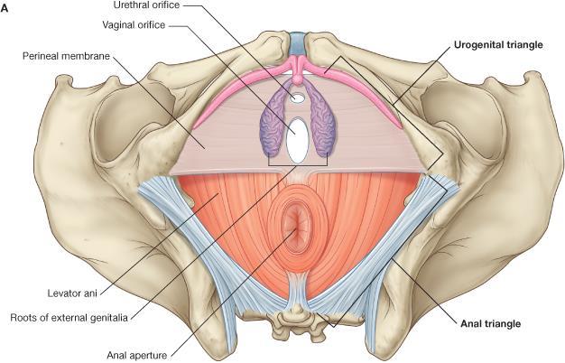
29 Perineum Two Triangles Anal triangle (posterior) contains the anus Urogenital triangle (anterior) contains the root of the scrotum and penis in males or the external genitalia in females The perineal membrane stretches between the two sides of the pubic arch and covers the anterior part of the outlet The perineal body is an irregular fibromuscular mass located between the anal canal and the perineal membrane
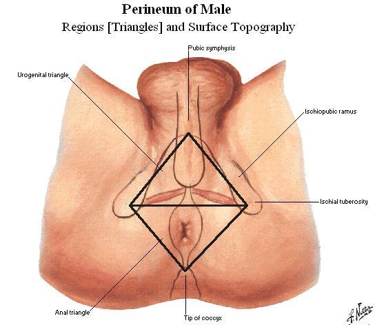
30 Triangles

31 Perineal Membrane

32 Perineal Body
33 Perineum-Female

34 Perineum - Male
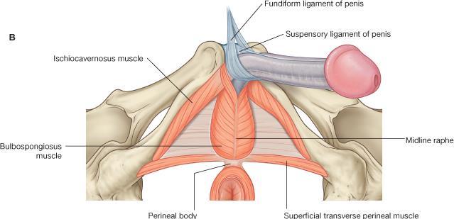
35 Perineum Muscles Bulbospongiosus Females O. central tendon of perineum, I. dorsum of clitoris, urogenital diaphragm; Males O. central tendon of perineum and bulb of penis; I. root of penis; A: compress vagina orifice / urethra; N: perineal nerve, pudenal nerve Ischiocavernosus O. ramus of ischium, I. near pubic symphysis; F: maintains erection of penis or clitoris
36 Muscles
 inhibits")
37 Perineum Muscles External anal sphincter (skeletal muscle) N: Inferior Anal Nerve (S2-S4) inhibits voiding Internal Anal Sphincter smooth muscle sympathetic supply (S2,S3,S4) inhibits voiding
38 Superficial Transverse Perineal O: Ischial tuberosity I: Perineal body A: Support perineal body N: Pudenal nerve
39 External Urethral Sphincter O: Ischial tuberosity I: surround urethra A: compress urethra to maintain urine continence N: Pudenal (S2-S4)
40 Deep Transverse Perineal O: Medial Aspect of Ischial Ramus I: Pineal Body A: Stabilizes position of the perineal body N: Pudenal N (S2-S4)

41 Ischianal fossa - male

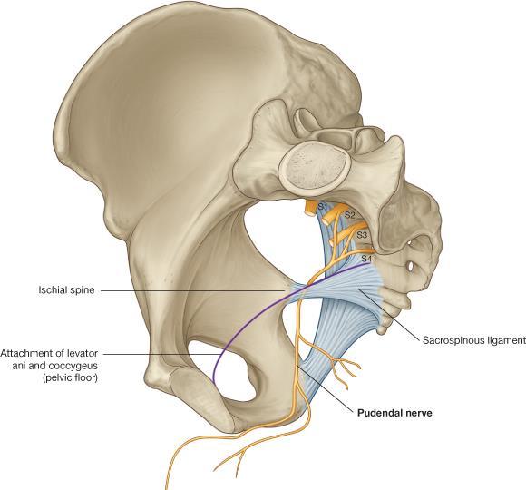
42 Pudendal canal

43 Pudendal canal & content
Abdomen. Retroperitoneal space
 Abdomen. Retroperitoneal space Abdominal cavity The space bounded by: Anterolateral abdominal wall Posterior abdominal wall Diaphragm Pelvic walls and pelvic floor. Subdivided into: True abdominal cavity
Pelvis MCQs. Block 1. B. Reproductive organs. C. The liver. D. Urinary bladder. 1. The pelvic diaphragm includes the following muscles: E.
 Pelvis MCQs Block 1 1. The pelvic diaphragm includes the following muscles: A. The obturator internus B. The levator ani C. The coccygeus D. The external urethral sphincter E. The internal urethral sphincter
Perineum. done by : zaid al-ghnaneem
 Perineum done by : zaid al-ghnaneem Hello everyone, this sheet will talk about 2 nd Lecture which is Perineum but there are some slides and info from 1 st Lecture. Everything included Slides + Pics Let
Dana Alrafaiah. - Amani Nofal. - Ahmad Alsalman. 1 P a g e
 - 2 - Dana Alrafaiah - Amani Nofal - Ahmad Alsalman 1 P a g e This lecture will discuss five topics as follows: 1- Arrangement of pelvic viscera. 2- Muscles of Pelvis. 3- Blood Supply of pelvis. 4- Nerve
REPRODUCTIVE SYSTEM By Dr.Ahmed Salman
 The University Of Jordan Faculty Of Medicine Anatomy Department REPRODUCTIVE SYSTEM By Dr.Ahmed Salman Assistant Professor of Anatomy &embryology Perineum It is the diamond-shaped lower end of the trunk
Table 2. First Generated List of Expert Responses. Likert-Type Scale. Category or Criterion. Rationale or Comments (1) (2) (3) (4)
 (2) (3) (4)") Table 2. First Generated List of Expert Responses. Likert-Type Scale Category or Criterion Anatomical Structures and Features Skeletal Structures and Features (1) (2) (3) (4) Rationale or Comments 1. Bones
Table 2. First Generated List of Expert Responses. Likert-Type Scale Category or Criterion Anatomical Structures and Features Skeletal Structures and Features (1) (2) (3) (4) Rationale or Comments 1. Bones
Pelvis Perineum MCQs. Block 1.1. A. Urinary bladder. B. Rectum. C. Reproductive organs. D. The thigh
 Pelvis Perineum MCQs Block 1.1 1. The pelvic diaphragm includes the following muscles: A. The coccygeus B. The levator ani C. The external urethral sphincter D. The internal urethral sphincter E. The obturator
Bony ypelvis. Composition: formed by coccyx, and their articulations Two portions
 Pelvis Bony ypelvis Composition: formed by paired hip bones, sacrum, coccyx, and their articulations Two portions Greater pelvis Lesser pelvis Terminal line ( pelvic inlet): formed by promontory of sacrum,
ORIENTING TO BISECTED SPECIMENS ON THE PELVIS PRACTICAL
 ORIENTING TO BISECTED SPECIMENS ON THE PELVIS PRACTICAL The Pelvis is just about as complicated as head and neck and considerably more mysterious. You have to be able to visualize (imagine) the underlying
Slide Read the tables it is about the difference between male & female pelvis.
 I didn t include the slides, this is only what the doctor read or said because he skipped a lot of things because we took it previously, very important to go back to the slides (*there is an edited version)
NOTES FROM GUTMAN LECTURE 10/26 Use this outline to study from. As you go through Gutman s lecture, fill in the topics.
 NOTES FROM GUTMAN LECTURE 10/26 Use this outline to study from. As you go through Gutman s lecture, fill in the topics. Anatomy above the arcuate line Skin Camper s fascia Scarpa s fascia External oblique
UROGENITAL SYSTEM By Dr.Ahmed Salman
 The University Of Jordan Faculty Of Medicine Anatomy Department UROGENITAL SYSTEM By Dr.Ahmed Salman Assistance Professor of Anatomy &embryology PELVIS Learning Objectives 1. Bony pelvis, its joints and
Urinary Bladder. Prof. Imran Qureshi
 Urinary Bladder Prof. Imran Qureshi Urinary Bladder It develops from the upper end of the urogenital sinus, which is continuous with the allantois. The allantois degenerates and forms a fibrous cord in
2. List the 8 pelvic spaces: list one procedure or dissection which involves entering that space.
 Name: Anatomy Quiz: Pre / Post 1. In making a pfannensteil incision you would traverse through the following layers: a) Skin, Camper s fascia, Scarpa s fascia, external oblique aponeurosis, internal oblique
ischium Ischial tuberosities Sacrotuberous ligament The coccyx
 Perineum General lfeatures Region of below pelvic diaphragm A diamond shape space whose boundaries are those of the pelvic outlet Lower border of symphysis pubis Rami of pubis and ischium Ischial tuberosities
Perineum. Dept. of Human Anatomy Zhou Hong Ying
 Perineum Dept. of Human Anatomy Zhou Hong Ying OUTLINE Subdivision The Layers Urogenital Diaphragm Main Structures inside Superficial & Deep Perineal Spaces Ischioanal Fossa Perineum A narrow region Urogenital
Gross Anatomy of the Urinary System
 Gross Anatomy of the Urinary System Lecture Objectives Overview of the urinary system. Describe the external and internal anatomical structure of the kidney. Describe the anatomical structure of the ureter
Yes, cranially with ovarian, caudally with vaginal. Yes, with uterine artery (collateral circulation between abdominal +pelvic source)
") Blood supply to internal female genitalia: uterine Internal iliac Sup. large branch: uterus, inf. Small branch: cervix+ sup. Vagina Yes, cranially with ovarian, caudally with vaginal Medially in base of
Blood supply to internal female genitalia: uterine Internal iliac Sup. large branch: uterus, inf. Small branch: cervix+ sup. Vagina Yes, cranially with ovarian, caudally with vaginal Medially in base of
-15. -Alaa Albandi. -Dr. Mohammad Almohtasib. 0 P a g e
 -15 -Alaa Albandi - -Dr. Mohammad Almohtasib 0 P a g e In this last lecture, we will talk about the sigmoid colon, rectum, and anal canal. Sigmoid colon It has a mesentery called pelvic mesocolon or sigmoidal
The Male and Female Internal Genitalia. Dr Oluwadiya Kehinde
 The Male and Female Internal Genitalia Dr Oluwadiya Kehinde www.oluwadiya.com Overview The reproductive role of the male is to produce sperm, deliver them to the female Primary sex organs are the gonads
STRUCTURAL BASIS OF MEDICAL PRACTICE EXAMINATION 3. October 16, 2015
 STRUCTURAL BASIS OF MEDICAL PRACTICE EXAMINATION 3 October 16, 2015 PART l. Answer in the space provided. (12 pts) 1. Identify the structures. (2 pts) A. B. A B C. D. C D 2. Identify the structures. (2
Pathogenesis of Chronic Pelvic Pain
 Pathogenesis of Chronic Pelvic Pain Yong-Chul Kim Department of anesthesia and pain medicine, Seoul National University College of Medicine 1 Overview Anatomy Nerve innervation CPP by pathology CPP by
TABLE OF CONTENTS. 1. Introduction I. 2. Lecturers I. 3. Timetable I. 4. Assessment I. 5. Study material II. 6. General information II.
 TABLE OF CONTENTS Page A. ORGANISATIONAL COMPONENT 1. Introduction I 2. Lecturers I 3. Timetable I 4. Assessment I 5. Study material II 6. General information II B. STUDY COMPONENT 1. Block XI: Session
STRUCTURAL BASIS OF MEDICAL PRACTICE EXAMINATION 3. October 17, 2014
 STRUCTURAL BASIS OF MEDICAL PRACTICE EXAMINATION 3 October 17, 2014 PART l. Answer in the space provided. (12 pts) 1. Identify the structures. (2 pts) A. B. A B C. D. C D 2. Identify the structures. (2
TABLE OF CONTENTS A. ORGANISATIONAL COMPONENT B. STUDY COMPONENT. Page. 1. Introduction I. 2. Lecturers I. 3. Timetable I. 4.
 TABLE OF CONTENTS Page A. ORGANISATIONAL COMPONENT 1. Introduction I 2. Lecturers I 3. Timetable I 4. Assessment I 5. Study material II 6. General information II B. STUDY COMPONENT 1. Block XI: Session
Anatomy Review File اللهم ال سهل إال ماجعلته سهال وأنت تجعل الحزن
 Anatomy Review File اللهم ال سهل إال ماجعلته سهال وأنت تجعل الحزن إذا شئت سهال This work was done by: Alanoud Abuhaimed Dania Alkelabi Ghada Alothaim Jawaher Abanumy Rawan AlWadee Wejdan Alzeid Mohanad
Anatomy & Physiology Pelvic Girdles 10.1 General Information
 Anatomy & Physiology Pelvic Girdles 10.1 General Information ICan2Ed, Inc. In human anatomy, the pelvis (plural pelves or pelvises) is the lower part of. The area of the body that is between the abdomen
The Female and Male External Genitalia. Prof Oluwadiya KS
 The Female and Male External Genitalia Prof Oluwadiya KS www.oluwadiya.com Anatomy of the female external genitalia This consists of : The vulva which is made up of: o The clitoris o Vestibular apparatus
B) cervix of uterus C) vagina D) rectum. 1. What number illustrates the adnexal area? (Fig. 4-64) A) 4 B) 5 C) 8 D) 9
 cervix of uterus C) vagina D) rectum. 1. What number illustrates the adnexal area? (Fig. 4-64) A) 4 B) 5 C) 8 D) 9") Pelvis Practice Problems 1. What number illustrates the adnexal area? (Fig. 4-64) A) 4 B) 5 C) 8 D) 9 2. What number illustrates the cervix? (Fig. 4-64) A) 4 B) 8 C) 5 D) 6 3. Which of the following is
Pelvis Practice Problems 1. What number illustrates the adnexal area? (Fig. 4-64) A) 4 B) 5 C) 8 D) 9 2. What number illustrates the cervix? (Fig. 4-64) A) 4 B) 8 C) 5 D) 6 3. Which of the following is
Benha University. Faculty of Medicine. Anatomy Department Course code (MED 0701) Model answer of Anatomy examination. (Abdomen,Pelvis and Thorax)
 Model answer of Anatomy examination. (Abdomen,Pelvis and Thorax)") 1 Benha University Faculty of Medicine Anatomy Department Course code (MED 0701) Model answer of Anatomy examination (Abdomen,Pelvis and Thorax) 1 st year 2 nd term Date :18 /5 /2013 2 I-Short account
1 Benha University Faculty of Medicine Anatomy Department Course code (MED 0701) Model answer of Anatomy examination (Abdomen,Pelvis and Thorax) 1 st year 2 nd term Date :18 /5 /2013 2 I-Short account
SUBJECTS 2nd year, 1st semester I. 1. Primitive gut - limits, derivatives 2. Foregut -limits, evolution, derivatives 3. Midgut -limits, evolution,
 SUBJECTS 2nd year, 1st semester I. 1. Primitive gut - limits, derivatives 2. Foregut -limits, evolution, derivatives 3. Midgut -limits, evolution, derivatives 4. Hindgut- limits, evolution, derivatives
Pelvic Angiogram - Male
 Pelvic Angiogram - Male Common iliac artery Internal iliac artery Lateral sacral artery Iliolumbar artery Posterior trunk of internal iliac artery Superior gluteal artery Internal pudendal artery External
Femoral Triangle and Adductor Canal. Dr. Heba Kalbouneh Associate Professor of Anatomy and Histology
 Femoral Triangle and Adductor Canal Dr. Heba Kalbouneh Associate Professor of Anatomy and Histology Femoral Triangle and Adductor Canal Femoral triangle Is a triangular depressed area located in the upper
Learning objectives. SGD on Functions of Testosterone. Class
 Learning objectives SGD on Functions of Testosterone Class 2016 14-1-2013 Discuss o Process of spermatogenesis. o Sex determination. o Process of maturation of sperms. o Physiology of mature sperms. Discuss
Lumbar Plexus. Ventral rami L1 L4 Supplies: Major nerves.. Abdominal wall External genitalia Anteromedial thigh
 Lower Limb Nerves Lectures Objectives Describe the structure and relationships of the plexuses of the lower limb. Describe the course, relationships and structures supplied for the major nerves of the
THE PELVIS VASCULAR AND NERVOUS SYSTEM SOMATIC AND AUTONOMIC NERVES
 THE PELVIS VASCULAR AND NERVOUS SYSTEM SOMATIC AND AUTONOMIC NERVES THE ABDOMINAL AORTA The abdominal aorta begins at the aor9c hiatus in the diaphragm at the level of the T12 vertebra and ends at the
END-SEMESTER EXAM 2018 ANATOMY, HISTOLOGY AND EMBRYOLOGY FACULTY OF MEDICINE, 2 ND SEMESTER
 University of Szeged, Faculty of Medicine Department of Anatomy, Histology and Embryology Chairman: Prof. Antal Nógrádi MD, PhD, DSc Kossuth L. sgt. 40., H-6724 Szeged, Hungary Tel.: +36-62-545-665 P.
The posterior abdominal wall. Prof. Oluwadiya KS
 The posterior abdominal wall Prof. Oluwadiya KS www.oluwadiya.sitesled.com Posterior Abdominal Wall Lumbar vertebrae and discs. Muscles opsoas, quadratus lumborum, iliacus, transverse, abdominal wall
Lecture 56 Kidney and Urinary System
 Lecture 56 Kidney and Urinary System The adrenal glands are located on the superomedial aspect of the kidney The right diagram shows a picture of the kidney with the abdominal walls and organs removed
ABDOMINAL WALL & RECTUS SHEATH
 ABDOMINAL WALL & RECTUS SHEATH Learning Objectives Describe the anatomy, innervation and functions of the muscles of the anterior, lateral and posterior abdominal walls. Discuss their functional relations
PELVIS II: FUNCTION TABOOS (THE VISCERA) Defecation Urination Ejaculation Conception
 Defecation Urination Ejaculation Conception") PELVIS II: FUNCTION TABOOS (THE VISCERA) Defecation Urination Ejaculation Conception REVIEW OF PELVIS I Pelvic brim, inlet Pelvic outlet True pelvis-- --viscera Tilt forward Mid-sagital views-- --how the
PELVIS II: FUNCTION TABOOS (THE VISCERA) Defecation Urination Ejaculation Conception REVIEW OF PELVIS I Pelvic brim, inlet Pelvic outlet True pelvis-- --viscera Tilt forward Mid-sagital views-- --how the
1. The part of the uterine wall which is not shed during menstruation is the: Endometrium Myometrium Mesometrium Cervical mucosa Rugae 2.
 1. The part of the uterine wall which is not shed during menstruation is the: Endometrium Myometrium Mesometrium Cervical mucosa Rugae 2. The extension of the vaginal lumen around the intravaginal part
Ureters, Urinary Bladder & Urethra
 Ureters, Urinary Bladder & Urethra Please check our Editing File هذا العمل ال يغني عن المصدر األساسي للمذاكرة Lecture 2 } و م ن ي ت و ك ع ل ا لل ه ف ه و ح س ب ه { Objectives o Describe the course of ureter
The Pelvis - Complied by Laurence Hattersley /3/2018. The Pelvis. Compiled by Laurence Hattersley
 5/3/2018 The Pelvis Compiled by Laurence Hattersley i Contents The Pelvis... i The Pelvis... 1 The Pelvic Cavity... 2 Sex differences... 2 Biiliac width... 4 Ligaments of pelvis... 5 Mechanism of the Pelvis...
Rama Nada. - Ensherah Mokheemer. - Ahmed salman. 1 P a g e
 - 5 - Rama Nada - Ensherah Mokheemer - Ahmed salman 1 P a g e We will continue talking about the urinary bladder The ligaments of the bladder: 1-Median umbilical ligament: Continuous with apex of the bladder
Functional anatomy of the female pelvic floor and lower urinary tract Stefano Floris, MD, PhD Department of Obstetrics and Gynaecology
 Functional anatomy of the female pelvic floor and lower urinary tract Stefano Floris, MD, PhD Department of Obstetrics and Gynaecology Ospedale San Giovanni di Dio, Gorizia, Italy ANATOMY URINARY CONTINENCE
GI module Lecture: 9 د. عصام طارق. Objectives:
 GI module Lecture: 9 د. عصام طارق Objectives: To list structures forming posterior abdominal wall. To follow aorta & its main branches. To describe IVC & its main tributaries. To list nerves of posterior
ANATYOMY OF The thigh
 ANATYOMY OF The thigh 1- Lateral cutaneous nerve of the thigh Ι) Skin of the thigh Anterior view 2- Femoral branch of the genitofemoral nerve 5- Intermediate cutaneous nerve of the thigh 1, 2 and 3 are
THE ABDOMEN SUPRARENAL GLANDS KIDNEY URETERS URINARY BLADDER
 THE ABDOMEN SUPRARENAL GLANDS KIDNEY URETERS URINARY BLADDER THE SUPRARENAL GLANDS The suprarenal (adrenal) glands lie immediately superior and slightly anterior to the upper pole of either kidney. Golden
ANATYOMY OF The thigh
 ANATYOMY OF The thigh 1- Lateral cutaneous nerve of the thigh Ι) Skin of the thigh Anterior view 2- Femoral branch of the genitofemoral nerve 5- Intermediate cutaneous nerve of the thigh 1, 2 and 3 are
DISSECTION 8: URINARY AND REPRODUCTIVE SYSTEMS
 8546d_c01_1-42 6/25/02 4:32 PM Page 38 mac48 Mac 48: 420_kec: 38 Cat Dissection DISSECTION 8: URINARY AND REPRODUCTIVE SYSTEMS Typically, the urinary and reproductive systems are studied together, because
Human Anatomy Unit 3 REPRODUCTIVE SYSTEM
 Human Anatomy Unit 3 REPRODUCTIVE SYSTEM In Anatomy Today Male Reproductive System Gonads = testes primary organ responsible for sperm production development/maintenan ce of secondary sex characteristics
Gross anatomy of the urinary system. Done by : razan krishan. slide in bold and book in green
 Gross anatomy of the urinary system Done by : razan krishan slide in bold and book in green Kidneys, ureters, urinary bladder & urethra Urine flows from each kidney, down its ureter to the bladder and
2/23/15 PRESENTERS ANATOMY OF THE PELVIC FLOOR
 ENHANCING PELVIC FLOOR FUNCTIONING THROUGH SEATING AND POSITIONING Carina Siracusa Majzun, PT, DPT Derrick Johnson, ATP PRESENTERS Carina Siracusa Majzun, PT, DPT Ohio Health, Columbus Ohio Pelvic Floor
Skeletal System Module 13: The Pelvic Girdle and Pelvis
 OpenStax-CNX module: m47993 1 Skeletal System Module 13: The Pelvic Girdle and Pelvis Donna Browne Based on The Pelvic Girdle and Pelvis by OpenStax College This work is produced by OpenStax-CNX and licensed
The thigh. Prof. Oluwadiya KS
 The thigh Prof. Oluwadiya KS www.oluwadiya.com The Thigh: Boundaries The thigh is the region of the lower limb that is approximately between the hip and knee joints Anteriorly, it is separated from the
Lecture 10 Arteries and veins of the upper limb
 Lecture 10 Arteries and veins of the upper limb 1. Identify the Subclavian, axillary, brachial (deep and superficial), radial and ulnar arteries and superficial/deep palmar arches 2. Describe the major
PELVIS & SACRUM Dr. Jamila El-Medany Dr. Essam Eldin Salama
 PELVIS & SACRUM Dr. Jamila El-Medany Dr. Essam Eldin Salama Learning Objectives At the end of the lecture, the students should be able to : Describe the bony structures of the pelvis. Describe in detail
Preview from Notesale.co.uk Page 1 of 34
 Abdominal viscera and digestive tract Digestive tract Abdominal viscera comprise majority of the alimentary system o Terminal oesophagus, stomach, pancreas, spleen, liver, gallbladder, kidneys, suprarenal
rotation of the hip Flexion of the knee Iliac fossa of iliac Lesser trochanter Femoral nerve Flexion of the thigh at the hip shaft of tibia
 Anatomy of the lower limb Anterior & medial compartments of the thigh Dr. Hayder The fascia lata encloses the entire thigh like a sleeve/stocking. Three intramuscular fascial septa (lateral, medial, and
Basic Body Structure
 Basic Body Structure The Cell All life consists of microscopic living structures called cells. They perform various functions throughout the body. All cells are similar in structure, but not identical.
ANATOMY OF PELVICAYCEAL SYSTEM -DR. RAHUL BEVARA
 1 ANATOMY OF PELVICAYCEAL SYSTEM -DR. RAHUL BEVARA 2 KIDNEY:ANATOMY OVERVIEW Kidneys are retroperitoneal, in posterior abdominal region, extending from T12 L3 Bean-shaped Right kidney is lower than left
The Hip (Iliofemoral) Joint. Presented by: Rob, Rachel, Alina and Lisa
 Joint. Presented by: Rob, Rachel, Alina and Lisa") The Hip (Iliofemoral) Joint Presented by: Rob, Rachel, Alina and Lisa Surface Anatomy: Posterior Surface Anatomy: Anterior Bones: Os Coxae Consists of 3 Portions: Ilium Ischium Pubis Bones: Pubis Portion
The Hip (Iliofemoral) Joint Presented by: Rob, Rachel, Alina and Lisa Surface Anatomy: Posterior Surface Anatomy: Anterior Bones: Os Coxae Consists of 3 Portions: Ilium Ischium Pubis Bones: Pubis Portion
Gluteal region DR. GITANJALI KHORWAL
 Gluteal region DR. GITANJALI KHORWAL Gluteal region The transitional area between the trunk and the lower extremity. The gluteal region includes the rounded, posterior buttocks and the laterally placed
LAB Notes#1. Ahmad Ar'ar. Eslam
 LAB Notes#1 Ahmad Ar'ar Eslam 1 P a g e Anatomy lab Notes Lower limb bones :- Pelvic girdle: It's the connection between the axial skeleton and the lower limb; it's made up of one bone called the HIP BONE
Group of students. - Rawan almujabili د. محمد المحتسب - 1 P a g e
 - 14 - Group of students - Rawan almujabili د. محمد المحتسب - 1 P a g e Nerves of the posterior abdominal wall The spinal cord gives off spinal nerves between the vertebrae. In the abdomen, through the
Biology Human Anatomy Abdominal and Pelvic Cavities
 Biology 351 - Human Anatomy Abdominal and Pelvic Cavities Please place your name and I.D. number on the back of the last page of this exam. You must answer all questions on this exam. Because statistics
Biology 224 Human Anatomy and Physiology II Week 9; Lecture 2; Wednesday Stuart Sumida. Development and Structure, of the Reproductive System
 Biology 224 Human Anatomy and Physiology II Week 9; Lecture 2; Wednesday Stuart Sumida Development and Structure, of the Reproductive System Don t forget the relationships of the structures of the layers
The Thoracic wall including the diaphragm. Prof Oluwadiya KS
 The Thoracic wall including the diaphragm Prof Oluwadiya KS www.oluwadiya.com Components of the thoracic wall Skin Superficial fascia Chest wall muscles (see upper limb slides) Skeletal framework Intercostal
Anatomy of the Large Intestine
 Large intestine Anatomy of the Large Intestine 2 Large Intestine Extends from ileocecal valve to anus Length = 1.5-2.5m = 5 feet Regions Cecum = 2.5-3 inch Appendix= 3-5 inch Colon Ascending= 5 inch Transverse=
TME and autonomic nerve preservation techniques: based on Video and Cadaveric anatomy
 TME and autonomic nerve preservation techniques: based on Video and Cadaveric anatomy Nam Kyu Kim M.D., Ph.D., FACS, FRCS, FASCRS Professor Department of Surgery Yonsei University College of Medicine Seoul,
Copyright 2003 Pearson Education, Inc. publishing as Benjamin Cummings. Dr. Nabil Khouri MD, MSc, Ph.D
 Dr. Nabil Khouri MD, MSc, Ph.D Pelvic Girdle (Hip) Organization of the Lower Limb It is divided into: The Gluteal region The thigh The knee The leg The ankle The foot The thigh and the leg have compartments
LAPAROSCOPIC REPAIR OF PELVIC FLOOR
 LAPAROSCOPIC REPAIR OF PELVIC FLOOR Dr. R. K. Mishra Elements comprising the Pelvis Bones Ilium, ischium and pubis fusion Ligaments Muscles Obturator internis muscle Arcus tendineus levator ani or white
First practical session. Bones of the gluteal region
 First practical session 2017 Bones of the gluteal region The Hip bone The hip bone is made of: 1 The ilium: superior in position 2 The ischium:postero-inferior in position 3 The pubis: antero-inferior
UNIVERSITY DEVELOPMENT CENTER. Course Specification 2015/2016 For the Anatomy (first year) Medicine Anatomy and Embryology Department 29/12/2015
 Medicine Anatomy and Embryology Department 29/12/2015") Course Specification 2015/2016 For the Anatomy (first year) Faculty : Department : Medicine Anatomy and Embryology Department Course Specification: Programme (s) on which the course is given : M.B.B.Ch
Course Specification 2015/2016 For the Anatomy (first year) Faculty : Department : Medicine Anatomy and Embryology Department Course Specification: Programme (s) on which the course is given : M.B.B.Ch
Abdomen: Introduction. Prof. Oluwadiya KS
 Abdomen: Introduction Prof. Oluwadiya KS www.oluwadiya.com Abdominopelvic Cavity Abdominal Cavity Pelvic Cavity Extends from the inferior margin of the thorax to the superior margin of the pelvis and the
The front of the thigh. Dr.Amjad shatarat
 The front of the thigh Femoral triangle (Scarpa s triangle) Is a triangular depressed area located in the upper part of the medial aspect of the thigh immediately below the inguinal ligament. Superiorly:
Adductor canal (Subsartorial) or Hunter s canal
 or Hunter s canal") Adductor canal (Subsartorial) or Hunter s canal John Hunter described the exposure and ligation of the femoral artery in this canal for aneurysm of the popliteal artery; this method has the advantage that
Adductor canal (Subsartorial) or Hunter s canal John Hunter described the exposure and ligation of the femoral artery in this canal for aneurysm of the popliteal artery; this method has the advantage that
Lecture 08 THIGH MUSCLES ANTERIOR COMPARTMENT. Dr Farooq Khan Aurakzai. Dated:
 Lecture 08 THIGH MUSCLES ANTERIOR COMPARTMENT BY Dr Farooq Khan Aurakzai Dated: 11.02.2017 INTRODUCTION to the thigh Muscles. The musculature of the thigh can be split into three sections by intermuscular
أحمد رواجبة- محمود الحربي- أحمد السالمان-
 -6 أحمد رواجبة- محمود الحربي- أحمد السالمان- 1 P a g e The Male Reproductive System The male genital system structures are divided into: Internal structures: 1- Prostate 3-Ejaculatory ducts External structures:
Anatomy of Reproductive System
 Anatomy of Reproductive System Anatomy of Reproductive System Lector: MD Ganna Pola Anatomy of Female Reproductive System The female reproductive system (or female genital system) contains two main parts:
The os coxae or hip bone consists of three flat bones, ilium, ischium and pubis, which fuse together to form the acetabulum.
 The os coxae The os coxae or hip bone consists of three flat bones, ilium, ischium and pubis, which fuse together to form the acetabulum. The ilium extends from the acetabulum upwards forming the lateral
ANATOMY OF THE SMALL & LARGE INTESTINES. Semester 1, 2011 A. Mwakikunga
 ANATOMY OF THE SMALL & LARGE INTESTINES Semester 1, 2011 A. Mwakikunga LEARNING OBJECTIVES 1. List the parts and anatomical regions of the small and large intestines 2. State anatomical relations of the
د. عصام طارق. Objectives:
 GI anatomy Lecture: 5 د. عصام طارق Objectives: To describe anatomy of stomach, duodenum & pancreas. To list their main relations. To define their blood & nerve supply. To list their lymph drainage. To
The Urinary System Pearson Education, Inc.
 26 The Urinary System Introduction The urinary system does more than just get rid of liquid waste. It also: Regulates plasma ion concentrations Regulates blood volume and blood pressure Stabilizes blood
The anatomy of the rectum and anal canal
 The anatomy of the rectum and anal canal Vishy Mahadevan Abstract Diseases of the rectum and anal canal, both benign and malignant, account for a very large part of colorectal surgical practice in the
Surface Anatomy. Location Shape Weight Role of Five Surfaces Borders Fissures Lobes Peritoneal Lig
 The Liver Functions Bile production and secretion Detoxification Storage of glycogen Protein synthesis Production of heparin and bile pigments Erythropoiesis (in fetus) Surface Anatomy Location Shape Weight
Copyright 2003 Pearson Education, Inc. publishing as Benjamin Cummings. Dr. Nabil Khouri
 Dr. Nabil Khouri Objectives: General objectives: - to identify the kidney s structures, function and location - to analyze the relationship between microscopic structure and function Specific objectives:
ANATOMY (7). INTRODUCTION TO THE PELVIS. MOHAMMAD ALLOUH. BNB. 1 P a g e
. INTRODUCTION TO THE PELVIS. MOHAMMAD ALLOUH. BNB. 1 P a g e") ANATOMY (7). INTRODUCTION TO THE PELVIS. MOHAMMAD ALLOUH. BNB 1 P a g e Introduction to the Pelvis: Pelvic Wall & Pelvic Cavity pelvis (L, basin, sink ), is The region of the trunk that lies inferoposterior
ANATOMY (7). INTRODUCTION TO THE PELVIS. MOHAMMAD ALLOUH. BNB 1 P a g e Introduction to the Pelvis: Pelvic Wall & Pelvic Cavity pelvis (L, basin, sink ), is The region of the trunk that lies inferoposterior
EMBRIOLOGY Intermediate Mesoderm. Becomes the suprarenal glands, gonads, kidneys and associated tubes and vasculature
 ANATOMY AND EMBRYOLOGY OF THE FEMALE GENITAL SYSTEM EMBRIOLOGY Intermediate Mesoderm Becomes the suprarenal glands, gonads, kidneys and associated tubes and vasculature THE VULVA 1. Mons : a pad of
ANATOMY OF THE PLEURA. Dr Oluwadiya KS
 ANATOMY OF THE PLEURA Dr Oluwadiya KS www.oluwadiya.sitesled.com Introduction The thoracic cavity is divided mainly into: Right pleural cavity Mediastinum Left Pleural cavity Pleural cavity The pleural
The abdominal Esophagus, Stomach and the Duodenum. Prof. Oluwadiya KS
 The abdominal Esophagus, Stomach and the Duodenum Prof. Oluwadiya KS www.oluwadiya.com Viscera of the abdomen Abdominal esophagus: Terminal part of the esophagus The stomach Intestines: Small and Large
Fig. A.1. Frontal. plane. Transverse. plane. Sagittal plane. Copyright McGraw-Hill Education. Permission required for reproduction or display.
 Fig. A.1 Frontal plane Transverse plane Sagittal plane McGraw-Hill Education/Joe DeGrandis Fig. A.2 (a) Sagittal section (b) Frontal section (c) Transverse section Table A.1 Fig. A.3 Cephalic r. (head)
Group B: Organ systems (digestive, respiratory, urinary, genital system, heart, glands and skin) green
 green") Group B: Organ systems (digestive, respiratory, urinary, genital system, heart, glands and skin) green Digestive system 1. Teeth Main points: external and internal structure of a tooth, fixation of a tooth
Group B: Organ systems (digestive, respiratory, urinary, genital system, heart, glands and skin) green Digestive system 1. Teeth Main points: external and internal structure of a tooth, fixation of a tooth
Urinary 1 Checklist Gross Anatomy of the Urinary System
 Urinary 1 Checklist Gross Anatomy of the Urinary System Urinary system Kidneys Parietal peritoneum Retroperitoneal Renal fascia The urinary system consists of two kidneys, two ureters, the urinary bladder,
ANATYOMY OF The thigh
 ANATYOMY OF The thigh 1- Lateral cutaneous nerve of the thigh Ι) Skin of the thigh Anterior view 2- Femoral branch of the genitofemoral nerve 1, 2 and 3 are From the lumber plexus 5- Intermediate cutaneous
Biology Human Anatomy Abdominal and Pelvic Cavities
 Biology 351 - Human Anatomy Abdominal and Pelvic Cavities You must answer all questions on this exam. Because statistics demonstrate that, on average, between 2-5 questions on every 100-point exam are
Axial Muscles of the Abdominal Wall, and Thorax *
 OpenStax-CNX module: m46485 1 Axial Muscles of the Abdominal Wall, and Thorax * OpenStax This work is produced by OpenStax-CNX and licensed under the Creative Commons Attribution License 4.0 By the end
GI anatomy Lecture: 2 د. عصام طارق
 GI anatomy Lecture: 2 د. عصام طارق Objectives: To define rectus sheath. To describe anatomy of inguinal canal. To relates types of inguinal hernia to the region. To explore spermatic cord. Rectus Abdominis
C. Bones of the Pelvic Girdle
 C. Bones of the Pelvic Girdle 1. 2 coxal bones (a.k.a hip bones): -bony pelvis is made up of hip bones, sacrum, & coccyx -pelvic bones are large & heavy & attach to the axial skeleton via sacrum/coccyx