Situs inversus. Dr praveena pulmonology- final year post graduate
|
|
|
- Eric Robert Snow
- 5 years ago
- Views:
Transcription
1 Situs inversus Dr praveena pulmonology- final year post graduate
2 Definiton History Types Cause Clinical features Diagnosis Treatment
3 Definition The term situs inversus is a short form of the latin phrase "situs inversus viscerum", meaning "inverted position of the internal organs
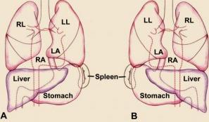
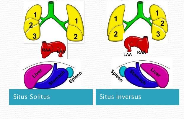
4 Situs inversus (also called situs transversus or oppositus) is a congenital condition in which the major visceral organs are reversed or mirrored from their normal positions. The normal arrangement of internal organs is known as situs solitus while situs inversus is generally the mirror image of situs solitus.
5
6 Situs inversus is found in about 0.01% [1] of the population, or about 1 person in 10,000. In the most common situation, situs inversus totalis, it involves complete transposition (right to left reversal) of all of the abdominal organs. Most people with situs inversus are asymptomatic and silent and hence remain undiagnosed. Recent advances and technology in medicine has enabled to diagnose situs inversus very easily.
7 History Dextrocardia (the heart being located on the right side of the thorax) was first seen and drawn by Leonardo da Vinci in , and then recognised by Marco Aurelio Severino in However, situs inversus was first clearly described more than a century later by Matthew Baillier

8
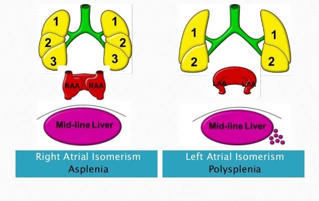
9 In rarer cases such as situs ambiguus or heterotaxy, situs cannot be determined. In these patients, the liver may be midline, the spleen absent or multiple, and the bowel malrotated. Often, structures are duplicated or absent altogether. This is more likely to cause medical problems than situs inversus totalis
10 Symptoms & signs In the absence of congenital heart defects,individuals with situs inversus are phenotypically normal, and can live normal healthy lives, without any complications related to their medical condition. There is a 5 10% prevalence of congenital heart disease in individuals with situs inversus totalis, most common transposition of the great vessels. The incidence of congenital heart disease is 95% in situs inversus with levocardia.

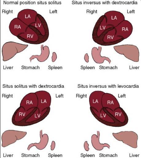
11 Cardiac orientation Relationship or axis of the base to the apex of heart (levocardia,dextrocardia,mesocardia) If the cardiac apex fails to shift,it may result in situs solitus with dextrocardia which is termed as dextorversion or situs inversus with levocardia called levoversion
12 Many people with situs inversus totalis are unaware of their unusual anatomy until they seek medical attention for an unrelated condition, such as a rib fracture or a bout of appendicitis. The condition may also be discovered during the administration of certain medicines or during tests such as a Barium meal or enema. The reversal of the organs may then lead to some confusion, as many signs and symptoms will be on the atypical side.
13 Thus, in the event of a medical problem, the knowledge that the individual has situs inversus can expedite diagnosis. People with this rare condition should inform their physicians before an examination, so the physician can redirect their search for heart sounds and other signs. Wearing a medical identification tag can help inform health care providers in the event the person is unable to communicate.
14 cause Situs inversus is generally an autosomal recessive genetic condition, although it can be X- linked or found in identical "mirror image" twins About 25% of individuals with situs inversus have an underlying condition known as primary ciliary dyskinesia(pcd).
15 PCD is a dysfunction of the cilia that manifests itself during the embryologic phase of development. Normally functioning cilia determine the position of the internal organs during early embryological development, and so embryos with PCD have a 50% chance of developing situs inversus.
16 SITUS INVERSUS WITH DEXTROCARDIA Incidence rate in general population is estimatedat 1/8000 to 1/25000 The heart and thoracic,abdominal viscera are mirror images of normal. The bronchi are inverted. The heart is right sided The right hemidiaphragm is lower than the left hemidiaphram. The descending aorta is on the right The ascending aorta,aortic knuckle,pulmonary trunk in their mirror image positions.
17
18 Situs solitus with dextrocardia The lungs and abdominal viscera are solitus. The heart is right thorax (dextrocardia) The ascending aorta,aortic knucle occupy their normal positions and the descending aorta runs its normal course along the left vertebral border. The major cardiac shadow lies to the right of midline (dextrocardia), the base to apex points to the right The right hemidiaphram is lower than the left hemidiaphragm
19 Situs inversus with levocardia The left hemidiaphragm is lower than the right hemidiaphragm because the apex is on the left Inversion of bronchi,coincides with inversion of atria and lungs The stomach is on right,and the liver is on left
20
21
22 Right isomerism Left isomerism Bilateral morphologic right atria Bilateral morphologic left atria Asplenia Bilateral bilobed lungs Bilateral trilobed lungs Interrupted inferior vena cava Symmetric liver Partial anomalous pulmonary venous return Total anomalous pulmonary venous return
23 Diagnosis History Clinical examination Radiology x-ray Ultrasound Contrast studies Ct scan MRI scan
24 Treatment No treatment required as the patient is otherwise healthy
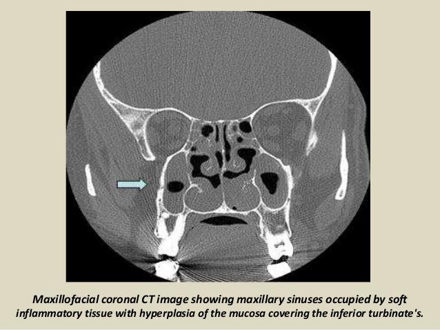
25 Kartageners syndrome Autosomal recessive primary ciliary dyskinesia leading to impaired mucociliary clearance Incidence_ 1: Characterised by clinical triad of Situs inversus Chronic sinusitis Bronchiectasis Other features include nasal polyposis Infertility in males Subfertility in females
26
27
28
29
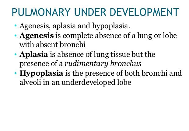
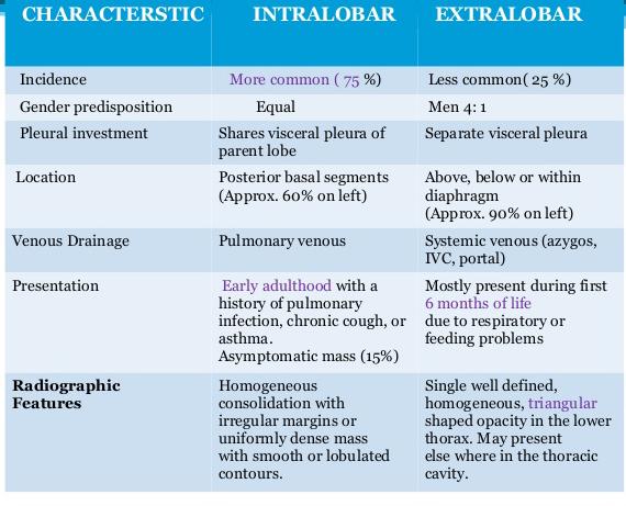
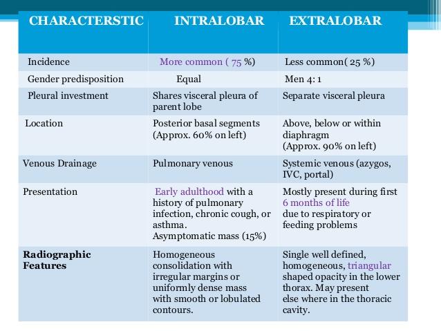
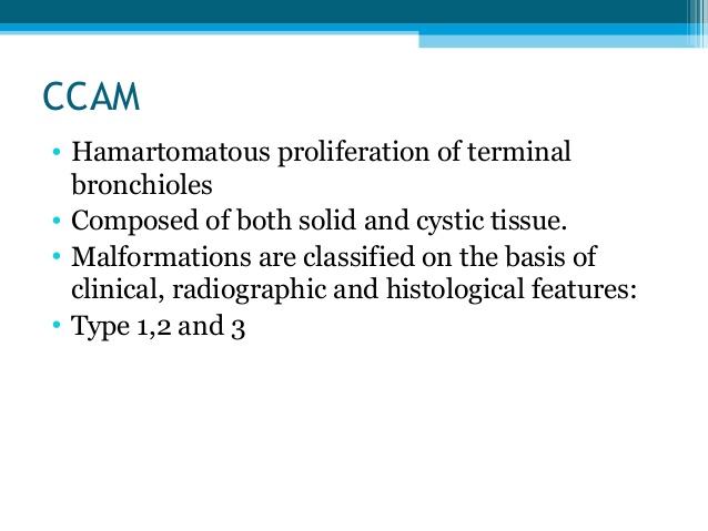
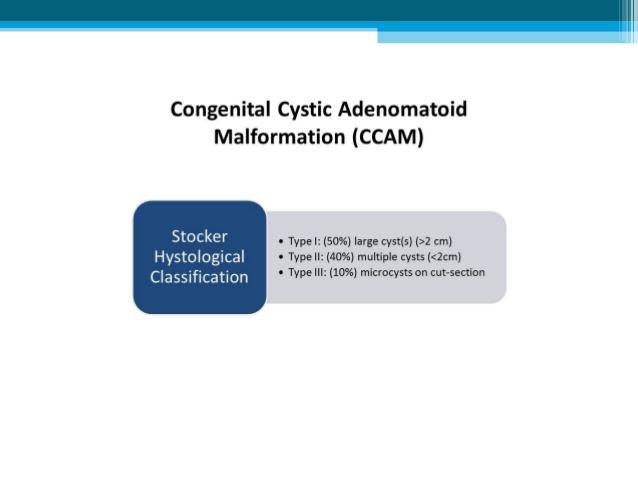
30 Congenital anamolies of lung Bronchopulmonary anomalies Combined lung & vascular anamolies Vascular anamolies Congenital bronchial atresia Congenital lobar emphysema Congenital cystic adenomatiod malformation Bronchogenic cysts Tracheal stenosis Hypogenetic lung (scimitar) syndrome Brochopulmonary sequestration Absence of main pulmonary artery Anomalous origin of the left pulmonary artery from the right Anomalous pulmonary venous drainage Pulmonary arteriovenous malformation Tracheal bronchus
31
32
33
34
35

36 Scimitar syndrome
37
38

39
40
41
42
43
44

45
46 ]
47
48
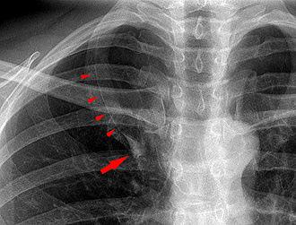
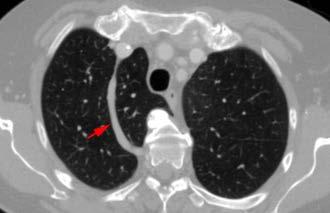
49 Azygos lobe Azygos lobe is a congenital variation of the right upper lobe. It is seen in 1% of the population. Embryologically, it arises from an anomalous lateral course of the azygos vein in a pleural septum within the apical segment of the right upper lobe or in other words an azygos lobe is formed when the right posterior cardinal vein, one of the precursors of the azygos vein, fails to migrate over the apex of the lung and penetrates it instead, carrying along two pleural layers that invaginates into the upper portion of the right upper lobe.
50 As it has no bronchi, veins and arteries of its own or corresponding alteration in the segmental architecture of the lung, so it is not a true (misnomer), or even accessory, pulmonary lobe, but rather an anatomically separated part of the upper lobe It is usually an incidental finding on chest x- ray or computed tomography and is as such not associated with any morbidity but can cause technical problems in thoracoscopic procedures.
51
52
53 Thank you
Cardiopulmonary Syndromes: Conditions With Concomitant Cardiac and Pulmonary Abnormalities
 Cardiopulmonary Syndromes: Conditions With Concomitant Cardiac and Pulmonary Abnormalities Carlos S. Restrepo M.D. Professor of Radiology The University of Texas HSC at San Antonio Cardiopulmonary Syndromes
Situs at the mirror: from situs inversus to situs ambiguus
 Situs at the mirror: from situs inversus to situs ambiguus Poster No.: C-0605 Congress: ECR 2013 Type: Educational Exhibit Authors: C. Maciel, J. Maciel, A. Silva, A. F. L. Carneiro ; Porto/PT, 1 2 1 1
Lung sequestration and Scimitar syndrome
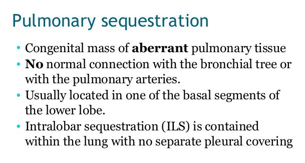
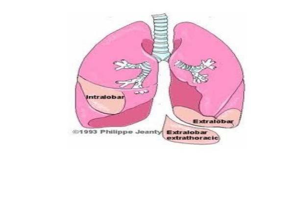
 Lung sequestration and Scimitar syndrome Imaging approaches M. Mearadji International Foundation for Pediatric Imaging Aid Rotterdam, The Netherlands Pulmonary sequestration Pulmonary sequestration (PS)
CASE OF HETEROTAXY SYNDROME WITH POLYSPLENIA AND INTESTINAL MALROTATION
 CASE OF HETEROTAXY SYNDROME WITH POLYSPLENIA AND INTESTINAL MALROTATION *Sagar H S, Basanta Manjari Swain, Jayashree Mohanty and Sasmita Parida Department of Radio diagnosis, S.C.B. Medical College, Cuttack
What do we know about Heterotaxy Syndrome? - An illustrated guide.
 What do we know about Heterotaxy Syndrome? - An illustrated guide. Poster No.: C-2369 Congress: ECR 2015 Type: Educational Exhibit Authors: M. C. Ageitos Casais, A. X. Martínez de Alegría Alonso, 1 1 2
This is the left, right?
 This is the left, right? Poster No.: C-1214 Congress: ECR 2013 Type: Educational Exhibit Authors: L.-L. Huang, L. Mitchell, S. Andronikou, F. Suleman, Z. I. Lockhat; Pretoria/ZA Keywords: Congenital, Diagnostic
Congenital Heart Disease Systematic Interpretation of CT Suhny Abbara, MD
 Congenital Heart Disease Systematic Interpretation of CT Suhny Abbara, MD Chief, Cardiothoracic Imaging Division Professor of Radiology UT Southwestern Medical Center, Dallas, TX Suhny.Abbara@UTSouthwestern.edu
A Rare Case Presentation of Meckel s Diverticulum with Situs Inversus Totalis
 A Rare Case Presentation of Meckel s Diverticulum with Situs Inversus Totalis Yazhini.V *, Kannan Thanikachalam Vol. 3 No. 5 (May 2011) International Journal of Collaborative Research on Internal Medicine
Congenital Heart Disease: a Pictorial Illustration of Putting Segmental Approach into Practice
 pissn 2384-1095 eissn 2384-1109 imri 2015;19:205-211 http://dx.doi.org/10.13104/imri.2015.19.4.205 Congenital Heart Disease: a Pictorial Illustration of Putting Segmental Approach into Practice Tse Hang
Segmental approach to normal and abnormal situs arrangement - Echocardiography -
 Segmental approach to normal and abnormal situs arrangement - Echocardiography - Jan Marek Great Ormond Street Hospital & Institute of Cardiovascular Sciences, University College London No disclosures
Journal of Radiology Case Reports
 Situs Ambiguous, Levocardia, Right Sided Stomach, Obstructing Duodenal Web, and Intestinal Tomas Mujo 1*, Tess Finnegan 2, Jonathan Joshi 1, Kathirene A. Wilcoxen 3, James C. Reed 1 1. Department of Radiology,
Chest X-ray Interpretation
 Chest X-ray Interpretation Introduction Routinely obtained Pulmonary specialist consultation Inherent physical exam limitations Chest x-ray limitations Physical exam and chest x-ray provide compliment
Lecturer: Ms DS Pillay ROOM 2P24 25 February 2013
 Lecturer: Ms DS Pillay ROOM 2P24 25 February 2013 Thoracic Wall Consists of thoracic cage Muscle Fascia Thoracic Cavity 3 Compartments of the Thorax (Great Vessels) (Heart) Superior thoracic aperture
Congenital Heart Disease. Disharmonious Patterns of Heterotaxy and Isomerism How Often Are the Classic Patterns Breached?
 Congenital Heart Disease Disharmonious Patterns of Heterotaxy and Isomerism How Often Are the Classic Patterns Breached? Deane Yim, MBchB; Hazumu Nagata, MD; Christopher Z. Lam, MD; Lars Grosse-Wortmann,
HOW TO IMAGE AND DESCRIBE CONGENITAL LUNG MALFORMATIONS
 HOW TO IMAGE AND DESCRIBE CONGENITAL LUNG MALFORMATIONS Paul Thacker, MD Assistant Professor Departments of Radiology and Pediatrics Medical University of South Carolina DISCLOSURES I have no relevant
PULMONARY VENOLOBAR SYNDROME. Dr.C.Anandhi DNB Resident, Southern Railway Headquarters Hospital.
 PULMONARY VENOLOBAR SYNDROME Dr.C.Anandhi DNB Resident, Southern Railway Headquarters Hospital. Presenting complaint: 10 yrs old girl with recurrent episodes of lower respiratory tract infection from infancy.
Heart and Soul Evaluation of the Fetal Heart
 Heart and Soul Evaluation of the Fetal Heart Ivana M. Vettraino, M.D., M.B.A. Clinical Associate Professor, Michigan State University College of Human Medicine Objectives Review the embryology of the formation
in PAEDIATRIC CARDIOLOGY
 IMAGES in PAEDIATRIC CARDIOLOGY Morrison ML, 1 Sands AJ, 1 Paterson A. 2 Primitive hepatic venous plexus in a child with scimitar syndrome and pulmonary 1 Department of Paediatric Cardiology, Royal Belfast
 CASE REPORT APPENDICITIS AND SITUS INVERSUS VISCERUM IN A 32-YEAR-OLD FEMALE NIGERIAN: A CASE REPORT A.E.O. Adeniyi1, Cynthia O. Akisanya2, O.S. Ogah3, Akinremi, Titilola O., Charles A. Erinle4 1. Department
J Somerville and V Grech. The chest x-ray in congenital heart disease 2. Images Paediatr Cardiol Jan-Mar; 12(1): 1 8.
: 1 8.") IMAGES in PAEDIATRIC CARDIOLOGY Images Paediatr Cardiol. 2010 PMCID: PMC3228330 The chest x-ray in congenital heart disease 2 J Somerville and V Grech Paediatric Department, Mater Dei Hospital, Malta Corresponding
IMAGES in PAEDIATRIC CARDIOLOGY Images Paediatr Cardiol. 2010 PMCID: PMC3228330 The chest x-ray in congenital heart disease 2 J Somerville and V Grech Paediatric Department, Mater Dei Hospital, Malta Corresponding
A rare case of situs ambiguous in an adult
 www.edoriumjournals.com CASE REPORT PEER REVIEWED OPEN ACCESS A rare case of situs ambiguous in an adult Niki Lama, Petros Maniatis, Dionisios Haralambos Antonatos, Dimitrios Fagkrezos, Charikleia Triantopoulou,
A Classic Case Of Polysplenia Syndrome With A Pancreatic Mass And SOLs In Liver
 ISPUB.COM The Internet Journal of Radiology Volume 13 Number 2 A Classic Case Of Polysplenia Syndrome With A Pancreatic Mass And SOLs In Liver V Gupta, N Agarwal Citation V Gupta, N Agarwal. A Classic
Congenital Lung Malformations: Radiologic-Pathologic Correlation
 Acta Radiológica Portuguesa, Vol.XVIII, nº 70, pág. 51-60, Abr.-Jun., 2006 Congenital Lung Malformations: Radiologic-Pathologic Correlation Marilyn J. Siegel Mallinckrodt Institute of Radiology, Washington
Heterotaxy Syndrome in a Young Adult
 36 A recent review of the article,, which appears in the American Journal of Clinical Medicine, Winter 2012, Volume 9, Number 1, revealed that parts of this article had been used without acknowledgement
Approach to Dextrocardia in Adults: Review
 AJR Integrative Imaging LIFELONG LEARNING FOR RADIOLOGY Approach to Dextrocardia in Adults: Review Pierre D. Maldjian 1 and Muhamed Saric 2 OBJECTIVE The educational objectives of this article are to describe
All You Need to Know About Situs and Looping Disorders: Embryology, Anatomy, and Echocardiography
 All You Need to Know About Situs and Looping Disorders: Embryology, Anatomy, and Echocardiography Helena Gardiner Co-Director of Fetal Cardiology, The Fetal Center, University of Texas at Houston Situs
Radiological Anatomy of Thorax. Dr. Jamila Elmedany & Prof. Saeed Abuel Makarem
 Radiological Anatomy of Thorax Dr. Jamila Elmedany & Prof. Saeed Abuel Makarem Indications for Chest x - A chest x-ray may be used to diagnose and plan treatment for various conditions, including: Diseases/Fractures
Comprehensive evaluation of complex congenital heart disease using the Van Praagh notation: step by step in MDCT
 Comprehensive evaluation of complex congenital heart disease using the Van Praagh notation: step by step in MDCT Poster No.: C-2050 Congress: ECR 2014 Type: Educational Exhibit Authors: Y.-P. Chang, Y.-T.
Pulmonary vascular anatomy & anatomical variants
 Review Article Pulmonary vascular anatomy & anatomical variants Asha Kandathil, Murthy Chamarthy Department of Radiology, University of Texas Southwestern Medical Center, Dallas, TX, USA Contributions:
Chest and cardiovascular
 Module 1 Chest and cardiovascular A. Doss and M. J. Bull 1. Regarding the imaging modalities of the chest: High resolution computed tomography (HRCT) uses a slice thickness of 4 6 mm to identify mass lesions
Left-Sided Acute Appendicitis With Situs Inversus Totalis In A Nigerian Male A Case Report And Review Of Literature
 ISPUB.COM The Internet Journal of Surgery Volume 30 Number 4 Left-Sided Acute Appendicitis With Situs Inversus Totalis In A Nigerian Male A Case Report And Review Of Literature O Ngim, L Adams, A Achaka,
Right Sided Aortic Arch and its rare Associations- A Case Series
 DOI: 10.7860/IJARS/2018/35392:2410 Radiology Section Case Series Right Sided Aortic Arch and its rare Associations- A Case Series Nidhi Aggarwal, Narender kumar kardam, Kushal Babu Gehlot ABSTRACT Right
The nomenclature, definition and classification of cardiac structures in the setting of heterotaxy
 Cardiol Young 2007; 17(Suppl. 2): 1 28 r Cambridge University Press ISSN 1047-9511 doi: 10.1017/S1047951107001138 Original Article The nomenclature, definition and classification of cardiac structures
Chest radiographic findings in children with asplenia syndrome
 Asian Biomedicine Vol. 4 No. 4 August 2010; 585-594 Original article Chest radiographic findings in children with asplenia syndrome Panruethai Trinavarat a, Kullana Tantiprawan a, Apichai Khongphatthanayothin
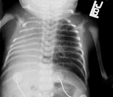
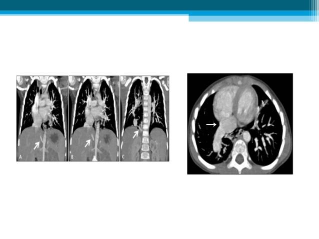
24. An infant with recurrent pneumonia underwent a frontal chest radiograph (Fig 24-A) followed by
 followed by") 24. An infant with recurrent pneumonia underwent a frontal chest radiograph (Fig 24-A) followed by diagnosis? ndings, what is the most likely A. Pulmonary sequestration B. Congenital pulmonary airway malformation
24. An infant with recurrent pneumonia underwent a frontal chest radiograph (Fig 24-A) followed by diagnosis? ndings, what is the most likely A. Pulmonary sequestration B. Congenital pulmonary airway malformation
DESCRIPTION: This is the part of the trunk, which is located between the root of the neck and the superior border of the abdominal region.
 1 THE THORACIC REGION DESCRIPTION: This is the part of the trunk, which is located between the root of the neck and the superior border of the abdominal region. SHAPE : T It has the shape of a truncated
An Approach to Cardiac Malposition and the Heterotaxy Syndrome Using 99mTc Sulfur Colloid Imaging
 An Approach to Cardiac Malposition and the Heterotaxy Syndrome Using 99mTc Sulfur Colloid Imaging P. M. FITZER A diagnostic approach to cardiac malposition and the heterotaxy syndrome is outlined. The
Distinguishing Right From Left: A Standardized Technique for Fetal Echocardiography
 Distinguishing Right From Left: A Standardized Technique for Fetal Echocardiography Timothy M. Cordes, MD, Patrick W. O'Leary, MD, James B. Seward, MD, and Donald J. Hagler, MD, Rochester, Minnesota Improved
Dextrocardia and Isolated Lavocardia
 Brit. Heart J., 1966, 28, 472. Dextrocardia and Isolated Lavocardia MAURICE CAMPBELL AND D. C. DEUCHAR From the Cardiac Department, Guy's Hospital, London S.E.J, and the Institute of Cardiology, London
Undergraduate Teaching
 Prof. James F Meaney Undergraduate Teaching Chest X-Ray Understanding the normal anatomical by reference to cross sectional imaging Radiology? It s FUN! Cryptic puzzle Sudoku (Minecraft?) It s completely
9/8/2009 < 1 1,2 3,4 5,6 7,8 9,10 11,12 13,14 15,16 17,18 > 18. Tetralogy of Fallot. Complex Congenital Heart Disease.
 Current Indications for Pediatric CTA S Bruce Greenberg Professor of Radiology Arkansas Children s Hospital University of Arkansas for Medical Sciences greenbergsbruce@uams.edu 45 40 35 30 25 20 15 10
Case Report Coexistent Congenital Diaphragmatic Hernia with Extrapulmonary Sequestration
 Canadian Respiratory Journal Volume 2016, Article ID 1460480, 4 pages http://dx.doi.org/10.1155/2016/1460480 Case Report Coexistent Congenital Diaphragmatic Hernia with Extrapulmonary Sequestration Nao
B-I-2 CARDIAC AND VASCULAR RADIOLOGY
 (YEARS 1 3) CURRICULUM FOR RADIOLOGY 13 B-I-2 CARDIAC AND VASCULAR RADIOLOGY KNOWLEDGE To describe the normal anatomy of the heart and vessels including the lymphatic system as demonstrated by radiographs,
Heart and Lungs. LUNG Coronal section demonstrates relationship of pulmonary parenchyma to heart and chest wall.
 Heart and Lungs Normal Sonographic Anatomy THORAX Axial and coronal sections demonstrate integrity of thorax, fetal breathing movements, and overall size and shape. LUNG Coronal section demonstrates relationship
Congenital Absence of IVC with Azygous Continuation
 Congenital Absence of IVC with Azygous Continuation M. J. Rauf ( Departments of Radiology, Liaquat National Postgraduate Medical Center, Karachi. ) K. R. Makhdoomi ( Departments of Vascular Surgery, Liaquat
Disclosures. Outline. Learning Objectives. Introduction. Introduction. Sonographic Screening Examination of the Fetal Heart
 Sonographic Screening Examination of the Fetal Heart Lami Yeo, MD Director of Fetal Cardiology Perinatology Research Branch of NICHD / NIH / DHHS Bethesda, MD and Detroit, Michigan, USA Professor, Division
INNOVATIVE JOURNAL OF MEDICAL AND HEALTH SCIENCE
 Innovative Journal Of Medical And Health Science 8:9(2018) Contents lists available at www.innovativejournal.in INNOVATIVE JOURNAL OF MEDICAL AND HEALTH SCIENCE Available online at http://www.innovativejournal.in/index.php/ijmhs
ISUOG Basic Training. Obtaining & Interpreting Heart Views Correctly Alfred Abuhamad, USA. Basic training. Editable text here
 ISUOG Basic Training Obtaining & Interpreting Heart Views Correctly Alfred Abuhamad, USA Learning Objectives 6, 7 & 8 At the end of the lecture you will be able to: describe how to assess cardiac situs
Dextrocardia and asplenia in situs inversus totalis in a baby: a case report
 Kumar et al. Journal of Medical Case Reports 2014, 8:408 JOURNAL OF MEDICAL CASE REPORTS CASE REPORT Open Access Dextrocardia and asplenia in situs inversus totalis in a baby: a case report Abnish Kumar
ISUOG Basic Training. Assessing the Neck & Chest Gihad Chalouhi, Lebanon
 ISUOG Basic Training Assessing the Neck & Chest Gihad Chalouhi, Lebanon Learning objectives 9 & 10 At the end of the lecture you will be able to: recognise the differences between the normal & most common
Pulmonary Sequestration
 July 26, 2004 Pulmonary Sequestration Jonathan Shaw, Harvard Medical School Year IV What do these two patients have in common? Patient 1: 50 y.o. non-smoking female with several months cough and hemoptysis;
Cholecystectomy in a patient with situs inversus
 CASE REPORT Trivedi et al. 1 PEER REVIEWED OPEN ACCESS Cholecystectomy in a patient with situs inversus Govind Trivedi, Rajeev Bhargava, Satish Gupta, Devashish Singh ABSTRACT Situs inversustotalis is
List by Region - Visceral Anomalies
 1 List by Region - Visceral Anomalies General Terms 10127 Situs inversus 80,00 10125 Aneurysm 68,42 10126Fluid-filled abdomen -35,00 Brain 10131 Hydrocephaly 10128 Dilated cerebral ventricle 20,00 10132
The External Anatomy of the Lungs. Prof Oluwadiya KS
 The External Anatomy of the Lungs Prof Oluwadiya KS www.oluwadiya.com Introduction The lungs are the vital organs of respiration Their main function is to oxygenate the blood by bringing inspired air into
List by Terms Visceral anomalies
 1 List by Terms Visceral anomalies Dilated 10128 Dilated cerebral ventricle 11 7 2 0 20,00 10201 Dilated aorta 9 8 2 1 5,26 10207 Dilated aortic arch 9 8 3 0 5,00 10213 Dilated carotid 3 12 4 1-47,37 10218
Chapter 2 Cardiac Interpretation of Pediatric Chest X-Ray
 Chapter 2 Cardiac Interpretation of Pediatric Chest X-Ray Ra-id Abdulla and Douglas M. Luxenberg Key Facts The cardiac silhouette occupies 50 55% of the chest width on an anterior posterior chest X-ray
Dr. Weyrich G07: Superior and Posterior Mediastina. Reading: 1. Gray s Anatomy for Students, chapter 3
 Dr. Weyrich G07: Superior and Posterior Mediastina Reading: 1. Gray s Anatomy for Students, chapter 3 Objectives: 1. Subdivisions of mediastinum 2. Structures in Superior mediastinum 3. Structures in Posterior
Anomalies of Visceroatrial Situs
 ardiopulmonary Imaging Pictorial Essay Ghosh et al. Visceroatrial Situs nomalies ardiopulmonary Imaging Pictorial Essay Downloaded from www.ajronline.org by 46.3.194.217 on 11/22/17 from IP address 46.3.194.217.
Segmental Analysis. Gautam K. Singh, M.D. Washington University School of Medicine St. Louis
 Segmental Analysis Gautam K. Singh, M.D. Washington University School of Medicine St. Louis Segmental Analysis Segmental Analysis: From Veins to Ventricles Segmental Approach to Evaluation of Congenital
Dana Alrafaiah. - Moayyad Al-Shafei. -Mohammad H. Al-Mohtaseb. 1 P a g e
 - 6 - Dana Alrafaiah - Moayyad Al-Shafei -Mohammad H. Al-Mohtaseb 1 P a g e Quick recap: Both lungs have an apex, base, mediastinal and costal surfaces, anterior and posterior borders. The right lung,
Right isomerism with complex cardiac anomalies presenting with dysphagia - A case report
 Right isomerism with complex cardiac anomalies presenting with dysphagia - A case report Himanshu Agarwal 1, Shireesh Kumar Mittal 1*, Chaitanya D Kulkarni 1, Ashok Kumar Verma 1, Saurabh Kumar Srivastava
Genetically Determined Variation in the Azygos Vein in the Mouse
 Genetically Determined Variation in the Azygos Vein in the Mouse FRED G. BIDDLE, JACOB D. JUNG, AND BRENDA A. EALES Departments of Pediatrics and Medical Biochemistry, University of Calgary, Calgary, Alberta,
F etal dextrocardia is a condition in which the major axis
 1590 CONGENITAL HEART DISEASE Fetal dextrocardia: diagnosis and outcome in two tertiary centres A Bernasconi, A Azancot, J M Simpson, A Jones, G K Sharland... See end of article for authors affiliations...
CT Chest. Verification of an opacity seen on the straight chest X ray
 CT Chest Indications: To assess equivocal plain x-ray findings Staging of lung neoplasm Merastatic workup of extra thoraces malignancies Diagnosis of diffuse lung diseases with HRCT Assessment of bronchietasis
Surgical indications: Non-malignant pulmonary diseases. Punnarerk Thongcharoen
 Surgical indications: Non-malignant pulmonary diseases Punnarerk Thongcharoen Non-malignant Malignant as a pathological term: Cancer Non-malignant = not cancer Malignant as an adjective: Disposed to cause
Lab #3. Mohammad Hisham Al-Mohtaseb. Jumana Jihad. Ammar Ramadan. 0 P a g e
 Lab #3 Mohammad Hisham Al-Mohtaseb Jumana Jihad Ammar Ramadan 0 P a g e Last anatomy lab: Lungs and structure on the mediastinal surfs: 1-the right lung: How do we know it s the right lung??? -the 3 lobes
CMS Limitations Guide - Radiology Services
 CMS Limitations Guide - Radiology Services Starting October 1, 2015, CMS will update their existing medical necessity limitations on tests and procedures to correspond to ICD-10 codes. This limitations
Assignable revenue codes: Explanation of services:
 computed tomography Chest/Cardiac Assignable revenue codes: Explanation of services: 0350 CT Scan General Classification 0351 CT Scan Head Scan 0352 CT Scan Body Scan 0359 CT Scan Other CT Scans Known
Large veins of the thorax Brachiocephalic veins
 Large veins of the thorax Brachiocephalic veins Right brachiocephalic vein: formed at the root of the neck by the union of the right subclavian & the right internal jugular veins. Left brachiocephalic
Fetal Heterotaxy with Tricuspid Atresia, Pulmonary Atresia, and Isomerism of the Right Atrial Appendages at 22 Weeks
 THIEME Case Report 97 Fetal Heterotaxy with Tricuspid Atresia, Pulmonary Atresia, and Isomerism of the Right Atrial Appendages at 22 Weeks Julia E. Solomon, MD 1 John H. Stock, MD 2 Randy R. Richardson,
The Respiratory System
 C h a p t e r 24 The Respiratory System PowerPoint Lecture Slides prepared by Jason LaPres North Harris College Houston, Texas Copyright 2009 Pearson Education, Inc., publishing as Pearson Benjamin Cummings
Case Based Fetal Lung Masses
 Case Based Fetal Lung Masses Advances in Fetal and Neonatal Imaging Course Orlando, Florida, January 28, 2017 Leann E. Linam, MD Associate Professor Radiology University of Arkansas for Medical Sciences/
Common Defects With Expected Adult Survival:
 Common Defects With Expected Adult Survival: Bicuspid aortic valve :Acyanotic Mitral valve prolapse Coarctation of aorta Pulmonary valve stenosis Atrial septal defect Patent ductus arteriosus (V.S.D.)
X-Rays. Kunal D Patel Research Fellow IMM
 X-Rays Kunal D Patel Research Fellow IMM The 12-Steps } 1: Name 2: Date 3: Old films 4: What type of view(s) 5: Penetration } Pre-read 6: Inspiration 7: Rotation Quality Control 8: Angulation 9: Soft tissues
CASE REPORT. 1. Assistant Professor. Department of Paediatrics, Vinayaka Missions Medical College, Karaikal
 A CASE OF KARTAGENER SYNDROME Pagadpally Srinivas 1. Assistant Professor. Department of Paediatrics, Vinayaka Missions Medical College, Karaikal CORRESPONDING AUTHOR: Pagadpally Srinivas, 72, Vellai Pillaiyar
Anomalous Systemic Venous Connection Systemic venous anomaly
 World Database for Pediatric and Congenital Heart Surgery Appendix B: Diagnosis (International Paediatric and Congenital Cardiac Codes (IPCCC) and definitions) Anomalous Systemic Venous Connection Systemic
Chapter 5: Other mediastinal structures. The Large Arteries. The Aorta. Ascending aorta
 Chapter 5: Other mediastinal structures The Large Arteries The Aorta The aorta is the main arterial trunk of the systemic circulation and in the healthy state its wall contain a large amount of yellow
Case report Esophageal lung: a rare case of communicating bronchopulmonary foregut malformation
 Case report Esophageal lung: a rare case of communicating bronchopulmonary foregut malformation 1 Dr.Varsha Rathi, 2 Dr. Saurabh Deshpande*, 3 Dr.Almas Nazim, 4 Dr.Shilpa Domkundwar 1 Professor, Department
PRACTICAL GUIDE TO FETAL ECHOCARDIOGRAPHY IC Huggon and LD Allan
 PRACTICAL GUIDE TO FETAL ECHOCARDIOGRAPHY IC Huggon and LD Allan Fetal Cardiology Unit, Harris Birthright Research Centre for Fetal Medicine, King's College Hospital, London, UK IMPORTANCE OF PRENATAL
Anatomy Lecture 8. In the previous lecture we talked about the lungs, and their surface anatomy:
 Anatomy Lecture 8 In the previous lecture we talked about the lungs, and their surface anatomy: 1-Apex:it lies 1 inch above the medial third of clavicle. 2-Anterior border: it starts from apex to the midpoint
Double Superior Vena Cava; A Benign Cause of Widened Mediastenum and Implication on Venous Central Access
 ISPUB.COM The Internet Journal of Endovascular Medicine Volume 2 Number 1 Double Superior Vena Cava; A Benign Cause of Widened Mediastenum and Implication on Venous H Enuh, A Patel, A Chaudry, K Diaz,
11.1 The Aortic Arch General Anatomy of the Ascending Aorta and the Aortic Arch Surgical Anatomy of the Aorta
 456 11 Surgical Anatomy of the Aorta 11.1 The Aortic Arch 11.1.1 General Anatomy of the Ascending Aorta and the Aortic Arch Surgery of the is one of the most challenging areas of cardiac and vascular surgery,
Basic Training. ISUOG Basic Training Examining the Upper Lip, Face & Profile
 ISUOG Examining the Upper Lip, Face & Profile Learning objectives At the end of the lecture you will be able to: Describe how to obtain the 3 planes required to assess the anatomy of the fetal face Recognise
KARTAGENER S SYNDROME: A CLASSICAL CASE
 Kartagener s Syndrome Arunabha D.C et al 363 CASE REPORT KARTAGENER S SYNDROME: A CLASSICAL CASE Arunabha DC 1, Sumit RT 1, Sourin B 2, Sabyasachi C 3, Subhasis M 4 ABSTRACT BACKGROUND: Recurrent lower
X-Rays. Prepared by Prof.Dr. Magda Hassab Allah Assist.lecturer Marwa Al Hady
 X-Rays Prepared by Prof.Dr. Magda Hassab Allah Assist.lecturer Marwa Al Hady CHEST X-RAYS Normal Chest X-ray Comments on chest X ray includes examination of 1- Bony cage (ribs,clavicles &vertebral column
ASSESSING THE PLAIN ABDOMINAL RADIOGRAPH M A A M E F O S U A A M P O F O
 ASSESSING THE PLAIN ABDOMINAL RADIOGRAPH M A A M E F O S U A A M P O F O Introduction The abdomen (less formally called the belly, stomach, is that part of the body between the thorax (chest) and pelvis,
Abnormalities of the spleen in relation to
 122 Department of Pediatric Cardiology, Children's Hospital of Pennsylvania, USA C Anderson R H Anderson J R Zuberbuhler Department of Pathology, Children's Hospital of Pennsylvania, USA W A Devine D E
SWISS SOCIETY OF NEONATOLOGY. Is every innocent murmur innocent?
 SWISS SOCIETY OF NEONATOLOGY Is every innocent murmur innocent? March 2010 2 Rüegger C, Malär R, Schraner T, Weber R, Arlettaz Mieth R, Clinic of Neonatology (RC, AMR), University Women s Hospital Zurich,
THE GOOFY ANATOMIST QUIZZES
 THE GOOFY ANATOMIST QUIZZES 7. LUNGS Q1. Fill in the blanks: the lung has lobes and fissures. A. Right, three, two. B. Right, two, one. C. Left, three, two. D. Left, two, three. Q2. The base of the lung
The Thoracic wall including the diaphragm. Prof Oluwadiya KS
 The Thoracic wall including the diaphragm Prof Oluwadiya KS www.oluwadiya.com Components of the thoracic wall Skin Superficial fascia Chest wall muscles (see upper limb slides) Skeletal framework Intercostal
Right lung. -fissures:
 -Right lung is shorter and wider because it is compressed by the right copula of the diaphragm by the live.. 2 fissure, 3 lobes.. hilum : 2 bronchi ( ep-arterial, hyp-arterial ), one artery mediastinal
Lung & Pleura. The Topics :
 Lung & Pleura The Topics : The Trachea. The Bronchi. The Brochopulmonary Segments. The Lungs. The Hilum. The Pleura. The Surface Anatomy Of The Lung & Pleura. The Root & Hilum. - first of all, the lung
The Triply Twisted Heart: Cyanosis in an Adult With Situs Inversus, Levocardia, Double Outlet Right Ventricle, and Malposition of the Great Arteries
 Elmer ress Case Report Cardiol Res. 2015;6(6):362-366 The Triply Twisted Heart: Cyanosis in an Adult With Situs Inversus, Levocardia, Double Outlet Right Ventricle, and Malposition of the Great Arteries
JlntSocPlastination, Vol4:16-22,
 JlntSocPlastination, Vol4:16-22, 1990 16 SECTIONAL ANATOMY: STANDARDIZED METHODOLOGY Alexander Lane, Coordinator of Anatomy and Physiology, Triton College, Visiting Associate Professor, University of Illinois
Theme 30. Structure, topography and function of the lungs and pleura. Mediastinum and its contents. X -ray films digestive and respiratory systems.
 Theme 30. Structure, topography and function of the lungs and pleura. Mediastinum and its contents. X -ray films digestive and respiratory systems. STRUCTURE, TOPOGRAPHY AND FUNCTІON OF LUNGS AND PLEURA.
Congenital Heart Defects
 Normal Heart Congenital Heart Defects 1. Patent Ductus Arteriosus The ductus arteriosus connects the main pulmonary artery to the aorta. In utero, it allows the blood leaving the right ventricle to bypass
ULTRASOUND OF THE FETAL HEART
 ULTRASOUND OF THE FETAL HEART Cameron A. Manbeian, MD Disclosure Statement Today s faculty: Cameron Manbeian, MD does not have any relevant financial relationships with commercial interests or affiliations
Amoebic liver abscess revealing a situs inversus totalis
 www.edoriumjournals.com CASE REPORT PEER REVIEWED OPEN ACCESS Amoebic liver abscess revealing a situs inversus totalis Papa Abdoulaye Bâ, Papa Amath Diagne, Thomas Marcel Mbar Wade, Babacar Mbengue, Balla
STUDY OF AZYGOS SYSTEM AND ITS VARIATIONS B. Vijaya Nirmala 1, Teresa Rani S 2
 STUDY OF AZYGOS SYSTEM AND ITS VARIATIONS B. Vijaya Nirmala 1, Teresa Rani S 2 HOW TO CITE THIS ARTICLE: B. Vijaya Nirmala, Teresa Rani S. Study of Azygos System and its Variations. Journal of Evolution
Early View Article: Online published version of an accepted article before publication in the final form.
 Early View Article: Online published version of an accepted article before publication in the final form. Journal Name: Edorium Journal of Anatomy and Embryology Type of Article: Case Report Title: Pulmonary