Mitotic spindle organization by dynein & kinetochores. Jonne Anne Raaijmakers
|
|
|
- Anissa Ward
- 6 years ago
- Views:
Transcription
1
2 Mitotic spindle organization by dynein & kinetochores Jonne Anne Raaijmakers
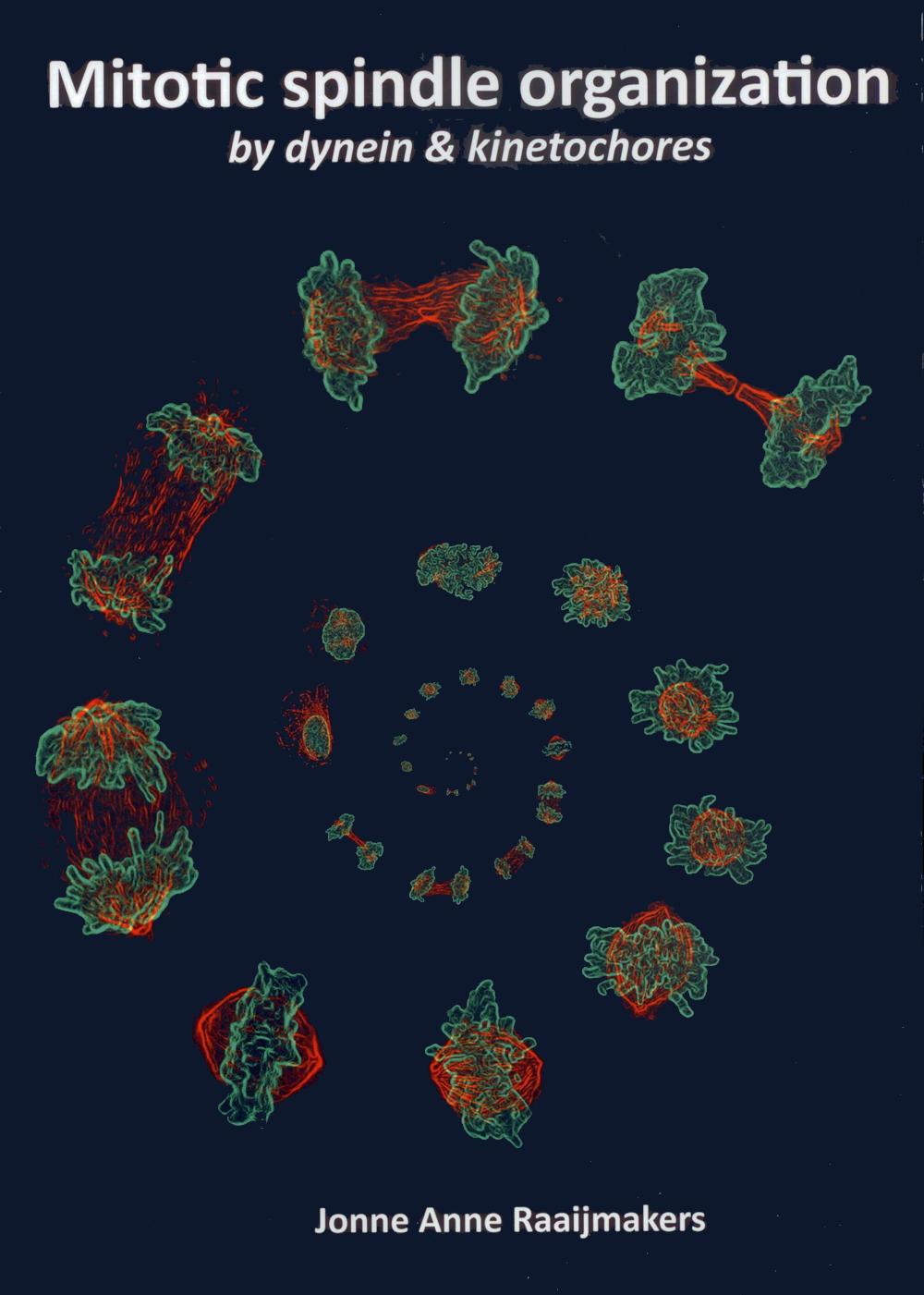
3 ISBN: Printed by: Gildeprint Copyright 214 J.A. Raaijmakers Cover: Immunofluorescent images of dividing U2OS cells assembled in a spiral. The microtubules are depicted in red and the DNA is depicted in green.
4 Mitotic spindle organization by dynein & kinetochores Spoelorganisatie in mitose door dynein & kinetochoren (met een samenvatting in het Nederlands) Proefschrift ter verkrijging van de graad van doctor aan de Universiteit Utrecht op gezag van de rector magnificus, prof. dr. G.J. van der Zwaan, ingevolge het besluit van het college voor promoties in het openbaar te verdedigen op donderdag 5 juni 214 des middags te uur. door Jonne Anne Raaijmakers geboren op 22 september 1984 te s-hertogenbosch
5 Promotor: Prof. dr. R.H. Medema
6 Table of Contents Chapter 1 General Introduction 7 Chapter 2 Function and regulation of dynein in mitotic chromosome segregation 19 Chapter 3 Systematic dissection of dynein regulators in mitosis 35 Chapter 4 Chapter 5 Chapter 6 Chapter 7 Nuclear envelope-associated dynein drives prophase centrosome separation and enables Eg5-independent bipolar spindle formation 53 Dynein/dynactin releases SAC-mediated APC/C inhibition to promote the metaphase-anaphase transition 69 Misaligned chromosomes cause spindle misorientation by interfering with cortical LGN localization 85 RAMA1 is a novel kinetochore protein involved in kinetochore-microtubule attachment 97 Chapter 8 Summary & General Discussion 113 Addendum References 124 Nederlandse Samenvatting 137 Curriculum Vitae 14 List of Publications 141 Dankwoord 142
7 6
8 Chapter 1 General Introduction J.A. Raaijmakers 1, R.H. Medema 1 1. Department of Cell Biology, The Netherlands Cancer Institute, Plesmanlaan 121, 166 CX, Amsterdam, Netherlands 7
9 1 General introduction Cell Division The human body contains an estimated 37.2 trillion cells 1. Although most cells in the body are non-dividing, approximately 3 billion new cells are produced every day. This is important for the maintenance of our tissues but also for the repair of tissues when they are damaged. To prepare for cell division, a cell goes through a cycle of four stages; G1-, S-, G2- and M-phase (Figure 1). In the G1-phase (Gap1-phase), cells grow to prepare for the replication of the complete genome that occurs during S-phase (Synthesis-phase). Upon the completion of DNA replication, the cell progresses into the second Gap-phase (G2-phase) followed by the actual division of the cells in M-phase (or mitosis). Although mitosis is the shortest phase, it is an extremely dynamic process, which is again divided in multiple stages. The individual stages of mitosis will be explained in more detail below (Also see Figure 1). 1. Prophase. The DNA starts to condense into small, compact structures to allow the individual chromosomes to move within the cell. At this stage, the duplicated sister chromatids are held together by chromatin cohesion. Furthermore, the centrosomes, which are the major microtubule nucleation structures, start to move apart to opposite sides of the nucleus. The centrosomes are pushed apart by microtubule motors, which will be explained in more detail below. 2. Prometaphase. At the start of prometaphase, the nuclear membrane breaks down to allow the microtubules to interact with the chromosomes. Specialized structures termed the kinetochores are formed pairwise on each set of chromatids. These structures form the main interaction platform for the spindle microtubules. Furthermore, a dense microtubule-network forms to build the typical diamond-shaped spindle and the centrosomes are pushed further apart. 3. Metaphase. At this stage, all chromosomes have made correct attachments to the mitotic spindle. This means that each sister chromatid within a pair is attached to microtubules originating from opposing sides of the spindle. As a consequence, all chromosomes line up in the middle of the cell. 4. Anaphase. When all the chromosomes are correctly attached to the mitotic spindle, the cohesion that holds the sister chromatids together is cleaved. This allows the sister chromatids to separate. Each chromatid is pulled towards opposite sides of the cell and both daughter cell ends up with an identical set of chromosomes. 5. Telophase. During this final stage of mitosis, the chromosomes decondense and the nuclear membrane reforms around the two newly formed nuclei. Furthermore, the cell membrane ingresses between the two nuclei, which is followed by cytokinesis where abscission takes place and the two cells are physically separated. After this stage, mitosis is completed and the individual cells can enter a new cell cycle. Obviously, this is a very brief description of an extremely complicated and dynamic process. How are large structures, such as the centrosomes and the chromosomes, moved within the cell? What are the proteins that are involved? How does a cell safeguard the equal division of the genomic material? How does a cell cope with mistakes? This thesis describes a number of novel insights into these important issues. In the following paragraphs we will introduce the relevant processes in more detail. Building the mitotic spindle The mitotic spindle is the master regulator of mitosis; it is responsible for chromosome movements and it drives the correct division of the two sets of chromosomes over the two new daughter cells. The mitotic spindle consists of a large set of microtubules; long hollow filaments that are made up from 8
10 S G2 G1 Mitosis Mitosis Prophase Prometaphase Metaphase Anaphase Telophase Figure 1. The cell cycle & mitosis A schematic representation of the several phases of the cell cycle. The cell cycle comprises four stages; S-phase where the DNA is replicated and M-phase or mitosis where the DNA is equally divided over the two new daughter cells. These phases are separated by two Gap -phases; G1 and G2. The microscopic images show representative examples of the different stages of mitosis in U2OS cells. The DNA is depicted in blue, the spindle microtubules in green and the actin-cytoskeleton in red. 1 General Introduction arrays of α/β-tubulin heterodimers that polymerize in a head-to-tail configuration. The centrosomes recruit large amounts of γ-tubulin, making them the major microtubule nucleation centers. γ-tubulin associates with several other proteins to form the γ-tubulin Ring Complex (γturc). The γturc-complex forms a seed for the nucleation of microtubules; therefore the non-growing microtubule minus-ends are embedded in the centrosome while the growing plus-ends protrude away from the centrosomes. The spindle contains three sets of microtubules; First, the kinetochore microtubules (k-fibers) that link the chromosomes to the spindle pole. Second, the interpolar microtubules, which extend all the way from the spindle pole into the other half of the spindle, thereby forming an antiparallel overlap at the spindle equator. Third, the astral microtubules, which reach from the spindle poles towards the cell cortex, thereby facilitating spindle positioning. The shape and size of the mitotic spindle is largely dictated by the presence of diverse set of microtubule-associated proteins (MAPs), and microtubule motors. Together these proteins determine the behavior of microtubules by crosslinking them, stabilizing or destabilizing them and by sliding them relative to each other. Prophase centrosome separation In prophase, the centrosomes are moved to opposite sides of the nucleus. The key player in prophase centrosome separation is the highly conserved motor protein Eg5 (kinesin-5) 2. Eg5 has a unique tetrameric configuration 3, which allows it to crosslink and walk on two microtubules simultaneously 4. Since Eg5 has plus-end directed activity, it can slide anti-parallel microtubules outward, resulting in the separation of the two centrosomes (Figure 2). That Eg5 is the main driver of prophase centrosome separation in mammalian cells is illustrated by the fact that inhibition of Eg5 results in a complete block in centrosome separation 5-7. In contrast, kinesin-5 does not seem to play a prominent role in prophase centrosome separation in some other organisms. For example, inhibition of the kinesin-5 Klp61F in Drosophila melanogaster (fruitfly) embryo s does lead to any defect in the separation of the centrosomes during prophase 8. Furthermore, Caenorhabditis elegans (roundworm) embryo s with a genetic disruption of the kinesin-5 gene BMK-1 are viable 9, and they display normal prophase centrosome separation (our own observations, A. Maia in collaboration with the laboratory of S. van den Heuvel). Rather, C. elegans and D. melanogaster depend on the minus-end directed motor dynein for full centrosome separation 1,11. Interestingly, recent data suggest that dynein also contributes to centrosome separation in human cells 12. Dynein acts by pulling on centrosome-derived microtubules while being anchored at the 9
11 1 nuclear envelope, thereby moving the centrosomes along the nuclear envelope (Figure 2). In chapter 2 and 3 of this thesis and in 13, the mechanism by which dynein can drive centrosome separation in mammalian cells is discussed in more detail. Bipolar spindle formation in (pro)metaphase; outward forces After nuclear envelope breakdown, the spindle continues to mature into a dense microtubule network with a bipolar configuration. Similar to prophase centrosome separation, inhibition of Eg5 leads to a failure to form a bipolar spindle. Rather, Eg5-inhibited cells get stuck in mitosis with a typical monopolar spindle, indicating that Eg5 generates a major outward force in prometaphase (Figure 3). This function of Eg5 is found to be conserved from yeast to human 5,14-17 (with the remarkable exception of C. elegans worms and Dictyostelium discoideum slime molds 9,18 ). Strikingly, inhibition of Eg5 after the centrosomes have separated, does not lead to a spindle collapse 5,6,19, indicating that Eg5 does not act alone in the maintenance of the bipolar spindle. Indeed, an additional plus-end directed motor was recently identified to act together with Eg5 in bipolar spindle assembly. This motor, termed Kif15 (or kinesin-12/ hklp2), is essential for bipolar spindle formation when Eg5 is partially inhibited 2,21. Furthermore, depletion of Kif15 results in a rapid collapse of the spindle when Eg5 is co-inhibited 2,21. Interestingly, when Prophase (Pro)metaphase Name Directionality Short Description Dynein Eg5 Kif15 - (Minus-end) + (Plus-end) + (Plus-end) Dynein is a multi-subunit microtubule motor with minus-end directed motility. In prophase, dynein is anchored to the nuclear envelope where it creates a pulling force on the individual centrosomes. In prometaphase, dynein can slide antiparallel microtubules, thereby generating an inward force in the spindle. A similar function has been described for kinesin-14, a minus-end directed kinesin. Eg5 is a kinesin-5 motor with plus-end directed motility. It has a unique tetrameric composition, allowing it to crosslink two microtubules simultaneously and sliding them apart. The capacity to slide microtubules is relevant in centrosome separation in both prophase and prometaphase Kif15 is a kinesin-12 motor with plus-end directed motility. Kif15 forms a dimer and can therefore only walk on one microtubule at the time. It uses another microtubule interacting protein; TPX2 to crosslink and slide microtubules apart. Kif15 cooperates with Eg5 in bipolar spindle assembly and is essential for the formation and maintenance of the bipolar spindle when Eg5-activity is repressed. Figure 2. Motor proteins in bipolar spindle assembly Prophase centrosome separation is mainly driven by Eg5-mediated antiparallel microtubule sliding. Dynein-mediated pulling forces from the nuclear envelope further stimulate this process. In prometaphase and metaphase, the outward forces in the spindle are generated by both Eg5 and Kif15. In contrast to prophase centrosome separation, during (pro) metaphase dynein acts antagonistically to Eg5 and Kif15 by sliding the antiparallel microtubules inward. Dynein could walk on one microtubule at the time but it has also been proposed that both heads coul walk on individual microtubules. The arrows indicate the direction of the relevant motor and the + indicates the polarity of the microtubule. 1
12 overexpressed, Kif15 can even fully compensate for the complete absence of Eg5 2 and in cells that grow independent of Eg5-activity 12. Since Kif15 forms a dimer, with both motor domains at one site, it needs an additional microtubule-binding domain to allow the sliding of antiparallel microtubules. This function appears to be mediated by TPX2, a molecule that can bind both Kif15 and microtubules simultaneously, thereby facilitating Kif15-mediated antiparallel microtubule sliding (Figure 2 and 2-22 ). However, the precise mechanism by which Kif15 promotes bipolar spindle assembly is not understood. Also, although Eg5 seems to have a more prominent role in the initial phases of centrosome separation and Kif15 is more essential during the latter, it is not clear what the advantage is of having two independent motors involved. Bipolar spindle formation in (pro)metaphase; antagonizing forces Although outward forces are essential for the formation of the bipolar spindle, it has been well established that the outward forces are balanced by antagonizing inward forces. The two major inward force generators in the spindle are dynein and kinesin-14, which both possess minus-end directed motility and can crosslink and slide antiparallel microtubules apart in vitro Although dynein seems to be the major factor that generates an inward force in mammalian cells and in Xenopus spindles 26-29, lower eukaryotes, such as yeast or Drosophila, depend more on kinesin-14 8,3-32. What is the relevance of having antagonizing forces present in the spindle? In the absence of dynein or kinesin-14, only slight defects are observed in intercentrosomal distance, suggesting that the main objective of having antagonistic motors is not to restrict spindle length 29,33. Rather, depletion of kinesin-14 or dynein leads to major defects in the organization of the spindle poles in a variety of systems 29,34-4. Interestingly, these defects can be rescued when kinesin-5 is co-inhibited, indicating that a correct balance of forces contributes to the organization of the spindle 26. Therefore, it is likely that antagonistic forces exist to create a properly organized and robust spindle. Whether the presence of antagonistic motors also contributes to other features in the spindle, such as the fidelity of kinetochore-microtubule interactions, needs further investigation. 1 General Introduction Establishing kinetochore-microtubule attachments The main goal in mitosis is to generate two daughter cells with an identical set of chromosomes. To be able to divide the DNA correctly, each chromosome needs to make bipolar attachments to the mitotic spindle. This means that each chromatid within a pair of sister chromatids needs to make attachments to microtubules originating from opposite sites of the spindle. How is this established? And how does the cells prevent mistakes in this process? The building blocks of the kinetochore At the onset of mitosis, each chromatid develops a macromolecular structure referred to as the kinetochore at its centromeric region that forms the major interaction site for the microtubules. Microtubules that attach to this structure are referred to as the kinetochore microtubules or K-fibers. While in budding yeast only a single microtubule interacts with each kinetochore 41, in human cells a K-fiber is made up out of 2-3 individual microtubules 42. Besides establishing interaction between chromosomes and the spindle, the kinetochores are also important sensors for the accuracy of these attachments. In the next section, we will discus the composition and the function of the kinetochores in more detail. The kinetochore has been extensively studied by electron microscopy and this has led to the definition of three separate components; two electron-dense structures termed the inner- and outer- plate and a fibrous corona which extends from the outer plate 43. Interestingly, the first molecular components of the kinetochore were identified by the use of auto-immune antibodies of patients suffering from a specific form of systemic scleroderma (CREST-syndrome) that specifically recognize the centromeric region of the chromosomes 44. These antibodies, referred to as anti-centromere antibodies (ACA), were found to recognize three components of the kinetochore; CENP-A, -B and C45. CENP-A is a histone H3 variant, which dictates the localization of the kinetochore by replacing histone H3 at the centromeric 11
13 1 regions 46. CENP-B recognizes and binds the typical α-satellite regions present in the centromeric regions, however, the presence of both α-satellite and the CENP-B gene are not important for centromere formation or activity 47,48. CENP-C was found as an interaction partner of CENP-A and together with 13 binding partners (CENP-H, CENP-I, CENP-K U) forms the centromere-associated network or CCAN. The CCAN is an important scaffold for the assembly of the outer kinetochore. Strikingly, ectopic recruitment of CENP-T and CENP-C to non-centromeric regions of the chromosomes is sufficient to build a complete, functional kinetochore 49. The most important outer kinetochore components that are recruited by the CCAN involves the KMN-network, consisting of Knl-1, the Mis12 complex and the Ndc8 complex. These complexes form the major interaction site with microtubules but they are also responsible for the recruitment of many adaptor proteins that fine-tune the kinetochore-microtubule interaction. Furthermore, the KMN-network is responsible for the recruitment of the kinetochore components that are involved in mitotic checkpoint signaling. More than 1 proteins have been identified to localize to the kinetochore. Details of the precise composition of the human kinetochore and on its different activities were extensively reviewed by Cheeseman and Desai 5. Attaching kinetochores to the mitotic spindle The kinetochores can form both lateral and end-on attachments to the spindle microtubules. The lateral interactions can stochastically occur when a kinetochore encounters a microtubule lattice and end-on attachments are the interactions that the kinetochores make with microtubule ends. Two motor proteins located at the kinetochore; dynein (minus-end) and CENP-E (plus-end) have been implicated in the translocation of chromosomes along the microtubule upon lateral interactions. Dynein-mediated translocation results in poleward chromosome movements. These poleward movements are suggested to bias the orientation of the other kinetochore on the paired sister chromatid toward the opposing spindle pole, thereby contributing to the formation of correct, bipolar kinetochore-microtubule interactions. Furthermore, the poleward movement might also contribute to the formation of end-on attachments as the microtubule-density is extremely high around the spindle poles. On the contrary, CENP-E-mediated movements result in chromosome movements towards the spindle equator. This is important for chromosome congression; the alignment of all chromosomes in the center of the cell. Unattached Attached Mad2 Mad1 KNL-1 Bub3 BubR1 Bub1 dynein/ dynactin Mps1 Spindly Ska-1-3 RZZ CENP-E NDC8-complex KNL-1 Mis12-complex CCAN CENP-A CENP-A CCAN Mis12-complex NDC8-complex Ska- 1-3 Ska- 1-3 KNL-1 NDC8-complex Figure 3. Molecular view of the kinetochore The kinetochore comprises over 1 components. This image shows the most important and best-characterized modules. The kinetochore is built on specific chromatin areas containing CENP-A histones on which the centromere-associated network (CCAN) binds (depicted in red). The CCAN forms a structural platform for several outer components. It recruits the KMN-network, which is depicted in yellow, consisting of Knl-1, the Mis12-complex and the Ndc8 complex. Together with the Ska-complex, these components are important for the generation and stabilization of kinetochore-microtubule interactions. The components of the mitotic checkpoint, depicted in grey, are also located to the outer kinetochore. Furthermore, the RZZ-complex recruits spindly and dynein, which drive the movement of chromosomes towards the spindle pole when they make a lateral attachment to a spindle microtubule. In contrast, CENP-E transports chromosomes towards the microtubule plus-ends, thereby promoting chromosome congression. When the kinetochore makes a stable attachment to the spindle, most outer-kinetochore components are removed. However, the KMN-network is present throughout mitosis to maintain the KT-MT attachments. This is important to keep the kinetochores coupled to the dynamic microtubule ends that rapidly depolymerize during anaphase. 12
14 End-on attachments need to be solid enough to transduce forces necessary for the physical separation of the chromosome in anaphase but dynamic enough to allow the tracking of growing and shrinking microtubules. The KMN-network comprises the main attachment factors of the kinetochore of which Knl-1 and Ndc8 (also known as Hec1) have direct microtubule binding capacity 51. A recently identified Ndc8-binding complex; the Ska-complex is proposed to bind to curved protofilaments specifically Curving of the microtubule occurs when the microtubule depolymerizes, and as such the Ska-complex plays an important role in the coupling of kinetochores to shrinking microtubules. Establishing bipolar attachments To ensure equal division of the genetic material over the two newly formed daughter cells, each sister chromatid within a pair needs to attach to microtubules originating from opposing spindle poles. We therefore refer to these attachments as bipolar or amphitelic attachments (Figure 4). However, in this process of chromosome bi-orientation, several erroneous attachments can occur transiently. These include monotelic attachments where only one sister chromatid is attached to a pole and the other is not attached or syntelic attachments where both sister chromatids attach to the same pole (Figure 4). Furthermore, a situation can occur where a single kinetochore makes attachments to both poles, also referred to as merotelic attachments (Figure 4). How does a cell make sure to only stabilize the correct, bipolar attachments? This involves an elegant error-correction machinery involving the centromerically localized kinase Aurora B. By phosphorylating outer kinetochore substrates such as members of the KMN-network, Aurora B reduces the affinity of these substrates for microtubules, thereby destabilizing microtubule-kinetochore attachments. The inner centromere localization of Aurora B spatially limits its ability to phosphorylate outer kinetochore components; In case of no attachments or erroneous attachments, there will be no tension between the sister kinetochores and Aurora B is able to phosphorylate the outer kinetochore components. However, since microtubule shrinkage creates a pulling force on the kinetochores which is antagonized by sister chromatid cohesion 55, tension will occur across the sister kinetochores only when a bipolar interaction is made. This tension results in stretching of the kinetochore which will lead to the physical separation of outer kinetochore components from Aurora B-activity at the inner centromere, thereby preventing the destabilization of correct attachments specifically 56. It is now clear that an antagonizing phosphatase becomes active when Aurora B-activity drops that allows the rapid dephosphoryation of Aurora B substrates at the outer kinetochore 57. Thus, spatial restriction of the Aurora B kinase from its substrates at the outer kinetochore specifically stabilizes the correct, tension-generating attachments while destabilizing faulty, non-tension generating attachments. 1 General Introduction Sensing attachment status: Mitotic checkpoint activation. The separation of sister chromatids in anaphase should only occur when all chromosomes have obtained stable, bioriented attachments to the mitotic spindle. Premature anaphase onset in the presence of erroneous attachments will lead to unequal division of the genetic content. Early observations indicated the existence of a checkpoint that prevents anaphase onset until the last chromosome is properly attached to the mitotic spindle58. This checkpoint is referred to as the spindle assembly checkpoint (SAC) or the mitotic checkpoint. The mitotic checkpoint involves a kinetochore-derived signal, as evidenced by experiments in which the mitotic checkpoint was rapidly relieved upon laser ablation of the Attachment status Monotelic Syntelic Merotelic Amphitelic Figure 4. Overview of possible kinetochore-microtubule interactions Schematic overview of the possible kinetochore-microtubule interactions that occur during (pro)metaphase. Monotelic, syntelic and merotelic attachments are undesirable attachment types that transiently occur. However, only correct amphitelic attachments, where each kinetochore is attached to opposite spindle poles, will be stabilized. 13
15 1 kinetochores of the last unaligned chromosome 59. What is this anaphase inhibitory signal? And how does it act to delay mitosis until all chromosomes are properly aligned? Figure 5 shows a schematic representation of the mitotic checkpoint, which will be explained in more detail below. Anaphase onset is determined by the degradation of cyclin B, resulting in the deactivation of Cdk1 and the degradation of Securin, which allows Separase (which cleaves chromatid cohesion) to become active. Cyclin B and Securin are targeted for degradation through ubiquitination mediated by the E3 ubiquitin ligase anaphase promoting complex/cyclosome or APC/C. The mitotic checkpoint acts by inhibiting the APC/C until all chromosomes are stably attached to the mitotic spindle. This inhibition is established through the formation of the mitotic checkpoint complex (MCC) consisting of Mad2, BubR1, Bub3. The formation of MCC is catalyzed by unattached kinetochores. The MCC acts by binding to and sequestering CDC2, the co-activator of the APC/C in mitosis 6. Other checkpoint proteins that are essential for proper mitotic checkpoint signaling but that are not part of the core MCC complex are Mad1 and the Mps1 kinase 61,62. These adaptor proteins are important for the localization and formation of the MCC at kinetochores and for the interaction dynamics of the MCC with kinetochores and the APC/C. Furthermore, inhibition of Bub1 or Aurora B leads to rapid anaphase onset with a high frequency of lagging chromosomes, indicating that they have an essential role in the mitotic checkpoint However, a direct role for Bub1 and Aurora B in de mitotic checkpoint has remained controversial and since Aurora B has an essential function in stabilizing kinetochore microtubule attachment, while Bub1 regulates the Prometaphase Metaphase Anaphase MCC formation at kinetochores CDC2 MCC CDC2 MCC MCC CDC2 MCC formation inhibited CDC2 CDC2 MCC CDC2 Sister chromatid separation Inhibited APC/C Cyclin B Securin MCC APC/C CDC2 Active APC/C Cyclin B Securin APC/C CDC2 Active APC/C APC/C CDC2 High cyclin B/Securin Separase Inhibited Cyclin B Securin Cyclin B/Securin degraded Separase released Ub Ub Ub Cyclin B Ub Ub Ub Securin Active Separase Figure 5. The mitotic checkpoint A Schematic representation of the mitotic checkpoint. The mitotic checkpoint prevents anaphase onset till all kinetochores are properly attached to the mitotic spindle. In prometaphase, unattached kinetochores catalyze the formation of mitotic checkpoint inhibitor complex or MCC. The MCC binds to the APC/C activator CDC2, thereby preventing APC/C-activity. As a consequence, cyclin B and Securin (the inhibitor of Separase) are stabilized. In metaphase, when all kinetochores are stably attached to the mitotic spindle, MCC formation is inhibited and CDC2 is now able to activate the APC/C. This results in the ubiquitination and degradation of cyclin B and Securin. As a result, the mitotic kinase Cdk1 is inactivated and Separase is able to destroy sister chromatid cohesion, thereby allowing anaphase onset. 14
16 localization of Aurora B to the inner centromere 66,67, the defect in checkpoint signaling might also reflect the hyper-stabilization of faulty attachments, thereby satisfying the mitotic checkpoint independent of tension. Sensing attachment status: Mitotic checkpoint inactivation When all kinetochores are correctly attached to the mitotic spindle, the mitotic checkpoint is inactivated to allow the APC/C to become active and to target cyclin B and Securin for degradation. The inactivation of the mitotic checkpoint is a multistep process. One of the first steps in checkpoint silencing is the removal of MCC components from the kinetochores. In mammalian and fly cells, an active checkpoint protein stripping pathway has been proposed that depends on cytoplasmic dynein 68,69. However, lower eukaryotes such as yeast (that do not have kinetochore dynein) and C. elegans depend largely on PP1γ recruitment by Knl-1 for efficient silencing of the mitotic checkpoint Since the recruitment of MCC components to the kinetochore in mammalian cells largely depends on phosphorylation events 73,74 and PP1γ is recruited in a similar fashion 57, it is not unlikely that dephosphorylation of critical kinetochore components contributes to the removal of checkpoint proteins from their kinetochores and for the silencing of the mitotic checkpoint in higher eukaryotes as well. Downstream of the kinetochore, other mechanisms are active to ensure timely checkpoint inactivation. One mechanism involves the MCC-inhibitory protein p31comet that is structurally related to Mad275. p31comet binds to Mad2 upon completion of chromosome attachments thereby antagonizing the Mad2-mediated inhibition of APC/C-CDC2 75,76. Consequently, depletion of p31comet leads to a delay in anaphase onset 76-79, while its overexpression leads to premature checkpoint silencing 77. Finally, ubiquitination of CDC2 has been proposed to be an important step in checkpoint silencing 8,81. Although relatively much is known about independent processes that act during mitotic checkpoint silencing, it remains elusive if and how these pathways crosstalk to each other to ensure a rapid exit from mitosis once stable attachment is achieved. 1 General Introduction 15
17 1 Thesis outline Mitosis is the shortest phase of the cell cycle and occurs within approximately 1 hour. Within this short time period the cell needs to build a complete mitotic spindle, generate stable and correct connections between the kinetochores and the mitotic spindle, spatially and temporally control protein localization and degrade/stabilize specific proteins that are important for mitotic progression. To accomplish all these processes within this narrow time-window (without making mistakes!), these processes must be extremely tightly controlled. Indeed, the list of proteins participating in mitosis is enormous and is still expanding. These include motor proteins, microtubule-associated proteins (MAP s), kinetochore proteins, DNA-binding proteins, kinases, membrane-associated proteins and cyclins. One previously characterized mitotic player that is the focus of this thesis is cytoplasmic dynein, a large minus-end-directed motor. Dynein consists of multiple subunits and interacts with multiple adaptor proteins. In mitosis, dynein is implicated in multiple processes, amongst which centrosome positioning, centrosome separation, spindle pole organization, spindle positioning and mitotic checkpoint silencing. In chapter 2 we give an overview of all previously described functions of dynein in mitosis. We discuss how dynein is regulated by its adaptor proteins in time and space and we discuss the major gaps in our understanding of dynein functions. In chapter 3, we describe an sirna-based mini-screen that was performed to gain more insight in the regulation of dynein in mitosis and the contribution of the individual subunits/adaptor proteins to each function of dynein in mitosis. Using this systematic approach, we were able to deplete each individual dynein subunit/adaptor protein and test their contribution to the different mitotic processes. Strikingly, we found that dynactin, previously thought to be essential for all dynein functions, was dispensable for dynein-mediated spindle organization. Furthermore, we identified the subunits that specifically contribute to the targeting of dynein to different subcellular structures and the subunits that are more important for the general regulation of dynein-activity. Together, we provide a comprehensive overview of role of each individual dynein subunit and adaptor proteins and how they function to promote orderly progression through mitosis. In the next part of this thesis, we emphasize selected functions of dynein in more detail. In chapter 4, we describe a novel function for dynein in prophase centrosome separation and how dynein cooperates with Eg5 to pull the centrosomes apart. In chapter 5, we study a previously proposed function for dynein in mitotic checkpoint silencing. Strikingly, we find that dynein is essential for APC/C activation rather then for checkpoint protein stripping from kinetochores, as was previously proposed. Finally, in chapter 6 we study the role of dynein in spindle positioning. To study this function of dynein in cultured mammalian cells, we used cells grown on adhesive micropatterns to restrain their geometrically environment and thereby control spindle orientation. We confirm a role for dynein in correct spindle positioning, but more importantly, we find that chromosomes that are closely positioned to the cell membrane disperse cortically localized dynein. As a consequence, conditions in which the chromosomes are not properly aligned to the metaphase plate but rather position closely to the cell cortex, cause problems with correct spindle orientation. In chapter 7, we describe the identification of a novel mitotic protein RAMA1 using an sirna screening approach. We find that RAMA1 is localized to the kinetochore where it regulates kinetochore-microtubule attachments. We provide a detailed description of the localization, recruitment and turnover of this protein at the kinetchores. Finally, in chapter 8 we summarize and discuss the data described in this thesis in relation to the currently available literature. 16
18 1 General Introduction 17
19 18
20 Chapter 2 Function and regulation of dynein in mitotic chromosome segregation J.A. Raaijmakers 1, R.H. Medema 1 1. Department of Cell Biology, The Netherlands Cancer Institute, Plesmanlaan 121, 166 CX, Amsterdam, Netherlands 19
21 Abstract 2 Cytoplasmic dynein is a large minus end directed microtubule motor complex, involved in many different cellular processes including intracellular trafficking, organelle positioning, and microtubule organization. Furthermore, dynein plays essential roles during cell division where it is implicated in multiple processes including centrosome separation, chromosome movements, spindle organization, spindle positioning, and checkpoint silencing. How is a single motor able to fulfill this large array of functions and how are these activities temporally and spatially regulated? The answer lies in the unique composition of the dynein motor and in the interactions it makes with multiple regulatory proteins that define the time and place where dynein becomes active. Here, we will focus on the different mitotic processes that dynein is involved in and how its regulatory proteins act to support dynein. Although dynein is highly conserved amongst eukaryotes (with the exception of plants), there is a significant variability in the cellular processes that depend on dynein in different species. In this review we concentrate on the functions of cytoplasmic dynein in mammals, but will also refer to data obtained in other model organisms that have contributed to our understanding of dynein function in higher eukaryotes. 2
22 General introduction Dynein is a minus-end directed microtubule motor that was first isolated from Tetrahymena pyriformis cilia in 1965 by Gibbons and Rowe 82. They termed the protein Dynein (Dyne = power/force in Greek) because of its ability to generate ciliary movement. Interestingly, it was only three years later that tubulin was identified from the axoneme of Sea Urchin spermatozoa 83 and 2 years later that the first kinesin was identified 84. In 1987, 12 years after the discovery of axonemal dynein, a cytoplasmic variant of dynein was isolated from bovine brain and from C. elegans adult worms 85,86. The dynein heavy chain (DHC) encodes the catalytic subunit of the dynein motor and to date there are 16 DHC genes identified in the human genome. Seven of these DHC genes belong to the axonemal dynein subclass and two belong to the cytoplasmic subclass 87. Cytoplasmic dynein 1 is involved in many different processes throughout the cell cycle including intracellular trafficking, organelle positioning, and microtubule organization. In contrast, the function of cytoplasmic dynein 2 is restricted to flagella, where it regulates intraflagellar transport and is required for the building and maintenance of cilia/flagella 88. In this review, we will focus on cytoplasmic dynein 1 (hereafter referred to as dynein) specifically and on its numerous roles in cell division. Composition of the dynein complex Dynein is a unique molecule for several reasons. Unlike most conventional microtubule motors, dynein consists of multiple subunits. The major components of the dynein motor are the two heavy chains that form a homodimer with a total mass of ~1MDa. The heavy chains contain a C-terminal motor domain that belongs to the AAA+ superfamily of ATPases. Unlike most members of the AAA+ superfamily, the six ATPase modules from dynein are built from a single polypeptide (Figure 1A and 89 ). Although the individual ATPase modules are structurally related to each other, the flexible regions that link the typical larger and smaller subdomain differ slightly from each other. Multiple modules possess nucleotide binding capacity 9,91, however, ATP-hydrolysis at the first and the third AAA domain was shown to contribute most to dynein motility 92. Hydrolysis at the ATPase modules triggers a conformational change in the dynein mechanical linker domain that locates across the ATPase domain (Figure 1B). This conformational change results in the relative sliding of two coiled coil domains within the stalk that resides between the fourth and the fifth AAA module, thereby affecting the microtubule binding capacity of the distally located microtubule binding domain 93. Within the fifth AAA module, another small coiled coil domain, termed the buttress 94, projects toward the stalk and might be involved in the transmission of the me- 2 Function and regulation of dynein in mitosis A B N- Tail Linker -C AAA+ ATPase/ Motor-domain Stalk MTBD Tail Intermediate chains Light chains Light intermediate chains Linker AAA1 AAA2 AAA3 AAA4 CC1 CC2 Nde1/L1 LIS1 MTBD buttress AAA5 AAA6 Dynactin Figure 1. Cytoplasmic dynein and its cofactors dynactin, Lis1 and Nde1/L1 A. Schematic depiction of the dynein heavy chain domains including the N-terminal tail, the linker, the six ATPase modules, the two coiled coil domains that form the stalk and the microtubule-binding domain (MTBD).Also note the location of the buttress within the AA5 domain. B. Schematic illustration of cytoplasmic dynein in complex with its intermediate-, light intermediate- and light chains. Also depicted are dynactin and Lis1/Nde1/NdeL1, which are the main general dynein regulators. 21
23 2 chanical conversions within the ATPase domain. The precise molecular details of the chemomechanical cycle that allows dynein to step along microtubules was recently reviewed extensively The N-terminal region of the dynein heavy chain is referred to as the tail. This domain is important for its homodimerization and forms a scaffold for several noncatalytic dynein subunits (Figure 1B). The cytoplasmic dynein 1 heavy chains (DYNC1H1) interact with a dimer of dynein intermediate chains (DYNC1I1/2), a dimer of light intermediate chains (DYNC1LI1/2) and three distinct light chain dimers (DYNLL1/2, DYNLT1/3, DYNLRB1/2) 98,99. The presence of these additional subunits is required to link dynein to a variety of cargos in the cells but were recently also shown to have an important structural function in maintaining complex integrity 1. Minus-end directed motility Another unique feature of the dynein motor is its minus-end directed motility. The majority of microtubule motors possess plus-end directed motility with the exception of a few kinesins that comprise a C-terminal motor domain 11. In vitro studies using artificially dimerized fragments of the yeast dynein motor have revealed processive movements of dynein along microtubules 12. Unlike processive kinesins, that walk in a head-over-head stepping cycle 13, dynein displays a rather uncoordinated stepping behavior with its two heads moving largely independent of each other 14,15. Experiments with optical tweezers have shown that dynein can conduct high load-bearing movements with a stall force of ~6-8 pn 16. Occasionally, dynein is found to take steps backwards towards the microtubule plus-end 12,17. This backward stepping is stimulated in conditions where increased load is applied to the dynein motor 18. However, the main direction of dynein is towards the microtubule minus-end and this is determined by the microtubule binding-domain (MTBD) 19. Regulation of dynein in mitosis by adaptor proteins Spatial regulation of the dynein motor In mitosis, dynein activity has been shown to be critical for a variety of different processes, which each will be discussed in more detail below. How can a single motor fulfill such a large range of processes? One important aspect of dynein regulation is the interactions that it makes with different adaptor proteins. A critical function of these adaptor proteins lies in the targeting of dynein to different subcellular structures. Indeed, dynein is found to localize to the kinetochores, centrosomes, cell cortex, nuclear envelope and the spindle microtubules and each structure requires its own unique recruitment factors. For example, NuMA is essential for the recruitment of dynein to the cell cortex 11,111 and Spindly recruits dynein to the kinetochores Together, the dynein recruitment factors determine from which sites dynein applies its force, which is particularly important for the movement of large structures such as the nucleus, centrosomes, chromosomes and even the entire spindle in the process of spindle positioning. Furthermore, the presence of unique adaptor proteins also allows temporal regulation of dynein function in mitosis. For each dynein function evaluated below, we will discuss the relevant adaptor proteins. A complete overview of all dynein adaptor proteins, including the ones that are involved in dynein functions in interphase, have been reviewed in 99. General regulators of the dynein motor Besides specific recruitment factors, there are several general regulators that influence dynein function. One of the best-characterized dynein adaptors is dynactin, another multisubunit complex that was first identified to allow dynein-mediated vesicle transport in vitro 115. Dynactin consists of 11 individual subunits which assemble in two unique structural domains; the actin-like rod domain and a projecting arm consisting of two p15glued subunits that bind to the dynein intermediate chains but can also directly interact with microtubules 116,117. Besides its stimulating role in dynein motility in vitro, dynactin also forms an important link between dynein and a large range of cargoes in vivo. The role of dynactin as an important adaptor for dynein is also supported by the fact that in evolution, the presence of dynactin 22
24 is always coupled to the presence of a dynein heavy chain gene 118. In mitosis, dynactin recruits dynein to kinetochores, the cell cortex and to the nuclear envelope 29,35,119. Overexpression of the dynactin subunit p5/dynamitin results in disruption of the dynactin complex, while overexpression of a coiled coil fragment of p15glued results in the dissociation of dynactin from dynein. Both strategies have been widely used as a strategy to perturb dynein function in vivo 35,12,121. These experiments contributed to the view that dynactin is required for most, if not all, dynein functions throughout the cell cycle. However, recent data using sirna-mediated depletion of dynactin suggests that there are also dynactin-independent functions for dynein in organizing the spindle during mitosis 29. This is further supported by the notion that a minimal dynein motor, that is unable to interact with dynactin, can produce significant forces in the mitotic spindle 25. Another important general regulator of the dynein motor is the Lis1-Nde1/NdeL1 complex. Lis1 was first identified as a gene linked to lissencephaly, a malformation of the brain cortex caused by a defect in neuronal migration 122. Genetics screens for nuclear migration defects in filamentous fungi classified Lis1 (NudF in A. nidulans) as a dynein regulator 123. Similar genetic approaches identified the fungal homologue of Nde1/L1 (RO11 in N. crassa, NudE in A. nidulans) to act in the same pathway 124,125. Lis1 and Nde1/L1 were found to bind to each other but also directly to dynein Based on in vitro experiments using dynein purified from yeast, it was recently proposed that Lis1 acts to prevent the release of dynein from microtubules during continuous ATP hydrolysis events 128. Such a mechanism might be of particular importance to prevent the release of dynein when high-load cargoes are transported. In accordance with this model, Lis1 was found to enhance the processivity and force generation capacity of mammalian dynein 129. Several lines of evidence from a variety of model systems suggest that Nde1/L1 acts by facilitating the binding of Lis1 to dynein 129,13, allowing Lis1 to function at lower concentrations 128, However, other reports suggest that the presence of Nde1/L1 rather suppresses the stimulating effect of Lis1 on dynein 129,134,135. Interfering with Nde1/L1 function in human cells and in Xenopus egg extracts reflects a dynein-depletion phenotype, suggesting that the presence of Nde1/L1 rather promotes than suppresses dynein activity in vivo 29,133, Interestingly, the p15glued component of dynactin and Nde1/L1 compete for the same interaction domain on the DIC of the dynein complex, suggesting that dynein is either in complex with dynactin or with LIS1 Nde1/L1 139,14. Recent data from Xenopus egg extracts suggests that Lis1 promotes the interaction between dynein and dynactin and that dynactin might act to release the Lis1-induced stall of dynein movement 13. These data suggest a tight interplay between dynactin, Lis1 and Nde1/L1 binding to the dynein molecule. Spatial and temporal regulation of the binding of dynein to its adaptor proteins will likely determine the exact composition of the dynein motor and define processivity and strength of the dynein complex in time and space. It needs to be noted that besides affecting dynein s chemomechanical properties, Lis1 and Nde1/L1 also contribute to the targeting of dynein to mitotic structures such as the kinetochores and the nuclear envelope 137. These properties will be discussed in more detail in the appropriate sections. 2 Function and regulation of dynein in mitosis Dynein in late G2/prophase Centrosome positioning/separation One of the earliest steps in the formation of the mitotic spindle is the separation of the centrosomes along the nuclear envelope (NE). This process is mainly driven by the plus-end directed kinesin Eg5, which pushes the centrosomes apart through antiparallel microtubule sliding (reviewed in 141 ). However, in several organisms, dynein was also found to contribute to the early separation of the centrosomes 1,11. Although in mammalian systems, the most prominent role for dynein in late G2/prophase is tethering the centrosomes to the nuclear envelope 119 and interkinetic nuclear migration in neuronal progenitor cells 142,143, dynein was recently also found to contribute to the separation of the centrosomes in prophase 12,13. Inhibition of specific pools of dynein have shown that it is the nuclear envelope(ne)-as- 23
25 2 sociated pool of dynein that is responsible for centrosome separation in late G2/prophase 12. Strikingly, in cells that divide independent of Eg5-activity, NE-associated dynein can even completely take over the role of Eg5 in prophase centrosome separation 12,13, suggesting that the ability of dynein to separate the centrosomes is highly conserved, but that its relative contribution differs between organisms. The recruitment of dynein to the NE in G2 depends on its interaction with BICD2, which is tethered to the nuclear pore via RANBP2 (Figure 2 and 119,144 ). Another dynein cofactor, Nde1/L1 is also recruited to the nuclear pores in prophase but through a different pathway involving CENP-F and Nup133 (Figure 2 and 137 ). Depletion of CENP-F or Nde1/L1 leads to detachment of the centrosomes from the nucleus, suggesting that this pathway contributes to dynein-activity at the NE 12,137. Importantly, the initial appearance of BICD2 and dynein at the nuclear envelope precedes the recruitment of CENP-F and Nde1/ L Therefore the role of the Nup133/CENP-F/Nde1/L1 pathway might be either to retain dynein molecules at the nuclear envelope 137 or to stimulate dynein s processivity by stimulating its interaction with Lis 112,129. In any case, the presence of this dual pathway has most likely evolved to allow the movement of large structures, such as the centrosomes or the nucleus, that requires enormous amounts of force 12,137,143. An important remaining question is how can dynein, which is homogenously distributed over the NE, generate a separating and therefore directional, force on the centrosomes? Dynein-dependent force generation has previously shown to act microtubule length-dependent in the positioning of asters in fish embryo s 145. Similarly, asymmetric microtubule outgrowth from the unseparated spindle poles in prophase could promote movement of the spindle poles away from each other. However, it is currently unclear whether such asymmetry exists and the exact mechanisms by which dynein promotes centrosome separation from the NE needs further investigation. Nuclear envelope breakdown Centrosome separation in prophase is followed by the rapid breakdown of the nuclear envelope to allow microtubules to interact with the chromosomes. The nuclear envelope consists out of a double lipid bilayer that is interrupted by a high number of nuclear pores. Nuclear envelope breakdown (NEB) requires dissolution of the filamentous network consisting of nuclear lamina that resides between the DNA and the lipid bilayer. In addition to phosphorylation driven disassembly of the nuclear lamina 146, mechanical sheering of the nuclear membranes by microtubules has shown to be a critical step in NEB A prominent role was demonstrated for dynein in the disassembly of the nuclear membrane by generating pulling forces on the nuclear membranes. Such pulling forces from dynein are highlighted Nup133 CENP-F RanBP2 BICD2 Nde1/L1 dynein dynactin + Figure 2. Nuclear envelope-associated dynein drives prophase centrosome separation In prophase, dynein (depicted in green) anchored to the nuclear pores pulls the centrosomes apart together with Eg5 (depicted in purple) that acts by pushing the centrosomes apart via antiparallel microtubule sliding. Inlay illustrates the players that act to recruit dynein to the nuclear envelope. One pathway involves BICD2 that is anchored to the nuclear pores via RanBP2. A secondary pathway that contributes to dynein-activity at the nuclear envelope involves CENP-F and Nde1/L1 that are recruited to the nuclear pores via Nup
26 by the presence of deep invaginations at the sites of the separating centrosomes during prophase When the invaginations form around each centrosome, other regions of the NE become stretched followed by the formation of physical holes 148. Importantly, in the absence of microtubules or in the absence of dynein activity at the NE, nuclear envelope breakdown still occurs, albeit with a delay ,151. This indicates that phosphorylation-driven disassembly of the NE is sufficient to drive NEB, but that dynein acts to speed up the process, possibly to allow robust spindle formation rapidly after NEB. Dynein in (pro)metaphase Dynein at the kinetochore; recruitment factors After nuclear envelope breakdown, the mitotic spindle is formed and microtubules start to interact with the chromosomes. The main attachment sites on the chromosomes are the kinetochores, large proteineaceous structures that assemble on the centromeric DNA. Dynein is recruited to the outer corona of the kinetochores that have not yet formed stable attachments. The targeting of dynein to kinetochores involves multiple pathways. The first pathway involves the RZZ complex and Spindly. RZZ is a three component complex consisting of Rod, Zwilch and Zw1152. The RZZ complex is recruited to the kinetochore via Zwint that is in turn recruited by the Mis12-KNL1 module 154. A direct interaction has been demonstrated between the Zw1 subunit of RZZ with the p5 subunit of dynactin by yeast two hybrid assays 155. In addition, a direct interaction of Zw1 with phosphorylated dynein intermediate chain has also been reported156. Besides recruiting dynein/dynactin, Zw1 has an essential function in the recruitment of Mad1 and Mad2, two essential players of the mitotic checkpoint 154,157. Furthermore, despite the direct interaction with dynein/dynactin, Zw1 is also responsible for the kinetochore localization of Spindly, another dynein/dynactin interacting protein Although interspecies differences have been observed, in mammalian cells Spindly is not involved in the recruitment of checkpoint proteins. However, Spindly also has functions in the formation of stable kinetochore-microtubule attachments independent of dynein/dynactin recruitment 158. A second pathway in the recruitment of dynein to the kinetochore involves CENP-F and Nde1/L1 126,136. Although Nde1/L1 directly binds dynein, the localization of dynactin to kinetochores is also perturbed upon interference with this pathway 126. Whether this CENP-F-Nde1/L1 pathway recruits a distinct pool of dynein or whether there is interplay between the two pathways needs further investigation. 2 Function and regulation of dynein in mitosis Dynein at the kinetochore; its role in chromosome alignment Since all dynein recruitment factors at the kinetochore have additional functions in either the mitotic checkpoint or in the formation of kinetochore-microtubule attachments, the possibilities for specific inhibition of KT-associated dynein without perturbing other processes are limited. Therefore the unraveling of dynein s function at the kinetochore has proven to be a challenge. Crude methods that inhibit dynein function, such as overexpression of p5 or injection of anti-dynein antibodies, have implicated dynein in the transport of chromosomes towards the spindle poles 159,16. This process is thought to bias the orientation of the sister kinetochore towards the opposing spindle poles which facilitates the formation of correct kinetochore-microtubule attachments. Furthermore, similar experiments using overexpression of p5 or dynein tail fragments implicated dynein in the stabilization of KT-MT attachments 35,161. As indicated, these data were obtained with crude methods for dynein inhibition and therefore it is impossible to assign these functions to a specific pool of dynein. Actually, a direct role of kinetochore-dynein in the formation of end-on attachments is debatable for several reasons. First, no defects in chromosome attachment are observed in Drosophila Rod and Zw1 mutants, in which dynein is not recruited to kinetochores 162,163. Furthermore, depletion of dynactin in human cells, which also prevents dynein recruitment to kinetochores does not result in any obvious defect in chromosome alignment 29. Finally, a Spindly mutant that contains 2 point-mutations in the highly conserved Spindly-box region fails to recruit dynein to the kinetochore yet these cells do not display a major defect in chromosome alignment
27 2 Spindly depletion and Spindly rescue experiments have proven to be a valuable tool in the understanding of the role of kinetochore dynein. Depletion of Spindly leads to a loss of dynein from the kinetochores accompanied by gross defects in chromosome alignment. Interestingly, depletion of ZW1 or ROD-1 in Spindly-depleted cells alleviates the chromosome misalignment phenotype in human cells and in C. elegans embryo s respectively, indicating that the main mechanism by which Spindly regulates chromosome alignment is by controlling RZZ function 113,164. It was recently shown in C. elegans embryo s that Spindly regulates the inhibitory function of RZZ on Ndc8 function 165. It was proposed that RZZ binds to the Nd8 tail thereby inhibiting the potency of the Ndc8 CH-domain to bind microtubules. This inhibition is normally relieved upon the transition from lateral to end-on attachments, since RZZ is then removed from the kinetochore. Thus, in the absence of Spindly, RZZ continuously inhibits the formation of stable end-on attachments by continuous inhibition of the ability of Ndc8 to form end-on attachments 165. Such regulation would explain the alleviation of chromosome alignment defects upon co-depletion of Spindly and RZZ, which would allow constitutive binding of Ndc8 to microtubules. Similar results are obtained with expression of a Spindly single point-mutant that cannot bind dynein, suggesting that dynein is the critical regulator of RZZ function. However, expression of Spindly mutants that lack the ability to recruit dynein in human cells does not result in a severe chromosome misalignment phenotype 158. Therefore, the exact contribution of dynein in mammalian cells to the alleviation of RZZ-mediated inhibition of Ndc8 upon end-on attachments is currently unclear. Dynein at the kinetochore; function in checkpoint silencing Once all kinetochores establish correct, stable attachments to the mitotic spindle, the checkpoint needs to be de-activated to allow chromosome segregation and mitotic exit. One critical step in checkpoint silencing is the removal of MCC components from the kinetochores. The importance of this removal is illustrated by the fact that constitutive targeting of Mad1 to kinetochores is sufficient to maintain an active checkpoint, even when all chromosomes achieved bipolar attachments 166. A role for dynein in this checkpoint protein removal is supported by the dynein-dependent accumulation of outer-kinetochore proteins in ATP-suppressed conditions 68. Also, streaming of outer-kinetochore proteins such as Rod and Mad2 towards the spindle poles has been observed in different model systems 167,168. Similar to the role of dynein in chromosome alignment, interfering with RZZ has been used as a strategy to study dynein function in checkpoint silencing, however, the direct role of the RZZ complex in recruiting spindle assembly checkpoint proteins make the results complicated to interpret. Also gross inhibitions of dynein as described above will lead to disorganized spindles with misaligned chromosomes and therefore cannot be used reliably as a tool to study checkpoint silencing. Thus far, depletion of Spindly has contributed most to the understanding of dynein s role in checkpoint inactivation. Spindly-depleted cells stain negative for checkpoint proteins such as Mad1, Mad2 and BubR1 at aligned chromosomes 114,158,164, indicating that dynein-independent processes must exist that remove checkpoint proteins from the kinetochores in Spindly-depleted cells. However, Spindly-mutant expressing cells that are unable to recruit dynein display high levels of checkpoint proteins at their kinetochores in metaphase 158. Similarly, expression of a Spindly construct that lacks the complete Spindly-box partially prevents the removal of Zw1 and of Mad2 from bi-oriented chromosomes 164. Since the continuous presence of Spindly prevents the removal of checkpoint proteins upon chromosome alignment, it was proposed that dynein-dependent removal of Spindly might be the first step in checkpoint protein removal from kinetochores and that this removal allows the subsequent removal of checkpoint proteins in a dynein-independent manner 114,158. This might involve an ancient pathway that also acts to silence the checkpoint in yeast that perform a closed mitosis and lack dynein at the kinetochore (nor do they express a Spindly homologue) 158,169,17. However, the exact mechanisms by which both dynein-dependent and dynein-independent removal occurs are not well understood. Taken together, kinetochore-associated dynein is associated with chromosome movements, checkpoint silencing, and in the regulation from lateral to end-on attachments. However, resources to specifically inhibit dynein at the kinetochore without perturbing other processes are limited. Recently developed tools to perturb dynein at the kinetochore more specifically, such as the Spindly mutants, have allowed 26
28 more specific inhibition and have proven to be a valuable tool in the understanding of dynein at the kinetochore. Development of additional specific methods to specifically perturb dynein at the kinetochore, for example mutations in dynein components itself that only perturb the recruitment of dynein to the kinetochore but not to other structures will add to the complete understanding of dynein s functions at the kinetochore. Dynein in spindle organization; antagonizing bipolar spindle formation The human mitotic spindle consists out of different populations of microtubules. The microtubules that emanate from the centrosomes towards the cell cortex are termed the astral microtubules. Besides, the large bundles of microtubules that emanate from each centrosome and interact with the kinetochores are termed the k-fibers. Finally, the spindle contains an antiparallel microtubule overlap that is formed between microtubules that originate from opposing centrosomes, called polar microtubules. Sliding of the antiparallel overlap is critical for the formation of the bipolar spindle. The key player in this antiparallel sliding is the highly conserved plus-end directed kinesin-5 motor Eg5 3. Eg5 forms a unique tetrameric conformation, which allows the binding and sliding of two antiparallel oriented microtubules (Figure 3 and 4). In the absence of Eg5, cells fail to build a bipolar spindle and become trapped in mitosis with a monopolar spindle 5. Strikingly, inhibition of dynein rescues bipolar spindle formation in the absence of Eg5, indicating that dynein counteracts Eg5-activity in (pro)metaphase In vitro data has shown that a minimal dimeric dynein motor, that lacks the tail and its accessory subunits, can slide microtubules apart 25. Unlike Eg5 that uses two motor domains on each microtubule to generate sliding activity, it was shown that dynein can walk with one motor domain on each microtubule simultaneously to generate sliding activity (Figure 3 and 25 ). + Figure 3. Antagonistic forces in bipolar spindle assembly During (pro)metaphase, dynein provides an inward force by sliding antiparallel microtubules inward. This sliding can occur by dynein walking on one microtubule with both motor domains while being anchored to the other. Alternatively, dynein can split its legs and produce a sliding force by walking on two individual microtubules simultaneously. In addition to dynein, kinesin-14 has also been shown to provide an inward force in the spindle. The plus-end directed motors Eg5 and Kif15 counteract the inward forces in the spindle. Eg5 is a kinesin-5 tetrameric motor that has motor domains on two sides, thereby allowing antiparallel microtubule sliding. On the contrary, Kif15 is a dimeric motor but uses an adaptor protein, Tpx2 to create forces in the spindle. The arrows indicate the direction of the relevant motor and the + indicates the polarity of the microtubule. 2 Function and regulation of dynein in mitosis + Although expression of a minimal dynein construct can also effectively antagonize Eg5 in vivo, depletion experiments have shown that native dynein does require the presence of additional subunits and dynein regulators such as Lis1 and Nde1/NdeL1 to generate sufficient force 27,29. These factors might be important for the stability of the dynein complex1 or allow dynein to generate sufficient amounts of force 128,129. Furthermore, it can not be excluded that the mechanism by which native dynein slides antiparallel microtubules apart differs from the mechanism observed in vitro and that the accessory 27
29 2 proteins form an additional microtubule binding site that serves in the crosslinking and sliding of antiparallel microtubules. In addition to Eg5- and dynein-mediated sliding activities, redundant motors have been identified that act together with dynein and Eg5 in the spindle. Firstly, the plus-end directed kinesin Kif15, together with its co-factor TPX2, was shown to be critical for bipolar spindle assembly when Eg5-activity is hampered (Figure 3 and 2,21. Furthermore, besides dynein, the minus-end directed kinesin-14 motor was found to antagonize the outward forces in the spindle in a variety of model systems 8,3-32,171. Kinesin-14 also possesses antiparallel microtubule sliding activity in vitro 23,24, suggesting that dynein and kinesin-14 might act via redundant mechanisms in sliding antiparallel microtubules inward. However, the relative contribution of each motor in microtubule sliding might have altered during evolution. Dynein in spindle organization; spindle pole focusing The spindle poles refer to the outer regions of the mitotic spindle where the microtubules minus-ends congregate, contributing to the typical diamond-shaped morphology of the spindle. In human cells, these regions are marked by the presence of a centrosome. The presence of centrosomes stimulates the formation of a focused spindle as illustrated by the gradual narrowing of the spindle pole region in the transition from meiotic to mitotic cell divisions in mouse oocytes, when centrosomes start to appear 172. However, systems where no functional centrosomes are present, such as centrosomin (cnn) mutant flies, spindles generated from Xenopus mitotic egg extracts and mammalian cells where centrosomes are destroyed by laser ablation show prominent spindle pole focusing, indicating that the presence of centrosomes is not absolutely required for the establishment of a functional, focused spindle 34,173,174. The first evidence for a role for dynein in spindle pole focusing comes from taxol-induced microtubule assembly experiments in mitotic frog extracts. Asters fail to properly assemble upon UV-mediated cleavage of dynein, a defect that could be rescued by the addition of purified dynein 175. Furthermore, addition of anti-dynein antibodies to Xenopus spindles build around DNA coated beads that lack both centrosomes and chromosomes leads to a failure in spindle pole focusing 34. Also in flies and mammalian cultured cells with functional centrosomes, a similar role for dynein was established in spindle pole focusing What is the underlying mechanism for dynein-dependent microtubule focusing? Addition of short polarity marked microtubule seeds showed that dynein was able to transport parallel oriented microtubules on pre-existing microtubule bundles toward the spindle poles 34,176. Similar observations of dynein-dependent transport of microtubule bundles have been made in LLCPK1 and PtK1 cells 177,178. Such parallel microtubule transport on pre-existing spindle-microtubules would cluster the minus-ends together, leading to a focused structure. Dynein-dependent microtubule transport would also explain the loss of the centrosomes from the spindle upon dynein-depletion since the centrosomal array of microtubules would disconnect from the chromosome-derived microtubules 37. The transport of parallel microtubules requires microtubule-binding affinity for dynein outside its motor domain. Although this interaction with microtubules could be mediated via one of the dynein core subunits, dynein also makes interactions with other microtubule-binding proteins. For example, the p15glued subunit of dynactin directly binds microtubules through its CAP-Gly domain and a region containing basic amino-acids 179,18. Indeed, mitotic asters generated in cell free extracts fail to organize properly in the absence of dynactin 36. Also, overexpression of the dynactin subunit p5/dynamitin, which results in a disrupted dynactin complex, leads to splayed poles in both mammalian cells and in Xenopus egg extracts 26,35,36. Furthermore, expression of a Glued null-allele in Drosophila results in a defect in spindle pole focusing in neuroblasts26. On the contrary, although depletion of dynactin was shown to lead to multipolar spindle formation in monkey fibroblasts 181, RNAi-mediated dynactin-depletion does not lead to obvious spindle pole focusing defects in human cells 29. These differences could be due to inter-species variation but might also be explained by the different mode of inhibition employed in the different studies. The exact contribution of dynactin in spindle pole focusing is therefore still under debate. 28
30 Besides dynactin, NuMA is also a likely candidate to link dynein to microtubules (extensively reviewed in 182 ). In vitro formation of microtubule asters or spindles generated from Xenopus egg extracts require the presence of NuMA for the organization of a focused microtubule array 183,184. Also, in mammalian cells, disruption of the microtubule-binding domain of the NuMa protein in primary cells leads to defects in spindle pole focusing 185. Furthermore, NuMa concentrates at the poles of the mitotic spindle in a dynein-dependent manner 186,187. As such it would be a perfect candidate to physically link dynein to microtubules. In vitro studies have demonstrated that NuMA has intrinsic microtubule-bundling capacity 188,189, Therefore, it is likely that NuMa drives inter-crosslinking of microtubules by itself, but its specific localization at the spindle poles is driven by dynein. Although parallel microtubule transport is likely an important mechanism in spindle pole focusing, there is also evidence that correct balancing of forces within the spindle contributes to the correct focusing of the spindle poles. Co-inhibition of dynein and Eg5 in Xenopus spindles not only restores spindle bipolarity, it also restores the focusing defects 26. Furthermore, a failure in the formation of taxol-induced microtubule asters in vitro when dynein is inhibited can be rescued by co-inhibition of Eg Thus, excess outward force in the spindle also results in spindle pole focusing defects. In parallel to dynein, the process of spindle pole focusing in most systems also requires the presence of another minus-end directed motor; kinesin-14. In both flies and Xenopus egg extracts, depletion or mutation of the kinesin-14 motor Ncd/XCTK2 leads to severe spindle pole focusing defects 39,4. In mammalian cells the contribution of HSET seems to be more controversial; although HSET is required for aster formation in vitro, depletion of HSET in intact cells does not lead to any major defect in spindle morphology 171,19. However, in the absence of centrosomes, for example in mouse oocytes, the role of HSET in pole focusing becomes critical 171. Interestingly, in centrosome-containing cells, spindle pole defects are more severe when kinesin-14 and dynein are co-inhibited 37,191. Similarly, in a minimal system involving taxol-induced aster formation in vitro, Eg5 inhibition recues aster formation in the absence of HSET or dynein but not in the absence of both minus-end directed motors 171. These data suggest that although kinesin-14 and dynein both contribute to spindle pole focusing, they most likely act via independent mechanisms. In line with this, it has been proposed that dynein performs processive transport of k-fibers towards the asters while kinesin-14 mediates initial capture and crosslinking of k-fibers 37. However, the exact mechanism by which dynein and kinesin-14 cooperate and the exact contribution of dynactin and NuMA in spindle pole focusing is currently not clear. A minimal system that allows aster formation in vitro would be a valuable tool to shed more light on the individual roles of each player. 2 Function and regulation of dynein in mitosis Dynein at the cortex; spindle positioning The position of the mitotic spindle defines the position of the future cleavage plane and thereby controls the relative size and position of the two daughter cells. In asymmetrically dividing cells, off center spindle positioning is key to generate two unequally sized daughter cells with correct distribution of critical cell fate determinants, which is essential in stem cell maintenance, cell differentiation and development 192. On the contrary, central positioning of the cleavage plane in symmetrically dividing cells is essential for the generation of two equally sized, correctly positioned cells to maintain tissue organization and integrity 193. In both symmetrical and asymmetrical dividing cells, correct spindle positioning relies on dynein-mediated pulling forces on microtubules emanating from the spindle poles. In vitro reconstitution assays involving isolated centrosomes and dynein bound to the walls of a microchamber suggest that cortical dynein can produce significant forces to center the asters by capturing and pulling on dynamic microtubules 194. Although the fundamental mechanisms in cortical dynein recruitment largely overlap between symmetrically and asymmetrically dividing cells, asymmetric cell divisions require additional layers of regulation to restrict the dynein pulling forces to selective regions. The mechanisms involved in asymmetric cell divisions have been studied extensively (reviewed in: 192, ). Symmetric cell divisions have been studied less extensively, but recently significant progress has been made in the understanding of the control of equal divisions. In this section we will therefore emphasize the role of dynein in symmetric cell divisions. 29
31 2 The recruitment of dynein to the cell cortex in mammalian cells requires a ternary complex consisting of Gαi, LGN and NuMA (Figure 4 and ). This recruitment pathway is highly conserved amongst metazoa but has been mostly studied in the context of asymmetric cell divisions in the C. elegans embryo 11,22,23. Gαi encodes a heterotrimeric G-protein subunit that is anchored in the cell membrane through myristoylation of its N-terminal tail. In its GDP-bound form, Gαi has high affinity for the C-terminal domain of LGN. In turn, the N-terminal domain of LGN binds to NuMa, thereby physically linking dynein to the plasma membrane 24. Replacement of GDP by GTP is regulated by the guanine exchange factor Ric-8A and leads to a disruption of the Gαi/LGN/NuMA complex 25. However, in the absence of Ric-8A, dynein fails to localize properly to the cell cortex and profound spindle orientation defects are observed 199. LGN Gαi NuMA Dynactin Dynein Figure 4. Regulation of dynein-mediated spindle positioning Gαi recruits LGN and NuMA to the cell membrane. LGN and NuMA in turn recruit dynein/dynactin (see inlay). The localization of LGN and NuMA is negatively regulated by the Ran-GTP gradient derived from the chromatin. The association of dynein/dynactin with LGN/NuMA is influenced by Plk-1 located at the spindle poles; when the spindle poles are in close proximity of the cell cortex, the recruitment of dynein/dynactin is disrupted. This negative feedback loop contributes to the correct centering of the spindle in metaphase. Regulated spindle positioning requires a non-homogenous distribution of dynein at the cell cortex. Indeed, dynein is found in distinct crescents at the cell cortex adjacent to the spindle poles 26. Multiple lines of evidence suggest that both intrinsic and extrinsic cues cooperate to concentrate dynein at the correct zones. A function for extrinsic regulation is clear from the observation that actin-rich retraction fibers (the fibers that adhere cells to their substrate) dictate spindle orientation in cells grown on adhesive micropatterns 27,28. However, the precise link between forces generated on the mitotic cell body and cortical dynein recruitment remains to be determined. Besides external forces, intrinsic signals have also been proposed to regulate spindle positioning. Firstly, high levels of RAN-GTP around chromatin was shown to be critical for the displacement of LGN/NuMA from the cell cortex, thereby preventing dynein localization to the regions of the cortex surrounding the metaphase plate (Figure 3 and 2,29 ). Another negative feedback signal has recently been proposed to be generated from the spindle poles and was shown to promote spindle centering in HeLa and in pig kidney LLC-Pk1 cells. As the spindle moves towards the crescent with most dynein molecules, the vicinity of spindle poles close to the cell cortex was shown to disrupt the recruitment of dynein/dynactin at cortical sites close to the pole (Figure 4 and 2,21 ). Loss of dynein from one side of the cell coincided with enhanced recruitment of dynein at the opposite side of the cell 2,21. This feedback loop causes spindle oscillations that ultimately position the spindle in the center of the cell. Plk1 was proposed to be the kinase responsible for these oscillations since it was shown that Plk1-activity disrupts the interaction between dynein/dynactin and LGN/NuMA 2. However, the precise target of Plk1 remains to be determined. 3
32 Dynein in anaphase Dynein in anaphase; chromosome movements Once all kinetochores established stable attachments to the spindle and the mitotic checkpoint is satisfied, the cell progresses into anaphase where the sister chromatids separate and move towards opposite sites of the cell. Anaphase consists of two distinct phases; during anaphase A the sister chromatids move towards opposite spindle poles while the spindle maintains a constant length. During the second phase, termed anaphase B, the chromatids are moved further apart by active spindle elongation. The initial movement of the chromatids towards the spindle poles during anaphase A is mainly driven by microtubule-depolymerizing activities at both the kinetochore and the spindle poles, while anaphase B depends primarily on midzone elongation 211. Interfering with dynein leads to defects in anaphase chromosome movement in several systems and multiple models have been proposed for dynein s mechanism of action. Although studies in both Drosophila and mammalian cells, where dynein recruitment to the kinetochore was prevented by interfering with RZZ function, proposed a function for dynein at the kinetochore in anaphase chromosome movements 155,16,163, several lines of evidence argue against a role for kinetochore-associated dynein. 1) Dynein can no longer be observed at kinetochores during anaphase ) Acute inhibition of dynein by antibody injection does not result in any defects in the rate or extent of anaphase chromosome movement in PtK1 cells ) Depletion of Ndc8, which perturbs KT-MT attachments, does not lead to a loss of dynein from kinetochores, yet its depletion leads to a complete failure in anaphase chromosome movement in Xenopus cells 214. Furthermore, although microspheres coated with flagellar dynein were shown to be able to track depolymerizing microtubules 215, similar properties were found for other kinetochore-associated proteins such as Ndc8 and the Ska-complex 52,216. Unlike dynein, these kinetochore-microtubule attachment factors are present at the kinetochore until the end of anaphase. Therefore, the coupling of the chromatids in anaphase to depolymerizing microtubules is more likely executed by factors regulating end-on attachment rather than by dynein. An alternative explanation for dynein function in anaphase comes from experiments in Xenopus egg extracts where dynein was shown to target essential depolymerizing proteins to the spindle poles 217. However, whether and how dynein contributes to chromosome movements in anaphase needs further exploration. Dynein in anaphase; spindle centering and elongation Proper positioning of the spindle during anaphase is of importance, since the spindle midzone will determine the position of the future cleavage furrow. In stark contrast to the dynamic, asymmetric cortical localization of dynein in (pro)metaphase, in anaphase dynein is found enriched at both regions juxtaposed to the spindle poles 21,218. This transformation in dynein distribution is accompanied by increased levels of NuMa at the cell cortex, which results from decreased Cdk1-activity as cells progress into anaphase 111, This NuMa-dependent recruitment pathway was shown to be anaphase-specific and to act independent of LGN and Gαi Rather, the association of NuMA with the cell cortex in anaphase depends on its interaction with members of the F-actin binding protein 4.1 family 218,219. The anaphase-specific interaction between cortical dynein and the mitotic spindle is important for spindle centering but it is also proposed to contribute to spindle elongation during anaphase B 111,218. Although pushing forces from the spindle midzone are the main separating force in anaphase B, it seems plausible that cortical anchoring of the spindle contributes to the elongation but also provides an anchor to prevent spindle collapse when large forces are generated by the depolymerizing k-fibers. However, the exact role of cortical dynein in anaphase remains controversial for several reasons. First, spindle elongation in budding and fission yeast is normal in dynein mutants 221,222. Furthermore, injection of anti-dynein antibodies in PtK1 cells was shown not to lead to major anaphase defects. In contrast, laser cutting experiments in another fungal model Ustilago maydis, provided strong proof that dynein can exert significant forces on the spindle to promote spindle elongation in anaphase B 223. Taken together, despite the prominent presence of dynein at the cell cortex in anaphase in mammalian cells, its exact contribution to spindle elongation remains uncertain. The recent discovery of the anaphase pathway 2 Function and regulation of dynein in mitosis 31
33 2 for dynein recruitment to the cell cortex involving protein 4.1 could provide further insight into the exact roles of dynein in spindle elongation. Furthermore, detailed analysis of acentrosomal spindles undergoing anaphase could shed more light on the contribution of cortical pulling forces. Concluding Remarks & Future Perspectives Although cytoplasmic dynein was identified more than 25 years ago, our understanding of the precise mechanisms by which the different pools of dynein contribute to the different mitotic processes remains incomplete. This is largely due to the pleiotropic functions of cytoplasmic dynein, combined with the limited methods to inhibit specific pools of dynein. Global inhibition of dynein-activity by antibody injection, overexpression of dominant negative protein fragments or genetic modifications results in a mitotic delay with disorganized spindles. The deleterious effect of dynein-depletion on the organization of the mitotic spindle has complicated studies of other functions of dynein, since spindle disorganization will indirectly contribute to defects in chromosome alignment, mitotic checkpoint silencing and spindle positioning. Interference with the function of specific dynein adaptor proteins allows the inactivation of selected dynein pools, an approach that already led to interesting new insights into the role of dynein at the nuclear envelope, kinetochores and at the cell cortex. However, these results also need careful interpretation as these adaptors often perform additional functions outside dynein recruitment/activation. Identification of additional dynein adaptor proteins as well as increased understanding of how the interaction between dynein and its adaptor proteins is spatially and temporally regulated will be of great benefit for the development of new, more selective strategies for dynein inhibition. Perturbing such interactions by minimal amino acid changes will greatly contribute to the complete understanding of dynein s widespread functions during mitosis. Also, development of small molecule inhibitors for dynein, such as ciliobrevin 224, which allow the acute inhibition of dynein, will be valuable for our understanding of dynein s contributions during the different stages of mitosis, without perturbing the functions of dynein during interphase or during the early stages of mitosis. Although dynein itself is highly conserved amongst eukaryotes, its functions are not always preserved between organisms. This can partially be explained by the presence of additional minus-end directed motors that can take over some of the functions of dynein. For example higher plants such as Arabidopsis completely lack dynein. Instead, the Arabidopsis genome contains 21 predicted minus-end directed motors 225 that together are thought accomplish the functions that in other cells are performed by dynein. Furthermore, in species that do contain a cytoplasmic dynein gene, the specific dynein adaptors are not always conserved along with dynein. For example, although all fungi possess a dynein gene, Spindly, Zw1 and Zwilch are absent from dikarya that lack kinetochore dynein such as S. cerevisiae and S. pombe17. However, other fungal classes such as Spizellomyces punctatus do possess a Spindly, Zw1 and Zwilch gene17 and are therefore likely to do recruit dynein to their kinetochores. It would be of interest to see if these fungi display additional defects in mitosis upon loss of dynein besides spindle positioning defects, which is the main defect observed in fungi lacking kinetochore dynein such as S. cerevisiae222. These kind of evolutionary evaluations combined with phenotypical analysis could also contribute to our understanding of the individual functions of different dynein pools. Finally, dynein is an unusual motor both in its composition as well as in its stepping behavior. In vitro experiments have shown that the behavior of dynein is heavily influenced by the presence of adaptor proteins such as Lis1, Nde1/L1 and dynactin 12,128,129,226. Furthermore, minimal in vitro reconstitution assays, such as aster positioning in microchambers 194 or in vitro microtubule sliding assays 25 have proven to be excellent tools to study the functional properties of the dynein motor. Further development of such minimal in vitro reconstitution assays combined with the addition of the different adaptor proteins could provide valuable insights in the mechanisms by which dynein controls its different functions and how the adaptor proteins act to optimize dynein s behavior. 32
34 2 Function and regulation of dynein in mitosis 33
35 34
36 Chapter 3 Systematic dissection of dynein regulators in mitosis J.A. Raaijmakers 1,2, M.E. Tanenbaum 2,3 and R.H. Medema 1,2 1. Department of Cell Biology, The Netherlands Cancer Institute, Plesmanlaan 121, 166 CX, Amsterdam, Netherlands 2. Department of Experimental Oncology and Cancer Genomics Center, University Medical Center Utrecht, 3584 CG Utrecht, The Netherlands 3. Department of Cellular and Molecular Pharmacology, University of California, San Francisco, United States. Journal of Cell Biology (213), 21 (2), pp
37 Abstract 3 Cytoplasmic dynein is a large minus end-directed motor complex with multiple functions during cell division. The dynein complex interacts with various adaptor proteins, including the dynactin complex, thought to be critical for most dynein functions. Specific activities have been linked to several subunits and adaptors, but the function of the majority of components has remained elusive. Here, we systematically address the function of each dynein/dynactin subunit and adaptor protein in mitosis. We identify the essential components that are required for all mitotic functions of dynein. Moreover, we find specific dynein recruitment factors, and adaptors, like Nde1/L1, required for activation, but largely dispensable for dynein localization. Most surprisingly, our data show that dynactin is not required for dynein-dependent spindle organization, but acts as a dynein recruitment factor. These results provide a comprehensive overview of the role of dynein subunits and adaptors in mitosis and reveal that dynein forms distinct complexes requiring specific recruiters and activators to promote orderly progression through mitosis. 36
38 Introduction Cytoplasmic dynein is a large minus end-directed microtubule motor complex, involved in many different cellular processes including intracellular trafficking, organelle positioning and microtubule organization. Mammalian cells express two cytoplasmic dynein complexes; cytoplasmic dynein 1 and cytoplasmic dynein 2. Cytoplasmic dynein 2 is mainly involved in intraflaggelar transport, a process involved in the building and maintenance of cilia/flagella 88. Unlike cytoplasmic dynein 2, cytoplasmic dynein 1 (hereafter referred to as dynein) is involved in many different processes throughout the cell cycle. Dynein is a homodimer of two heavy chains comprising a ring of six AAA domains, which binds and hydrolyzes ATP, a stalk required for microtubule binding and an N-terminal tail. The tail of the dynein heavy chain is important for homodimerization and forms a scaffold for several non-catalytic dynein subunits. The cytoplasmic dynein 1 heavy chains (DHC s) interact with two dynein intermediate chains (DIC s), four light intermediate chains (LIC s) and three different light chain dimers (LL1/2, Roadblock-1/2 and TCTex1/1L) 99,227. In mitosis, dynein has been implicated in chromosome movements, spindle organization, spindle positioning and checkpoint silencing 68,159,161. In line with this large array of functions, dynein localizes to a variety of subcellular structures during G2 and mitosis including the nuclear envelope (NE), centrosomes, kinetochores (KTs), spindle microtubules and the cell cortex 2, The dynein motor complex interacts with multiple adaptor proteins, which are thought to be required for correct localization and activation of the complex 99. The dynein activator or dynactin complex is the best-characterized interactor of dynein 115,116,232. Dynactin consists of a long actin-like Arp1 filament which is capped on one site by the capping proteins CAPZA/B and interacts with the actin-related protein Arp11 and three fairly uncharacterized proteins; p25, p27 and p62 at the opposing site 116. The flexible arm of the dynactin complex consists out of two large p15glued subunits, which interact directly with the DIC s 117. The p15glued arm is linked to the Arp1 backbone through four p5 (dynamitin) and two p24/22 subunits 233. P15glued can bind to microtubules directly through its CAP-Gly domain and a region containing basic amino acids 179,18. The interaction of dynein with dynactin is important to link dynein to a large array of cargoes in interphase Furthermore, dynactin can enhance the processivity of dynein in vitro 226,237. Overexpression of dynamitin or a fragment of p15glued, which disrupts the interaction between dynein and dynactin, is widely used as a strategy to inhibit dynein in both interphase and mitosis 12,121, suggesting that dynactin is indeed essential for most if not all functions of dynein 116,238. However, these approaches may have additional effects on dynein activity, therefore the role of dynactin and its subunits during cell division remains largely unknown. Besides dynactin, dynein interacts with numerous other adaptor proteins. A complex of dynein, LIS1 and Nde1/NdeL1 promotes transport of high-load cargoes 129. It has recently been shown that LIS1 binds to the AAA2 and AAA3 domains of the dynein motor domain and the association of LIS1 with dynein prevents the release of the microtubule-binding domain upon ATP-hydrolysis 128. This allows dynein to remain associated with microtubules for prolonged periods, which might especially be important for high-load dynein transport. Accordingly, interfering with LIS1 function results in defects in both cell migration and cell division (reviewed in 99 ). Besides acting as a regulator for dynein activity, LIS1 has roles in the initiation of dynein-driven transport and in the recruitment of dynein to the NE 144,239. LIS1 is also involved in the recruitment of dynein to MT plus ends in mammalian cells and in budding yeast 24,241 but not other model organisms 242,243. The exact roles for Nde1 and NdeL1 remain controversial. Nde1 and NdeL1 have found to both suppress and enhance the effect of LIS1 on dynein 128,129,134. In most cases, depletion of Nde1 or NdeL1 in cells leads to dynein inhibition-like phenotypes although there seems to be selectivity for either Nde1 or NdeL1 in a subset of dynein-dependent processes and during different developmental stages 133,136. Furthermore, Nde1/NdeL1 have also been shown to be essential for the targeting of dynein to KTs 126,136,244 and the NE 137. Recent insights have revealed that the p15glued component of dynactin and Nde1/L1 compete for the same interaction domain on the DIC of the dynein complex, suggesting that dynein is either in complex with dynactin or with LIS1-Nde1/L1 139,14. Thus, it is possible that multiple distinct dynein complexes are formed with specialized functions. Although this is 3 Systematic dissection of dynein regulators in mitosis 37
39 3 an attractive model, there is currently little in vivo evidence to support this model. In addition to dynactin, LIS1 and Nde1/L1, numerous dynein-binding proteins have been identified to be important to target dynein to different subcellular structures. ZW1 155,156, hspindly 112,113,158 and CENPF 136 all contribute to the targeting of dynein/dynactin to KTs. Furthermore, dynein is recruited to the NE in G2/prophase in a BICD2-dependent manner 12,119. The complexity of the dynein complex itself, the large number of interaction partners and its broad localization pattern, suggests that its activity and localization are tightly controlled. However, it is largely unclear if and how different subunits and adaptors of the dynein complex contribute to distinct dynein functions. In addition, dynein localizes to many distinct sites in the cell, and it has proven very difficult to assign functions to specific dynein pools, which is key to obtain a mechanistic understanding of dynein s activity. Possibly, dynein forms different subcomplexes with its regulatory proteins, each performing a unique function, and this may have been overlooked, as commonly used approaches to perturb dynein activity in cells (e.g. antibody injections, expression of dominant negative proteins) do not address the role of individual subunits. More recently, the use of sirna s has evolved as a more specific way to study individual dynein subunits 245. We use a similar sirna-based screening approach to determine the contribution of each dynein subunit, dynactin subunit and adaptor protein to the different functions of dynein in mitosis. Using this approach, we identified a set of dynein subunits, LIS1 and Nde1/L1 that are essential for all dynein functions in mitosis. Unexpectedly, we found that, while dynactin contributes to dynein recruitment to the NE and KTs, it is not required for dynein s function in organizing spindle microtubules. This surprising finding not only demonstrates that during cell division dynactin acts as a dynein targeting factor, rather than a general activator, it also allows us to assign separate functions to distinct dynein pools. 38

40 Results An RNAi-based screening approach to study dynein functions in mitosis To get more insight into the regulation of dynein in mitosis and to the contribution of the individual subunits, we designed an sirna library containing pools of 4 single sirna s targeting each individual subunit of the dynein/dynactin complex and a number of dynein adaptor proteins. A complete overview of all the selected proteins, their accession number, gene ID and protein size is listed in Table S1. Several dynein/dynactin subunits have multiple isoforms which could act functionally redundant 227. Alternatively, the different isoforms might play roles in distinct processes and thereby contribute to dynein s specificity 246,247. To reveal possible redundancies or functional differences between multiple isoforms, we included several combinations of sirna s in our library. As determined by qrt-pcr analysis, the knockdown efficiency of the individual sirna pools ranged between 8-99%, 24 hours post-transfection (Fig. 1A). The qrt-pcr analysis showed no detectable expression of DIC1 mrna in our cell system, consistent with earlier reports that its expression is limited to neuronal tissue 248. Therefore, we excluded DIC1 from further analysis. For the subset of proteins for which we could obtain working antibodies, we also tested knockdown efficiency 72 hours posttransfection on protein level and confirmed a substantial reduction in protein levels in all cases (Fig. 1B). Notably, depletion of p15glued led to a decrease in p5 levels and vice versa. Similarly, depletion of p22/24 led to decreased levels of p15glued and to a lesser extent to decreased levels of p5. This indicates that depletion of either p15glued, p5 or p22/24, leads to destabilization of the projecting arm of the dynactin complex. Importantly, depletion of components from the projecting arm of dynactin also led to reduced levels of ARP1, indicating that loss of the projecting arm can affect the stability of the complete dynactin complex (Fig. 1B and 249 ). Finally, depletion of p62, p25 and p27, three components associated with the dynactin rod, led to decreased ARP1 levels, but did not affect the stability of p15glued or p5 (Fig. 1B) A B Relative mrna levels α-dic α-lis1 α-actin sidhc sidic2 * Relative mrna levels qrt-pcr Dynein subunits Dynactin subunits Dynein regulators Not expressed Control DHC DNCH2 DIC1 DIC2 DLIC1 DLIC2 D2LIC DLC1 DLC2 Roadblock-1 Roadblock-2 TCTEX1 TCTEX1L p15glued p5 ARP1 ARP1B Arp11 p62 p25 p27 p22/24 CAPZA CAPZB LIS1 Nde1 NdeL1 BICD2 Spindly ZW1 kd α-dic α-dlic1 α-dlic2 sidlic1 sidlic2 sidlic1/2 sip15glued sip5 siarp1 siarp11 sip62 sip25 sip27 sip22/24 sicapza sicapzb simock * silis1 sinde1 kd kd kd 7 α-p15gl 13 α-bicd2 * α-p5 55 α-spindly α-arp1 α-lis1 35 sindel1 sinde1/l1 sibicd2 sispindly simock 3 Systematic dissection of dynein regulators in mitosis α-h2ax 15 α-h2ax 15 α-h2ax 15 * = BICD1 Figure 1. Validation of sirna library A. Effects of sirna pools on target gene down-regulation determined by qrt-pcr. cdna was obtained from HeLa cells 24h post-transfection with sirna pools targeting the indicated gene and mrna levels were determined relative to the expression of this gene in control () depleted cells. Levels are normalized against β-actin levels. Bars represent an average of a triplicate from a single experiment. B. Western blot analysis to show effect of indicated sirna s on total protein levels from mitotic HeLa cell lysates 72 hours post-transfection. 39
41 3 Anchoring of centrosomes to the NE in prophase During late G2 and prophase, the centrosomes are positioned in close proximity of the nucleus in a dynein-dependent manner 1,11,119. Dynein exerts a pulling force on the centrosomes once it is recruited to the NE specifically in late G2. This G2-specific activation of dynein at the NE is thought to contribute to the separation of the centrosomes 12 and to the tearing of the nuclear envelope when cells enter mitosis 149. The dynein-dependent pulling forces are antagonized by kinesin As a result, centrosomes are actively pushed away from the nucleus in late G2 when dynein is depleted. Indeed, in prophase cells depleted of DHC, centrosomes were displaced from the NE by an average distance of 18.84μm (±1.72) (Fig. 2A,B). Also depletion of DIC2 or Roadblock-1 led to a large increase in centrosome-nuclear distance in prophase. Depletion of DLIC1 and DLIC2 individually did not result in a severe phenotype, but combining the sirna s directed against both DLIC1 and DLIC2 resulted in very pronounced centrosome mispositioning, similar to DHC depletion. Thus, while different functions have been assigned to DLIC1 and DLIC2 in a variety of processes 245,247, our results imply that during prophase, DLIC1 and DLIC2 act redundantly to position the centrosome close to the NE. Consistent with previously published result, we could confirm an essential function for dynactin in anchoring the centrosomes to the NE 119 (Fig. 2A,B). Besides dynein/dynactin subunits, we also identified several adaptor proteins to be essential for the centrosome anchoring function of dynein: depletion of LIS1 led to a severe centrosome detachment phenotype. Furthermore, co-depletion of Nde1 and NdeL1 resulted in a pronounced increase in centrosome-ne distance, whereas depletion of Nde1 or NdeL1 individually resulted in mild detachment or no phenotype at all, suggesting that, like DLIC1 and 2, Nde1 and NdeL1 act redundantly in centrosome positioning. Finally, a minor defect in centrosome anchoring was observed upon BICD2 depletion. BICD2 was described previously to recruit dynein to the NE as well as the antagonizing kinesin-1 motor, which explains the moderate defect in centrosome detachment 119,137. Similar results were obtained in HeLa cells for a subset of dynein/dynactin subunits and for LIS1 and Nde1/L1 (Fig. S2A). Taken together, these results show that a large array of dynein and dynactin subunits, as well as multiple adaptor proteins, are required for dynein function at the NE. To distinguish between activators and recruiters of the dynein motor complex, we tested multiple subunits that are essential for centrosome anchoring for their involvement in the targeting of dynein/ dynactin to the NE in G2/prophase. To visualize dynein at the NE, we used HeLa cells expressing a BAC-clone encoding mouse DHC with a GFP-tag 25. We confirmed localization of both dynein and dynactin (p15glued) at the NE specifically during G2 (Fig. 2C, first panel). We found that dynein and dynactin are interdependent for localization to the NE (Fig. 2C and 144 ). Furthermore, we found that depletion of DHC, DIC2, p15glued, p5, p62 and LIS1 resulted in a clear reduction or loss of both DHC-GFP and p15glued from the NE in G2/prophase cells, similar to depletion of BICD2 (Fig. 2C,D). Thus, the centrosome detachment in prophase that is observed upon depletion of these proteins can be explained by loss of dynein from the NE. In contrast, although depletion of Roadblock-1 or Nde1/ L1 led to severe centrosome detachment in prophase (Fig. 2 A,B), we did not observe a decrease in the levels of either DHC-GFP or p15glued at the NE (Fig. 2C,D). This suggests that Roadblock-1 and Nde1/L1 may be dynein activators rather than recruiters. It should be noted that we treated cells with nocodazole to be able to visualize dynein and dynactin localization to the NE, so we cannot exclude that Roadblock-1 and Nde1/L1 may be required for maximal accumulation of dynein/dynactin in the presence of microtubules. Analysis of mitotic progression reveals distinct functions for dynein and dynactin After nuclear envelope breakdown (NEB), dynein is essential for correct spindle formation 175,213. Furthermore, dynein has been implicated in silencing the spindle assembly checkpoint (SAC) by transporting checkpoint proteins from attached KTs to the spindle poles68,69. Using systematic depletion of all dynein/dynactin and adaptor proteins, combined with semi-automated microscopic analysis 251, we determined the effect of protein depletion on mitotic progression in both HeLa and U2OS cells (Fig. 3A and Fig. S1A). Depletion of DHC resulted in an increased mitotic index, with 24% mitotic cells compared to 5% in GAPDH-depleted HeLa cells. Depletion of several other dynein subunits, 4
42 A dynein subunits dynactin subunits dynein regulators sidhc sidic2 siroadblock-1 sip15glued sip5 sip62 silis1 sinde1/l1 DAPI α-tubulin γ-tubulin 1μM B C D Distance centrosome - NE (μm) DAPI p15glued DHC-GFP Merge Linescan Intensity (a.u.) Mock sidhc sidic2 Average distance centrosome-nucleus Dynein subunits Dynactin subunits Dynein regulators sidlic1 sidlic2 C1: sidlic1/2 sidlc1 sidlc2 siroadblock-1 siroadblock-2 C2: Roadblock-1/2 sitctex1 TCTEX1L C3: sitctex1/1l sidnch2 sid2lic sip15glued sip5 siarp1 siarp1b siarp11 sip62 sip25 sip27 sip22/24 sicapza sicapzb C4: sicapza/b silis1 sinde1 sindel1 C5: sinde1/l1 sibicd2 sispindly sizw1 dynein subunits dynactin subunits dynein regulators sidic2 siroadblock-1 sip15glued sip5 sip62 silis1 1μM % of prophase cells DHC-GFP p15glued 2.4 μm 1% 8% 6% 4% 2% % Intensity (a.u.) DHC-GFP at Nuclear Envelope sidic2 DHC-GFP p15glued 2.4 μm siroadblock-1 sip15glued Intensity (a.u.) DHC-GFP p15glued 2.4 μm Intensity (a.u.) sip5 sip62 silis1 sinde1/l1 sibicd2 DHC-GFP p15glued 2.4 μm Intensity (a.u.) negative low high DHC-GFP p15glued 2.4 μm % of prophase cells Intensity (a.u.) 1% 8% 6% 4% 2% % DHC-GFP p15glued 2.4 μm p15glued at Nuclear Envelope sidic2 siroadblock-1 Intensity (a.u.) sip15glued DHC-GFP p15glued 2.4 μm Intensity (a.u.) sinde1/l sip5 sip62 silis1 sinde1/l1 sibicd2 DHC-GFP p15glued 2.4 μm sibicd2 negative low high Figure 2. Dynein mediated centrosome anchoring to the NE requires dynactin, LIS1 and Nde1/L1. A. Representative images of centrosome detachment in prophase U2OS cells upon depletion of the indicated sirna s. Centrosomes are stained with y-tubulin, microtubules with α-tubulin and the nucleus is visualized using DAPI. The dotted line indicates the outline of the nucleus. Arrows indicate the centrosomes. Scale bars: 1μM B. Quantification of centrosome-nuclear distance in prophase cells 72 hours post-transfection with indicated sirna s. Bars represent average of 3 experiments (n=2 centrosomes/experiment). Error bars represent SEM. C. Representative images of nuclear envelope localization of DHC-GFP and p15glued after depletion of indicated sirna s. Prophase cells were selected based on DNA condensation status. Linescans represent DHC-GFP (green line) and p15glued (red line) intensity at the NE. Scale bars: 1μM D. Quantification of C (n>2cells/condition). Intensity (a.u.) DHC-GFP p15glued 2.4 μm 3 Systematic dissection of dynein regulators in mitosis 41
43 3 including DIC2, LIC1/2 and Roadblock-1 and a number of dynactin subunits; p5, ARP1, p62 and p22/24, as well as the adapter proteins LIS1, Nde1/L1 and Spindly also resulted in a significantly increased mitotic index (p>.5) (Fig. 3A). All subunits that resulted in a significantly higher mitotic index and resulted in an average mitotic index of at least two times standard deviation over upon depletion were selected for more detailed analysis. Firstly, chromosome alignment defects were tested using HeLa cells arrested in mitosis using the proteasome inhibitor MG132. Strikingly, the increased mitotic index observed after interference with dynactin function is most likely not due to major problems in chromosome alignment as are observed after depletion of dynein subunits (Fig. 3B,C). Live-cell imaging of HeLa cells stably expressing H2B-YFP revealed that the mitotic delay after depletion of different dynein subunits coincides with a gross defect in chromosome alignment (Fig. 3D,E), while depletion of all the dynactin subunits we tested (p5, p62, p22/24 or ARP1) did not result in chromosome alignment defects (Fig. 3D,E). Instead, dynactin-depleted cells spend long periods of time in metaphase, without obvious loss of chromosomes from the metaphase plate (although in cells delayed >35 minutes, chromosomes eventually start to scatter, consistent with progressive loss of chromosome cohesion during a prolonged mitosis (see Fig. S1B and 252,253 ). In line with the metaphase delay observed in the dynactin-depleted cells, we also observed long periods of mitotic delay with aligned chromosomes in the few cells depleted of different dynein subunits that do manage to align their chromosomes (Fig. 3D). In contrast to depletion of dynactin subunits, depletion of LIS1 and Nde1/L1 led to a dramatic defect in chromosome alignment (Fig. 3D,E). Finally, in line with previous findings 112,158, we observed an increase in mitotic index after depletion of Spindly, characterized by a large increase in misalignments (Fig. 3A-E). Dynactin is dispensable for correct MT-KT attachments Although chromosome alignment is readily established in the dynactin-depleted cells, this does not exclude that the fidelity of the MT-KT attachments is perturbed, resulting in the prolonged metaphaselike arrest. Therefore, we set out to determine the quality of the KT-MT attachments in more detail. Firstly, we studied the presence of cold-stable MT s in dynactin-depleted cells as a read-out for the amount of stable K-fibers. Dynactin-depleted cells showed no reduction in the amount of cold-stable MT s compared to GAPDH-depleted cells (Fig. 4A,B). Hec1, a well-established regulator of KT-MT attachments, was used as a positive control (Fig. 4A,B). Next, we studied the presence of Astrin on KTs, which only localizes to KTs that are under tension254. Indeed, nocodazole-treated cells display very low levels of Astrin, whereas GAPDH-depleted control cells showed high Astrin levels on all KTs (Fig. 4C). Although DHC-depleted cells showed decreased Astrin localization to a subset of KTs, dynactindepleted cells displayed high astrin levels, indicating that all KT-pairs are under tension (Fig. 4C). In line with these results, we measured no decrease in inter-kt tension upon depletion of different dynactin subunits (Fig. 4D). As a final assay to test the fidelity of the MT-KT attachments, we filmed HeLa cells stably expressing H2B-YFP transfected with GAPDH sirna, ARP1 sirna, DHC sirna or a low dose of Taxol (1nM) as a positive control. After 18 minutes, cells were forced into anaphase by addition of the Mps1 inhibitor Mps1-IN-1255 and the amount of missegregating chromosomes in anaphase was scored (Fig. 4E,F). Forcing cells treated with a low dose of taxol or transfected with DHC sirna into anaphase, resulted in an increase in the amount of missegregating chromosomes, indicating that not all KTs were properly bi-oriented. In striking contrast, ARP1-depleted cells showed no increase in missegregating chromosomes. Although we cannot exclude that there are minor errors in KT-MT attachment in the ARP1-depleted cells, this result indicates that all KTs are bi-oriented properly in the mitotic arrest induced by the inhibition of dynactin, indicative of a true metaphase arrest. Furthermore, all ARP1- and DHC-depleted cells rapidly proceeded to anaphase upon Mps1 inhibition, indicating that the mitotic arrest is checkpoint-dependent in both cases. Dynein has previously been implicated in silencing the mitotic checkpoint by stripping checkpoint proteins from kinetochores 68,69. To test whether a failure of stripping checkpoint proteins of the KTs is the cause of the observed metaphase arrest in dynactin-depleted cells, we stained for several checkpoint proteins (Fig. S3A-D). We synchronized cells by releasing them from RO-336, a potent CDK1 inhibitor
44 A B D mitotic index (%) % of mitotic cells Dynein subunits Cell number Dynactin subunits Mitotic Index HeLa Dynein subunits Dynactin subunits Dynein regulators Mock sidhc sidic2 sidlic1 sidlic2 C1: sidlic1/2 sidlc1 sidlc2 siroadblock-1 siroadblock-2 C2: Roadblock-1/2 sitctex1 TCTEX1L C3: sitctex1/1l sidnch2 sid2lic sip15glued sip5 siarp1 siarp1b siarp11 sip62 sip25 sip27 sip22/24 sicapza sicapzb C4: sicapza/b silis1 sinde1 sindel1 C5: sinde1/l1 sibicd2 sispindly sizw1 2 4 sidhc Chromosome alignment defects HeLa + 3 min MG132 6 sidic2 Time (minutes) 8 C1: sidlic1/2 Unaligned Aligned 1 siroadblock-1 sip5 siarp1 2 sip62 4 sip22/24 6 Time (minutes) silis1 8 1 C5: sinde1/l1 sispindly Time (minutes) 8 C 1 α-tubulin DAPI ph3 3 min MG132 sidhc sip5 silis1 Figure 3. Analysis of mitotic progression reveals different functions for dynein and dynactin Unaligned Unaligned Unaligned Unaligned Aligned Aligned Aligned Aligned A. HeLa cells were transfected with the sir NA library and mitotic index was determined 72 hours after transfection, as described Time (minutes) Time (minutes) Time (minutes) Time (minutes) in the Materials and Methods section. The silis1 sinde1/l1 sispindly dotted line indicates the average x standard deviation. Bars represent an average of 4 individual experiments and error bars display SEM. Student s t-test was performed to determine statistical significance. B. HeLa cells were transfected Unaligned Unaligned Unaligned Unaligned Aligned Aligned Aligned Aligned 5 with indicated sirnas. Cells were fixed hours post-transfection and stained with α-tubulin to visualize the microtubules, Time (minutes) Time (minutes) Time (minutes) Time (minutes) phospho-histon 3 (ph3) to detect mitotic cells and DAPI to visualize the DNA. Bars represent the percentage of mitotic cells displaying chromosomes that are not aligned on the metaphase plate. N=3 experiments (5 cells/experiments) and error bars represent SEM. C. Representative images of cells quantified in B. Scale bar: 1μM. D. Quantification of mitotic timing and chromosome alignment of HeLa cells stably expressing YFP- H2B. Cells were transfected with indicated sirna s and blocked in thymidine 48 hours post-transfection. After 16 hours, cells were released from the thymidine block. Live cell imaging started 6 hours after the release for the duration of 18 hours. Because of the effect of the sirna on cell survival, cells depleted for Spindly were imaged 48 hours post-transfection. Images were acquired every 6 minutes. Bars in the graph represent total time spent in mitotis for individual cells from a single experiment. White bars indicate time spent with unaligned chromosomes and coloured bars indicate time spent with full chromosome alignment. Starting point is NEB and end of the bar represents either anaphase or cell death in mitosis. E. Average time from NEB to full chromosome alignment from the cells in C. Error bars represent standard deviation. Dynein regulators Cell number Cell number sidhc sidic2 sidlic1+sidlic2 siroadblock-1 sip5 4 4 ** * * Cell number Cell number Cell number *** Live analysis of mitotic progression (HeLa cells) ** * ** ** ** Unaligned Aligned Cell number Unaligned Aligned Time (minutes) sip62 sip22/24 siarp1 Cell number Cell number Cell number Cell number Control Cell number Unaligned Aligned E * sidhc sidic2 sidlic1/2 siroadblock-1 Significance p<.5 * ** p<.1 *** p<.1 sip5 siarp1 sip62 sip22/24 silis1 sinde1/l1 sispindly * *** Average time from NEB to metaphase alignment Time (minutes) 3 Systematic dissection of dynein regulators in mitosis 43
45 3 A dynein regulator dynactin subunits dynein + control - control silis1 siarp1 sip62 sip15gl sidhc sihec1 α-tubulin CREST Merge B relative ratio tubulin/crest D Interkinetochore distance (μm) Average interkinetochore distance sidhc (n=126) (n=157) +noco (n=15) Coldshock tubulin/crest sihec1 sidhc siarp1 sip15glued sip62 silis1 siarp1 (n=14) sip15glued (n=155) sip62 (n=152) sispindly aligned (n=211) sispindly unaligned (n=48) C dynein regulator dynactin subunits dynein + control - control sispindly siarp1 sip62 sip15gl sidhc + Noco CREST Astrin Merge Inlay CREST Astrin Merge CREST Astrin Merge 1. CREST 2. Astrin Merge CREST Astrin Merge CREST Astrin Merge CREST Astrin Merge 1. CREST 2. Astrin Merge CREST Astrin Merge CREST Astrin Merge E siarp1 MPS1-IN-1 addition min 138 min 144 min 15 min 162 min 18 min 198 min 228 min min 72 min 78 min 84 min 16 min 18 min 198 min 228 min and we used nocodazole-treated cells as a control. Surprisingly, we could not observe BubR1, MAD1, CDC2 or Spindly at the KTs of ARP1-depleted cells. On the contrary, although most KTs in DHC-depleted cells are negative for the tested checkpoint proteins, some KTs displayed detectable levels (see inlays in Fig. S3A-D). Thus, we find no obvious stripping defect of checkpoint proteins at KTs, but the arrest is due to persistent checkpoint signaling. We next studied dynein recruitment to KTs (Fig. 5A,B). Using HeLa cells stably expressing DHC-GFP, we could confirm the involvement of Spindly and ZW1 in the recruitment of dynein to KTs 112,113,155,156. Although we do observe a small reduction in dynein levels, we could not confirm a significant role for Nde1 in dynein recruitment to the KT whereas we do find a small but significant reduction upon F % anaphase cells % chromosome missegregations upon checkpoint inhibition (n=47) 1nM Taxol (n=39) siarp1 (n=53) sidhc (n=69) >4 missegr. 1-4 missegr. no missegr. Figure 4. Dynactin is dispensable for correct MT-KT attachments A. Analysis of cold-stable microtubules. HeLa cells were transfected with indicated sirna s for 72 hours. Cells were treated with cold medium (4 C) for 2 minutes before fixation. Cells were subsequently fixed and stained for α-tubulin and CREST. Images are maximum projections of deconvolved z-stacks. Scale bar represents 1 μm. Inlays are magnified views of single kinetochore pairs selected from a single z-slice. Scale bars in the inlays represent 1 μm. B. The average α-tubulin signal was quantified and normalized against CREST after background correction (n=1 cells per condition). C. Analysis of Astrin localization at KTs. HeLa cells were transfected with indicated sirna s for 72 hours. Nocodazole-treated cells were used as a positive control. Cells were fixed and stained for CREST and Astrin. Inlays are enlargements of individual KT-pairs. Scale bar represents 1 μm. Scale bars in the inlays represent 1 μm. D. Inter-KT distance. HeLa cells were transfected with indicated sirna s and treated with MG132 for 1 hour before fixation. After fixation, cells were stained with CREST antibody. The distance between CREST signals was measured. E. HeLa cells stably expressing YFP-H2B were transfected with sirna against GAPDH or against ARP1. Cells were imaged every 6 minutes. After 18 minutes, cells were forced into anaphase by inhibiting the mitotic checkpoint by addition of 1μM of the Mps1 inhibitor Mps1-IN-1. Two representative examples are shown. Scale bar represents 1 μm. F. Cells were transfected with indicated sirna s. The amount of missegregating chromosomes in anaphase was scored for the indicated amount of cells per condition. 44
46 depletion of NdeL We find that approximately 8% of dynein retains at the KTs in Nde1/L1 depletions, suggesting that other pathways act redundant with Nde1/L1 in dynein recruitment. Furthermore, in contrast to previous studies, interfering with LIS1 resulted in severely reduced dynein at the KT (Fig. 5A,B and 127,24 ). In addition, we found that DIC2, Roadblock-1 and TCTEX1/1L all contribute to recruitment of the dynein complex to the KT as well as the dynactin subunits p15glued, p5, ARP1, p62, p25 and p22/24. We found similar results in prometaphase cells, not treated with nocodazole (Fig. S4). It should be noted that we find a small pool of dynein retained at KTs when dynactin is depleted (Fig. 5B). Arguably, this residual dynein pool promotes chromosome alignment in the dynactin-depleted cells. However, depletion of different dynein subunits, under which DIC2 and DLIC1/2, results in a similar or even less prominent displacement of dynein from the KT, but unlike dynactin-depletion, these cells have major chromosome congression defects (Fig. 3B-E and Fig. 5). Taken together, these results suggest that the function of dynein in chromosome alignment does not depend on its presence at kinetochores. Moreover, these data suggest that the major role of KT-dynein is to silence the SAC. A B CREST DHC-GFP Merge dynein subunits dynactin subunits dynein regulators sidlic1+ siroadblock-1 sip15 sidic2 sidlic2 sitctex1 glued sip5 siarp1 sip62 sip25 sip22/24 silis1 sinde1/l1 sispindly sizw1 5μM *** Average kinetochore intensity DHC-GFP relative to Crest Dynein subunits Dynactin subunits Dynein regulators * * * ** ** *** *** *** *** *** *** *** Significance p<.5 p<.1 p<.1 sidic2 sidlic1 sidlic2 C1: sidlic1/2 sidlc1 sidlc2 siroadblock-1 siroadblock-2 C2: Roadblock-1/2 sitctex1 TCTEX1L C3: sitctex1/1l sidnch2 sid2lic sip15glued sip5 siarp1 siarp1b siarp11 sip62 sip25 sip27 sip22/24 sicapza sicapzb C4: sicapza/b silis1 sinde1 sindel1 C5: sinde1/l1 sibicd2 sispindly sizw1 Figure 5. Dynein recruitment to kinetochores A. DHC-GFP expressing HeLa cells were transfected with indicated sirna s. 48 Hours post-transfection, cells were treated with nocodazole overnight. Cells were fixed and stained with anti-gfp antibody and a CREST antibody to visualize centromeres. Scale bar represents 5 μm. Scale bars in the inlays represent.5 μm. B. The relative level of DHC at the KTs was determined from maximum projections for all KT-pairs (>5 KTs/cell) in a cell for >5 cells/condition and corrected for cytoplasmic levels. Student s t-test was performed to determine statistical significance. * ** *** *** *** 3 Systematic dissection of dynein regulators in mitosis Dynactin is dispensable for dynein-dependent force generation in the mitotic spindle A key player in bipolar spindle assembly is the plus-end directed motor Eg5 (kinesin-5). Eg5 forms homotetramers, allowing it to slide anti-parallel microtubules apart 3,4. Without Eg5 activity, human cells fail to separate their centrosomes and form a monopolar spindle 3,5,17. Previous studies from our lab and others have demonstrated that dynein can antagonize the outward force in the spindle generated by Eg5 as inhibition of dynein function rescues spindle bipolarity in Eg5-inhibited cells To identify the components essential for the dynein-mediated force generation in the spindle, we used our sirna collection to screen for a rescue of spindle bipolarity in Eg5 inhibited cells. The majority of HeLa cells treated with the small molecule Eg5 inhibitor STLC 257, form a monopolar spindle. However, when DHC 45
47 3 is depleted (Fig. 6A and Fig. S2B), a large fraction of cells form a bipolar spindle, consistent with our previously published work 27. Also depletion of the dynein subunits DIC2, Roadblock-1 and DLIC1/DLIC2 led to a prominent rescue of spindle bipolarity in STLC-treated cells. Interestingly, these are the same subunits that are essential for mitotic progression and centrosome anchoring to the NE (Fig. 3A and Fig. 2B), suggesting that, at least for these functions, there is no specificity between the different dynein subunits themselves. The STLC-induced monopolar spindles could also be rescued by depletion of LIS1 or Nde1/NdeL1. Strikingly, we did not observe a rescue of spindle bipolarity upon depletion of any dynactin subunit (Fig. 6A and Fig. S2B), demonstrating that dynactin is not essential for dynein-mediated force generation in the spindle. Thus, consistent with the results on chromosome alignment, a complex of dynein together with LIS1 and Nde1/L1 controls force generation in the spindle, independent of dynactin. To resolve if the force-generating function of dynein in the spindle affects spindle elongation, we studied the average spindle length in HeLa cells. In line with the results obtained in the Eg5-antagonism assay, we found that depletion of DHC, DIC2 or Roadblock-1 led to a small but significant increase in average spindle length (Fig. 6B). Depletion of LIS1 or Nde1/L1 also led to a small increase. However, no defects in spindle length were observed after depletion of different dynactin subunits. These results are consistent with a role for dynein, LIS1 and Nde1/L1 in inward force generation in the mitotic spindle and show that dynactin is dispensable for this function. A % bipolar spindles Spindle bipolarity HeLa cells +1.5 μm STLC Dynein subunits Dynactin subunits Dynein regulators Mock sidhc sidic2 sidlic1 sidlic2 C1: sidlic1/2 sidlc1 sidlc2 siroadblock-1 siroadblock-2 C2: Roadblock-1/2 sitctex1 TCTEX1L C3: sitctex1/1l sidnch2 sid2lic sip15glued sip5 siarp1 siarp1b siarp11 sip62 sip25 sip27 sip22/24 sicapza sicapzb C4: sicapza/b silis1 sinde1 sindel1 C5: sinde1/l1 sibicd2 sispindly sizw1 B spindle length (μm) Average metaphase spindle length HeLa cells ** ** * simock sidhc sidic2 siroadblock-1 sip15glued sip5 sip62 silis1 sinde1/l1 * * Dynein subunits Dynactin subunits Dynein regulators * p<.5 ** p<.1 Figure 6. Dynactin is dispensable for dynein-dependent force generation in the mitotic spindle A. Quantification of the percentage of bipolar spindles in HeLa cells treated with 1,5μM STLC. Remaining spindles are all monopolar. Bars are an average of 3 independent experiments (n=5cells/experiment). Error bars represent SEM. B. Average mitotic spindle length. Cells were transfected with indicated sirna s. 72 Hours post-transfection, cells were fixed and stained with α-tubulin to visualize the mitotic spindle. The length of the spindle was determined for >2 cells/condition in two independent experiments. Error bars represents standard deviation. Dynein-dependent focusing of spindle poles does not depend on dynactin The ability of dynein to focus microtubules in the pole region of the spindle is an important aspect of dynein function in spindle organization 34,37. Consistent with these studies, depleting DHC in U2OS or HeLa cells results in spindles with large arrays of unfocused microtubules and/or spindles that lose the attachments to their centrosomes (Fig. 7A). We next tested a subset of dynein/dynactin components and adaptor proteins for their involvement in spindle microtubule organization and found that depletion of three different dynein subunits results in severe spindle pole focusing defects (Fig. 7B and Fig. S2C). Similar defects were observed after depletion of LIS1 or Nde1/NdeL1. Strikingly, although the 46
48 A B γ-tubulin α-tubulin merge DAPI focused spindle mild focusing defects sidhc severe focusing defects 1μM Figure 7. Dynein-mediated spindle pole focusing does not depend on dynactin A. Immunofluorescent images of spindle pole focusing defects upon DHC depletion. Cells were stained with α-tubulin to visualize microtubules; y-tubulin to visualize the centrosomes and the DNA was stained with DAPI. Scale bar: 1μM B. Quantification of spindle pole focusing defects in U2OS cells 72 hours post transfection with indicated sirna s. n= 75 cells/condition in three independent experiments. Error bars represent SD. amount of DHC-GFP at the spindle poles is slightly reduced in dynactin-depleted cells (Fig. S5), no defects in pole focusing were observed after depletion of three different dynactin subunits (Fig. 7B and Fig. S2C). This suggests that also for dynein-dependent microtubule focusing, dynactin is completely dispensable. Thus, similar to results described above, dynein acts together with LIS1 and Nde1/L1, but independently of dynactin to control spindle organization. % mitotic cells sidhc Spindle focusing defects U2OS cells sidic2 siroadblock-1 Dynein subunits sip15glued sip5 sip62 silis1 sinde1/l1 Dynactin subunits Dynein regulators mild severe 3 Systematic dissection of dynein regulators in mitosis 47
49 Discussion 3 Components of the dynein motor essential for dynein function in mitosis The dynein complex itself contains a variety of subunits. The DHC is essential for the stability of the complete complex and depletion of the DHC is therefore expected to result in loss of all dynein functions. Indeed, DHC RNAi produced a phenotype in all of the assays we used to study dynein functions. In addition, we found that depletion of the DIC2, which is not essential for complex stability 245, is also essential for all mitotic functions of dynein tested here (see Figs 2,3,6,7). The DIC is a major binding platform for multiple dynein adaptor proteins including Nde1/L1 and dynactin, which explains its central role in dynein function. Besides DIC2, we found that the dynein LIC s are essential for all dynein functions in mitosis. Interestingly, DLIC1 and DLIC2 act almost completely redundant, since depletion of the individual DLIC s resulted in minor mitotic defects, but combining sirna s targeting both DLIC1 and DLIC2 led to severe phenotypes, comparable to depletion of the DHC. Furthermore, we found that the Roadblock-1 light chain is required for all dynein functions studied here. Roadblock-1 forms a homodimer that can bind directly to the intermediate chains of the dynein complex 258,259. Despite the extensive knowledge acquired on the structure and binding of Roadblock , the contribution of Roadblock-1 to dynein function in mitosis has remained elusive. Since we find that Roadblock-1 depletion leads to a complete loss of dynein function in mitosis, without affecting its localization (in case of the NE; Fig. 2B-D), we suggest that this light chain is an essential component of the dynein motor complex required for its full activity in vivo. Depletion of LIS1 results in mitotic defects that are very similar to defects observed after depletion of DHC, DIC, DLIC or Roadblock-1. LIS1 was previously shown to promote dynein-mediated force-generation in vitro 128,129. We find that binding to the regulatory proteins LIS1 and Nde1/NdeL1 is essential for most mitotic dynein functions, but that LIS1 also plays critical roles in targeting the dynein complex to different subcellular structures in mitosis. In contrast, Nde1/L1 is not required for localization of dynein to the majority of sites, but is critical for all dynein functions tested. In vitro data has suggested that the presence of Nde1/L1 might enhance the effects that LIS1 executes on dynein activity 128,129. In line with this, we find that the presence of Nde1/L1 is important for all dynein-dependent functions tested here, which are all processes that require a high load bearing state of the dynein complex. Dynactin-independent functions of dynein The interaction of dynein with the multisubunit dynactin complex has been thought to be critical for most, if not all, dynein functions. In this study we find that both components from the arm (p15glued, p5 and p22/24) and from the rod (ARP1 and p62) are critical for all dynactin-dependent functions tested here. Depletion of p25 results in reduced ARP1 levels (Fig. 1B). In line with this, we also observe defects when p25 is depleted in most assays. Although depletion of p27 also leads to decreased ARP1 levels, albeit a bit less compared to p25 depletion, we observe only minor defects upon p27 depletion. It seems most likely that the amount of protein depletion might be critical for loss of dynactin function and that we are simply not sufficiently reducing p27 protein levels to observe major defects in our assays. Unfortunately, we have no antibodies to test this possibility. Finally, depletion of the capping proteins CAPZA and CAPZB does not result in any major mitotic phenotype, suggesting that these proteins are not critical for dynactin function in mitosis. Although we find that the dynactin complex contributes to the targeting of dynein to the NE and KTs, we find that dynactin is completely dispensable for dynein mediated MT-organization within the spindle. This unexpected discrimination in dynactin-dependency allows assignment of specific functions of dynein to separate motor complexes of distinct composition, providing important mechanistic insights into dynein s numerous functions. The finding that perturbation of alignment, spindle pole focusing and force generation in the spindle do not correlate with the displacement of dynein from KTs (compare p5 to DIC2 depletion; Fig 5, Fig 3B-E, Fig 6 and Fig 7), suggest that KT-dynein is not involved in these processes. If so, then this would also imply that the gross defects in chromosome alignment observed after dynein depletion are indirectly due to defects in spindle microtubule organization and should 48
50 not be taken as evidence for a role for the KT-associated pool of dynein in chromosome congression. However, we cannot exclude that the residual KT-dynein pool contributes to chromosome congression in the dynactin-depleted. While our data indicate that dynactin is not required for spindle formation, a role for dynactin in spindle organization was previously suggested based on experiments in Xenopus extracts 26,261 and COS-7 cells 35. However, these conclusions were based on experiments in which excess p5/dynamitin was necessary to generate the spindle defects, levels that are 1-fold higher than the levels needed for biochemical interruption of the dynactin complex 261. In this respect it is of interest to note that excess p5 causes disruption of the dynactin complex, leading to enhanced levels of free p15glued 261. This means that the commonly applied method of overexpression of p5 to block dynactin function is clearly distinct from depletion of p5, since overexpression of p5 leads to release of the p15glued subunit but not to its degradation 249,262,263. Since p15glued can bind to dynein directly, the release of p15glued from the dynactin complex could prevent other proteins to bind to the same domain on the DIC, such as Nde1/L1 139, necessary for high load-bearing dynein activity. We and others demonstrate that depletion of p5 results in decreased p15glued levels (Fig. 1B and249), preventing such possible dominant negative effects. This not only provides an explanation for the different conclusions drawn on dynactin s involvement in spindle organization between different studies, it also demonstrates how different inhibition methods (i.e. protein overexpression versus protein depletion) can result in different outcomes. PROPHASE Centrosome anchoring: DHC, DIC2, DLIC1/2, Roadblock-1 p15glued, Arp1, p62, p27, p25,p22/24 LIS1,Nde1/NdeL1 Nuclear envelope localization*: DIC2 p15glued, p5, p62 LIS1, BICD2 + Pole focusing*: DHC, DIC2, Roadblock-1 LIS1, Nde1/NdeL1 Force generation: DHC, DIC2, DLIC1/2, Roadblock-1 LIS1, Nde1/NdeL1 (PRO)METAPHASE Chromosome alignment*: DHC, DIC2, DLIC1/2, Roadblock-1 LIS1, Nde1/NdeL1, Spindly Kinetochore localization: DIC2, DLIC1/2, Roadblock-1, TCTEX1 p15glued, p5, Arp1, p62, p25, p22/24 LIS1, Nde1/NdeL1, Spindly, ZW1 Mitotic progression: DHC, DIC2, DLIC1/2, Roadblock-1 p5, Arp1, p62, p22/24 LIS1, Nde1/NdeL1, Spindly Figure 8. Summary of results Overview of results obtained in this study. The stars indicate phenotypes that were only studied for a subset of the sirna library. Dynein subunits are indicated in dark-blue, dynactin subunits in light-blue and the adaptor proteins in green. 3 Systematic dissection of dynein regulators in mitosis Dynactin-dependent functions of dynein Depletion of dynactin leads to a prominent delay in metaphase without obvious defects in spindle organization or chromosome congression (Fig. 4). We find that the mitotic delay is checkpointdependent, as inhibition of Mps1 resulted in rapid exit from mitosis in both dynein- and dynactindepleted cells (Fig. 4E and data not shown). Importantly, chromosome segregation was completely normal, supporting the notion that all KT were properly bioriented and the defect is in SAC silencing. Although a role for dynein has previously been described in checkpoint silencing by stripping checkpoint proteins from correctly attached MT s 68,69 we do not observe residual checkpoint proteins at the KTs of ARP1-depleted cells upon bi-orientation (Fig. S3). As dynactin acts as an anchor for dynein at the KT (Fig. 5), we use dynactin-depletion as a tool to study the role of KT-dynein in the silencing of the mitotic checkpoint without perturbing KT-MT attachments. The mechanism by which the checkpoint proteins are removed from the KTs in dynactin-depleted cells remains unclear and we cannot exclude 49
51 3 that this is executed by a residual dynein pool at the KT that is recruited via a dynactin-independent pathway (ZW1, Nde1/NdeL1). Alternatively, there might still be a stripping defect, but the levels of the different checkpoint proteins could be below the detection limit in our assays. Finally, dynein/dynactin might play a role in the p31comet pathway, as depletion of this checkpoint antagonist leads to a very similar metaphase delay with mature KT-MT attachments without sustained checkpoint signalling from the KTs 77. Taken together, our data provide further support for a role of dynein/dynactin in checkpoint silencing. However, more work is required to resolve the underlying mechanism, which may or may not act through kinetochore stripping. Microtubule organization requires a high load-bearing state of dynein Our results favour a model in which dynein needs to be in complex with LIS1 and Nde1/L1 to execute its functions in spindle organisation. Binding to LIS1 and Nde1/L1 generates a motor complex with high load-bearing activity 128,129, implying that prominent dynein-dependent forces are required to organize the mitotic spindle. This is not entirely unexpected, since dynein needs to antagonize significant outward forces produced by other motor proteins, such as Eg5 and Kif15 3,17,2,21. Our data show that dynactin is not required for this, although we do find that dynactin plays an important role in targeting dynein to various subcellular structures in mitosis. These data imply that during mitosis, rather than acting as a processivity factor, dynactin acts to recruit dynein to selected sites to perform a particular function. Together, our data suggest that the role that dynein plays in spindle organisation does not depend on selective recruitment by dynactin, but instead demands dynein to produce sufficient power to antagonize the forces produced by other motors and the highly dynamic microtubules. In summary, we have been able to resolve the contribution of the individual dynein subunits, the dynactin complex and several dynein-adaptor proteins to the numerous functions of the dynein motor complex in mitosis. Our data reveal which subunits are essential for dynein function in mitosis. In addition, our data show which of the single recruiters and/or activators are required for a specific dynein function in mitosis (for a summary see Fig. 8). This makes it possible to selectively interfere with a particular function of dynein in mitosis, and will be of great benefit for future studies to resolve how the distinct mitotic functions of dynein are differentially regulated. Materials and Methods Cell culture, transfection and drug treatment U2OS and HeLa cells were cultured in DMEM (Gibco) with 6% FCS, 1 U/ml penicillin and 1 μg/ml streptomycin. sirna (Dharmacon OTP pools) were transfected using reverse transfection with Hiperfect (Qiagen) according to the manufacturer s guidelines. STLC and nocodazole were dissolved in DMSO and used with a final concentration of 1.5 μm and 25 ng/ml respectively. Immunofluorescence Cells were grown on 1 mm glass coverslips and fixed in 3.7% formaldehyde/.5% Triton X in PBS for 2 min. All primary antibodies were incubated at 4 degrees overnight and secondary antibodies were incubated for 2 hours at room temperature. The following antibodies were used: anti-α-tubulin (Sigma) (1:1), anti-γ-tubulin (Sigma) (1:5), anti-crest (Cortex Biochem)(1:1), anti-ph3 (Upstate) (1:1) and anti-gfp (custom made) (1:5). Secondary antibodies for immunofluorescence were Alexa-488, Alexa-568 and Alexa-647 (Molecular Probes). DAPI was added to all samples before mounting using Vectashield mounting fluid (Vectorlabs). Confocal images were acquired on a Zeiss LSM51 META (Carl Zeiss) with a Plan Apochromat 63x NA 1.4 objective with 1 μm z-stacks. Images in Figure 4A,C and Figure 5A were acquired on a Deltavision Elite Microscope (Applied Precision), taking 2 nm z-stacks using a PlanApo N 6x NA 1.42 objective (Olympus) and a Photometrics Coolsnap HQ2 camera. Images were analyzed after deconvolution using SoftWoRx (Applied Precision Instrument). Figures were generated by maximum intensity projection of entire cells using Softworx and ImageJ. Brightness and contrast were adjusted with Photoshop 6. (Adobe). Mitotic indexes were determined using automated 5
52 image acquisitioning. Cells were grown in 96-well plates (Viewplate-96, Perkin Elmer) in 1 μl of culture medium. Cells were fixed by addition of 5 μl of an 11% formaldehyde/1.5% Triton X solution for 15 minutes to prevent loss of mitotic cells. Cells were then washed with PBS and stained with antiph3 antibody and DAPI. Image acquisition was performed using a Cellomics ArrayScan VTI (Thermo Scientific) using a 1x.5NA objective and five images were acquired per well, which contained around 1-2 cells in total. Image analysis was performed using Cellomics ArrayScan HCS Reader (Thermo Scientific). Cells were identified on the basis of DAPI staining and they were scored as mitotic if the ph3 staining reached a preset threshold. All images and automated image quantifications were visually checked. Time-lapse microscopy HeLa cells stably expressing YFP-H2B were reverse transfected with sirna and plated in a 96-well plate (BD Falcon). Images were obtained on a Leica SP5 (Leica Microsystems CMS GmbH, Am Friedensplatz 3, Mannheim, D Germany) confocal system, in a permanently heated chamber in Leibovitz L15 CO2-independent medium. Images were acquired every 6 minutes using a Plan Apo 2x NA.7 objective (Leica). Z-stacks were acquired with 2 μm intervals. Images were processed using ImageJ software. RNA isolation and qrt-pcr analysis For RNA preparations, HeLa cells were seeded in 96-well plates and transfected as described above. Cells were harvested 24h post-transfection by trypsinization. Total RNA was extracted from the cells using InviTrap RNA Cell HTS 96 Kit and quantified using NanoDrop (Thermo scientific). cdna was synthesized using SuperScript III reverse transcriptase, Random Primers (Promega) and 1 ng of total RNA according to manufacturer s protocol. Primers were designed with a Tm close to 6 degrees to generate 9-12 bp amplicons, mostly spanning introns (see Supplemental Table 2 for primer sequences). cdna was amplified for 4 cycles on a Bio-Rad CFX96 cycler using SYBR Green PCR Master Mix (Applied Biosystems) Target cdna levels were analyzed by the comparative cycle (Ct) method and values were normalized against β-actin expression levels. Western blotting Cells were transfected with indicated sirna s. After 72 hours, cells were harvested and lysed using Laemmli buffer (12 mm Tris (ph 6.8), 4% SDS, 2% glycerol). Equal amounts of protein were seperated on a polyacrylamide gel and subsequently transferred to nitrocellulose membranes. Membranes were probed with the following primary antibodies: anti-dynein Intermediate Chain (7.1, Sigma) 1:1, anti-lis1 (a gift from Dr. O. Reiner) 1:2, anti-p15glued (Transduction Laboraties) 1:1, anti-p5 (BD) 1:1, anti-actin (Santa Cruz) 1:15, anti-dlic1 and anti-dlic2 (gift from Dr. M. McCaffrey) 1:2, anti-h2ax (Upstate) 1:1, anti-arp1 (gift from Dr. T. Schroer) 1:2, anti-spindly (Bethyl Laboratories) 1:25 and anti-bicd2 (gift from Dr. A. Akhmanova) 1:1. HRP-coupled secondary antibodies (DAKO) were used in a 1:25 dilution. The immunopositive bands were visualized using ECL western blotting reagent (GE Healthcare). 3 Systematic dissection of dynein regulators in mitosis Acknowledgements We thank A. Akhmanova for sharing reagents and experimental help. We would like to thank B. van den Broek for technical assistance with the microscopes. Furthermore, we would like to thank G. Kops for sharing reagents. We would like to thank O. Reiner for the LIS1 antibody, M. McCaffrey for the DLIC1 and DLIC2 antibodies, A. Musacchio for the MAD1 antibody and T. Schroer for the ARP1 antibody. We would like to thank T. Hyman for the HeLa cells stably expressing the BAC plasmid encoding GFP-tagged dynein heavy chain. Finally, we would like to thank A. Janssen and R. van Heesbeen for critically reading this manuscript. This work was supported by the Netherlands Genomic Initiative of NWO and a ZonMW TOP project ( ) to R.H.M. 51
53 52
54 Chapter 4 Nuclear envelope-associated dynein drives prophase centrosome separation and enables Eg5-independent bipolar spindle formation J.A. Raaijmakers 1,2,4, R.G.H.P. van Heesbeen 1,2,4, J.L. Meaders 1, E.F. Geers 1, B. Fernandez-Garcia 1, R.H. Medema 1,2,5 and M.E. Tanenbaum 1,3,5 1. Department of Experimental Oncology and Cancer Genomics Center, University Medical Center Utrecht, 3584 CG Utrecht, The Netherlands 2. Department of Cell Biology, The Netherlands Cancer Institute, Plesmanlaan 121, 166 CX, Amsterdam, Netherlands 3. Department of Cellular and Molecular Pharmacology, University of California, San Francisco, United States. 4. These authors contributed equally to this manuscript 5. These senior authors contributed equally to this manuscript EMBO Journal (212), 31 (21), pp
55 Abstract 4 The microtubule motor protein kinesin-5 (Eg5) provides an outward force on centrosomes, which drives bipolar spindle assembly. Acute inhibition of Eg5 blocks centrosome separation and causes mitotic arrest in human cells, making Eg5 an attractive target for anti-cancer therapy. Using in vitro directed evolution, we show that human cells treated with Eg5 inhibitors can rapidly acquire the ability to divide in the complete absence of Eg5 activity. We have used these Eg5-independent cells to study alternative mechanisms of centrosome separation. We uncovered a pathway involving nuclear envelope (NE)-associated dynein that drives centrosome separation in prophase. This NE-dynein pathway is essential for bipolar spindle assembly in the absence of Eg5, but also functions in the presence of full Eg5 activity, where it pulls individual centrosomes along the NE and acts in concert with Eg5-dependent outward pushing forces to coordinate prophase centrosome separation. Together, these results reveal how the forces are produced to drive prophase centrosome separation and identify a novel mechanism of resistance to kinesin-5 inhibitors. 54
56 Introduction Successful chromosome segregation in mitosis requires the formation of a bipolar spindle. In mammalian cells, spindle organisation is to a large extent dominated by the centrosomes. In prophase, centrosomes move to opposite sides of the nucleus along the NE 141. After nuclear envelope breakdown (NEB), microtubules interact with chromosomes and the bipolar spindle is formed 264. A key player driving centrosome separation and bipolar spindle assembly is the microtubule motor protein kinesin-5 (Eg5 in humans). Eg5 has a unique tetrameric configuration, which allows it to crosslink and slide microtubules apart 3,4. In this way, Eg5 is thought to push centrosomes apart, thereby promoting bipolar spindle formation. This function of Eg5 is conserved from yeast to humans 265,266, and inhibition of Eg5 activity was shown to inhibit centrosome separation in prophase 6,27, and block bipolar spindle assembly in prometaphase 3,5-7,27. Consequently, inhibition of Eg5 arrests cells in mitosis with a monopolar spindle 265 and results in cell death 19,271. Because of this essential role of Eg5 in bipolar spindle assembly, much attention has focussed on Eg5 as a drug target for cancer therapy. While Eg5 is clearly a key player in bipolar spindle assembly, recent studies identified a second kinesin, kinesin-12 (known as Kif15/Hklp2 in humans), which acts together with Eg5 in bipolar spindle assembly 2,21. Normally, kinesin-12 activity is not sufficient for bipolar spindle formation, as acute inhibition of Eg5 results in monopolar spindles. Nonetheless, the existence of such redundant pathways for bipolar spindle assembly has major implications, not only for our understanding of the mechanism of spindle assembly, but also for the use of Eg5 inhibitors as anti-cancer agents. To address whether redundant pathways can take over the functions of Eg5, we asked if human cells could be established that bypass the need for Eg5 in spindle assembly. To this end, we used an in vitro directed evolution approach to obtain human cells that can grow in the complete absence of Eg5 activity. Characterization of these Eg5-Independent Cells (EICs) reveals that centrosome separation occurs relatively normal, both in prophase and in prometaphase. We show that bipolar spindle assembly in EICs depends on kinesin-12 in prometaphase, but that prophase centrosome separation does not. Rather, we show that a pathway involving dynein drives prophase centrosome separation in EICs and find that this pathway is essential for Eg5-independent bipolar spindle assembly. Surprisingly, the NE-associated pool of dynein, rather than the well-studied cortical pool of dynein, is required for Eg5-independent prophase centrosome separation. Finally, we show that in the parental cells, where Eg5 is fully active, NE-associated dynein acts in concert with Eg5 to coordinate prophase centrosome separation. Thus, our data have uncovered a pathway of centrosome separation in human cells that is driven by NE-associated dynein and may play an important role in the resistance to Eg5 inhibitors. 4 NE-dynein drives prophase centrosome separation 55
57 Results 4 Generation and characterization of cells that can divide independently of Eg5 In an attempt to generate human cells that grow independently of Eg5, we treated HeLa cells for several weeks with increasing concentrations of the Eg5 inhibitor S-trityl-L-cysteine (STLC, 257. Using this method, we generated three different EIC clones that can grow in the presence of a high dose (2 μm) of STLC, sufficient to fully inhibit Eg5 activity 272. Colony formation assays confirmed that proliferation was efficiently blocked upon STLC treatment in parental HeLa cells (hereafter referred to as parental cells), while the newly derived EICs survived in the presence of STLC (Fig.1A). Further analysis of EICs indicated that the majority of cells in all three EIC clones were able to assemble a bipolar spindle (Fig.1B,C) (EICs were always cultured in the presence of 2 μm STLC unless stated otherwise). To confirm that EICs A Clone #1 Clone #2 Clone #3 2 μm STLC Parental EICs B Parental Untreated 2 μm STLC α-tubulin α-tubulin DAPI C 1 Monopolar spindles (%) DMSO 5hr 2 μm STLC Clone #1 2 μm STLC 5hr STLC washout Clone #2 DMSO 5hr 2 μm STLC 2 μm STLC 5hr STLC washout Clone #3 DMSO 5hr 2 μm STLC 2 μm STLC 5hr STLC washout Parental EICs E EICs 2 μm STLC α-eg5 Par. EICs sieg kda 15 D 6 Clone #1 4 Clone #2 4 Clone #3 α-actin 5 Mitotic index (%) Parental EICs sihec1 α-hec1 Par. EICs kda 7 sieg5 sihec1 sieg5 sihec1 sieg5 sihec1 α-actin 5 F Time (minutes) Time from NEB to anaphase DMSO 2μM STLC 2μM STLC Parental EICs Figure 1. Characterization of cells that grow in the absence of kinesin-5 activity. (A) Colony formation assays of three different HeLa clones. Both parental and EICs were left untreated or treated for 5 days with 2 μm STLC, fixed with methanol and stained with crystal violet. (B) Representative images of parental and EICs (clone #1) treated as indicated. Cells were stained for α-tubulin to visualize spindles and DAPI was used to stain DNA. (C) Quantification of the percentage of monopolar spindles from (B) (n=3 per condition). (D) Parental and EIC clones were treated for 48 hours with either control (GAPDH), Eg5, or Hec1 sirna and stained for phospho-h3. Mitotic index was determined as described in the materials and methods section. (E) Parental and EICs (clone #1) were transfected with the indicated sirna s and 48 hours after transfection, cells were harvested and protein levels were analyzed by western blot. (F) Parental and EICs (clone #1) were treated as indicated and analyzed by time-lapse microscopy. Time from NEB to anaphase was determined based on DIC (n=15 per condition). Results in (C), (D) and (F) are averages of at least three independent experiments. Error bars represent standard deviations (SD). 56
58 acquired resistance to STLC by bypassing Eg5 function, rather than via mutations in Eg5 or upregulation of multi-drug resistance genes, we depleted Eg5 from both parental and EICs by sirna. Knockdown of Eg5 in parental cells resulted in a dramatic increase of the mitotic index, while it did not affect EICs (Fig.1D,E), demonstrating that EICs are truly Eg5-independent. As a control, kinetochore disruption by Hec1 depletion increased the mitotic index similarly in both cell lines, indicating that the EICs are not impaired in the ability to maintain a mitotic arrest (Fig.1D). While EICs can form bipolar spindles, mitotic timing was increased and they proliferated slightly slower than parental cells (Fig.1F and data not shown). Together, these results show that cells can be generated that form a bipolar spindle and proliferate in the absence of Eg5 activity, indicating that redundant pathways can take over all essential functions of Eg5. Kinesin-12 is essential for bipolar spindle assembly in EICs Recently, we and others showed that the plus-end-directed motor kinesin-12 (Kif15/Hklp2 in humans) cooperates with Eg5 in bipolar spindle assembly 2,21. We therefore tested whether kinesin-12 is required for Eg5-independent bipolar spindle assembly in the EICs. Indeed, depletion of kinesin-12 resulted in a dramatic increase in the percentage of monopolar spindles in all three clones of EICs, while it had no effect on parental cells (Fig.2A). Thus, kinesin-12 becomes essential for bipolar spindle assembly in human cells that divide independent of Eg5. We therefore tested if Eg5-independent growth of EICs is due to kinesin-12 overexpression. Interestingly, although clone #1 and #3 do not upregulate kinesin-12, clone #2 showed a clear upregulation in kinesin-12 protein levels (Fig.2B). Thus, upregulation of kinesin-12 protein levels may contribute to Eg5-independent cell growth, but additional mechanisms must exist. A Monopolar spindles (%) B α-kinesin-12 α-eg5 α-actin Clone #1 Clone #2 Clone #3 sikinesin sikinesin-12 kda Clone #1 Parental Clone #1 EICs Clone #2 Parental Clone #2 EICs Clone #3 Parental Clone #3 EICs sikinesin-12 Parental EICs Figure 2. Kinesin-12 is essential for bipolar spindle assembly in EICs. (A) Parental and EIC clones were treated for 48 hours with control (GAPDH) or kinesin-12 sirna. Spindles were stained for α-tubulin and DAPI was used to visualize the DNA. The percentage of monopolar spindles was determined (n = 3 per condition). Results are averages of at least three independent experiments. Error bars represent SD. (B) Western blot analysis of protein levels in parental and EIC clones. 4 NE-dynein drives prophase centrosome separation The dynein complex drives prophase centrosome separation in EICs Eg5 was shown to be essential for prophase centrosome separation in human cells 6,27, and Fig.3A,B). Surprisingly, all three clones of EICs separated their centrosomes in prophase in the absence of Eg5 ac- 57
59 4 tivity to almost the same extent as parental cells (Fig.3A,B and Fig.S1A-C), indicating that an Eg5-independent pathway takes over prophase centrosome separation in these cells. Interestingly, washout of STLC in EICs resulted in excessive prophase centrosome separation (Fig.3A,B), suggesting that EICs have hyperactivated the Eg5-independent pathway for prophase centrosome separation. In contrast to bipolar spindle assembly after NEB, we found that prophase centrosome separation is not dependent on kinesin-12 (Fig.3C), consistent with the fact that kinesin-12 does not act before NEB 2,21,273. Previous studies implicated the minus-end directed motor dynein in prophase centrosome separation in certain cell types 1,11,213. Furthermore, NE-associated dynein can transport nuclei along microtubules, indicating it is capable of producing significant forces on microtubules 274. Therefore, we tested whether dynein was involved in centrosome separation in the EICs. Indeed, depletion of dynein completely blocked prophase centrosome separation in all three clones of EICs (Fig.3C and Fig.S1A-C). In contrast, robust centrosome separation was observed after dynein depletion in all three parental HeLa clones (Fig.3C and Fig.S1A,C), although a small decrease in centrosome separation was observed after dynein RNAi in one of the three clones (p=<.1, Fig.S1B). Similarly, depletion of Lis1 or dynein intermediate chain 2 (DIC), two other proteins essential for dynein function 99, completely blocked prophase centrosome separation in EICs, while they did not inhibit centrosome separation in parental cells (Fig.S1A). Together, these results show that dynein is required for prophase centrosome separation in the absence of Eg5 activity. The NE-associated pool of dynein drives prophase centrosome separation How can dynein promote prophase centrosome separation? Dynein localizes to several distinct intracellular compartments 99,141, including the cortex, intracellular vesicles, microtubules plus-ends and the NE 99,231 and could therefore exert force from distinct locations. Recent studies identified BICD2 and CENPF as independent specific recruiters of dynein and the dynein-activating proteins Nde1/L1 at the NE, respectively 119,137, allowing us to address if the NE-associated pool of dynein is involved in centrosome separation. Indeed, we were able to confirm that depletion of BICD2 and CENPF resulted in loss of dynein and Nde1/L1 from the NE, respectively (Fig. S2A,B). Depletion of BICD2 and CENPF does not affect localization of dynein to the centrosomes (Fig. S2C). Furthermore, Nde1/NdeL1 are not found at the centrosomes during prophase, indicating that its dynein activating function is restricted to the NE during prophase (Fig. S2C). Nde1/NdeL1 localization is not affected by BICD2 depletion (Fig.S2A,B.), consistent with previous findings 137. Surprisingly, we did not observe a detectable decrease in DIC or p15glued levels at the NE upon CENPF depletion, while we were able to effectively deplete CENPF, as judged by western blot and by the strongly decreased Nde1/L1 levels at the NE after CENPF depletion (Fig. S2A,B and S3A). Furthermore, we found that CENPF depletion resulted in an increased distance between centrosomes and the NE, confirming that CENPF is likely required for dynein activity at the NE (Fig. S3B, 137 ). It should be noted that, in contrast to Bolhy et al., 211, our experiments where done in the presence of nocodazole to better visualize NE-dynein, and this may explain the difference between the two studies. In any case, these results validate sirnas targeting BICD2 and CENPF as good tools to specifically inactivate dynein at the NE, either by preventing dynein recruitment to the NE, preventing NE-dynein activation or both. Strikingly, depletion of either BICD2 or CENPF resulted in an almost complete block of prophase centrosome separation in EICs, similar to depletion of dynein itself (Fig.3D,E and Fig.S1B,C). These results indicate that specifically the NE pool of dynein drives prophase centrosome separation. Interestingly, inhibition of NE-dynein by depletion of either BICD2 or CENPF not only blocked prophase centrosome separation, but also robustly inhibited bipolar spindle assembly in EICs (Fig.3F), demonstrating the importance of prophase centrosome separation for Eg5-independent bipolar spindle assembly. Note that dynein depletion itself did not increase the fraction of cells that formed a monopolar spindle, likely because dynein has a second, independent function in prometaphase in pulling centrosomes together This result also confirms that depletion of BICD2 or CENPF does not perturb dynein function in general, but specifically inhibits NE-associated dynein activity. Since depletion of kinesin-12 or removal of dynein from the NE in EICs results in a dramatic increase 58
60 A γ-tubulin Parental ph3 EICs γ-tubulin ph3 DAPI γ-tubulin ph3 γ-tubulin ph3 DAPI B Inter-centrosomal distance in prophase 2 2 μm STLC Untreated C Inter-centrosomal distance in prophase 15 Distance in µm 1 5 STLC Washout 2 μm STLC Parental EICs D γ-tubulin Parental ph3 Distance in µm Untreated 2 μm STLC 2 μm STLC STLC Washout Parental EICs EICs γ-tubulin ph3 DAPI γ-tubulin ph3 γ-tubulin ph3 DAPI 4 E Distance in µm F Monopolar spindles (%) sikinesin-12 sidhc sikinesin-12 sidhc Inter-centrosomal distance in prophase sidhc sidhc sibicd2 sicenpf sidhc sibicd2 sicenpf % Monopolar spindles sibicd2 sicenpf sidhc sibicd2 sicenpf Parental EICs sidhc sibicd2 sicenpf Parental EICs H G sikinesin-12 sibicd2 sicenpf Parental Relative colony formation EICs sikinesin-12 Colony formation sibicd2 sicenp-f sikinesin-12 sibicd2 sicenp-f Parental EICs Figure 3. NE-Dynein is required for prophase centrosome separation in EICs. (A) Representative images of parental and EICs (clone #1) treated as indicated. Cells were stained for γ-tubulin to visualize the centrosomes, phospho-h3 (ph3) to mark prophase cells and DAPI to visualize the DNA. Arrowheads mark the centrosomes. (B) Quantification of inter-centrosomal distance in prophase from (A) (n = 45 per condition). (C) Parental and EICs (clone #1) were treated for 48 hours with either control (GAPDH), kinesin-12 or dynein heavy chain (DHC) sirna. Inter-centrosomal distance in prophase was calculated as in (B) (n = 45 per condition). (D) Parental and EICs (clone #1) were treated for 72 hours with either control (GAPDH), DHC, BICD2, or CENPF sirna. Cells were stained for centrosomes (γ-tubulin), ph3 (prophase cells) and DNA (DAPI). Arrowheads mark the centrosomes. (E) Quantification of inter-centrosomal distance in prophase from (D) (n = 45 per condition). (F) Parental and EICs were treated as in (D), stained for α-tubulin and DAPI to visualize the DNA and the percentage of monopolar spindles was determined (n = 3 per condition). (G) Colony formation of parental and EICs (clone #1). Cells were treated for 7 days with either control (GAPDH), kinesin-12, BICD2, CENPF sirna and stained as in Fig. 1A. Re-transfection was performed every 3 days. (H) Quantification of the colony formation in (G). Results in (B), (C), (E), (F) and (H) are averages of at least three independent experiments. Error bars represent SD. Scale bars represent 1 μm. See also Figure S1, S2, S3, S4. NE-dynein drives prophase centrosome separation 59
61 in monopolar spindles, these pathways may be key to survival of EICs. Indeed, depletion of kinesin-12 or removal of NE-associated dynein in EICs potently blocked their proliferation, while having no substantial effect on parental cells (Fig.3G,H). These results show that NE-associated dynein-dependent centrosome separation during prophase and subsequent kinesin-12 activity during prometaphase drive bipolar spindle assembly and long-term cellular proliferation in cells lacking Eg5 activity. 4 The balance of motor activities at the NE is altered in EICs Previous studies found that during late G2 and prophase a balance of NE-dynein and kinesin-1 activity controls proper localization of centrosomes relative to the nucleus 119. Depletion of dynein results in detachment of centrosomes from the nucleus in normal cells, which is dependent on kinesin-1 activity (Fig.3D and Fig.S4A and 119. Surprisingly, centrosome detachment from the nucleus was strongly reduced in EICs depleted of dynein compared to parental cells (Fig.S4A) Importantly, there was no apparent difference in the depletion of dynein when comparing both cell lines (Fig,S4B) and the difference effect was also not due to STLC treatment of EICs (Fig.S4A and S4C,D). These results indicate that the balance of forces that link the centrosomes to the NE is altered in EICs (i.e. either kinesin-1 activity is reduced in EICs or NE-dynein activity is increased) and could explain why dynein is able to drive centrosome separation in the absence of Eg5 in EICs, while it is unable to do so in parental HeLa cells. To obtain further insights into the altered motor balance in EICs, we first tested the involvement of kinesin-1 in prophase centrosome separation. Depletion of kinesin-1 did not significantly affect prophase centrosome separation in EICs, nor did it affect prophase centrosome separation in U2OS cells treated with a low dose of STLC (Fig.S5A,B). However, depletion of kinesin-1 in cells treated with a high dose of STLC (2μM) promoted centrosome separation in U2OS cells (p=.24, Fig.S5C), consistent with the model that dynein promotes centrosome separation and is counteracted by kinesin-1. We also examined the levels of several components of the dynein complex at the NE, including DIC, p15glued, CENPF and BICD2, to test if the EICs have altered the expression of these dynein components at the NE. However, we found that NE levels of these proteins were unchanged (Fig.S6A,B), suggesting that the altered motor balance is due to a change in NE-dynein activity rather than an increase in protein levels. Dynein cooperates with Eg5 to drive prophase centrosome separation Since NE-associated dynein drives prophase centrosome separation in EICs, we wondered if a similar pathway is active in parental cells. Depletion of dynein does not result in a significant block in centrosome separation in most of the parental cell lines (Fig.3C and 27, suggesting that high Eg5 activity may compensate for a lack of dynein activity during prophase centrosome separation. To test this, Eg5 activity was partially inhibited using increasing concentrations of STLC in U2OS and HeLa cells (Fig.4A and Fig. S7A). Strikingly, concentrations as low as 25nM of STLC, which barely affected centrosome separation in control cells, almost completely eliminated prophase centrosome separation in cells lacking dynein. Similar results were observed for depletion of Lis1 or DIC (Fig.S7B and Fig.S8), confirming the importance of the dynein complex for prophase centrosome separation. These results indicate that, while dynein is not essential for prophase centrosome separation in cells with full Eg5 activity, it becomes essential when Eg5 activity is slightly compromised. Similar to dynein depletion, BICD2 and CENPF depletion also potently blocked prophase centrosome separation in parental cells when Eg5 activity is reduced (Fig.4B,C and Fig.S7C), confirming the involvement of the NE pool of dynein. The block in centrosome separation was observed with 4 different sirna s targeting CENPF and in case of BICD2 RNAi, the block could be reverted by expression of an RNAi-insensitive HA-tagged BICD2 (Fig 4D,E). To obtain more insights into the kinetics of centrosome separation and bipolar spindle assembly, timelapse imaging was performed to follow prophase centrosome separation and spindle assembly in living cells. Consistent with fixed cell experiments, live-cell imaging revealed that depletion of dynein, BICD2 or CENPF inhibited prophase centrosome separation when Eg5 was slightly inhibited (Fig.5A). Furthermore, depletion of BICD2 or CENPF also increased the fraction of cells that form a monopolar spindle, 6
62 A D 1.5 µm STLCDistance in µm HA-Negative HA-Positive sibicd2 HA-Positive HA-Negative Inter-centrosomal distance in prophase HA-BICD2 sidhc 1. µm STLC.75 µm STLC.5 µm STLC.25 µm STLC.1 µm STLC Untreated.25 um STLC γ-tubulin γ-tubulin HA DAPI B sidhc sibicd2 sicenpf E Distance in µm.25μm STLC γ-tubulin phh3 γ-tubulin phh3 DAPI Inter-centrosomal distance in prophase 1.25 µm STLC HA - HA + HA - HA + sibicd2 consistent with the notion that robust prophase centrosome separation is important for subsequent spindle bipolarity (Fig.5B,C and Fig.3F). Again, dynein depletion did not increase the fraction of cells that formed a monopolar spindle (Fig.5C), likely due to loss of the inward force produced by dynein in the spindle in prometaphase To test the contribution of Eg5- and dynein-dependent pathways in non-transformed cells, we inhibited Eg5 and/or dynein activity in RPE-1 cells. Surprisingly, in contrast to HeLa and U2OS cells, inhibition of Eg5 in RPE-1 cells did not fully block prophase centrosome separation (Fig.4F) whereas it did block bipolar spindle formation after NEB (data not shown). This Eg5-independent centrosome separation depends on NE-associated dynein, as depletion of DHC, BICD2 and CENPF resulted in a substantial decrease in inter-centrosomal distance in cells lacking Eg5 activity (Fig.4F). Taken together, these results show that NE-associated dynein cooperates with Eg5 to drive prophase centrosome separation in human cells, but the relative contribution of each pathway appears to differ per cell type. Consistent with this, depletion of dynein resulted in a decrease in prophase centrosome separation in one of the parental HeLa clones (Fig. S1B). F Distance in µm 5 C Inter-centrosomal distance in prophase RPE-1 cells sidhc Distance in µm sibicd2 sicenpf Inter-centrosomal distance in prophase.25 µm STLC sidhc sibicd2 sicenpf Untreated 2 μm STLC sidhc sibicd2 sicenpf Figure 4. NE-Dynein cooperates with Eg5 to drive prophase centrosome separation. (A) STLC titration curve. U2OS cells were transfected with sirnas targeting either control (GAPDH) or DHC. The indicated concentrations of STLC were added to the cells 16 hours before fixation (n = 3 per condition). (B) U2OS cells were treated for 72 hours with either control (GAPDH), DHC, BICD2, or CENPF sirna. 16 hours before fixation, cells were treated with.25 μm STLC. Centrosomes (γ-tubulin), prophase cells (ph3) and DNA (DAPI) were stained. (C) Quantification of inter-centrosomal distance in prophase from (B) (n = 3 per condition). (D) U2OS cells were treated for 72 hours with either control (GAPDH) or BICD2 sirna. 24 hours after sirna transfection, cells were transfected with HA-BICD2. 16 Hours before fixation, cells were treated with.25 μm STLC. Cells were stained for centrosomes (γ-tubulin), DNA (DAPI) and HA. Arrowheads mark the centrosomes. (E) Quantification of inter-centrosomal distance in prophase from (D) (n = 3 per condition). (F) RPE-1 cells were transfected with indicated sirna s. Cells were left untreated or treated with 2 μm STLC 6 hours before fixation. Inter-centrosomal distance in prophase was determined (n = 45 per condition). Results in (A), (C), (E) and (F) are averages of at least three independent experiments. Error bars represent SD. Scale bars represent 1 μm NE-dynein drives prophase centrosome separation 61
63 4 Dynein pulls on individual centrosomes, while Eg5 acts on centrosome pairs Eg5 is known to slide two anti-parallel microtubules apart, and this activity is thought to allow Eg5 to exert an outward pushing force on both centrosomes simultaneously. In contrast, we hypothesize that NE-associated molecules of dynein, by continuously walking towards the minus ends of microtubules emanating from either the one or the other centrosome, generate a pulling force on individual centrosomes. To test this, we generated cells possessing only one centrosome by depletion of Plk4 to inhibit centriole duplication hours post-transfection more than 92% of Plk4-depleted prophase cells contained only one centrosome (Fig.S9A,B). Time-lapse imaging of prophase centrosome movement in these cells showed that a large fraction of the individual centrosomes moved substantial distances along Figure the 5. nuclear Live analysis envelope of centrosome (Fig.6A,B and separation Movie S1). defects Importantly, treatment with a high dose of STLC did A B.25 μm STLC Distance in µm sidhc sicenpf sibicd2 Inter-centrosomal distance at NEB Live analysis +.25 µm STLC H2B Tubulin sidhc -2: -2: -2: -2: sibicd2 sicenpf -1: -1: -1: -1: NEB C Monopolar spindles (%) : : : : : 1: 1: 1: % monopolar spindle.25 μm STLC sidhc sibicd2 3: 3: 3: 3: sicenpf not significantly affect these movements (p=.251, Student s t-test), demonstrating that Eg5 does not act on individual centrosomes (Fig.6A,B). In contrast, depleting DHC or BICD2 in cells with a single centrosome resulted in a substantial reduction of the total observed movement of the centrosome (p<.1, Students t-test) (Fig.6A,B and Movie S2,S3). Together, these results show that, while NE-associated dynein and Eg5 cooperate in prophase centrosome separation, they act mechanistically different; NE-associated dynein pulls on individual centrosomes, while Eg5 pushes centrosome pairs apart. Figure 5. Live-cell analysis of prophase centrosome separation in U2OS cells. (A) U2OS cells stably expressing mcherry-α-tubulin and GFP-H2B were transfected with the indicated sirna s and imaged using time-lapse microscopy. Images were acquired every 2.5 minutes. Arrow indicates NEB, arrowheads mark the centrosomes. (B) Quantification of the inter-centrosomal distance from the movies in (A). Inter-centrosomal distance was measured one frame (2.5 min) before NEB (n = 3 per condition). (C) Quantification of the number of monopolar spindles from the movie in (A).(n = 15 per condition). 62
64 A siplk4 siplk4 + STLC siplk4 + sidhc siplk4 + sibicd2 Y X B % of centrosomes Discussion -2μm Total centrosomal movement in 2 minutes 2-6μm 6-1μm 1-14μm 14-18μm siplk4 siplk4 + 2μM STLC siplk4 + sidhc siplk4 + sibicd2 Figure 6. NE-Dynein moves individual centrosomes. (A-B) Cells stably expressing mcherry-α-tubulin were depleted of Plk4 and centrosome movements were analyzed by time-lapse microscopy. Time interval for each tracking point is 6 seconds for the duration of 2 minutes prior to NEB. Movements were corrected for nuclear movement for the duration of the movie. (A) Representative tracks from individual centrosome movements are shown (B) Histogram of the total movements of individual centrosomes from (A) (n >2 cells per conditions). See also Fig. S9 and Movie S1, S2, S3. Eg5 is thought to be the key regulator of centrosome separation and bipolar spindle assembly, making it an attractive drug target for cancer therapy. However, relatively little is known about Eg5-independent pathways that contribute to spindle bipolarity. Here, we have generated human cells that divide and proliferate in the absence of Eg5 activity. Using this approach, we show that kinesin-12 is essential for bipolar spindle assembly in the absence of Eg5. Furthermore, we identify a pathway involving NE-associated dynein that can substitute for Eg5 in prophase centrosome separation and find that this pathway is essential for bipolar spindle assembly in the absence of Eg5. Importantly, the action of this NE-dynein pathway is not restricted to EICs, but is also seen in Eg5-dependent cells, where it acts together with Eg5 to coordinate prophase centrosome separation. Mechanisms of prophase centrosome separation Initial centrosome separation occurs in late G2/prophase with the migration of the two centrosomes to opposite sides of the nucleus, allowing spindle assembly in prometaphase to initiate with separated centrosomes. However, the mechanism of prophase centrosome separation has remained unclear, in part due to conflicting data from various organisms concerning the involvement of different motors 1,11,27,32,213,276. In this study, we show that in human cells dynein and Eg5 act together to drive prophase centrosome separation. Interestingly, while both Eg5 and dynein are involved in prophase centrosome separation in all cell types we tested, the relative importance of each pathway differs between cell types. Perhaps a similar effect might also explain why studies have found contradictory results regarding the involvement of these motor proteins in prophase centrosome separation in different organisms. Future experiments in other systems involving double inhibition of dynein and Eg5 will hopefully address this intriguing notion and lead to a unifying model of prophase centrosome separation. While Eg5 and dynein have redundant functions in prophase centrosome separation, their activities are mechanistically distinct. Our results show that Eg5 specifically generates an outward force on centrosome pairs, likely by cross-linking and sliding antiparallel inter-centrosomal microtubules apart (Fig.7). In contrast, dynein pulls on single microtubules emanating from centrosomes, enabling dynein to gen- 4 NE-dynein drives prophase centrosome separation 63
65 4 erate forces specific to each individual centrosome (Fig.7). An interesting question is how NE-dynein can generate asymmetric pulling forces on centrosomes, required for juxtaposed movement of the two centrosomes along the NE. One possibility, albeit speculative, is that productive microtubule interactions with NE-dynein occur stochastically, resulting in random walk of individual centrosomes. In this respect it is of interest to note that we observed discrete periods of prolonged unidirectional movement of single centrosomes (Fig.6A). In cells with two centrosomes, it is possible that microtubules emanating from one centrosome physically collide with MTs from the other aster, resulting in catastrophes specifically between the two centrosomes 277. This would result in biased MT growth away from the opposing aster and could result in an asymmetric distribution of pulling forces and movement of centrosomes away from each other. Addition of the Eg5-dependent outward sliding force between centrosomes will further skew movement of centrosomes in opposite directions. Mechanisms for prophase and prometaphase centrosome separation are redundant While most cells separate centrosomes in prophase (Figs. 3,4 and 278, in certain HeLa clones a fraction of cells does not undergo prophase centrosome separation and these cells can still form a bipolar spindle 279. However, without prophase centrosome separation, subsequent chromosome segregation becomes more error-prone 28. Furthermore, the prophase centrosome separation becomes essential for spindle bipolarity, when spindle assembly in prometaphase is put under stress 279. Consistent with this, we found that inhibition of prophase centrosome separation in cells with reduced Eg5 activity also decreased spindle bipolarity (Fig.5). Thus, it is likely that prophase and prometaphase mechanisms for centrosome separation act redundantly to allow robust bipolar spindle assembly. When such redundancy is removed, as is the case in EICs, grown in the presence of Eg5 inhibitor, bipolar spindle assembly becomes less robust and all remaining pathways become essential. A Eg5 Dynein Centrosome BICD2 CENP-F Figure 7. Model of NE-dynein and Eg5 function in prophase centrosome separation. Both Eg5-dependent antiparallel microtubule sliding, as well as minus-end-directed microtubule pulling forces generated by NE-dynein mediate centrosome separation. During late G2/prophase, CENPF and BICD2 are recruited to the NE and in turn recruit/activate dynein at the NE. NE-dynein can bind centrosomal microtubules and pull centrosomes apart through its minus-end-directed motility. Blue and orange arrows indicate the direction of Eg5 and dynein motility, respectively. Red arrows indicate the movement of the centrosomes. Mechanism of resistance to Eg5 inhibitors How can EICs build a bipolar spindle in the absence of Eg5 activity? Previously, we reported that 5- to1- fold overexpression of kinesin-12 is sufficient to establish spindle bipolarity in the absence of Eg5 2. However, EICs can form a bipolar spindle without kinesin-12 overexpression, so other mechanisms must exist. Dynein-dependent prophase centrosome separation in EICs allows spindle assembly in prometaphase to initiate with highly separated centrosomes, a situation which likely results in a strong bias towards spindle bipolarity 2,28 and thus, relatively low kinesin-12 activity might be sufficient to tip the balance towards spindle bipolarity. Consistent with this, endogenous kinesin-12 activity is not sufficient for spindle bipolarity in parental HeLa cells treated with Eg5 inhibitors, as these cells enter prometaphase with unseparated centrosomes. However, other changes have likely occurred in EICs as well that could enhance spindle bipolarity in prometaphase, and future work will be directed towards identification of these additional changes. None- 64
66 theless, eliminating prophase centrosome separation in EICs by depletion of BICD2 or CENPF, results in a dramatic decrease in spindle bipolarity, clearly pointing at an important role for prophase centrosome separation as a modulator of bipolar spindle assembly in the absence of Eg5 activity. Although the exact mutations/changes that allow dynein to drive prophase centrosome separation in EICs are currently unknown, these might involve increased activation of NE-dynein or weakening of the linkage that holds centrosomes together 281, and could be different for each clone. Taken together, the development of EICs has allowed us to delineate multiple levels of redundancy in centrosome separation and bipolar spindle assembly. This approach will be useful to identify additional pathways involved in spindle assembly that have previously been overlooked due to redundancy. A similar approach can easily be adopted for other motor and non-motor proteins, allowing widespread identification of redundancy, as well as potential resistance to clinically relevant drugs. Materials and Methods Cell culture, transfection and drug treatment HeLa and U2OS cells were cultured in Dulbecco s modified Eagle s medium (GIBCO) with 6% fetal calf serum, 1 U/ml penicillin, and 1 μg/ml streptomycin. sirna was transfected with HiPerFect (QIA- GEN) according to the manufacturer s guidelines. DNA was transfected using X-tremeGENE (Roche) according to the manufacturer s guidelines. The following sirnas were used in this study: GAPDH OTP SMARTpool (Dharmacon), Eg5 OTP SMARTpool (Dharmacon), Hec1 OTP SMARTpool (Dharmacon), dynein HC AAGGAUCAAACAUGACGGAAU 282 (Dharmacon), kinesin-12 OTP SMARTpool (Dharmacon), Lis1 GAGTTGTGCTGATGACAAG 283 (Dharmacon), dynein IC OTP GUAAAGCUUUGGACAACUA (Dharmacon), BICD2 SMARTpool and OTP AGACGGAGCGCGAACAGAA (Dharmacon), CENPF SMARTpool and OTP GAAGCUAUGCUAAGAAAUA (Dharmacon), Kinesin-1 OTP CGAGGAACGUCUAAGAGUA (Dharmacon) and Plk4 OTP GGACUUGGUCUUACAACUA (Dharmacon). The following expression construct was used: Mouse HA-BICD Generation of EICs Different HeLa clones were treated with increasing concentrations of STLC over the course of 5 weeks. Cells were washed three times a week and passaged into increased STLC concentrations twice a week. After reaching the final concentration of 2 μm STLC, Eg5 RNAi was used to test if the cells were truly dividing independently of Eg5. Immunofluorescence Cells were grown on 1 mm glass coverslips and fixed with either 4% formaldehyde with 1% triton X-1 in PBS at room temperature or ice cold methanol for 15 min. α-tubulin antibody (Sigma) was used at 1:1., γ-tubulin (Abcam) was used at 1:5, phospho-h3 (Ser1) (Millipore) was used at 1:1.5, BICD2 antibody 119 was used 1:3, P15glued antibody (BD) was used 1:5, CENPF (Novus) was used 1:5, Dynein Intermediate Chain (7.1, Sigma) was used 1:1., Nde1/NdeL1 126 was used 1:1, HA (HA11, Covance) was used 1:1., Lamin B (Santa Cruz) and Lamin A/C (Santa Cruz) antibodies were used 1:2. Primary antibodies were incubated overnight at 4 C and secondary antibodies (Alexa 488 and 561, Molecular Probes) were incubated for 1h at room temperature. DAPI was added before mounting using Vectashield (Vectorlabs). Prophase cells were selected based on phospho-h3 (Ser1) signal, DNA condensation status and the lack of cytoplasmic proteins in the nucleus and inter-centrosomal distances were measured in 3D images using Zeiss LSM 51 confocal software. Images were acquired on a Zeiss LSM51 META confocal microscope (Carl Zeiss) with a Plan Apochromat 63x NA 1.4 objective. Brightness and contrast were adjusted with Adobe Photoshop. Mitotic indexes were determined using automated image acquisition and analysis based on the phh3 signal using a Cellomics ArrayScan VTI (Thermo Scientific) as described previously NE-dynein drives prophase centrosome separation 65
67 Time-lapse microscopy U2OS cells stably expressing mcherry-α-tubulin and GFP-H2B were plated on 8-well glass-bottom dishes (Labtek). Cells were imaged in a Zeiss Axiovert 2M microscope equipped with a Plan-Neofluar 4x/1.3 NA oil objective in a permanently heated chamber in Leibovitz L15 CO 2 -independent medium. Images were acquired every 6 seconds using a Photometrics Coolsnap HQ charge-coupled device (CCD) camera (Scientific) and GFP/mCherry filter cube (Chroma Technology Corp.). Z-stacks were acquired with 3.33 μm intervals between Z-slices. Images were processed using Metamorph software (Universal Imaging). Centrosome movement was tracked in 3D using ImageJ. Statistical analysis was carried out using Prism 5 (Graphpad Software Inc.) 4 Colony formation Parental and STLC-resistant HeLa cells were seeded at a density of 5. cells per well in a 96-well plate. Cells were treated with the indicated concentrations of STLC and sirnas and grown for 7 days. sirna transfections were repeated every 72 hours. Cells were fixed with methanol and stained with crystal violet. Colony density was quantified using ImageJ. Western blotting Cells were lysed with Laemmli buffer (12mM Tris (ph 6.8), 4% SDS, 2% glycerol). Protein levels were analysed by western blot. Kinesin-12 antibody 2 was used 1:5, Actin antibody (Santa Cruz) was used 1:1.5, BICD2 antibody 119 was used 1:1., Eg5 antibody (Abcam) was used 1:5, Hec1 antibody (Genetex) was used 1:1., CENPF antibody (Novus) was used 1:5 and Dynein Intermediate Chain (7.1, Sigma) was used 1:1.. Actin levels served as a loading control. Acknowledgements We thank Livio Kleij for maintaining the microscopes, Richard Vallee for the Nde1/NdeL1 antibody, Anna Akhmanova and Casper Hoogenraad for the mouse HA-BICD2 expression construct. We also thank Pierre Gonzcy and members of the Medema, Kops and Lens labs for helpful discussion. We thank Ron Vale, Peter Bieling, Benjamin Rowland and Aniek Janssen for critical reading of the manuscript. This work was supported by the Netherlands Genomic Initiative of NOW and a ZonMW TOP project ( ) to R.H.M.. M.E.T. was supported by fellowships from the European Molecular Biology Organization (EMBO) and the Dutch Cancer Society (KWF). 66
68 4 NE-dynein drives prophase centrosome separation 67
69 68
70 Chapter 5 Dynein/dynactin releases SAC-mediated APC/C inhibition to promote the metaphase-anaphase transition J.A. Raaijmakers 1, M. Vleugel 2, M.E. Tanenbaum 3, A.T. Saurin 2, G.J.P.L. Kops 2, R.H. Medema 1 1. Department of Cell Biology, The Netherlands Cancer Institute, Plesmanlaan 121, 166 CX, Amsterdam, Netherlands 2. Department of Experimental Oncology and Cancer Genomics Center, University Medical Center Utrecht, 3584 CG Utrecht, The Netherlands 3. Department of Cellular and Molecular Pharmacology, University of California, San Francisco, US. 69
71 Abstract 5 The spindle assembly checkpoint (SAC) safeguards the equal division of the genetic material over two new daughter cells. The SAC is activated by unattached kinetochores and results in the generation of mitotic checkpoint complex (MCC). Once all kinetochores obtain bipolar attachments, the SAC-signal needs to be silenced to allow progression into anaphase. One of the early steps in checkpoint silencing is the removal of SAC components from kinetochores. Previously, a critical role was proposed for cytoplasmic dynein in active checkpoint protein stripping from attached kinetochores. However, since dynein also has essential roles in chromosome congression, the exact contribution of dynein to checkpoint silencing has remained controversial. Here, we have used depletion of dynactin as a tool to study the role of dynein in checkpoint silencing. While dynactin is essential for dynein recruitment to kinetochores, its depletion does not lead to spindle defects or errors in kinetochore-microtubule attachment formation. Surprisingly, dynactin-depleted cells severely delay in metaphase, even though checkpoint protein removal occurs with normal dynamics. Moreover, we find that cyclin B degradation is severely delayed in dynactin-depleted cells, as well as in somatic cell extracts upon the addition of dynein/ dynactin inhibitory antibodies. Forcing disassembly of the MCC by p31 comet overexpression or timed inhibition of BubR1 overcomes this delay, indicating that the release of MCC-mediated APC/C-inhibition is perturbed. CDC2-dependent activation of the APC/C is perturbed. Taken together, these data imply that the critical role of dynein/dynactin in checkpoint silencing is not in checkpoint protein stripping but is further downstream in the activation of the APC/C. 7
72 Introduction Correct segregation of the duplicated genome into newly formed daughter cells is essential for the maintenance of genome integrity. Mistakes in this process lead to an imbalance in the genomic material, which interferes with organismal growth and fitness 285. A surveillance mechanism termed the spindle assembly checkpoint (SAC, also termed the mitotic checkpoint or MC) is active to avoid such chromosome missegregations; it prevents mitotic exit until all chromosomes are properly bi-oriented 286. The SAC is triggered by unattached kinetochores, which facilitate the generation of the mitotic checkpoint complex (MCC). The MCC consists of Mad2, BubR1 and Bub3 and functions by binding to the anaphase promoting complex/cyclosome (APC/C) activator CDC2, thereby inhibiting substrate recognition by the APC/C In this way, the MCC prevents the destruction of mitotic proteins such as cyclin B and securin and as such blocks the onset of anaphase 291,292. Once all kinetochores establish correct, stable attachments to the mitotic spindle, the checkpoint needs to be de-activated to allow chromosome segregation and mitotic exit. The extinction of the SAC is a multifaceted process. One of the first steps in checkpoint silencing is the removal of MCC components from the kinetochores. In mammalian and fly cells, an active checkpoint protein stripping pathway has been proposed that depends on cytoplasmic dynein 68,69. Cytoplasmic dynein is a large minus-end directed motor protein that is specifically recruited to unattached kinetochores in prometaphase. Multiple factors, including dynactin, the RZZ complex, Spindly, CENP-F, LIS1 and Nde1/L1, contribute to the recruitment of dynein to kinetochores in prometaphase 29,112,113,136,155,158. Interfering with these recruitment pathways has resulted in significant insights into the role of kinetochore dynein (KT-dynein) in checkpoint silencing; we previously showed that depletion of dynactin results in a loss of dynein from the KT s and this loss coincides with a prolonged metaphase duration 29. Furthermore, depletion of Spindly, an essential dynein/dynactin recruitment factor, results in a mitotic delay with misaligned chromosomes 112,114. Interestingly, expression of a Spindly mutant, that lacks the ability to recruit dynein, rescues the alignment phenotype but can not silence the checkpoint efficiently 158. Although kinetochores of aligned chromosomes in Spindly-depleted cells are negative for checkpoint proteins such as Mad1, Mad2 and BubR1 114,158, Spindly-mutant expressing cells display high levels of checkpoint proteins at their kinetochores 158. This suggests that dynein-independent processes exist that can remove checkpoint proteins from the kinetochores when Spindly is absent. However, in the presence of Spindly (at least in presence of the Spindly-mutant) checkpoint proteins cannot be removed. It is therefore proposed that dynein-dependent removal of Spindly might be the first step in checkpoint protein removal from the kinetochore and that this removal allows the subsequent removal of other checkpoint proteins in either a dynein-dependent or independent mechanism 114,158. Besides dynein-mediated stripping, there is also evidence from multiple model organisms that suggests that phosphatase activity is essential to ensure SAC silencing. Studies in yeast and C. elegans have shown that the recruitment of PP1γ by Spc7/Spc15/Knl1 in late mitosis is an essential step in the silencing of the SAC Also in vertebrate cells it was shown that PP1γ counteracts Aurora B activity at the kinetochore 57. Since the recruitment of virtually all critical checkpoint proteins is dependent on kinase activity 73,74, it is likely that dephosphorylation of kinetochore components contributes to the loss of checkpoint proteins once stable attachment is achieved. However, the exact targets of PP1γ that contribute to checkpoint silencing remain to be determined. Downstream of the kinetochore, other mechanisms are active to ensure timely checkpoint inactivation. APC/C-dependent ubiquitination of CDC2 has been shown to be essential for the rapid disassembly of the MCC and its dissociation from the APC/C 8,81,293. This ubiquitination is driven by the E2 ligase UbcH1 and is counteracted by the de-ubiquitinating enzyme USP44 when the checkpoint is active 8,81. In addition, the MCC-inhibitory proteins p31 comet binds preferentially to the closed form of Mad2, thereby antagonizing the Mad2-mediated inhibition of APC/C-CDC2 75,76. Consequently, depletion of p31 comet leads to a delay in anaphase onset 76-79, without detectable checkpoint proteins present at the kinetochores 77. Finally, phosphorylation of the APC15 subunit of the APC/C subunit has been shown to be important for continuous turnover of the MCC 294. In the absence of APC15, MCC is locked on the APC/C resulting in a 5 Dynein/dynactin silence the mitotic checkpoint 71
73 delay of cyclin B degradation coinciding with a delay in anaphase onset 294. Although many steps of the SAC-silencing pathway have been elucidated, how these pathways are intertwined and how they are temporally tightly controlled is not well understood. Especially the role of dynein in SAC-silencing has been difficult to address since its depletion causes severe spindle defects and chromosome misalignment 29, resulting in activation of the SAC. Depletion of specific dynein-recruitment factors such as Spindly and RZZ have produced interesting insights, however, interpretation of these results has proven difficult because of their additional functions in chromosome alignment and checkpoint protein recruitment respectively. Here, we study the role of dynein in checkpoint silencing in both intact human cells and somatic cell extracts from mitotic HeLa cells. We find that dynactin depletion results in an extensive metaphase delay and that the defect is downstream of checkpoint protein stripping from kinetochores. Rather, we propose a model in which dynein/dynactin drive MCC turnover, thereby contributing to the timely activation of the APC/C. 5 Results Depletion of dynactin leads to a metaphase delay Since depletion of dynein heavy chain (DHC) or other subunits of the cytoplasmic dynein complex results in defects in mitotic spindle assembly and chromosome alignment, accompanied by a delay in mitosis ( 29 and Figure 1A,B), we used depletion of the dynein-cofactor dynactin as a tool to study the role of dynein in SAC silencing. Although depletion of the dynactin subunits ARP1 and p15glued leads to loss of dynein from the KT s, it does not result in obvious defects in spindle organization or chromosome alignment ( 29 and Figure 1A,B). However, both HeLa and HT29 cells depleted from ARP1 or p15glued, severely delayed in metaphase (Figure 1A,B). This suggests that the SAC is not silenced efficiently in the absence of dynactin, though all chromosomes are fully aligned. The metaphase delay was dependent on the presence of critical checkpoint components as sirna-mediated depletion of Mad2 and Mps1 efficiently shortened the mitotic timing in cells depleted of ARP1, similarly to cells that were treated with the microtubule-depolymerizing drug nocodazole (Figure 1C). Since cells depleted of essential checkpoint components by sirna also entered mitosis with defects in checkpoint establishment, we also tested whether the arrest is checkpoint-dependent by addition of a specific Mps1 inhibitor (NMS-5) during the mitotic delay. Addition of NMS-5 to Dynein Heavy Chain (DHC)-, p15glued- or ARP1-depleted HeLa cells forced a rapid exit from mitosis, similarly to cells in which the checkpoint was triggered by the addition of the microtubule-stabilizing drug Taxol (Figure 1D). Thus, depletion of dynactin leads to an Mps1- and Mad2-dependent metaphase arrest. Removal of checkpoint proteins is independent of dynactin and microtubules, but is rather regulated by dephosphorylation of kinetochore components. Dynein has been implicated in the removal of checkpoint proteins from kinetochores in a variety of model organisms. Therefore, one obvious explanation for the observed metaphase delay observed upon dynactin-depletion would be a defect in checkpoint protein stripping. However, surprisingly, no residual BubR1, Mad2, Spindly or CDC2 were found at the kinetochores in dynactin-depleted metaphase cells (Figure 2A). Also, using antibodies raised against an Mps1-phosphorylated MELT-like sequence in KNL1, we could no longer detect pknl1 at kinetochores of dynactin-depleted metaphase cells (Figure 2A, right panel). Thus, Mps1-activity at kinetochores is effectively silenced in these cells and opposing phosphatases are activated. To test for a possible defect in the dynamics of checkpoint protein removal, we visualized the removal of LAP-BubR1 in a live setup. On average, LAP-BubR1 disappeared from the last kinetochore after minutes in control cells versus minutes in siarp1-depleted cells (Figure 2B,C). This difference was statistically significant (p<.1) and is in line with the difference we observed in prometaphase timing between GAPDH-depleted control cells and ARP1-depleted cells in both HeLa and Ht29; minutes vs minutes (p<.1) and minutes versus 22.8 (p<.48) respectively (Figure 72
74 1 A,B). We reasoned that the delay in prometaphase in dynactin-depleted cells is due to the role of dynein/dynactin in the anchoring of the centrosomes to the nuclear envelope in prophase 1-12,119, causing a delay in chromosome alignment. If so, then the observed difference in the dynamics of BubR1 removal should be abolished in cells released from a nocodazole-induced mitotic arrest. Indeed, BubR1 removal occurred with similar dynamics in GAPDH- and ARP1-depleted cells released from a nocodazole induced mitotic arrest; minutes versus minutes respectively (p=.9) (Figure 2D). A Time (minutes) D individual cells individual cells Average mitotic timing HeLa cells Prometaphase Metaphase simock sidhc siarp1 sip15glued + DMSO Time in mitosis (minutes) Time in mitosis (minutes) Time (minutes) B Taxol Average mitotic timing HT29 cells Prometaphase Metaphase + NMS-5 simock sidhc siarp1 sip15glued Time in mitosis (minutes) siarp1 + DMSO + NMS Time in mitosis (minutes) C Time NEB-mitotic exit (minutes) Mitotic timing checkpoint inhibition Time in mitosis (minutes) Time in mitosis (minutes) simps1 simad2 + nocodazole Time NEB-mitotic exit (minutes) sidhc + DMSO + NMS-5 Mitotic timing checkpoint inhibition Prometaphase Metaphase Time in mitosis (minutes) sip15glued + DMSO + NMS Time in mitosis (minutes) simps1 simad2 + siarp1 Figure 1. Depletion of dynactin leads to a checkpoint-dependent metaphase delay A. HeLa cells stably expressing H2B-YFP were transfected with the indicated sirna s. Cells were released from a thymidine block and were filmed 72 hours post-transfection. Images were made every 5 minutes. Time from nuclear envelope breakdown till full chromosome alignment (prometaphase) and time from full alignment till anaphase or mitotic cell death (metaphase) were determined. n<3 cells/condition, B. Ht29 cells stably expressing H2B-YFP were treated as in A. n<3 cells/condition. C. Left panel: HeLa cells stably expressing H2B-YFP were transfected with indicated sirna s, cells were released from a thymidine block and were filmed 48 hours post-transfections. Nocodazole was added to all conditions before the start of the movie. N=3 cells/condition. D. HeLa H2B-YFP cells were transfected with ARP1 sirna. 24 hours later, cells were re-transfected with sirna s against GAPDH, Mps1 or Mad2. Cells were released from a thymidine block and were filmed 48 hours post-transfection. N=3 cells/condition. Error bars in A-C represent standard deviation D. Hela H2B-YFP expressing cells were released from a thymidine block and filmed 9 hours post-release with 5 minutes time interval. The beginning of each grey bar indicates a cell entering mitosis. After 25 minutes, either DMSO or 1 nm of NMS-5 (Mps1 inhibitor) was added to the cells. The end of the grey bar indicates mitotic exit (n=3 cells/condition). 5 Dynein/dynactin silence the mitotic checkpoint 73
75 5 Strikingly, although MOCK-transfected cells rapidly proceeded to anaphase once all kinetochores were BubR1-negative, ARP1-depleted cells remained in mitosis for a prolonged period, even though all kinetochores were negative for BubR1 (Figure 2B-D). These results imply that dynactin is essential for checkpoint silencing, and that this function is most likely downstream of kinetochore protein stripping since no residual checkpoint proteins were found at kinetochores in metaphase cells that were depleted of dynactin. So how are the checkpoint proteins removed in a dynactin-depleted situation? Previous studies have shown that checkpoint proteins are recruited to unattached kinetochores in a kinase-dependent and microtubule-independent manner 73,74,295. Therefore, we reasoned that the removal of checkpoint proteins might also happen in the absence of microtubules by preventing novel recruitment through dephosphorylation of essential scaffold proteins at the kinetochore. To test this, we treated cells with nocodazole to allow initial checkpoint protein recruitment. We subsequently added the proteasome inhibitor MG132 combined with inhibitors against two major centromeric/kinetochore kinases, Aurora B and Mps1. In line with previous results, we find that the checkpoint proteins BubR1 and Mad2 are completely removed when Mps1 and Aurora B are inhibited (Figure 2E and 73,74,295 ). Furthermore, we find that the more structural components such as Spindly and dynactin itself are also lost completely when Aurora B and Mps1 are inhibited (Figure 2E). These results argue that microtubules are not absolutely essential for the removal of kinetochore proteins and that checkpoint protein removal is most likely regulated by shifting the balance between kinase and phosphatase activity. The APC/C is not efficiently activated in dynactin-depleted cells. So if the checkpoint proteins are normally removed in the absence of dynactin, why do these cells not exit mitosis? To test the activity of the APC/C in dynactin-depleted conditions, we studied the degradation of two APC/C target proteins; cyclin A and cyclin B. Cyclin A is an early target of the APC/C and although it is targeted in a CDC2-dependent manner, it has such high affinity for CDC2 that it escapes the SAC 296. We found no defects in cyclin A degradation in ARP1-depleted cells, indicating that general APC/C activity is not perturbed (Figure 3A). In contrast, cyclin B degradation, which also occurs in a CDC2-dependent manner but only when the MCC is no longer inhibiting the APC/C, was clearly affected in dynactin-depleted cells (Figure 3B). Strikingly, the majority of ARP1-depleted cells displayed a slow rate of cyclin B degradation, in contrast to cells where the checkpoint is activated by nocodazole treatment, which completely stabilize cyclin B (Figure 3C). The slow degradation rate indicates that the APC/C is not fully blocked by the MCC in dynactin-depleted cells. Recent work has shown that the checkpoint displays a graded response; high activity in cells where no KT-MT attachments are established and low activity in cells where only few KT s are unattached 297,298. To rule out that the slow turnover of cyclin B observed in dynactin-depleted cells was due to minimal residual checkpoint activation by few misattached KT s, we compared the rate of cyclin B degradation to cells with a limited amount of misattached chromosomes. To this end, we treated cells with a low dose of the mild microtubule poison noscapine 299. Noscapine-treated cells form a normal metaphase plate in HeLa cells, with few misaligned chromosomes 3. We observed that a low dose of noscapine delayed cells in mitosis, but eventually all cells aligned their chromosomes and entered anaphase. Strikingly, these cells stabilized cyclin B until the last chromosome attached, followed by a rapid switch in cyclin B degradation when full alignment was established (Figure 3D). This is in stark contrast to the defect observed in the dynactin-depleted cells where cyclin B was continuously, but slowly degraded, arguing against a role for single misattached kinetochores in the mitotic delay observed in dynactin-depleted cells. Rather, our data indicate that although full alignment is established, the APC/C does not function at its full activity. To test whether MCC turnover has a role in the observed mitotic delay, we forced MCC disassembly by overexpressing the Mad2-binding protein p31 comet 77,31. Strikingly, overexpression of p31 comet forced both dynein (sidhc)- and dynactin (siarp1)-depleted cells out of mitosis with similar dynamics as taxol-treated cells (Figure 3C,D). Thus, forcing MCC disassembly rescues the mitotic delay induced by the depletion of dynein or dynactin. 74
76 A BUBR1 BUBR1/ CREST Mad2 MAD2/ CREST Spindly Spindly/ CREST CDC2 CDC2/ CREST pknl-1 pknl1/ CREST siarp1 Nocodazole B E Nocodazole+MG132 DMSO NMS-5 + ZM siarp BUBR BUBR1/ CREST MAD2 MAD2/ CREST Spindly Spindly/ CREST p15glued Figure 2 Removal of checkpoint proteins is independent of dynactin and microtubules, but is rather regulated by dephosphorylation of kinetochore components. A. HeLa cells were transfected with either sirna against GAPDH or ARP1. After 69 hours, cells were treated with the Cdk1 inhibitor RO-336 for 3 hours. Subsequently, cells were washed 5 times and release in either normal medium or medium supplemented with 25ng/mL nocodazole for 2 hours before fixation. Representative images are shown of cells stained with antibodies against BubR1, Mad2, Spindly, CDC2 or an antibody raised against a phosphorylated MELT-repeat in Knl-1 (pt18). Scale bar; 1μM. Insets show an enlarge image of a kinetochore pair. B. HeLa cells stably expressing low levels of LAP-BubR1 were transfected with GAPDH or ARP1 sirna for 72 hours. Cells were released from a thymidine block and filmed 9 hours post-release with 5 minutes time interval. Two representative cells are shown. Scale bar; 1μM. C. Quantification of LAP-BubR1 removal in control cells (n=66) and ARP1-depleted cells (n=43). Green bar indicates average time spent in mitosis with at least 1 kinetochore positive for LAP-BubR1. Grey bar indicates average time spent in mitosis with completely negative kinetochores. Cells that showed re-appearance of LAP-BubR1 after >4 minutes were excluded from the analysis because of cohesion fatigue occurrence. Error bars represent standard deviation. D. Cells were treated as in B-C, but instead cells were released from nocodazole. Again time till the last removal of LAP-BubR1 and time spent with LAP-BubR1 kinetochores were determined. Error bars represent standard deviation. D. HeLa cells were treated with nocodazole for 5 hours. Subsequently, cells were treated for 3 minutes with the proteasome inhibitor MG132 followed by 3 minutes of MG132 with DMSO or MG132 with ZM447439/NMS-5. After fixation cells were stained with anti CREST serum to visualize the centromeres combined with antibodies against BubR1, Mad2, Spindly, p15glued or p5. Scale bar; 1μM. Insets show an enlarge image of a single kinetochore pair. C Time (minutes) D LAP-BUBR1 removal from kinetochores (n=66) Time (minutes) p15glued/ CREST LAP-BUBR1 pos LAP-BUBR1 neg (n=31) siarp1 (n=43) LAP-BUBR1 removal from kinetochores Noco washout LAP-BUBR1 pos LAP-BUBR1 neg p5 siarp1 (n=28) p5/ CREST 5 Dynein/dynactin silence the mitotic checkpoint 75
77 A Relative Cyclin A-Venus levels NEB simock Time (minutes) Cyclin A degradation Individual cells Average Relative Cyclin A-Venus levels NEB siarp1 Individual cells Average Time (minutes) 5 B C Relative Cyclin B-YFP levels Relative Cyclin B-YFP levels NEB simock NEB Time (minutes) Individual cells Average Cyclin B degradation Individual cells Average 18 2 nocodazole Time (minutes) Relative Cyclin B-YFP levels NEB Individual cells Average D Relative Cyclin B-YFP levels NEB siarp Time (minutes) Individual cells noscapine Time (minutes) E YFPp31 comet siarp1 DIC + YFPp31 comet YFPp31 comet YFP-negative cell t(min)=-24 t(min)=-6 t(min)= t(min)=36 t(min)=96 t(min)=276 YFP-positive cell (medium expression) t(min)=-24 t(min)=-6 t(min)= t(min)=12 t(min)=18 t(min)=36 F Time in mitosis (minutes) Average mitotic timing HeLa cells + p31 comet overexpression p31 comet NEG p31 comet LOW expression p31 comet MED expression p31 comet HIGH expression DIC + YFPp31 comet simock Taxol siarp1 sidhc Figure 3. The APC/C is not efficiently activated in dynactin-depleted cells. A. HeLa cells stably expressing low levels of GFP-tagged cyclin A were mock transfected or transfected with sirna against ARP1. 68 hours post-transfection, cells were filmed with 5 minute time-intervals. Cyclin A levels were determined relative to the levels of cycin A at the moment of nuclear envelope breakdown (NEB) after background subtraction. Light grey lines indicate individual cells and green line represents the average cyclin A degradation. The end of the lines represents mitotic exit or cell death in mitosis, which occasionally occurs in ARP1-depleted cells. B. Hela cells with endogenously YFP-tagged cyclin B were treated as in A. Light grey lines indicate individual cells and purple line represents the average cyclin B degradation. The end of the lines represents mitotic exit or cell death in mitosis. C and D. Cyclin B degradation in nocodazole- or noscapine-treated HeLa cells with endogenously YFP-tagged cyclin B was determined as in A-B. The end of the line in nocodazole-treated cells represents the end of the movie, the end of the lines in noscapine treated cells represent anaphase. E. HeLa cells were transfected with sirna against ARP1. After 24 hours cells were transfected with YFP-tagged p31comet. After 48 hours cells were filmed with 6 minutes time intervals. A representative example of an YFP-negative and an YFP-positive cell is shown. F. Quantification of mitotic timing of cells from E and of cells that were treated similarly but instead were mock-transfected, transfected with sirna against DHC or treated with taxol. Cells were categorized based on YFP expression levels. Error bars represent standard deviation. 76
78 The dynein/dynactin-mediated delay in cyclin B degradation can be alleviated by addition of inhibitory BubR1 antibodies but not by antibodies against more upstream scaffolds. Mitotic cell extracts have previously proven to be a robust method to study APC/C-dependent substrate degradation and SAC silencing 32,33. These extracts lack the presence of chromosomes and therefore allow the analysis of cyclin B degradation in the absence of KT-MT attachments. To further investigate the role of dynein in checkpoint silencing downstream of the kinetochore, we used mitotic cell extracts from HeLa S3 cells to study cyclin B degradation. Therefore, we developed a system to visualize cyclin B degradation without the necessity to perform western blotting procedures (Figure 4A,B). In short; N-terminal fragments of cyclin B were produced in E. coli and in vitro coupled to fluorescent labels allowing in gel detection using a fluorescent imaging detector system. Degradation of the fluorescently labeled cyclin B wild type probe occurred with similar dynamics compared to endogenous cyclin B, indicating that visualizing the degradation of this probe is a reliable method to study cyclin B degradation (Figure 4B). The observed degradation of cyclin B was dependent on APC/C-activity since addition of antibodies against CDC2 inhibited the degradation of cyclin B completely (Figure 4C). Also, addition of the mitosis-specific E2 ligase UbcH1 markedly increased the speed of degradation (Figure 4B) and 33. Finally, a cyclin B probe with a mutation in the D-box, showed almost complete stabilization, consistent with APC/C-dependent degradation (Figure 4B). Thus, this in vitro system allows us to study cyclin B degradation in the absence of KT-MT attachments. We tested whether cyclin B degradation in the extracts was dependent on dynein/dynactin by adding affinity-purified antibodies against dynein heavy chain (DHC), dynein intermediate chain (DIC) or p15glued. In all cases, cyclin B degradation was severely hampered (Figure 4C). In contrast, cyclin B degradation in extracts made from G1-arrested HeLa cells was not prevented by the addition of dynein/dynactin antibodies (Figure 4D). This suggests that dynactin is specifically required for the alleviation of MCC-dependent inhibition of the APC/C rather than stimulating general APC/C-activity (Figure 4D). If the mitotic delay observed in dynactin-depleted cells is indeed caused by a defect in MCC turnover, then targeting core components of the MCC should overcome this arrest. In contrast, targeting scaffold proteins that are involved in the production of new MCC, such as BUB1 or Mad1, should not be able to overcome the mitotic delay. However, sirna-mediated depletion of these proteins in cells would preclude the establishment of a potent checkpoint at the onset of mitosis, rendering it impossible to define the contribution of the MCC and MCC scaffold proteins in an in vivo setting. Conversely, the cellfree extracts allow acute inactivation of these different proteins by antibody addition. Addition of antibodies against the core MCC-component BubR1 or against the MCC scaffold proteins Mad1 and Bub1 resulted in a significant acceleration of cyclin B degradation in control extracts (Figure 4E). In contrast, only addition of BubR1 antibodies was able to enhance the degradation of cyclin B in dynein-inhibited extracts (Figure 4E). Thus, inhibiting core components of the MCC can overcome the DIC antibody-induced cyclin B stabilization whereas inhibition of MCC scaffold proteins does not. Taken together, these results indicate that the major role of dynein/dynactin in silencing the SAC is to drive the turnover of the MCC rather then stripping checkpoint proteins from the kinetochore to prevent new MCC formation. Discussion Depletion of dynactin leads to a metaphase delay A role for dynein in checkpoint silencing was first proposed based on its function in protein transport from kinetochores in ATP-suppressed PtK1 cells 68 and in Drosophila embryo s 69. However, it has proven difficult to address the precise role of dynein in checkpoint silencing because of its direct role in chromosome alignment 159,161. To prevent checkpoint activation due to misattached kinetochores, we used depletion of dynactin as a tool to study the role of dynein in checkpoint silencing. Dynactin is essential for dynein recruitment to the kinetochore, yet its depletion does not lead to spindle defects nor does it lead to errors in KT-MT attachment 29. We show here that depletion of dynactin leads to a severe mitotic 5 Dynein/dynactin silence the mitotic checkpoint 77
79 A Workflow Lysates HeLa S3 cells (grow in suspension) Thymidine release in nocodazole B C Mitotic lysates + UbcH Mitotic lysates Time (Hr) Cyclin B (endogenous) Cyclin B 1-21 (wt) Cyclin B 1-21 (R42A;nd) Cell lysis with hypotonic buffer + sheering of cells with needle CycB (wt) CycB (nd) Time (Hr) control Centrifuge: 15.g/1 hour + CDC2 Ab s add fluorescent Cyclin B probes + DIC Ab s 5 SDS-page + analysis of Cyclin B degradation using Odyssey Imaging System + DHC Ab s + p15gl Ab s D CycB (wt) CycB (nd) 1 G1 lysates Time (min) control + CDC2 Ab s E CycB (wt) CycB (nd) Mitotic lysates control +DIC Ab s Time (Hr) control + BubR1 Ab s + DIC Ab s + Bub1 Ab s + DHC Ab s + Mad1 Ab s Figure 4. Cyclin B degradation in mitotic cell lysates is APC/C and dynein/dynactin dependent. A. Workflow for the analysis of cyclin B degradation in somatic cell extract using cyclin B fragments covalently bound to a fluorescent tag. B. Analysis of cyclin B degradation in mitotic cells extracts from HeLa S3 cells. Upper panel shows endogenous cyclin B levels determined by western blotting, lower panel shows degradation of cyclin B fragments by direct gel imaging. Addition of UbcH1 speeds up both endogenous cyclin B degradation and degradation of the wt cyclin B fragment (aa1-21). Note that the non-degradable cyclin B fragment (R42A) is far more stable then the wt fragment. Experiment was done in the presence of a high dose of nocodazole (75ng/mL). C. Addition of.1mg/ml CDC2 antibody or antibodies against DIC, DHC or p15glued to mitotic cell extracts prevents degradation of wt cyclin B fragments. Experiment was done in the presence of a high dose of nocodazole (75ng/mL). D. Similar experimental setup as C. However cell extracts were made from cells arrested in S/G2 by the addition of thymidine. Degradation of cyclin B was determined in the presence of a high dose of nocodazole (75ng/mL). E. Degradation of cyclin B fragments was analyzed in control mitotic lysates or mitotic lysates treated with an antibody against DIC. Lysates were supplemented with antibodies against BubR1, Bub1 or Mad1 and a high dose of nocodazole (75ng/mL) was added to all conditions. delay in cells with fully aligned chromosomes, indicative for a defect in checkpoint silencing (Figure 1A- D). Also, addition of antibodies against dynein or dynactin subunits severely delays cyclin B degradation in mitotic cell extracts (Figure 4C). Although we loose dynein from the KT when we deplete dynactin, dynein is also lost from other cellular structures upon dynactin depletion 29. Furthermore, it is known that dynactin affects the general processivity of the dynein motor complex 226. Thus, while it is attractive to attribute the defect of checkpoint silencing to the loss of dynein from the kinetochore, it is certainly possible that other pools of dynein fulfill a function in checkpoint silencing. In fact, our data indicate that dynein is also required for checkpoint silencing in the absence of kinetochores (Figure 4), making it likely that checkpoint silencing is mediated by a non-kt pool of dynein. 78
80 Checkpoint protein removal occurs independent of KT-dynein or dynactin One important step in checkpoint silencing has proven to be the removal of checkpoint proteins from the kinetochore. The importance of this removal is illustrated by the fact that constitutive targeting of Mad1 to kinetochores is sufficient to maintain an active checkpoint, even when all chromosomes achieve bipolar attachments 166. A role for dynein in this checkpoint protein removal is supported by the dynein-dependent accumulation of outer-kinetochore proteins in ATP-suppressed conditions 68. Also streaming of outer-kinetochore proteins such as Rod and Mad2 towards the spindle poles has been observed in different model systems 167,168. However, we show here that this function of kinetochore-associated dynein/dynactin is not essential for the removal of checkpoint proteins; depletion of dynactin leads to a metaphase arrest but all kinetochores stain negative for BuBR1, Mad2, Spindly and CDC2 (Figure 2A). We also show that BuBR1 is removed from kinetochores with kinetics comparable to control-depleted cells (Figure 2B-D). This leads us to conclude that stripping of checkpoint proteins from kinetochores can occur independently of KT-dynein. This notion is corroborated by earlier work involving Spindly, since depletion of Spindly leads to a loss of dynein/dynactin from kinetochores, while aligned chromosomes stain negative for Zwilch, ZW1, Mad1, Mad2 and BubR1 114,158. We propose that the removal of checkpoint proteins is rather controlled by a change in phosphatase-activity. We find that co-inhibition of the essential checkpoint kinases Mps1 and Aurora B in nocodazole-treated cells leads to a loss of all checkpoint proteins tested (Figure 2E). We find that not only components of the MCC such as Mad2 and BubR1 are lost when Mps1 and Aurora B are inhibited, but also more structural components such as Spindly, which was previously proposed to be removed in a dynein-dependent manner. Finally, we find that the localization of dynactin itself is dependent on kinase-activity, which is in line with a role for Aurora B in the recruitment of ZW1 and its downstream targets to the kinetochore 34. Together, this not only indicates that checkpoint proteins can be removed independent of dynein/dynactin, it also suggest that their removal does not absolutely rely on the presence of microtubules. However, in an unperturbed mitosis, microtubules perform an essential role in tension-generation across sister-kinetochores, thereby inhibiting kinase-activity at the outer-kinetochore 56. Furthermore, in C. elegans it has been proposed that KNL1-dependent microtubule attachments make an independent contribution to checkpoint silencing besides the recruitment of PP1γ 72. Taken together, we show that checkpoint protein removal occurs independent of dynein/dynactin but largely depends on the reversal of protein phosphorylation at the outer-kinetochore. Dynein/dynactin are required for efficient APC/C activation Although checkpoint proteins are absent from kinetochores in dynactin-depleted cells, these cells are still not able to proceed to anaphase. So why can these cells not silence their checkpoint? Detailed analysis of the activity of the APC/C in both intact cells and in cell-free somatic cell extracts shows an essential role for dynein in APC/C-CDC2-dependent degradation of cyclin B. The slow kinetics of cyclin B degradation in dynactin-depleted cells indicate a defect in efficient APC/C activation that is unlikely due to persistent checkpoint activation by few unattached kinetochores. However, the observed defect in APC/C-activation is dependent on the presence of MCC since forcing the disassembly of the MCC by overexpressing p31 comet or by interfering with BubR1 or Mad2, two core components of the MCC, overcomes the observed mitotic delay (Figure 1C, 3E,F and 4E). Similarly, inhibition of Mps1 also overcomes the observed mitotic delay (Figure 1C,D). This might be counterintuitive since there is no Mps1-activity found at the kinetochores of dynactin-depleted metaphase cells and all kinetochore components that localize dependent on Mps1-activity, such as Mad2 and CDC2, are yet removed (Figure 2A). Therefore, we conclude that other pools of Mps1, outside the kinetochore, also play a critical role in regulating the stability of the MCC. This might involve cytoplasmic activity or activity at other cellular localizations of Mps1, for example at the spindle poles 35,36. In contrast to forced MCC disassembly, the delay in cyclin B degradation cannot be overcome by inhibition of Bub1 or Mad1, two components that function upstream of MCC formation (Figure 4E). This strongly suggests that it is not continuous formation of MCC in cells depleted of dynactin that causes the defect in cyclin B degradation, but rather a defect in the turnover/disassembly of the MCC. 5 Dynein/dynactin silence the mitotic checkpoint 79
81 Taken together, we find that dynein/dynactin play a critical role in checkpoint silencing that is downstream of checkpoint protein removal from kinetochores. Rather, dynein/dynactin play a role in the disassembly of the MCC. How and where dynein/dynactin precisely regulate MCC turnover is currently unclear. Possibly, dynein/dynactin regulate the activity of other proteins involved in MCC disassembly, such as P31 comet or UbcH1. However, no direct interaction between dynein/dynactin and the APC/C or the MCC has been reported to date. Finding such interactions could shed more light on the precise mechanism by which dynein and dynactin activate the APC/C at the end of mitosis. 5 8
82 Materials and Methods Cell culture, transfection, and drug treatment HeLa and Ht29 cells were cultured in DMEM (Gibco) with 6% FCS, 1 U/ml penicillin, and 1 μg/ml streptomycin. sirna transfections were performed using RNAiMax (Invitrogen) in a reverse transfection protocol following the manufacturer s guidelines. The following sirna s were used: GAPDH OTP SMARTpool, ARP1 OTP smartpool, DHC (AAGGAUCAAACAUGACGGAAU) 282, Mps1 (GACAGAUGAUUCA- GUUGUA), Mad2 (UACGGACUCACCUUGCUUG), p15glued (GUAUUUGAAGAUGGAGCAG). All sirna were purchased at Thermo Scientific and were used at a final concentration of 2 nm. Inhibitors were dissolved in DMSO and used with a final concentration of: 1 μm Taxol, 25ng/mL nocodazole, 12.5 μm noscapine, 2 μm ZM447439, 1 nm NMS-5, 5 μm MG132 and 7.5 μm RO336. Transient transfection of the YFP-P31 comet plasmid (gift Jagesh Shah) was done using XtremeGENE (Roche) following manufacturer s guidelines. HeLa-Cylin B1-eYFP cells were obtained by adeno-associated virus (AAV)-mediated homologous recombination. The targeting cassette was synthesized to contain 959bp of homology immediately 5 from the CCNB1 stop codon, followed by the coding sequence of eyfp (lacking the startcodon) and 1253bp of homology immediately 3 of the CCNB1 stop codon. This was inserted into the AAV vector using the NotI restriction site. The method of CCNB1 targeting was essentially as described 37. Following FACS sorting, single cell clones were expanded and correctly targeted clones where identified by expression of Cyclin B1-eYFP. 5 Immunofluorescence Cells were grown on 1-mm glass coverslips and fixed in 3.7% formaldehyde/.5% Triton X-1 in PBS for 2 min. All primary antibodies were diluted in PBST and incubated at 4 degrees overnight. Secondary antibodies were incubated for 2 h at room temperature. The following antibodies were used: anti-crest (Cortex Biochem, 1:5.), anti-bubr1 (Bethyl, A3-386a, 1:1.), anti-mad2 (custom rabbit serum, 1:5), anti-spindly (Bethyl, A31-354A, 1:1.), anti-cdc2 (Santa Cruz, sc-13162, 1:5), anti-pknl1 (custom rabbit serum against pt18), anti-p15glued (BD Biosciences, BD 61474, 1:5) and anti-p5 (BD Biosciences, BD6113, 1:5). Secondary antibodies for immunofluorescence were Alexa Fluor 488, Alexa Fluor 568, and Alexa Fluor 647 (Molecular Probes). Samples were mounted using Vectashield mounting fluid (Vector Laboratories). Images were acquired on DeltaVision microscope (Deltavision Elite; Applied Precision), taking 2-nm z-stacks using a PlanApo N 6x/NA 1.42 objective (Olympus) and a CCD camera (Coolsnap HQ2; Photometrics). Images were deconvolved using SoftWoRx (Applied Precision). Figures were generated by maximum intensity projection of entire cells using Softworx or ImageJ (National Institutes of Health). Brightness and contrast were adjusted in Photoshop 6. (Adobe). Time-lapse microscopy Images were obtained on a DeltaVision microscope (Deltavision Elite; Applied Precision), in a permanently heated chamber. Cells were filmed in Leibovitz L15 CO2-independent medium (Gibco). For mitotic timing experiments using H2B-YFP expressing cells, images were acquired every 5 to 6 minutes using a 2x/NA.75 air objective (Olympus). Z-stacks were acquired with 2.5 μm intervals. The p31comet, cyclin A or cyclin B expressing cells HeLa cells were filmed every 5 or 6 minutes with a 4x/NA1.3 oil objective taking z-stacks every 2.5 μm. LAP-BubR1 expressing cells were filmed every 5 minutes with a 6x/NA1.42 oil objective with 2 μm z-stacks. Images were processed using ImageJ software. Dynein/dynactin silence the mitotic checkpoint Somatic cell extract preparation HeLa S3 cells were grown in suspension culture in MEM (Gibco) with 8% FCS, 1 U/ml penicillin, 1 μg/ml streptomycin, 2mM L-glutamine and non-essential amino acids (Sigma). Cells were arrested in early S-phase for 24 h by addition of 2 mm thymidine, and released for 16 h in medium containing 83 nm nocodazole. Cells were harvested, washed 2x with PBS and resuspended in swelling buffer (2 mm Hepes ph7.5, 5mM KCl, 1.5 mm MgCl 2, 1mM DTT and protease inhibitors) containing E-mix (.15 mm 81
83 Creatine phosphate, 1 mm ATP,.1 mm EGTA and 1 mm MgCl 2 ) 38. After 3 incubation on ice, cells underwent 3 freeze (liquid nitrogen) thaw (3 C) cycles. Cells were sheared with a needle and spun down for 5 at 5k rpm and for 6 at 13.2k rpm to remove debris. Soluble extracts were snap-frozen in liquid nitrogen and stored at -8 C. Extract assays Fluorescent GST-Cyclin B xHIS fragments were produced in Dh5a BL21 DE3 bacteria and purified using glutathione-agarose. After PreScission cleavage, Cyclin B xHIS was released and captured on NiNTA-agarose and subsequently labeled with DyLight Maleimide probes according to the manufacturer s protocol (Thermo Scientific). Cyclin B fragments were added to somatic cell extracts, together with Ubiquitin (1.5 mg/ml) and E-mix. Degradation assays were performed at RT for the indicated times in the presence of the indicated antibodies (.1mg/ml) or recombinant UbcH1. 5 Acknowledgements We would like to thank J. Shah for the HeLa YFP-H2B cell line and the p31 comet plasmid, A. Musacchio for the Mad1 antibody, A. Janssen for generation of the LAP-BubR1 expressing cells and R. Wolthuis for the cyclin A2-venus construct. This work was supported by the Netherlands Genomic Initiative of NWO and a ZonMW TOP project ( ) to R.H. Medema. 82
84 5 Dynein/dynactin silence the mitotic checkpoint 83
85 84
86 Chapter 6 Misaligned chromosomes cause spindle misorientation by interfering with cortical LGN localization J.A. Raaijmakers 1, M. Tame 1, M.E. Tanenbaum 2, A. Lindqvist 3, R.H. Medema 1 1. Department of Cell Biology, The Netherlands Cancer Institute, Plesmanlaan 121, 166 CX, Amsterdam, Netherlands 2. Department of Cellular and Molecular Pharmacology, University of California, San Francisco, United States. 3. Department of Cell and Molecular Biology, Karolinska Institutet, Solnavägen 1, Solna, Sweden 85
87 Abstract Cortical pulling forces on astral microtubules are essential to position the mitotic spindle. These pulling forces are generated by cortical dynein, a minus-end directed motor protein complex. The recruitment of dynein to the cell cortex is performed via a highly conserved ternary complex composed of Gαi, LGN and NuMA. In symmetrically dividing mammalian cells, dynein is asymmetrically localized to the cell cortex. This asymmetric localization of dynein was shown to depend on the position of the spindle pole (negative regulation by PLK1) as well as by the position of the DNA (negative regulation by Ran-GTP). Over the past few years, numerous proteins have been suggested to have roles in spindle positioning. However, in most cases, these proteins have additional roles in mitosis for example in chromosome alignment. Since it is currently unclear what the effect is of such defects on the orientation of the spindle, it is possible that the observed spindle positioning defects are an indirect consequence of the mitotic defects. To test this, we induced chromosome alignment defects in mammalian cells grown on rectangular-shaped micropatterns that display regulated spindle orientation. Importantly, we find that misaligned chromosomes negatively influence LGN localization to the cell cortex, resulting in misoriented spindles. 6 86
88 Introduction The mitotic spindle not only ensures the equal division of genetic material, it also determines the position of the future cleavage plane. In asymmetric cell divisions this is key to determine the relative size of the daughter cells and to ensure appropriate segregation of cell fate determinants 192. But also in symmetrically dividing cells, central positioning of the cleavage plane is essential to generate two equally sized, correctly positioned cells to maintain tissue organization and integrity 193. Studies in asymmetrically dividing model systems such as C. elegans embryo s and Drosophila neuroblasts have elucidated the key players that function to put the spindle in its correct position. This involves a highly conserved ternary complex consisting of Gαi, LGN and NuMA, which are found to function both in asymmetrically and symmetrically dividing cells 24,39. This complex acts by recruiting the minus-end directed motor protein cytoplasmic dynein to selective regions at the cell cortex 11, Cortically-anchored dynein can exert forces on the astral microtubules emanating from the spindle poles 194, thereby moving the spindle towards the site with most dynein molecules. These dynein-dependent pulling forces need to be tightly regulated both spatially and temporally to secure correct spindle positioning and orientation in mitosis. Multiple lines of evidence suggest that both intrinsic and extrinsic cues cooperate to ensure correct spindle positioning. This is illustrated by the fact that retraction fibers (the fibers that adhere cells to their substrate) dictate spindle orientation in cells grown on adhesive micropatterns 27. Laser cutting of retraction fibers leads to repositioning of the spindle along the cell axis where most force is exerted on the mitotic cell body, indicating a defined relation between external forces and internal regulation of spindle orientation cues 28. However, the precise link between forces generated on the mitotic cell body and cortical dynein recruitment remains to be determined. Besides external forces, intrinsic signals have been proposed to regulate spindle positioning. In symmetrically dividing HeLa cells, the spindle-pole localized kinase Plk1 was found to negatively regulate dynein localization, resulting in spindle oscillations until the spindle is positioned in the center of the cell 2,21. Although multiple proteins, such as NuMA, p15glued and p5, were found to be phosphorylated by Plk1, the exact target of Plk1 that regulates the interaction between dynein/dynactin and its upstream partners is currently unknown. A similar negative regulation for NuMA binding to the cell cortex is proposed for Cdk1. In contrast to Plk1, Cdk1 provides a more general negative signal throughout mitosis that is eliminated in anaphase, when cyclin B is degraded by the APC/C. The cyclin B degradation results in increased NuMA/ dynein localization in anaphase, which is proposed to be important for anaphase spindle movements and spindle centering 111,21,218. In addition to Cdk1 and Plk1, several other kinases have been proposed to fulfill a similar negative regulation. Using an sirna screening approach, ABL1 was recently identified to phosphorylate NuMA and negatively regulate the localization of LGN/NuMA to the cell cortex in both HeLa cells and in the mouse epidermis 31. In polarized MDCK cells, an important role is described for atypical protein kinase C (apkc) in preventing LGN localization at the apical membrane through phosphorylation 311,312. Besides phosporegulation, chromatin has also been proposed to negatively regulate the localization of dynein/lgn/numa at the cell cortex 2. It is proposed that a chromosome-derived Ran-GTP signal is responsible for this negative regulation. However, the exact role of chromosomes and Ran-GTP in the binding of dynein/dynactin and its upstream factors to the cell cortex is poorly understood and needs further investigation. Together with the increased knowledge in spindle positioning mechanisms, the list of involved proteins is also rapidly expanding. However, a direct link between these novel players and dynein, the ternary complex, astral microtubules or to the cell cortex is often lacking. Furthermore, many of these proteins have additional roles in mitosis, like chromosome alignment or regulation of spindle pole integrity 114, Since spindle positioning has proven to be a very sensitive and tightly coordinated process that is in part regulated by signals that derive from the spindle and chromosomes itself, it is not unlikely that defects in mitotic cell division will lead to spindle misorientation. However, the consequence of such distinct mitotic defects on spindle orientation has previously not been explored. Here, we combine cell micropatterning experiments with live cell imaging to study the effect of mitotic defects on spindle 6 Misaligned chromosomes perturb spindle orientation 87


 sidhc (n=5) 3 3 % cells 2 % cells 2 6 Live cell imaging C 1 1 15-9 -75-45 -3-15 3 45 6 75 9-6 15-6 -9-75 -45-3 -15 3 45 6 75 9 angle of")

89 orientation. Importantly, we find that chromosome alignment defects result in local perturbation of LGN localization at the cortex, resulting in severe spindle positioning defects. Results Dynein regulates spindle orientation in cells grown on micropatterns It has been previously established that non-polarized, symmetrically dividing cells grown on adhesive micropatterns position their spindle parallel to the long axis of the shape 27. This system has revealed that the forces exerted on the mitotic cell body are the upstream determinant that guides spindle orientation and directional cell division 28. However, how these forces are translated to the cellular components that are required for correct spindle positioning remains elusive. Likely, re-distribution A B Single cell micropatterning (n=235) sidhc (n=5) 3 3 % cells 2 % cells 2 6 Live cell imaging C angle of spindle (degrees) angle of spindle (degrees) siroadbl-1 sip5 silis1 sinuma Tubulin H2B DHC-GFP D siroadblock-1 (n=5) sip5 (n=125) X ( 3 3 X=... % cells 2 1 % cells 2 1 Figure 1. Dynein regulates spindle orientation in cells grown on micropatterns A. Overview of the live single cell patterning setup. U2OS cells stably expressing H2B-YFP and mcherry-tubulin were transfected with sirna for the total duration of 48 or 72 hours. The day before filming, cells were synchronized in thymidine. In the morning, cells were released into fresh medium and plated sparsely on a coverglass containing rectangular shaped micropatterns coated with fibronectin. Thirty minutes after plating, non-adhered cells were washed away and the remaining cells were incubated on the micropatterns for an additional 8 hours before the start of the movie. During the movie, images were taken every 8 minutes and the angle of the spindle % cells angle of spindle (degrees) silis1 (n=92) angle of spindle (degrees) % cells angle of spindle (degrees) sinuma (n=74) angle of spindle (degrees) was determined by measuring the angle of the metaphase plate relative to the short site of the rectangle before anaphase onset. B. Histograms of spindle angle of cells transfected with either GAPDH or DHC sirna filmed on rectangular micropatterns as describe in A. C. Representative live cell microscopic images of HeLa cells stably expressing DHC-GFP from a bacterial artificial chromosome25. Cells were transfected with indicated sirna s for 72 hours. One hour before images were taken, cells were treated with the Eg5 inhibitor STLC to arrest them in mitosis with a monopolar spindle and to allow visualization of cortical dynein. D. Histograms of spindle angle measured in micropatterned U2OS cells transfected with indicated sirna s and treated as described in A. 88
90 of cortical dynein upon changes in cortical tension is responsible for spindle rotations. In line with this hypothesis, micromanipulation experiments in non-polarized rat cells have revealed a critical role for dynein in spindle re-orientation after cell shape distortion 318. To test whether spindle orientation in human cells grown under geometrically constrained conditions also depends on cytoplasmic dynein, we developed a system where we combine live cell imaging of U2OS cells grown on adhesive micropatterns with sirna-mediated depletion of different dynein or dynein-associated proteins (Figure 1A and Material and Methods). By measuring the angle of the metaphase plate of U2OS cells grown on rectangular shapes relative to the short axis of the shape, we confirmed that control-depleted () cells preferentially position their spindle along the long axis of the pattern (Figure 1B). This orientation was indeed dependent on A % cells B Time NEB-anaphase (minutes) D F % cells (n=235) angle of spindle (degrees) DHC-GFP Time NEB-anaphase (minutes) Mitotic timing sispindly % cells sispindly (n=96) angle of spindle (degrees) E sispindly siclip-17 sicenpe sicenpe (n=3) sispindly Mitotic timing sicenpe sicenpe angle of spindle (degrees) % cells C G DAPI/pH3/tubulin DHC-GFP Time NEB-anaphase (minutes) siclip-17 mitotic defect (n=3) angle of spindle (degrees) sispindly Mitotic timing siclip-17 siclip-17 defective % cells siclip-17 no defect siclip-17 no mitotic defect (n=61) Figure 2. Chromosome misalignments cause spindle misorientation A. Histograms of spindle angle of U2OS cells transfected with either GAPDH or Spindly sirna filmed on rectangular micropatterns. GAPDH-depleted cells were filmed 72 hours post-transfection and Spindly-depleted cells were filmed 48 hours post-transfection. B. Average time in mitosis (nuclear envelope breakdown till anaphase) of cells from A. C. Representative image of the mitotic phenotype observed after Spindly-depletion. Cells were transfected with either GAPDH or Spindly sirna for 48 hours. After fixation, cells were stained with α-tubulin, ph3 and DAPI. D. Representative live cell microscopic images of HeLa cells stably expressing DHC-GFP from a bacterial artificial chromosome. Cells were transfected with indicated sirna s for 48 hours. One hour before images were taken, cells were treated with the Eg5 inhibitor STLC to arrest angle of spindle (degrees) them in mitosis with a monopolar spindle and to allow visualization of cortical dynein. F. Mitotic timing of control-depleted U2OS cells (GAPDH) or CENPE-depleted cells. The histogram of spindle angles corresponds to the CENPE-depleted cells specifically. Spindle angle was determined 32 minutes post-nuclear envelope breakdown. G. Mitotic timing of control-depleted U2OS cells (GAPDH) or CLIP-17-depleted cells. CLIP-17-depeleted cells are split in two categories; mitotic defect and no mitotic defect, relating to the observed chromosome misalignment phenotype. The histogram of spindle angles is shown for the two independent categories of CLIP-17-depleted U2OS cells. 6 Misaligned chromosomes perturb spindle orientation 89
91 dynein activity, since depletion of dynein heavy chain (DHC) led to an almost complete random distribution of spindle orientation (Figure 1B). To support this finding we also depleted three proteins that are important for the recruitment of dynein to the cell cortex including the dynein light chain roadblock-1, the dynactin subunit p5, the dynein cofactor LIS1 and dynein s cortical recruitment factor NuMA (Figure 1C). Depletion of all four subunits, led to randomization of spindle orientation, supporting a role for cortical dynein in guiding spindle orientation in cells grown under geometrical constrains (Figure 1D). 6 Chromosome misalignments cause spindle misorientation Previously, another dynein regulator termed Spindly was proposed to regulate dynein-dependent spindle positioning 114. Spindly is a coiled-coil domain-containing protein that recruits dynein to unattached kinetochores in prometaphase ,158. This dynein-recruitment function plays a critical role in the removal of checkpoint proteins from attached kinetochores and concomitant efficient silencing of the mitotic checkpoint 112,158,164. Furthermore, human Spindly has a key function in chromosome alignment 114,164, a function that is most likely independent of its role in dynein-recruitment since it can be rescued by expression of a Spindly mutant that is unable to recruit dynein to the kinetochores 158. Because it was found that depletion of Spindly solely affects dynein recruitment to kinetochores but not to other structures, it was proposed that kinetochore dynein has a function in spindle positioning 114. However, a precise model of how Spindly or KT-dynein could affect spindle positioning is currently non-existing. To study the role of Spindly in spindle positioning in more detail, we filmed U2OS cells grown on rectangular shaped micropatterns depleted of Spindly and measured the angle of the spindle relative to the rectangular shape. In line with previous results, we could confirm a critical role for Spindly in spindle positioning since spindle angles showed an almost completely random distribution (Figure 2 and 114 ). Functional inactivation of Spindly after sirna-mediated knockdown was confirmed by an increase in mitotic timing (Figure 2B), a defect in chromosome alignment (Figure 2C) and loss of dynein from kinetochores (Figure 2D). Also in line with previous data, we found no effect of Spindly depletion on the localization of dynein to the cell cortex (Figure 2D and 114 ). So how can a kinetochore protein affect spindle orientation? Since the most striking phenotype of a Spindly-depleted cell is a defect in chromosome alignment (Figure 2C) and it was previously described that chromosome-derived signals can perturb the localization of cortical factors involved in spindle positioning 2, we set out to test whether there is a causal function for misaligned chromosomes in the misorientation of the mitotic spindle. We depleted two other factors, CLIP-17 and CENPE, that both have important functions in chromosome alignment through the formation of stable kinetochore-microtubule interactions or by facilitating chromosome transport, respectively We selected these proteins since their depletion leads to relatively mild phenotypes, and cells are still able to form a normal bipolar spindle. Importantly, depletion of CENPE or CLIP-17 did not lead to a defect in dynein localization to the kinetochore and, more importantly, to the cell cortex (Figure 2E). Depletion of CENPE in U2OS cells resulted in a chromosome alignment defect, with multiple chromosomes positioned near the spindle poles. This resulted in an increase in mitotic timing due to spindle checkpoint activation (Figure 2F). Strikingly, CENPE-depleted cells displayed an almost completely random distribution of spindle angle (Figure 2F). We next tested the effect of CLIP-17-depletion on spindle positioning. The phenotype was not very penetrant with only 33% of cells presenting a clear phenotype in chromosome alignment, marked by an increase in mitotic timing (Figure 2G). The other 67% of cells displayed normal chromosome alignment with normal mitotic timing (Figure 2G). We therefore decided to split the CLIP-17-depleted cells in two categories; defective and non-defective. Strikingly, CLIP-17-depleted cells that normally aligned their chromosomes ( non-defective ), no defect in spindle orientation was observed. In contrast, CLIP-17-depleted cells that did display a chromosome alignment defect ( defective ), were not able to position their spindles in the correct orientation (Figure 2G). Taken together, we found that depletion of three proteins that are involved in chromosome alignment; Spindly, CENPE and CLIP-17, all resulted in spindle positioning defects. Since dynein recruitment to the cell cortex is not perturbed after depletion of these proteins and they act in different pathways for 9
 simock siran 16 14 12 1 8 6 4 2 LGN-GFP Merge + H2B LGN-GFP Merge + H2B simock siran Cortical LGN-GFP localization Line scan 1 2 3 4 5 6 7 8 9 1 11 12 13 14 15 16 17 18 19 2 Noscapine-treated Time")
 Axis Upper Lower mcherry-arp1 cortical intensit (a.u.")
92 A LGN-GFP B mcherry-arp1 LGN-GFP kymograph Time mcherry-arp1 kymograph Time C D LGN-GFP cortical intensity (a.u.) simock siran LGN-GFP Merge + H2B LGN-GFP Merge + H2B simock siran Cortical LGN-GFP localization Line scan Noscapine-treated Time (minutes) Axis Upper Lower mcherry-arp1 cortical intensit (a.u.) Cortical mcherry-arp1 Localization Line scan Time (minutes) 1 min 25 min 4 min 55 min 7 min 85 min 1 min 1 min 25 min 4 min 55 min 12 min 245 min 365 min HURP tubulin Merge + DAPI Axis Upper Lower Figure 3. The localization of LGN is negatively regulated by spindle poles and chromosomes A and B. Kymographs showing LGN-GFP and mcherry-arp1 switching in a single HeLa cell. Images were taken every 5 minutes. As an example a cell was selected that took almost 2 minutes to complete mitosis to be able to show multiple switching events. Line scans were made from the kymographs of both the upper and the lower cortical region. Values are intensity value per pixel, corrected for background fluorescence. C. HeLa cells stably expressing LGN-GFP and transiently expressing RFP-H2B were either mock-transfected or transfected with sirna against RAN for 48 hours μg/ml noscapine was added to the cells prior to filming to keep cells in mitosis with misaligned chromosomes. Cells were imaged every 5 minutes. Two representative cells are shown as an example. The time is relative to nuclear envelope breakdown. The white arrows indicate the regions where the chromosomes are in close proximity to the cell boundaries, accompanied by the loss of LGN from the cortex. D. U2OS cells were mock-transfected or transfected with RAN sirna for 48 hours. After fixation, cells were stained with α-tubulin, HURP and DAPI. Two representative cells are shown. 6 Misaligned chromosomes perturb spindle orientation 91
93 chromosome alignment, we conclude that the misaligned chromosomes are the common cause of the spindle position defects observed under these conditions. 6 Cortical localization of LGN is negatively regulated by chromosomes So how does chromosome misalignment lead to a defect in spindle positioning? It was previously suggested that a Ran-GTP gradient derived from the chromosomes can negatively regulate the localization of LGN and its downstream binding partners NuMA, dynein and dynactin to the cell cortex 2. To test whether chromosome misalignments are able to generate such a negative signal that can potentially lead to a spindle orientation defect, we tested the behavior of cortically associated LGN in more detail. To get more insight into the regulation of LGN and its downstream binding partners at the cell cortex in an unperturbed mitosis, we filmed HeLa cells expressing GFP-tagged LGN and mcherry-tagged ARP1 (a dynactin subunit) with high temporal and spatial resolution (Figure 3A,B). In line with previously published data, we found that ARP1 was asymmetrically localized to the cell cortex (Figure 3B and 2 ). Spindle pole association of ARP1 allowed the tracking of spindle movements and revealed clear spindle rocking throughout mitosis. Similarly to Kiyomitsu and Cheeseman 2, we observed switching of cortical ARP1 when the spindle poles were in close proximity of the cell cortex, a process that contributes to spindle centering. This switching event was shown to be regulated by Plk1-mediated phosphorylation of cortical substrates, resulting in their dissociation. Since LGN showed a more symmetrical localization pattern in their setup, which was unaffected by Plk1-activity, it was proposed that the regulation by the spindle poles was a least downstream of LGN. However, in our setup, we found that LGN is asymmetrically enriched at the cell cortex and the enrichment clearly switched sites during mitosis, similarly to ARP1 (Figure 3A). Thus, switching of dynein at the cell cortex during mitosis is regulated upstream or at the level of LGN rather than downstream of LGN. Since ARP1 and LGN displayed a similar behavior in mitosis, we henceforth decided to use LGN as a readout for dynein-localization at the cell cortex. We next set out to study the effect of misaligned chromosomes to cortically localized LGN. To this end, we filmed cells expressing LGN-GFP and H2B-RFP to visualize the chromosomes. To generate misaligned chromosomes we treated cell with a low dose of noscapine, an opium alkaloid that interferes with microtubule dynamics, resulting in a relatively mild chromosome misalignment phenotype 299,323. Control-depleted cells treated with noscapine showed a clear negative correlation between the presence of chromosomes near the cell cortex and the intensity of LGN at these sites (Figure 3C). The position of the spindle could be directly extrapolated from the position of the metaphase plate. Strikingly,the distribution of LGN was extremely dynamic in these cells and appeared to rotate around the cortex. This movement of LGN localization was accompanied by continuous rotation of the metaphase plate, indicating that the spindle reorients towards the sites with highest LGN intensity, most likely because dynein is pulling on the astral microtubules from these positions. However, the chromosomes that position near the spindle poles create a negative signal, thereby displacing LGN as soon as they are in close proximity. In line with the result we obtained with Spindly, CENPE and CLIP-17-depletion experiments, this indicates that a spindle with misaligned chromosomes can never obtain a stable orientation within the cell, since it will always be counteracted by negative signals deriving from polar localized chromosomes. So what is this negative signal that perturbs the recruitment of LGN to the cell cortex? As mentioned before, Ran-GTP was previously found to negatively regulate LGN localization to the cell cortex 2. To test whether Ran-GTP was indeed creating a negative signal from mispositioned chromosomes, we eliminated the Ran-gradient by depleting Ran itself using sirna-mediated depletion. Loss of HURP from the k-fibers confirmed functional inactivation of Ran (Figure 3D and 324,325 ). Strikingly, in the early phases of mitosis LGN localization was severely reduced in Ran-depleted cells compared to mock-depleted cells in line with the effect that the Ran-inhibitor importazole has on LGN localization (Figure 3C and 29 ). Since NuMA is a Ran-regulated protein, inhibition of the Ran-GTP pathway prevents the release of NuMA from importin beta 326,327. Possibly, this results in reduced affinity of NuMA for LGN and, since LGN and NuMa are interdependent for localization to the cell cortex, to reduced LGN cortical localization 24. Interestingly, the amount of LGN at the cell cortex progressively increased during the mitotic arrest. Since NuMA localization is negatively regulated by Cdk1 activity, the increased LGN binding could be 92
94 a consequence of a decline in Cdk1-activity during the mitotic delay, since cyclin B is slowly degraded during a prolonged mitosis 328. Interestingly, when cortical LGN levels start to rise, the localization is rather dispersed and no negative effect was observed from the chromosomes, supportive for a role of Ran-GTP in the removal of LGN from the cell cortex. Furthermore, spindles with misaligned chromosomes in Ran-depleted cells did not rotate as much as in control conditions. Thus, Ran-GTP negatively regulates cortical LGN localization and depletion of Ran can rescue spindle rotations in cells with misaligned chromosomes. Discussion Dynein translates extrinsic cues into correct spindle orientation Cell patterning experiments have previously shown that forces exerted on the cell body by the retraction fibers dictate the orientation of the mitotic spindle during symmetric cell divisions 27,28. Although it is currently unclear how such forces are transmitted to the cell interior precisely, it is clear that they result in increased astral pulling forces at the sites of most tension. Because of the well studied role of dynein in mammalian spindle positioning, it is likely that the most downstream effect of these signals is the recruitment of dynein to the cell cortex 194,199-21,21,318. Indeed, depletion of dynein itself or the dynein light chain Roadblock-1 led to an almost completely random distribution of spindle orientation in cells grown on rectangular micropatterns. Similar results were obtained with knockdown of the dynactin subunit p5 or the dynein adaptor protein LIS1. Knockdown of the dynein recruitment factor NuMA also led to more random spindle orientation pattern, confirming that it is specifically cortical-associated dynein that guides spindle orientation in these cells. In addition to extrinsic cues, there are also several cell intrinsic signaling pathways identified that regulate the association of dynein to the cell cortex. The fact that dynein switches sites during mitosis 2,21 suggests that the intrinsic signals are dominant over the cell extrinsic signals. Most likely, the extrinsic cues result in a positive signal that determines at which sites dynein should be deposited and the intrinsic cues make dynein switch between these sites by negative regulation. Future studies are required to gain more understanding of the crosstalk between the cell extrinsic cues and the internal regulation of spindle positioning pathways. Misaligned chromosomes interfere with cortical LGN localization and result in spindle positioning defects It was previously established that the chromatin emits a negative signal for cortical-associated LGN, which is critical for the displacement of LGN from the regions of the cortex surrounding the metaphase plate 2. However, it has never been investigated whether individual chromosomes that fail to align to the metaphase plate have a similar perturbing effect on cortical LGN and whether this can lead to spindle positioning defects. We show here that interfering with three independent proteins; Spindly, CLIP- 17 and CENPE, each with a separate role in chromosome alignment, is accompanied by severe spindle positioning defects. We find that cortical LGN is excluded from the cortex when chromosomes are in close vicinity. Therefore the balanced asymmetric localization of LGN, that normally only localizes to the cortical regions surrounding the spindle poles/astral microtubules, is perturbed. As a consequence, the spindle repositions towards the sites with most LGN molecules. However, mispositioned chromosomes will again perturb LGN localization once they are in close proximity of the newly formed LGN crescent, thereby continuously causing the spindle to reorient. In contrast to previously published data, we show here that the spindle poles also negatively regulate LGN localization to the cell cortex, even when all chromosomes are properly aligned (Figure 3B and 2 ). This negative signal from the spindle poles might as well contribute to the exclusion of LGN from cortical sites during spindle reorientation. What is this negative signal derived from the chromosomes? In line with previous data, we show a potential role for Ran-GTP in the negative regulation of LGN 2,29. However, because of the essential role of Ran-GTP in the activation of NuMA early in mitosis, we were only able to study the effect of 6 Misaligned chromosomes perturb spindle orientation 93
95 Ran-depletion in late mitotic cells. In these cells, LGN localized in a non-polarized fashion and no clear negative effect could be observed from the chromosomes. These results should however be interpreted with care because of the long mitotic delay, the altered cyclin B levels that go with such a delay and the more global functions of Ran in spindle assembly and function. Also, negative regulation by Ran-GTP is also somewhat counterintuitive because high levels of Ran-GTP normally result in the release of importin bound substrates, thereby rather stimulating then inhibiting local activation of substrates. Since the Ran-derived signals from the chromosomes rather have a negative effect on dynein/lgn localization, it suggests that there are likely additional levels of regulation present. Possibly the presence of Ran-GTP on chromosomes leads to the release of an currently unidentified factor that in turn inhibits or competes with one of the components of the ternary complex. The use of small molecule inhibitors that allow timely inactivation of important mitotic signaling cascades could contribute to the unraveling of the exact mechanism and the responsible players that create the negative signal from the chromosomes. 6 Taken together, we show here that misaligned chromosomes create a continuous negative feedback to cortically localized LGN, thereby preventing stable spindle positioning. A number of proteins that function in chromosome alignment were previously described to also function in spindle positioning. Importantly, our data suggest that rather than a direct role in spindle positioning mechanisms, these spindle positioning defects are more likely a side effect of the chromosome misalignment phenotypes. Besides the chromosomes, also spindle poles negatively regulate dynein association with the cell cortex. Therefore, interfering with any protein that has a function in spindle pole integrity, astral microtubule dynamics or chromosome alignment will potentially result in spindle orientation defects, highlighting the importance of careful data interpretation. Although in cell culture these spindle orientation defects are of uncertain significance, in vivo phenotypes such as loss of tissue integrity or even tumor formation that is observed when numerical CIN is induced by creating chromosome misalignments in for example CENPE +/- or HEC1 overexpression mouse models (for review see 329 ), could potentially be enhanced by the additional spindle orientation defects 33 caused by chromosome alignment defects. Materials and Methods Cell culture, transfection, and drug treatment HeLa and U2OS cells were cultured in DMEM (Gibco) with 6% FCS, 1 U/ml penicillin, and 1 μg/ml streptomycin. sirna transfections were performed using RNAiMax (Invitrogen) in a reverse transfection protocol following the manufacturer s guidelines. The following sirna s were used: GAPDH OTP SMARTpool, DHC: AAGGAUCAAACAUGACGGAAU 282, Roadblock-1 OTP SMARTpool, p5 OTP SMARTpool, LIS1 (GAGTTGTGCTGATGACAAG) 283, NuMA OTP SMARTpool and Ran OTP: CUAGGAAGCUCAUUGGAGA. All sirna s were purchased at Thermo Scientific and were used at a final concentration of 2 nm. Noscapine was dissolved in DMSO and used with a final concentration of 12.5 μm. Transient expression of H2B-RFP was achieved by transducing HeLa cells with a modified baculovirus expressing Histone 2B-RFP (CellLight BacMam 2., Molecular Probes) following manufacturer s guidelines. Immunofluorescence Cells were grown on 1-mm glass coverslips and fixed in 3.7% formaldehyde/.5% Triton X-1 in PBS for 2 min. All primary antibodies were incubated at 4 C overnight and secondary antibodies were incubated for 2h at room temperature. The following antibodies were used: anti α-tubulin (1:1.; Sigma-Aldrich), anti-hurp (1:5 custom made 325 ) anti-ph3 (1:1.; EMD Millipore). Secondary antibodies for immunofluorescence were Alexa Fluor 488 and Alexa Fluor 568 (Molecular Probes). DAPI was added to all samples before mounting using Vectashield mounting fluid (Vector Laboratories). Images were acquired on a DeltaVision microscope (Deltavision Elite; Applied Precision), taking 2-nm z-stacks using a PlanApo N 6 /NA 1.42 objective (Olympus) and a high resolution CCD camera (Coolsnap HQ2; Photometrics). Images were deconvolved using SoftWoRx (Applied Precision). Figures were 94
96 generated by maximum intensity projection of entire cells using Softworx and ImageJ (National Institutes of Health). Brightness and contrast were adjusted with Photoshop 6. (Adobe). Live cell microscopy For cortical dynein localization, HeLa cells stably expressing DHC-GFP were plated in eight-well glass bottom dishes (Labtek) in Leibovitz L15 CO 2 -independent medium (Gibco). Images were acquired on a Zeiss LSM51 META confocal microscope (Carl Zeiss) with a Plan Apochromat 63x 1.4NA objective with 1 μm z-stacks. Images were processed using ImageJ software. For LGN-GFP, mcherry-arp1 and H2B-RFP localization experiments, cells were filmed on a DeltaVision microscope (Deltavision Elite; Applied Precision), in a permanently heated chamber. Cells were filmed in Leibovitz L15 CO2-independent medium (Gibco). Images were acquired every 5 minutes using a 6x/NA1.42 oil objective (Olympus) and a high resolution CCD camera (Coolsnap HQ2; Photometrics). Z-stacks were acquired with 2.5 μm intervals. Images were processed using ImageJ software. Micropatterning on glass Adhesive fibronectin micropatterns on 42 mm glass coverslips were produced using deep-ultraviolet illumination through a photomask according to a previously described protocol 331. The photomask was custom-made (Delta Mask, the Netherlands), and was printed with rectangular shapes with a dimension of 2x8μm. For live cell imaging of micropatterned cells, cells were synchronized at the G1/S-phase using a single thymidine block for 24 hours and then removed from the dish by trypsinization. Cells were re-suspended in fresh media and deposited on the micropatterned coverslip that was inserted in a POC cultivation system (POC-R2, Pecon) at a density of 3x1 4 cells/cm 2. For live cell imaging, the media was replaced with Leibovitz L15 CO 2 -independent medium and cells were maintained in a heated chamber at 37 C. Images were taken every eight minutes using a Zeiss Axiovert 2M microscope equipped with a 4x/1.3NA oil objective. Images were acquired every 8 minutes using a CCD camera (Coolsnap HQ2; Photometrics). Z-stacks were acquired with 3.33mm intervals and images were processed using Metamorph software (Universal Imaging). Acknowledgements We would like to thank R. Klompmaker and L. Kleij for maintenance of the microscopes. Furthermore we would like to thank I. Poser and A. Hyman for the DHC-GFP HeLa cell line, Erich Nigg for the Hurp antibody. We would also like to thank I. Cheeseman for the LGN-GFP and mcherry-arp1 expressing HeLa cell line and for helpful discussion. This work was supported by the Netherlands Genomic Initiative of NWO and a ZonMW TOP project ( ) to R.H. Medema. 6 Misaligned chromosomes perturb spindle orientation 95
97 96
98 Chapter 7 RAMA1 is a novel kinetochore protein involved in kinetochore-microtubule attachment J.A. Raaijmakers 1,2,3, M.E. Tanenbaum 1,3, A.F. Maia 1 and René H. Medema 1,2 1. Department of Medical Oncology, University Medical Center Utrecht, Utrecht, The Netherlands 2. Department of Cell Biology, The Netherlands Cancer Institute, Plesmanlaan 121, 166 CX, Amsterdam, Netherlands 3. These authors contributed equally to this manuscript Journal of Cell Science (29), 122 (14), pp
99 Abstract During mitosis kinetochores need to attach to microtubules emanating from spindle poles. Several protein complexes have been shown to mediate the kinetochore-microtubule interaction. However, with the continuously growing number of newly identified kinetochore proteins, it is unclear if all major components of the kinetochore-microtubule interface have been identified. We therefore performed a high-throughput RNAi screen to identify additional factors involved in kinetochore-microtubule attachment and identified RAMA1 as a novel regulator of this process. Depletion of RAMA1 results in severe chromosome alignment defects and a checkpoint-dependent mitotic arrest and we show that this is due to reduced kinetochore-microtubule attachments. RAMA1 localizes to the spindle and to outer kinetochores throughout all phases of mitosis and is recruited to kinetochores by the core kinetochore-microtubule attachment factor Hec1. Interestingly, unlike Hec1, the association of RAMA1 with kinetochores is highly dynamic, suggesting that it is not a structural component of the kinetochore. Consistent with this, all other kinetochore proteins tested do not require RAMA1 for their kinetochore localization. Taken together, these results identify RAMA1 as a novel kinetochore protein and suggest that RAMA1 may have a direct role in mediating kinetochore-microtubule interactions. 7 98
100 Introduction During mitotic entry, the relatively stable interphase microtubule array breaks down and is replaced by a highly dynamic mitotic spindle. Spindle microtubules nucleated at the centrosomes go through repeated cycles of growth and shrinkage until they are captured by a kinetochore in a process termed search-and-capture 332. In addition, microtubules are nucleated in the vicinity of chromatin and at kinetochores and these microtubules are integrated with centrosomally derived microtubules to rapidly form stable kinetochore-microtubule bundles 264. In human cells, each kinetochore can interact with ~25 microtubules in an end-on fashion, in which the plus-ends of the microtubules are embedded in the outer plate of the kinetochore 5,333,334. Initially, kinetochores are thought to interact with the lattice of microtubules and this promotes dynein-dependent transport to the spindle pole (or Kar3-dependent in yeast) 16, These lateral microtubule interactions are then converted to end-on microtubule attachments through an ill-defined mechanism. Many proteins have been implicated in the initial interaction with microtubules, as well as in the stable end-on attachment. In the current view, the NDC8 complex plays a central role in stable kinetochore-microtubule attachment 51, The NDC8 complex is a four-subunit complex (Spc24, Spc25, Nuf2 and Hec1 in humans) of which the Hec1 and Nuf2 subunits possess microtubule binding activity 51, Interestingly, although individual Hec1 molecules have relatively low microtubule binding affinity, this affinity is strongly increased when Hec1 is in complex with other NDC8 subunits, KNL-1 and the Mis12 kinetochore complex 51. In addition to the NDC8 complex, several other proteins that localize to kinetochores have microtubule binding activity, including both motor and non-motor microtubule associated proteins (MAPs) and many of these have been shown to be involved in chromosome alignment and kinetochore-microtubule attachment 5,333. These proteins can roughly be divided into three subgroups. First, several microtubule binding proteins are recruited specifically to unattached kinetochores, including dynein and the microtubule plus-end tracking protein CLIP-17, and based on their transient recruitment to kinetochores, these proteins have been implicated in the initial interaction of kinetochores with microtubules 16,282,32,346. A second group of proteins appears to bind to kinetochores only when they are attached to microtubules. This group includes the plus-end tracking proteins APC and EB1 282, , the Ska1-Ska2 complex 35 and the yeast Dam1 complex 351, although a clear human counterpart of the latter complex has not yet been identified. This group of proteins is likely involved in the maintenance of the dynamic kinetochore-microtubule interaction. Finally, several microtubule binding proteins have been shown to localize to kinetochores throughout mitosis, including the microtubule plus-end tracking proteins CLASP1/2 352 and the microtubule motor Cenp-E 353. While CLASPs have a major role in regulating kinetochore-microtubule dynamics 352,354, Cenp-E has been suggested to be a microtubule capture factor 322, as well as a motor driving chromosome congression 355. It is clear from these studies that kinetochore-microtubule interactions are very complex, likely involving many different regulators that act during different phases of the kinetochore-microtubule interaction. Furthermore, the list of proteins that localizes to kinetochores is continuously expanding, suggesting that major attachment factors might still remain undiscovered. We therefore set out to identify additional proteins required for kinetochore-microtubule attachment. Using a RNAi library targeting 18 microtubule-associated proteins, we identified RAMA1 as a novel regulator of kinetochore-microtubule attachment. RAMA1 localizes to kinetochores and the spindle throughout mitosis and loss of RAMA1 results in clear defects in kinetochore-microtubule attachments. Interestingly, we find that RAMA1 is recruited to kinetochores by the NDC8 complex, suggesting that this complex not only plays an important role in kinetochore-microtubule attachments through direct binding of microtubules, but also acts indirectly by recruiting additional attachment factors to the kinetochore. 7 RAMA1 promotes microtubule attachment 99
101 Results Identification of RAMA1 as a novel regulator of mitosis To search for novel proteins that could link kinetochores to spindle microtubules, we generated an sirna library targeting 18 proteins that were previously shown to associate with the mitotic spindle 356 (Table S1). Each protein was targeted by a pool consisting of 4 sirnas that were chemically modified to reduce the chance of off-target effects 357. In addition, we have previously established a high-throughput platform to screen for proteins required for mitosis (M.E. Tanenbaum and R.H. Medema, unpublished) and we used this approach to systematically test these 18 proteins for an essential role in mitosis. Using this setup, we identified 8 proteins for which the knockdown resulted in a mitotic index above 15% in HeLa cells (Fig. 1A). The strongest accumulation of mitotic cells was observed after knockdown of an uncharacterized protein called C13Orf3/RAMA1 (hereafter referred to as RAMA1) and we therefore decided to investigate this protein further. To ensure that the observed effects were not due to off-target effects, the 4 sirnas targeting RAMA1 were tested individually. Indeed, all 4 independent sirnas resulted in a potent accumulation of mitotic cells (Fig. 1B), demonstrating that RAMA1 is indeed required for proper mitotic progression. In addition, depletion of RAMA1 also resulted in a significant increase in the mitotic index in a second cell line (U2OS) (Fig. 1C), indicating that the role of RAMA1 in mitosis is not cell type specific. As a final proof that RAMA1 is required for mitosis, we performed RNAi rescue experiments. For this, RAMA1 A HeLa cells 6 RAMA1 7 Mitotic index (%) 4 2 SART1 CDC5L FAM29A LRRC5 TBRG4 FLJ4629 KIAA841 B 6 Individual sirnas HeLa cells C 14 U2OS cells D 35 RNAi rescue Mitotic index (%) Mitotic index (%) % mitotic cells in anaphase sirna1 sirna2 sirna3 sirna4 sirama1 + GFP sirama1 sirama1 + + GFP GFP-RAMA1 sirama1 Figure 1. Identification of RAMA1 as a novel regulator of mitotic progression. (A) HeLa cells were transfected with pooled sirnas targeting 18 spindle-associated proteins using reverse transfection (See supplemental table S1) and the mitotic index was determined 56h after transfection, as described in the material and methods section. Panel (A) shows the 18 proteins that displayed an increased mitotic index compared to control. (B) All four individual sirnas targeting RAMA1 give rise to a significant increase in the mitotic index (3 independent experiments). (C) RAMA1 depletion also results in an increased mitotic index in U2OS cells (3 independent experiments). (D) Loss of RAMA1 blocks entry into anaphase and this is restored by co-expression of a sirna-resistant version of GFP-RAMA1 (3 independent experiments). Error bars represent standard deviations. 1


 (Fig. 1D).")


 GFP-RAMA1 localizes to centrosomes in")


 GFP-RA- MA1 localizes adjacent to")


102 was cloned from a U2OS cdna library and N-terminally tagged with GFP. Furthermore, two silent point mutations were introduced in the cdna to render the cdna resistant to sirna-mediated depletion. Control cells and cells depleted of RAMA1 were then transfected with either GFP or GFP-RAMA1 and the percentage of cells that entered anaphase was determined as a measure for mitotic progression. In control cells, 28 ± 5% of mitotic cells were in anaphase/telophase and this was reduced to only 3 ± 1% in RAMA1-depleted cells, consistent with a potent block in mitotic progression after loss of RAMA1. However, when RAMA1-depleted cells were transfected with GFP-RAMA1 the percentage of cells that had entered anaphase/telophase was substantially increased, demonstrating that the observed mitotic defects were indeed due to loss of RAMA1 (14 ± 3%) (Fig. 1D). Taken together, these results show that RAMA1 is essential for mitotic progression. A GFP-RAMA1 α-tubulin Merge + DAPI Figure 2. RAMA1 localizes to kinetochores and to the mitotic spindle throughout mitosis. (A) GFP-RAMA1 localizes to centrosomes in interphase and to the spindle poles in mitosis. In addition, GFP-RAMA1 is observed in distinct foci that co-localize with chromosomes from NEB to telophase. (B) GFP-RA- MA1 localizes adjacent to CREST signals at chromosomes, indicating that it is a kinetochore protein. All images were acquired using confocal microscopy. Scale bars indicate 5 µm. Interphase Prophase B Telophase Anaphase Metaphase Prometaphase DAPI GFP-RAMA1 CREST Merge CREST RAMA1 7 RAMA1 promotes microtubule attachment 11
103 RAMA1 is a novel kinetochore and spindle protein Next, we expressed GFP-RAMA1 in asynchronous growing cells, to determine its localization throughout the cell cycle. In interphase, RAMA1 was observed diffusely in the cytoplasm, but also specifically at the centrosome (Fig. 2A, Interphase). In prophase, when separated centrosomes had initiated the assembly of microtubule asters, RAMA1 co-localized with the two forming microtubule asters (Fig. 2A, Prophase). Strikingly, after nuclear envelope breakdown (NEB) RAMA1 was not only observed on the spindle, but also accumulated strongly at foci on the chromosomes (Fig. 2A, Prometaphase). These foci were observed throughout prometaphase and metaphase (Fig. 2A, Prometaphase and Metaphase), and were also present in anaphase at similar levels (Fig. 2A, Anaphase). However, in late telophase, when chromosomes started to decondense and the nuclear envelope reformed, the dot-like staining on chromosomes disappeared (Fig. 2A, Telophase). This type of staining is highly reminiscent of kinetochore localization, and indeed, RAMA1 foci were always observed in close proximity of CREST staining, a marker for centromeres (Fig. 2B). Interestingly, RAMA1 staining did not overlap with CREST, but localized substantially more outward (Fig. 2B), suggesting that it is a component of the outer kinetochore. We therefore conclude that RAMA1 is a constitutive component of the kinetochore and spindle. We also attempted to detect endogenous RAMA1 at kinetochores, but unfortunately our antibodies were unable to reliably detect the endogenous protein. 7 RAMA1 is recruited to kinetochores by the NDC8 complex To confirm that RAMA1 is an outer kinetochore component, cells expressing GFP-RAMA1 were costained for CREST and Hec1, a known outer kinetochore component. Indeed, GFP-RAMA1 largely overlapped with Hec1 staining (Fig. 3A), confirming that RAMA1, like Hec1 is a component of the outer kinetochore. Furthermore, like Hec1, RAMA1 recruitment to kinetochores did not depend on microtubule attachment, since RAMA1 could clearly be observed at kinetochores in cells treated with nocodazole (Fig. 3B, lower panel). Next, we tested whether Hec1 was required to recruit RAMA1 to kinetochores. Hec1 was depleted as described previously 27, which resulted in a very strong defect in chromosome alignment (Fig. 3C). Strikingly, RAMA1 was completely lost from kinetochores after depletion of Hec1, but still localized to the spindle (Fig. 3C and D). Similarly, knockdown of Nuf2, another component of the NDC8 complex, also prevented RAMA1 recruitment to kinetochores (Fig. 3C and D). We also attempted to co-immunoprecipitate RAMA1 with components of the NDC8 complex. However, we were unable to detect an interaction, suggesting that the interaction is either indirect or very unstable (as described below). Taken together, these results show that RAMA1 is an outer kinetochore component whose recruitment to kinetochores depends on the NDC8 complex. RAMA1 is a highly dynamic component of the outer kinetochore The outer most components of the kinetochore, like CLIP-17 and CLASP, interact with kinetochores very dynamically 32,358, while structural components of the kinetochore are expected to bind to kinetochores much more stably. Indeed, the NDC8 complex was shown to have an extremely low turnover at kinetochores 359. We therefore measured the turnover of RAMA1 at kinetochores by recording fluorescence recovery after photobleaching (FRAP). Surprisingly, RAMA1 recovered very rapidly on metaphase kinetochores after photobleaching with a half life of ~15 seconds (Fig. 3E and F). These results indicate that RAMA1 is not a structural component of the kinetochore. Furthermore, the very dynamic association of RAMA1 with kinetochores, in contrast to the stable association of Hec1 and Nuf2, suggests that these proteins do not form a stable complex. Rather, the NDC8 complex might form a scaffold onto which RAMA1 is recruited, directly or indirectly. RAMA1 is not required for recruitment of other attachment factors to kinetochores Since RAMA1 is a kinetochore protein and the kinetochore is structured in a highly hierarchical fashion, we also tested whether depletion of RAMA1 results in loss of other kinetochore components, which, in turn, could result in the observed defects in mitotic progression. Transfection of HeLa cells with RAMA1 12


")


 Cells were transfected with Hec1 or")


 Single kinetochores in U2OS cells stably")

.")

104 A DAPI CREST RAMA1 HEC1 Merge B DAPI GFP-RAMA1 CREST Merge CREST RAMA1 nocodazole C GFP-RAMA1 CREST Merge + DAPI CREST RAMA1 E sinuf2 sihec1 GFP-RAMA1 t= t= t=2 t=6 D Relative kinetochore fluorescence GFP-RAMA1 sihec1 sinuf2 F Relative fluorescence intensity t½= 15 s Time (seconds) Figure 3. RAMA1 is a dynamic component of the outer kinetochore and requires the NDC8 complex for kinetochore localization. (A) Cells stably expressing GFP-RAMA1 were treated with MG132 for 1 hour and stained for Hec1 and CREST. RAMA1 co-localizes with Hec1 at kinetochores, but localizes more outward than CREST. (B) GFP-RAMA1 localizes to kinetochores independently of microtubules. Cells were treated with nocodazole overnight to depolymerize all microtubules and fixed and stained for CREST. (C and D) Cells were transfected with Hec1 or Nuf2 sirna, fixed 48 hours after transfection and stained for CREST. Quantification of GFP-RAMA1 levels at kinetochores was performed by comparing GFP intensities at the spindle with the intensity adjacent to CREST dots. Fluorescent intensities were determined using Metamorph software (n=14 kinetochores in 4 cells). (E-F) Single kinetochores in U2OS cells stably expressing GFP-RAMA1 were bleached and post-bleaching images were acquired every 5 seconds to follow fluorescence recovery (arrow indicates bleached kinetochore). (F) Fluorescence intensity quantifications and background subtraction were performed using Metamorph software. Graph represents average of 14 kinetochores in 2 independent experiments. Error bars represent either standard deviation (D) or standard error (F). Images were acquired using the DeltaVision (A) or confocal microscope (B-F). Scale bars indicate 5 µm. Relative fluorescence (a.u.) FRAP 7 RAMA1 promotes microtubule attachment 13


 HeLa cells were transfected with either")


 Cells were lysed and total RNA was extracted for")
 Cells were stained for CLIP-17 (B and C), Cenp-E")
 or CREST (D and F) as an internal control.")

105 A sirama1 RAMA1 β-actin B DAPI Cenp-E CENP-A Merge Cenp-A Cenp-E C Relative Cenp-E intensity 1.5 sirama sirama1 D DAPI CLIP-17 CREST Merge E 7 sirama1 CREST CLIP Relative CLIP-17 intensity sirama1 F DAPI Hec1 CREST Merge CREST Hec1 G Relative Hec1 intensity 1.2 sirama1.8.4 sirama1 Figure 4. Assembly of the outer kinetochore is not dependent on RAMA1 (A-F) HeLa cells were transfected with either GAPDH sirna of RAMA1 sirna by reverse transfection. After 24 hours cells were treated with nocodazole overnight. In all experiments one RAMA1-depleted sample was not treated with nocodazole to ensure RAMA1 knockdown had occurred efficiently (as determined by the strong accumulation of cells in mitosis). (A) Cells were lysed and total RNA was extracted for RT-PCR as described in the materials and methods.(b-g) Cells were stained for CLIP-17 (B and C), Cenp-E (D and E) or Hec1 (F and G) and co-stained for either Cenp-A (B) or CREST (D and F) as an internal control. All images were acquired and analyzed using the DeltaVision microscope. Relative fluorescence intensities were calculated by comparing the ratios of Cenp-A/CREST with CLIP-17, Cenp-E or Hec1 after background correction. 5 kinetochores were analyzed per condition and error bars represent standard deviations. Scale bars indicate 5 µm. 14
106 sirna resulted in a strong reduction of RAMA1 mrna levels (Fig. 4A), demonstrating the effectiveness of knockdown. Similarly, GFP-RAMA1 was sufficiently depleted by RAMA1 sirna (Figure S1). Since the levels of many proteins vary at kinetochores depending on the number of microtubules bound to it, we treated cells with nocodazole to allow for a better comparison. First, we found that Cenp-E was recruited normally to kinetochores of RAMA1-depleted cells as compared to control cells (Fig. 4B and C). Similarly, we did not observe a significant difference in kinetochore localization of CLIP-17 after RAMA1 RNAi (Fig. 4D and E). Furthermore, although Hec1 was required to localize RAMA1 to kinetochores, loss of RAMA1 did not significantly reduce the levels of Hec1 at kinetochores (Fig. 4F and G), indicating that Hec1 is upstream of RAMA1 in kinetochore assembly. Finally, we found that Cenp-A, the dynactin subunit p15glued and the spindle checkpoint protein BubR1 were all recruited to kinetochores normally in RAMA1-depleted cells (data not shown). Although we cannot exclude that other kinetochore components are mis-localized, these results strongly suggest that general kinetochore structure and several well known attachment factors do not require RAMA1 for their kinetochore localization. Together with the highly dynamic binding of RAMA1 to kinetochores, these results suggest that RAMA1 does not have a structural role at kinetochores. RAMA1 is required for chromosome alignment Loss of RAMA1 results in a very strong accumulation of cells in mitosis and a concomitant decrease in the fraction of anaphase cells (See Fig. 1). To better understand the role of RAMA1 in mitosis, we analyzed the loss of function phenotype in more detail. A significant fraction of RAMA1-depleted cells (68%) showed severe chromosome alignment defects (>5 misaligned chromosomes), even after 1 hour treatment with the proteasome inhibitor MG132 to block progression to anaphase, while an additional 18% displayed a metaphase plate with 1-5 misaligned chromosomes (Fig. 5A and B). In contrast, in cells transfected with GAPDH sirna, we did not observe any cells that had severe alignment defects and only 4% of cells had a metaphase plate with 1-5 misaligned chromosomes after 1 hour MG132 treatment (Fig. 5A and B). To analyze the dynamics of chromosome alignment in more detail, we followed RAMA1-depleted and GAPDH-depleted HeLa cells stably expressing YFP-H2B by time-lapse analysis (Fig.5C). In GAPDH-transfected cells >9% of cells had formed a metaphase plate within 3 minutes after NEB and cells entered anaphase 18 ± 4 minutes after full chromosome alignment (Fig. 5D). In contrast, RAMA1-depleted cells showed a strong delay before reaching full alignment, with 45% of cells never reaching anaphase for the duration of the film (1 hours) (Fig. 5C and D). Furthermore, the time from full alignment to anaphase onset was significantly delayed and we often observed cells that reached metaphase alignment in which chromosomes fell out of the metaphase plate (Fig. 5D). The delay in mitosis observed in RAMA1-depleted cells was dependent on the spindle checkpoint, since co-depletion of the essential checkpoint component BubR1 completely overcame the mitotic delay (Fig. 5E). Together these results show that loss of RAMA1 inhibits efficient chromosome alignment and, in addition, suggest that chromosomes that have aligned at the metaphase plate have reduced kinetochore-microtubule attachments and therefore often fall out of the plate. Furthermore, the results show that these defects induce a checkpoint-dependent delay in mitosis. Loss of RAMA1 results in defects in kinetochore-microtubule attachment and tension The results of the time-lapse analysis suggest that RAMA1-depleted cells have reduced kinetochore-microtubule attachments. To test this directly, we quantified the amount of cold-stable microtubules in RAMA1-depleted cells, as kinetochore microtubules are more stable at 4 C compared to unattached microtubules 36. Indeed, RAMA1-depleted cells showed a significant reduction in the amount of cold-stable microtubules, although this effect was less severe as cells depleted for Hec1 (Figure 6A and B). To determine the defects in attachment in more detail, we stained cells for p15glued or CLIP-17, both markers for unattached kinetochores 212,32. In control cells, p15glued- and CLIP-17-positive kinetochores were observed in all prometaphase cells as expected, while they were mostly absent from metaphase cells (Fig. 6C, D and data not shown). Misaligned chromosomes in RAMA1-depleted cells displayed very high 7 RAMA1 promotes microtubule attachment 15
107 A DAPI α-tubulin Merge B + 1h MG132 1% 8% 6% 4% severe misalignments (>5) mild misalignments (1-5) no misalignments sirama1 2% % n=5 sirama1 n=5 C t= t=1 t=2 t=35 t=45 sirama1 t= t=1 t=2 t=24 t=72 7 D X X X X X X X X X X X X X X X X X X X X X X X X X X X X X X X X X X X X X X X X X X Mitotic timing X unaligned aligned sirama1 unaligned sirama1 aligned anaphase X Time (minutes) X X X X 6 E Time from NEB to anaphase (min) sirama1 + sibubr1 sirama1 + sibubr1 Figure 5. RAMA1 is required for chromosome alignment. (A and B) HeLa cells were transfected with either GAPDH sirna or RAMA1 sirna and fixed 36h after transfection. Spindles and DNA were stained and the number of cells with no misaligned chromosomes ( no misalignments ), 1-5 misaligned chromosomes ( mild misalignment ) and >5 misaligned chromosomes ( severe misalignment ) was scored (B) (3 independent experiments). (C-E) HeLa cells stably expressing YFP-H2B were transfected as above, but were subsequently analyzed by time-lapse microscopy. Images were acquired every 5 minutes. Time in (D) is indicated in minutes. (E) Time from NEB to metaphase and from metaphase to anaphase was determined. Cells were followed for 6 minutes, X indicates entry into anaphase. Light blue and red indicate time in which misaligned chromosomes were observed and dark blue and red bars indicate time in which a metaphase plate was observed. Chromosomes in RAMA1-depleted cells were often observed to reach a metaphase plate, and subsequently fall out of the plate (indicated by a light red bar following a dark red bar). (F) Average time from NEB to anaphase was determined after transfection with indicated sirnas. ~3 cells were analyzed per condition. Images in (A) were acquired using confocal microscopy. Error bars represent standard deviations. Scale bars indicate 5 µm. 16
108 p15glued and CLIP-17 staining at kinetochores, demonstrating that they were indeed unattached (Fig. 6C, D and data not shown). These results confirm that the chromosomes that failed to align at the metaphase plate lack kinetochore-microtubule attachments. We next investigated chromosomes within the metaphase plate, since the results of the time-lapse analysis suggested that kinetochores of fully aligned chromosomes in RAMA1-depleted cells contain reduced kinetochore-microtubule attachments. Indeed, while only 6% of controls cells that appeared to be in metaphase showed >2 kinetochores that were positive for p15glued, this was the case in 38% of RAMA1-depleted cells (Fig. 6C and D). Taken together, these results show that chromosome alignment defects in RAMA1-depleted cells are due to lack of stable microtubule attachments and show, consistent with time-lapse analysis, that the fraction of cells that is able to align all chromosomes at the metaphase plate delays in a metaphase-like state due to a few unattached kinetochores. Since RAMA1-depleted cells show increased numbers of unattached kinetochores, not only on misaligned chromosomes, but also on kinetochores of chromosomes that had aligned at the metaphase plate, we assessed whether these attachment defects also resulted in decreased inter-kinetochore tension. To determine the tension per kinetochore pair, we measured the distance between sister-kinetochores (as determined by Hec1 staining) in cells blocked in mitosis for 1 hour with the proteasome inhibitor MG132 (Fig. 6E). In control cells, the average inter-kinetochore distance was 1.21 µm ±.18 (n=382 kinetochore pairs in 1 cells), while in RAMA1-depleted cells that inter-kinetochore distance was significantly reduced to 1.1 µm ±.18 (p<.1) (n=456 kinetochore pairs in 1 cells). Importantly, for these experiments only cells were chosen that showed full chromosome alignment. Consistent with the p15glued staining, the decrease in average inter-kinetochore distance in RAMA1-depleted cells was largely due to a relatively small amount of kinetochore pairs in each cell that were under very low tension. While in control cells 2.3% of kinetochore pairs per cell displayed an inter-kinetochore tension of less than.86 µm (2x the standard deviation below average), this was the case for on average 11.6% of kinetochore pairs in each RAMA1-depleted cell (Fig. 6F). Taken together, these data show that RAMA1 is necessary for the formation of stable kinetochore-microtubule attachments and the generation of inter-kinetochore tension. 7 Discussion Using a systematic high-throughput sirna screen, we have identified RAMA1 as a novel regulator of chromosome alignment. We show that RAMA1 localizes to kinetochores and to the spindle and we find that loss of RAMA1 results in severe chromosome alignment defects. Kinetochores of unaligned chromosomes are unattached, suggesting that defects in chromosome alignment are due to reduced kinetochore-microtubule attachment. Indeed, even chromosomes that were able to congress to the metaphase plate showed decreased kinetochore-microtubule attachments, reduced inter-kinetochore tension and these chromosomes were often observed to fall out of the metaphase plate. Furthermore, we find that RAMA1 is a highly dynamic component of the kinetochore and we show that several other key attachment factors still localize to kinetochores in the absence of RAMA1, suggesting that the observed phenotypes are not simply a consequence of mis-localization of other essential kinetochore components. Taken together, these results show that RAMA1 is a novel regulator of kinetochore-microtubule attachments. Depletion of RAMA1 results in severe chromosome alignment defects. Nonetheless, we did find cells that had fully aligned their chromosomes to the metaphase plate in both live and fixed cell populations. It is possible that these cells only had a partial knockdown for RAMA1, or alternatively, parallel attachment pathways could compensate for loss of RAMA1 in these cells. However, many of these cells were also delayed in the metaphase-to-anaphase transition and careful examination revealed that these metaphase-like cells contained an increased frequency of unattached kinetochores and reduced inter-kinetochore tension, explaining why anaphase onset was delayed. These results show that loss of RAMA1 reduces the number of stable kinetochore-microtubule attachments, which, depending on the RAMA1 promotes microtubule attachment 17


 HeLa cells were transfected with sirna targeting GAPDH or RAMA1")

.")


109 A 4 C α-tubulin CREST sirama1 sihec1 B Relative fluorescence (a.u.) Tubulin signal normalized against CREST sirama1 sihec1 7 C sirama1 aligned sirama1 unaligned DAPI p15glued Merge D % cells per category Number of p15glued positive kinetochores per cell aligned sirama1 aligned sirama1 unaligned 1-2 > 2 E CREST HEC1 CREST HEC1 sirama1 F 2 16 % of KT per cell under low tension sirama1 Figure 6. Alignment defects in RAMA1-depleted cells are due to impaired kinetochore-microtubule attachments (A) Analysis of cold-stable microtubules. HeLa cells were transfected with GAPDH sirna, RAMA1 sirna or Hec1 sirna. After 48 hours, cells were incubated at 4 C for 15 minutes and subsequently fixed and stained for α-tubulin and CREST. (B) The average α-tubulin signal was quantified and normalized against CREST signal after background correction (n=8 cells per condition). (C) HeLa cells were transfected with sirna targeting GAPDH or RAMA1 and fixed 36 hours after transfection. Cells were stained for p15glued to visualize unattached kinetochores (C and D) (Arrowheads point at unattached kinetochores). (D) The number of p15glued-positive kinetochores per mitotic cell was quantified. (E) Cells were treated as in (C), but were treated with MG132 for 1 hour and stained for CREST and Hec1. Images were acquired and processed using the DeltaVision microscope and the distance between sister kinetochores was determined by measuring the distance between the center of the Hec1 dots. Control cells were used to determine the average inter-kinetochore distance and standard deviation. A cut-off of 2x the standard deviation below the average was chosen and the percentage of kinetochore pairs per cell that displayed inter-kinetochore tension that was under this cut-off was determined (E). Error bars represent standard deviations. Scale bars indicate 5 µm. 18
110 severity of the effect, results in misaligned chromosomes and a delay in a metaphase-like state. GFP-RAMA1 was not only observed at kinetochores, but also on the mitotic spindle, indicating that RAMA1 has an affinity for microtubules, either directly or indirectly through binding to a microtubule-associated protein. This suggests that RAMA1 could act as a linker protein to physically tether kinetochores to microtubules, similar to what has been proposed for CLIP-17, Cenp-E and Hec1 51,282,32,321,34, It will be important to determine in the future if RAMA1 binds microtubules directly or indirectly. If the latter is true, it is possible that RAMA1 links kinetochores to microtubules through proteins associated with the tips of kinetochore-microtubules. Several proteins have been suggested to localize specifically to the microtubule-kinetochore interface, including EB1, APC, the Ska1/Ska2 complex and in yeast the Dam1 complex. It will be interesting to determine if RAMA1 could act as the kinetochore anchor for any of these proteins. Although several proteins have been implicated in the regulation of kinetochore-microtubule attachment, loss of only very few outer kinetochore proteins results in complete loss of attachments. The NDC8 complex has a well established role in the formation of stable kinetochore-microtubule attachments from yeast to mammals and loss of the NDC8 complex from kinetochores results in an almost complete loss of kinetochore-microtubule attachments in mammalian cells 341,342,361. However, our data shows that loss of the NDC8 complex also displaces RAMA1 from kinetochores. Similarly, Cenp-E no longer localizes to kinetochores after depletion of the NDC8 component Nuf It is therefore possible that the severity of the NDC8 loss of function phenotype could be explained by the fact that multiple attachment pathways are eliminated simultaneously, while loss of RAMA1, CLIP-17 or Cenp-E does not perturb the localization of the other kinetochore attachment factors. Future work will hopefully resolve the contribution of each individual component to the formation and maintenance of stable kinetochore-microtubule attachments and the generation of tension across kinetochore pairs. 7 RAMA1 promotes microtubule attachment 19
111 Materials and Methods Cell culture, transfection and drug treatments U2OS and HeLa cells were cultured in DMEM (Gibco) with 6% FCS, 1 U/ml penicillin and 1 mg/ml streptomycin. sirna was transfected using reverse transfection with Hiperfect (Qiagen) according to manufacturers guidelines. Gene names and sirna sequences of the sirna library are listed in supplemental table S1. Additional sirnas used in this study were: RAMA1 AAUCCAGGCUCAAUGAUAA, Hec1 and Nuf2 OTP SMART pool (Dharmacon) and BubR1 AGATCCTGGCTAACTGTTC, DNA transfections were performed using Fugene 6 (Roche) according to manufacturers guidelines. For FRAP experiments, U2OS cell lines created stably expressing GFP-RAMA1 using zeocin (Invitrogen) selection. STLC and MG132 (Sigma) were dissolved in DMSO and were both used at 5µM final concentration. Cloning of GFP-RAMA1 RAMA1 was amplified from a U2OS-derived cdna library by PCR using Takara LA polymerase (Takara Bio) with the following primers: forward CAGACCCTATCCGGAGCTTCTGCGGGGAAG and reverse TCAGTTTTCTTTGTTGCTGACATCTC and cloned into the pgem-t vector. RAMA1 was then digested with SacII-Not1 and cloned into a modified version of pcdna4-to vector (Invitrogen), which includes an N-terminal biotinylation-tag and a GFP (pton-begfp). The plasmid was then fully sequenced and silent mutations were inserted by site-directed mutagenesis. 7 Immunofluorescence Cells were grown on 1 mm glass coverslips and fixed with 3.7% formaldehyde with 1% triton X-1, washed once with PBS and post-fixed with cold methanol. The following antibodies were used: α-tubulin antibody (Sigma) (1:75), anti-phistone H3 (Upstate) (1:1), Hec1 (Genetex) (1:5), CLIP- 17 antibody #236 (1:1) 365, p15glued (BD) (1:5), Cenp-E (a gift from G. Kops) (1:5), CREST anti-serum (Cortex Biochem) (1:1) and anti-gfp (custom made) (1:5). Primary antibodies were incubated overnight at room temperature and secondary antibodies (Alexa 488, 561 and 647, Molecular Probes) were incubated for 1h at room temperature. DAPI was added to all samples before mounting in Vectashield mounting fluid (Vectorlabs). Images were acquired either on a Zeiss LSM51 META confocal microscope (Carl Zeiss) with a Plan Apochromat 63x NA 1.4 objective with 1 µm intervals between Z-planes or on a DeltaVision RT system (Applied Precision) with a 6X/1.42NA PlanApoN objective (Olympus) using SoftWorx software. Images acquired on the DeltaVision are maximum projections of deconvolved images, unless stated otherwise. Statistic analysis of the interkinetochore distance was performed using SPSS software version 11. Automated analysis of mitotic index Cells were grown in 96-well plates (Viewplate-96, Perkin Elmer) in 1µl of culture medium. Cells were fixed by addition of 5µl of a 1% formaldehyde solution to the medium to prevent loss of mitotic cells. Cells were then washed with PBS and post-fixed with cold methanol. Wells were stained with anti-phistoneh3 antibody and DAPI. Image acquisition was performed using a Cellomics ArrayScan VTI (Thermo Scientific) using a 1x.5 NA objective and 5 images were acquired per well, which contained around 1-2 cells in total. Image analysis was performed using Cellomics ArrayScan HCS Reader (Thermo Scienctific). In short, cells were identified based on DAPI staining and they were scored as mitotic if the phistone H3 staining reached a pre-set threshold. All images and automated image quantifications were visually checked. Time-lapse microscopy HeLa cells stably expressing YFP-H2B were plated on 4/8-well glass-bottom dishes (Labtek). Slides were imaged on a Zeiss Axiovert 2M microscope equipped with a Plan-Neofluar 63x/1.25 Oil in a permanently heated chamber in Leibovitz L15 CO 2 -independent medium. Images were acquired every 5 minutes using a Photometrics Coolsnap HQ charged-coupled device (CCD) camera (Scientific, Tucson, AZ) 11
112 and an YFP filter cube (Chroma Technology Corp.). Z-stacks were acquired with 2 µm interval between Z-stacks. Images were processed using Metamorph software (Universal Imaging, Downington, PA). FRAP U2OS stably expressing GFP-RAMA1 were grown in 8-well glass bottom dishes (Labtek). FRAP analysis was performed on a ZEISS LSM51 META confocal microscope. 2 images were acquired before bleaching and these were averaged to give the starting fluorescence. Bleaching was preformed by scanning an area of 3 by 3 pixels with 1% laser power of the 488 nm laser with 5 iterations and images were acquired every 1s after bleaching. Fluorescence intensities of the bleached area were measured over time using Metamorph software and total cell fluorescence was also measured to correct for bleaching. RT-PCR HeLa cells lysates were prepared 24h after sirna transfection and whole cell RNA was purified using the Qiagen RNA easy kit according to manufacturers guidelines. cdna was synthesized using Superscript II (Invitrogen). 1 µg cdna was used per reaction and products were amplified in 25 cycles. Real-time PCR primer pairs were designed with T m of close to 6 C to generate 2 3 bp amplicons mostly spanning introns. Primers used were RAMA1 forward CAGATCCCTCTTCACCTACGA and reverse TCAACGTTTAAAG- GGGGACA and b-actin forward GGCATCCTCACCCTGAAGTA and reverse GGGGTGTTGAAGGTCTCAAA Western blot HeLa cells were transfected with sirna targeting either GAPDH or RAMA1. After 8 hours cells were transfected with GFP-RAMA. 24 hours cells were harvested and lysed using Laemmli buffer (12 mm Tris (ph 6.8), 4% SDS, 2% glycerol). Equal amounts of protein were seperated on a polyacrylamide gel and subsequently transferred to nitrocellulose membranes. Membranes were probed with the following primary antibodies: anti-gfp (custom made) 1:1 and anti-actin (Santa Cruz) 1: 25. HRP-coupled secondary antibodies (DAKO) were used in a 1:25 dilution. The immunopositive bands were visualized using ECL western blotting reagent (GE Healthcare). 7 Acknowledgements We thank Anna Akhmanova for the plasmid containing the biotinylation-tag-gfp construct, Niels Galjart for the CLIP-17 antibody, Geert Kops for the Cenp-E and GFP antibodies and Jagesh Shah for the HeLa YFP-H2B cell line. We would also like to thank Joost Vermaat for help with statistical analysis and Livio Kleij for maintaining the microscopes. Furthermore we would like to thank the members of the Medema, Kops and Lens labs for helpful discussion. This work was supported by the Dutch Organization for Scientific Research (NWO-VICI, ZonMw and NWO-ALW, 81823) and a UMC Internationalizering grant. R.H.M. was funded by the Netherlands Genomics Initiative of the Netherlands Organization for Scientific Research. Note added in proof During the final revision of this manuscript two additional studies identified RAMA1 as novel kinetochore component involved in kinetochore-microtubule attachment. In these studies the same protein was called Rama1 52, C13orf3 363 and Ska RAMA1 promotes microtubule attachment 111
113 112
114 Chapter 8 Summary & General Discussion J.A. Raaijmakers 1 and R.H. Medema 1 1. Department of Cell Biology, The Netherlands Cancer Institute, Plesmanlaan 121, 166 CX, Amsterdam, The Netherlands 113
115 Summary 8 Cell division is a highly dynamic and stringently orchestrated process. In a brief time period, the cell needs to build a dynamic mitotic spindle, attach the chromosomes to this spindle in a bipolar fashion and move the paired sister chromatids to opposite sites of the cell. Many proteins cooperate to coordinate these processes. In this thesis, we focused on the function of an important mitotic regulator; the minus-end directed motor cytoplasmic dynein. Although dynein was previously associated to many mitotic processes, not a lot was known about its precise regulation in both time and space. In chapter 3, we systematically addressed the contribution of each dynein subunit and for each dynein adaptor protein to the different mitotic functions of dynein. With this approach, we uncovered that dynactin is dispensable for dynein-mediated spindle organization and for chromosome alignment. This was a rather unexpected finding, since dynactin was previously thought to be important for all dynein functions. However, we do find that dynactin is an important recruitment factor for dynein to the nuclear envelope and the kinetochores. This discrepancy in dynactin-dependency made us conclude that kinetochore-associated dynein has no role in chromosome alignment or spindle organization. In the following chapters, we focused more on the individual functions of dynein in mitosis, extending all the way from prophase to anaphase. In chapter 4, we identified a previously uncharacterized function for dynein in prophase centrosome separation. Although dynein was previously found to be important for the anchoring of the centrosomes to the nuclear envelope, it was never found to contribute to centrosome separation. However, by generating cells that can grow independent of Eg5-activity, we revealed a critical role for dynein in pulling the centrosomes apart in prophase. Furthermore, we found that also in normal, Eg5-dependent cells, dynein cooperates with Eg5 in separating the centrosomes in prophase. In chapter 5 we focused on the role of dynein and its cofactor dynactin in mitotic checkpoint silencing. It was previously suggested that dynein contributes to mitotic checkpoint silencing by stripping essential mitotic checkpoint proteins from the kinetochore towards the spindle poles. However, we found that dynactin-depleted cells, where dynein is absent from kinetochores, cells severely delay in metaphase, while all checkpoint proteins were normally removed from the kinetochores. Therefore, we conclude that dynein is not involved in the removal of checkpoint proteins from the kinetochore. Rather, we found a defect in the timely activation of the APC/C that explains the observed mitotic delay upon depletion of dynactin. In the final dynein-related section, chapter 6, we studied dynein-mediated spindle orientation in non-polarized cultured cells. We found that the localization of the dynein-recruitment factor LGN is sensitive to spindle pole-derived signals as well as chromosome-derived signals. As a consequence, we found that chromosomes that fail to align to the metaphase plate, but rather position close to the cell cortex, perturb the localization of LGN at the cell cortex. As a result, we find severe spindle positioning defects. We identify the chromosome-derived RAN-GTP gradient as a possible negative regulator that is responsible for the disruption of cortically localized LGN/dynein at the cell cortex. Together, the data described in chapter 3-6 contributed to a more complete view of the multiple modes of action of dynein in mitosis. Besides identifying novel functions for dynein in mitosis, we also obtained critical insides into the regulation of dynein that is essential for orderly progression through mitosis. In chapter 7, we describe the identification of a previously uncharacterized protein RAMA1 through an sirna-based screening approach. We found that RAMA1 localizes to the mitotic spindle and to kinetochores where it regulates the formation of stable kinetochore-microtubule interactions. 114
116 General Discussion Faithful cell division requires the tight coordination of multiple processes performed by numerous players. In this thesis, we mainly focused on the function and regulation of cytoplasmic dynein, a large minus-end directed motor protein complex. Furthermore, we focus on the identification of novel proteins that are essential for mitotic progression and we describe the identification of RAMA1, a kinetochore protein essential for the formation of stable kinetochore-microtubule interactions. In this section I will review the findings described in this thesis in light of the current literature and I will propose future research directions. Dynactin is not a general regulator of cytoplasmic dynein In mitosis, dynein activity has been shown to be critical for a variety of different processes including chromosome movements, spindle organization, spindle positioning, and check- point silencing. How can a single motor fulfill such a broad range of processes? One important aspect of dynein regulation is the interactions that it can make with different adaptor proteins. These dynein adaptor proteins contribute to the targeting of dynein to different subcellular structures, couple dynein to a large collection of cargoes and contribute to dynein s ability to produce substantial forces. The best-characterized dynein adaptor is dynactin, a large multisubunit complex that was first identified to allow dynein-mediated vesicle transport 115. Dynactin was later found to enhance dynein s processivity in vitro 226. Overexpression of p5, one of the dynactin subunits, or of a small fragment of p15glued, is widely used as a method to inhibit dynein activity 35,12,121. The defects observed upon overexpression of these fragments resemble a dynein-inhibition phenotype and therefore it has been generally accepted that dynactin is a general dynein adaptor that is essential for all dynein functions. However, using sirna-mediated depletion of all individual dynactin subunits we identified a dynactin-independent function for dynein in spindle organization (chapter 3 and 29 ). Importantly, besides qrt-pcr and western blot analysis, functional knockdown of dynactin was confirmed by several findings. First; dynein was lost from the nuclear envelope in prophase accompanied by severe defects in centrosome anchoring. Second; dynein localization to kinetochores in prometaphase was completely abolished. Nevertheless, in contrast to depletion of dynein, depletion of dynactin could not rescue monopolar spindle formation in the absence of Eg5-activity. Moreover, no defects were observed in spindle pole focusing upon depletion of dynactin. Therefore, we conclude that dynein acts independent of dynactin in spindle organization/microtubule sliding. This conclusion is supported by the fact that no dynein accessory proteins are required to allow processive movement of a minimal dynein motor in vitro 12. Furthermore, a similar minimal dynein motor, which is unable to interact with dynactin, was shown to functionally antagonize Eg5 in bipolar spindle assembly 25. In contrast to dynactin, we find that the ability of dynein to organize the spindle does depend on the presence of Lis1 and Nde1/L1, two adaptor proteins that make dynein more processive under load 129. Therefore, we propose that Lis1 and Nde1/L1, rather then dynactin, regulate dynein-activity that is necessary to organize the spindle and that dynactin is not a general regulator of the dynein motor in mitosis but rather acts as dynein recruitment factor. Prophase centrosome separation; Eg5 and dynein cooperate At the start of mitosis, the centrosomes move apart to opposite sides of the nucleus. This is important to facilitate bipolar spindle formation and for the formation of correct microtubule-kinetochore interactions after nuclear envelope breakdown 28. The main driver of prophase centrosome separation in mammalian cells is Eg5 that pushes the centrosomes apart by sliding the antiparallel microtubules that reside between the two centrosomes. Although Eg5 was long thought to be the sole player in this process, we found that cells that are resistant to the Eg5 inhibitor STLC (and grow in a completely Eg5-independent manner) are able to efficiently separate their centrosomes in prophase. Detailed analysis of these Eg5-independent cells revealed that they completely depend on dynein to drive prophase centrosome separation (chapter 4 and 12 ). Furthermore, we found that dynein also cooperates with Eg5 in prophase centrosome separation in normal, Eg5-dependent cells but its relative contribution differs 8 General Discussion 115
117 8 between cell types. The involvement of dynein is maybe not completely surprising as dynein has been previously found to drive prophase centrosome separation in C. elegans embryo s and in Drosophila embryo s 1,11. However, this was the first time that dynein was found to participate in prophase centrosome separation in a mammalian system, thus far thought to depend solely on Eg5. How can dynein, a motor with minus-end directed motility, move the centrosomes apart? Although the cortical pool of dynein has been proposed to be responsible for centrosome movements in Drosophila embryo s 32,366,367, in mammalian cells this is not very likely since dynein is only recruited to the cortex after nuclear envelope breakdown 26. Rather, in mammalian cells dynein becomes highly enriched at the nuclear envelope (NE) in G2 specifically 119. Indeed, interfering with the recruitment of dynein to the NE resulted in a complete block of centrosome separation in the Eg5-independent cells, indicating that the nuclear envelope-associated pool of dynein is responsible for centrosome movements in human cells. One important question that remains is: how can dynein that is homogeneously distributed over the NE, provide an asymmetric pulling force on the individual centrosomes, which is required for the centrosomes to move away from each other? One attractive hypothesis is that the MT-asters that derive from the asters are non-symmetrical. It has been previously proposed that dynein exerts microtubule length-dependent pulling forces, meaning that dynein exerts more force on a long microtubule compared to a short microtubule 145. During prophase, the centrosomal microtubules will have different lengths and the longer the microtubule, the more dynein molecules will interact. In this way, variances in centrosomal microtubule length will translate into differential forces that act on each centrosome. If the aster is non-symmetric, this would result in a net imbalance of forces and the centrosome would move towards the longest microtubules. So how would this result in a net outward force between the centrosomes? It was shown previously that polymerizing microtubules show a dramatic increase in catastrophe frequency when they collide against barriers 277. In case of two centrosomes that need to separate, microtubules nucleated from one centrosome might collide into the dense microtubule-network emanating from the other centrosome, resulting in more frequent catastrophies for the microtubules that grow toward the opposing centrosome. Consequently, an asymmetric centrosomal microtubule-network with longer MTs growing away from the other centrosome will emerge. This asymmetry would result in a net outward force leading to separation of the two centrosomes. Although this is an attractive model, it is currently unclear what the exact mechanism is by which nuclear envelope-associated dynein drives prophase centrosome separation. Detailed analysis of the microtubule network in prophase is required in order to see if symmetry-breaking events of microtubule growth indeed occur. Furthermore, in vitro reconstitution experiments could potentially give more insight into the mechanism by which dynein drives centrosome separation in prophase. Prometaphase centrosome separation; dynein acts antagonistically to Eg5 and Kif15 During prometaphase, after the nuclear envelope has dissolved, the spindle further develops into a dense diamond-shaped microtubule network. Also here, Eg5 plays a major role in establishing the bipolar array as inhibition of Eg5 leads to monopolar spindle formation 5,7. Besides Eg5, another plus-end directed kinesin, Kif15, was recently identified to cooperate in bipolar spindle formation and maintenance 2,21. In cells that grow independently of Eg5-activity, Kif15 can fully compensate for the lack Eg5, indicating that Eg5 and Kif15 have overlapping functions in bipolar spindle formation (chapter 4 and 12 ). Besides these outward forces in the spindle, it is clear that there are minus-end directed forces active in the spindle that effectively antagonize Eg5 and Kif15. The first evidence for such antagonisms in the spindle comes from genetic experiments in A. nidulans; a temperature sensitive mutation in the kinesin-5 homologue bimc could be suppressed by deletion of the minus-end directed kinesin KlpA gene 3. Similarly, lethality upon the disruption of the kinesin-5-related genes Cin8 and Kip1 in budding yeast was shown to be suppressed by mutations in the minus-end directed motor Kar3 31. It is now well established that similar antagonistic relations between plus-end and minus-end directed motors in spindle assembly also exist in mammalian cells. Inhibition of dynein rescues bipolar spindle formation in the absence of Eg5, indicating that dynein counteracts Eg5-activity 27-29,368. Besides, similar results have been obtained with co-inhibition of Eg5 and HSET, a minus-end directed kinesin-14 motor
118 What is the relevance of having antagonizing forces present in the spindle? Intuitively, one could reason that antagonistic motors exist to control spindle length. However, only a minor increase can be seen in spindle length upon dynein-depletion in Drosophila 33,38 and in human cells (chapter 3 and 29. Furthermore, the elongated spindles can be explained through the loss of essential microtubule depolymerases at the spindle poles upon depletion of dynein 217. Rather, instead of increased spindle length, the major defect observed in spindle morphology upon inhibition of dynein is splayed spindle poles; the microtubule minus-ends fail to cluster (chapter 3 and 29,34,35,37,38,4. Interestingly, in Xenopus spindles, the focusing defects are rescued when kinesin-5 is co-inhibited, indicating that a correct balance of forces contributes to the correct organization of the spindle poles 26. Whether the splayed spindle poles in dynein-depleted cells can also be rescued upon inhibition of Eg5 in human cells still needs to be tested but based on the data obtained in Xenopus, this will be very likely. Why would excess outward force in the mitotic spindle lead to spindle pole focusing defects? It has been proposed that spindle pole focusing is achieved through microtubule capturing and transport mediated by minus-end directed motors 37,178. Possibly, tilting the balance in the spindle towards excess outward force prevents the efficient capturing of microtubule ends by the minus-end directed motors as they are simply pushed too far out. However, the exact mechanism by which the microtubule minus-ends fail to focus in the absence of dynein requires further study. Investigating the process of spindle assembly in the absence of all in- and outward forces in the spindle (thus besides Eg5 and dynein also deplete Kif15 and HSET) would be the ultimate experiment to reveal the necessity of having antagonistic forces present in the spindle. Dynein at the kinetochore: is it important at all? Crude methods to inhibit dynein function such as p5 overexpression, have implied a role for dynein in stabilizing kinetochore-microtubule attachments in (pro)metaphase 35,161. Indeed, we confirm a role for dynein in the formation/stability of kinetochore-microtubule attachments using sirna-mediated depletion of dynein (chapter 3 and chapter 5). Although the earlier studies were based on crude inhibition of all dynein pools present in the cell, it is tempting to attribute the role of dynein in the stabilization of kinetochore-microtubule interactions specifically to kinetochore-associated pool of dynein. However, a direct role for kinetochore-dynein in the formation of end-on attachments is debatable for several reasons. First, although dynein is recruited to kinetochores via the RZZ-complex 155, no defects in chromosome attachment are observed in Drosophila RZZ-mutants 162,163. Furthermore, depletion of dynactin, which also prevents dynein recruitment to kinetochores, does not result in chromosome alignment defects and kinetochore-microtubule attachments appear normal (chapter 3 and 29 ). Finally, a Spindly mutant that contains 2 point-mutations in the highly conserved Spindly-box region fails to recruit dynein to the kinetochore yet no major defects in chromosome alignment can be observed in cells expressing this mutant 158. Thus, although inhibition of dynein leads to defects in chromosome alignment, the kinetochore-pool of dynein is most likely not involved in this process. So which pool of dynein is responsible? Since depletion of dynein leads to major spindle organization defects, we hypothesize that the formation and stabilization of correct kinetochore-microtubule attachments is hampered by the defects in spindle organization rather than by a defect at the kinetochore. This would suggest that the spindle-associated pool of dynein is responsible rather than the kinetochore-associated pool. Since it is currently unclear how dynein is targeted to the spindle precisely and whether it involves a specific adaptor protein, it is currently impossible to specifically inhibit spindle-associated dynein without perturbing other pools of dynein. Nonetheless, our finding that dynein-dependent force generation in the spindle occurs independently of dynactin (Chapter 3) and that depletion of dynactin does not perturb KT-MT interactions, is consistent with a role for spindle-associated dynein. It would be of interest to see whether the defects in chromosome alignment upon dynein-inhibition can be rescued by co-inhibition of Eg5, since this has been shown to lead to a rescue of spindle morphology, at least in Xenopus egg extracts 26. If so, this would be a good indication that the chromosome alignment defect indeed occurs as a side-effect of defects in spindle-organization rather than by a direct defect at the kinetochore. 8 General Discussion Besides stabilizing kinetochore-microtubule attachments, dynein has been implicated in the silencing 117
119 8 of the mitotic checkpoint upon bipolar attachment. The first evidence for a function of dynein in mitotic checkpoint silencing was based on its function in protein transport from kinetochores in ATP-suppressed PtK1 cells 68 and in Drosophila embryo s 69. However, these results need careful interpretation since depletion of dynein also results in a failure to form correct attachments as discussed above. To prevent checkpoint activation due to misattached kinetochores, we used depletion of dynactin to study the role of dynein in checkpoint silencing. Depletion of dynactin results in a loss of dynein from the kinetochores, yet its depletion does not lead to any defect in the formation of kinetochore-microtubule attachments (chapter 3). We confirmed a role for dynein in checkpoint silencing, as cells depleted of dynactin present a severe metaphase delay (chapter 3 and 5). Although it was proposed that the main function for dynein in mitotic checkpoint silencing is the stripping of checkpoint proteins upon correct attachment, we find no residual checkpoint proteins at the kinetochores in the dynactin-depleted cells that arrest in metaphase. We therefore conclude that checkpoint protein removal is not dependent on active protein stripping by kinetochore-dynein. Rather, we propose that the removal of checkpoint components from the kinetochore is solely regulated through de-phosphorylation of critical kinetochore components upon bi-orientation of the chromosomes (chapter 5). Thus, kinetochore-dynein is not important for the removal of checkpoint proteins. So why are dynactin-depleted cells not able to silence their checkpoint? We find that dynactin-depleted cells fail to efficiently degrade cyclin B, suggesting that the APC/C is not fully active. We can overcome this defect by directly targeting the MCC via overexpression of p31 comet or by interfering with Mad2 or BuBR1 function. In contrast, the delay in cyclin B degradation cannot be overruled by inhibition of Bub1 or Mad1, two components that function upstream of MCC formation. This strongly suggests that it is not continuous formation of MCC in cells depleted of dynactin that causes the defect in cyclin B degradation, but rather a defect in the turnover or disassembly of the MCC. Since dynactin is a recruitment factor for dynein to the kinetochore, it is tempting to attribute this function of dynein to the kinetochore-pool. However, it needs to be noted that dynein is also lost from other cellular structures upon dynactin depletion (chapter 3 and 29 ). Furthermore, dynactin affects the general processivity of the dynein motor complex in vitro 226. Thus, while it is attractive to attribute the defect of checkpoint silencing to the loss of dynein from the kinetochore, it is certainly possible that other pools of dynein fulfill a function in checkpoint silencing. In fact, our data indicate that dynein is also required for checkpoint silencing in mitotic lysates that lack the presence of kinetochores (chapter 5), making it likely that a non-kinetochore pool of dynein mediates checkpoint silencing. Taken together, in this thesis we provide strong evidence that the kinetochore-pool of dynein is not important for the formation of kinetochore-microtubule attachments, nor is it involved in mitotic checkpoint silencing (at least not in checkpoint protein stripping). So what is dynein s role at the kinetochore? The possibilities for specific inhibition of KT-associated dynein without perturbing other processes are limited. Therefore, the unraveling of dynein s function at the kinetochore has proven to be challenging. Recently developed tools to perturb dynein at the kinetochore more specifically, such as Spindly-motif mutants, have proven to be a valuable tool in the understanding of dynein at the kinetochore 158. A recent study in C. elegans using such a dynein-recruitment deficient Spindly-mutant, proposed a role for dynein in the transition from lateral to end-on attachment through regulation of the function of the RZZ-complex 165. However, whether a similar function for kinetochore-dynein is conserved in human cells is currently unclear. Development of additional methods to specifically perturb dynein at the kinetochore, for example mutations in dynein components itself that only perturb the recruitment of dynein to the kinetochore but not to other structures, will add to the complete understanding of dynein s functions at the kinetochore. Tracking depolymerizing microtubules; RAMA1 does the job The attachments between microtubules and kinetochores need to be robust but also flexible enough to track the dynamic microtubule ends, as they continuously grow and shrink. Although the Ndc8-complex was long thought to be the major microtubule-binding factor at the kinetochore in mammalian 118
120 cells 369, the Ndc8 complex is not sufficient for the formation of stable kinetochore-microtubule interactions in yeast: depletion of Dam1-complex subunits results in a failure to form proper attachments 37. Dam1 was found to assemble in ring-like structures around microtubules and was found to move along with the depolymerizing microtubule tip in vitro 371,372. The ability of Dam1 to track depolymerizing microtubules even occurs under high load and over long range distances 373, making the Dam1-complex a likely candidate to couple kinetochores to the dynamic microtubule ends. It needs to be noted though that no ring-structures have been identified in vivo and the formation of the ring is not required for the processive tracking in vitro 374. Thus, although the Dam1-complex is clearly important for the tracking of depolymerizing microtubules, the mechanism by which it does is not fully understood. Importantly, no Dam1 homologue has been identified outside yeast thus far and for years the search for a functional homologue of Dam1 in higher eukaryotes has been ongoing. In chapter 6, using an sirna screening approach, we identified RAMA1 as a potential functional homologue of Dam1 in mammals 251. RAMA1 localizes downstream of the Ndc8 complex to the kinetochores and depletion of RAMA1 leads to a checkpoint-dependent mitotic arrest caused by a failure to establish stable kinetochore-microtubule attachments. Five additional studies identified RAMA1 (also referred to as C13orf3 or Ska3) as a novel kinetochore-component 52,363,364,375,376 of which three confirmed a role for RAMA1 in the formation of stable kinetochore-microtubule attachments 52,364,375. RAMA1 was found to be a component of a three-subunit subcomplex termed the Ska-complex. Similar to the Dam1-complex, this complex was found to possess microtubule depolymerization-driven motility in vitro 52,53. However, structural analysis of the Ska-complex failed to identify a ring-shaped macrostructure 53,54,377. Rather, unlike the Ndc8-complex that preferentially binds to straight protofilaments 378, the Ska-complex was found to have affinity for both straight and curved protofilaments 53,54. Taken together, the ability of the Ska-complex to track depolymerizing microtubules in vitro together with its essential role in the formation of stable kinetochore-microtubule interactions in vivo makes it very likely that this complex is the functional homologue of the Dam1 complex in mammals. Dynein at the cortex: how mispositioned chromosomes displace the spindle In contrast to the role of dynein at the kinetochore, the role of dynein at the cell cortex is much better understood. A specific recruitment pathway involving Gαi, LGN and NuMA recruits dynein to the cell cortex in mitosis where dynein drives spindle positioning. This pathway is studied extensively in context of asymmetric cell divisions, such as the first cell division in C. elegans embryo s 11,22, polarized epithelial cells 312,379 and Drosophila neuroblasts 38,381. However, recently it became evident that this dynein recruitment pathway is also critically involved in the central positioning of the spindle in non-polarized, equally dividing cells in tissue culture In chapter 6, we confirm a role for cortical dynein in correct spindle positioning in mammalian non-polarized cells by growing cells on rectangular micropatterns. While control cells preferentially position their spindle toward the long axis of the micropattern, dynein-depleted cells display an almost completely random distribution of spindle orientation. Similar results were obtained with depletion of the dynein light chain Roadblock1, or the dynein adaptor proteins p5, LIS1 or NuMA, of which the latter specifically recruits dynein to the cell cortex. Correct positioning of the mitotic spindle was shown to require a non-homogenous distribution of dynein at the cell cortex 2. Indeed, we and others observe distinct crescents of dynein at the cell cortex during mitosis (chapter 6 and ,21 ). It was previously shown that spindle pole-derived signals as well as chromatin-derived signals negatively regulate dynein localization to the cell cortex. The signal from the spindle poles was proposed to be Plk1-related and was shown to contribute to spindle centering in HeLa and in pig kidney LLC-Pk1 cells 2,21. Although the precise target of Plk1 remains to be determined, we find here that it most likely acts on the level of LGN recruitment (chapter 6). Furthermore, it was previously shown that high levels of RAN-GTP around chromatin results in the displacement of LGN/NuMA from the cell cortex, thereby preventing dynein localization to the regions of the cortex surrounding the metaphase plate 2,29. The existence of these cell-intrinsic pathways allows rapid spindle repositioning upon cellular changes however they also make spindle positioning a delicate process that is sensitive to subtle changes in spindle morphology or behavior. It is therefore likely that mitotic defects that af- 8 General Discussion 119
121 fect spindle integrity or chromosome positioning lead to spindle positioning defects. Indeed, we find that mispositioned chromosomes negatively influence LGN at the cell cortex and consequently, severe spindle positioning defects are observed. In line with previously published data, we find a potential role for RAN-GTP in the displacement of LGN from the cortex (chapter 6 and 2,29 ). However, since RAN is important for NuMA-activation in early mitosis and RAN has additional functions in spindle assembly, these results should be interpreted with care. The use of small molecule inhibitors could contribute to the identification of the exact signaling cascade that is initiated by the mispositioned chromosomes, that is responsible for the displacement of LGN and dynein from the cell cortex. Furthermore, the finding that single chromosome misalignments interfere with the correct positioning of the mitotic spindle is important for the interpretation of defects observed in vivo. For example, loss of tissue integrity or even tumor formation that is observed when chromosome instability is induced by chromosome alignment defects in for example CENPE +/- or HEC1 overexpression mouse models (for review see 329 ), could potentially be enhanced by the additional spindle orientation defects caused by chromosome alignment defects 33. Concluding remarks The work presented in this thesis provides novel insights into the multiple processes that are essential for the faithful segregation of the chromosomes over the two new daughter cells. Special emphasis is put on the role of cytoplasmic dynein in the process of bipolar spindle assembly, mitotic checkpoint regulation and spindle positioning. We provide a comprehensive overview of the many roles that this complex motor fulfills during cell division, we describe previously uncharacterized functions and we present novel insights about dynein regulation by its numerous adaptor proteins. 8 12
122 8 General Discussion 121
123 122
124 Addendum References Nederlandse samenvatting Curriculum Vitea Publications Dankwoord 123
125 & References 1. Bianconi, E. et al. An estimation of the number of cells in the human body. Ann Hum Biol 4, (213). 2. Ferenz, N. P., Gable, A. & Wadsworth, P. Mitotic functions of kinesin-5. Semin Cell Dev Biol 21, (21). 3. Kashina, A. S. et al. A bipolar kinesin. Nature 379, (1996). 4. Kapitein, L. C. et al. The bipolar mitotic kinesin Eg5 moves on both microtubules that it crosslinks. Nature 435, (25). 5. Blangy, A. et al. Phosphorylation by p34cdc2 regulates spindle association of human Eg5, a kinesin-related motor essential for bipolar spindle formation in vivo. Cell 83, (1995). 6. Whitehead, C. M. & Rattner, J. B. Expanding the role of HsEg5 within the mitotic and post-mitotic phases of the cell cycle. J. Cell. Sci. 111 ( Pt 17), (1998). 7. Mayer, T. U. et al. Small molecule inhibitor of mitotic spindle bipolarity identified in a phenotype-based screen. Science 286, (1999). 8. Sharp, D. J., Yu, K. R., Sisson, J. C., Sullivan, W. & Scholey, J. M. Antagonistic microtubule-sliding motors position mitotic centrosomes in Drosophila early embryos. Nat. Cell Biol. 1, (1999). 9. Bishop, J. D., Han, Z. & Schumacher, J. M. The Caenorhabditis elegans Aurora B kinase AIR-2 phosphorylates and is required for the localization of a BimC kinesin to meiotic and mitotic spindles. Mol. Biol. Cell 16, (25). 1. Gonczy, P., Pichler, S., Kirkham, M. & Hyman, A. A. Cytoplasmic dynein is required for distinct aspects of MTOC positioning, including centrosome separation, in the one cell stage Caenorhabditis elegans embryo. J. Cell Biol. 147, (1999). 11. Robinson, J. T., Wojcik, E. J., Sanders, M. A., McGrail, M. & Hays, T. S. Cytoplasmic dynein is required for the nuclear attachment and migration of centrosomes during mitosis in Drosophila. J. Cell Biol. 146, (1999). 12. Raaijmakers, J. A. et al. Nuclear envelope-associated dynein drives prophase centrosome separation and enables Eg5-independent bipolar spindle formation. The EMBO Journal 31, (212). 13. van Heesbeen, R. G. H. P., Raaijmakers, J. A., Tanenbaum, M. E. & Medema, R. H. Nuclear envelope-associated dynein cooperates with Eg5 to drive prophase centrosome separation. cib 6, e23841 (213). 14. Hagan, I. & Yanagida, M. Novel potential mitotic motor protein encoded by the fission yeast cut7+ gene. Nature 347, (199). 15. Hoyt, M. A., He, L., Loo, K. K. & Saunders, W. S. Two Saccharomyces cerevisiae kinesin-related gene products required for mitotic spindle assembly. J. Cell Biol. 118, (1992). 16. Heck, M. M. et al. The kinesin-like protein KLP61F is essential for mitosis in Drosophila. J. Cell Biol. 123, (1993). 17. Sawin, K. E., LeGuellec, K., Philippe, M. & Mitchison, T. J. Mitotic spindle organization by a plus-end-directed microtubule motor. Nature 359, (1992). 18. Tikhonenko, I., Nag, D. K., Martin, N. & Koonce, M. P. Kinesin-5 is not essential for mitotic spindle elongation in Dictyostelium. Cell Motil. Cytoskeleton 65, (28). 19. Kapoor, T. M., Mayer, T. U., Coughlin, M. L. & Mitchison, T. J. Probing spindle assembly mechanisms with monastrol, a small molecule inhibitor of the mitotic kinesin, Eg5. J. Cell Biol. 15, (2). 2. Tanenbaum, M. E. et al. Kif15 cooperates with eg5 to promote bipolar spindle assembly. Curr. Biol. 19, (29). 21. Vanneste, D., Takagi, M., Imamoto, N. & Vernos, I. The role of Hklp2 in the stabilization and maintenance of spindle bipolarity. Curr. Biol. 19, (29). 22. Wittmann, T., Boleti, H., Antony, C., Karsenti, E. & Vernos, I. Localization of the kinesin-like protein Xklp2 to spindle poles requires a leucine zipper, a microtubule-associated protein, and dynein. J. Cell Biol. 143, (1998). 23. Fink, G. et al. The mitotic kinesin-14 Ncd drives directional microtubule-microtubule sliding. Nature Cell Biology 11, (29). 24. Braun, M., Drummond, D. R., Cross, R. A. & McAinsh, A. D. The kinesin-14 Klp2 organizes microtubules into parallel bundles by an ATP-dependent sorting mechanism. Nature Cell Biology 11, (29). 25. Tanenbaum, M. E., Vale, R. D. & McKenney, R. J. Cytoplasmic dynein crosslinks and slides anti-parallel microtubules using its two motor domains. elife 2, e943 e943 (213). 26. Mitchison, T. J. et al. Roles of polymerization dynamics, opposed motors, and a tensile element in governing the length of Xenopus extract meiotic spindles. Mol. Biol. Cell 16, (25). 27. Tanenbaum, M. E., Macurek, L., Galjart, N. & Medema, R. H. Dynein, Lis1 and CLIP-17 counteract Eg5-dependent centrosome separation during bipolar spindle assembly. EMBO J. 27, (28). 28. Ferenz, N. P., Paul, R., Fagerstrom, C., Mogilner, A. & Wadsworth, P. Dynein antagonizes eg5 by crosslinking and sliding antiparallel microtubules. Curr. Biol. 19, (29). 29. Raaijmakers, J. A., Tanenbaum, M. E. & Medema, R. H. Systematic dissection of dynein regulators in mitosis. The Journal of Cell Biology 21, (213). 3. O Connell, M. J., Meluh, P. B., Rose, M. D. & Morris, N. R. Suppression of the bimc4 mitotic spindle defect by deletion of klpa, a gene encoding a KAR3-related kinesin-like protein in Aspergillus nidulans. J. Cell Biol. 12, (1993). 31. Hoyt, M. A., He, L., Totis, L. & Saunders, W. S. Loss of function of Saccharomyces cerevisiae kinesin-related CIN8 and KIP1 is suppressed by KAR3 motor domain mutations. Genetics 135, (1993). 32. Sharp, D. J. et al. Functional coordination of three mitotic motors in Drosophila embryos. Mol. Biol. Cell 11, (2). 124
126 33. Goshima, G., Wollman, R., Stuurman, N., Scholey, J. M. & Vale, R. D. Length control of the metaphase spindle. Curr. Biol. 15, (25). 34. Heald, R. et al. Self-organization of microtubules into bipolar spindles around artificial chromosomes in Xenopus egg extracts. Nature 382, (1996). 35. Echeverri, C. J., Paschal, B. M., Vaughan, K. T. & Vallee, R. B. Molecular characterization of the 5-kD subunit of dynactin reveals function for the complex in chromosome alignment and spindle organization during mitosis. J. Cell Biol. 132, (1996). 36. Gaglio, T. et al. Opposing motor activities are required for the organization of the mammalian mitotic spindle pole. J. Cell Biol. 135, (1996). 37. Goshima, G., Nedelec, F. & Vale, R. D. Mechanisms for focusing mitotic spindle poles by minus end-directed motor proteins. J. Cell Biol. 171, (25). 38. Morales-Mulia, S. & Scholey, J. M. Spindle pole organization in Drosophila S2 cells by dynein, abnormal spindle protein (Asp), and KLP1A. Mol. Biol. Cell 16, (25). 39. Endow, S. A., Chandra, R., Komma, D. J., Yamamoto, A. H. & Salmon, E. D. Mutants of the Drosophila ncd microtubule motor protein cause centrosomal and spindle pole defects in mitosis. J. Cell. Sci. 17 ( Pt 4), (1994). 4. Walczak, C. E., Verma, S. & Mitchison, T. J. XCTK2: a kinesin-related protein that promotes mitotic spindle assembly in Xenopus laevis egg extracts. J. Cell Biol. 136, (1997). 41. Biggins, S. & Walczak, C. E. Captivating capture: how microtubules attach to kinetochores. CURBIO 13, R449 6 (23). 42. McEwen, B. F., Heagle, A. B., Cassels, G. O., Buttle, K. F. & Rieder, C. L. Kinetochore fiber maturation in PtK1 cells and its implications for the mechanisms of chromosome congression and anaphase onset. J. Cell Biol. 137, (1997). 43. McEwen, B. F., Dong, Y. & VandenBeldt, K. J. in Methods in Cell Biology 79, (Elsevier, 27). 44. Moroi, Y., Peebles, C., Fritzler, M. J., Steigerwald, J. & Tan, E. M. Autoantibody to centromere (kinetochore) in scleroderma sera. Proc. Natl. Acad. Sci. U.S.A. 77, (198). 45. Earnshaw, W. C. & Rothfield, N. Identification of a family of human centromere proteins using autoimmune sera from patients with scleroderma. Chromosoma 91, (1985). 46. Sullivan, K. F., Hechenberger, M. & Masri, K. Human CENP-A contains a histone H3 related histone fold domain that is required for targeting to the centromere. J. Cell Biol. 127, (1994). 47. Hudson, D. F. et al. Centromere protein B null mice are mitotically and meiotically normal but have lower body and testis weights. J. Cell Biol. 141, (1998). 48. Amor, D. J. & Choo, K. H. A. Neocentromeres: role in human disease, evolution, and centromere study. Am. J. Hum. Genet. 71, (22). 49. Gascoigne, K. E. et al. Induced Ectopic Kinetochore Assembly Bypasses the Requirement for CENP-A Nucleosomes. Cell 145, (211). 5. Cheeseman, I. M. & Desai, A. Molecular architecture of the kinetochore-microtubule interface. Nat Rev Mol Cell Biol 9, (28). 51. Cheeseman, I. M., Chappie, J. S., Wilson-Kubalek, E. M. & Desai, A. The conserved KMN network constitutes the core microtubule-binding site of the kinetochore. Cell 127, (26). 52. Welburn, J. P. et al. The human kinetochore Ska1 complex facilitates microtubule depolymerization-coupled motility. Dev. Cell 16, (29). 53. Schmidt, J. C. et al. The Kinetochore-Bound Ska1 Complex Tracks Depolymerizing Microtubules and Binds to Curved Protofilaments. Dev. Cell 1 13 (212). doi:1.116/j.devcel Abad, M. A. et al. Structural basis for microtubule recognition by the human kinetochore Ska complex. Nature Communications 5, 1 14 (214). 55. Oliveira, R. A. & Nasmyth, K. Getting through anaphase: splitting the sisters and beyond. Biochem. Soc. Trans. 38, 1639 (21). 56. Liu, D., Vader, G., Vromans, M. J., Lampson, M. A. & Lens, S. M. Sensing chromosome bi-orientation by spatial separation of aurora B kinase from kinetochore substrates. Science 323, (29). 57. Liu, D. et al. Regulated targeting of protein phosphatase 1 to the outer kinetochore by KNL1 opposes Aurora B kinase. The Journal of Cell Biology 188, (21). 58. Rieder, C. L., Schultz, A., Cole, R. & Sluder, G. Anaphase onset in vertebrate somatic cells is controlled by a checkpoint that monitors sister kinetochore attachment to the spindle. J. Cell Biol. 127, (1994). 59. Rieder, C. L., Cole, R. W., Khodjakov, A. & Sluder, G. The checkpoint delaying anaphase in response to chromosome monoorientation is mediated by an inhibitory signal produced by unattached kinetochores. J. Cell Biol. 13, (1995). 6. Visintin, R. CDC2 and CDH1: A Family of Substrate-Specific Activators of APC-Dependent Proteolysis. Science 278, (1997). 61. De Antoni, A. et al. The Mad1/Mad2 complex as a template for Mad2 activation in the spindle assembly checkpoint. Curr. Biol. 15, (25). 62. Stucke, V. M., Silljé, H. H. W., Arnaud, L. & Nigg, E. A. Human Mps1 kinase is required for the spindle assembly checkpoint but not for centrosome duplication. EMBO J. 21, (22). 63. Kallio, M. J., McCleland, M. L., Stukenberg, P. T. & Gorbsky, G. J. Inhibition of aurora B kinase blocks chromosome segregation, overrides the spindle checkpoint, and perturbs microtubule dynamics in mitosis. CURBIO 12, 9 95 (22). 64. Hauf, S. The small molecule Hesperadin reveals a role for Aurora B in correcting kinetochore-microtubule attachment and in maintaining the spindle assembly checkpoint. The Journal of Cell Biology 161, (23). 65. Jeganathan, K., Malureanu, L., Baker, D. J., Abraham, S. C. & van Deursen, J. M. Bub1 mediates cell death & Addendum 125
127 & in response to chromosome missegregation and acts to suppress spontaneous tumorigenesis. The Journal of Cell Biology 179, (27). 66. Kawashima, S. A., Yamagishi, Y., Honda, T., Ishiguro, K. I. & Watanabe, Y. Phosphorylation of H2A by Bub1 Prevents Chromosomal Instability Through Localizing Shugoshin. Science 327, (21). 67. Ricke, R. M., Jeganathan, K. B., Malureanu, L., Harrison, A. M. & van Deursen, J. M. Bub1 kinase activity drives error correction and mitotic checkpoint control but not tumor suppression. The Journal of Cell Biology 199, (212). 68. Howell, B. J. et al. Cytoplasmic dynein/dynactin drives kinetochore protein transport to the spindle poles and has a role in mitotic spindle checkpoint inactivation. J. Cell Biol. 155, (21). 69. Wojcik, E. et al. Kinetochore dynein: its dynamics and role in the transport of the Rough deal checkpoint protein. Nat. Cell Biol. 3, (21). 7. Rosenberg, J. S., Cross, F. R. & Funabiki, H. KNL1/Spc15 recruits PP1 to silence the spindle assembly checkpoint. Curr. Biol. 21, (211). 71. Meadows, J. C. et al. Spindle checkpoint silencing requires association of PP1 to both Spc7 and kinesin-8 motors. Dev. Cell 2, (211). 72. Espeut, J., Cheerambathur, D. K., Krenning, L., Oegema, K. & Desai, A. Microtubule binding by KNL-1 contributes to spindle checkpoint silencing at the kinetochore. The Journal of Cell Biology 196, (212). 73. Maciejowski, J. et al. Mps1 directs the assembly of Cdc2 inhibitory complexes during interphase and mitosis to control M phase timing and spindle checkpoint signaling. The Journal of Cell Biology 19, 89 1 (21). 74. Sliedrecht, T., Zhang, C., Shokat, K. M. & Kops, G. J. P. L. Chemical Genetic Inhibition of Mps1 in Stable Human Cell Lines Reveals Novel Aspects of Mps1 Function in Mitosis. PLoS ONE 5, e1251 (21). 75. Yang, M. et al. p31comet Blocks Mad2 Activation through Structural Mimicry. Cell 131, (27). 76. Xia, G. et al. Conformation-specific binding of p31(comet) antagonizes the function of Mad2 in the spindle checkpoint. EMBO J. 23, (24). 77. Hagan, R. S. et al. p31comet acts to ensure timely spindle checkpoint silencing subsequent to kinetochore attachment. Molecular Biology of the Cell 22, (211). 78. Westhorpe, F. G., Tighe, A., Lara-Gonzalez, P. & Taylor, S. S. p31comet-mediated extraction of Mad2 from the MCC promotes efficient mitotic exit. J. Cell. Sci. 124, (211). 79. Jia, L. et al. Defining pathways of spindle checkpoint silencing: functional redundancy between Cdc2 ubiquitination and p31(comet). Mol. Biol. Cell 22, (211). 8. Reddy, S. K., Rape, M., Margansky, W. A. & Kirschner, M. W. Ubiquitination by the anaphase-promoting complex drives spindle checkpoint inactivation. Nature 446, (27). 81. Stegmeier, F. et al. Anaphase initiation is regulated by antagonistic ubiquitination and deubiquitination activities. Nature 446, (27). 82. Gibbons, I. R. & Rowe, A. J. Dynein: A Protein with Adenosine Triphosphatase Activity from Cilia. Science 149, (1965). 83. Mohri, H. Amino-acid composition of Tubulin constituting microtubules of sperm flagella. Nature 217, (1968). 84. Vale, R. D., Reese, T. S. & Sheetz, M. P. Identification of a novel force-generating protein, kinesin, involved in microtubule-based motility. Cell 42, 39 5 (1985). 85. Paschal, B. M., Shpetner, H. S. & Vallee, R. B. MAP 1C is a microtubule-activated ATPase which translocates microtubules in vitro and has dynein-like properties. J. Cell Biol. 15, (1987). 86. Lye, R. J., Porter, M. E., Scholey, J. M. & McIntosh, J. R. Identification of a microtubule-based cytoplasmic motor in the nematode C. elegans. Cell 51, (1987). 87. Yagi, T. Chapter 1 - Bioinformatic Approaches to Dynein Heavy Chain Classification. Methods in Cell Biology: VOLUME 92 92, 1 9 (Elsevier, 29). 88. Mikami, A. et al. Molecular structure of cytoplasmic dynein 2 and its distribution in neuronal and ciliated cells. J. Cell. Sci. 115, (22). 89. Neuwald, A. F., Aravind, L., Spouge, J. L. & Koonin, E. V. AAA+: A class of chaperone-like ATPases associated with the assembly, operation, and disassembly of protein complexes. Genome Res. 9, (1999). 9. Tang, W. Y. & Gibbons, I. R. Photosensitized cleavage of dynein heavy chains. Cleavage at the V2 site by irradiation at 365 NM in the presence of oligovanadate. J. Biol. Chem. 262, (1987). 91. King, S. M., Haley, B. E. & Witman, G. B. Structure of the alpha and beta heavy chains of the outer arm dynein from Chlamydomonas flagella. Nucleotide binding sites. J. Biol. Chem. 264, (1989). 92. Kon, T., Nishiura, M., Ohkura, R., Toyoshima, Y. Y. & Sutoh, K. Distinct functions of nucleotide-binding/hydrolysis sites in the four AAA modules of cytoplasmic dynein. Biochemistry 43, (24). 93. Kon, T. et al. Helix sliding in the stalk coiled coil of dynein couples ATPase and microtubule binding. Nature Structural & Molecular Biology 16, (29). 94. Carter, A. P., Cho, C., Jin, L. & Vale, R. D. Crystal structure of the dynein motor domain. Science 331, (211). 95. Cho, C. & Vale, R. D. The mechanism of dynein motility: insight from crystal structures of the motor domain. Biochim. Biophys. Acta 1823, (212). 96. Roberts, A. J., Kon, T., Knight, P. J., Sutoh, K. & Burgess, S. A. Functions and mechanics of dynein motor proteins. Nat Rev Mol Cell Biol 1 14 (213). doi:1.138/nrm Kikkawa, M. Big steps toward understanding dynein. The Journal of Cell Biology 22, (213). 98. Pfister, K. K. Cytoplasmic dynein nomenclature. The Journal of Cell Biology 171, (25). 99. Kardon, J. R. & Vale, R. D. Regulators of the cytoplasmic dynein motor. Nat Rev Mol Cell Biol 1, (29). 126
128 1. Trokter, M., Mücke, N. & Surrey, T. Reconstitution of the human cytoplasmic dynein complex. Proc. Natl. Acad. Sci. U.S.A. (212). doi:1.173/pnas Kim, A. J. & Endow, S. A. A kinesin family tree. J. Cell. Sci. 113 Pt 21, (2). 12. Reck-Peterson, S. L. et al. Single-molecule analysis of dynein processivity and stepping behavior. Cell 126, (26). 13. Yildiz, A., Tomishige, M., Vale, R. D. & Selvin, P. R. Kinesin walks hand-over-hand. Science 33, (24). 14. DeWitt, M. A., Chang, A. Y., Combs, P. A. & Yildiz, A. Cytoplasmic dynein moves through uncoordinated stepping of the AAA+ ring domains. Science 335, (212). 15. Qiu, W. et al. Dynein achieves processive motion using both stochastic and coordinated stepping. Nature Structural & Molecular Biology 19, (212). 16. Toba, S., Watanabe, T. M., Yamaguchi-Okimoto, L., Toyoshima, Y. Y. & Higuchi, H. Overlapping hand-overhand mechanism of single molecular motility of cytoplasmic dynein. Proc. Natl. Acad. Sci. U.S.A. 13, (26). 17. Ross, J. L., Wallace, K., Shuman, H., Goldman, Y. E. & Holzbaur, E. L. Processive bidirectional motion of dynein-dynactin complexes in vitro. Nat. Cell Biol. 8, (26). 18. Gennerich, A., Carter, A. P., Reck-Peterson, S. L. & Vale, R. D. Force-induced bidirectional stepping of cytoplasmic dynein. Cell 131, (27). 19. Carter, A. P. et al. Structure and functional role of dynein s microtubule-binding domain. Science 322, (28). 11. Nguyen-Ngoc, T., Afshar, K. & Gönczy, P. Coupling of cortical dynein and G alpha proteins mediates spindle positioning in Caenorhabditis elegans. Nat. Cell Biol. 9, (27) Kotak, S., Busso, C. & nczy, P. G. O. NuMA phosphorylation by CDK1 couples mitotic progression with cortical dynein function. The EMBO Journal 1 13 (213). doi:1.138/emboj Griffis, E. R., Stuurman, N. & Vale, R. D. Spindly, a novel protein essential for silencing the spindle assembly checkpoint, recruits dynein to the kinetochore. The Journal of Cell Biology 177, (27) Gassmann, R. et al. A new mechanism controlling kinetochore-microtubule interactions revealed by comparison of two dynein-targeting components: SPDL-1 and the Rod/Zwilch/Zw1 complex. Genes Dev. 22, (28) Chan, Y. W. Mitotic control of kinetochore-associated dynein and spindle orientation by human Spindly. The Journal of Cell Biology 185, (29) Schroer, T. A. & Sheetz, M. P. Two activators of microtubule-based vesicle transport. J. Cell Biol. 115, (1991) Schroer, T. A. Dynactin. Annu. Rev. Cell Dev. Biol. 2, (24) Vaughan, K. T. & Vallee, R. B. Cytoplasmic dynein binds dynactin through a direct interaction between the intermediate chains and p15glued. J. Cell Biol. 131, (1995) Hammesfahr, B. R. & Kollmar, M. Evolution of the eukaryotic dynactin complex, the activator of cytoplasmic dynein. BMC Evolutionary Biology 12, 1 1 (212) Splinter, D. et al. Bicaudal D2, dynein, and kinesin-1 associate with nuclear pore complexes and regulate centrosome and nuclear positioning during mitotic entry. 8, e135 (21). 12. Burkhardt, J. K., Echeverri, C. J., Nilsson, T. & Vallee, R. B. Overexpression of the dynamitin (p5) subunit of the dynactin complex disrupts dynein-dependent maintenance of membrane organelle distribution. J. Cell Biol. 139, (1997) Quintyne, N. J. et al. Dynactin is required for microtubule anchoring at centrosomes. J. Cell Biol. 147, (1999) Reiner, O. et al. Isolation of a Miller-Dieker lissencephaly gene containing G protein beta-subunit-like repeats. Nature 364, (1993) Xiang, X., Osmani, A. H., Osmani, S. A., Xin, M. & Morris, N. R. NudF, a nuclear migration gene in Aspergillus nidulans, is similar to the human LIS-1 gene required for neuronal migration. Molecular Biology of the Cell 6, (1995) Minke, P. F., Lee, I. H., Tinsley, J. H., Bruno, K. S. & Plamann, M. Neurospora crassa ro-1 and ro-11 genes encode novel proteins required for nuclear distribution. Mol. Microbiol. 32, (1999) Efimov, V. P. & Morris, N. R. The LIS1-related NUDF protein of Aspergillus nidulans interacts with the coiledcoil domain of the NUDE/RO11 protein. J. Cell Biol. 15, (2) Stehman, S. A., Chen, Y., McKenney, R. J. & Vallee, R. B. NudE and NudEL are required for mitotic progression and are involved in dynein recruitment to kinetochores. The Journal of Cell Biology 178, (27) Tai, C. Y., Dujardin, D. L., Faulkner, N. E. & Vallee, R. B. Role of dynein, dynactin, and CLIP-17 interactions in LIS1 kinetochore function. J. Cell Biol. 156, (22) Huang, J., Roberts, A. J., Leschziner, A. E. & Reck-Peterson, S. L. Lis1 Acts as a ``Clutch between the ATPase and Microtubule-Binding Domains of the Dynein Motor. Cell 15, (212) McKenney, R. J., Vershinin, M., Kunwar, A., Vallee, R. B. & Gross, S. P. LIS1 and NudE induce a persistent dynein force-producing state. Cell 141, (21). 13. Wang, S. et al. Nudel/NudE and Lis1 promote dynein and dynactin interaction in the context of spindle morphogenesis. Mol. Biol. Cell 24, (213) Efimov, V. P. Roles of NUDE and NUDF proteins of Aspergillus nidulans: insights from intracellular localization and overexpression effects. Molecular Biology of the Cell 14, (23) Wang, S. & Zheng, Y. Identification of a novel dynein binding domain in nudel essential for spindle pole organization in Xenopus egg extract. Journal of Biological Chemistry 286, (211) Zylkiewicz, E. et al. The N-terminal coiled-coil of Ndel1 is a regulated scaffold that recruits LIS1 to dynein. J. Cell Biol. 192, (211). & Addendum 127
129 & 134. Yamada, M. et al. LIS1 and NDEL1 coordinate the plus-end-directed transport of cytoplasmic dynein. The EMBO Journal 27, (28) Torisawa, T. et al. Functional dissection of LIS1 and NDEL1 towards understanding the molecular mechanisms of cytoplasmic dynein regulation. Journal of Biological Chemistry 286, (211) Vergnolle, M. A. & Taylor, S. S. Cenp-F links kinetochores to Ndel1/Nde1/Lis1/dynein microtubule motor complexes. Curr. Biol. 17, (27) Bolhy, S. et al. A Nup133-dependent NPC-anchored network tethers centrosomes to the nuclear envelope in prophase. The Journal of Cell Biology 192, (211) Lam, C., Vergnolle, M. A. S., Thorpe, L., Woodman, P. G. & Allan, V. J. Functional interplay between LIS1, NDE1 and NDEL1 in dynein-dependent organelle positioning. J. Cell. Sci. 123, (21) McKenney, R. J., Weil, S. J., Scherer, J. & Vallee, R. B. Mutually Exclusive Cytoplasmic Dynein Regulation by NudE-Lis1 and Dynactin. Journal of Biological Chemistry 286, (211). 14. Nyarko, A., Song, Y. & Barbar, E. Intrinsic disorder in dynein intermediate chain modulates its interactions with NudE and dynactin. Journal of Biological Chemistry (212). doi:1.174/jbc.m Tanenbaum, M. E. & Medema, R. H. Mechanisms of centrosome separation and bipolar spindle assembly. Dev. Cell 19, (21) Tsai, J.-W., Lian, W.-N., Kemal, S., Kriegstein, A. R. & Vallee, R. B. Kinesin 3 and cytoplasmic dynein mediate interkinetic nuclear migration in neural stem cells. Nat. Neurosci. 13, (21) Hu, D. J.-K. et al. Dynein Recruitment to Nuclear Pores Activates Apical Nuclear Migration and Mitotic Entry in Brain Progenitor Cells. Cell 154, (213) Splinter, D. et al. BICD2, dynactin, and LIS1 cooperate in regulating dynein recruitment to cellular structures. Molecular Biology of the Cell 23, (212) Wuhr, M., Tan, E. S., Parker, S. K., Detrich, H. W. 3. & Mitchison, T. J. A Model for Cleavage Plane Determination in Early Amphibian and Fish Embryos. Curr. Biol. (21). doi:1.116/j.cub Foisner, R. & Gerace, L. Integral membrane proteins of the nuclear envelope interact with lamins and chromosomes, and binding is modulated by mitotic phosphorylation. Cell 73, (1993) Georgatos, S. D., Pyrpasopoulou, A. & Theodoropoulos, P. A. Nuclear envelope breakdown in mammalian cells involves stepwise lamina disassembly and microtubule-drive deformation of the nuclear membrane. J. Cell. Sci. 11 ( Pt 17), (1997) Beaudouin, J., Gerlich, D., Daigle, N., Eils, R. & Ellenberg, J. Nuclear envelope breakdown proceeds by microtubule-induced tearing of the lamina. Cell 18, (22) Salina, D. et al. Cytoplasmic dynein as a facilitator of nuclear envelope breakdown. Cell 18, (22). 15. Robbins, E. & Gonatas, N. K. The Ultrastructure of a Mammalian Cell During The Mitotic Cycle. J. Cell Biol. 21, (1964) Hebbar, S. et al. Lis1 and Ndel1 influence the timing of nuclear envelope breakdown in neural stem cells. The Journal of Cell Biology 182, (28) Karess, R. Rod-Zw1-Zwilch: a key player in the spindle checkpoint. Trends Cell Biol. 15, (25) Starr, D. A. et al. HZwint-1, a novel human kinetochore component that interacts with HZW1. J. Cell. Sci. 113 ( Pt 11), (2) Kops, G. J. et al. ZW1 links mitotic checkpoint signaling to the structural kinetochore. J. Cell Biol. 169, 49 6 (25) Starr, D. A., Williams, B. C., Hays, T. S. & Goldberg, M. L. ZW1 helps recruit dynactin and dynein to the kinetochore. J. Cell Biol. 142, (1998) Whyte, J. et al. Phosphorylation regulates targeting of cytoplasmic dynein to kinetochores during mitosis. The Journal of Cell Biology 183, (28) Buffin, E., Lefebvre, C., Huang, J., Gagou, M. E. & Karess, R. E. Recruitment of Mad2 to the kinetochore requires the Rod/Zw1 complex. CURBIO 15, (25) Gassmann, R. et al. Removal of Spindly from microtubule-attached kinetochores controls spindle checkpoint silencing in human cells. Genes Dev. 24, (21) Sharp, D. J., Rogers, G. C. & Scholey, J. M. Cytoplasmic dynein is required for poleward chromosome movement during mitosis in Drosophila embryos. Nat. Cell Biol. 2, (2). 16. Yang, Z., Tulu, U. S., Wadsworth, P. & Rieder, C. L. Kinetochore dynein is required for chromosome motion and congression independent of the spindle checkpoint. Curr. Biol. 17, (27) Varma, D., Monzo, P., Stehman, S. A. & Vallee, R. B. Direct role of dynein motor in stable kinetochore-microtubule attachment, orientation, and alignment. J. Cell Biol. 182, (28) Karess, R. E. & Glover, D. M. rough deal: a gene required for proper mitotic segregation in Drosophila. J. Cell Biol. 19, (1989) Williams, B. C., Karr, T. L., Montgomery, J. M. & Goldberg, M. L. The Drosophila l(1)zw1 gene product, required for accurate mitotic chromosome segregation, is redistributed at anaphase onset. J. Cell Biol. 118, (1992) Barisic, M. et al. Spindly/CCDC99 is required for efficient chromosome congression and mitotic checkpoint regulation. Mol. Biol. Cell 21, (21) Cheerambathur, D. K., Gassmann, R., Cook, B., Oegema, K. & Desai, A. Crosstalk Between Microtubule Attachment Complexes Ensures Accurate Chromosome Segregation. Science (213). doi:1.1126/science Maldonado, M. & Kapoor, T. M. Constitutive Mad1 targeting to kinetochores uncouples checkpoint signalling from chromosome biorientation. Nature Cell Biology 13, (211) Basto, R. et al. In vivo dynamics of the rough deal checkpoint protein during Drosophila mitosis. CURBIO 14, (24) Howell, B. J., Hoffman, D. B., Fang, G., Murray, A. W. & Salmon, E. D. Visualization of Mad2 dynamics at kinetochores, along spindle fibers, and at spindle poles in living cells. J. Cell Biol. 15, (2). 128
130 169. Barisic, M. & Geley, S. Spindly switch controls anaphase: Spindly and RZZ functions in chromosome attachment and mitotic checkpoint control. Cell Cycle 1, (211). 17. Vleugel, M., Hoogendoorn, E., Snel, B. & Kops, G. J. P. L. Evolution and Function of the Mitotic Checkpoint. Dev. Cell 23, (212) Mountain, V. et al. The kinesin-related protein, HSET, opposes the activity of Eg5 and cross-links microtubules in the mammalian mitotic spindle. J. Cell Biol. 147, (1999) Courtois, A., Schuh, M., Ellenberg, J. & Hiiragi, T. The transition from meiotic to mitotic spindle assembly is gradual during early mammalian development. The Journal of Cell Biology 198, (212) Megraw, T. L., Kao, L. R. & Kaufman, T. C. Zygotic development without functional mitotic centrosomes. CURBIO 11, (21) Khodjakov, A., Cole, R. W., Oakley, B. R. & Rieder, C. L. Centrosome-independent mitotic spindle formation in vertebrates. Current Biology 1, (2) Verde, F., Berrez, J. M., Antony, C. & Karsenti, E. Taxol-induced microtubule asters in mitotic extracts of Xenopus eggs: requirement for phosphorylated factors and cytoplasmic dynein. J. Cell Biol. 112, (1991) Heald, R., Tournebize, R., Habermann, A., Karsenti, E. & Hyman, A. Spindle assembly in Xenopus egg extracts: respective roles of centrosomes and microtubule self-organization. J. Cell Biol. 138, (1997) Rusan, N. M., Tulu, U. S., Fagerstrom, C. & Wadsworth, P. Reorganization of the microtubule array in prophase/prometaphase requires cytoplasmic dynein-dependent microtubule transport. J. Cell Biol. 158, (22) Khodjakov, A. Minus-end capture of preformed kinetochore fibers contributes to spindle morphogenesis. The Journal of Cell Biology 16, (23) Waterman-Storer, C. M., Karki, S. & Holzbaur, E. L. The p15glued component of the dynactin complex binds to both microtubules and the actin-related protein centractin (Arp-1). Proc. Natl. Acad. Sci. U.S.A. 92, (1995). 18. Culver-Hanlon, T. L., Lex, S. A., Stephens, A. D., Quintyne, N. J. & King, S. J. A microtubule-binding domain in dynactin increases dynein processivity by skating along microtubules. Nat. Cell Biol. 8, (26) Yeh, T. Y., Quintyne, N. J., Scipioni, B. R., Eckley, D. M. & Schroer, T. A. Dynactin s pointed-end complex is a cargo-targeting module. Molecular Biology of the Cell 23, (212) Fant, X., Merdes, A. & Haren, L. Cell and molecular biology of spindle poles and NuMA. Int. Rev. Cytol. 238, 1 57 (24) Gaglio, T., Saredi, A. & Compton, D. A. NuMA is required for the organization of microtubules into aster-like mitotic arrays. J. Cell Biol. 131, (1995) Merdes, A., Ramyar, K., Vechio, J. D. & Cleveland, D. W. A complex of NuMA and cytoplasmic dynein is essential for mitotic spindle assembly. Cell 87, (1996) Silk, A. D., Holland, A. J. & Cleveland, D. W. Requirements for NuMA in maintenance and establishment of mammalian spindle poles. J. Cell Biol. 184, (29) Gaglio, T., Dionne, M. A. & Compton, D. A. Mitotic spindle poles are organized by structural and motor proteins in addition to centrosomes. J. Cell Biol. 138, (1997) Merdes, A., Heald, R., Samejima, K., Earnshaw, W. C. & Cleveland, D. W. Formation of spindle poles by dynein/dynactin-dependent transport of NuMA. J. Cell Biol. 149, (2) Merdes, A. & van Haren, L. Direct binding of NuMA to tubulin is mediated by a novel sequence motif in the tail domain that bundles and stabilizes microtubules. 1 1 (22) Du, Q., Taylor, L., Compton, D. A. & Macara, I. G. LGN blocks the ability of NuMA to bind and stabilize microtubules. A mechanism for mitotic spindle assembly regulation. CURBIO 12, (22). 19. Cai, S., Weaver, L. N., Ems-McClung, S. C. & Walczak, C. E. Kinesin-14 family proteins HSET/XCTK2 control spindle length by cross-linking and sliding microtubules. Mol. Biol. Cell 2, (29) Walczak, C. E., Vernos, I., Mitchison, T. J., Karsenti, E. & Heald, R. A model for the proposed roles of different microtubule-based motor proteins in establishing spindle bipolarity. Curr. Biol. 8, (1998) Knoblich, J. A. Mechanisms of Asymmetric Stem Cell Division. Cell 132, (28) Morin, X. & Bellaïche, Y. Mitotic spindle orientation in asymmetric and symmetric cell divisions during animal development. Dev. Cell 21, (211) Laan, L. et al. Cortical Dynein Controls Microtubule Dynamics to Generate Pulling Forces that Position Microtubule Asters. Cell 148, (212) Knoblich, J. A. Asymmetric cell division: recent developments and their implications for tumour biology. Nat Rev Mol Cell Biol 11, (21) Siller, K. H. & Doe, C. Q. Spindle orientation during asymmetric cell division. Nature Cell Biology 11, (29) Li, R. The Art of Choreographing Asymmetric Cell Division. Developmental Cell 25, (213) Inaba, M. & Yamashita, Y. M. Asymmetric Stem Cell Division: Precision for Robustness. Stem Cell 11, (212) Woodard, G. E. et al. Ric-8A and Gi alpha recruit LGN, NuMA, and dynein to the cell cortex to help orient the mitotic spindle. Molecular and Cellular Biology 3, (21). 2. Kiyomitsu, T. & Cheeseman, I. M. Chromosome- and spindle-pole-derived signals generate an intrinsic code for spindle position and orientation. Nature Cell Biology 14, (212). 21. Kotak, S., Busso, C. & Gonczy, P. Cortical dynein is critical for proper spindle positioning in human cells. The Journal of Cell Biology 199, (212). 22. Couwenbergs, C. et al. Heterotrimeric G protein signaling functions with dynein to promote spindle positioning in C. elegans. J. Cell Biol. 179, (27). 23. Galli, M. & van den Heuvel, S. Determination of the Cleavage Plane in Early C. elegansembryos. Annu. Rev. Genet. 42, (28). & Addendum 129
131 & 24. Du, Q. & Macara, I. G. Mammalian Pins is a conformational switch that links NuMA to heterotrimeric G proteins. Cell 119, (24). 25. Tall, G. G. & Gilman, A. G. Resistance to inhibitors of cholinesterase 8A catalyzes release of Galphai-GTP and nuclear mitotic apparatus protein (NuMA) from NuMA/LGN/Galphai-GDP complexes. Proc. Natl. Acad. Sci. U.S.A. 12, (25). 26. Busson, S., Dujardin, D., Moreau, A., Dompierre, J. & De Mey, J. R. Dynein and dynactin are localized to astral microtubules and at cortical sites in mitotic epithelial cells. Curr. Biol. 8, (1998). 27. Théry, M. et al. The extracellular matrix guides the orientation of the cell division axis. Nature Cell Biology 7, (25). 28. Fink, J. et al. External forces control mitotic spindle positioning. Nature Cell Biology 13, (211). 29. Bird, S. L., Heald, R. & Weis, K. RanGTP and CLASP1 cooperate to position the mitotic spindle. Molecular Biology of the Cell 24, (213). 21. Collins, E. S., Balchand, S. K., Faraci, J. L., Wadsworth, P. & Lee, W. L. Cell cycle-regulated cortical dynein/dynactin promotes symmetric cell division by differential pole motion in anaphase. Molecular Biology of the Cell 23, (212) Maiato, H. & Lince-Faria, M. The perpetual movements of anaphase. Cell. Mol. Life Sci. 67, (21) King, J. M., Hays, T. S. & Nicklas, R. B. Dynein is a transient kinetochore component whose binding is regulated by microtubule attachment, not tension. J. Cell Biol. 151, (2) Vaisberg, E. A., Koonce, M. P. & McIntosh, J. R. Cytoplasmic dynein plays a role in mammalian mitotic spindle formation. J. Cell Biol. 123, (1993) Vorozhko, V. V., Emanuele, M. J., Kallio, M. J., Stukenberg, P. T. & Gorbsky, G. J. Multiple mechanisms of chromosome movement in vertebrate cells mediated through the Ndc8 complex and dynein/dynactin. Chromosoma 117, (28) Lombillo, V. A., Stewart, R. J. & McIntosh, J. R. Minus-end-directed motion of kinesin-coated microspheres driven by microtubule depolymerization. Nature 373, (1995) McIntosh, J. R. et al. Fibrils connect microtubule tips with kinetochores: a mechanism to couple tubulin dynamics to chromosome motion. Cell 135, (28) Gaetz, J. & Kapoor, T. M. Dynein/dynactin regulate metaphase spindle length by targeting depolymerizing activities to spindle poles. J. Cell Biol. 166, (24) Kiyomitsu, T. & Cheeseman, I. M. Cortical Dynein and Asymmetric Membrane Elongation Coordinately Position the Spindle in Anaphase. Cell 154, (213) Seldin, L., Poulson, N. D., Foote, H. P. & Lechler, T. NuMA localization, stability and function in spindle orientation involves 4.1 and Cdk1 interactions. Mol. Biol. Cell (213). doi:1.191/mbc.e Zheng, Z., Wan, Q., Meixiong, G. & Du, Q. Cell cycle-regulated membrane binding of NuMA contributes to efficient anaphase chromosome separation. Mol. Biol. Cell (213). doi:1.191/mbc.e Saunders, W. S., Koshland, D., Eshel, D., Gibbons, I. R. & Hoyt, M. A. Saccharomyces cerevisiae kinesin- and dynein-related proteins required for anaphase chromosome segregation. J. Cell Biol. 128, (1995) Yeh, E., Skibbens, R. V., Cheng, J. W., Salmon, E. D. & Bloom, K. Spindle dynamics and cell cycle regulation of dynein in the budding yeast, Saccharomyces cerevisiae. J. Cell Biol. 13, (1995) Fink, G., Schuchardt, I., Colombelli, J., Stelzer, E. & Steinberg, G. Dynein-mediated pulling forces drive rapid mitotic spindle elongation in Ustilago maydis. EMBO J. 25, (26) Firestone, A. J. et al. Small-molecule inhibitors of the AAA+ ATPase motor cytoplasmic dynein. Nature 1 5 (212). doi:1.138/nature Reddy, A. S. & Day, I. S. Kinesins in the Arabidopsis genome: a comparative analysis among eukaryotes. BMC Genomics 2, 2 (21) King, S. J. & Schroer, T. A. Dynactin increases the processivity of the cytoplasmic dynein motor. Nat. Cell Biol. 2, 2 24 (2) Pfister, K. K. et al. Genetic analysis of the cytoplasmic dynein subunit families. PLoS Genet 2, e1 (26) Dujardin, D. L. & Vallee, R. B. Dynein at the cortex. Curr. Opin. Cell Biol. 14, (22) Pfarr, C. M. et al. Cytoplasmic dynein is localized to kinetochores during mitosis. Nature 345, (199). 23. Steuer, E. R., Wordeman, L., Schroer, T. A. & Sheetz, M. P. Localization of cytoplasmic dynein to mitotic spindles and kinetochores. Nature 345, (199) Tanenbaum, M. E., Akhmanova, A. & Medema, R. H. Dynein at the nuclear envelope. EMBO reports 11, 649 (21) Gill, S. R. et al. Dynactin, a conserved, ubiquitously expressed component of an activator of vesicle motility mediated by cytoplasmic dynein. J. Cell Biol. 115, (1991) Amaro, I. A., Costanzo, M., Boone, C. & Huffaker, T. C. The Saccharomyces cerevisiae homolog of p24 is essential for maintaining the association of p15glued with the dynactin complex. Genetics 178, (28) Holleran, E. A. et al. beta III spectrin binds to the Arp1 subunit of dynactin. J. Biol. Chem. 276, (21) Johansson, M. et al. Activation of endosomal dynein motors by stepwise assembly of Rab7-RILP-p15Glued, ORP1L, and the receptor betalll spectrin. J. Cell Biol. 176, (27) Muresan, V. et al. Dynactin-dependent, dynein-driven vesicle transport in the absence of membrane proteins: a role for spectrin and acidic phospholipids. Molecular Cell 7, (21) Kardon, J. R., Reck-Peterson, S. L. & Vale, R. D. Regulation of the processivity and intracellular localization of Saccharomyces cerevisiae dynein by dynactin. Proc. Natl. Acad. Sci. U.S.A. 16, (29) Karki, S. & Holzbaur, E. L. Cytoplasmic dynein and dynactin in cell division and intracellular transport. Curr. 13
132 Opin. Cell Biol. 11, (1999) Egan, M. J., Tan, K. & Reck-Peterson, S. L. Lis1 is an initiation factor for dynein-driven organelle transport. The Journal of Cell Biology 197, (212). 24. Faulkner, N. E. et al. A role for the lissencephaly gene LIS1 in mitosis and cytoplasmic dynein function. Nat. Cell Biol. 2, (2) Li, J., Lee, W.-L. & Cooper, J. A. NudEL targets dynein to microtubule ends through LIS1. Nature Cell Biology 7, (25) Lenz, J. H., Schuchardt, I., Straube, A. & Steinberg, G. A dynein loading zone for retrograde endosome motility at microtubule plus-ends. EMBO J. 25, (26) Zhang, J., Han, G. & Xiang, X. Cytoplasmic dynein intermediate chain and heavy chain are dependent upon each other for microtubule end localization in Aspergillus nidulans. Mol. Microbiol. 44, (22) Liang, Y. et al. Nudel modulates kinetochore association and function of cytoplasmic dynein in M phase. Molecular Biology of the Cell 18, (27) Palmer, K. J., Hughes, H. & Stephens, D. J. Specificity of cytoplasmic dynein subunits in discrete membrane-trafficking steps. Mol. Biol. Cell 2, (29) Tai, A. W., Chuang, J. Z., Bode, C., Wolfrum, U. & Sung, C. H. Rhodopsin s carboxy-terminal cytoplasmic tail acts as a membrane receptor for cytoplasmic dynein by binding to the dynein light chain Tctex-1. Cell 97, (1999) Tynan, S. H., Purohit, A., Doxsey, S. J. & Vallee, R. B. Light intermediate chain 1 defines a functional subfraction of cytoplasmic dynein which binds to pericentrin. J. Biol. Chem. 275, (2) Crackower, M. A. et al. Cloning and characterization of two cytoplasmic dynein intermediate chain genes in mouse and human. Genomics 55, (1999) Jacquot, G., Maidou-Peindara, P. & Benichou, S. Molecular and functional basis for the scaffolding role of the p5/dynamitin subunit of the microtubule-associated dynactin complex. Journal of Biological Chemistry 285, (21). 25. Poser, I. et al. BAC TransgeneOmics: a high-throughput method for exploration of protein function in mammals. Nat Meth 5, (28) Raaijmakers, J. A., Tanenbaum, M. E., Maia, A. F. & Medema, R. H. RAMA1 is a novel kinetochore protein involved in kinetochore-microtubule attachment. J. Cell. Sci. 122, (29) Daum, J. R. et al. Cohesion fatigue induces chromatid separation in cells delayed at metaphase. Curr. Biol. 21, (211) Stevens, D., Gassmann, R., Oegema, K. & Desai, A. Uncoordinated loss of chromatid cohesion is a common outcome of extended metaphase arrest. PLoS ONE 6, e22969 (211) Schmidt, J. C. et al. Aurora B kinase controls the targeting of the Astrin-SKAP complex to bioriented kinetochores. The Journal of Cell Biology 191, (21) Kwiatkowski, N. et al. Small-molecule kinase inhibitors provide insight into Mps1 cell cycle function. Nat Meth 6, (21) Vassilev, L. T. et al. Selective small-molecule inhibitor reveals critical mitotic functions of human CDK1. Proc. Natl. Acad. Sci. U.S.A. 13, (26) Debonis, S. et al. In vitro screening for inhibitors of the human mitotic kinesin Eg5 with antimitotic and antitumor activities. Mol Cancer Ther 3, (24) Hall, J., Song, Y., Karplus, P. A. & Barbar, E. The crystal structure of dynein intermediate chain-light chain roadblock complex gives new insights into dynein assembly. J. Biol. Chem. 285, (21) Susalka, S. J. et al. The roadblock light chain binds a novel region of the cytoplasmic Dynein intermediate chain. J. Biol. Chem. 277, (22). 26. Song, J., Tyler, R. C., Lee, M. S., Tyler, E. M. & Markley, J. L. Solution structure of isoform 1 of Roadblock/LC7, a light chain in the dynein complex. Journal of Molecular Biology 354, (25) Wittmann, T. & Hyman, T. Recombinant p5/dynamitin as a tool to examine the role of dynactin in intracellular processes. Methods Cell Biol. 61, (1999) Eckley, D. M. et al. Analysis of dynactin subcomplexes reveals a novel actin-related protein associated with the arp1 minifilament pointed end. J. Cell Biol. 147, (1999) Melkonian, K. A., Maier, K. C., Godfrey, J. E., Rodgers, M. & Schroer, T. A. Mechanism of dynamitin-mediated disruption of dynactin. J. Biol. Chem. 282, (27) Walczak, C. E. & Heald, R. Mechanisms of mitotic spindle assembly and function. Int. Rev. Cytol. 265, (28) Ferenz, N. P., Gable, A. & Wadsworth, P. Mitotic functions of kinesin-5. Seminars in Cell and Developmental Biology 21, (21) Roostalu, J. et al. Directional switching of the kinesin Cin8 through motor coupling. Science 332, (211) Bertran, M. T. et al. Nek9 is a Plk1-activated kinase that controls early centrosome separation through Nek6/7 and Eg5. The EMBO Journal 3, (211) Smith, E. et al. Differential control of Eg5-dependent centrosome separation by Plk1 and Cdk1. The EMBO Journal 3, (211) Woodcock, S. A. et al. Tiam1-Rac signaling counteracts Eg5 during bipolar spindle assembly to facilitate chromosome congression. Curr. Biol. 2, (21). 27. Sawin, K. E., LeGuellec, K., Philippe, M. & Mitchison, T. J. Mitotic spindle organization by a plus-end-directed microtubule motor. Nature 359, (1992) Sakowicz, R. et al. Antitumor activity of a kinesin inhibitor. Cancer Res. 64, (24) Skoufias, D. A. et al. S-trityl-L-cysteine is a reversible, tight binding inhibitor of the human kinesin Eg5 that specifically blocks mitotic progression. J. Biol. Chem. 281, (26). & Addendum 131
133 & 273. Boleti, H., Karsenti, E. & Vernos, I. Xklp2, a novel Xenopus centrosomal kinesin-like protein required for centrosome separation during mitosis. Cell 84, (1996) Reinsch, S. & Karsenti, E. Movement of nuclei along microtubules in Xenopus egg extracts. Curr. Biol. 7, (1997) Habedanck, R., Stierhof, Y.-D., Wilkinson, C. J. & Nigg, E. A. The Polo kinase Plk4 functions in centriole duplication. Nat. Cell Biol. 7, (25) Saunders, A. M., Powers, J., Strome, S. & Saxton, W. M. Kinesin-5 acts as a brake in anaphase spindle elongation. Curr. Biol. 17, R453 4 (27) Janson, M. E., de Dood, M. E. & Dogterom, M. Dynamic instability of microtubules is regulated by force. J. Cell Biol. 161, (23) Magidson, V. et al. The Spatial Arrangement of Chromosomes during Prometaphase Facilitates Spindle Assembly. Cell 146, (211) Toso, A. et al. Kinetochore-generated pushing forces separate centrosomes during bipolar spindle assembly. J. Cell Biol. 184, (29). 28. Kaseda, K., McAinsh, A. D. & Cross, R. A. Dual pathway spindle assembly increases both the speed and the fidelity of mitosis. Biology Open 1, (211) Mardin, B. R. et al. Components of the Hippo pathway cooperate with Nek2 kinase to regulate centrosome disjunction. Nature Cell Biology 12, (21) Draviam, V. M., Shapiro, I., Aldridge, B. & Sorger, P. K. Misorientation and reduced stretching of aligned sister kinetochores promote chromosome missegregation in EB1- or APC-depleted cells. EMBO J. 25, (26) Shu, T. et al. Ndel1 operates in a common pathway with LIS1 and cytoplasmic dynein to regulate cortical neuronal positioning. Neuron 44, (24) Matanis, T. et al. Bicaudal-D regulates COPI-independent Golgi-ER transport by recruiting the dynein-dynactin motor complex. Nat. Cell Biol. 4, (22) Torres, E. M., Williams, B. R. & Amon, A. Aneuploidy: Cells Losing Their Balance. Genetics 179, (28) Foley, E. A. & Kapoor, T. M. Microtubule attachment and spindle assembly checkpoint signalling at the kinetochore. Nat Rev Mol Cell Biol 14, (213) Fang, G., Yu, H. & Kirschner, M. W. The checkpoint protein MAD2 and the mitotic regulator CDC2 form a ternary complex with the anaphase-promoting complex to control anaphase initiation. Genes & Development 12, (1998) Fraschini, R. et al. Bub3 interaction with Mad2, Mad3 and Cdc2 is mediated by WD4 repeats and does not require intact kinetochores. EMBO J. 2, (21) Chao, W. C. H., Kulkarni, K., Zhang, Z., Kong, E. H. & Barford, D. Structure of the mitotic checkpoint complex. Nature 484, (212). 29. Han, J. S. et al. Catalytic Assembly of the Mitotic Checkpoint Inhibitor BubR1-Cdc2 by a Mad2-Induced Functional Switch in Cdc2. Molecular Cell 51, (213) Sudakin, V. et al. The cyclosome, a large complex containing cyclin-selective ubiquitin ligase activity, targets cyclins for destruction at the end of mitosis. Molecular Biology of the Cell 6, (1995) King, R. W. et al. A 2S complex containing CDC27 and CDC16 catalyzes the mitosis-specific conjugation of ubiquitin to cyclin B. Cell 81, (1995) Varetti, G., Guida, C., Santaguida, S., Chiroli, E. & Musacchio, A. Homeostatic Control of Mitotic Arrest. Molecular Cell 44, (211) Mansfeld, J., Collin, P., Collins, M. O., Choudhary, J. S. & Pines, J. APC15 drives the turnover of MCC-CDC2 to make the spindle assembly checkpoint responsive to kinetochore attachment. Nature Cell Biology 13, (211) Saurin, A. T., van der Waal, M. S., Medema, R. E. H., Lens, S. M. A. & Kops, G. J. P. L. Aurora B potentiates Mps1 activation to ensure rapid checkpoint establishment at the onset of mitosis. Nature Communications 2, (211) Wolthuis, R. et al. Cdc2 and Cks Direct the Spindle Checkpoint-Independent Destruction of Cyclin A. Molecular Cell 3, (28) Dick, A. E. & Gerlich, D. W. Kinetic framework of spindle assembly checkpoint signalling. Nature Cell Biology 15, 1 13 (213) Collin, P., Nashchekina, O., Walker, R. & Pines, J. The spindle assembly checkpoint works like a rheostat rather than a toggle switch. Nature Cell Biology 15, 1 11 (213) Ye, K. et al. Opium alkaloid noscapine is an antitumor agent that arrests metaphase and induces apoptosis in dividing cells. Proc. Natl. Acad. Sci. U.S.A. 95, (1998). 3. Zhou, J. Minor Alteration of Microtubule Dynamics Causes Loss of Tension across Kinetochore Pairs and Activates the Spindle Checkpoint. Journal of Biological Chemistry 277, (22). 31. Teichner, A. et al. p31comet Promotes disassembly of the mitotic checkpoint complex in an ATP-dependent process. Proc. Natl. Acad. Sci. U.S.A. 18, (211). 32. Rape, M. & Kirschner, M. W. Autonomous regulation of the anaphase-promoting complex couples mitosis to S-phase entry. Nature 432, (24). 33. Rape, M., Reddy, S. K. & Kirschner, M. W. The processivity of multiubiquitination by the APC determines the order of substrate degradation. Cell 124, (26). 34. Kasuboski, J. M. et al. Zwint-1 is a novel Aurora B substrate required for the assembly of a dynein-binding platform on kinetochores. Mol. Biol. Cell 22, (211). 35. Kasbek, C., Yang, C.-H. & Fisk, H. A. Antizyme restrains centrosome amplification by regulating the accumulation of Mps1 at centrosomes. Mol. Biol. Cell 21, (21). 36. Majumder, S., Slabodnick, M., Pike, A., Marquardt, J. & Fisk, H. A. VDAC3 regulates centriole assembly by 132
134 targeting Mps1 to centrosomes. Cell Cycle 11, (212). 37. Mata, J. F., Lopes, T., Gardner, R. & Jansen, L. E. T. A rapid FACS-based strategy to isolate human gene knockin and knockout clones. PLoS ONE 7, e32646 (212). 38. Deibler, R. W. & Kirschner, M. W. Quantitative reconstitution of mitotic CDK1 activation in somatic cell extracts. Mol. Cell 37, (21). 39. Srinivasan, D. G. A complex of LIN-5 and GPR proteins regulates G protein signaling and spindle function in C. elegans. Genes & Development 17, (23). 31. Matsumura, S. et al. ABL1 regulates spindle orientation in adherent cells and mammalian skin. Nature Communications 3, (212) Hao, Y. et al. Par3 controls epithelial spindle orientation by apkc-mediated phosphorylation of apical Pins. Curr. Biol. 2, (21) Zheng, Z. et al. LGN regulates mitotic spindle orientation during epithelial morphogenesis. The Journal of Cell Biology 189, (21) Maier, B., Kirsch, M., Anderhub, S., Zentgraf, H. & Krämer, A. The novel actin/focal adhesion-associated protein MISP is involved in mitotic spindle positioning in human cells. Cell Cycle 12, (213) Zhu, M. et al. MISP is a novel Plk1 substrate required for proper spindle orientation and mitotic progression. The Journal of Cell Biology 2, (213) Bompard, G. et al. P21-activated kinase 4 (PAK4) is required for metaphase spindle positioning and anchoring. Oncogene 32, (212) Dunsch, A. K. et al. Dynein light chain 1 and a spindle-associated adaptor promote dynein asymmetry and spindle orientation. The Journal of Cell Biology 198, (212) Delaval, B., Bright, A., Lawson, N. D. & Doxsey, S. The cilia protein IFT88 is required for spindle orientation in mitosis. Nature Cell Biology 13, (211) O Connell, C. B. & Wang, Y. L. Mammalian spindle orientation and position respond to changes in cell shape in a dynein-dependent fashion. Molecular Biology of the Cell 11, (2) Dujardin, D. et al. Evidence for a role of CLIP-17 in the establishment of metaphase chromosome alignment. J. Cell Biol. 141, (1998). 32. Tanenbaum, M. E., Galjart, N., van Vugt, M. A. & Medema, R. H. CLIP-17 facilitates the formation of kinetochore-microtubule attachments. EMBO J. 25, (26) Wood, K. W., Sakowicz, R., Goldstein, L. S. & Cleveland, D. W. CENP-E is a plus end-directed kinetochore motor required for metaphase chromosome alignment. Cell 91, (1997) Putkey, F. R. et al. Unstable kinetochore-microtubule capture and chromosomal instability following deletion of CENP-E. Dev. Cell 3, (22) Landen, J. W. et al. Noscapine alters microtubule dynamics in living cells and inhibits the progression of melanoma. Cancer Res. 62, (22) Koffa, M. D. et al. HURP is part of a Ran-dependent complex involved in spindle formation. Curr. Biol. 16, (26) Sillje, H. H., Nagel, S., Korner, R. & Nigg, E. A. HURP is a Ran-importin beta-regulated protein that stabilizes kinetochore microtubules in the vicinity of chromosomes. Curr. Biol. 16, (26) Wiese, C. Role of Importin-beta in Coupling Ran to Downstream Targets in Microtubule Assembly. Science 291, (21) Nachury, M. V. et al. Importin beta is a mitotic target of the small GTPase Ran in spindle assembly. Cell 14, (21) Brito, D. A. & Rieder, C. L. Mitotic checkpoint slippage in humans occurs via cyclin B destruction in the presence of an active checkpoint. CURBIO 16, (26) Janssen, A. & Medema, R. H. Genetic instability: tipping the balance (212). doi:1.138/ onc Pease, J. C. & Tirnauer, J. S. Mitotic spindle misorientation in cancer - out of alignment and into the fire. J. Cell. Sci. 124, (211) Azioune, A., Storch, M., Bornens, M., Théry, M. & Piel, M. Simple and rapid process for single cell micro-patterning. Lab Chip 9, (29) Mimori-Kiyosue, Y. & Tsukita, S. Search-and-capture of microtubules through plus-end-binding proteins (+TIPs). Journal of Biochemistry 134, (23) Maiato, H., DeLuca, J., Salmon, E. D. & Earnshaw, W. C. The dynamic kinetochore-microtubule interface. J. Cell. Sci. 117, (24) Rieder, C. L. The formation, structure, and composition of the mammalian kinetochore and kinetochore fiber. Int. Rev. Cytol. 79, 1 58 (1982) Rieder, C. L. & Alexander, S. P. Kinetochores are transported poleward along a single astral microtubule during chromosome attachment to the spindle in newt lung cells. J. Cell Biol. 11, (199) Tanaka, K., Kitamura, E., Kitamura, Y. & Tanaka, T. U. Molecular mechanisms of microtubule-dependent kinetochore transport toward spindle poles. J. Cell Biol. 178, (27) Tanaka, K. et al. Molecular mechanisms of kinetochore capture by spindle microtubules. Nature 434, (25) Ciferri, C., Musacchio, A. & Petrovic, A. The Ndc8 complex: hub of kinetochore activity. FEBS Letters 581, (27) Ciferri, C. et al. Implications for kinetochore-microtubule attachment from the structure of an engineered Ndc8 complex. Cell 133, (28). 34. Deluca, J. G. et al. Kinetochore microtubule dynamics and attachment stability are regulated by Hec1. Cell 127, (26) McCleland, M. L. et al. The highly conserved Ndc8 complex is required for kinetochore assembly, chromosome congression, and spindle checkpoint activity. Genes Dev. 17, (23). & Addendum 133
135 & 342. Wigge, P. A. & Kilmartin, J. V. The Ndc8p complex from Saccharomyces cerevisiae contains conserved centromere components and has a function in chromosome segregation. J. Cell Biol. 152, (21) Guimaraes, G. J., Dong, Y., McEwen, B. F. & Deluca, J. G. Kinetochore-microtubule attachment relies on the disordered N-terminal tail domain of Hec1. Curr. Biol. 18, (28) Miller, S. A., Johnson, M. L. & Stukenberg, P. T. Kinetochore attachments require an interaction between unstructured tails on microtubules and Ndc8(Hec1). Curr. Biol. 18, (28) Wei, R. R., Al-Bassam, J. & Harrison, S. C. The Ndc8/HEC1 complex is a contact point for kinetochore-microtubule attachment. Nat Struct Mol Biol 14, (27) Green, R. A., Wollman, R. & Kaplan, K. B. APC and EB1 function together in mitosis to regulate spindle dynamics and chromosome alignment. Mol. Biol. Cell 16, (25) Fodde, R. et al. Mutations in the APC tumour suppressor gene cause chromosomal instability. Nat. Cell Biol. 3, (21) Kaplan, K. B. et al. A role for the Adenomatous Polyposis Coli protein in chromosome segregation. Nat. Cell Biol. 3, (21) Tirnauer, J. S., Canman, J. C., Salmon, E. D. & Mitchison, T. J. EB1 targets to kinetochores with attached, polymerizing microtubules. Mol. Biol. Cell 13, (22). 35. Hanisch, A., Sillje, H. H. & Nigg, E. A. Timely anaphase onset requires a novel spindle and kinetochore complex comprising Ska1 and Ska2. EMBO J. 25, (26) Cheeseman, I. M., Enquist-Newman, M., Muller-Reichert, T., Drubin, D. G. & Barnes, G. Mitotic spindle integrity and kinetochore function linked by the Duo1p/Dam1p complex. J. Cell Biol. 152, (21) Maiato, H. et al. Human CLASP1 is an outer kinetochore component that regulates spindle microtubule dynamics. Cell 113, (23) Yen, T. J. et al. CENP-E, a novel human centromere-associated protein required for progression from metaphase to anaphase. EMBO J. 1, (1991) Maiato, H., Khodjakov, A. & Rieder, C. L. Drosophila CLASP is required for the incorporation of microtubule subunits into fluxing kinetochore fibres. Nat. Cell Biol. 7, (25) Kapoor, T. M. et al. Chromosomes can congress to the metaphase plate before biorientation. Science 311, (26) Sauer, G. et al. Proteome analysis of the human mitotic spindle. Mol Cell Proteomics 4, (25) Jackson, A. L. et al. Position-specific chemical modification of sirnas reduces off-target transcript silencing. Rna 12, (26) Pereira, A. L. et al. Mammalian CLASP1 and CLASP2 cooperate to ensure mitotic fidelity by regulating spindle and kinetochore function. Mol. Biol. Cell 17, (26) Hori, T., Haraguchi, T., Hiraoka, Y., Kimura, H. & Fukagawa, T. Dynamic behavior of Nuf2-Hec1 complex that localizes to the centrosome and centromere and is essential for mitotic progression in vertebrate cells. J. Cell. Sci. 116, (23). 36. Rieder, C. L. The structure of the cold-stable kinetochore fiber in metaphase PtK1 cells. Chromosoma 84, (1981) Deluca, J. G., Moree, B., Hickey, J. M., Kilmartin, J. V. & Salmon, E. D. hnuf2 inhibition blocks stable kinetochore-microtubule attachment and induces mitotic cell death in HeLa cells. J. Cell Biol. 159, (22) Liu, D. et al. Human NUF2 interacts with centromere-associated protein E and is essential for a stable spindle microtubule-kinetochore attachment. J. Biol. Chem. 282, (27) Theis, M. et al. Comparative profiling identifies C13orf3 as a component of the Ska complex required for mammalian cell division. The EMBO Journal 28, (29) Gaitanos, T. N. et al. Stable kinetochore-microtubule interactions depend on the Ska complex and its new component Ska3/C13Orf3. EMBO J. (29) Coquelle, F. M. et al. LIS1, CLIP-17 s Key to the Dynein/Dynactin Pathway. Molecular and Cellular Biology 22, (22) Cytrynbaum, E. N., Scholey, J. M. & Mogilner, A. A force balance model of early spindle pole separation in Drosophila embryos. Biophys J 84, (23) Cytrynbaum, E. N., Sommi, P., Brust-Mascher, I., Scholey, J. M. & Mogilner, A. Early spindle assembly in Drosophila embryos: role of a force balance involving cytoskeletal dynamics and nuclear mechanics. Mol. Biol. Cell 16, (25) Florian, S. & Mayer, T. U. The Functional Antagonism between Eg5 and Dynein in Spindle Bipolarization Is Not Compatible with a Simple Push-Pull Model. CellReports 1 9 (212). doi:1.116/j.celrep Tooley, J. & Stukenberg, P. T. The Ndc8 complex: integrating the kinetochore s many movements. Chromosome Res 19, (211). 37. Jones, M. H., He, X., Giddings, T. H. & Winey, M. Yeast Dam1p has a role at the kinetochore in assembly of the mitotic spindle. Proc. Natl. Acad. Sci. U.S.A. 98, (21) Miranda, J. L., Wulf, P. D., Sorger, P. K. & Harrison, S. C. The yeast DASH complex forms closed rings on microtubules. Nature Structural & Molecular Biology 12, (25) Westermann, S. et al. Formation of a Dynamic Kinetochore- Microtubule Interface through Assembly of the Dam1 Ring Complex. Molecular Cell 17, (25) Asbury, C. L., Gestaut, D. R., Powers, A. F., Franck, A. D. & Davis, T. N. The Dam1 kinetochore complex harnesses microtubule dynamics to produce force and movement. Proc. Natl. Acad. Sci. U.S.A. 13, (26) Gestaut, D. R. et al. Phosphoregulation and depolymerization-driven movement of the Dam1 complex do not require ring formation. Nature Cell Biology 1, (28) Ohta, S. et al. The Protein Composition of Mitotic Chromosomes Determined Using Multiclassifier Combina- 134
136 torial Proteomics. Cell 142, (21) Daum, J. R. et al. Ska3 is required for spindle checkpoint silencing and the maintenance of chromosome cohesion in mitosis. Curr. Biol. 19, (29) Jeyaprakash, A. A. et al. Structural and Functional Organization of the Ska Complex, a Key Component of the Kinetochore-Microtubule Interface. Molecular Cell 1 13 (212). doi:1.116/j.molcel Alushin, G. M. et al. The Ndc8 kinetochore complex forms oligomeric arrays along microtubules. Nature 467, (21) Morin, X., Jaouen, F. & Durbec, P. Control of planar divisions by the G-protein regulator LGN maintains progenitors in the chick neuroepithelium. Nat. Neurosci. 1, (27). 38. Schaefer, M., Shevchenko, A., Shevchenko, A. & Knoblich, J. A. A protein complex containing Inscuteable and the Galpha-binding protein Pins orients asymmetric cell divisions in Drosophila. CURBIO 1, (2) Cai, Y., Yu, F., Lin, S., Chia, W. & Yang, X. Apical complex genes control mitotic spindle geometry and relative size of daughter cells in Drosophila neuroblast and pi asymmetric divisions. Cell 112, (23). & Addendum 135
137 & 136
138 Nederlandse Samenvatting Celdeling Het menselijk lichaam bestaat uit ongeveer 37.2 biljoen cellen. Al deze cellen komen voort uit één enkele cel, de bevruchte eicel. Deze eicel deelt zich in tweeën, vervolgens delen deze cellen zich weer zodat er vier cellen ontstaan, daarna acht cellen enzovoort. Dit gaat door totdat er een volledig organisme is gevormd. Zodoende zijn alle cellen in het menselijk lichaam een kopie van één enkele bevruchte eicel. Ook na de zogenaamde embryonale fase blijven er celdelingen plaatsvinden om de groei van de mens te bevorderen. Echter, ook nadat we uitgegroeid zijn, vinden er dagelijks nog miljarden celdelingen plaats in ons lichaam. Dit is belangrijk voor bijvoorbeeld wondgenezing maar ook om bestaande weefsels te onderhouden. De binnenwand van onze darmen vernieuwt zich bijvoorbeeld volledig iedere 3-7 dagen. Goede en foute celdelingen Alvorens een celdeling plaats kan vinden moeten alle 46 chromosomen verdubbeld worden. De verdubbelde chromosomen moeten vervolgens gelijk verdeeld over de twee nieuw te vormen dochtercellen zodat iedere cel een identieke set chromosomen ontvangt. De chromosomen bevatten de genetische informatie van de cel en het is dus zeer belangrijk dat dit proces zonder fouten verloopt. Toch gaat er regelmatig iets mis. Het komt bijvoorbeeld voor dat 1 chromosoom naar de verkeerde cel gaat. Dit resulteert dan in een genetische disbalans ook wel aneuploïdie genoemd. Een bekend voorbeeld hiervan is het syndroom van Down. Patiënten met het syndroom van Down hebben een extra kopie van chromosoom 21 ten gevolge van een foutieve chromosoomverdeling tijdens de meiotische delingen van de ei- of zaadcel alvorens deze bevrucht wordt. Foutieve delingen van de chromosomen komen ook regelmatig voor in een gezonde, volwassen persoon. In de meeste gevallen leidt een verkeerde deling van de chromosomen tot celdood. Op deze manier zorgt je lichaam er zelf voor dat enkel de gezonde cellen blijven leven. Echter, af en toe ontsnapt een cel aan dit zelfbeschermingsmechanisme. Er wordt gedacht dat dit een van de defecten is die bijdraagt aan het ontstaan van kanker. Ongeveer 9% van alle tumoren is aneuploïd, oftewel deze tumoren hebben een verkeerde hoeveelheid chromosomen. Mitose: het proces van chromosoomseparatie Tijdens de celdeling moeten de verdubbelde chromosomen naar twee nieuw gevormde cellen worden gebracht. Hiervoor gebruikt de cel speciale trekdraden, ook wel microtubuli genoemd. Deze microtubuli worden voornamelijk gevormd vanuit de centrosomen, twee gespecialiseerde organellen die belangrijk zijn voor de organisatie van de microtubuli. Zodoende vormt zich een ruitvormige structuur, ook wel de mitotische spoel genoemd. In Figuur 1 zijn de verschillende stappen van de celdeling zowel schematisch weergegeven als in fotografische opnames van een delende cel, gekleurd voor de microtubuli in groen en de chromosomen in blauw. Mitose begint in profase met het bewegen van de twee centrosomen naar tegengestelde kanten van de celkern. Daarnaast verdwijnt gedurende deze fase de membraan die om de celkern zit. Als de membraan volledig is opgelost kunnen de trekdraden contact maken met de chromosomen in prometafase. Het contact tussen de trekdraden en de chromosomen vindt plaats op de kinetochoor: een speciale structuur die specifiek op de chromosomen wordt gebouwd tijdens het proces van celdeling. Deze kinetochoren bestaan uit wel 1 verschillende bouwstenen of eiwitten, ieder met een unieke functie. Een aantal van deze eiwitten zorgt voor het fysieke contact tussen de microtubuli en de chromosomen. Andere eiwitten zorgen er juist voor dat deze interactie weer wordt verbroken op het moment dat er een onjuist contact wordt gemaakt. Een verbinding is correct als van ieder setje verdubbelde chromatiden er een verbonden is met trekdraden van de linkercentrosoom en een aan de trekdraden van de tegenovergestelde centrosoom. Het moment waarop alle chromosomen correct zijn verbonden met de spoelfiguur wordt ook wel metafase genoemd. In de opvolgende anafase worden alle zusterchromatiden uit elkaar getrokken, ieder naar een eigen kant van de cel. Zodoende krijgen beide dochtercellen eenzelfde set chromosomen. Tijdens telofase worden de twee dochtercellen fysiek van elkaar gescheiden. Hierna is de cel klaar met delen of kan er weer een nieuwe celcyclus gestart worden. & Addendum 137


139 Profase Prometafase Metafase Anafase Telofase Figure 1. De verschillende fases van mitose Een schematische alsmede een realistische weergave van mitose. Mitose begint in profase met het bewegen van de twee centrosomen naar tegengestelde kanten van de celkern. Vervolgens verdwijnt de membraan die om de celkern zit. Als de membraan volledig is opgelost kunnen de trekdraden contact maken met de chromosomen in prometafase. Het moment waarop alle chromosomen correct zijn verbonden met de spoelfiguur wordt ook wel metafase genoemd. In de opvolgende anafase en telofase worden alle zusterchromatiden uit elkaar getrokken en ieder naar een eigen kant van de cel getrokken. & Motoreiwitten/dynein Mitose duurt gemiddeld een uur van het begin tot het eind en is daarmee een van de kortste fases van de celcyclus. Er moet binnen dit tijdbestek een enorme hoeveelheid werk worden verricht zoals hierboven beschreven is. Om dit proces zo goed mogelijk te laten verlopen werken er honderden eiwitten mee aan dit proces. Een belangrijke groep eiwitten zijn de zogenaamde motoreiwitten. Deze eiwitten zijn in staat om te binden en te lopen op de microtubuli en dragen op deze manier bij aan het genereren van krachten nodig om de centrosomen en de chromosomen door de cel te bewegen maar bijvoorbeeld ook om de oriëntatie van de celdeling te bepalen. Een van de belangrijkste motoreiwitten in onze cellen is dynein. Dynein is belangrijk voor bijna alle stappen van mitose waaronder transport van chromosomen, centrosoompositionering en het organiseren van de microtubuli. Om deze taken te vervullen maakt dynein gebruik van een heel scala aan hulpeiwitten. In dit proefschrift De verschillende studies beschreven in dit proefschrift richten zich dan ook voornamelijk op dynein en dynein-hulpeiwitten. Om duidelijk inzicht te krijgen in de precieze taken van dynein en van welke hulpeiwitten dynein precies gebruik maakt in de verschillende mitotische processen hebben wij in hoofdstuk 3 een systematische studie gedaan. Door individuele eiwitten te remmen in de cel hebben we in kaart kunnen brengen bij welke taken de specifieke hulpeiwitten betrokken zijn. Een belangrijke vinding hier was dat dynactin, een hulpeiwit waarvan lang gedacht werd dat deze betrokken was bij zo ongeveer elke functie van dynein, niet belangrijk is voor het genereren van krachten en voor het organiseren van de microtubuli binnen de mitotische spoel. In hoofdstuk 4 beschrijven wij de identificatie van een nieuwe functie van dynein in centrosoomseparatie gedurende profase. Hoewel dynein in prometafase een inwaartse kracht genereert tussen de centrosomen, beschrijven wij in dit hoofdstuk hoe dynein een uitwaartse kracht genereert gedurende profase door aan de microtubuli te trekken vanuit de kernmembraan. In hoofdstuk 5 bestuderen we een eerder beschreven functie van dynein en dynactin in het uitschakelen van het mitotische checkpoint die ervoor zorgt dat een cel pas gaat delen zodra alle chromosomen correct zijn verbonden met de trekdraden. In lijn met eerdere bevindingen vinden wij dat dynein en dynactin belangrijk zijn voor de tijdige uitschakeling van dit zogenaamde mitotische checkpoint. Echter suggereren wij een ander werkingsmechanisme dan eerder beschreven. In hoofdstuk 6 bestuderen we vervolgens nog een eerder beschreven functie van dynein in het correct positioneren van de mitotische spoel binnen de cel. De richting van celdeling is onder andere erg belan- 138
140 grijk voor de differentiatie en organisatie van verschillende weefsels. Dynein bewerkstelligt de correcte oriëntatie van de mitotische spoel door vanuit de celmembraan te trekken aan de microtubuli die vanuit de centrosomen naar de cortex positioneren. In dit proefschrift beschrijven wij een belangrijk feedbackmechanisme tussen de positie van de chromosomen en de uiteindelijke positie van de mitotische spoel door de beïnvloeding van dyneinlokalisatie op de celcortex. Het laatste hoofdstuk, hoofdstuk 7, is voornamelijk gericht op de identificatie van nieuwe eiwitten met een essentiële functie gedurende mitose. Wij beschrijven hier de ontdekking van RAMA1, een niet eerder beschreven eiwit welke onderdeel uitmaakt van de kinetochoor. Remming van RAMA1 resulteert in een defect in de interactie tussen de chromosomen en de trekdraden, wat suggereert dat RAMA1 belangrijk is voor het vormen of stabiliseren van deze interacties. In hoofdstuk 8 tenslotte worden de studies die in dit proefschrift zijn beschreven samengevat en bediscussieerd in relatie tot de huidige beschikbare literatuur. & Addendum 139

141 Curriculum Vitea Jonne Anne Raaijmakers werd geboren op 22 september 1984 te s-hertogenbosch. In 22 behaalde zij haar VWO diploma aan het Ds. Pierson College te s-hertogenbosch. In datzelfde jaar startte zij met de opleiding Biomedische Wetenschappen aan de Radboud Universiteit te Nijmegen. Gedurende haar bachelor opleiding deed zij een 4 maanden durende stage op de afdeling Medische microbiologie/parasitologie van het Nijmegen Centre of Molecular Life Sciences (NCMLS) waar zij onderzoek deed naar nieuwe diagnostische testen voor Malaria infecties in het laboratorium van Prof. dr. Robert Sauerwein. Na het behalen van haar bachelorsdiploma in 25 startte zij haar masteropleiding waarbij zij Pathobiologie als hoofdrichting verkoos. Gedurende deze periode doorliep zij 2 stages. In het laboratorium van Prof. dr. Peter Koopman in het Institute for Molecular Bioscience (IBM) in Brisbane, Australië, werkte zij aan de identificatie van nieuwe genen in muizen met een potentiele betrokkenheid bij de geslachtsbepaling gedurende de embryonale fase onder begeleiding van dr. Annemiek Beverdam. Ze voltooide haar master met een 8 maanden durende stage op de afdeling Farmacologie van Organon Biosciences, Oss onder begeleiding van Prof. dr. Fred van Dijck, waar zij de relatie tussen hormoon- en groeifactor signaaltransductie in borstkankercellen heeft onderzocht. In september 28 is zij begonnen aan haar promotieonderzoek, zoals beschreven in dit proefschrift, onder begeleiding Prof. dr. René Medema op de afdeling Medische Oncologie van het Universitair Medisch Centrum Utrecht. In januari 212 is het laboratorium verhuisd naar het Nederlands Kanker Instituut te Amsterdam waar zij haar promotieonderzoek heeft voortgezet. & 14
Cell Cycle, Mitosis, and Microtubules. LS1A Final Exam Review Friday 1/12/07. Processes occurring during cell cycle
 Cell Cycle, Mitosis, and Microtubules LS1A Final Exam Review Friday 1/12/07 Processes occurring during cell cycle Replicate chromosomes Segregate chromosomes Cell divides Cell grows Cell Growth 1 The standard
Regulators of Cell Cycle Progression
 Regulators of Cell Cycle Progression Studies of Cdk s and cyclins in genetically modified mice reveal a high level of plasticity, allowing different cyclins and Cdk s to compensate for the loss of one
Mitotic spindle assembly:
 Mitotic spindle assembly: May the force be with you Roy van Heesbeen Van_Heesbeen.indd 1 9-7-2015 11:10:00 ISBN: 978-94-6233-037-5 Printed by: Gildeprint Copyright 2015 R.G.H.P. van Heesbeen Cover: Mitosis
Molecular Cell Biology - Problem Drill 22: The Mechanics of Cell Division
 Molecular Cell Biology - Problem Drill 22: The Mechanics of Cell Division Question No. 1 of 10 1. Which of the following statements about mitosis is correct? Question #1 (A) Mitosis involves the dividing
Mitosis and the Cell Cycle
 Mitosis and the Cell Cycle Chapter 12 The Cell Cycle: Cell Growth & Cell Division Where it all began You started as a cell smaller than a period at the end of a sentence Getting from there to here Cell
Biology is the only subject in which multiplication is the same thing as division
 Biology is the only subject in which multiplication is the same thing as division The Cell Cycle: Cell Growth, Cell Division 2007-2008 2007-2008 Getting from there to here Going from egg to baby. the original
APGRU4L1 Chap 12 Extra Reading Cell Cycle and Mitosis
 APGRU4L1 Chap 12 Extra Reading Cell Cycle and Mitosis Dr. Ramesh Biology is the only subject in which multiplication is the same thing as division 2007-2008 The Cell Cycle: Cell Growth, Cell Division 2007-2008
Biology is the only subject in which multiplication is the same thing as division
 Biology is the only subject in which multiplication is the same thing as division 2007-2008 The Cell Cycle: Cell Growth, Cell Division 2007-2008 Where it all began You started as a cell smaller than a
Biology is the only subject in which multiplication is the same thing as division
 Biology is the only subject in which multiplication is the same thing as division 2007-2008 The Cell Cycle: Cell Growth, Cell Division 2007-2008 Getting from there to here Going from egg to baby. the original
The Cell Cycle. Packet #9. Thursday, August 20, 2015
 1 The Cell Cycle Packet #9 2 Introduction Cell Cycle An ordered sequence of events in the life of a dividing eukaryotic cell and is a cellular asexual reproduction. The contents of the parent s cell nucleus
Mitosis THE CELL CYCLE. In unicellular organisms, division of one cell reproduces the entire organism Multicellular organisms use cell division for..
 Mitosis THE CELL CYCLE In unicellular organisms, division of one cell reproduces the entire organism Multicellular organisms use cell division for.. Development from a fertilized cell Growth Repair Cell
BIOLOGY - CLUTCH CH.12 - CELL DIVISION.
 !! www.clutchprep.com CONCEPT: CELL DIVISION Cell division is the process by which one cell splits into two or more daughter cells. Cell division generally requires that cells produce enough materials,
Chapter 12 The Cell Cycle: Cell Growth, Cell Division
 Chapter 12 The Cell Cycle: Cell Growth, Cell Division 2007-2008 Where it all began You started as a cell smaller than a period at the end of a sentence And now look at you How did you get from there to
Name. A.P. Biology Chapter 12 The Cell Cycle
 A.P. Biology Chapter 12 The Cell Cycle Name Living species MUST possess the ability to r if they are to flourish. The Cell Cycle follows the life of a cell from its o until its d. The Key Roles Of Cell
Campbell Biology in Focus (Urry) Chapter 9 The Cell Cycle. 9.1 Multiple-Choice Questions
 Chapter 9 The Cell Cycle. 9.1 Multiple-Choice Questions") Campbell Biology in Focus (Urry) Chapter 9 The Cell Cycle 9.1 Multiple-Choice Questions 1) Starting with a fertilized egg (zygote), a series of five cell divisions would produce an early embryo with how
Campbell Biology in Focus (Urry) Chapter 9 The Cell Cycle 9.1 Multiple-Choice Questions 1) Starting with a fertilized egg (zygote), a series of five cell divisions would produce an early embryo with how
Cell division functions in 1. reproduction, 2. growth, and 3. repair
 Cell division functions in 1. reproduction, 2. growth, and 3. repair What do you think you are looking at here??? Can something like you or I do this??? Fig. 12.1 How did you start out? How did you grow?
Microtubule Forces Kevin Slep
 Microtubule Forces Kevin Slep Microtubules are a Dynamic Scaffold Microtubules in red, XMA215 family MT polymerase protein in green Some Microtubule Functions Cell Structure Polarized Motor Track (kinesins
BIOLOGY 4/6/2015. Cell Cycle - Mitosis. Outline. Overview: The Key Roles of Cell Division. identical daughter cells. I. Overview II.
 2 Cell Cycle - Mitosis CAMPBELL BIOLOGY TENTH EDITION Reece Urry Cain Wasserman Minorsky Jackson Outline I. Overview II. Mitotic Phase I. Prophase II. III. Telophase IV. Cytokinesis III. Binary fission
LECTURE PRESENTATIONS
 LECTURE PRESENTATIONS For CAMPBELL BIOLOGY, NINTH EDITION Jane B. Reece, Lisa A. Urry, Michael L. Cain, Steven A. Wasserman, Peter V. Minorsky, Robert B. Jackson Chapter 12 The Cell Cycle Lectures by Erin
Mitosis vs. microtubule
 Mitosis vs. microtubule Anaphase-promoting complex/cyclosome (APC/C) Duplicated centrosomes align and begin separating in prophase Relation of centrosome duplication to the cell cycle. Parent centrioles
Biology is the only subject in which multiplication is the same thing as division
 Biology is the only subject in which multiplication is the same thing as division 2007-2008 The Cell Cycle: Cell Growth, Cell Division 2007-2008 Where it all began You started as a cell smaller than a
Biology is the only subject in which multiplication is the same thing as division. AP Biology
 Biology is the only subject in which multiplication is the same thing as division Chapter 12. The Cell Cycle: Cell Growth, Cell Division Where it all began You started as a cell smaller than a period at
Origin of replication. Septum
 Bacterial cell Bacterial chromosome: Double-stranded DNA Origin of replication Septum 1 2 3 Chromosome Rosettes of Chromatin Loops Chromatin Loop Solenoid Scaffold protein Scaffold protein Chromatin loop
Bacterial cell. Origin of replication. Septum
 Bacterial cell Bacterial chromosome: Double-stranded DNA Origin of replication Septum 1 2 3 Chromosome Rosettes of Chromatin Loops Scaffold protein Chromatin Loop Solenoid Scaffold protein Chromatin loop
Ploidy and Human Cell Types. Cell Cycle and Mitosis. DNA and Chromosomes. Where It All Began 11/19/2014. Chapter 12 Pg
 Ploidy and Human Cell Types Cell Cycle and Mitosis Chapter 12 Pg. 228 245 Cell Types Somatic cells (body cells) have 46 chromosomes, which is the diploid chromosome number. A diploid cell is a cell with
Cellular Reproduction, Part 1: Mitosis Lecture 10 Fall 2008
 Cell Theory 1 Cellular Reproduction, Part 1: Mitosis Lecture 10 Fall 2008 Cell theory: All organisms are made of cells All cells arise from preexisting cells How do new cells arise? Cell division the reproduction
Chapter 8 The Cell Cycle
 What molecule stores your genetic information or determines everything about you? DNA a nucleic acid How are DNA molecules arranged in the nucleus? As you can see DNA is: Chapter 8 The Cell Cycle 1. Arranged
BIOLOGY. Cell Cycle - Mitosis. Outline. Overview: The Key Roles of Cell Division. identical daughter cells. I. Overview II.
 2 Cell Cycle - Mitosis CAMPBELL BIOLOGY TENTH EDITION Reece Urry Cain Wasserman Minorsky Jackson Outline I. Overview II. Mitotic Phase I. Prophase II. III. Telophase IV. Cytokinesis III. Binary fission
LECTURE PRESENTATIONS
 LECTURE PRESENTATIONS For CAMPBELL BIOLOGY, NINTH EDITION Jane B. Reece, Lisa A. Urry, Michael L. Cain, Steven A. Wasserman, Peter V. Minorsky, Robert B. Jackson Chapter 12 The Cell Cycle Lectures by Erin
The Cell Cycle. Chapter 12. Biology Eighth Edition Neil Campbell and Jane Reece. PowerPoint Lecture Presentations for
 Chapter 12 The Cell Cycle PowerPoint Lecture Presentations for Biology Eighth Edition Neil Campbell and Jane Reece Lectures by Chris Romero, updated by Erin Barley with contributions from Joan Sharp Copyright
Creating Identical Body Cells
 Creating Identical Body Cells 5.A Students will describe the stages of the cell cycle, including DNA replication and mitosis, and the importance of the cell cycle to the growth of organisms 5.D Students
Biology is the only subject in which multiplication is the same thing as division
 Biology is the only subject in which multiplication is the same thing as division 2007-2008 The Cell Cycle: Cell Growth, Cell Division Ch. 10 Where it all began You started as a cell smaller than a period
The Cell Cycle CAMPBELL BIOLOGY IN FOCUS SECOND EDITION URRY CAIN WASSERMAN MINORSKY REECE
 CAMPBELL BIOLOGY IN FOCUS URRY CAIN WASSERMAN MINORSKY REECE 9 The Cell Cycle Lecture Presentations by Kathleen Fitzpatrick and Nicole Tunbridge, Simon Fraser University SECOND EDITION Overview: The Key
How Cells Divide. Chapter 10
 How Cells Divide Chapter 10 Bacterial Cell Division Bacteria divide by binary fission. -the single, circular bacterial chromosome is replicated -replication begins at the origin of replication and proceeds
The Cell Cycle. Chapter 12. PowerPoint Lecture Presentations for Biology Eighth Edition Neil Campbell and Jane Reece
 Chapter 12 The Cell Cycle PowerPoint Lecture Presentations for Biology Eighth Edition Neil Campbell and Jane Reece Lectures by Chris Romero, updated by Erin Barley with contributions from Joan Sharp Overview:
The Cell Cycle CHAPTER 12
 The Cell Cycle CHAPTER 12 The Key Roles of Cell Division cell division = reproduction of cells All cells come from pre-exisiting cells Omnis cellula e cellula Unicellular organisms division of 1 cell reproduces
Unduplicated. Chromosomes. Telophase
 10-2 Cell Division The Cell Cycle Interphase Mitosis Prophase Cytokinesis G 1 S G 2 Chromatin in Parent Nucleus & Daughter Cells Chromatin Daughter Nuclei Telophase Mitotic Anaphase Metaphase Use what
CH 9: The Cell Cycle Overview. Cellular Organization of the Genetic Material. Distribution of Chromosomes During Eukaryotic Cell Division
 CH 9: The Cell Cycle Overview The ability of organisms to produce more of their own kind best distinguishes living things from nonliving matter The continuity of life is based on the reproduction of cells,
The Cell Cycle. Dr. SARRAY Sameh, Ph.D
 The Cell Cycle Dr. SARRAY Sameh, Ph.D Overview When an organism requires additional cells (either for growth or replacement of lost cells), new cells are produced by cell division (mitosis) Somatic cells
LECTURE PRESENTATIONS
 LECTURE PRESENTATIONS For CAMPBELL BIOLOGY, NINTH EDITION Jane B. Reece, Lisa A. Urry, Michael L. Cain, Steven A. Wasserman, Peter V. Minorsky, Robert B. Jackson Chapter 12 The Cell Cycle Lectures by Erin
Why do cells divide? Cells divide in order to make more cells they multiply in order to create a larger surface to volume ratio!!!
 Why do cells divide? Cells divide in order to make more cells they multiply in order to create a larger surface to volume ratio!!! Chromosomes Are made of chromatin: a mass of genetic material composed
2014 Pearson Education, Inc.
 2 The Cell Cycle CAMPBELL BIOLOGY TENTH EDITION Reece Urry Cain Wasserman Minorsky Jackson The Key Roles of Cell Division The ability of organisms to produce more of their own kind best distinguishes living
10-2 Cell Division. Chromosomes
 Cell Division In eukaryotes, cell division occurs in two major stages. The first stage, division of the cell nucleus, is called mitosis. The second stage, division of the cell cytoplasm, is called cytokinesis.
(a) Reproduction. (b) Growth and development. (c) Tissue renewal
 Reproduction. (b) Growth and development. (c) Tissue renewal") 100 µm 200 µm 20 µm (a) Reproduction (b) Growth and development (c) Tissue renewal 1 20 µm 2 0.5 µm Chromosomes DNA molecules Chromosome arm Centromere Chromosome duplication (including DNA synthesis)
100 µm 200 µm 20 µm (a) Reproduction (b) Growth and development (c) Tissue renewal 1 20 µm 2 0.5 µm Chromosomes DNA molecules Chromosome arm Centromere Chromosome duplication (including DNA synthesis)
BIOLOGY. The Cell Cycle CAMPBELL. Reece Urry Cain Wasserman Minorsky Jackson. Lecture Presentation by Nicole Tunbridge and Kathleen Fitzpatrick
 CAMPBELL BIOLOGY TENTH EDITION Reece Urry Cain Wasserman Minorsky Jackson 12 The Cell Cycle Lecture Presentation by Nicole Tunbridge and Kathleen Fitzpatrick The Key Roles of Cell Division The ability
The Cell Cycle 4/10/12. Chapter 12. Overview: The Key Roles of Cell Division
 LECTURE PRESENTATIONS For CAMPBELL BIOLOGY, NINTH EDITION Jane B. Reece, Lisa A. Urry, Michael L. Cain, Steven A. Wasserman, Peter V. Minorsky, Robert B. Jackson Chapter 12 The Cell Cycle Lectures by Erin
10-2 Cell Division. Slide 1 of 38. End Show. Copyright Pearson Prentice Hall
 1 of 38 Cell Division In eukaryotes, cell division occurs in two major stages. The first stage, division of the cell nucleus, is called mitosis. The second stage, division of the cell cytoplasm, is called
Lecture 10. G1/S Regulation and Cell Cycle Checkpoints. G1/S regulation and growth control G2 repair checkpoint Spindle assembly or mitotic checkpoint
 Lecture 10 G1/S Regulation and Cell Cycle Checkpoints Outline: G1/S regulation and growth control G2 repair checkpoint Spindle assembly or mitotic checkpoint Paper: The roles of Fzy/Cdc20 and Fzr/Cdh1
Cell Division (Mitosis)
") Cell Division (Mitosis) Chromosomes The essential part of a chromosome is a single very long strand of DNA. This DNA contains all the genetic information for creating and running the organism. Each chromosome
Cell Division (Mitosis) Chromosomes The essential part of a chromosome is a single very long strand of DNA. This DNA contains all the genetic information for creating and running the organism. Each chromosome
Mitosis. AND Cell DiVISION
 Mitosis AND Cell DiVISION Cell Division Characteristic of living things: ability to reproduce their own kind. Cell division purpose: When unicellular organisms such as amoeba divide to form offspring reproduction
Outline Interphase Mitotic Stage Cell Cycle Control Apoptosis Mitosis Mitosis in Animal Cells Cytokinesis Cancer Prokaryotic Cell Division
 The Cell Cycle and Cellular Reproduction Chapter 9 Outline Interphase Mitotic Stage Cell Cycle Control Apoptosis Mitosis Mitosis in Animal Cells Cytokinesis Cancer Prokaryotic Cell Division 1 2 Interphase
Name: Date: Block: 10-2 Cell Division Worksheet
 10-2 Cell Division Worksheet W hat do you think would happen if a cell were simple to split into two, without any advance preparation? Would each daughter cell have everything it needed to survive? Because
Mitosis Notes AP Biology Mrs. Laux
 I. Cell Cycle-includes interphase and mitosis (IPPMAT) A. Interphase 1. accounts for 90% of the cycle 2. cell grows and copies its chromosomes in preparation for cell division 3. produces proteins and
10-2 Cell Division mitosis. cytokinesis. Chromosomes chromosomes Slide 1 of 38
 In eukaryotes, cell division occurs in two major stages. The first stage, division of the cell nucleus, is called mitosis. The second stage, division of the cell cytoplasm, is called cytokinesis. Chromosomes
CELL CYCLE INTRODUCTION PART I ANIMAL CELL CYCLE INTERPHASE
 CELL CYCLE INTRODUCTION The nuclei in cells of eukaryotic organisms contain chromosomes with clusters of genes, discrete units of hereditary information consisting of double-stranded DNA. Structural proteins
General Biology. Overview: The Key Roles of Cell Division The continuity of life is based upon the reproduction of cells, or cell division
 General Biology Course No: BNG2003" Credits: 3.00 " " " 8. The Cell Cycle Prof. Dr. Klaus Heese Overview: The Key Roles of Cell Division The continuity of life is based upon the reproduction of cells,
Chapter 12. living /non-living? growth repair renew. Reproduction. Reproduction. living /non-living. fertilized egg (zygote) next chapter
 next chapter") Chapter 12 How cells divide Reproduction living /non-living? growth repair renew based on cell division first mitosis - distributes identical sets of chromosomes cell cycle (life) Cell Division in Bacteria
Chapter 12 How cells divide Reproduction living /non-living? growth repair renew based on cell division first mitosis - distributes identical sets of chromosomes cell cycle (life) Cell Division in Bacteria
Chapter 2. Mitosis and Meiosis
 Chapter 2. Mitosis and Meiosis Chromosome Theory of Heredity What structures within cells correspond to genes? The development of genetics took a major step forward by accepting the notion that the genes
CELL CYCLE INTRODUCTION PART I ANIMAL CELL CYCLE INTERPHASE EVOLUTION/HEREDITY UNIT. Activity #3
 AP BIOLOGY EVOLUTION/HEREDITY UNIT Unit 1 Part 3 Chapter 12 Activity #3 INTRODUCTION CELL CYCLE NAME DATE PERIOD The nuclei in cells of eukaryotic organisms contain chromosomes with clusters of genes,
MITOSIS AND THE CELL CYCLE PowerPoint Notes
 1 Name: Date: MITOSIS AND THE CELL CYCLE PowerPoint Notes THE FUNCTIONS OF CELL DIVISION 1. Cell division is vital for all. living organisms This is the only process that can create. new cells 2. Cell
meiosis asexual reproduction CHAPTER 9 & 10 The Cell Cycle, Meiosis & Sexual Life Cycles Sexual reproduction mitosis
 meiosis asexual reproduction CHAPTER 9 & 10 The Cell Cycle, Meiosis & Sexual Sexual reproduction Life Cycles mitosis Chromosomes Consists of a long DNA molecule (represents thousands of genes) Also consists
The Cell Cycle. Chapter 12. Biology. Edited by Shawn Lester. Eighth Edition Neil Campbell and Jane Reece. PowerPoint Lecture Presentations for
 Chapter 12 The Cell Cycle Edited by Shawn Lester PowerPoint Lecture Presentations for Biology Eighth Edition Neil Campbell and Jane Reece Lectures by Chris Romero, updated by Erin Barley with contributions
Bis2A 15.0 The Cell Cycle *
 OpenStax-CNX module: m56084 1 Bis2A 15.0 The Cell Cycle * Mitch Singer Based on The Cell Cycle by OpenStax This work is produced by OpenStax-CNX and licensed under the Creative Commons Attribution License
The Cell Cycle and How Cells Divide
 The Cell Cycle and How Cells Divide 1 Phases of the Cell Cycle The cell cycle consists of Interphase normal cell activity The mitotic phase cell divsion INTERPHASE Growth G 1 (DNA synthesis) Growth G 2
Chapter 10 Cell Growth and Division
 Chapter 10 Cell Growth and Division 10 1 Cell Growth 2 Limits to Cell Growth The larger a cell becomes, the more demands the cell places on its DNA. In addition, the cell has more trouble moving enough
Cellular Reproduction, Part 2: Meiosis Lecture 10 Fall 2008
 Mitosis & 1 Cellular Reproduction, Part 2: Lecture 10 Fall 2008 Mitosis Form of cell division that leads to identical daughter cells with the full complement of DNA Occurs in somatic cells Cells of body
General Biology. Overview: The Key Roles of Cell Division. Unicellular organisms
 General Biology Course No: BNG2003 Credits: 3.00 8. The Cell Cycle Prof. Dr. Klaus Heese Overview: The Key Roles of Cell Division The continuity of life is based upon the reproduction of cells, or cell
Chapter 12. The Cell Cycle
 Chapter 12 The Cell Cycle The Key Roles of Cell Division The ability of organisms to produce more of their own kind is the one characteristic that best distinguishes living things from nonliving things.
Prentice Hall Biology Slide 1 of 38
 Prentice Hall Biology 1 of 38 2 of 38 In eukaryotes, cell division occurs in two major stages. The first stage, division of the cell nucleus, is called mitosis. The second stage, division of the cell cytoplasm,
The Process of Cell Division
 Lesson Overview 10.2 The Process of Cell Division THINK ABOUT IT What role does cell division play in your life? Does cell division stop when you are finished growing? Chromosomes What is the role of chromosomes
Essential Questions. Why are cells relatively small? What are the primary stages of the cell cycle? What are the stages of interphase?
 Essential Questions Why are cells relatively small? What are the primary stages of the cell cycle? What are the stages of interphase? Cellular Growth Vocabulary Review selective permeability New cell cycle
-The cell s hereditary endowment of DNA -Usually packaged into chromosomes for manageability
 Binary Fission-Bacterial Cell Division -Asexual reproduction of prokaryotes -No mitosis -Circular DNA and organelles replicate, the copies migrate to opposite sides of the elongating cell, and the cell
Chapter 12 The Cell Cycle
 Chapter 12 The Cell Cycle Objectives Describe how cell reproduction contributes to repair and growth. Compare and contrast prokaryotic and eukaryotic cell division. Compare and contrast asexual and sexual
The Cell Cycle 4/10/12. Chapter 12. Overview: The Key Roles of Cell Division
 LECTURE PREENTATION For CAMPBELL BIOLOGY, NINTH EDITION Jane B. Reece, Lisa A. Urry, Michael L. Cain, teven A. Wasserman, Peter V. Minorsky, Robert B. Jackson Chapter 12 The Cell Cycle Overview: The Key
Cell Cycle. Trends in Cell Biology
 Cell Cycle Trends in Cell Biology Cell Cycle The orderly sequence of events by which a cell duplicates its contents and divides into two Daughter Cells Activities of a cell from one cell division to the
The Cell Cycle. Chapter 12. Biology Eighth Edition Neil Campbell and Jane Reece. PowerPoint Lecture Presentations for
 Chapter 12 The Cell Cycle PowerPoint Lecture Presentations for Biology Eighth Edition Neil Campbell and Jane Reece Lectures by Chris Romero, updated by Erin Barley with contributions from Joan Sharp 1
Mitotic spindle perturbations. Mihoko Amy Tame
 Mitotic spindle perturbations Mihoko Amy Tame Mitotic spindle perturbations Perturbatie van de mitotische spoel (met een samenvatting in het Nederlands) Proefschrift ter verkrijging van de graad van doctor
Cell Cycle and Mitosis
 Cell Cycle and Mitosis Name Period A# THE CELL CYCLE The cell cycle, or cell-division cycle, is the series of events that take place in a eukaryotic cell between its formation and the moment it replicates
Biology is the only subject in which multiplication is the same thing as division
 The Cell Cycle Biology is the only subject in which multiplication is the same thing as division Why do cells divide? For reproduction asexual reproduction For growth one-celled organisms from fertilized
Chapter 12 The Cell Cycle. Lecture Outline
 Chapter 12 The Cell Cycle Lecture Outline Overview: The Key Roles of Cell Division The ability of organisms to reproduce their kind is the one characteristic that best distinguishes living things from
Mitosis and Cellular Division. EQ: How do the cells in our body divide?
 Mitosis and Cellular Division EQ: How do the cells in our body divide? Cell division is the process by which cellular material is divided between two new daughter cells. 1 Mother Cell 2 Daughter cells.
Breaking Up is Hard to Do (At Least in Eukaryotes) Mitosis
 Mitosis") Breaking Up is Hard to Do (At Least in Eukaryotes) Mitosis Chromosomes Chromosomes were first observed by the German embryologist Walther Fleming in 1882. Chromosome number varies among organisms most
Breaking Up is Hard to Do (At Least in Eukaryotes) Mitosis Chromosomes Chromosomes were first observed by the German embryologist Walther Fleming in 1882. Chromosome number varies among organisms most
Major concepts: Notes: Cell Reproduction: From One Cell to Two. Why do Cells Reproduce?
 Grade 7 Standard: Life Science 1e Students know cells divide to increase their numbers through a process of mitosis, which results in two daughter cells with identical sets of chromosomes. Major concepts:
Cytoskelet Prednáška 6 Mikrotubuly a mitóza
 Cytoskelet Prednáška 6 Mikrotubuly a mitóza Polarity of tubulin polymerization Nuclei Tubulin > C C Preferential addition of tubulin ar (+) ends Tubulin < C C Preferential loss of tubulin ar (+) ends Kinesin
Cell Division. During interphase, a cell s DNA is in a loose form called. It condenses into tightly coiled structures called chromosomes during.
 Cell Division The is a cell s total DNA. Prokaryotes DNA is found mostly in a single called the and also in small circles called. Eukaryotes have several DNA double helices packaged into. During interphase,
Cell Division and Mitosis
 Chromatin-Uncoiled DNA during interphase Cell Division and Mitosis Chromosomes-Tightly coiled DNA Chromatid-One half of a duplicated chromosome. Each is identical and called sister chromatids Centromere-The
The Cell Cycle. Chapter 12. Key Concepts in Chapter 12. Overview: The Key Roles of Cell Division. Video: Sea Urchin Embryonic Development (time-lapse)
") Chapter 12 The Cell Cycle Dr. Wendy era Houston Community College Biology 1406 Key Concepts in Chapter 12 1. Most cell division results in genetically identical daughter cells. 2. The mitotic phase alternates
Chapter 12 The Cell Cycle Dr. Wendy era Houston Community College Biology 1406 Key Concepts in Chapter 12 1. Most cell division results in genetically identical daughter cells. 2. The mitotic phase alternates
Genetics and Cellular Function
 Genetics and Cellular Function DNA replication and the cell cycle Mitosis Mitosis Mitosis: division of cells that results in daughter cells with the same the genetic information that the original cell
Breaking Up is Hard to Do (At Least in Eukaryotes) Mitosis
 Mitosis") Breaking Up is Hard to Do (At Least in Eukaryotes) Mitosis Prokaryotes Have a Simpler Cell Cycle Cell division in prokaryotes takes place in two stages, which together make up a simple cell cycle 1. Copy
Breaking Up is Hard to Do (At Least in Eukaryotes) Mitosis Prokaryotes Have a Simpler Cell Cycle Cell division in prokaryotes takes place in two stages, which together make up a simple cell cycle 1. Copy
Chapter 8: Cellular Reproduction
 Chapter 8: Cellular Reproduction 1. The Cell Cycle 2. Mitosis 3. Meiosis 2 Types of Cell Division 2n 1n Mitosis: occurs in somatic cells (almost all cells of the body) generates cells identical to original
U3.2.3: Eukaryotic chromosomes are linear DNA molecules associated with histone proteins. (Oxford Biology Course Companion page 151).
.") Cell Division Study Guide U3.2.3: Eukaryotic chromosomes are linear DNA molecules associated with histone proteins. (Oxford Biology Course Companion page 151). 1. Describe the structure of eukaryotic DNA
Cell Division Study Guide U3.2.3: Eukaryotic chromosomes are linear DNA molecules associated with histone proteins. (Oxford Biology Course Companion page 151). 1. Describe the structure of eukaryotic DNA
8.4 The cell cycle multiplies cells. 8.4 The cell cycle multiplies cells
 8.4 The cell cycle multiplies cells! Cell division is a highly orchestrated process! The cell cycle is an ordered sequence of events that extends from the time a cell is first formed from a dividing parent
The Cell Cycle and Cell Division
 Content Vocabulary Directions: On each line, write the term from the word bank that correctly replaces the underlined words in each sentence. NOTE: You may need to change a term to its plural form. cell
Monday, October 6 Put these items into the appropriate category:
 Monday, October 6 Put these items into the appropriate category: Active Transport Facilitated Diffusion Osmosis Simple Diffusion The smell of rotten eggs spreading through the room Requires ATP expenditure
基醫所. The Cell Cycle. Chi-Wu Chiang, Ph.D. IMM, NCKU
 基醫所 The Cell Cycle Chi-Wu Chiang, Ph.D. IMM, NCKU 1 1 Introduction to cell cycle and cell cycle checkpoints 2 2 Cell cycle A cell reproduces by performing an orderly sequence of events in which it duplicates
Chapter 10. Cell Cycle - Mitosis
 Chapter 10 Cell Cycle - Mitosis WHAT CELL REPRODUCTION ACCOMPLISHES Cell division plays important roles in the lives of organisms. Cell division replaces damaged or lost cells permits growth allows for
Cell Division Mitosis Notes
 Cell Division Mitosis Notes Cell Division process by which a cell divides into 2 new cells Why do cells need to divide? 1.Living things grow by producing more cells, NOT because each cell increases in
Mitosis. An Introduction to Genetics. An Introduction to Cell Division
 Mitosis An Introduction to Genetics An Introduction to Cell Division DNA is Packaged in Chromosomes Cell Cycle Mitosis and Cytokinesis Variations in Cell Division Cell Division and Cancer An Introduction
The Cell Cycle. Chapter 12. Lecture Outline. Overview: The Key Roles of Cell Division
 Chapter 12 The Cell Cycle Lecture Outline Overview: The Key Roles of Cell Division The ability of organisms to produce more of their own kind is the one characteristic that best distinguishes living things