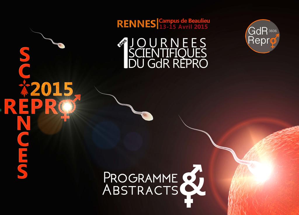
1 ères Journées ReproSciences ères Journées ReproSciences ères Journées ReproSciences ères Journées ReproSciences 2015
|
|
|
- Emery Randall
- 6 years ago
- Views:
Transcription
1

2


3 ReproSciences


 Structure fédérative de")

4 Cette réunion n aurait pu être organisée sans le soutien d un certain nombre d organismes et d Instituts. Nous les remercions très sincèrement. GDR 3606 Repro. Université de Rennes 1. Rennes Métropole. Institut National de la Recherche Agronomique. Institut de Recherche en Santé Environnement et Travail (IRSET, U1085) Structure fédérative de recherche Biosit. Université Européenne de Bretagne. Centre National de la Recherche Scientifique. Institut de recherche sur la santé l environnement et le travail 3
5 Bienvenue à Rennes Au nom du comité scientifique et du comité local d organisation, c est avec grand plaisir que nous vous accueillons à Rennes à l occasion des premières journées du GDR 3606 REPRO organisées sur le campus de Beaulieu de l Université de Rennes 1. ReproSciences 2015 est la première édition d un événement appelé à être réorganisé tous les deux ans dans d autres villes françaises. L objectif est de rassembler une communauté française parfois trop dispersée avec pour ambition de stimuler les discussions et les interactions entre les différents acteurs de la recherche dans le domaine de la reproduction. Avec l aide d un comité scientifique national extrêmement large, nous nous sommes efforcés d établir un programme aussi riche que possible et reflétant les différentes facettes de la fonction physiologique qui nous rassemble. Le domaine est particulièrement riche et nous voudrions ici remercier tous ceux qui nous ont aidé dans cette tâche. Nous remercions également tous ceux qui ont accepté de prendre la parole et dire à ceux qui n auront pas le privilège de nous présenter leur travail qu il y aura d autres occasions. Compte tenu de la situation financière des organismes, nous avons choisi d organiser la réunion avec le souci d épargner au maximum les crédits du GDR, de manière à pouvoir développer d autres actions. Nous avons fait le choix de privilégier la participation des jeunes chercheurs en leur offrant l inscription gratuite et des bourses de voyage. Par ailleurs, une session jeunes chercheurs avec des présentations flash a été incorporée au programme qui, d autre part, laisse de larges espaces de discussions devant les posters. L organisation de ces journées n aurait pas été possible sans le soutien financier de l Université de Rennes 1, de Rennes Métropole, du CNRS, de l INRA, de l INRIA, de l Université Européenne de Bretagne, de l IRSET, de la SFR Biosit et du GDR Repro. Qu ils en soient très sincèrement remerciés. Notre gratitude va également à tous ceux qui, à des titres divers, se sont impliqués dans l organisation de cette réunion. Le comité d organisation 4
6 5
7 SOMMAIRE Présentation Page 4 Les comités Page 7 Quelques Informations Générales Page 10 Programme détaillé Page 22 Résumés des communications Page 28 Liste des auteurs Page 86 6
8 Comité Scientifique National Isabelle Allemand Julien Bobe Xavier Bonnefont François Brion Thierry Charlier Frédérique Clément Joëlle Cohen-Tannoudji Yves Combarnous Corinne Cotinot Nathalie Dejucq-Rainsford Nicolas de Roux Nathalie di Clemente Sylvie Dufour Anne Duittoz Joëlle Dupont Stéphane Fabre Patricia Fauque Pascal Favrel Florian Guillou René Habert Hélène Jammes Bernard Jégou Olivier Kah Catherine Labbé Emmanuel Lemazurier 7 Gabriel Livera Micheline Misrahi Danielle Monniaux Eric Pailhoux Catherine Patrat Vincent Prévot Celia Ravel Eric Reiter Sophie Rousseaux Olivier Sandra Valérie Simonneaux Hervé Tostivint Daniel Vaiman François Vialard Catherine Viguié
9 Comité Local d Organisation Milissia Ben Maamar (Graphisme) Blandine Blanchard (Gestion) Julien Bobe Joel Cano-Nicolau Frédéric Chalmel Thierry Charlier Pascal Coumailleau Nathalie Dejucq-Rainsford Jean-Pierre Dussol (Site paiement) Pierre Gaudriault Marie-Madeleine Gueguen Yann Guiguen Bernard Jégou Olivier Kah Catherine Labbé Jean-Jacques Lareyre Florence Le Gac M. Legavre et toute son équipe (Restauration) Laurianne Lesné Christèle Lethimonier-Desdoits Séverine Mazaud-Guittot Rémy Morand (Gestion des amphis) Anne Moreau (Gestion) Catherine Nouyrigat (Gestion) Jean Louis Novo (Services techniques) Elisabeth Pellegrini Patricia Pilot Rousseau (Secrétariat Marylène Sauvée (Gestion des amphis) Colette Vaillant Véronique Villalon (Gestion) 8
10 9
11 Informations Générales 10
 Team NEED, Case 1302. Université de Rennes 1 Olivier.kah@univ-rennes1.")
12 ReproSciences 2015 se tient à l'université de Rennes 1, sur le Campus de Beaulieu, 263 avenue du Général Leclerc à Rennes les 13, 14 et 15 avril Contact: Olivier Kah Research Institute in Health, Environment and Occupation (INSERM U1085) Team NEED, Case Université de Rennes 1 Olivier.kah@univ-rennes1.fr Patricia Pilot Rousseau (Secrétariat) Patricia.pilot@univ-rennes1.fr Enregistrement: Lundi 13 avril 2015 de 11 heures 30 à 13 heures 30 Au club des professeurs (à gauche après le hall de l entrée principale de l Université). 11
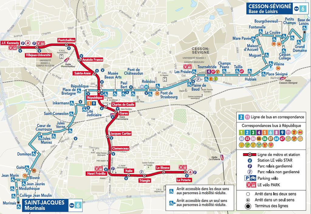
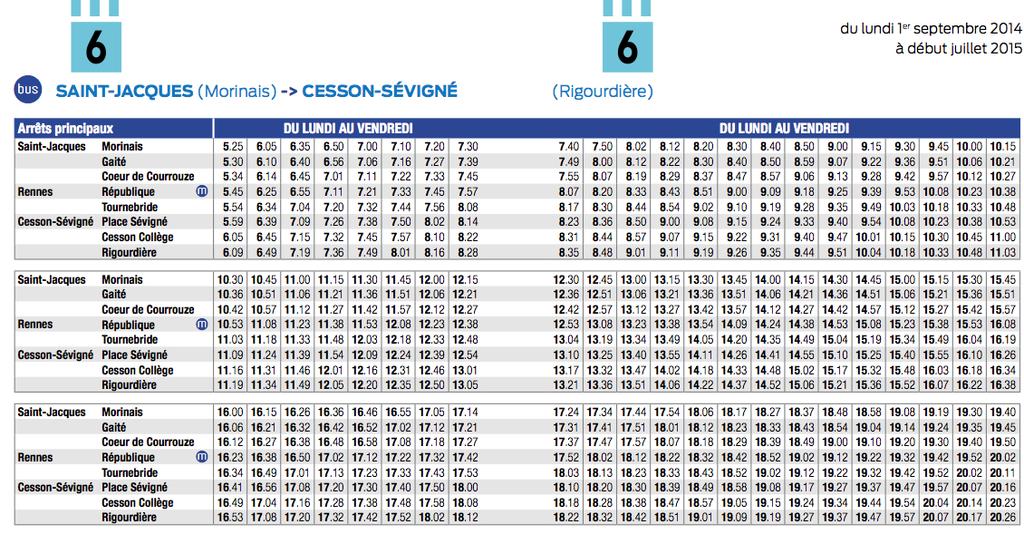
13 1 ères Journées ReproSciences ères Journées ReproSciences ères Journées ReproSciences ères Journées ReproSciences 2015 Comment venir sur le Campus de Beaulieu? Le Campus de Beaulieu est situé au 263 avenue du Général Leclerc à Rennes. Arrêt de bus Tournebride. Coordonnées GPS : ; Le Campus est très facile d accès : En bus (réseau STAR) : Environ minutes du centre ville: A partir des arrêts «Place de Bretagne», République où Musée des Beaux Arts, (Voir plan ciaprès) Bus ligne n 6, Bus ligne n C4 et Bus ligne 40 (express) L arrêt Tournebride est situé juste en face de l entrée principale du campus de Beaulieu. Le bâtiment N 2 est juste en face de l arrêt Tournebride à environ 100 mètres de l autre côté de la rue (Voir plan ci-après) En voiture: Il y a un grand parking gratuit devant le bâtiment principal. En vélo: Le Vélo STAR est le système de vélo en libre service proposé par Rennes Métropole : 900 vélos disponibles 24h/24 et 7j/7 dans 83 stations, positionnées en grande majorité près d une station de métro, d un arrêt de bus, de car ou de la gare. A pied: Si vous voulez vous dégourdir les jambes, vous pouvez marcher et longer la rivière (environ 40 minutes du centre en marchant bien). Très agréable. Les locaux sont accessibles aux personnes handicapées. Espace non fumeur. Accès wifi (Réseau EDUROAM disponible) 12


14 1ères Journées ReproSciences ères Journées ReproSciences ères Journées ReproSciences ères Journées ReproSciences 2015 Auberge de jeunesse Campus de Beaulieu, Bât. 2 Centre ville: Zone où se trouvent les hôtels Place de Bretagne 100m Tournebride Musée des Beaux Arts République 400m Gare Itinéraire des bus 6 et C4 Arrêts des bus C4 et 6 Bon à savoir Le C4 passe toutes les 8 minutes Le 40 est un express qui s arrête à République et Musée des Beaux Arts puis va direct à Tournebride De la Gare à République il y a un métro. Vous pouvez prendre le bus avec le même ticket 13


15 14
16 15

17 16
18 17

19 18

20 19

21 20

22 21
23 Programme 22
24 LUN DI 13 AVRIL 2015 Après midi 11h30-13h30 Enregistrement et café d accueil 13h30-13h50 Introduction générale Olivier Kah (Rennes). Session 1: Modérateurs Corinne Cotinot et François Vialard 13h50-14h20 14h20-14h40 14h40-15h00 15h00-15h20 15h20-16h50 Julien Bobe (Rennes) Mécanismes moléculaires impliqués dans la qualité des oeufs de poissons. Véronique Duranthon (Jouy-en-Josas) Effets de l environnement sur l embryon de Mammifères, conséquences pour le phénotype adulte. Virginie Maillard (Nouzilly) Métabolisme lipidique et reproduction femelle: rôle au niveau ovarien. Samir Hamamah (Montpellier) The regulation of the human cumulus-oocyte complex: The MicroRNAs CAFÉ ET POSTERS. Session 2: Modérateurs Daniel Vaiman et Charlotte Moretti 16h50-17h10 17h10-17h30 17h30-17h50 17h50-18h45 18h45-21h00 Thierry Fournier (Paris) Développement du placenta humain: Rôle du récepteur nucléaire PPARg et des gènes cibles. Olivier Sandra (Jouy en Josas) De l existence d un senseur endométrial chez les mammifères. Marie-Noëlle Dieudonné (Montigny le Bretonneux) Rôles du facteur pré-implantatoire (PIF) à l interface fœto-maternelle Conférence plénière. Modérateur Hervé Tostivint Pierre-Henri Gouyon (Paris) Sexe, Conflits et Coopérations COCKTAIL DÎNATOIRE (Hall) 23
25 MARDI 14 AVRIL 2015 Matin Session 3: Modérateurs Valérie Simonneaux et Pascal Coumailleau 09h00-09h30 Nicolas de Roux (Paris) Apport de la génétique dans la compréhension de l activation neuroendocrine de l axe gonadotrope de la vie foeœtale à la puberté. 09h30-09h50 Hervé Tostivint (Paris) Impact des tétraploïdisations sur le répertoire des hormones reproductrices des vertébrés. 09h50-10h10 Paolo Giacobini (Lille) Shaping the reproductive system: lesssons from semaphorins 10h10-10h30 Paul Klosen (Strasbourg) Mélatonine et TSH dans le contrôle saisonnier de la reproduction chez les rongeurs 10h30-11h00 PAUSE CAFÉ Session 4: Modérateurs Joëlle Cohen-Tannoudji et Antoine D. Rolland 11h00-11h20 11h20-11h40 11h40-12h00 12h00-12h20 12h20-14h00 Matthieu Keller (Nouzilly) Contrôle olfactif de la fonction de reproduction. Denis Tagu (Rennes) Changer de mode de reproduction: Le cas des pucerons pour l étude de l équilibre entre génétique et environnement. Frédéric Chalmel (Rennes) Génomique intégrative de la spermatogenèse chez les mammifères. Aurélien Capitan (Paris). Apports du modèle bovin et des données de génotypage et de séquençage à haut-débit dans la connaissance du contrôle génétique de la fertilité. BUFFET ET POSTERS. 24
26 MARDI 14 AVRIL 2015 Après midi Session 5: Modérateurs Danielle Monniaux et Céline Guigon 14h00-14h30 14h30-14h50 14h50-15h10 15h10-15h30 15h30-15h50 15h50-17h20 Nathalie di Clemente (Paris) Hormone anti-müllérienne et fonction ovarienne normale et pathologique Gabriel Livera (Fontenay aux Roses) Contrôle de la transition mitose/méiose Norbert Ghyselink (Strasbourg) Role of retinoic acid receptor (RAR) signalling in post-natal male germ cell differentiation Philippe Michel (Lyon) Modélisation mathématique du développement folliculaire basal Philippe Touraine ( Paris) Génétique des insuffisances ovariennes primitives CAFÉ ET POSTERS. Session 6: Modérateurs Nicolas de Roux et Yves Tillet 17h20-19h00 20h30-22h00 SESSION JEUNES CHERCHEURS Conférence Grand Public, Professeur Jean-Pierre Bourguignon, Endocrinologie Pédiatrique Liège La puberté: le grand chambardement de la tête aux pieds? Espace des Sciences, les Champs libres, 10 Cours des alliés, Rennes 25
.")
 Effets des fongicides azolés sur le système endocrinien et la reproduction.")
.")
27 MERCREDI 15 AVRIL 2015 Matin Session 7: Modérateurs François Brion et Catherine Viguié 09h00-09h30 09h30-09h50 09h50-10h10 10h10-10h30 10h30-11h00 Sakina Mhaouty-Kodja (Paris). Effets et mécanismes d action du bisphenol A dans les réponses neuroendocrines et comportementales liées à la reproduction chez la souris. Nathalie Hinfray (Verneuil-en-Halatte) Effets des fongicides azolés sur le système endocrinien et la reproduction. Marie Postel (Paris) Modélisation multi-échelle de la folliculogenèse terminale Louis Bujan (Toulouse) Projet GAMATOX: Effets des traitements du cancer sur le gamète mâle humain. PAUSE CAFÉ Session 8: Modérateurs Célia Ravel et Guillaume Halet 11h00-11h20 Arnaud Reignier (Nantes). Time lapse monitoring: a tool fot clinical research and embryo assessment. 11h20-11h50 Saadi Khochbin (Grenoble) Bases moléculaires de la programmation post-méiotique du génome mâle. 11h50-12h30 PRIX ET CLÔTURE 26
28 27
29 RÉSUMÉS 28
30 29
31 Présentations Orales Classement par ordre alphabétique Pa 30
32 What makes a good egg? Molecular mechanisms defining egg developmental competence in teleost fish. Julien Bobe1, Aurélien Bouleau1,2, Caroline Cheung1, Ozlem Yilmaz1, Thaovi Nguyen1, Amine Bouchareb1, Amélie Juanchich1, Daniel Zarski1, Iratxe Rojo1, Stéphanie Gay1, Aurélie Lecam1, Jérôme Montfort1, Hélène Rime1, Christian Fauvel2, Violette Thermes1 1 Equipe différenciation Sexuelle et Ovogenèse, INRA LPGP, F Rennes 2 IFREMER, LALR, F Palavas Les Flots julien.bobe@rennes.inra.fr Egg quality (i.e. the egg s ability to be fertilized and subsequently develop into a normal embryo) is highly variable in the wild or under aquaculture conditions. Yet, the egg components and associated molecular processes responsible for its developmental competence remain poorly understood. In teleost fish, in which a high fecundity can be observed in comparison to other vertebrate models, it is possible to sample individual egg clutches in which both developmental success assessment and analytical studies can be performed in parallel. Several types of approaches have been conducted to draw the molecular portrait of a developmentally competent fish egg by studying its composition in terms of maternal mrnas, mirnas, and proteins. Correlative studies have shown a link between the abundance of specific mrnas and/or proteins in the eggs and the overall developmental success of the corresponding egg clutches. In rainbow trout (Oncorhynchus mykiss), a correlation between developmental success and the abundance of nucleoplasmin (npm2) mrna in the egg was previously established. In zebrafish (Danio rerio), a model species with transparent eggs and rapid development, a knockdown (KD) approach showed that maternally-inherited npm2 mrna was crucial to allow developmental success beyond zygotic genome activation (ZGA). Similar approaches at the proteome and mirna repertoire levels have yielded interesting results that are currently being further analyzed. Taking advantage of the wide diversity of fish models (over species), future studies will be designed to identify key molecular mechanisms that could be shared by evolutionary distant species. 31 GAMATOX Project: impact of cancer treatments on semen characteristics, sperm DNA fragmentation and sperm aneuploïdy: a multicenter prospective study from the CECOS Network Louis Bujan1 and Marie Walschaerts1, Nathalie Rives2, Sylvianne Hennebicq3, Guillaume Martinez3, Véronique Duchesne2, Jacqueline Saias4, Florence Brugnon5, Jacques Auger6, Isabelle Berthaut7, Ethel Szerman8, Nathalie Moinard1, Myriam Daudin1 1 Université de Toulouse; UPS; Groupe de Recherche en Fertilité Humaine (EA 3694, Human Fertility Research Group) and CECOS, Toulouse, France; and following CECOS centers and research team associated : 2Rouen, 3Grenoble, 4Marseille, 5Clermont-Ferrand, 6Paris Tenon, 7 Paris Cochin, 8Caen. Bujan.l@chu-toulouse.fr Testicular Germ Cell Tumor (TGCT) is the most common cancer in young men and TGCT incidence has increased in several countries over the past 50 years. Hodgkin lymphoma (HL) and non-hodgkin lymphoma (NHL) affect also young men who wish to procreate. Prognosis of these cancers has improved very markedly over the last decades due to the treatment mainly based on chemotherapy and radiotherapy. Several late adverse effects of chemotherapy or radiotherapy have been described but mainly in retrospective studies and very few studies, with discrepant results, have explored sperm DNA fragmentation and sperm aneuploïdy following such treatments. In this context, we performed the national multicenter prospective research project GAmete MAle TOXicity (GAMATOX I) which enrolled patients with testicular germ cell tumors (n= 129), patients with Hodgkin Lymphoma or no Hodgkin lymphoma (n=75). Patients performed semen samples before cancer treatment and 3, 6, 12, 24 months after the treatment ending. Routine semen analyses were performed according to the WHO recommendations in each center while specific analyses for the sperm DNA fragmentation and the sperm aneuploïdy were centralized in Toulouse, Grenoble and Rouen centers. All samples were registered with the GERMETHEQUE biobank (France). Results were explored according to each cancer type and each treatment regimen. Predictive factors for sperm recovery following treatment were studied by multivariate analyses. Treatments have drastic effects on spermatogenesis and the capacity and the time needed to recover were dependant of the type of treatment and of pretreatment sperm characteristics. Compared to control group of normal men pretreatment alterations existed in certain cancer groups. Sperm DNA fragmentation and sperm aneuploïdy were increased following treatment but were also increased before treatment in lymphoma group. The results of the GAMATOX I project were relevant for the counseling of cancer patients, before and after treatment, about the risks for the male gamete and the progeny. However, the new genome and epigenome technology explorations will be applied in the next project: GAMATOX II, in order to define more precisely the alterations induced by such treatment and to evaluate the period safety after the end of treatment. This work was supported by a grant from the French Ministry of Health, PHRC N Regulatory and ethical submissions were performed by the University Hospital of Toulouse.
33 Genetic tools to improve reproduction traits in dairy cattle Capitan A.1,2*, Michot P.1,2, Baur A.1,2, Saintilan R. 1,2, Hozé C. 1,2, Valour D. 1,4, Guillaume F.3, Boichon, D.5, Barbat A. 2, Boichard D.2, Schibler L.1 and Fritz S.1,2 1UNCEIA, 149 rue de Bercy, Paris, France 2INRA, UMR1313 Génétique Animale et Biologie Intégrative, Domaine de Vilvert, Jouy-en-Josas, France 3EVOLUTION, 69 rue de la Motte Brûlon, Rennes, France 4INRA, UMR 1198 Biologie du Développement et Reproduction, Domaine de Vilvert, Jouy-en-Josas, France 5MIDATEST, Les Nauzes, Soual, France Fertility is a major concern in dairy cattle industry and has been the subject of numerous studies over the last twenty years. Surprisingly, most of them focused on rough female phenotypes and despite their important role in reproductive success, male and embryo related traits have been poorly studied. In recent years, the rapid and important evolution of technologies in genetic research led to the development of genomic selection. In a chain reaction, the generalization of this method combined with the extreme achievement of the artificial insemination industry have led to the constitution of large data bases of genotyping and sequencing data as well as refined phenotypes and pedigree records. These resources offer unprecedented opportunities in term of fundamental and applied research. Here we present five examples of them with a focus on reproduction related traits i.e. the detection of QTL for male fertility and semen quality traits (i), and, for refined phenotypes associated with female fertility (ii); the identification of recessive embryonic lethal mutations by depletion of homozygous haplotypes (iii) or by mining whole genome sequencing data (iv); and finally the contributions of HD SNP chips, whole genome sequencing and imputation to the increase of the power of QTL detection methods and to the identification of their causal variants (v). Integrative genomics and mammalian spermatogenesis Frédéric Chalmel1,*, Antoine Rolland1,*, Bertrand Evrard1, Sophie Chocu1, Nolwen Hernio1, Emmanuelle Com1, Charles Pineau1, Michael Primig1, Nathalie Rioux-Leclercq, Nathalie Dejucq-Rainsford2 & Bernard Jégou1 1 Inserm U1085-Irset, Rennes, France 2 CHU Pontchailloux, 2, rue Henri Le Guillou, Rennes cedex 9, France. frederic.chalmel@inserm.fr Spermatogenesis is a complex and tightly regulated process leading to the continuous production of male gametes, the spermatozoa. Within the testes, male germ cells first proliferate to amplify their number, next shuffle and reduce their genome through two consecutive meiotic divisions, and finally differentiate dramatically into cells specialized for mobility and fecundation. This developmental process requires the sequential and coordinated expression of thousands of genes, including many that are testis-specific. The molecular networks underlying normal and pathological spermatogenesis have been widely investigated in recent decades, and many high-throughput expression studies have studied genes and proteins important for male fertility. During this presentation, I will focus on studies that have attempted to link the transcriptome and proteome in spermatogenesis or have combined transcriptomic and proteomic data to gain insight into testicular functions and germ cell biology. Supported by the Institut national de la santé et de la recherche médicale (Inserm), the Université de Rennes 1, the Agence nationale de sécurité sanitaire de l'alimentation, de l'environnement et du travail [ANSES n EST to F.C.], the Fondation pour la recherche médicale [FRM n DBI to F.C.], and the European Union [FEDER to F.C]. 32
34 Apport of Human genetics in the understanding of the neuroendocrine control of the gonadotropic axis Nicolas de Roux Inserm U1141. Université Paris Diderot. Labaratoire de Biochimie Hormonale. Hopital Robert Debré. 48 Bld Sérurier Paris. The development of the neuroendocrine control of the gonadotropic axis is complex. It starts during the fetal life, inhibited at the end of gestation, reactivated after birth for few weeks, this axis is then inhibited during childhood until a second reactivation around 10 years which marks the start of the puberty. This sequence of activation-inhibition is fundamental to develop a normal reproduction function, but the mechanisms remain poorly understood. Human genetics of rare disorders of gonadotropic axis activation has led to major advances to understand the functional plasticity of this axis. Initially focused on isolated or syndromic gonadotropin deficiency, novel perspectives have recently emerged with the description of genetic defects causing central precocious puberty. In addition to the description of novel neuropeptides, current studies try to characterize the molecular mechanisms controlling the plasticity of the GnRH neuronal network. The most recent results on human genetics of pubertal disorders will be presented. Anti-Müllerian hormone and ovarian function Nathalie di Clemente1 1. Univ Paris Diderot, Sorbonne Paris Cité, Biologie Fonctionnelle et Adaptative (BFA), F Paris, France; CNRS UMR 8251, F Paris, France; Physiologie de l'axe gonadotrope INSERM U1133, F Paris, France. nathalie.diclemente-besse@univ-paris-diderot.fr Anti-Müllerian hormone (AMH) is a 140 kda glycoprotein belonging to the TGF-β family. Its existence was postulated by Pr Alfred Jost in the early fifties to explain the regression in male fetuses of Müllerian ducts, the anlagen of uterus and tubes in females. AMH must be cleaved to allow its C- terminal fragment to bind AMH specific type II receptor, AMHR-II. Then, AMH activates the same signalling pathway than Bone Morphogenetic Proteins (BMPs): the type I receptors Alk 2, 3 and 6 and the Smad1,5, 8 proteins. In males, AMH expression starts when Sertoli cells begin to differentiate, decreases at puberty mainly under the influence of androgens, but stays detectable in adults. The only pathology due to a defect of AMH synthesis or sensitivity is the persistent Müllerian duct syndrome, a rare case of male pseudohermaphroditism characterized by the presence of uterus and tubes in otherwise virilized males. In addition, serum AMH is a valuable marker for the diagnostic of sexually ambiguous babies, for the follow-up of boys puberty or the treatment of men with hypogonadotropic hypogonadism. In the eighties, AMH was shown to be also synthesized in females by granulosa cells of growing follicles of the ovary where AMH represses both primordial follicle recruitment and FSH-dependent follicle maturation. The discovery in years 2000 that serum AMH was a marker of ovarian reserve brought light to ovarian AMH. Since that time, many groups have extended this result and showed that serum AMH was also a prognostic marker of ovarian stimulation, making serum AMH a useful tool in assisted reproductive technology. Because despite this growing interest in ovarian AMH, its regulation and mechanism of action were still unclear, these last years, our group has made use of numerous and complementary tools to fill this lack of data. We have shown that AMH expression is stimulated by FSH and BMPs, and regulated differentially by estradiol depending on estrogen receptors. We have also demonstrated that AMH signals through Alk3 type I receptor and Smad 1 and 5 proteins in granulosa cells and identified a new AMH target gene, Inhibitor of differentiation/deoxyribonucleic Acid-Binding 3. In addition, we have studied how AMH could be involved in the polycystic ovary syndrome (PCOS), the main cause of women infertility, which is characterized by high serum AMH levels. We have shown that both AMH and AMHR-II are overexpressed in granulosa cells from PCOS women and that this is partly due to a dysregulation of these genes by LH. 33
35 PIF, a major actor for pregnancy Hadia MOINDJIE1, Esther DOS SANTOS1,2, Florence BOITRELLE1,2, Valérie SERAZIN1,2, Nathalie MELAINE3, Eytan BARNEA4, François VIALARD1,2, Marie Noëlle DIEUDONNE1, 1 : GIG EA2493, UFR des sciences de la santé Simone Veil, UVSQ, Montigny le bretonneux, France2 : Medical biology laboratory, CHI de Poissy, Poissy, France. 3: Biogenouest, Rennes, France 4: BioIncept, LLC, Cherry Hill NJ, USA. marie-noelle.dieudonne@uvsq.fr The preimplantation factor (PIF) is a 15aa peptide secreted very early by viable mammalian embryo. First identified in 1995, its impact has been clearly shown since Afterwards, many results have been obtained, showing its pleiotropic effects and implications in the immune and inflammatory processes. Concerning the reproductive biology, more elements indicate a major role of PIF during pregnancy. PIF, embryo secreted, has been shown to have an autocrine positive effect on embryo development. Recently, using mass spectrometry, a low PIF level in IVF embryo culture media has been detected. If a correlation between PIF level and IVF success was established, PIF could be considered as a new non invasive biomarker for IVF. PIF has been also detected in maternal blood circulation. Recently, using bovine model, it was shown that PIF detection in maternal circulation was correlated with live birth in early pregnancy. Furthermore, it has been reported that PIF could modulate disrupting immune and apoptosis pathways, cells proliferation and adhesion in human endometrial stromal cells. Moreover, an intense PIF immunostaining was observed in human trophoblastic cells from first trimester placentas. A decrease of PIF labeling was observed at term. Finally, we recently showed that PIF promotes invasion in human primary extravillous trophoblasts (EVT), confirming its pro-invasive effect initially described in the cell line HTR-8/SVneo in The proinvasive regulatory effect of PIF in EVT was associated with a modulation of metalloproteinase activity and mrna integrin expressions mediating by multiple signaling pathways. Further analyses are currently in progress in our laboratory to precise the molecular mechanisms implicated in the PIF effects on human placenta and endometrium. In conclusion; PIF appears to be a key factor of foeto-maternal interface. Effects of the environment on early mammalian embryo. Consequences for adult phenotype. Véronique Duranthon. INRA, UMR1198 Biologie du Développement et Reproduction, F Jouy-en-Josas, France veronique.duranthon@jouy.inra.fr In Mammals, fetal environment is known to affect adult health, giving rise to the DOHaD concept (Developmental Origin of Health and Disease). More recently, this concept of sensitivity to the environment with long term consequences has been extended to the periconceptional period. Especially, preimplantation period of development has been shown to be very sensitive to environmental conditions. This was quite unexpected since in most mammalian species, preimplantation development has been obtained in vitro and is compatible with full term development after transfer to a recipient mother. One of the most specific examples of such long term effect has been developed in the mouse model where females were fed a low protein diet for 3.5 days from fertilization onwards. This maternal diet skewed cell allocation to the first embryonic lineages at the blastocyst stage and induced a compensatory fetal and perinatal growth positively correlated to cardivascular, metabolic and behavioural abnormalities (1). In vitro development of mammalian embryo has also been reported to have long term effects, which is particularly worrisome within the framework of Assisted Reproductive Technologies. To analyze the effect of different environments on epigenetics modifications and on gene expression during the preimplantation period of development, we used the rabbit embryo as a model of early mammalian embryo with a delayed onset of embryonic genome activation, which is the case in most mammals (including human) except the mouse. In the rabbit, in vivo developed embryos can be easily recovered at each stage of development. We showed that the kinetics of embryonic genome de-methylation during the preimplantation development varies with embryo culture conditions and differs from in vivo development (2). We also modified the embryo environment in vivo by feeding rabbit females with an hyperlipidic/hypercholesterolemic diet. We showed that gene expression and trophoblast function were altered as soon as the blastocyst stage in such females (3,4). 1. Sun C, 2014 Development. 141(5): Reis e Silva AR, Epigenetics. 7(5): Picone O, 2011 Theriogenology. 75(2): Tarrade A, PLoS One. 8(12):e Supported by: Agence de la Biomédecine, INRA-PHASE, Labex Revive 34
36 Role of PPAR-gamma and of its target genes in placental development. Thierry Fournier UMR-S1139, Inserm-Paris Descartes, Faculté de Pharmacie, Paris, France Fondation PremUp Grossesse et Prématurité, Paris, France DHU Risques et Grossesse, Maternité Port Royal, Paris, France The peroxisome proliferator-activated receptor-γ (PPAR γ) is a member of the nuclear receptor superfamily that controls in a ligand-dependent manner the expression of a large array of genes involved in the control of energy homeostasis, cell differentiation, proliferation, apoptosis, and the inflammatory process. Unexpectedly, genetic studies performed in mice established that PPAR γ is essential for placental development. During pregnancy, the placenta ensures multiple functions, which are directly involved in the initiation, outcome of gestation and foetal growth. In the human placenta, PPAR γ is highly and specifically expressed in the two trophoblast subtypes i.e. the villous trophoblast that represents the endocrine and exchange tissue and the invasive extravillous cytotrophoblasts (EVCT) involved in implantation, immune-tolerance and uterine artery remodelling. Activation of PPAR γ induces accumulation of lipids, villous trophoblast differentiation and inhibits EVCT invasiveness. Oxidized LDLs that contain potential PPAR γ ligands, but not native LDLs, induce PPAR γ transcriptional activity and inhibit trophoblast invasion in vitro. Recently, human cytomegalovirus (HCMV) was shown to activate trophoblastic PPAR γ for its own replication and consequently inhibits invasiveness of infected cytotrophoblasts. Analysis of PPAR γ target genes revealed trophoblastic factors described to control trophoblast invasiveness and surprisingly chorionic gonadotropin hormone (hcg), known to be mainly produced by the endocrine villous trophoblast. Analysis of hcg gene expression revealed opposite regulation by PPAR γ in the two trophoblast subtypes. Finally, a hyperglycosylated form of hcg (hcg-h) only produced by invasive EVCT was shown to promote trophoblast invasion and angiogenesis through a TGFß signalling pathway and independently to its binding to the LH-hCG receptor. Together, these data underscore the major role of PPAR γ and its target genes, such as hcg and hcg-h, in the control of human trophoblast differentiation and invasion, and suggest that over-activation of this nuclear receptor following HCMV infection or by excess of ligands at the maternal foetal interface could impair implantation and placentation and therefore embryonic development. Role of Retinoic Acid Receptor (RAR) signalling in post-natal male germ cell differentiation Norbert B. Ghyselinck1, Aurore Gely-Pernot1, Mathilde Raverdeau1, Nadège Vernet1, Betty Féret1, Muriel Klopfenstein1, Christine Dennefeld1, Marius Teletin1,2, Manuel Mark1,2 1Institut de Génétique et de Biologie Moléculaire et Cellulaire, Département de Génétique Fonctionnelle et Cancer, CNRS UMR7104, INSERM U964, Université de Strasbourg, Illkirch, France 2Hopitaux Universitaires de Strasbourg, France norbert@igbmc.fr All-trans retinoic acid (ATRA), the active metabolite of vitamin A, is synthesised by dedicated enzymes called retinaldehyde dehydrogenases (ALDH1A1 to A3). Then it acts either through activating nuclear receptor heterodimers made of ATRA receptors (RARA, RARB, RARG) and rexinoid receptors (RXRA, RXRB and RXRG), or through non genomic effects. It is known for decades that ATRA is instrumental to male germ cell differentiation, but its origin and its mechanism of action in the seminiferous epithelium remained elusive. To address these questions, we have analysed the phenotypes of mice lacking either ATRA-synthesizing activities in Sertoli cells (SC), the supporting cells of the germ cell lineage, or retinoid receptors (RAR and RXR) in spermatogonia (SG). We demonstrate that (i) ALDH1Adependent synthesis of ATRA by SC is indispensable during the first spermatogenic cycle to initiate differentiation of SG; (ii) RARA in SC mediates the effects of ATRA, notably through activating expression of MAFB transcription factor, whose Drosophila homologue is mandatory to germ cell differentiation; (iii) ablation of RXR in SG recapitulates the set of defects observed both upon ablation of RAR in SG and upon vitamin A deficiency; (iv) ATRA enhances expression of the SALL4A transcription factor in SG. This effect depends on activation of RARG and RXRA bound to a conserved regulatory region located in the Sall4 gene. This indicates that RAR/RXR heterodimers are the functional units in spermatogonia driving the ATRA-induced transition from the undifferentiated to the differentiating state. Moreover, they cast light on the long-searched mechanism through which ATRA cell-autonomously controls expression of the KIT tyrosine kinase receptor to trigger this transition. Our data also establish for the first time that the effects of ATRA on SG differentiation in the seminiferous epithelium are indirect, via SC. Supported by the RAPSSODI, MOLMECHMEIOSIS and ARGONADS ANR projects 35
37 Shaping the reproductive system: lessons from semaphorins Paolo Giacobini 1 1 Inserm, Development and Plasticity of the Neuroendocrine Brain, Jean- Pierre Aubert Research Center, U1172, Lille, France paolo.giacobini@inserm.fr Reproductive competence in mammals depends on the projection of gonadotropin-releasing hormone (GnRH) neurons to the hypothalamic median eminence (ME) and the timely release of GnRH into the hypothalamic pituitary gonadal axis. In adult rodents, GnRH neurons and the specialized glial cells named tanycytes, periodically undergo cytoskeletal plasticity. During the ovarian cycle, under conditions of low gonadotropin output, GnRH-secreting axon terminals are distant from the pericapillary space of the ME, thus impairing the access of the neurohormone to the pituitary portal circulation, but they undergo extensive axonal growth toward the vascular wall at the onset of the preovulatory surge, when massive GnRH release has to occur to trigger ovulation. However, the mechanisms that regulate this plasticity are still largely unknown. This talk summarizes recent studies analysing the contribution of specific guidance molecules named semaphorins in the development and adult function of gonadotropin-releasing hormone (GnRH) neurons. These studies started to shed light on the molecular mechanisms responsible for the progression of the estrous cycle in rodents and suggest that this phenomenon relies on the antagonistic effects of two semaphorins whose expression in the median eminence is periodically influenced by circulating sex hormones Le sexe : conflits et coopération" Pierre-Henri Gouyon, Professeur au Muséum National d Histoire Naturelle, à l ENS, à l AgroParisTech et à Sciences Po. pierre-henri.gouyon@mnhn.fr La sexualité, c'est l'échange. L'échange de matériel génétique entre deux organismes qui en produisent un nouveau procédant des deux. Dans ce sens large, la sexualité se trouve dans tous les groupes d'organismes vivants, bactéries, archées, eucaryotes (plantes, animaux, champignons...). Pourquoi les êtres vivants ontils adopté une caractéristique si compliquée? Comment procèdentils? Les modes de sexualité observés dans la nature sont d'une diversité incroyable. Pourquoi des mâles et des femelles, ou des hermaphrodites? Certaines espèces ont abandonné le sexe. Les femelles (parthénogénétiques) se débrouillent seules. Pourquoi font-elles ça? Et pourquoi pas les autres? En effet, il semble bien que les femelles aient tout à gagner à abandonner le sexe ; de ce point de vue, le fait qu elles le pratiquent s apparente à de l altruisme. Elles favorisent la diversité de la population au détriment de leur propre reproduction. Une telle caractéristique semble alors sélectionnée à l échelle de la lignée évolutive et non pas à l échelle individuelle. Chez les humains par exemple, le sexe pourrait-il disparaître? Le sexe est au centre d un réseau ce situations de conflits et de coopération qui montrent toute la complexité de nous pose une multitude de questions tant biologiques que sociologiques. Comme on le dit pour l'amour (et les maths), on ne peut pas le faire en public, mais on peut en parler... Supported by the ANR-14-CE RoSes and GnRH 36
38 The regulation of the human cumulus-oocyte complex: The MicroRNAs S Hamamah INSERM U 1203 Human early embryo development and pluripotency Arnaud de Villeneuve hospital, Montpellier, France s- hamamah@chu- montpellier.fr An enormous amount of knowledge about the human oocyte and CCs have been generated over the last years, due in part to the recent advances in gene expression technologies using microarray, CGH array and high-fidelity RNA amplification. Numerous small endogenous non-coding transcripts, termed micrornas (mirnas), have been found to execute key functions in silencing expression of specific target genes in plant, animal and human systems. Changes in mirna expression profiles have been linked to pathologies such as cancer and infertility: female mice with global mirna deficiency are sterile from several causes, including defects in oocyte function. In addition, the messenger RNA (mrna) expression in mice and bovine during oogenesis shows that a large proportion of maternal genes are under the control of mirnas. Thus, mirna profiling offers an effective means of acquiring novel and valuable information regarding the regulation of transcripts involved in human reproduction. The mirnas study of oocyte-cumulus complex offers a promising opportunity, by a non-invasive method, to evaluate ovarian failure and pregnancy outcome Azole fungicides in zebrafish: new effects for old molecules Nathalie Hinfray1, Rüdiger W. Schulz2, Yann Guiguen3, François Brion1 1INERIS, DRC/VIVA, Ecotoxicology unit, Verneuil-en-Halatte, France.2Utrecht University, Reproductive Biology Group, Utrecht, The Netherlands.3INRA, LPGP, Sexual differentiation and oogenesis unit, Rennes, France nathalie.hinfray@ineris.fr Azole is a class of diverse compounds discovered several decades ago and essentially used as antifungals in agriculture and medicine. Their primary mode of action is to inhibit the fungal enzyme 14α-demethylase, which produces ergosterol, an important component of the cell membranes of fungi. Despite this specific mode of action, azoles are also characterized by their capacity to disrupt the endocrine system of vertebrate through multiple mechanisms notably by altering steroidogenesis, a key physiological process responsible for the biosynthesis of steroidal hormones. For instance, azole compounds affect both expression and enzymatic activities of several steroidogenic enzymes in vertebrate models, including fish, leading to reproductive disorders. Because of their uses, their presence in the aquatic environment (surface waters of rivers, lakes and estuaries; sewage sludge) has been recently reported in different industrialized countries raising the need to assess hazard and risk posed to aquatic organisms. In this context, several experiments have been performed to explore the effects of the pharmaceutical azole, clotrimazole, on the endocrine system in the zebrafish. In males, we found that clotrimazole was able to affect the testicular physiology by affecting steroidogenesis, androgen release and spermatogenesis (Hinfray et al., 2011, Baudiffier et al. 2012, 2013). However, the most striking effect was observed in females. Indeed, we found that exposure of adult female zebrafish to clotrimazole led to a dramatic masculinisation as revealed by the complete sex-reversal of the phenotypic sex. Remarkably, this sex-reversal occurred rapidly leading to well-differentiated testicular tissue after 42 days of exposure. By using cyp19a1a-gfp transgenic zebrafish, we further demonstrated that clotrimazole led to a time-dependent inhibition of GFP expression in ovary which preceded the histological differentiation of testis demonstrating the crucial role played by aromatase in the process of masculinisation. Altogether, our study demonstrates that clotrimazole significantly affect the gonad endocrinology and physiology of fish revealing new and striking effects on its ability to reverse the phenotypic sex of adult female. Based on our data, it is clear that further studies are needed to address the issue raised by the presence of azoles in the aquatic environment as regards to their potential impact on wild population of fish. Supported by the 190 program of the French ministry of environment and the post-grenelle program NEMO. 37
39 Olfactory control of reproductive function Matthieu Keller1, Chantal Moussu1, Didier Chesneau1, Laura Szymanski1, Mélanie Jouhanneau1 & Pablo Chamero1 1Physiologie de la Reproduction & des Comportements, UMR 7247 INRA/CNRS/Université de Tours/IFCE, Nouzilly, France In many vertebrate species, olfactory informations exchanged during social interactions have profound consequences on both reproductive physiology and behavior. Indeed, olfactory cues can virtually affect all the steps of the reproductive cycle. These olfactory informations are processed by various olfactory sub-systems and especially by the main and the accessory (or vomeronasal) olfactory systems. In rodents, it is quite well established that the chemosignals affecting reproductive physiology and behavior are mainly dependent on olfactory cues processed through the accessory olfactory pathway which is closely connected to the hypothalamus, thereby controlling reproductive function. To illustrate the role of male olfactory chemosignals on female mice, we will present here data on the control of puberty onset and sexual behavior. Indeed, we have shown that male soiled bedding contains various androgen-dependent chemosignals such as (1R, 5S, 7S)-3,4-dehydro-exo-brevicomin, 6- hydroxy-6-methyl-3-heptanone or (S)-2-sec-butyl-4,5-dihydrothiazole that advance vaginal opening and enhance uterus weight in prepubertal females. By using surgical approaches and the use of mice with conditional cell-specific ablation of the vomeronasal G protein Gαo, we have shown that the olfactory compounds contained in male bedding are processed by the vomeronasal olfactory system and especially the vomeronasal type 2 receptors (V2Rs) which are located in the basal layer of the vomeronasal epithelium. Then, using c-fos as a marker of cellular activation, we have delineated the neural network involved in the processing of male chemosignals. We show a significant effect of odor on c-fos-expression in areas mainly receiving olfactory information from the vomeronasal system, thus showing that these areas may be responsible for communicating odor information that drives puberty acceleration. Finally, we provide evidence that the peripubertal exposure to male odors has also longterm behavioral consequences as it promotes an early preference for male odors in adulthood. Molecular basis of post-meiotic male genome programing Emilie Montellier1, Hitoshi Shiota1, Sophie Barral1, Thierry Buchou1, Afsaneh Goudarzi1, Fayçal Boussouar1, Jonathan Gaucher1, Matthieu Gérard2, Yingming Zhao3, Sophie Rousseaux1, Saadi Khochbin1 1 - INSERM, U823; Université Joseph Fourier - Grenoble 1; Institut Albert Bonniot, Grenoble, F France 2 - Laboratoire d'etude du Métabolisme des Médicaments,, DSV / ibitec-s / SPI, CEA Saclay, Gif sur Yvette, Cedex, France 3 - Ben May Department of Cancer Research, The University of Chicago, Chicago, IL 60637, USA. khochbin@ujf-grenoble.fr In mammals, post-meiotic male genome reorganization and compaction can be considered as conceptually related to sporulation in lower eukaryotes or pollen formation in plants, since all these processes consist in preparing the genome to confront the hostile external environment. All involve post-meiotic genome compaction mechanisms of unclear nature. In mammals, the current knowledge implies a post-meiotic stepwise replacement of histones by transition proteins and protamines, which finally pack the genome into the mature spermatozoid. Our recent investigations on the molecular basis of post-meiotic male genome programming have demonstrated that not only hyperacetylation-dependent histone replacement but also the meiotic and post-meiotic gene transcription programs are largely controlled by a single member of the BET double bromodomain family, Brdt. Our parallel investigations of histone variants show that in post-meiotic cells, histone hyperacetylation and Brdt s action are not sufficient for the replacement of histones and that a prior global incorporation of testisspecific H2A and H2B histone variants is required. Finally, we also demonstrate that the whole male germ cell expression program is directed by new and yet uncharacterized histone post-translational modifications which shape the male genome and drive the meiotic and post-meiotic male-specific gene expression program. We have therefore discovered unique and essential regulators of male germ cell differentiation, which, in a developmentally controlled manner, first drive a specific spermatogenic gene expression program and later control the tight packaging of the male genome. Supported by the ANR JC PHEROSEX. 38
40 Melatonin and TSH in the seasonal control of reproduction in rodents Paul Klosen1, Marion Ciancia1, Sébastien Milési1, Marie-Emilie Sébert1, Kamontip Rasri1,2, Marie-Pierre Laran-Chich1, Valérie Simonneaux1 1INCI, CNRS UPR 3212, Strasbourg 2 Fac Medecine, Thammasat University, Bangkok, Thailand klosen@inci-cnrs.unistra.fr Many animal species synchronize their reproductive activity with the seasons in order to have their offspring being born at a favourable moment of the seasonal cycle. The nocturnal secretion of the pineal hormone melatonin is known to be the key synchronizing cue for seasonal physiology. Melatonin controls the production and secretion of the thyroid stimulating hormone (TSH) by the pars tuberalis of the adenohypophysis. In 2008, the tanycytes, highly specialized glial cells of the hypothalamus, have been recognized as the main target of the pars tuberalis TSH for the seasonal control of reproduction. TSH stimulates the production of Deiodinase 2 (Dio2) by the tanycytes. This enzyme activates tetraiodothyronine T4 to its active form triiodothyronine T3. The current consensus is that this local T3 production then controls the gonadotropic axis through a neuroendocrine pathway that remains to be uncovered. We have shown that a chronic intracerebroventricular (icv) infusion of TSH is able to fully reactivate the photoperiodically inhibited gonadotropic axis of hamsters exposed to a short photoperiod. This reactivation coincides with the restauration of a long day pattern in the expression of the RFamides kisspeptin and RFRP, both potent regulators of GnRH neuron activity. This suggests that TSH acts through these neurons to control the gonadotropic axis. However, currently no precise signalling pathway between tanycytes and RFamide neurons has been described. During the photoperiodic reactivation of the hamster gonadotropic axis by a long day photoperiod, we noticed that the secretion of LH, and thus of GnRH, was increased before the expression of RFamides started to rise, an observation that questions the initial hypothesis. Furthermore, acute icv infusion of TSH in sexually active Djungarian hamsters induces an increase in circulating testosterone without notably affecting RFamide immunostaining. These observations suggest the existence of a signalling pathway independent of RFamides through which TSH is able to control the gonadotropic axis. This pathway might also explain the circadian rhythm in circulating sex steroids observed in various species. Control of the mitotic/meiotic switch Marie-Justine Guerquin, Virginie Rouiller-Fabre, René Habert, Benoit Souquet, Emilie Abby, Ronan Le Bouffant, Sébastien Messiaen, Sophie Tourpin, Delphine Moison, Clotilde Duquenne, Gabriel Livera Laboratory of development of the gonads, UMR967 INSERM/CEA/University Paris Diderot, Sorbonne Paris Cité & University Paris XI, Fontenay aux Roses, France gabriel.livera@cea.fr The timing of meiotic entry is a crucial event in the life of all sexually reproducing organisms. In Mammals, embryonic germ cells follow a sexually dichotomic fate with female germ cells entering meiosis and male ones escaping this differentiation process. In the mouse fetal ovary all germ cells enter rapidly into meiosis between 13.5 and 15.5 days post-conception. During the same period, in the testis germ cells progressively stop proliferating and enter into quiescence. Over the recent years, many intrinsic regulators of these events have progressively been identified while the upstream regulators are still a matter of debates. Based on original organ cultures, co-cultures and cell sorting experiments we evidenced that the timing of meiosis in fetal germ cells does not depend on their chromosomal constitution but rather from signals providing from the surrounding somatic cells. Those experiments allowed defining various activities based on secreted testicular substance some inhibiting meiosis and other slowing down proliferation. The role of retinoic acid, that has been proposed as a key factor for governing meiotic entry, was then investigated and surprisingly it appears to have a modest impact on the mitotic/meiotic switch in both rodents and human ovaries. Even more strikingly, we propose that part of the meiotic program is likely retinoic acid-independent based on the identification of new regulators of the meiotic entry. It thus appears that the abrupt switch from mitosis to meiosis requires a more complex interplay than anticipated relying on both retinoic acid-dependant and retinoic acid independent signalling. 39
41 Lipid metabolism and female reproduction: role at the ovary level. Virginie Maillard1, Sébastien Elis1, Sandrine Fréret1, Valérie Labas1,2, Ana-Paula Teixeira-Gomes2,3, Véronique Cadoret1,4, Philippe Monget1 and Svetlana Uzbekova1,2. 1 Team BINGO, PRC, UMR , INRA-CNRS-Université de Tours- IFCE, Nouzilly, France 2 Laboratoire de Spectrométrie de masse, PAIB, INRA PRC, Nouzilly, France3 ISP, INRA UMR1282, Nouzilly, France 4 CHRU de Tours, LBR, Tours France. virginie.maillard@tours.inra.fr Besides their role of energy sources, intracellular lipids and their derivatives are well-known to be essential components of biological membranes, cell-to-cell interaction, and in regulation of different cellular processes as proliferation, apoptosis, hormone synthesis... In mammals, oocytes develop inside the ovarian follicles; this process is strongly supported by the surrounding follicular cells (cumulus, granulosa and theca cells) and follicular fluid. Folliculogenesis and final oocyte maturation are regulated at the endocrine and paracrine levels and are strongly influenced by dietary fat supplementation and lipid metabolism. The BINGO team investigates the roles of lipid metabolism and dietary n-3 polyunsaturated fatty acid (FA) supplementation at ovarian level and the molecular factors involved in these processes. Firstly, our data showed in bovine that several genes of lipid metabolism (lipolysis, lipogenesis, FA transport and oxidation) are upregulated in cumulus cells at different times of in vitro maturation relating to stages of oocyte meiosis progression. We showed that inhibition of FA oxidation in cumulus cells strongly influences meiosis progression and survival of enclosed oocytes. Moreover in bovine granulosa cells, FA synthesis and oxidation were found to regulate cell proliferation and steroidogenesis. Currently several components of lipid metabolism in follicular cells are analysed during basal follicular growth thanks to a model of ovine cultured follicles in vitro. Secondly, using mass spectrometry imaging and transcript analysis, we observed differences in spatial distribution of lipids and in expression of several lipid metabolism genes between the compartments of the porcine ovary follicles, emphasizing the potential lipogenic and lipolytic activity of oocyte and theca cells, respectively. Thirdly, we showed in dairy cows that early post-partum application of n-3 marine polyunsaturated FA enriched diet tended to improve fertility compared to control diet. This effect of FA supplementation suggests that either oocyte competence to develop or uterine and oviductal compartments could be affected. Our present project aims to explore the effects of n-3 marine FA enriched diet on oocyte competence and/or embryo quality in cattle, to identify ovarian tract cell targets of these n-3 FAs (oocytes and follicular cells) and finally to understand the involved mechanisms. Supported by INRA, Apis-gène and Région Val de Loire. Effects of oral exposure to bisphenol A on neuroendocrine and behavioral responses related to reproduction in male and female mice Sakhina Mhaouty-Kodja Neuroscience Paris Seine, Team Neuroplasticity of Reproductive Behaviors Université Pierre et Marie Curie, INSERM U 1130, CNRS UMR 8246; F75005, Paris, France sakina.mhaouty-kodja@snv.jussieu.fr There are human reproduction concerns associated with extensive use of bisphenol A (BPA)-containing plastic, and in particular, the leaching of BPA into food and beverages. In this context, we investigated whether and how exposure to oral BPA at reference doses interferes with sex steroids in the developmental organization and adult activation of neural structures underlying the expression of sexual behavior and regulation of the hypothalamus-pituitary-gonad axis in mice. Indeed, testosterone and its neural metabolite estradiol play a key role in the permanent masculinization and defeminization of these neural areas in males during the perinatal period. During this period, the ovaries are inactive and the female brain is protected from the potential masculinizing effects of estradiol. In adulthood, testosterone and estradiol are important in the activation of male and female responses. Developmental exposure of mice to BPA at the no-observed-adverseeffect-level (NOAEL, 5 mg/kg body weight.day) and tolerable daily intake (TDI, 50 µg/kg body weight.day) doses induced sex-dependent effects. In exposed males, testosterone levels, sexual behavior and the neuroanatomical organization of brain areas underlying these responses were unchanged. In female mice, BPA at TDI dose increased sexual behavior, kisspeptin cell number in the preoptic area and estradiol levels. Adult exposure of male mice to BPA at TD, but not NOAEL dose, reduced sexual behavior without affecting circulating levels of testosterone or olfactory preference. Analyses of the potential mechanisms underlying BPA effects suggest that this compound exacerbates effects of estradiol in the female postnatal/prepubertal brain, whereas it acts as an anti-androgenic compound in the adult male brain. These findings will be discussed in the context of current knowledge of the roles of neural androgen and estrogen receptors and potential mechanisms of BPA effects in male and female reproduction. 40
42 MULTISCALE MODEL-BASED INSIGHT ON OVARIAN FOLLICULAR DEVELOPMENT F. Clement1, P. Michel2, D. Monniaux3 et T. Stiehl4 1 INRIA Paris-Rocquencourt Research Centre, Domaine de Voluceau Rocquencourt, Le Chesnay, France. 2 Université de Lyon, CNRS, Ecole Centrale de Lyon, Institut Camille Jordan, Ecully Cedex, France. 3 INRA, UMR85 Physiologie de la Reproduction et des Comportements, F Nouzilly, France; CNRS, UMR7247, F Nouzilly, France; Université François Rabelais de Tours, F Tours, France; IFCE, F Nouzilly, France 4 Interdisciplinary Center for Scientific Computing (IWR), Heidelberg University, Heidel- berg, Germany. philippe.michel@ec-lyon.fr We present a stochastic individual-based model describing the first stages of follicular development (the initiation of follicular development from the pool of resting follicles), where the somatic cell population is structured with respect to age (progression within the cell cycle) and space (radial distance from the oocyte). The model accounts for the molecular dialogue existing between the oocyte and granulosa cells. The model accounts for the molecular dialogue existing between the oocyte and granulosa cells, as well as the three-dimensional morphogenesis of follicles : (i) detailed spatial distribution of individual granulosa cells, (ii) organization as concentric layers or functional cell clones, and (iii) increase in the follicle size. The model can help to explain pathological situations of imbalance between oocyte growth and follicular cell proliferation. This work is part of the Inria Large Scale Initiative REGATE (Regulation of the GonAdoTropE axis) A Multiscale model for the terminal development of ovarian follicles Benjamin Aymard1,2, Frédérique Clément2, Danielle Monniaux3, Marie Postel1,2 1 UPMC - Paris 06, Laboratoire Jacques-Louis Lions 2 INRIA Paris-Rocquencourt. 3 INRA, UMR85 Physiologie de la Reproduction et des Comportements, Nouzilly. marie.postel@upmc.fr This talk will present a mathematical model of the selection process in ovarian follicles, which determines the number of ovulations occurring during each ovarian cycle, together with a numerical method dedicated to the quantitative calibration of its main parameters. The ovulatory follicle(s) is (are) selected within a cohort of growing follicles which compete with each other for FSH resource. The purpose of the study is a better understanding of the selection process and the identification of mechanisms that can promote multiple ovulations. The follicles recruited for the latest stages of development start from a quite homogenous state (comparable number of granulosa cells and maturity), and their trajectories progressively diverge according to their differential response to FSH in terms of cell proliferation and final differentiation. In turn, FSH secretion is modulated on the pituitary level by the secretion of ovarian hormones cumulating the contribution of all follicles, weighted by their individual maturity. The endpoint of the selection process occurs when estradiol levels reach a threshold and trigger the hypothalamic surge of GnRH, followed by the LH surge and subsequent ovulation of the selected follicles The mathematical model has multiscale features: on the microscopic scale, a system of coupled transport equations, whose unknowns are the cell densities, describes the evolution in space and time of the distribution of cells within each follicle, according to their age (progression in or exit from the cell cycle) and maturity. The functional domain is divided into zones corresponding to different cell states (proliferation, differentiation, sensitivity to apoptosis) and cell cycle phases. On the mesoscopic scale, the follicle individual maturity and cell number are obtained by integrating the cell density to obtain different aggregated quantities. Finally, on the macroscopic scale, the ovarian maturity is obtained by summing the individual maturities. The dynamic feedback-loop between the hypothalamo-pituitary axis and the ovaries is accounted for through interactions between variables defined on the different scales. 41
43 Time lapse monitoring: a tool for clinical research and embryo assessment Arnaud Reignier1,2, Jenna Lammers1,2, Carole Splingart1,2, Aurore Catteau1, Laurent David2, Thomas Fréour1,2 1 Service de médecine et de biologie de la reproduction, CHU de Nantes, France 2 UMR 1064, INSERM, Nantes, France thomas.freour@chu-nantes.fr Among all the strategies available in order to improve success rates in IVF cycles, a lot of work has been done on embryo culture conditions and embryo quality evaluation. Most IVF centres use conventional incubators and select embryo according to punctual morphological evaluation, but this strategy has several limitations. Recently developed commercial devices associating more stable culture conditions and time lapse observation of embryo development provide new insights into early embryo development in IVF cycles. One of the main benefits of these systems resides in the use of different models or algorithms known to improve clinical outcomes by predicting embryo viability even if more studies (random prospective trials) have confirm its specificity in selection of embryos with high reproductive potential Moreover, mammalian preimplantation embryo development is a complex process in which knowing the exact timing and sequence of events can be a source of many useful information in the field of research, notably in the study of exogenous factors on embryo development or in the evaluation of some of the cofactors of infertility. Talking about an endometrial biosensor in mammals Olivier Sandra INRA, UMR1198 Biologie du Développement et Reproduction, Jouy-en- Josas, France olivier.sandra@jouy.inra.fr In mammals, the birth of a viable and healthy progeny involves a continuum of complex biological processes and several checkpoints (or hurdles) that have to be passed successfully. For a long time, successful pregnancy has been thought to be restricted to embryo quality. Nevertheless, recent data have shown that endometrium (the tissue layer covering the internal part of the uterus) can elicit a tailored biological response to embryos presenting distinct post-implantation fates. Indeed biological functions (e. g. metabolism and immune function), molecular pathways (e.g. oxidative phosphorylation) and individual genes are affected in endometrium facing various types of embryos (produced by artificial insemination, in vitro-fertilization or somatic cell nuclear transfer) and may affect the issue of pregnancy. These findings have led to the concept that endometrium is an early biosensor of embryo developmental potential, useful for the prediction of pregnancy issues. This biological property first evidenced in cattle has been recently applied to human species then has been extended to selection of embryos by the endometrium. Hence mammalian endometrium appears as a dynamic and reactive tissue whose physiology can be negatively affected by environmental factors or types of embryos. This compromised endometrial quality can affect embryo development during implantation with consequences on pregnancy outcome and long-term health of the offspring. 42
44 Changing of reproductive mode, a balanced affair between genetics and environment. Exemple taken for aphids TAGU, D.1; LEGEAI, F.1 ; JAQUIERY, J.1; MIEUZET, L.1; MAHEO, F1. ; LETERME, N.1; BONHOMME, J.1 ; NOUHAUD, P.1; RISPE, C.1; LAROSE, C. ;1 GAGGIOTTI, O.2; STOECKEL, S.1 ; SIMON, J-C1. 1 INRA, UMR1349 IGEPP, F Le Rheu, France 2 LECA UMR CNRS 5553, Université Joseph Fourier BP Grenoble, France denis.tagu@rennes.inra.fr One key issue for the success of pest management is the understanding of mechanisms involved in pest adaptations to environmental pressures. Aphids are among the main insect pests in countries of temperate and continental climates. They feed from phloem sap and provoke damage on plants. The success of aphids as pests is related to their peculiar life history traits, in particular their reproductive mode alternating asexual parthenogenesis and sexual reproduction. An additional complexity is the presence of lines or populations that have become entirely asexual. This work aims at integrating population genomics, quantitative genetics and transcriptomics to get comprehensive insights into these variations of reproductive mode in aphids, by linking phenotypic plasticity (molecular bases of clonal and sexual phases within a given genotype) and polymorphism (co-existence of sexual and asexual lines within a given species). We first identified loci of the pea aphid genome involved in differences in reproductive mode by genome scanning of multiple sexual and asexual populations. Since the variation of reproductive mode is shaped by climate factors, we sampled sexual populations in regions that have a cold winter and asexual populations in regions that have a mild winter. To identify the genomic regions linked to reproductive phenotypes, we genotyped 124 individuals at 378 microsatellite markers chosen to cover different scaffolds of the referenced annotated genome. We detected 5 genomic regions under divergent selection loci between asexual and sexual populations. The second step was to identify quantitative trait loci (QTLs) for the reproductive mode in the pea aphid. We i) generated F1 and F2 individuals from F0 that present contrasted phenotype for the reproductive mode, ii) genotyped these individuals, iii) assessed the phenotype (i.e. reproductive mode), and iv) constructed a genetic linkage map. Interestingly, the major QTL corresponds to the locus identified from the genome scan approach. This opens hypthesis on the function underlying this locus. Supported by ANR GW_Aphid and ANR mirnadapt projects Impact of tetraploidization events in the repertoire of the reproductive hormones in vertebrates Hervé Tostivint1, 1 Evolution des Régulations Endocriniennes. CNRS UMR Muséum National d Histoire Naturelle. Paris hotstivi@mnhn.fr It is now well established that two rounds of whole genome duplication (2R) took place in the vertebrate lineage after its separation from invertebrate chordates, about 500 million years ago. This means that vertebrates initially possessed four copies of each gene inherited from their chordate ancestor. Even though a large part of these copies were subsequently lost during evolution, a number of them were preserved and are still present in living vertebrate species. The aim of our presentation will be to show the impact of 2R in the repertoire of the reproductive hormones, namely kisspeptins (Kiss), gonadotropin-releasing hormones (GnRH) and glycoprotein hormones (LH and FSH), and their receptors. An important conclusion of our talk will be that, when compared with other vertebrates such as fish for example, mammals including human are far from possessing the richest repertoire of these molecules. 43
45 Genetics of Premature Ovarian Insufficiency Anne Bachelot 1,2 and Philippe Touraine1,2 1 Service d Endocrinologie et Médecine de la reproduction, IE3M, Hôpitaux Universitaires Pitié-Salpêtrière Charles Foix; Centre des Maladies Rares de la Croissance; Centre des pathologies Gynécologiques Rares 2 Université Pierre et Marie Curie, Paris 6 philippe.touraine@psl.aphp.fr Premature ovarian insufficiency (POI) is a disorder affecting approximately 1% of women under 40 years of age. POI encompasses a heterogeneous spectrum of conditions, through two major mechanisms: follicle dysfunction and follicle depletion. Although causes such as autoimmunity, monosomy X and environmental factors play a role in POI, the aetiology in most cases remains unknown. These last 10 years, genetic studies have been set up top better understand the role of either chromosome X or genes located on autosomal genes. The welldescribed association between Xfra permutation and POI leads to the current practice for searching such anomaly in any POI patient. Emphasis has also been put to search for gene candidates based on animal models leading to POI; therefore multiples genes have been identified as potentially involved in POI. However, clinical presentations of POI are heterogeneous and not systematically similar to the phenotypes observed in certain animal models. Since these mutations are most frequently described in clinical case reports, the opportunity which is now discussed is to get new approaches including GWS or exomic studies. All these aspects will be discussed. 44
46 45
47 Posters Classement par ordre alphabétique 1 er auteur 46
48 A role for estrogen in the development of KNDY neurons? Caroline Alfaïa, Mélanie Faure, Vincent Robert and Isabelle Franceschini UMR Physiologie de la reproduction et des comportements Nouzilly Kndy neurons express the Kiss1 gene encoding kisspeptin (Kp), a potent neuropeptide secretagogue of GnRH that plays a fundamental role in sexual differentiation and regulation of reproductive life cycles. Considering that prenatal exposure to estrogenic compounds can produce adverse effects on sexual differentiation and reproductive function and lead to altered patterns of Kiss1 expression postnatally, we hypothesize that developing Kndy neurons could represent an early target of estrogens during fetal life. The purpose of this study is to understand how Kndy neurons are set up during fetal development and if estrogens could interfere with this development. We took advantage of a knock-in mouse expressing GFP under control of the Kiss-1 promoter. The anatomical distribution and antigenic phenotype of GFP-immunoreactive (ir) cells was studied by immunohistochemistry from embryonic day E12.5 to E16.5 with a variety of antibody markers. GFP-ir cells were first detected at E13.5 in the mantle layer of the tuberal hypothalamus. At E14.5 GFP-ir cells were clustered on either side of the infundibular recess and extended posteriorly along the ventral midline up to the mammillary recess; at E16.5 they were less widespread along the postero-anterior axis, accumulating around the infundibulum. Moreover, the number of GFP-ir cells tripled between E13.5 and Spatiotemporal differences in the antigenic phenotype of GFP-ir cells were further noted: Erα-ir was first detected at E14.5 in over half GFPir cells. At this stage, most Erα-ir cells in the hypothalamus expressed GFP. The posterior GFP-ir cell population displayed a more immature profile than the anterior one, as suggested by sox-2-ir and Erα-ir. In addition, Kp-ir increased between E14.5 and E16.5 when it labeled nearly all GFP-ir cells. At E16.5, proximity between Kp-ir fibers and GnRH neurons and between GnRH-ir fibers and GFP/kp-ir neurons was noted. Preliminary data also suggest the onset of sex differences in the antigenic phenotype of GFP-ir cells after E13.5. Taken together, these results are consistent with the hypothesis that developing Kndy neurons may undergo a sex-specific differentiation during fetal life and represent one of the earliest cellular targets of estrogens in the developing hypothalamus. SETting the stage for GnRH signaling: evidence for a functionnal interplay between GnRH receptor and its regulatory partner SET Charlotte Avet, Ghislaine Garrel, Chantal Denoyelle, Joëlle Cohen- Tannoudji, Violaine Simon Univ Paris Diderot, Sorbonne Paris Cité, Unité Biologie Fonctionnelle et Adaptative (BFA), CNRS UMR 8251; Equipe: Physiologie de l'axe gonadotrope INSERM U1133, F Paris, France. violaine.colson@rennes.inra.fr Reproductive function is under the control of the hypothalamic neurohormone Gonadotropin-Releasing Hormone (GnRH), which activates a G-protein coupled receptor (GnRHR) expressed in pituitary gonadotrope cells. Mechanisms regulating GnRHR coupling to its signaling pathways are still elusive. Recently, we identified the first interacting partner of GnRHR, the proto-oncogene SET (1), which was initially known as an inhibitor of protein phosphatase 2A and a regulator of gene expression. We demonstrated that SET induces a signaling switch of GnRHR from calcium towards camp pathway and also showed using sirna and cell permeable peptides in at3-1 gonadotrope cells that GnRHR couples to the camp pathway through interaction of SET with the first intracellular domain (ICL1) of the receptor. We demonstrated, using GST pull down assays, that both N- and C-terminal domains of SET directly interact with ICL1. Interestingly, GnRH agonist (GnRHa) treatment rapidly decreases SET expression in at3-1 gonadotrope cells as early as 30 minutes and until 24 hours, highlighting for the first time a role of GnRH in regulating SET protein. This regulation was not accompanied by any change in SET mrna content as evidenced by real time PCR. Our results highlight two mechanisms driving SET downregulation by GnRHa: a post-traductionnal regulation involving the proteasomal pathway and a post transcriptionnal regulation targeting mrna SET into the RISC complex. Our data suggest that GnRH not only regulates SET expression but also modulates its phosphorylation state thereby influencing its activity notably by regulating its interaction with other proteins and its subcellular localization. Altogether, our work shows that GnRH may regulate its own signaling by acting on SET level and phosphorylation and suggests that a regulatory loop between GnRHR and SET fine-tunes GnRHR coupling to the camp pathway in gonadotrope cells. (1) Avet et al. J. Biol. Chem., 2013, 288(4):
49 Effect of season and steroid on RFRP3 expression in ewe: a three dimensions analysis of neurons distribution and neurotransmitter markers. Julien Bartzen-Sprauer, Hugues Dardente, Karine Anger, Vincent Robert, Caroline Decourt, Massimiliano Beltramo UMR Physiologie de la Reproduction et des Comportements (INRA, UMR85; CNRS, UMR7247; Université François Rabelais Tours; IFCE) F Nouzilly, France. jbs67@hotmail.fr A neuropeptide of the RF-amides family, RFRP3 (RF-amide related peptides-3), has been implicated in the central control of reproduction in mammals. Initially identified by homology to GnIH, that inhibit LH secretion in bird, its physiological functions in mammals appear more complex and variable. It has been reported that RFRP3 expression is influenced by season and sexual hormones. For example in ewe RFRP3 neurons are less abundant during the breeding season. We investigated the effect of a combination of progesterone analog (flugestone acetate, FA) treatment and season on RFRP3 gene expression by in situ hybridization (ISH) on Ile de France ewes (n=5 per group). Different rostrocaudal levels of the hypothalamus were analyzed and neurons labeled by ISH counted on microscope images using a Mercator Software. Labeled neurons were present in the dorsomedial hypothalamus (DMH) and more scattered neurons observed in the nearby hypothalamic regions. Under FA treatment neurons expressing RFRP3 in the DMH were slightly less abundant during breeding season compared to non-breeding season. To define if there is a subpopulation of neurons that is most affected by the reproductive status and FA treatment we perform a tridimensional analysis of neurons distribution. A grid was applied on photomicrographs and RFRP-3 neurons were counted in each case. Our analysis showed a subpopulation in DMH core that account for the seasonal difference observed and is possibly less affected by FA treatment. At present it is unknown which other neurotransmitters are present in RFRP3 neurons. To assess possible coexpression we performed double ISH using glutamate and GABA neuron markers (vglut2 and gad65). Preliminary results suggest that GAD65 and Vglut2 are not expressed in RFRP-3 neurons. Further studies are in progress to establish if RFRP3 neurons contain other neurotransmitters and/or progesterone and corticosteroid receptors. Funding : ANR Repramide / Bourse region Centre Spermatogonial Stem Cells: the Gdnf-Gfra1 pathway regulation is spermatogenetic dependent in trout, and differs from that in mouse. Johanna Bellaiche, A.Sophie Goupil, Elisabeth Sambroni, J.Jacques Lareyre, Florence Le Gac Fish Physiology and Genomics INRA, BIOSIT, Biogenouest, Campus de Beaulieu, Rennes. florence.legac@rennes.inra.fr We recently characterized putative spermatogonial stem cells (SSCs) in trout spermatozoa [Bellaiche et al 2014 a and 2014b]. What makes these cells selfrenew or differentiate to produce spermatozoa is barely understood, in particular in non-mammalian species. Our research explores possible regulations of the spermatogonial stem cell niche in teleost, locally by paracrine factors and peripherally by hormonal regulation. In the present study, we focused on the Gdnf/Gfra1 pathway, known to play a major role in SSC self-renewal in rodents. Using qpcr measurements in purified testicular cell populations, the gdnfb was found expressed in testicular somatic cells and in spermatogonia. In contrast, the transcript of the gdnf receptor, gfra1a, was specifically expressed in a population of undifferentiated-spermatogonia (und-spg) purified by centrifugal elutriation. Transplantation studies demonstrated that this particular cell population had a high stemness potential in terms of gonadal colonization and production of fertile spermatozoa [Bellaiche et al 2014a]. It also preferentially expressed nanos2, a putative SSC marker in trout. Furthermore, by flow cytometry and immunohistochemistry we find that only a sub-fraction of the und-spg (12%-20%) expressed gfra1a and nanos2. In trout, spermatogenesis develops along a strict annual cycle. We show that gdnfb and its receptor were expressed in a spermatogenetic activity dependent manner. Interestingly, a dramatic increase of the gdnfb transcript towards the end of the reproductive cycle coincided with the progressive cessation of differentiated spermatogonia proliferation. These results suggest that, in trout, Gdnfb is involved in the repression of und-a-spg differentiation. In rodents, Fsh was found to up regulate Gdnf. We demonstrate that in trout, in vitro Fsh treatment stimulated the expression of the receptor gfra1a1, but not of its ligand, gdnfb. Fsh treatment also stimulated the proliferation of und-spg co-cultured with testicular somatic cells. [Bellaiche et al 2014b] Based on those results we propose that the Gfra1 positive cells correspond to the putative SSCs in rainbow trout and that the balance between SSC selfrenewal and differentiation during the trout spermatogenetic cycle is possibly under paracrine regulation by Gdnfb and under peripheral regulation by Fsh via the control of gfra1 expression. Bellaiche J., Lareyre J.J., Cauty C., Yano A., Allemand I., Le Gac F. 2014a. Biol Reprod, Bellaïche J., Goupil AS., Sambroni E., Lareyre J.J., Le Gac F. 2014b. Biol Reprod, Supported by EU LIFECYCLE project and CRB-Anim (Infrastructure ANR) 48
50 No evidence of LH secretion inhibition by RFRP treatment in ewe Beltramo Massimiliano, Karine Anger, Robert Vincent, Julien-Bartzen- Sprauer, Lomet Didier, Caroline Decourt UMR Physiologie de la Reproduction et des Comportements (INRA, UMR85; CNRS, UMR7247; Université François Rabelais Tours; IFCE) F Nouzilly, France. The discovery of the mammalian orthologue (RFRP) of the avian gonadotropin inhibiting factor (GnIH) created the expectation that this was the long sought-after inhibitory system controlling GnRH release. However, data obtained in rodent showed that pending on species, sex, and reproductive status (breeding vs non-breeding) administration of RFRP could decrease, increase or have no effect on LH secretion. Contrasting data were also reported in ewe with either an inhibitory (Clarke 2008 & 2011) or no effects observed (Caraty et al 2013). In order to clarify this contentious finding we performed additional experiments in ewe (Ile de France breed). To assess effect on basal and stimulated LH level in anoestrus season we infused intravenously, for 4 hours, either RFRP3 at different concentrations (250, 500 and 1000 µg hour -1 ) or saline (n=6 per group). Two hours after perfusion initiation LH secretion was stimulated by an intravenous bolus injection of kisspeptin 10 (5 nmoles/ewe). No difference was observed between RFRP3 treated animals and control on either basal or stimulated LH level. Considering that in non-breeding season a high inhibitory tone could have masked the inhibitory effect of RFRP3 we decided to test RFRP3 effect on preovulatory LH surge in the oestrus season. To this aim 12 ewes were ovariectomized and implanted with 2 cm estradiol implant. To simulate an artificial luteal phase a vaginal sponge containing flugestone acetate was inserted for 14 days. To induce an LH surge 50 µg of estradiol benzoate were injected 24 hours after sponge withdrawal. RFRP3 (500 µg hour -1 ) or saline perfusion started 10 hours after estradiol benzoate injection and lasted for 24 hours. No significant difference was observed either on the maximal LH level achieved during the surge or on the timing of LH surge between RFRP3-treated and saline-treated ewes. Our results lend further support to the hypothesis that in ewe RFRP3 does not play a significant role in controlling LH secretion but do not exclude over possible role for example in food intake and/or stress and anxiety modulation. Supported by ANR (Repramide grant). 49 Ibuprofen targets several cell types in the human fetal testis in vitro Millissia Ben Maamar1,2, Laurianne Lesné1,2, Antoine Rolland1,2, Isabelle Coiffec1,2, Christèle Desdoits-Lethimonier1,2, David M. Kristensen3, Vincent Lavoué4, Nathalie Dejucq-Rainsford1,2, Séverine Mazaud- Guittot1,2 & Bernard Jégou1,2,5 1Inserm (Institut national de la santé et de la recherche médicale), IRSET, U1085, SFR Biosit, Campus de Beaulieu, Rennes CEDEX, France 2Université de Rennes I, Campus de Beaulieu, Rennes CEDEX, France 3Department of Biomedical Sciences, Faculty of Health Sciences, University of Copenhagen, Copenhagen, Denmark. 4CHU Rennes, Service Gynécologie et Obstétrique, Rennes, France. 5EHESP - School of Public Health, Avenue du Professeur Léon Bernard, Rennes, France millissia.benmaamar@gmail.com Many drugs are prohibited during pregnancy because birth defects issues go along with their consumption. Epidemiological studies have shown an association between exposure of the human fetus during pregnancy to paracetamol and/or other mild analgesics and an increased risk of cryptorchidism, but none of them have singled out ibuprofen. The aim of this study is to determine whether ibuprofen can disrupt the endocrine balance of the human fetal testis through an in vitro system based on the culture of human fetal testes exposed or not to different concentrations of ibuprofen (from 10-7 to 10-4 M). Morphology, hormonal production and cell markers by qpcr were assessed. No morphological changes were observed on the Leydig and Sertoli cells, unlike the germ cells which seem to diminish with the highest doses of ibuprofen. A decrease in the production of testosterone, AMH and the prostaglandin PGE 2 was shown in the youngest fetuses ( developmental weeks). This was confirmed by qpcr with repression of Sertoli cell markers like SOX9, AMH and DHH but not KRT18; Leydig cell markers like CYP17A1 but not DLK1; and germ cell markers like LIN28A but not POU5F1. Ibuprofen, like other mild analgesics and at concentrations close to the human peak plasma concentration (around 10-4 M) cause endocrine disturbances in the human fetal testis. We suggest here that ibuprofen targets several cell types in the human fetal testis with a critical age window of sensitivity. Unlike other mild analgesics tested in the laboratory (paracetamol, indomethacin and aspirin), ibuprofen is the first drug showing an effect on germ cells. Funded by INSERM, Rennes 1 University. ANSM (AAP )
51 Role of Gata6 during mouse preimplantation development Sylvain Bessonnard, Pierre Pouchin, Claire Chazaud. GReD, UMR CNRS 6293/INSERM 1103/Université Clermont-Auvergne, Clermont-Ferrand, France At E3.5, the mouse embryo is composed of a monolayer of trophectoderm surrounding the inner cell mass (ICM). The ICM is heterogeneous, composed of Epiblast (Epi) and Primitive Endoderm (PrE) precursor cells. The heterogeneity is visualized by the exclusive expression of Nanog, an Epi marker, and Gata6, a PrE marker in a salt and pepper pattern. Our aim is to understand the mechanisms regulating the differentiation between Epi and PrE within the ICM. The transcription factor Gata6 is suggested to have an important role in PrE formation. Indeed, Gata6 is required for the induction of PrE in ES cells embryoid bodies and E4.5 mutant embryos are lacking the PrE epithelium. Thus, is Gata6 sufficient for PrE determination and differentiation? In addition how does it relate to Nanog and the RTK pathway activities? Hypogonadism associated with Cyp19a1 (Aromatase) posttranscriptional upregulation in Celf1-KO mice Gaella Boulanger a,b, Marie Cibois a,b, Justine Viet a,b, Alexis Fostier c, Stéphane Deschamps a,b, Sylvain Pastezeur a,b, Catherine Massart d, Bernhard Gschloessl a,b, Luc Paillard a,b and Carole Gautier-Courteille a,b a Université de Rennes 1; b CNRS UMR 6290, IFR 140, Rennes, France c INRA, UR1037, LPGP IFR 140, Rennes, France d Centre d'investigation Clinique Inserm 0203, CHU Rennes carole.gautier@univ-rennes1.fr CELF1 is a multifunctional RNA-binding protein that controls several aspects of RNA fate. The targeted disruption of the Celf1 gene in mice causes male infertility due to impaired spermiogenesis, the post-meiotic differentiation of male gametes. Here, we investigated the molecular reasons that underlie this testicular phenotype. By measuring sex hormone levels, we detected low concentrations of testosterone in Celf1-null mice. We investigated the effect of Celf1 disruption on the expression levels of steroidogenic enzyme genes. We observed that Cyp19a1 was upregulated, whereas the expression of other genes was not modified. Cyp19a1 encodes aromatase, which transforms testosterone into estradiol. Administration of testosterone or the aromatase inhibitor Letrozole partly rescued the spermiogenesis defects, indicating that a lack of testosterone associated with excessive aromatase contributes to the testicular phenotype. In vivo and in vitro interaction assays demonstrated that CELF1 binds to Cyp19a1 mrna, and reporter assays supported the conclusion that CELF1 directly downregulates Cyp19a1. We conclude that CELF1 represses Cyp19a1/Aromatase posttranscriptionally to achieve high concentrations of testosterone compatible with spermiogenesis completion. 50
52 Estrogen receptors and effects of 17-b estradiol on human spermatozoa motility. Vanessa Brouard1, Emeline Bovet-Courtois2, Ethel Szerman-Poisson2, Hélène Bouraima-Lelong1 et Christelle Delalande1. 1 : EA2608 USC INRA, OeReCa, University of Caen Basse-Normandie, esplanade de la paix, CS14032, Caen cedex 5, France. 2 : CECOS, CHU de Caen, Caen cedex 9, France Christelle.delalande@unicaen.fr It is now clearly established that estrogens are implicated in male reproduction. Their production and the expression of estrogen receptors by testicular cells allow them to act locally. The estrogen receptors ESR1 and ESR2 were also retrieved on spermatozoa in different species and recently a new transmembrane estrogen receptor (GPER) was described in murine germ cells. This receptor would be implicated in non genomic actions of estrogens by the activation of signaling pathways. Many studies suggest that estrogens could be implicated in spermatozoa s quality; in fact, 17bestradiol and xenoestrogens stimulated the capacitation and acrosomic reaction of pig, mouse and human spermatozoa whereas they decreased stallion spermatozoa s motility. Our objective was to determine if human spermatozoa expressed GPER by western blot, to localize it and ESR1 and ESR2 by confocal microscopy, to quantify the expression of ESR1 and ESR2 by cytometry and to study the effects of 17b-estradiol on spermatozoa s motility by computer assisted sperm analysis (CASA).Sperm samples are obtained at the CECOS (CHU, Caen) after patient consent. In addition to ESR1 and ESR2, some spermatozoa samples expressed GPER. Two forms of 38 kda and 42 kda were observed, the last form could correspond to the N-glycosylated protein. GPER is localized at the base of the head whereas ESR1 and ESR2 are localized on the flagellum. For ESR1, sometimes, the signal is more important on the mid piece of the flagellum. The percentage of positive spermatozoa for ESR1 was higher than that expressing ESR2. 17b-estradiol at 10-9 M added during 5 minutes induced an elevation of the rapid and/or progressive motility in normal samples. This result seemed not to be related to the number of positive spermatozoa for ESR1 and ESR2 between normosperm and asthenozoosperm. Therefore, estrogens and estrogen receptors could be implicated in spermatozoa s quality parameters like the motility; the use of agonists and antagonists of the different estrogen receptors will allow us to determine the nature of receptors implicated. 51 Impact of Bisphenol A on spermatogenesis s establishment in rats Vanessa Brouard 1,2, Hélène Bouraima-Lelong 1,2, Christelle Delalande 1,2 1 EA2608, Laboratoire OEstrogènes, Reproduction, Cancer; Université de Caen Basse Normandie, CS 14032, Caen, France ; 2 INRA USC, CS 14032, Caen, France. vanessa.brouard@unicaen.fr Several chemical compounds present in our environment are endocrine disruptors. Among these compounds, Bisphenol A (BPA) is able to bind estrogen receptors and can activate signaling pathways. Estrogens play roles in reproductive male functions as spermatogenesis and final maturations of sperm. Rats exposure to BPA at low dose and at period of establishment of first spermatogenesis will allow estimating the impact of BPA on events regulated by estrogens. Prepuberal S.D. rats of 15 days post-partum (dpp) were randomized in two groups: Bisphenol A group (BPA: 50µg/kg/day) and control group (DMSO) (8 animals/group). A phytoestrogens free food (SDS-Dietex) was used form the weaning. Treatments were administered by intra-peritoneal injection for 14 days. At the end of the treatment, animals were anesthetized, the blood was collected and testes and epididymis were isolated and weighed. Tissues were then fixed in 3% PFA for histological analysis or frozen for genes expression analysis. The relative testes weights, the percent of seminiferous tubules (ST) with lumen and with acrosomal vesicles show an increase for the BPA group. No modification of estradiol and testosterone levels is observed. Number of apoptotic cells per ST decreases for BPA group but number of ST stained isn t different between BPA and control group. Expression of specific genes of germ cells (C-Kit), Sertoli cells (SCF) and of specific Blood-Testis-Barrier (BTB) proteins: Occludine and β-catenine, is decreased in BPA group. Testes of prepubertal rats exposed to BPA present an increase of ST with lumen and with acrosomal vesicles; this can reflect an advanced spermatogenesis. Decreases of genes expression of specific cells (germ cells and Sertoli cells) and of BTB can led to a disruption of BTB and spermatogenesis. These results are in accordance with Li et al. (2009) who describe a decrease of gene expression of BTB proteins in adults rats exposed to BPA. This disruption doesn t seem to depend of hypothalamushypophysis axis because we don t observe a modification of hormonal levels. So BPA exposure can affect the local regulation of spermatogenesis in prepubertal rats.
53 Characterization of the adult hypothalamic neurogenic niche in sheep and influence of an environmental factor: the photoperiod Lucile Butruille1, Martine Batailler1, Martine Migaud1 1 INRA, UMR PRC, Nouzilly, France Lucile.Butruille@tours.inra.fr In mammals recent studies have demonstrated the presence of an adult neurogenic niche in the hypothalamus, a key region that controls physiological functions, such as reproduction. In sheep, a long lived mammalian model, the existence of a neurogenic niche has also been shown in the hypothalamus and DCX-positive cells were found in the vicinity of this hypothalamic neurogenic niche, indicating the presence of numerous adult-born neurons in this structure (Batailler et al, 2014). In this seasonal model, reproduction is characterized by alternation of two periods: a period of reproduction during short days and a period of sexual rest during long days. We have recently reported seasonal increases in both proliferation rates and DCX s expression in the hypothalamus during short days (Migaud et al, 2011, Batailler et al., submitted). This study aims at evaluating (i) whether the markers linked to the various niche cell types (neural stem cells, oligodendrocytes, microglia, etc.) are also sensitive to photoperiod by comparing their expression between short and long days (ii) the migratory potential of the sheep hypothalamic neuroblasts. Through an immunohistochemical approach, we assessed the variation in the expression of the niches components by estimating the expression of various markers in the short and the long photoperiods. We showed a variation in the density of labeling for neural stem cells and basal lamina markers according to photoperiod. However, there is no change in the expression of markers of oligodendrocytes and microglia. This data suggest that photoperiod drives cytoarchitectural rearrangements within the sheep hypothalamic neurogenic niche. Electron microscopy technique will be used to determine more specifically to which extend the cytoarchitecture of the cells lining the third ventricle is affected by photoperiod. Next, a neuroimaging approach using micron-sized iron oxide particles (MPIOs) injection was developed to explore the migratory potential of the sheep hypothalamic neuroblats (DCX+ cells). In a pilot experiment we showed that MPIOs are incorporated by cells lining the third ventricle by endocytosis and can be detected by Magnetic-Resonance Imaging (MRI), indicating that the use of MPIOs and RMI for the detection of a possible migration route in the hypothalamus is feasible in sheep. Once the hypothalamic migratory path determined we will identify the phenotype of the newly born neurons by immunohistochemistry approach. 52 Brain derived neurotrophic factor in the ovary of zebrafish Pietro Cacialli1,2, Livia D angelo1, Paolo De Girolamo1, Carla Lucini1, Elisabeth Pellegrini2, Olivier Kah2, Luciana Castaldo1 1Department of Veterinary Medicine and Animal Productions, University of Naples Federico II, via Veterinaria, 1, Napoli, Italy 2Team NEED, IRSET, IFR 140, Rennes, France pietro.cacialli@unina.it Brain derived neurotrophic factor (BDNF) is a member of the neurotrophin family, whose other components are nerve growth factor (NGF), neurotrophin (NT) 3, NT 4/5 and NT 6/7. BDNF has been highly conserved molecule during the vertebrate evolution. It has been demonstrated that the DNA-deduced amino-acid sequence of the processed mature BDNF of the teleost fish Xiphophorus maculatum shows 90% identity with the mouse sequence. Also, the primary amino acid sequences of zebrafish (Danio rerio) and human BDNF are 91% identical. It is largely known that BDNF in the nervous system promotes neuronal growth, differentiation, survival and synaptogenesis. However, BDNF, similar to other neurotrophins, acts on several peripheral organs. In the ovary, BDNF is involved in mammalian oocyte development, early embryo cleavage and blastocyst formation. However, to date, there are no data concerning BDNF in teleost fish ovary. Thus, this study aims to investigate the presence and distribution of BDNF in the ovary of zebrafish, a teleost fish widely used as vertebrate model. The identification of the different stages of oocytes was carried out by morphological basis and BDNF was investigated by immunohistochemistry, in situ hybridization and qpcr. Our results showed BDNF expression in follicle cell layer in later stage. In conclusion, these preliminary findings demonstrated that BDNF is synthesized and stored in the ovary of zebrafish, suggesting an involvement of this neurotrophin in oocyte development.
54 Development of preantral follicles in culture: Cellular characterization and gene expression Véronique Cadoret1,2,3,4, Cynthia Frapsauce1,2,3,4, Peggy Jarrier1,2,3, Virginie Maillard1,2,3, Agnès Bonnet5, Alain Roulet6, Philippe Monget1,2,3, Danielle Monniaux1,2,3, Dominique Royère1,2,3,4, Yann Locatelli1,2,3,7, Fabrice Guerif1,2,3,4 1 INRA, UMR85; 2 CNRS, UMR7247; PRC, Nouzilly, France. 3 Université François Rabelais, Tours, France. 4 CHRU de Tours, LBR, Tours, France 5 INRA, UMR1388 GenPhySE, 6 GeT-PlaGe; Castanet-Tolosan, France 7 MNHN, Réserve de la Haute Touche, Obterre, France. cadoret@tours.inra.fr In the context of fertility preservation, ovarian tissue cryopreservation followed by culture of isolated follicles is a promising approach. It is then crucial to develop a culture system preserving the integrity of the oocytegranulosa-theca interactions while allowing an optimal follicular maturation in order to produce in vitro an oocyte of good quality for subsequent fertilization. Therefore, appropriate parameters must be determined to evaluate the best conditions for follicular in vitro development. The animal model chosen was the prepubertal lamb. After isolation from ovarian cortex strips, preantral follicles were cultured individually in microdrops under oil in αmem+ medium supplemented with insulin. Then, we compared follicles cultured for 1, 6, 12 and 18 days to follicles of similar sizes developed in vivo. In this culture system, the follicles developed from the preantral stage ( µm) until the antral stage (up to 800µm) over an 18 days period. Follicular development involved oocyte growth (x1.3), follicular cell proliferation (x20), and appearance and development of the antrum (from day 6). However, through comparison with in vivo development, quantitative differences in growth parameters were found. Particularly, oocyte growth and antrum development were significantly reduced, but cell proliferation was enhanced in cultured follicles (p<0.001). These differences in follicular morphogenesis were accompanied by a lower expression of genes encoding factors of the AMH, BMP and KIT systems, but a higher expression of differentiation gene markers such as steroidogenic enzymes, FSH, LH and estradiol receptors. In conclusion, despite a harmonious follicle development in culture, the observed patterns of gene expression suggested an early maturation of the cultured follicles. Supported by INRA and Région Centre. Contribution of the Toulouse Genomic Platform 53 Introducing the International Network of Young Researchers in Male Fertility Pierre Calvel1, Yoni Baert2, Ida Björkgren3, Jennifer Borgmann4, Judit Castillo5, Chiara Chianese6, Dorte Louise Egeberg7, Anne Jørgensen7, Michelle Welsh8, Alexandra Amaral9 1Department of Developmental and Stem Cell Biology, Institut Pasteur, Paris, France; 2Embryology and Genetics (EMGE), Vrije Universiteit Brussel, Brussels, Belgium; 3University of Turku, Turku, Finland; 4Center of Reproductive Medicine and Andrology, Münster, Germany; 5Lead Pharma Medicine B. V., Nijmegen, The Netherlands; 6Department of Experimental and Clinical Biomedical Sciences, University of Florence, Florence, Italy; 7University Department of Growth and Reproduction, Rigshospitalet, Copenhagen, Denmark; 8College of Medical, Veterinary and Life Sciences, University of Glasgow, Glasgow, UK; 9Max Planck Institute for Molecular Genetics, Berlin, Germany. Pierre.calvel@pasteur.fr The International Network for Young Researchers in Male Fertility (INYRMF is a non-profitable, European association that aims at promoting interactions and collaborations between young scientists in the field of male reproduction and fertility. Through our website and social networks, we provide an easy-to-use platform where our members can communicate, exchange advices and discuss about projects and methodologies. We also inform our members about scientific events in our research field, job opportunities and other relevant news, both through the webpage and by Newsletters. We organize annual meetings where members can meet, present and discuss their work and get constructive feedbacks from experienced scientists in a very friendly and informal environment. For the year 2015, the annual INYRMF meeting will be held in Florence, Italy. Also of importance for the French community of researchers in reproductive science is that both the annual INYRMF meeting and the European Testis Workshop will be held in France in These events will be a great occasion to present and promote the contribution of French research to our field. Therefore we encourage any young French scientist in the field to visit our webpage or meet us during the Reprosciences meeting in Rennes to make a free registration to our network.
55 Estrogenic effects of two progestins, Levonorgestrel and Norethindrone, in zebrafish Joel Cano-Nicolau1, François Brion2, Olivier Kah1 1 Team NEED, IRSET, IFR 140, Rennes, France 2 Unité d Ecotoxicologie in vitro et in vivo, INERIS, Verneuil-en-Halatte, France. joelc.nicolau@gmail.com Brain aromatase (cyp19a1b) is a gene that is expressed in radial glial cells of adult zebrafish and that is very sensitive to estrogens; thus, cyp19a1b is commonly used as an endpoint to monitor the potentially estrogenic effects of endocrine disruptors (EDs) found in the environment. The estrogendependent cyp19a1b up-regulation requires the presence of functional estrogen receptors (ERs) and, interestingly, occurs in glial cell context only. Previous studies have shown that, while progesterone has no effects on cyp19a1b expression in the brain of zebrafish some progestins, notably Levonorgestrel and Norethindrone, stimulate cyp19a1b expression in the brain of adult zebrafish, an effect blocked by the anti-estrogen inhibitor ICI In order to decipher the mechanisms underlying those effects, i.e. direct binding of progestins to ERs or their metabolization into estrogenic compounds, we an in vitro glial cell-based assay using cyp19a1b as the target gene (Le Page et al. 2006). To this end, the ER-negative glial cell line U251-MG was transfected with the three zebrafish ER subtypes (zferα, -β1, and -β2) and the cyp19a1b promoter linked to luciferase reporter gene. Using estradiol (E2) as a positive control, we analyzed the doseresponse (10-9 to 10-6M) effects of natural progesterone (P4) and two synthetic progestins: Levonorgestrel and Norethindrone. As expected, P4 has no effects on the activation of the cyp19a1b promoter at any concentration However, Norethindrone and to a lesser extent Levonorgestrel caused a dose-dependent expression of luciferase. All these effects were suppressed by ICI Using ER binding assays, we demonstrated that the affinity of Levonorgestrel and Norethindrone for the three zfers was very weak, only at doses superior to 10-6M. This suggests that the activity of progestin in vivo and in vitro is probably due to metabolization through 5α-reductase activity. However, we failed to block the luciferase fold induction of progestins using the 5α-reductase inhibitor finasteride. All together, these data provide evidences that Levonorgestrel and Norethistrone have estrogenic activity in vivo and in vitro. However, the underlying mechanisms ate still not completely clear. Supported by the PROOF project. Correlation between chromosomes localization and DNA oxidative damage in sperm nucleus. Alexandre Champroux1, Joëlle Henry-Berger1, Rachel Guiton1, Fabrice Saez1, Joël Drevet1, Ayhan Kocer1. 1GReD laboratory, CNRS UMR6293-INSERM U1103-Clermont University, team Mechanisms of mammalian post-testicular infertility BP80006, Aubière cedex, France. ayhan-kocer@univ-bpclermont.fr Sperm DNA oxidative damage (SDOD) is one of the causes frequently associated with reproductive failures in natural or artificial conception. We generated earlier transgenic mice models showing oxidative stress in the epididymis compartment and spermatozoa with high level of DNA oxidation that were associated with reproductive failures. Using such spermatozoa we demonstrated that SDOD is localized in discrete regions of the sperm nucleus (peripheral and basal nuclear domains; Noblanc et al., 2013). To characterize more precisely the sperm nuclear regions concerned by oxidation we developed a DNA immunoprecipitation strategy using an anti-7,8-dihydro-8-oxo-2 - déoxyguanine (8-OHdG) antibody coupled with genome wide sequencing. We show that all chromosomes are not affected homogenously by oxidation and that smaller chromosomes (such as chromosomes 19 and Y) show higher susceptibility to oxidative damage. Chromosome localization within the sperm nucleus using Fluorescence In Situ Hybridization (FISH) and immuno- FISH assays partly explain that chromosome susceptibility to oxidative damage is correlated with their position in the sperm nucleus. Supported by the Region Auvergne 54
56 Epigenetic transgenerational actions of the widely used herbicide Atrazine. Chunxiang Hao1*, Aurore Gely-Pernot1*, Emmanuelle Becker1, Christine Kervarrec1, Bernard Jégou1,2 and Fatima Smagulova1# 1Inserm U1085 IRSET, 263 Avenue du Général Leclerc, 35042, Rennes, France. 2EHESP, Avenue du Professeur Léon-Bernard, 35043, Rennes, France. * These authors made an equal contribution chunxiang.hao@univ-rennes1.fr The environmental toxicants have recently been shown to promote an epigenetic transgenerational inheritance of disease states including testis diseases and male infertility. We hypothesize that pesticides change the DNA methylation and histone modifications in germ line and these changes can be transferred to several generations via germline cells. To test our hypothesis outbred (CD1) and inbred (C57BL/6J) strains of mice were treated by atrazine during gestation period from embryonic day E5.5 until E15.5. The male progeny of treated mice were crossed with untreated females for a several generations. Mice from F3 generation were sacrified, reproductive organs and some other tissue were dissected and saved for analysis. The effects of atrazine were evaluated by analyzing the cells morphology and physiology (H&E staining, FACS analysis, germ cells count, TUNEL assay ) of testis tissue. To analyze a genome wide distribution of pivotal marks of actively transcribed genes and meiosis we performed high throughput sequencing of H3K4me3 histone marks. To analysis the effect on genes expression we performed high throughput RNA-sequencing using Illumina technology. Our preliminary results showed that atrazine doesn t affect the weight of reproductive organs and testicular morphology. The RNA-seq analysis showed that 58 transcripts are differentially expressed in testis of F3 males progeny of treated females. Out of differential transcripts 35 are genes with known functions and the roles of rest 23 transcripts are not elucidated yet. Further work will address the question if gene expressions are affected in other organs including brain and liver. High throughput sequencing of H3K4me3 histone marks showed that at least 1000 genomic loci have changed H3K4me3 marks in testis of F3 generation of mice. Our preliminary data demonstrates that atrazine can cause the epigenetic transgenerational phenotype in mouse. 55 Novel Role for Anti-Müllerian Hormone in the Regulation of GnRH Neuron Excitability and Hormone Secretion Irene Cimino1,2, Filippo Casoni1,2, Xinhuai Liu3, Andrea Messina1,2, Jyoti Parkash1,2, Soazik P Jamin4, Sophie Catteau-Jonard1,2,5, Francis Collier2,5, Marc Baroncini 1,2, Didier Dewailly1,2,5, Allan E Herbison3, Vincent Prevot1,2, Paolo Giacobini1,2 1 Inserm, Jean-Pierre Aubert Research Center, U1172, Development and Plasticity of the Postnatal Brain, Lille, France 2 Université de Lille 2, Institut de Medecine Predictive et de Recherche Therapeutique (IMPRT-IFR114), Lille, France 3 Centre for Neuroendocrinology and Department of Physiology, University of Otago School of Medical Sciences, Dunedin 9054, New Zealand. 4 Inserm U1085-IRSET, Université de Rennes 1, Rennes, France. 5 Service de Gynécologie Endocrinienne et Médecine de la Reproduction, Hôpital Jeanne de Flandre, CHU de Lille, Lille, France irene.cimino@inserm.fr Polycystic ovary syndrome (PCOS) is the most common cause of female infertility affecting up to 10% of all women worldwide. The reproductive dysfunction involves persistently rapid gonadotropinreleasing hormone (GnRH) pulsatility, which favors the pituitary synthesis of luteinizing hormone (LH) over follicle-stimulating hormone (FSH), indicating that abnormal GnRH release is an important pathophysiological feature in many cases of PCOS, although the origin of this dysregulation remains unknown. Another hallmark of PCOS is elevated levels of circulating anti-müllerian hormone (AMH). Besides the ovaries, AMH and its receptors are expressed in multiple areas of the murine brain; however, the possible extra-ovarian effects of AMH on the hypothalamicpituitary-gonadal axis have never been investigated. Here, we show that AMH is an important regulator of GnRH neuronal function, stimulating the firing activity of GnRH neurons as well as the secretion of GnRH and LH, highlighting how aberrant AMH signaling in GnRH neurons might culminate in PCOS. Supported by ANR-2010-JCJC project
57 Reference preparations and bioassays for gonadotropin preparations: why do pharmacopeas ignore scientific data? Yves Combarnous, Julie Mariot, Camille Jouanny, Sandrine Rafert, Danièle Klett & Claire Cahoreau INRA-CNRS Physiologie de la Reproduction et des Comportements Nouzilly (France) The classical bioassay for natural pituitary FSHs is the ovarian weight stimulation in immature rats in the presence of a low and constant dose of hcg (LH activity). Given the short half-life of natural FSHs, 6 injections are performed over three days in order to get a sustained FSH stimulation of ovaries (Steelman & Pohley, 1953) The equine chorionic gonadotropin (ecg; PMSG) like hcg possesses a carboxy-terminal peptide (CTP) and therefore exhibits a prolonged half-life. Moreover it exhibits both LH and FSH activity. In consequence of these two properties, no additional hcg is required for the measurement of its ovarian weight stimulation and due to its long half-life, one single injection is sufficient to exert its full activity over several days (Cole & Erway, 1941). This assay has been successfully used by all researchers in the field but for a completely unknown reason the european pharmacopea states that for the assay of PMSG, 5-6 injections must be performed as it is the case for FSHs that exhibit short half-lives. This procedure is particularly risky as PMSG preparations with degraded carbohydrate moieties and therefore with shorten half-lives will appear as active as intact preparations. The bfsh fused with hcg CTP exhibits only FSH activity (like natural FSHs) but has a long half-life (like natural ecg). Therefore the FSH assay of Steelman & Pohley was modified as to take into account the expected long half-life of bfsh fused with CTP (Combarnous et al, 2012 unpubl.). Therefore, only one single injection was performed instead of 6 but still in the presence of a low and constant dose of hcg (LH activity). This procedure is strongly recommended as the presence of CTP dramatically increases the in vivo bioactivity of FSH through the augmentation of its half-life. This effect is moderately observed when multiple injections are carried out. These exemples clearly illustrate the fact that the number of injections must be reduced to only one when the effect of prolonged half-life has to be taken in consideration in in vivo gonadotropins bioassays. Isolation and molecular characterization of Sertoli and undifferentiated spermatogonial cell populations using GFPexpressing transgenic zebrafish lines. Edouard Curran, Aude Gautier, Florence Le Gac, Jean-Jacques Lareyre INRA UPR1037 (Fish Physiology and Genomics), BIOSIT, Ouest- Genopole, Campus de Beaulieu, Rennes, France. edouard.curran@hotmail.fr The preservation of the genetic resources (mitochondrial and nuclear genomes) and the regeneration of individuals are important economic and ecological issues for the sustainability of the aquaculture sector and conservation of the biodiversity, respectively. In fish, the cryopreservation of the ovocytes and embryos is impossible but the transplantation of cryopreserved diploid spermatogonial stem cells (SSC) in pre-hatching recipient embryos is a promising approach fulfilling the requirements. However, the rareness, and our incapacity to efficiently purify and amplify in vitro the SSCs limit the use and the dissemination of this procedure. In order to unravel mechanisms involved in the control of SSC self-renewal, we have undertaken the identification of paracrine factors that are released from the Sertoli cells within the germinal niche and could act on their cognate membrane bound receptors expressed on SSC. In the present study, we have produced transgenic zebrafish lines that express GFP specifically in the Sertoli cells (Dr_Gsdf:eGFP) or in spermatogonia (Dr_Vasa:eGFP) using promoter fragments of cell-specific genes (gsdf and vasa respectively). The expression pattern of the transgene was validated using whole mount RNA in situ hybridization and immunocytochemistry techniques. Note that transgene expression was not observed in the primordial germ cells (PGCs) of the developing Dr_vasa:eGFP embryos but was mainly detected in isolated or doublets of undifferentiated spermatogonial cells in the testis of pubertal males. The fluorescent cell populations were isolated from adult testes using a differential plating approach followed by a fluorescent assisted cell sorting. The enrichment of the candidate cell types was determined using real time quantitative PCR and a panel of 15 genes representative of different testicular cell types and germ cell stemness. We demonstrate that the transgenic zebrafish lines are valuable models to purify highly enriched populations of Sertoli cells or germ cells expressing high levels of certain but not all germ stem cell markers previously proposed in mammals and in fish. 56
58 Maternal factors: their expression, control and role in oocytecumulus dialogue, oocyte maturation and early embryo development Rozenn Dalbies-Tran1-3, Svetlana Uzbekova1-3, Véronique Cadoret1-4, Fabrice Guérif1-4 1 INRA, UMR85 Phsiologie de la Reproduction et des Comportements, Nouzilly. 2 CNRS, UMR7247, Nouzilly. 3 Université François Rabelais de Tours, Tours. 4 Service de Médecine et Biologie de la Reprodction, CHRU de Tours, Tours dalbies@tours.inra.fr The ultimate function of the ovary is to produce oocytes that can be fetilized and generate viable embryos. This requires prior optimal evolution of the oocyte within the follicle. Oocyte development is refected in and influenced by its surrounding cumulus, as a close and essential dialog exists between the two compartments. Our studies im at describing the cellular and molecular events that accompany optimal differentiation of the oocyte-cumulus complex, and how they re altered by physiological or environmental factors that can affect ubsequent embryo development. We have developed and applied microgenomics technologies (microarray, RNAseq, microfluidic qpcr, proteomics) and revealed gene expression profiles in the bovine and human oocyte and/or cumulus in various physiological situations, according to meiotic stage, oocyte potential to develop into a blastocyst, age or metabolic status as the basis to unravel underlying regulations. In particular, in the fully grown oocyte, expression relies mostly on post-transcriptional regulation of maternal RNA, including selective cytoplasmic polyadenylation, under control of sequence elements in the 3 untranslated region; it may also involve long and short non coding RNA. In parallel, known and novel (Breast Cancer Anti-Estrogen Resistance 4, Stem-Loop Binding Protein 2 ) mammalian oocyte-specific genes have been isolated from a library. Functional genomics experiments are underway to investigate their role in oocyte-cumulus dialogue, maturation and/or embryo development. BCAR4 was indeed characterized as a novel maternal-effect gene in bovine. Mutation or deregulation of such genes may underlie certain cases of so far unexplained infertility in women. The rabbit as a model for the study of gene pathways involved in gonad differentiation in foetal development Nathalie Daniel-Carlier, Erwana Harscoët, Dominique Thépot, Aurélie Auguste, Eric Pailhoux, Geneviève Jolivet. UMR INRA-ENVA Biologie du Développement et Reproduction, Jouy-en-Josas, France genevieve.jolivet@jouy.inra.fr In mammals, gonad differentiation initiates early in foetal life. The molecular mechanisms that underlie gonad differentiation through foetal and neonatal life until puberty have already been studied intensively in mouse species. A cascade of genes has thus been described as the main actors, which regulate specific steps. It is now accepted that early events, which occur during foetal life profoundly determine the sequence of further gonad development. However, foetal life is extremely short in mice when compared with other mammalian species (especially livestock species such as cows, goats, sheep and pigs) and humans. Even though the same events punctuate overall differentiation, the different milestones are not reached at the same time and probably do not have the same effect in each species. It is therefore difficult to rely on data obtained in the mouse to further explain the origin of aberrations in gonad development that may be encountered in other species, especially in humans. The aim of the present study was to report a first insight into gonad differentiation in the rabbit that is an attractive species because of its 31-day long gestation, the timing of female meiosis around birth and the 15-day delay between gonadal switch and the onset of meiosis in the female. The study was achieved by measuring the expression of genes already characterized as being important actors in sex determination and gonad differentiation in mouse species such as the SRY, Aromatase, STRA8, DMC1 and NANOS2 genes. At the same time, gonads were analysed histologically throughout the developmental period, alongside immunohistological localizations of proteins when this was possible. Interestingly, our data highlighted some rabbit-specific findings with respect to the gonad differentiation process. 57
59 Induction of synchronized fertile ovulation by a new selective agonist of kisspeptin receptor Decourt Caroline1, Robert Vincent1, Lomet Didier1, Anger Karine1, Galibert Mathieu2, Madinier Jean-Baptiste2, Marceau Philippe2, Delmas Agnès2, Caraty Alain1, Aucagne Vincent 2, Beltramo Massimiliano1. 1UMR Physiologie de la Reproduction et des Comportements (INRA, UMR85; CNRS, UMR7247; Université François Rabelais Tours; IFCE) F Nouzilly, France; 2Centre de Biophysique Moléculaire (CNRS UPR 4301) F Orléans cedex 2, France. Caroline.Decourt@tours.inra.fr or caroline.decourt@free.fr The neuropeptide kisspeptin (Kp) is the most potent stimulator of GnRH secretion and therefore an interesting target to develop treatments to manage reproduction and heal related pathologies. Endogenous Kp isoforms (54, 14, 13 and 10 amino acids in length) have short in vivo half-life and continuous administration is required to obtain the wanted effects. Single injection, however, is preferable to continuous administration. To meet this need we designed analogs of the 10 amino acid isoform (Kp10) with improved pharmacokinetics and pharmacodynamics. Kp10 scaffold was customized by combining modifications improving resistance to degradation and reducing renal clearance. Combination of various modifications leads to analogs having equal efficacy, similar or better potency, and a prolonged half-life in blood serum compared to Kp10. Compounds were active by intramuscular injection at very low doses (5 to 15 nmoles/ewe). Nine hours after analog injection, LH level were still three-fold higher than basal. When injected in oestrus ewes pretreated with flugestone acetate (FA) for 14 days, the best Kp analog produced a superior synchronization of LH surge, and consequently of ovulation, compared to available treatment (injection of pregnant mare serum gonadotrophin). In presence of a ram treated ewes showed all behavioral signs of oestrus. Ovulations triggered by Kp analog were fertile as indicated by the rate of pregnancy (7 out of 10) obtained after servicing. In conclusion we generated Kp analogs with improved pharmacokinetics and pharmacodynamics capable, after a single intramuscular injection, to induce synchronized fertile ovulation in ewe pretreated with FA. These molecules hold a strong potential to improve management of livestock reproduction and to treat human reproductive disorders. Supported by Région Centre (Reprokiss grant). 58 Molecular and cellular characterization of bovine extraembryonic primary cultured cells: an in vitro model to study trophoblast, endoderm and mesoderm crosstalks before implantation Séverine A. Degrelle1-4, Thierry Fournier 2-3, Christophe Richard1,5, Pierre Adenot1,6, Danièle Evain-Brion2-4, Isabelle Hue1 1 INRA UMR1198 BDR, Jouy-en-Josas, France 2 INSERM UMR S1139, U767, FSPB, Paris, France 3 UPD, SPC, Paris, France 4 PremUP, Paris, France 5 INRA, UE331 UCEA, Leudeville, France 6 INRA, MIMA2, Jouy-en-Josas, France isabelle.hue@jouy.inra.fr In mammals, extra-embryonic tissues are essential to support embryo patterning but also embryo survival, especially in late implanting species. These tissues are composed of 3 cell types: trophoblast, endoderm and mesoderm, each one being affected by somatic cell nuclear transfer (SCNT). At present it is unclear how these cells interact. In this study, we have established primary cell cultures of extra-embryonic tissues from bovine embryos collected at day-18 after artificial insemination. After trypsic digestions and percoll gradients, these extra-embryonic cells were cultured for up to a week on a plastic dish (endoderm, mesoderm) or matrigel/collageniv coated dish (trophoblast). We validated each culture by comparing it to the corresponding cells from in vivo micro-dissected embryos (microarrays, immunofluorescence). These primary cell cultures were a powerful tool to start studying their cellular properties (CYTOO TM chip) and will further allow in vitro studies on cellular interactions among extra-embryonic tissues (among control cells or control vs. SCNT derived cells), and potentially between extra-embryonic vs. embryonic tissues. PremUp foundation (S.A.D. post-doctoral fellowship), INRA funding: ACI PHASE 2008, 2009, AIP AGROBI to I. Hue & EMBIC European network of excellence headed at INRA by O. Sandra
60 Ibuprofen disrupts hormonal profiles within the human adult testis Christèle Desdoits-Lethimonier 1,2, Laurianne Lesné 1,2, Séverine Mazaud-Guittot1,2, Millissia Ben Maamar 1,2, David M. Kristensen3, Karim Bensalah4, Christian Platel4, Nathalie Dejucq-Rainsford1,2& Bernard Jégou1,2,5 1Inserm(Institut National de la Santé et de la Recherche Médicale), IRSET U1085, SFR Biosit, Campus de Beaulieu, Rennes, France ; 2Université de Rennes I, Campus de Beaulieu, Rennes CEDEX, France ; 3Department of Biomedical Sciences, Faculty of Health Sciences, University of Copenhagen, Denmark ; 4Centre Hospitalier universitaire Pontchaillou, 2 rue Henri Le Guilloux, Rennes, France. 5EHESP, School of public health, av. du Pr Léon Bernard, Rennes CEDEX. Christele.desdoits-lethimonier@univ-rennes1.fr Ibuprofen is one of the most used Non Steroidal Anti-Inflammatory Drugs (NSAIDs) worldwide. It is used for its analgesic, anti-pyretic and anti-inflammatory properties. Recent study from our laboratory has demonstrated that paracetamol and two NSAIDs - aspirin and indomethacin - can exert endocrine disrupting effects within the human adult testis ex vivo (Albert et al., 2013). The present study aimed at investigating the possible endocrine disruptive capabilities of ibuprofen in the human testis. Using the Testis Explant Assay previously described (Desdoits-Lethimonier et al., 2012), we showed that after 24 or 48h of exposure, ibuprofen at concentrations lower or equivalent to its peak plasma concentration significantly decreased testosterone production, whereas insulin like factor-3 (INSL3) production was not affected. Ibuprofen also displayed antiprostaglandinic properties (PGD2 and PGE2) after 24h of exposure which can be compared to other NSAIDs as follows: indomethacin > ibuprofen > aspirin > paracetamol. Furthermore, ibuprofen also affected Sertoli cell function. Thus, it inhibited both Anti-Mullerian Hormone (AMH) and inhibin B levels. In conclusion, ibuprofen can be considered as an endocrine disruptor with multiple targets in the human adult testis ex vivo. Further experiments are needed to decipher the mechanisms underlying these endocrine disruptive properties in the adult human testis. Supported by Inserm, University of Rennes 1, and grants from the Agence Nationale de Sécurité du Médicament et des Produits de Santé (Ansm; AAP ). VITRIFICATION OVOCYTAIRE EN DON D OVOCYTES: Constitution d une banque d ovocytes. Deville M 2, Lopes M1, Griveau JF1, Jouve G1, Veau S1, Morcel K2,Leveque J2, Kazdar N1, Viard P1, Ravel C1 1 Service de Biologie de la reproduction-cecos, CHU de Rennes, 16 Bd de Bulgarie, Rennes 2 Service de Gynécologie,CHU de Rennes, 16 Bd de Bulgarie, Rennes celia.ravel@chu-rennes.fr Contexte: Grâce à la mise en place de la technique de vitrification (technique de congélation ultra-rapide des ovocytes) dans les centres d AMP, l organisation des traitements inhérents au don d ovocytes a été bouleversée, offrant désormais un meilleur confort aux receveuses puisque le transfert embryonnaire peut à présent être programmé, selon les mêmes principes que ceux du transfert d un embryon congelé. Matériel et Méthodes: Dans notre laboratoire, au CECOS de Rennes, la vitrification a été introduite dès 2012 et, depuis janvier 2014, tous les ovocytes destinés au don sont vitrifiés. Nous utilisons un système de vitrification fermé, ce qui garantit une sécurité sanitaire optimale. Le système utilisé est le RAPID-I VITRIFICATION SYSTÈME (VITROLIFE) associé au milieu de cryoconservation VitKit - Freeze/Thaw (Irvine Scientific) selon les recommandations du fabricant. Deux ovocytes intacts sont attribués à chaque don sur 3 dons ce qui correspond à 6 ovocytes intacts au total par receveuse. Résultats: Sur 779 ovocytes vitrifiés, le taux de survie des ovocytes réchauffés est de 78%, le taux de fécondation des ovocytes ayant survécu au réchauffement est de 72.5% et le taux de clivage des ovocytes fécondés est de 98%. Le taux de grossesse clinique en don d ovocytes par transfert d embryons issus d ovocyte vitrifié est de 22% alors que le taux de grossesse clinique en don d ovocytes par transfert d embryons issus d ovocytes frais est de 22.8%. Conclusion: Grâce à la vitrification, une véritable banque d ovocytes a pu être constituée, sur le même principe que la banque de sperme. Nous pouvons proposer aujourd hui une prise en charge optimale à nos patientes, en conservant les mêmes taux de grossesse avec des ovocytes vitrifiés qu avec des ovocytes frais. La vitrification ovocytaire permet ainsi une meilleure gestion de la liste d attente, les délais étant particulièrement longs en France. Financements : Agence de BioMédecine 59
61 Expression of Adipokines and several Lipid Metabolism Actors in Adipose Tissue and Granulosa Cells during lactation in dairy cows. Diot Mélodie1,2,3,4, Ramé Christelle1,2,3,4, Touzé Jean-Luc1,2,3,4, Briant Eric5, Dupont Mickaël5, Dupont Joëlle1,2,3,4. 1INRA, UMR85 Physiologie de la Reproduction et des Comportements, F Nouzilly, France. 2CNRS, UMR7247, F Nouzilly, France. 3Université François Rabelais de Tours, F Tours, France. 4IFCE, F Nouzilly, France. 5INRA, UEPAO 1297, Nouzilly France. In dairy cows, the energetic cost of milk production during early lactation is greater than energy consumed resulting in a prolonged period of negative energy balance (NEB) and consequent mobilization of body tissue reserves. The amount of body tissue mobilized negatively affects reproductive performance. Some evidence indicates that molecules produced by adipose tissue named adipokines are not only expressed in adipose tissue but also in the reproductive tract and more precisely in ovary. Our laboratory has previously shown that these adipokines can regulate ovarian functions including steroidogenesis. In this study we have investigated the expression of adipokines and their receptors and some lipid metabolism actors in subcutaneous adipose tissue and granulosa cells in nine dairy cows at different times of body fat mobilization (1, 2, 5 months and 305 days after calving). Biopsies of adipose tissue were performed at the dewlap. Granulosa cells from small follicles purified on a percoll gradient were obtained after ovum pick up. As expected, in adipose tissue, we have showed by RT-qPCR that expression of several actors involved in lipogenesis were decreased after one or two months post-partum and increased after 5 months post-partum or during the dry period. Opposite results were observed for some actors involved in the lipolysis. Concerning the adipokines, adiponectin, visfatin, leptin and apelin mrna expression increased in adipose tissue during the lactation whereas resistin and chemerin increased until 2 months postpartum and then decreased at 5 months postpartum and at the dry period. Interestingly, we have observed similar variation of messengers of these adipokines and most of actors of the lipid metabolism in granulosa cells except for leptin where the expression was unchanged.taken together, our data suggest that negative energy balance is associated not only to variation of adipokine and lipid metabolism actor expression in adipose tissue but also in granulosa and consequently could affect steroidogenesis and fertility. Supported by the Région Centre Adipofertikines project 60 Expression and effect of NAMPT (visfatin) on progesterone secretion in hen granulosa cells Mélodie Diot1,2,3,4, Maxime Reverchon1,2,3,4, Christelle Ramé1,2,3,4, Yannick Baumard5, Joëlle Dupont1,2,3,4 1INRA, UMR85 Physiologie de la Reproduction et des Comportements, F Nouzilly, France. 2CNRS, UMR7247, F Nouzilly, France 3Université François Rabelais de Tours, F Tours, France 4IFCE, F Nouzilly, France. 5INRA, UE 1295, Unité Expérimentale Pôle d Expérimentation Avicole de Tours, F Nouzilly, France. melodie.diot@tours.inra.fr In mammals, NAMPT is an adipokine mainly produced by adipose tissue and exists in intracellular and extracellular compartments. The intracellular form of NAMPT is a nicotinamide phosphoribosyltransferase whereas the extracellular form is considered as an adipokine. In human, NAMPT regulates metabolism but also reproduction and more precisely ovarian functions. In hens, no study has yet identified and investigated the role of NAMPT in ovary. Here, we investigated the presence of NAMPT in hen ovarian follicles and its role in granulosa cells. By RT-PCR, Western-Blot and immunocytochemistry, we found mrna transcript and protein of NAMPT in theca and granulosa cells from preovulatory follicles. By RTqPCR, we observed that mrna NAMPT levels were higher in granulosa than in theca cells and they decreased during follicle development in theca cells where it remained unchanged in granulosa cells. NAMPT protein quantities were significantly higher in theca as compared to granulosa cells and were unchanged during follicular development. Plasma NAMPT levels, determined by ELISA and immunoblot, were significantly reduced in adult as compared to young hen. In vitro, treatment with human recombinant NAMPT (100 ng/ml, 48h) halved basal and IGF1-induced progesterone secretion and this was associated with a reduction in STAR and HSD3B protein levels and MAPK3/1 phosphorylation levels by granulosa cells. These effects were abolished by the addition of FK866, a specific inhibitor of NAMPT enzymatic activity. Moreover, NAMPT had no effects on granulosa cell proliferation. In conclusion, NAMPT is present in chicken ovarian cells and inhibits progesterone production in granulosa cells. Supported by the Région Centre Adipofertikines project
62 Cardiovascular Alterations in Preeclampsia Aurélien Ducat1, Ludivine Doridot1, Rosamaria Calicchio1, Céline Méhats1, Jean-Luc Vilotte2, Johann Castille2, Sébastien Jacques1, Franck Letourneur1, Christophe Buffat, Paul Laissue4, Francisco Miralles1, Daniel Vaiman1. 1 INSERM U1016, Institut Cochin, Paris, France. 2 INRA UMR1313, Génétique Animale et Biologie Intégrative, Jouy-en-Josas, France. 3 Laboratoire de biologie moléculaire, Genetique Oncologique et Endocrinienne, Hôpital de la Conception - AP-HMINRA, Marseille. 4 Universidad del Rosario, Genetics/GENIUROS Group, Colombia aurelien.ducat@inserm.fr Preeclampsia (PE) is a complication of human pregnancy characterized by a gestational hypertension associated with proteinuria occurring from midgestation. Worldwide, this syndrome affects ~5% of pregnant women, and is a leading cause of maternal mortality. Endothelial and cardiac dysfunctions are well-described characteristics of preeclampsia, but up to now, studying the molecular effects of preeclampsia on the maternal cardiovascular system has not been possible, owing to the obvious difficulty of analysing maternal tissue. We recently developed a mouse model of preeclampsia by transgenesis of the transcription factor STOX1, discovered in 2005 as a protein involved in the pathophysiology of preeclampsia. Crossing transgenic males with WT females induces a severe preeclamptic phenotype in the females, with gestational hypertension, proteinuria, increased plasma levels of seng and sflt1, and kidney histological anomalies reminiscent of those visible in human patients. We focused on the question of cardiovascular alterations, using purified endothelial cells from the limb muscles of mice carrying either WT or transgenic embryos, and carried out for a first time an RNAseq analysis of these cells. We discovered very significant alterations of gene networks involved in inflammation, cell cycle and cardiac hypertrophy. Studying the heart of mice, we could observe an abnormal cardiac hypertrophy accompanied by histological anomalies. In addition, we performed human experiments by exposing endothelial cells (HUVEC) to plasma from either normotensive or preeclamptic pregnant women. Bioinformatics analysis revealed very significant similarities between the in vivo alterations of the murine endothelium and the gene expression profile of the HUVEC. These data pave the way to understanding the molecular effects of preeclampsia in the cardiovascular system of women. Adiponectin in human villous trophoblast cells: opposite effects on energetic metabolism and mitochondrial function Fabien Duval1, Esther Dos Santos1,2, François Vialard1,3, Philippe de Mazancourt1,4, Marie-Noëlle Dieudonné1 1 : GIG-EA 2493, Université de Versailles-St Quentin, UFR des Sciences de la Santé, Montigny le Bretonneux, France. 2 : Service de Biologie Médicale, CHI de Poissy-Saint-Germain, Poissy, France. 3 : Département de Biologie de la Reproduction, Cytogénétique, Gynécologie et Obstétrique, CHI de Poissy-Saint-Germain, Poissy, France. 4 : Service de Biochimie et Génétique Moléculaire, Hôpital A. Paré, Boulogne, France. fabien.duval1991@gmail.com The placenta provides nutrient exchanges between the mother and the fetus and presents an important metabolic activity. Adiponectin is a cytokine produced principally by the adipose tissue which controls glucose and lipid metabolisms. It is well known that maternal serum adiponectin is inversely related to birth weight, suggesting a negative effect of adiponectin on fetal growth. These effects seem to be associated with a control of nutrient transporters in human placenta. However, the molecular mechanisms are presently not clearly elucidated. In this work, we studied the direct effect of adiponectin on human primary cytotrophoblasts from first trimester placenta. We have shown that in these cells, adiponectin i) inhibits the expression of the two principal glucose transporters (GLUT1 and GLUT12) ii) enhances glucose consumption and total ATP production but decreases lactate production iii) down-regulates the expression of genes involved in mitochondrial biogenesis (PGC1α, ERRγ, SIRT1, NRF1) and the number of functional mitochondria. Adiponectin appears to regulate human placental functions by promoting aerobic glycolysis and by inhibiting mitochondrial biogenesis. These opposite effects suggest a pro-apoptotic role of adiponectin as described in other cell types. These findings may help us to better understand the role of adiponectin in placental physiopathology like intrauterine growth restriction. 61
63 Dmxl2 is involved in follicle formation in the mouse. Maëva Elzaiat1, Clara Gobé1, Bruno Passet2, Nicolas Meunier3, Marjolaine André1, Luc Jouneau1, Patrice Congar3, Aurélie Allais-Bonnet1, Eric Pailhoux1 and Maëlle Pannetier1. 1INRA, UMR 1198, Biologie du Développement et Reproduction, F Jouy-en-Josas, France. 2INRA, UMR1313 Génétique Animale et Biologie Intégrative, F Jouy-en-Josas, France. 3INRA, UR1197 Neurobiologie de l Olfaction, F Jouy-en-Josas, France. m.elzaiat@gmail.com We identified DMXL2 from RNA-sequencing data as a gene preferentially expressed in differentiating-ovaries in the goat species. In order to determine its role during ovarian morphogenesis, we investigated functional studies in mice. Dmxl2 invalidation (KO) is lethal within the few hours following birth. The analysis of its expression in several tissues let us show that the heart, olfactory mucosa and brain expressed Dmxl2 during development. Our results further suggest that the lethality may be due to an alteration of the synaptic transmission. Besides, both male and female gonads express Dmxl2. Notably in the ovary, its expression increases few days before follicle formation. We show that DMXL2 protein is dynamically expressed in germ cells and granulosa cells; DMXL2 is strongly detected in the oocytes whereas its expression in the granulosa cells fluctuates depending on the stage of folliculogenesis. To study Dmxl2 implication during follicle formation, and to bypass the issue of lethality at birth, we performed allograft of KO ovaries on nude recipient mice. KO grafts show an alteration in follicle formation, followed by ovarian dysgenesis. To conclude, our work shows that Dmxl2 is involved in follicle formation in mice, and makes it a new candidate to explain some cases of premature ovarian failure. The conditional knock-out of Dmxl2 in germ cells or in granulosa cells is in progress in the laboratory (please refer to Clara Gobé s poster) and will permit to precise its specific role in each cell types. Supported by ANR and FRM BPS and BPF are not safe alternatives to BPA Soria Eladak1, Tiphany Grisin1, Delphine Moison1, Marie-Justine Guerquin1, Stéphanie Pozzi-Gaudin2, Alexandra Benachi2, Gabriel Livera1, Virginie Rouiller-Fabre1, and René Habert1. 1: Laboratoire de Développement des Gonades, Unité de Stabilité génétique, cellules souches et radiations. INSERM U CEA - Université Paris Diderot Fontenay-aux-Roses, France. 2: Service de Gynécologie-Obstétrique et Médecine de la Reproduction, Hôpital A. Béclère, Université Paris Sud Clamart, France. rené.habert@cea.fr Bisphenol A (BPA) is a widely studied typical endocrine-disrupting chemical, and one of the major new issues is the safe replacement of this commonly used compound. Bisphenol S (BPS) and bisphenol F (BPF) are already or are planned to be used as BPA alternatives. Recently, using the culture system that we historically developed and named Fetal Testis Assay (FeTA), we brought the first experimental evidence showing that 10 nmol/l BPA reduces basal testosterone secreted by human fetal testis explants and that the susceptibility to BPA is at least 100-fold lower in rat and mouse species [1]. Here, using again the FeTA system without LH, i.e. the experimental conditions in which mouse and human fetal testes are most sensitive to BPA [2], we found that, as for BPA, 10 nmol/l BPS or BPF are sufficient to decrease basal testosterone secretion by human fetal testes. The three bisphenols exhibited often non-monotonic doseresponse curves. With fetal mouse testes, we observed that the minimum effective concentrations were 1,000 nmol/l for BPA and BPF and 100 nmol/l for BPS. Finally, 10,000 nmol/l BPA, BPS or BPF reduced Insl3 expression in cultured mouse fetal testes. This is the first report describing BPS and BPF adverse effects on one physiological function in humans and rodents [2]. Supported by University Paris Diderot, INSERM, CEA, MEDDE (Antiopes), ANSES (PNR-EST). References 1. Tumba-Byn T, Moison D, Lacroix M, Lecureuil C, Lesage L, Prud homme SM, Pozzi-Godin S, Frydman R, Benachi A, Livera G, Rouiller-Fabre V, Habert R. Differential effects of bisphenol A and diethylstilbestrol on human, rat and mouse fetal Leydig cell function. PLoS One 2012;7: e Eladak S, Grisin T, Moison D, Guerquin MJ, N'Tumba-Byn T, Pozzi-Gaudin S, Benachi A, Livera G, Rouiller-Fabre V, Habert R. A new chapter in the bisphenol A story: bisphenol S and bisphenol F are not safe alternatives to this compound. Fertil Steril. 2015; 103:
64 Study of LC26A8 in HUMAN ASTHENOZOOSPERMIA Elma EL KHOURI, Thassadite DIRAMI, Baptiste RODE, Nathalie DA SILVA, Patrick LORES, Jean-Philippe WOLF, Gérard GACON, Thierry BIENVENU, Emmanuel DULIOUST and Aminata TOURE. Institut Cochin, Paris. INSERM U1016, CNRS UMR 8105, Université Paris Descartes. Ion fluxes play an essential role in the control of sperm motility and capacitation (a maturation event occurring in the female genital tract and required for fertilization); in particular, calcium, chloride and bicarbonate are essential for both processes by activating camp-pka-dependent phosphorylation pathways. We previously described SLC26A8, also known as Testis Anion Transporter 1 (SLC26A8), as a male germ cell-specific member of a large family of anion transporters known as SLC26 (Solute Linked Carrier 26) proteins. Most SLC26 proteins display transport activity towards monovalent and/or divalent anions (including sulfate, chloride, bicarbonate, iodide, oxalate) and in humans, genetic abnormalities of SLC26A2, SLC26A3, SLC26A4, and SLC26A, have been causally associated with diastrophic dysplasia, chloride diarrhoea, Pendred syndrome and deafness respectively. We showed that homozygous deletion of Slc26A8 in the mouse leads to male sterility due to the total lack of sperm motility, impairment of sperm capacitation and severe structural defects of the flagellum (midpiece disorganization, hairpin-like bending of the flagellum and atrophy of the annulus). Consistent with this phenotype, SLC26A8 in mature sperm localizes at the equatorial segment of the head and at the annulus, a ring shape structure at the junction between the midpiece and the principal piece of the flagellum. In various epithelia, members of the SLC26 family (SLC26A3, A4, A5, A6 and A9) complex with the Cystic Fibrosis Transmembrane conductance Regulator, the chloride/bicarbonate channel mutated in cystic fibrosis, and are able to regulate CFTR transport activity. Interestingly, the CFTR channel has been shown to localize at the equatorial segment of the head and at the flagellum (midpiece) of mature sperm, and to be required for sperm motility and capacitation. We will report our findings about SLC26A8 and CFTR functional cooperation in the sperm and the identification of novel SLC26A8 mutations associated with human asthenozoospermia. Acknowledgments This work was supported by the Ministère de l Education Nationale et de la Recherche Scientifique, Institut National de la Santé et de la Recherche Médicale, Centre National de la Recherche Scientifique, Université Paris Descartes, Agence Nationale de la Recherche (ANR-07-JCJC-0099; ANR-12-BSV MUCOFERTIL) and Association Vaincre la Mucoviscidose (RF Inflammatory response of testicular cells is modified by metformin Faure Mélanie1, Alves Sabine1, Guibert Edith1, Michailidis Georgios2, Anastasiadou Maria2, Brillard Jean-Pierre1, Froment Pascal1 1 INRA, UMR85 Physiologie de la Reproduction et du Comportement, F NOUZILLY 2 Laboratory of Physiology of Reproduction of Farm Animals, Department of Animal Production, School of Agriculture, Aristotle University of Thessaloniki, Greece Melanie.faure@tours.inra.fr It is well established that infections of the male reproductive tract contribute to alteration of fertility. The testis is considered as an immune-privileged site, and testicular Sertoli cells have been identified as key players for conferring this immune privilege. These cells present immune properties such as immune factors secretion, phagocytosis and constitute also the blood testicular barrier which limit damage of germ cells. Metformin is an insulin-sensitizer which presents anti-inflammatory properties and improved the oxidative status. Our purpose was to better understand the inflammatory response induced by metformin in Sertoli cells. We have shown its activity on chicken testicular cells and more particularly on the protective cells of germ cells, called Sertoli cells. First results have shown that metformin increased phagocytosis induced by dead germ cells in Sertoli cells, measured by oil-o-red staining. In addition, we have analysed the Sertoli cells stimulation in presence of a kinetic of stimulation by LPS (lipopolysaccharide used to mimic a bacterial infection), conducted using 0, 6, 12, 24, 48 hours. The expression measured by rtqpcr of some interleukins (secreted when the immune system is activated) were increased (IL-1β, IL-6, IL-8) after LPS or after metformin stimulation. As known, LPS activates the NFkappaB pathway and cells pretreated by metformin presented a synergic effect on NFkappaB activation induced by LPS and measured by reporter gene expressing luciferase. Despite metformin stimulated several receptors of the immune system called Tolls like Receptors (TLR), it did not modify expression of the TLR4. TLR4 is the receptor bound by LPS and which stimulate NFkappaB pathway. We can conclude that chicken Sertoli cells possess the set of immune system, but their response following a bacterial infection was slower than the circulating immune cells. Metformin influence the response of Sertoli cells in presence of bacterial infection. 63
65 Study of the reproduction for the development of biomarkers in ecotoxicology in the fresh water zebra mussel Dreissena polymorpha Alban Franco1, Gabrielle Magniez1, Jean-Marc Porcher2, Sandrine Joachim2, Laurence Delahaut1, Isabelle Bonnard1, Alain Geffard1, Marc Bonnard1 1 UMR I-02 SEBIO, Université de Reims Champagne Ardennes 2 UMR I-02 SEBIO, Verneuil en Halatte alban_franco@yahoo.fr During the last two decades, pharmaceuticals compounds have been detected in several aquatic ecosystems and their potential impacts on the reproduction of non-target organisms have to be evaluated. Zebra mussels D. polymorpha, commonly distributed in Europe is frequently used for the biomonitoring of freshwater systems. In view to define relevant biomarkers we considered three important steps of the reproductive process: - Spermatogenesis. The aim was to obtain a rapid measure of spermatogenesis disturbance using flow cytometry. Quantification in germ cells of the DNA content by staining with propidium iodide and analysis by flow cytometry was performed. The percentage of cells in Q, G0G1, S and G2M measured, was used to calculate an index of maturity for each samples. A correlation was found between the stage determinated by histological observation and this value. This tool was used to evaluate the impact of the exposure at carbamazepine (0.05, 0.5, 5µg/L) during six months in mesocosms. After a three month exposure (July) and 6 months (September), a significant delayed maturity was measured at the highest concentration. - Spermatozoa quality. Different responses were investigated as DNA integrity (Comet assay), viability, acrosome integrity, fertility. Concerning carbamazepine, three times (1h, 3h and 6h) and 3 concentrations (0.1, 1 and 10µg/L) were tested ex vivo. No effect was observed on viability and acrosome integrity but DNA integrity tested showed an increase of DNA damage after 1h, 3h and 6h at each concentration suggesting a possible effect on the genetic information transfer. In vivo exposure of mussels in mesocosms revealed genotoxic effect of carbamazepine on spermatozoa after 3 months. - Larval development. In a transgenerational point of view, it s necessary to control the embryo-larval development of progeny. Optimal conditions to obtain larval development in the labs are in the process of validation. Supported by the TRECOPOLEM and DOREMIPHARM project Ontogenesis of estradiol synthesis and its regulation by gonadotropins in prepubertal female mice François CM1, Magre S1, Giton F2, Petit F1, Cohen-Tannoudji J1, Guigon CJ1 1 Equipe PAG, Université Paris 7, unité BFA, CNRS UMR 8251, Inserm U1133, Paris. 2 Inserm U955, Hôpital Henri Mondor, Créteil charlotte.francois02@gmail.com In mice, the follicles that start to grow after birth constitute the first follicular waves. They develop rapidly to the antral stage and then undergo a massive depletion after 14 days post-natal (dpn), so that few of them ovulate at puberty (30 dpn). These follicles may ensure the endocrine ovary capacity during the prepubertal period, including estradiol (E2) production. However, little is known about the ontogenesis of E2 synthesis in the prepubertal female mouse and its possible regulation by the gonadotropins FSH and LH. The aim of this study is to analyze the ontogenesis of E2 synthesis and to determine whether it could be regulated by gonadotropins, as in cycling females. Circulating E2 was detectable by HPLC/MS as early as 7 dpn, and reached maximal concentrations around 14 dpn. Analyses of steroidogenic enzymes involved in E2 synthesis (cyp11a1, cyp17a1, cyp19a1, hsd3b) and of FSH (fshr) and LH receptors (lhcgr) revealed that their relative expression levels increased progressively in the ovary until 14 dpn to decrease thereafter, except for those of hsd3b and fshr that further increased. Plasma LH and FSH levels were high as early as 7 dpn, peaked at dpn, and then dropped after 17 dpn. The fact that the levels of circulating E2 and ovarian steroidogenesis markers became maximal at dpn when plasma LH and FSH levels were high, strongly suggest that gonadotropins are already involved in E2 synthesis in this period. Preliminary studies on organotypic cultures of 12 dpn ovaries supported this hypothesis, as co-treatment by LH and FSH stimulated steroidogenic enzymes expression. Noteworthy, in situ hybridization studies showed that up to 14 dpn, early antral follicles displayed characteristics of preovulatory follicles, as shown by the expression of lhcgr in granulosa cells, usually observed at this stage in thecal cells in cycling females. In conclusion, this study reveals that the ovarian endocrine capacity develops rapidly after birth to become significant around dpn, when circulating gonadotropins are high. We propose that the unique endocrine features of the initial follicular waves may contribute to such a rapid ovarian maturation. 64
66 The transferrin expression by Sertoli cell was critical to the early morphological events of testis cord formation. Betty Fumel, Stéphanie Roy, Amina Sow, Isabelle Fontaine, Florian Guillou INRA-CNRS-IFCE-Université de Tours Joint Research Unit for Reproductive and Behavioural Physiology, Tours Research Centre, France. Sertoli cells play a central role in early testicular differentiation and male sex determination as well as in germ cell development in adult life. Sertoli cells are somatic cells located in the seminiferous epithelium of the testis and without their physical and metabolic support germ-cell differentiation, meiosis and transformation into spermatozoa could not occur. A failure in Sertoli cells malfunction or poor development causes disorders in spermatogenesis and leads to impaired semen quality and possibly testis cancer. Although mammalian spermatogenesis is absolutely dependent of the cord formation and survival of Sertoli cells, little is known about the mechanisms by which drive the cord testis formation and survive of seminiferous tubules through adult life. Transferrin (Trf), a serum glycoprotein, plays an essential role in the transport of iron from sites of absorption and storage to iron-requiring cells. It is synthesized primarily in the liver and at low levels in other organs, such as the brain, mammary glands, and testes. Trf is one of the most abundant proteins secreted by Sertoli cells. Trf receptors are present at the cell surface of germ cells, particularly in spermatogonia and spermatocytes and in lower quantities in round spermatids. The Trf role in spermatogenesis and controls of the production of germ cells remains still unknown. In this issue we have investigate if down expression of Trf by Sertoli cells decrease the efficiency of mouse spermatogenesis. We have generated transgenic mice expressing a specific Trf shrna in Sertoli cells. We shown that Trf down expression in Sertoli cells bring about the misformation of testis cord. This result suggested that the Trf level expressed by the Sertoli cells was critical to the early morphological events of testis cord formation. A newly cyp11c1-gfp transgenic zebrafish model to study corticosteroidogenesis and its perturbation by endocrine active substances at early developmental stages Clémentine Garoche1, Marie Picot1, Nathalie Hinfray1, Jean-Marc Porcher1, Olivier Kah2, François Brion1 1INERIS, DRC/VIVA, Ecotoxicology unit, Verneuil-en-Halatte, France 2IRSET, Team NEED, Rennes, France clementine.garoche-etudiant@ineris.fr In fish, 11β-hydroxylase (11βH) is the enzyme responsible for the biosynthesis of cortisol, the main corticosteroid in fish, which is implicated in the regulation of multiple physiological processes including stress response, energy metabolism, hydromineral balance, reproduction and the immune system. The gene coding for 11βH, namely cyp11c1, is expressed early in interrenal cells during the development participating to the emergence of the corticosteroid axis. To further investigate the expression of cyp11c1 and its potential deregulation by stressors and pharmaceutical ligands, we established a transgenic zebrafish line that expresses Green Fluorescent Protein (GFP) under the control of the zebrafish cyp11c1 promoter. Using in vivo fluorescence imaging techniques on whole larvae, we showed an expression of the reporter gene from 4 to 7 days postfertilization (dpf) in an anatomical region where interrenal cells are known to be present. Tissue sections and immunostaining with specific 11βH antibodies revealed that GFP is co-localized with the endogenous cyp11c1 protein in interrenal cells. This well agrees with the expression and localization of other steroidogenic interrenal marker genes such as star and cyp11a1 in wild-type zebrafish at this stage of development (To et al. 2007). To further characterize the cyp11c1-gfp zebrafish model, GFP was quantified in 6-dpf old zebrafish after exposure to glucocorticoids receptor (GR) agonists (Dexamethasone, DEX) and antagonists (Mifepristone, MIF). Exposure to DEX (0.1µM, 1µM and 10µM from 4 to 6 dpf) increased GFP intensity in a concentration dependent manner, suggesting that GR is involved in the transcriptional regulation of cyp11c1 during development. This effect was abolished when zebrafish larvae were co-exposed with MIF. Interestingly, exposure to DEX induced whole-body cortisol in a concentration-dependent manner which is consistent with the effect seen on 11βH. Altogether, these data suggest that the newly developed transgenic cyp11c1-gfp zebrafish is an interesting model to study the expression of 11β-hydroxylase gene in interrenal cells and its perturbation by endocrine active compounds acting on the HPI axis, which is critical for cortisol homeostasis. 65
67 Acknowledgments: This research was supported by the ANR PROO Study of the dimerization of GPR147, the RfRP3 receptor; Influence on his cell signalling Gassem Rami, Foulon-Gauze Florence, Noly Alicia, Xavier CAYLA PRC, UMR6175, INRA centre Val de Loire, Nouzilly, FRANCE The GPR147, also known as Neuropeptide FF1 Receptor (Bonini et al., 2000), is a seven-transmembrane G protein-coupled receptor that binds the Gonadotropin inhibitory hormone (GnIH) or the RFamide-related peptide-3 (RfRP3) peptide (Smith & Clarke, 2010). Ligand binding on this receptor signals through Gαi and inhibits camp production (Ubuka & Tsutsui, 2014). GPR147 is present in GnRH hypothalamic neurons in which he could meet GnRH receptor as well as with GPR54, the kisspetine receptor, then participating altogether to the regulation of the hypothalamicpituitary-gonadal axes (HPG). Today, in addition to the known neuronal crosstalk of GnRH, Kisspetin (KnDy) and RfRP3 neurons. In mammalian, the effect of RfRP3 on GnRH seems depend of the species, sex and season and is still a matter of debate. Furthermore, we don t clearly know if these receptors physically interact in a cell and what could be the consequences of these interactions. This research project, dedicated to the study of the influence GPR147 dimerization on his action, is indeed part of my current masters-ii training and which implies cellular imagery studies. My first goal is to demonstrate the functionality of the fluorescent chimeric receptors produced in the laboratory (with full or partial fluorescent protein), then I will study the consequences of receptors dimerization by following their signaling in living cells with the help of signaling sensor. I will perform ratiometric FRET (Förster Resonance Energy Transfer) experiments with reporters able to evaluate kinases (PKA, PKC and ERK with AKAR, CKAR, EKAR sensors respectively) and second messenger (camp with ICUE sensor) levels in living cells. First results indicate that the chimeric receptors are functional and furthermore with the help of Bimolecular Fluorescence Complementation (BiFC) (Kerpolla, 2008) we are able to detect some homo- and heteroreceptor dimerizations. References: Bonini et al., J. Biol. Chem 2000; 275: Kerppola TK; Ann Rev Biophys 2008; 37: Smith JT. & Clarke IJ. Trends Endocrinol Metab.2010; 21(4) : Ubuka T. & Tsutsui K. Gen Comp Endocrinol. 2014; 209: Supported by ANR Repramide project 66 Materno-fetal toxicokinetics of bisphenol A and its main metabolite, BPA-glucuronide Glenn Gauderat, Nicole Picard-Hagen, Pierre-Louis Toutain, Marlène Lacroix, Catherine Viguié, Sylvie Puel, Véronique Gayrard. UMR 1331 TOXALIM - E3 GPE : Gestation Perturbateurs Endocriniens Ecole Nationale Vétérinaire de Toulouse, 23, chemin des Capelles - BP F TOULOUSE cedex 3, France g.gauderat@envt.fr Previous studies in experimental animals have shown that maternal exposure to bisphenol A (BPA) during late pregnancy leads to high plasma concentrations of glucuronoconjugated BPA (BPAG) in fetus compared to mother due to its inabilty to cross the placental barrier. In this context, our project aimed at addressing the issue of the possible reemergence of bisphenol A by deglucuronidation reactions, at the level of the fetoplacental unit through an in vivo toxicokinetic approach in fetal sheep. Eight 12-mo old Lacaune pregnant ewes were used. One fetus per ewe was chronically catheterized between 107 and 118 days of gestation with catheters placed in one carotid artery and one jugular vein. During the first part of the experiment, arterial fetal blood samples were collected at regular intervals after fetal intravenous administrations of the same molar dose of 21.9 µmol/kg BPA or BPAG (fetal body weight around 2.5kg), 3 days apart to monitor the temporal decay of fetal BPA and BPAG plasma concentrations. In parallel, maternal blood samples were simultaneously collected and total urine was collected every 3-4-hours for 36 h after BPA and BPAG administration to the fetus. During the second part of the experiment that took place at least 15 days after cesarean, the ewes received intravenous administrations of BPA or BPAG (at 21.9 µmol/kg and 8.8 µmol/kg respectively) 3 days apart in order to determine maternal toxicokinetic parameters. Our results suggest that about 30% of the BPAG dose administrated to fetus was transferred to maternal circulation over 72h. This fraction of BPAG was of the same order than the estimated value of the percentage of BPAG converted to BPA by the fetus during the same period, calculated from the area under the fetal plasma concentration time curve of BPA, obtained after BPAG administration. While BPA administered to the mother was rapidly eliminated in urine as BPAG, the inability of BPAG produced by the fetus to cross the placenta associated to the BPA-BPAG conjugationdeconjugation futile cycle at the level of the fetal unit could act as a mechanism prolonging the fetal exposure to the active form of BPA. Supported by the ANR (13-CESA0007-1, Modelexpo) and Région Midi- Pyrénées
68 Effects of caffeine and its metabolites on the human fetal testis ex-vivo. Gaudriault Pierre1,2, Mazaud-Guittot Séverine1,2, Lavoué Vincent3, Coiffec Isabelle1,2, Jégou Bernard1,2,4 1Irset-Inserm UMR1085, SFR Biosit,, Rennes, France 2Université de Rennes 1, Campus de Beaulieu F , Rennes CEDEX, France 3CHU Rennes, Service Gynécologie et Obstétrique, F Rennes, France. 4EHESP-School of public health, Avenue du Professeur Léon Bernard, F Rennes, France. Caffeine is a natural member of the alkaloid family, found in more than 63 plant species (coffee, cocoa beans, kola nuts, tea leaves ). It is the most consumed psychoactive compound in the world, and more than 85% of the world s population use it daily 1, including pregnant women 2. There are experimental evidences suggesting that caffeine and its metabolites may be able to interfere with normal Leydig cell development and testosterone production in rat fetal testis 3. In the present study, the possible impact of caffeine and of 2 of its metabolites, theophylline which have been used to pharmaceuticals drug and theobromine which is naturally found in chocolate 4, was investigated on the human fetal testosterone production and human fetal testis morphology. Our first series of experiments evidence that caffeine at doses between 10-9 and 10-5 M, and theophylline at doses between 10-5 and 10-7 M, decrease fetal testosterone production ex vivo in a dose-dependent manner after 72h of exposure. Chemicals were dissolved in the DMSO which is use control during the culture. These effects were found in the absence of any gross morphology changes in the testis. In conclusion, we demonstrate that under our experimental conditions, caffeine and at least one of its metabolite exert anti-androgenic effects. Further experiments will investigate effects of paraxanthine, a third metabolite of caffeine and attempt to identify the main pathways involved in caffeine anti-androgenic effects. 1 : Harris M., The buzz on caffeine. Veg Times., : Stanto CK., Effects of caffeine consumption on delayed conception. Am J Epidemiol, : Pollard I., Influence of caffeine administered during pregnancy on the early differentiation of fetal rat ovaries and testes. J Dev Physiol., : Winston J Craig., Caffeine and theobromine levels in cocoa and carob products. Journal of Food Science, 1984 Supported by Fondation pour la Recherche Médicale, Inserm, Université de Rennes 1, EHESP-School of public health and by the Agence nationale de sécurité sanitaire de l alimentation, de l environnement et du travail (ANSES Estradiol decreases the motility of ejaculated stallion spermatozoa via a GPER- mediating pathway. Camille Gautier1, Isabelle Barrier-Battut2, Christelle Delalande1, Hélène Bouraïma-Lelong1 1 EA2608-USC INRA2006 OeReCa, Université de Caen Basse-Normandie, Caen, France. 2 IFCE, site du Haras du Pin, La Jumenterie du Pin, Exmes, France. Helene.bouraima@unicaen.fr The stallion is the mammal producing the largest amount of testicular estrogens (Raeside, 1932). These steroid hormones are synthetized mainly by the Leydig cells in the testis, but also by the epididymis. Recently, we showed the presence of the three estrogen receptors (ERα, ERβ and GPER) on ejaculated stallion spermatozoa, identifying them as a putative target for estradiol. Indeed, during this course in male and female genital tracts, spermatozoa are exposed to estradiol. Estradiol could be one of the factors which regulate the different steps of spermatozoa maturations undertaken to acquire the fertilizing ability: survival, motility, capacitation and acrosomic reaction. In order to determine if estradiol could regulate some stallion spermatozoa s maturations, we have tested the effects of estradiol on motility and capacitation of ejaculated spermatozoa. The samples were obtained from stallions from Jumenterie du Pin (IFCE) during breeding season. Spermatozoa were incubated in Tyrode buffer supplemented or not with NaHCO 3 and BSA with and without E2 to evaluate the effect of estradiol on capacitation. The induction of capacitation was evaluated by flow cytometry, with the measure of tyrosine phosphorylation, with monoclonal antibody 4G10. To evaluate the potential effect on motility, spermatozoa were incubated in Tyrode s buffer supplemented or not with E M or fulvestrant 10-7 M (ERα and ERβ specific antagonist) during 10, 20, and 30 minutes and then to determine the receptor implicated, spermatozoa were incubated with the agonist of ERα (PPT 10-7 M), ERβ (DPN, 10-7 M) and GPER (G1, 10-6 M) during 20 min. Motility was evaluated with computer assist sperm analysis (Ivos, Hamilton-Thorne). We showed that capacitation is not regulated by estradiol in opposite to the effect described in porcine spermatozoa (Ded et al., 2012). Some parameters of motility of freshly ejaculated spermatozoa were significantly decreased after 20 minutes incubation with estradiol. Incubation with the specific agonists of the estrogen receptors showed that this effect is exclusively mediated by GPER. 67
69 Analysis of molecular events characteristics of spermatogenesis initiation in stallion Camille Gautier1, Isabelle Guenon1, Christelle Delalande1, Hélène Bouraïma-Lelong1 1 Université de Caen Basse-Normandie, EA2608-USC INRA2006 OeReCa, Campus 1, esplanade de la Paix CS14032, Caen camille.gautier@unicaen.fr Initiation of spermatogenesis requires an active steroidogenesis and the establishment of blood-testis barrier (BTB). Recently the importance of estrogens in regulating testicular functions has been established. The stallion puberty happens from month 12th to 18th but the specific chronology of molecular events and BTB formation remains to be described. To determine if there is a link between estrogen synthesis and implementation of BTB at puberty, we have examined the gene expression of steroidogenic enzymes (Steroidogenic acute regulatory protein, STAR; 3β-hydroxysteroid dehydrogenase, 3βHSD ; Aromatase encoding by cyp19, steroid receptors (Androgen receptor, AR ; estrogen receptors, ESR1, ESR2), BTB genes (βcatenin ; Connexin 43, Cx43 ; tight junction protein1, Tjp1) and cell maturation markers (c-kit ; anti-mullerian hormone, AMH) in prepubertal, peripubertal, postpubertal and adult equine testis. Testes were collected by routine castration (La Clinique de Livet, La Jumenterie du Pin and l INRA de Jouy en Josas), from a total of 33 horses. Animals were divided into 4 groups: prepubertal (9-11 month), peripubertal (12-18 month), postpubertal (19 month-3 years) and adult (4-14 years). Testes were snap frozen and stored at -80 C before mrna isolation and quantitative real time PCR analysis. STAR, 3βHSD and CYP19 gene expression reach their maximum after 3 years, suggesting that steroidogenesis is maximal after 3 years. ESR1 and ESR2 gene expression show large intragroup variations, the latter could be due to an effect independent of age. AR gene expression increases with age. βcatenin and Tjp1 mrna levels increase with age and are higher after 3 years. Cx43 gene expression increases from 18 month and is higher after 3 years. Expression of AMH gene is reduced after 18 months, corresponding to differentiation of Sertoli cells and activation of steroidogenesis. C-kit mrna level increases from 18 months, suggesting that the germ line becomes functional at this age. High CYP19 mrna level are found at the same period of establishment of BTB, thus estrogen synthesis could be correlated with events linked to spermatogenesis development in the equine testis. Characterization of the spermatogonial stem cells in the catshark Scyliorhinus canicula: new insights into the evolution of the niche? Aude Gautier, Adrien Bosseboeuf and Pascal Sourdaine UMR BOREA, UCBN, IBFA, Caen, France pascal.sourdaine@unicaen.fr Spermatogonial stem cells (SSCs) are crucial for the maintenance of a continuous spermatogenesis. The self-renewal of SSC is controlled in a close microenvironment called niche. Spermatogonia resulting from SSCs divisions, leave the niche and gradually differentiate while proliferating. The phylogenetic position of the catshark among Chondrichthyes at the root of Vertebrates makes it a valuable model for comparative studies. In addition, the anatomical structure of its testis allow an easy access to the germinative area. Located between the tunica albuginea and the main testicular blood vessel, this area contains single, paired and clusters of undifferentiated spermatogonia surrounded by Sertoli cell precursors. Upon leaving this zone, both spermatogonia and Sertoli cell precursors proliferate and progressively form a cyst composed of spermatoblasts in which one Sertoli cell is initially associated with one spermatogonium. This cyst formation coincides with the end of Sertoli cell divisions and further four mitosis of differentiated spermatogonia lead to preleptotene spermatocytes. Our aim was to characterize molecular markers to differentiate the spermatogonial subpopulations, including SCCs, in the catshark. Furthermore, an in vitro model of the testicular germinative area was established. cdna sequences were identified for GDNF Family Receptor alpha1 (GFRα1), Promyelocytic Leukemia Zinc Finger protein (PLZF), Pou2, high mobility group box protein 3 (HMGB3) and mini-chromosome maintenance protein 6 (MCM6). On the basis of the transcript expression analyses of those factors by RT-PCR and in situ hybridization, four subpopulations were defined: (i) pou2+/gfrα1+/plzf+/hmgb3- /mcm6- single and paired spermatogonia; (ii) pou2+/gfrα1+/plzf+/hmgb3+/mcm6+ undifferentiated spermatogonia; (iii) pou2- /gfrα1+/plzf+/hmgb3+/mcm6+ differentiating spermatogonia and (iv) pou2-/gfrα1- /plzf+/hmgb3+/mcm6+ differentiated spermatogonia. The primary co-culture of undifferentiated spermatogonia and somatic cells from the germinative area has been successful for long term maintenance of spermatogonia. Addition of GDNF has promoted the development of clones of spermatogonia expressing stem-cell markers. In conclusion, chondrichthyan SSCs seem to share common molecular markers and regulation pathways with other vertebrates. Transplantation of potential SSCs in the catshark is now a challenge to take up to further characterize those cells. Supported by the PEPTISAN project (CRBN and FEDER) 68
70 The widely used herbicide atrazine affects epigenetic processes during meiosis in male mice Aurore Gely-Pernot1*, Chunxiang Hao1*, Emmanuelle Becker1, Igor Stuparevic1,3, Christine Kervarrec1, Frédéric Chalmel1, Michael Primig1, Bernard Jégou1,2 and Fatima Smagulova1# 1Inserm U1085 IRSET, 263 Avenue du Général Leclerc, 35042, Rennes, France. 2EHESP, Avenue du Professeur Léon-Bernard, 35043, Rennes, France, 3Present address: University of Zagreb, Faculty of Food Technology and Biotechnology, 10000, Zagreb, Croatia. * These authors made an equal contribution. aurore.gely-pernot@univ-rennes1.fr Atrazine is a widely used agricultural herbicide. It is the most common contaminant present in groundwater in many countries. Many studies using rodent models showed that the administration of atrazine affects steroidogenesis and gonad function in both sexes. Atrazine demasculinizes male gonads and induces a partial or complete feminization in fish, amphibians and reptiles. In mammals, this compound induces reductions in androgen level and induction of estrogen synthesis that lead to a reproductive toxicity. However, the effects of atrazine on meiosis, a key step of reproduction, are largely unknown. We hypothesize that atrazine can affect meiosis via interfering histone modifying enzymes and chromatin remodelling events. To test this hypothesis, we treated C57black6J mice with atrazine at 25mg/kg/day during 3 weeks and two weeks without pesticide introduction. Mice were sacrified, effects of atrazine were evaluated by analyzing cells morphology and physiology (H&E staining, FACS analysis, Cell count, TUNEL assay ) and by molecular biology techniques using Affymetrix microarrays, Immunohistochemistry of chromosomal spreads and testis sections. To understand if atrazine has an impact on histones H3k4me3 marks distribution we performed Genome wide sequencing of H3K4me3 histone marks. We found a meiotic delay in treated mice, associated with an increased number of cells having persistent meiotic DNA double strand breaks (DSBs) and histone modifications marks associated with the presence of the breaks. Genome wide analysis of H3K4me3 marks revealed altered patterns in 823 genomic loci. H3K4me3 marks are changed in genes involved in a wide range of cellular functions including GTPase activity, apoptosis, ubiquitin pathways and regulation of steroid hormone function. We found that H3K4me3 marks are enriched in regions where meiotic recombination is initiated via the formation of DSBs and depleted within the pseudoautosomal region of the X chromosome. Our data demonstrate that atrazine exposure results in impaired chromosome synapsis and disruption of meiotic DSB repair progression leading to delayed meiosis and reduced numbers of spermatozoa. 69 Invalidation of Dmxl2 in female germ cells and fertility trouble. Clara Gobé, Maëva Elzaiat, Marjolaine André, Aurélie Allais-Bonnet, Eric Pailhoux, Maëlle Pannetier INRA, UMR 1198, Biology of Development and Reproduction, F Jouy-en-Josas, France. clara.gobe@jouy.inra.fr Recent researches in our laboratory showed the implication of Dmxl2 gene in follicle formation in mouse ovaries (refer to Maëva Elzaiat s poster). In the differentiating ovary Dmxl2 is expressed both by germ cells and somatic cells. Then, when folliculogenesis starts, the protein is detected in oocyte cytoplasm at every folliclar stages; but also in granulosa cells, faintly in primordial or primary follicles and strongly in follicle of later stages. Dmxl2 encodes for a scaffold protein presenting several protein/protein interaction domains, which appear to be involved in cell trafficking. In the female gonad, this protein could play a specific role in each cell types together with being involved in oocyte and granulosa cells communication. To determine the role of Dmxl2 in germ cells and in supporting cells, conditional invalidation of this gene is realized thanks to 2 transgenic lines of mice: one expressing the Cre recombinase under the control of the Vasa gene promoter in germ cells (Vasa-Cre); the other under the control of the AMH receptor II promoter in female supporting cells (Amhr2-Cre). Up to now, only mice invalidated for Dmxl2 in germ cells have been obtained. Regarding to our first results, females (Vasa-Cre; Dmxl2 flox/flox ) seem to be able to reproduce. However, ongoing fertility tests tend to show a lower fertility, (decreasing with age). Histological analyses of these mice ovaries at 7 weeks showed important mass cells and a few antrum follicles. Analyses at older stages are pursued. To conclude, invalidation of Dmxl2 in the germ cell line has no effect on initial follicle formation, but seems to be involved in ovarian function maintenance at adult stages. Supported by FRM
71 1alpha,25-dihydroxyvitamin D3 effects on 30-day-old rat Sertoli cells: gamma-glutamyl transpeptidase activity and glucose uptake Renata Gonçalves 1,2, Ana Paula Zanatta1,2, Leila Zanatta3, Christelle Delalande2, Fátima Regina Mena Barreto Silva1, Hélène Bouraïma- Lelong2. 1Universidade Federal de Santa Catarina, Florianópolis, Brasil 2Université de Caen Basse-Normandie, Caen, France 3Universidade Comunitária da Região de Chapecó, Chapecó, Brasil Sertoli cells have an important role in the male reproductive system, maintaining the microenvironment for germ cells development by, for example, secreting nutrients as lactate, the main energetic substrate for germ cells. The gamma-glutamyl transpeptidase (GGTP) is a membranebound enzyme considered a Sertoli cell marker, having an important role in reproduction since animals deficient on GGTP are infertile. 1alpha,25- dihydroxyvitamin D 3 (1,25D 3) the active form of Vitamin D, is critical for the maintenance of normal reproduction since reduced fertility is observed in male rats deficient on vitamin D. The aim of this work was to study the effect of 1,25D 3 on GGTP activity, glucose uptake and lactate secretion in 30-day-old rat Sertoli cells. Sertoli cells were obtained from 30-day-old Wistar rats by sequential enzymatic digestion. For the glucose uptake experiments, cells were incubated with 14 C-DG (0.1 microci/ml) in the presence or absence of 1,25D 3 for 1 h (10-9 M). For the GGTP assay and lactate determination, cells were incubated for 1, 3, 6, 24, 48 or 72 h in the presence or absence of 1,25D 3 (10-9 M). Inhibitors, when used, were added 30 min before the hormone. After incubation time, the cells were homogenized in cold 0.1 M Tris buffer, ph 8.5 for the enzyme assay and the medium was collected for lactate measurement. 1,25D 3 was able to increase the GGTP activity after 6 h (132%) and 72 h (113%) of hormone exposure. H-89 and KT-5720 were able to inhibit this effect (42% and 45%, respectively) while Stearoilcarnitine and Ro did not modify the hormone stimuli. 1,25D 3 also stimulated glucose uptake (124%) and lactate secretion after 3 h (118%), 6 h (115%) and 72 h (113%). These findings demonstrate that the 1,25D 3 increase testicular GGTP activity via PKA, and stimulates glucose uptake and lactate secretion in 30-day-old rat Sertoli cells, suggesting that the hormone may be involved in male reproductive functions. Supported by CNPq, CAPES, FINEP, PPGBQA-UFSC. Sex- and photoperiod-dependant variations in the Syrian hamster RFRP system Jo B Henningsen1, Vincent-Joseph Poirel1, Jens D Mikkelsen2, Francois Gauer1 and Valérie Simonneaux1 1Institut des Neurosciences Cellulaires et Intégratives, CNRS, University of Strasbourg, Strasbourg, France 2Neurobiology Research Unit, Rigshospitalet, Copenhagen University Hospital, Denmark simonneaux@inci-cnrs.unistra.fr Photoperiodic signals are transduced into the rhythmic release of the pineal hormone melatonin, which synchronizes reproduction with season. RFrelated peptide (RFRP)-3 is considered to play a role in the seasonal regulation of reproduction and its expression is strongly down-regulated by melatonin in short photoperiod (SP). RFRP-3 regulates reproductive activity unclear. In this study we investigated the photoperiod-dependent variations in the RFRP system in female and male Syrian hamster and furthermore evaluated the effects of chronic intracerebroventricular RFRP- 3 administration in female Syrian hamster. We found no differences in the neuroanatomical distribution of RFRPimmunoreactivity and GPR147 (RFRP-3 receptor) mrna between female and male Syrian hamster. Both GPR147 and RFRP3 nerve terminals are found in similar areas: mainly in hypothalamic (POA/OVLT, MPN/AVPV, PVN, AH, VMH and ARC) and thalamic areas (BST, Hb and PVT). However, RFRP-neuronal expression and GPR147 mrna levels are significantly higher in female than in male. Furthermore the SP inhibition of GPR147 mrna levels as well as RFRP-neuronal expression is a stronger in female as compared to male. In females as well, RFRP-fiber density in the MPN/AVPV is increased in SP as compared to long photoperiod (LP). Intracerebroventricular RFRP-3 administration in sexually active LP female hamsters decreased gonadal weight, whereas in SP-adapted females RFRP-3 significantly stimulates gonadal size despite photoinhibitory conditions RFRP and its receptor GPR147 are regulated by photoperiod in both male and female Syrian hamster. Our results suggest a more sensitive RFRP system in females, possibly playing a role in modulating the pre-ovulatory LH surge. The effects of RFRP-3 depend on the reproductive state of the female and interestingly RFRP-3 potently stimulates reproductive activity despite photoinhibitory conditions Supported by the Université de Strasbourg (Idex) and ANR Repramide. 70
72 The conserved 3-5 exoribonuclease Exosc10 is essential for embryogenesis and spermatogenesis in the mouse Soazik P. Jamin, Fabrice G. Petit, Christine Kervarrec, Michael Primig Inserm U1085-IRSET, Université de Rennes 1, Rennes, France Exosc10 encodes a conserved 3'-5' exoribonuclease important for RNA processing and degradation. The yeast and fly orthologs (Rrp6) are essential for normal growth and development. However, little is known about the role of Exosc10 in mammals, apart from sirna interference data generated in vitro, indicating that the gene might not be absolutely required for cell division. To investigate the function of Exosc10 in vivo, we generated a mouse gene deletion model. Interestingly, Exosc10-/- mice display an early embryonic lethal phenotype, which shows that the protein is important for the onset of cell division during initial stages of embryogenesis. Therefore, we have generated conditional mutant mice using the Cre-lox system to delete Exosc10 specifically in mitotically growing spermatogonia. Initial results reveal that both testis and epididymis are smaller and germ cell development is affected in these mutants. Morphokinetic study of early embryo development using time-lapse microscopy. Nadia Kazdar1, Patricia Viard1, Célia Ravel1 1CHU de Rennes, Service de Biologie de la Reproduction, Rennes, France. Nadia.Kazdar@chu-rennes.fr One particular challenge of Assisted Reproductive Technologies (ART) is the selection of in vitro-fertilized embryos with the highest success rate of implantation. Recently, kinetic analysis of early embryo development has been proposed as a novel evaluation tool for the classification of embryos with the best potential for implantation. However, the reliability of the criteria defined thus far is still controversial. Here, we examined new kinetic and morphologic parameters which aim to best predict in vitro-fertilized embryos implantation. In particular we investigated the potential relevance of the morphology of the zona pellucida (ZP).The study was conducted on 66 patients aged 21-40, with blood levels of AMH > 1ng/ml, and who benefited from one mono-embryo transfer. The successive cleavages of embryos fertilized by Intracytoplasmic Sperm Injection (ICSI) was followed by time-lapse video-microscopy. We have examined the duration of the transitions between the 2-3 cells (CC2) and the 3-4 cells (S2) stages, together with the time required for the embryos to achieve the 5-cells stage in vitro (t5; expressed in hours post-icsi: hpi). The thickness of the ZP was calculated as an average of 10 measures.we have found that the thickness of the ZP can vary between patients from 14 to 21µm. A global analysis performed on all the embryos included in the study, show that a thinner ZP (16,5 ± 0,70 µm; n=22 versus 18,5 ± 3,32 µm; n=44) was correlated with a higher implantation rate, which is in agreement with the assisted hatching strategy frequently used in ART laboratories. The implantation rate of embryo who developed with an early t5 (< 48.8hpi), and therefore classified as C+ according to Meseguer et al (Hum Reprod 2011; 26, ), was slightly higher than for embryos classified A+, ie with an expected t5 between 48.8 and 56.6 hpi. As expected, embryos classified as A+ exhibited a better implantation rate with a thinner ZP (50% n= 6 for zp 15.5µm vs 20% n=10 for zp>15.5µm). Surprisingly, C+ embryos were equally successful in implanting with a zp 15.5µm ( 44% n=9) or >15.5µm (39% n=14). This preliminary result suggests that, unexpectedly, embryos classified on the basis of their developmental kinetics only as C+ have, in fact, the best potential for implantation. Therefore, we propose that the thickness of the ZP is a pertinent criteria for the selection of the embryos for transfer, in order to offer the best chances of a successful pregnancy. 71
73 Intact Cell MALDI Mass Spectrometry and Top Down proteomic approaches to identify potential biomarkers of oocyte maturation and quality in surrounding cumulus cells in human and bovine. Valérie Labas2, Véronique Cadoret1,4, Ana-Paula Teixeira-Gomes2,3, Audrey Gargaros2, Fabrice Guerif 1,4, and Svetlana Uzbekova1,2. 1 Team BINGO (Biologie Integrative de l Ovaire), INRA PRC (Physiologie de la Reproduction et des Comportements), UMR7247, Nouzilly, France 2 Laboratoire de la Spectrométrie de Masse, plate-forme PAIB2, INRA, Nouzilly 3 INRA, Infectiologie et Santé Publique, UMR1282, Nouzilly 4 Service de la Reproduction, CHRU, Tours svetlana.uzbekova@tours.inra.fr valerie.labas@tours.inra.fr Reproducible fingerprints in mass range < 20 kda could be obtained by Intact Cell MALDI-TOF Mass Spectrometry (ICM-MS) on different microorganisms and eukaryotic cells. We adapted ICM-MS on few cells biopsies of cumulus cells (CC) surrounding bovine and human oocytes before in vitro fertilization (IVF). CC gathered from individual immature or mature oocytes were analysed by ICM-MS with sinapic acid matrix using MALDI-TOF mass spectrometer (Waters) operating in positive linear mode. Spectral profiles were collected in the mass range m/z and analysed with MassLynx software. Alignment, peak intensity quantification and statistics were done using Progenesis-MALDI (Nonlinear Dynamics). More than 130 peaks were detected in CC in both species, and several of them varied between different physiological conditions (p<0.01) in relation with either oocyte maturation (immature vs mature) or oocyte competence to develop after IVF into viable embryo (blastocyst vs arrested embryo). In order to identify m/z peaks, Top Down proteomic approach consisting in direct fragmentation of intact molecular species was carried out. Protein/peptide extracts from human and bovine CC were enriched for small molecular species and analysed by tandem MS using LTQ Velos Orbitrap. 26 and 11 major peaks were formerly identified using ProSight PC in bovine and human CC, respectively. Among them different histones, ubiquitin, vimentin, heat shock and thymosin family proteins and others were identified. In conclusion, m/z peaks detected in CC by differential and quantitative ICM-MS profiling could be identified using top down proteomic approach and might be used as potential biomarkers of oocyte competence in assisted reproduction technology protocols. Supported by INRA and Val-de-Loire Region. 72 Targeting MT1 and MT2 melatonin receptors with fluorescent ligands: assay on cell cultures and brain tissue. C. Lagaraine1, V. Bozon1, M. Beltramo1, P. Delagrange2, G. Guillaumet3, F. Suzenet3, L. Dufourny1 1PRC, INRA-CNRS-Univ. Tours-IFCE, Centre INRA Val de Loire, Nouzilly, France. 2Institut de Recherches SERVIER, Croissy sur Seine, France 3ICOA, CNRS, Université d Orléans, Orléans, France laurence.dufourny@tours.inra.fr Melatonin is the main synchronizer of circadian and seasonal functions and is also involved in numerous others physiological processes. In mammals, melatonin binds to 2 high affinity G-protein coupled receptors, MT1 and MT2. These 2 receptors have been cloned in several mammals. The paucity of mrna for these receptors did not allow determining the location and identity of MT1 and MT2 expressing cells using in situ hybridization. Furthermore, many attempts to develop primary antibodies against these receptors failed. The study of melatonin receptors is complicated by the lack of selective pharmacological molecule to determine the specific involvement of each subtype in cellular and physiological functions. Therefore the aim of the present study was to develop new fluorescent agonists to localize MT1 and MT2 receptors in cells and tissue. For this purpose, melatonin was linked to fluorescent molecules or transformed into a fluorescent molecule. These 2 strategies lead to the synthesis of more than 50 different ligands that were first pharmacologically characterized on membrane preparations. Those having Ki in nanomolar range for MT1 and/or MT2 receptors were subsequently tested on different cell cultures expressing MT1 or MT2 receptor subtype. Tests were also performed after melatonin preincubation to check for staining specificity. Finally, the most promising fluorescent ligands were tested on brain tissue sections from ewes killed in short photoperiod to check for the presence of fluorescence at the level of the pars tuberalis, the region having the most intense concentration of melatonin receptors, and of the mediobasal hypothalamus. Among the fluorescent ligands tested, several provided good pharmacogical data but none gave specific labelling on cells. The most suitable for microscopic observations were linked to cyanin and Bodipy. Future studies will focus on more discriminative ligands in order to map these receptors on tissue. Supported by Région Centre.
74 A microrna-132/212 pathway mediates GnRH activation of FSH expression Jérôme Lannes, David L hôte, Ghislaine Garrel, Jean-Noël Laverrière, Joëlle Cohen-Tannoudji, Bruno Quérat. Université Paris-Diderot, Sorbonne Paris Cité, Biologie Fonctionnelle et Adaptative (BFA), F Paris, France; Centre National pour la Recherche Scientifique (CNRS) UMR 8251, Paris, France; Institut National de la Santé et de la Recherche Médicale (INSERM) U1133 Physiologie de l axe gonadotrope, Paris, France lannes.jerome.bfa@gmail.com Gonadotropin-releasing hormone (GnRH) plays a key role in the vertebrate reproductive system by stimulating biosynthesis and secretion of pituitary gonadotropins. Although a wealth of knowledge has accumulated on transcriptional control of gonadotropin subunit genes, the potential involvement of micrornas (mirnas) has still to be explored. During the last decade, mirnas emerged as critical regulators of gene expression by modulating target mrna availability (degradation or inhibition of translation) through their capture into the RNA-induced silencing complex (RISC). In this study, we investigated the role of two mirnas that target the same transcripts, mirna-132 and mirna-212 on the GnRH-induced Follicle-Stimulating Hormone (FSH) expression. Both mirnas are encoded by the same intronic sequence and are activated by GnRH. We first showed in rat pituitary cells that blocking mir-132/212 action by locked nucleic acid overexpression reduced the activation of FSH secretion by GnRH. It also abolished the GnRH stimulation of Fshb mrna steady state level. The mechanism of this mediation was then explored in mouse gonadotrope LβT2 cells. The GnRH stimulation of Fshb mrna was reproduced in these cells by overexpressing one or both mirnas and was prevented by blocking both mirnas together. Sirt1 deacetylase mrna is a potential target of mir-132/212. We showed that GnRH treatment induced a capture into RISC of mir-132 and Sirt1 mrna, resulting in a lowered level of SIRT1 deacetylase and a concomitant increase in the acetylated form of Forkhead Box O1 (FOXO1), a transcriptional repressor of Fshb. Blocking mir132/212 prevented GnRH-induced FOXO1 acetylation. Over-expression of an acetylated-mimicking mutant of FOXO1 induced an increase in Fshb mrna expression, likely via its observed exit from the nucleus and consequently, the release of the inhibitory action on Fshb promoter. Overall, we show that GnRH increases mir-132/212 that target Sirt1 mrna into the RISC complex. The lower level of SIRT1 deacetylase results in an increase in the acetylated form of FOXO1 and a decrease in its transcriptional inhibitory action on Fshb subunit gene. This is the first demonstration of an obligatory microrna pathway in the GnRH-regulated expression of a gonadotropin gene. Lannes J. is a recipient of a PhD grant from Université Paris-Diderot. Overexpression of hyaluronidase-1 oppositely alters cell growth and camp-stimulated estrogen synthesis in human granulosa KGN cells. Jérémy Le Masson1,2,, Marion Vanneste 1,2, Jérôme Levallet 1,2,3, Isabelle Guénon1,2,3, Pierre-Jacques Bonnamy 1,2,3 1 ComUE Normandie Univ. 2 UNICAEN, OeReCa, F-14032Caen, France 3 INRA, USC 1377, F-14032Caen, France.. pierre-jacques.bonnamy@unicaen.fr Hyaluronan is a linear, high molecular weight, and non-sulfated glycosaminoglycan and is widely distributed in the extracellular matrix. Despite the well-defined role of hyaluronan in the expension of cumulus-oocyte complex at the time of ovulation, both expression and biological functions of hyaluronancatabolizing enzymes (namely hyaluronidases 1 and 2) in ovarian tissue are still largely unknown. To explore potential effects of HA-catabolizing activity in ovarian functions, we have induced a stable overexpression of hyaluronidase1 (HYAL1) in human granulosa KGN cells. As compared with wild-type (WT) and control plasmid-transfected (CTRL) cells, HYAL1 overexpressing cells (OVER) cultured for 96h in MEM medium supplemented with 5% charcoal-stripped fetal calf serum displayed a two-fold increase in cell growth associated with the acquisition of a more epitheloid morphology. Increase in cell growth appeared as the consequence of increasing expression of CCND1 (cyclin D1) as determined by real-time PCR. HYAL-1 overexpression was also associated a 2-fold expression of CD44 and some genes encoding HA-metabolizing enzymes such as HYAL2 and HAS2. Overexpression also markedly inhibited camp-induced estradiol synthesis (-60%) through transcriptional repression of CYP19A1. Addition of dextran sulfate (DxS), an inhibitor of hyaluronidase activity, not only abolished the repression of camp-induced CYP19A1 expression in OVER cells but also markedly increased that expression in all cell types, suggesting the involvement of endogenous hyaluronidase in the repression of aromatase expression. Endogenous hyaluronidase was also involved in KGN cell growth as demonstrated by the DxS-induced exit of cell cycle in all cell types. The use of different chemical inhibitors of intracellular signalling pathways revealed that the repression of PI3K (Phosphatidylinositol-3-Kinase) and JNK (c-jun N-terminal kinase) pathways mimicked the DxS effects. All these data suggest that endogenously activated JNK and PI3K pathways in KGN cells promote cell growth and repress camp-stimulated CYP19A1 expression that are further amplified by hyaluronidase overexpression. In conclusion, HYAL-1 could play a key role in folliculogenesis (early terminal growth, phenotypic differentiation between cumulus and antral granulosa cells) by oppositely regulating granulosa cell growth and steroidogenic response to FSH. 73
75 Has Uranium an endocrine disruptor-like activity after chronic exposure? A focus on the rat male reproductive function Audrey Legendre1, Margaux Tymen1, Philippe Lestaevel1, Stéphane Grison1, Maâmar Souidi1, Karine Tack1 1 Institut de radioprotection et de sûreté nucléaire (IRSN), PRP-HOM, SRBE, LRTOX, Fontenay aux Roses, France. audrey.legendre@irsn.fr Some environmental contaminants interact with hormones and may exert adverse effects which could explain the increased risk of fertility problems and reproductive dysfunctions observed since several years. Uranium is a heavy metal naturally found in the environment and its many uses in civil or military technologies give cause of concern about human health risks, particularly for their reproductive function. In complement to reproductive studies, a recent experimental study has suggested that uranium will be an endocrine-disrupting chemical, causing estrogen receptor-dependent responses in female mice. The objective of our project is to identify and evaluate endocrine activity and reproductive effects of low dose of natural uranium on male reproductive parameters. We compare general parameters (body and reproductive organ weights) and RTQPCR analysis in two models of chronic contamination by drinking water (40 mg/l, supraenvironmental and non toxic concentration) after in utero and postnatal exposure of male Sprague-Dawley rats. We show that uranium increased significantly testis weights after in utero exposure although no effect was observed in postnatal exposed animals. RTQPCR analysis evidenced a significant differences in genetic expressions of cyp11a1 (- 36%; ø), cyp19a1 (- 11%; ø), HSD17B3 (- 21%; ø), HSD3B1(- 47%; + 75%) and AR (- 43%; + 60%) RNAs for in utero and postnatal models, respectively. These preliminary results suggest a modulation of androgen and estrogen balance in two groups of exposed animals to low dose of uranium, which could be more deleterious for in utero exposed animals. Focus on reproductive function markers, which are hormonally regulated, confirms that mrna expression of some spermatogenesis and BTB integrity markers are modulated. Further analysis with (immuno)histological observations, hormonal assays are necessary to complete these first results and study if adult animals exposed during critical windows have reproductive disorders. Together, these data will provide a first study of endocrine-disruptor activity of low doses of natural uranium on male reproductive function after chronic contamination. Indeed, identify endocrine disruptor properties of low dose of uranium is a real challenge for risk assessment and could suggest this involvement in the increase of male reproductive dysfunctions. Gametogenesis of triploid Pacific Oyster Crassostrea gigas Christophe Lelong1, Anne Sophie Martinez1, Beatrice Adeline1, Guillaume Rivière1, Nolwenn Dheilly1, Aude Jouaux1, Pascal Favrel1, Pierre Boudry2, Clothilde Heude Berthelin1, Kristell Kellner1 1- UMR BOREA. UCBN, CNRS-7208 UPMC, MNHN, IRD-207, Caen cedex. 2- Unité PFOM, UMR 6539 LEMAR, Ifremer, Brest christophe.lelong@unicaen.fr Triploidy can occur in many animal species but is often lethal. In invertebrates, amphibians and fishes, triploids are viable but they are often sterile, or unfertile. Although triploid oysters Crassostrea gigas have generally been considered to be sterile, gametogenesis events are sometimes recorded. A classification of gametogenesis stages has been established allowing the description of gametogenesis in triploid oysters but also comparison with diploid animals (Jouaux et al., 2010): The α-pattern corresponds to animals displaying numerous proliferating gonia (PCNA labelling at stage I), resulting in abundant gametes at stage III whereas the β-pattern is associated with locked gametogenesis (only few mature gametes at sexual maturity) with accumulation of abnormal gonia from stage I to III. The granular aspect of TUNEL labelling in the perinuclear area of stage Iα animals suggests the occurrence of apoptotic events. Reproductive effort was evaluated by quantitative approaches as well. We also used a microarray analysis to compare female and male gonad transcriptomes of 2n, 3nβ and 3nα during their gametogenesis (Dheilly et al, 2014). In comparison to 2n, we observed a difference of gene expression related to DNA repair, apoptosis, cell division. A misregulation of the cell cycle checkpoint during mitosis may be involved in the successful, but delayed development of gonad in 3nα individuals. Moreover, the sterility of 3nβ individuals is associated with the disruption of sex differentiation mechanisms. Indeed, 3nβ females express male-specific genes and 3nβ males express female-specific genes. Some of these genes have previously been characterized in C gigas as sex-determining genes in 2n oysters, based in particular on their early gonadic cell expression. In 3n, some of them also exhibit a peak of expression in stage III, the timewindow for sex determination already defined in 2n oysters (Santerre et al., 2014). This study in triploid Pacific oysters provides evidence that a disruption of sex differentiation and mitosis may be responsible for the impaired gametogenesis. 74
76 Elements of the germinal niche in an alternative hermaphrodite: the mollusc Crassostrea gigas Christophe Lelong1, Sébastien Chong1, Romain Travers1, Béatrice Adeline1, Didier Goux2, Kristell Kellner1 and Clothilde Heude Berthelin1. 1- UMR BOREA. UCBN, CNRS-7208 UPMC, MNHN, IRD-207, Caen cedex. 2- CMABio, SF ICORE, UCBN, Caen cedex The lophotrochozoan mollusc, Crassostrea gigas, is an alternative hermaphrodite. Germinal niche in this species can lead to the reversible development of male or female line with every annual reproductive cycle. We have studied the ultrastructure of the gonad at early stages of gametogenesis, especially the early niche. It is defined by an organization with early germ cells that show specific stem cell characteristics (as chromatoid body or low nucleoplasmic ratio) and surrounded by somatic cells. Germinal stem cells markers have been identified from recent transcriptomic analysis of C. gigas, and the expression by RTqPCR and in situ hybridization of Vasa and Piwi homologs was strictly localized on the early niche. Functional tools are currently developed in order to explore the functioning of the germinal niche. Strategies of cell enrichments led to obtain a population with both germ and somatic cells, and specific populations with either early germ cells or somatic cells. These different cell fractions were qualified by molecular expression of specific markers. A first cell sorting of putative germ stem cells was developed on the basis of aldehyde dehydrogenase activity in the germinal population. We aim now to study the fine molecular and cellular regulation on these germ and somatic cells during the early reinitiation of gametogenesis, but also the mechanisms involved in the maintenance of the undifferentiated state of stem cells in this molluscan species. Role of Melatonin receptors in the synthesis of melatonin in the pineal gland. Julie Lépinay1, Dominique Gennetay1, Didier Chesneau1, Philippe Delagrange2, Catherine Taragnat1, Véronique Bozon1 1-UMR7247, Physiologie de la Reproduction et des Comportements, Nouzilly, France. 2-Institut de Recherche Servier, Croissy sur Seine, France veronique.bozon@univ-tours.fr Pineal gland is the main site of melatonin synthesis (MLT), neurohormone involved in mammalian reproduction. Night duration is the major factor controlling MLT synthesis. MLT binds two GPCR with high affinity, MT1 and MT2. Cogé and al. showed that the ovine pineal gland expressed mrna encoding both MT receptors. The presence of the ligand and its receptors in the same organ suggests the regulation of MLT synthesis by MT receptors. The aim of our work is to study functional properties of MT receptors and their role in the MLT synthesis in ovine pineal gland. Primary cultures of pinealocytes were realized from sheep s pineal glands aged less than 6 months. To decrease the endogenous MLT in culture supernatant, two different treatments were used: the first was to wash the cells before pharmacological stimulations, and the second was to inhibit tryptophan hydroxylase, enzyme involved in MLT synthesis, with 4-chloro- L-phenylalanine (PCPA) and to empty the vesicles of 5-HT with reserpine. The efficiency of inhibition is determined by incubating the cells with the isoproterenol (ISO, adrenergic receptor agonist) and measuring the amount of MLT secreted by RIA. The activation of the MAP kinase Erk 1/2 pathway and the Gi-protein were studied by incubating the cells with specific pharmacological agents of α1, β adrenergic receptors, MT receptors (luzindole, inverse agonist) and with 0.1 µm MLT +/- 5 ng/ml of PTX (Giprotein inhibitor). The expression of MT receptors on the surface of cells, the binding affinity and the internalization mechanism were studied with the radioligand, 2-[125I] iodomelatonin, at various concentrations and with different incubation times. MT receptors are functional in pineal gland and are able to transduce an intracellular signal through the activation of Gi-protein and MAP kinase Erk 1/2 pathway activation. More, MT receptors would appear to regulate negatively the MLT synthesis and finally, their number expressed on the membrane would depend of the breeding or anoestrus period. During short days, the binding affinity of MT receptors is higher than in long days, and the number of sites is decreased Supported by Institut de Recherche Servier 75
77 FOXL2 is a new progesterone-regulated gene in the endometrium Audrey Lesage-Padilla1*, Caroline Eozenou1*, Gareth Healey2, Takashi Shimizu3, Jean-François Oudin1, Daniel Vaiman4, Akio Myamoto3, I Martin Sheldon2, Pierrette Reinaud1, Gilles Charpigny1, Maëlle Pannetier1, Olivier Sandra1 1 INRA, UMR1198 Biologie du Développement et Reproduction, Jouy-en- Josas, France 2 Centre for Reproductive Immunology, Swansea University, Swansea, UK 3 Obihiro University of Agriculture and Veterinary Medicine, Hokkaido, Japan 4 Institut Cochin, INSERM U1016, Paris, France. co-first authors; audrey.lesage@jouy.inra.fr In mammals, mutual actions of estrogens and progesterone on their uterine receptors are essential for endometrium receptivity and conceptus implantation. In cattle we showed that FOXL2 -a key gene for ovarian differentiation and maintenance- is expressed and regulated in endometrium during oestrous cycle, a finding confirmed in murine and human endometrium. The present study aims to determine if FOXL2 is a progesterone-target gene in the bovine endometrium. Using various experimental models in cattle, our results indicated (i) a negative correlation between FOXL2 gene expression and progesterone (P4) blood levels (ii) a significant reduction of FOXL2 transcript level in ovariectomized cows supplemented with P4 for 6 days (2.2-fold vs. control ovariectomized cows, P < 0.05) (iii) a significant decrease in FOXL2 mrna level in bovine endometrial explants incubated with P4 (10-5 M) for 48h (2.4-fold vs. control explants, P < 0.05). No impact of oestradiol on FOXL2 gene expression was detected in these conditions. In order to confirm the regulation of FOXL2 promoter by P4, COS7 cells were transfected with a caprine FOXL2 reporter gene and progesterone receptor (PR) A or B expression vectors. In the presence of PRA and PRB, P4 (10-7 M) stimulated the activity of FOXL2 promoter (2.8-fold). Mutation of the P4 Response Element (PRE) in the caprine FOXL2 promoter abrogated the activity of this promoter in P4-treated COS7 cells overexpressing PRA/PRB. Collectively, our data show that reduced FOXL2 expression in the endometrium during the luteal phase results from the down-regulation of PRA/B known to occur in the presence of P4. Determining the biological actions of FOXL2 will be mandatory to define the contribution of this transcription factor in the regulation of sensor and driver properties of the endometrium. Supported by ANR-08-GENM-037, INRA PHASE division and MESR Topaz1: a new actor of the spermatogenesis Alix Luangpraseuth-Prosper1, Fanny Husson1, Elodie Lesueur1, Corinne Cotinot1, Béatrice Mandon-Pépin1 1INRA, UMR 1198 Biologie du Développement et Reproduction, F Jouy en Josas, France beatrice.pepin@jouy.inra.fr fhusson@jouy.inra.fr Our laboratory have pointed out and characterized a new gene which was called TOPAZ1 (Testis and Ovary-specific PAZ domain gene 1) (Baillet el al, 2011). Because this gene is highly conserved in Vertebrates and is specifically expressed in germ cells, the role of TOPAZ1 has been studied during gametogenesis. For this, the mouse Topaz1 gene has been disrupted via a knockin model. Whereas the fertility of invalidated female mice is not disturbed, homozygous Topaz1-/- male are sterile. They have a reduced testis size compared to wild type testis and histological analysis highlighted an absence of spermatids (round and elongated) and spermatozoa in Topaz1-/- testis. These analyses also revealed that the perturbation of spermatogenesis takes place between 15 and 20 dpp. The lumen of their epididym is also completely devoid of spermatozoa. Thus, meiosis seems to stop before haploid germ cells formation. Moreover, we showed that neither retrotransposon repression in germ cells nor chromosome pairing during prophase I of meiosis are disturbed in Topaz1-/- testis (Luangpraseuth-Prosper et al., in preparation). Transcriptomic analysis have been realized from two developmental stages (15 and 20 dpp) of controls and Topaz1-/- testis and showed gene expression variations. The validation of the differential expressed genes by quantitative PCR and the characterization of the biological pathways disturbed in absence of Topaz1 are on the way in the laboratory. All of these analyzes should point out a better understanding of Topaz1 function in mammals spermatogenesis. 76
78 HIV-1 interacts with human testicular germ cells Dominique Mahé1, 2, 3, Giulia Matusali1, 2, 3*, Claire Deleage1, 2, 3*, Raquel Alvarenga4, Anne-Pascale Satie1, 2, 3, Karim Bensalah5, Laurence Guézenec6, Bernard Jégou1, 2,3, Nathalie Dejucq-Rainsford1, 2,3 1 INSERM U1085-IRSET, Campus de Beaulieu, Rennes CEDEX F-35042, France ; 2 Université de Rennes I, Rennes CEDEX F-35042, France ;3 SFR Biosit, Rennes, France ;4 Laboratoire de Biologie Cellulaire, ICB- UFMG, Brésil ;5 Centre Hospitalier Universitaire de Pontchaillou, Service Urologie, 2 rue Henri Le Guilloux, Rennes CEDEX , France ;6 Centre Hospitalier Universitaire de Pontchaillou, Centre de coordination des prélèvements, 2 rue Henri Le Guilloux, Rennes CEDEX , France ; * equal contribution. nathalie.dejucq-rainsford@inserm.fr Recent reports of the endogenisation of SIV in primates demonstrate that lentiviruses can infect the germinal lineage. Testicular germ cells (TGC) of both infected men and macaques have been shown to harbor HIV/SIV nucleic acids in situ by several teams including ours (Le Tortorec A, PloS ONE 2008). Although HIV binds but cannot enter human spermatozoa (Ceballos A, J Exp Med, 2009), viral DNA has been detected in a few spermatozoa from HIV-1 infected men, suggesting a clonal infection of their progenitors, the TGC. In this context, we aimed at investigating the ability of human TGC to interact with HIV-1. TGC were isolated from normal human testes obtained at autopsy or following orchidectomy. TGC preparations, composed of haploid spermatids, tetraploid spermatocytes and diploid spermatogonia and spermatocytes cells, were on average 94% pure and contained less than 4% of contaminating testicular somatic cells and 2% of CD45+ leukocytes, as revealed by flow cytometry. As expected, TGC were devoid of CD4. However, they expressed at their membrane alternate HIV-1 receptors (heparan sulfate proteoglycans, the mannose receptor and galactocerebroside), as well as CCR3. HIV-1 binding on untreated or pronasetreated TGC, was evaluated by p24 ELISA quantification and show HIV-1 attachment of both HIV-1 R5 (SF162) and X4 (IIIB) strains in a dose dependent manner, involving.cellular heparin sulfate and to a lesser extent, the mannose receptor. The viral envelope gp120 was partly involved in this binding. The seminoma T-cam2 cell line, which displays the receptor described above, was used to assess whether the male germ line can support viral entry and further steps of viral cycle.. HIV-1 entry and reverse transcription could be detected in infected T-cam2 as revealed by confocal imaging for the viral protein p24 detection and qpcr for HIV DNA. In addition, the use of VSV-G pseudotyped virus, which increases the efficiency of viral entry, indicates that T- cam2 can also support further steps of viral cycle, e.g. integration of viral DNA into host DNA. The development of primary culture of spermatogonia and their infection is underway to determine whether HIV-1 can enter these cells. Acknowledgements : This work was funded by Inserm, ANRS and Sidaction. Analysis of mutations involved in Disorders of Sexual Development using a new model of mouse supporting cells culture. Namya Mellouk1, Pierre Calvel1, Anu Bashamboo1, Ken McElreavey1 1Human Developmental Genetics Unit, Department of Developmental and Stem Cell Biology, Institut Pasteur, Paris, France namya.mellouk@pasteur.fr Disorders of sexual development (DSDs) are rare and heterogeneous disorders characterized by a complete or partial mismatch between genetic and phenotypic sex. In particular, 46, XY DSD with complete gonadal dysgenesis and 46, XX testicular DSD are genetic disorders in which the development and differentiation of the embryonic testes or ovaries, respectively, are affected. The development of high throughput genomic tools such as CGH arrays or exome sequencing is beginning to provide deeper insights into the genetic architecture associated with human DSD. Nevertheless, their aetiology still remains unknown in almost 50% of the cases, mainly because of the lack of broad, rapid and reproducible functional assays to validate the candidate pathogenic variants issued from genomic analyses. To tackle this issue, we recently developed a model of mouse supporting cells culture to study pathogenic variants associated to DSD. From mice bearing SF1 prom-gfp transgene, we were able to dissect, dissociate and grow total cells from male or female embryonic gonads and transfect those with wild type or mutant versions of DSD associated genes fused with a C-term Cherry. This protocol enable us to specifically purify GFP+/Cherry+ cells (i.e. supporting, transfected cells) by FACS, in order to monitor the effects of the candidate genes on male and female genetic programs by qpcr. The data collected so far by testing human variants of steroidogenic factor 1 (SF1) tend to show that primary culture of mouse supporting cells is a viable technique to functionally validate the pathogenicity of DSD-associated genetic variants. 77
79 PreImplantation Factor (PIF) Promotes Human Trophoblast Invasion Hadia Moindjie1, Esther Dos Santos1,2, Laurence Loeuillet3, Philippe de Mazancourt1,5, Eytan R. Barnea6, François Vialard1,4 and Marie-Noëlle Dieudonne1. 1. GIG-EA 2493, Université de Versailles-St-Quentin, U.F.R Sciences de la Santé, France. 2. Service de Biologie Médicale, Centre Hospitalier de Poissy-Saint Germain, France. 3. Service d Anatomo-pathologie, Centre Hospitalier de Poissy-Saint Germain, France. 4. Département de Biologie de la Reproduction, Cytogénétique, Gynécologie et Obstétrique, Centre Hospitalier de Poissy-Saint Germain, France. 5. Service de Biochimie et Génétique Moléculaire, Hôpital Ambroise Paré, Boulogne, France. 6. BioIncept, LLC, Cherry Hill NJ, USA. PIF* Proprietary hadia.moindjie@gmail.com Successful human embryo implantation depends on a deep and highly controlled invasion of extravillous trophoblast (EVT) in the maternal endometrium. Invasion process is regulated, in part, by matrix metalloproteinase (MMP) activity and integrin expression. PreImplantation Factor (PIF) is a peptide secreted by viable mammalian embryos. Moreover, it is detected in placenta and maternal blood circulation. Recently, it was shown that PIF promotes invasion in trophoblast cell lines. The present study was undertaken to assess the presence of PIF in human placenta during pregnancy and to characterize its effects on primary human trophoblast invasion. At the fetomaternal interface, intense PIF labelling was detected during early gestation in trophoblastic cells. However, a decrease of PIF labelling was observed at term. Furthermore, PIF significantly promoted invasion of human EVT. This pro-invasive effect of PIF in EVT was associated with (i) increased matrix metalloproteinase MMP-9 activity and (ii) reduced tissue inhibitor of metalloproteinase-1 (TIMP-1) mrna expressions. PIF also regulated αv and α1 integrin mrna expressions. Using a genome microarray, we have shown that p53 signaling pathway is activated by PIF in EVT and could be implicated in this proinvasive effect. In summary, this work describes the direct positive effect of PIF on the control of human trophoblastic invasion by modulating MMP/TIMP balance and integrin expressions. Moreover, these results provide insight into the possible role of PIF in pathological pregnancy characterized by insufficient or excessive trophoblast invasion like pre-eclampsia or intrauterine growth restriction. Exploring the function of spindlin genes during mouse spermiogenesis. Charlotte Moretti1, Maria-Elisabetta Serrentino1, Julie Cocquet1 1INSERM U1016, Institut Cochin, Paris, France; CNRS, UMR8104, Paris, France; Université Paris Descartes, Sorbonne Paris Cité, Faculté de Médecine, Paris, France. charlotte.moretti@inserm.fr In the mouse, deletions of the long arm of the Y chromosome (MSYq-) are responsible for the deregulation of hundreds of genes, many of which are encoded by the sex chromosomes; this deregulation leads to spermiogenesis defects and male infertility. Sly gene, a member of the Sycp3 superfamily, is one of the five multicopy genes present on MSYq and Sly deficiency has been shown to be at the basis of the gene deregulation and sperm defects observed in MSYq- males via changes at the chromatin level. Another member of the Sycp3 family and found to be up-regulated in Sly-deficient mice is Slx, an X-linked multicopy gene. Intriguingly, the absence of SLX leads to a down-regulation of spermiogenic XY genes and male infertility. SLX and SLY are therefore thought to be cornerstones in the regulation of XY genes during mouse spermiogenesis. However, the phenotype observed in Sly-deficient mice is less severe than in MSYq-, paving the way for the implication of others regulators. Our favorite candidate is Ssty, another multicopy gene bore by the MSYq segment which encodes a spermatid-specific protein and appears to interact with SLY/SLX proteins. SSTY protein shares ~70% homology with Spin1, an ubiquitous nucleolar protein shown to control gene expression at the level of chromatin and to interact with the trimethylated histone H3K4. SSTY and Spin1 are part of the Spindlin family which encompasses three other members on the X chromosome (Spin2c, Spin2d and Spin4). As of today, nothing is known about these other members. The aim of the present study is to investigate the role of Ssty and other Spindlin members in spermiogenesis and in the underlying chromatin regulation processes. We found that Ssty and Spin2d are specifically expressed in round spermatids, and that SSTY protein co-localizes with the X or Y chromatin. We are currently developing antibodies, mouse and cellular models to further investigate their localization and molecular mechanism. Specifically, we are setting up experiments to establish if they are all able to interact with histone post-translational modifications and with SLY/SLX proteins. We are also developing coimmunoprecipitations followed by mass spectrometry analyses with the intent to discover new partners. Knowing that several Spindlin members are highly conserved in rodents and humans, this project could be particularly relevant for the study of human male infertility. Funded by ANRJC EPICHROMXY 78
80 Genome-wide analyses of the X and Y chromosomes during mouse spermiogenesis Charlotte Moretti1, Frederic Tores2, Maria-Elisabetta Serrentino1, Julie Cocquet1 1INSERM U1016, Institut Cochin, Paris, France; CNRS, UMR8104, Paris, France; Université Paris Descartes, Sorbonne Paris Cité, Faculté de Médecine, Paris, France. 2Plateforme de Bioinformatique, Université Paris Descartes - Sorbonne Paris Cité, Institut Imagine, Paris, France. julie.cocquet@inserm.fr In mammals, the X and Y chromosomes are transcriptionally silenced by an epigenetic process named MSCI for meiotic sex chromosome inactivation. It is initiated at pachytene stage by phosphorylation of histone H2AX by ATR and MDC1-mediated spreading of this signal over the sex chromosomes, and followed by recruitment of repressive histone marks such as trimethylated lysine 9 of histone H3 (H3K9me3) and heterochromatin proteins. Because some of these repressive marks and proteins are still visible on the sex chromosomes after meiosis, it was initially thought that the X and Y chromosomes remain mostly silenced in spermatids and therefore do not carry genes important for spermiogenesis. This dogma has been challenged by studies showing a significant number of X genes (re)activated after meiosis, coinciding with accumulation of active chromatin marks on the X and Y chromosomes, such as histone lysine crotonylation (Kcr) and trimethylated lysine 4 of histone H3 (H3K4me3). To conclude on the expression and regulation of XY genes during mouse spermiogenesis, we have re-analyzed expression datasets (RNAsequencing) from purified mouse germ cells at different stages of spermatogenesis, using the last version of the mouse genome mm10. We show that over a 1000 of XY genes are highly expressed in round and elongating spermatids, many of which with relevant putative roles in sperm differentiation. We then sought to better characterize the chromatin composition of the X and Y in spermatids via chromatin immunoprecipitation-sequencing analyses for 10 different chromatin marks (histone variants and histone post translational modifications). With this approach, we show that the coverage of several active chromatin marks does not differ between the sex chromosomes and autosomes, contrary to conclusions drawn from immunofluorescence analyses. There are however significant differences in other marks which could be at the basis of the co-regulation of XY genes during sperm differentiation. This is particularly pertinent since deregulation (up- or down-regulation) of XY genes after meiosis leads to male infertility. Supported by ANRJC EPICHROMXY 79 Characterization of a de novo complex chromosomal rearrangement with five breakpoints associated with azoospermia A. Mouka 1, 2, V. Izard 3, S. Brisset 1, 2, L. Drévillon 1, G. Tachdjian 1, 2, L. Tosca 1, 2 1AP-HP, Service d Histologie, Embryologie et Cytogénétique, Hôpitaux Universitaires Paris-Sud, Hôpital Antoine Béclère, 92140, Clamart, France. 2Université Paris-Sud, Le Kremlin-Bicêtre cedex, France 3AP-HP, Service de Gynécologie-Obstétrique et Médecine de la Reproduction, Hôpitaux Universitaires Paris-Sud, Hôpital Antoine Béclère, 92140, Clamart, France. aurelie.mouka@hotmail.fr Complex chromosomal rearrangements (CCRs) are balanced or unbalanced structural aberrations involving three or more chromosomal breakpoints with exchange of genetic material between two or more chromosomes. CCRs are rather rare event in the general population and carriers display various phenotypes (normal phenotype, infertility, malformations, mental retardation and/or congenital abnormalities). Male carriers are at risk of reproductive failure as a result of spermatogenesis disruption. Indeed, CCRs can be associated with abnormal segregation of derivative chromosomes and production of chromosomally abnormal sperm. In this report, we describe a rare and de novo CCR associated with azoospermia in a 36 years-old man. The CCR was characterized by karyotype, FISH (fluorescence in situ hybridization) and array-cgh (microarray Comparative Genomic Hybridization) Agilent 1M 2.1Kb resolution assays. Successive analyses by FISH allowed to estimate the location of breakpoints and thus a gene cartography of these regions. Results showed that the rearrangement was more complex than initially assumed with a two-step CCR and five breakpoints were identified. The first event, insertion of a part of an inversed chromosome 12 into the short arm of a chromosome 7, involved three breakpoints located on 12p11.1q11/12q13.11, 12q21.2/12q21.33 and 7p The second event, pericentric inversion of chromosome 12, involved two others breakpoints located on 12p p13.2 and 12q21.33/qter. The cartography associated with these breakpoints showed several genes of potential interest involved in the reproductive function such as ARL4, FOXJ2, NEDD1, SYCP3 and NFYB. Array-CGH analysis did not highlighted DNA copy number variation on estimated breakpoints or elsewhere. This case report underlined the potential of conventional and molecular technics to precise mechanism of CCRs formation and to identify breakpoint DNA sequence
81 Anti-Müllerian hormone expression is differentially regulated by oestradiol and dehydrotestosterone in control women and dysovulatory women with the polycystic ovarian syndrome Chrystèle Racine1, Joëlle Taieb1,2, Michaël Grynberg1,2, Hady El Hachem1,2, Renato Fanchin1,2, Joëlle Cohen-Tannoudji1, Nathalie di Clemente1, Alice Pierre1 1. Univ Paris Diderot, Sorbonne Paris Cité, Biologie Fonctionnelle et Adaptative (BFA), F Paris, France; CNRS UMR 8251, F Paris, France; Physiologie de l'axe gonadotrope INSERM U1133, F Paris, France. 2. AP-HP, Service de Biochimie Hormonologie Gynecologie Obstetrique, Hopital Antoine Beclere, Clamart F-92140, France. Contex: Anti-Müllerian hormone (AMH) is a member of the transforming growth factor family which exerts a repressive role on folliculogenesis. AMH and its specific receptor AMHR-II are expressed by granulosa cells of growing follicles. These last years, serum AMH has been recognized as a reliable marker of the ovarian follicular status. In particular, serum AMH is elevated in women with the polycystic ovarian syndrome (PCOS), the most common cause of female infertility, which is characterized by an increased number of small follicles. We had previously shown that AMH and AMHR-II expression are up-regulated in granulosa cells from PCOS women undergoing in vitro fertilization treatment and that oestradiol repressed AMH expression in granulosa cells from control women. Objectives: In this work, using real-time RT-PCR experiments, we compared the regulation by oestradiol (E2) and dehydrotestosterone (DHT) of AMH and AMHR-II expression in primary culture of granulosa cells (GCs) from control and dysovulatory PCOS women and we studied the expression level of estrogen and androgen receptors. Results: We confirmed that E2 inhibited AMH mrna in GCs from control women. We showed that in GCs from PCOS women, DHT up-regulated AMH mrnas (+68,8 % p=0.0488, n=10) and that E2 had no effect. ER alpha was overexpressed in these cells (+ 99%, p=0.0068) compared to those from control women. AMHR-II expression was not modified by either DHT or E2 treatments in both groups of patients. Conclusion: Our results demonstrate that DHT and E2 regulation of AMH expression is altered in PCOS GCs, in a way which promotes AMH overexpression. Because we had previously shown that the effect of E2 on AMH transcription is stimulatory via ER alpha and an inhibitory through ER beta, overexpression of ER alpha in GCs from PCOS women could explain why E2 did not repress AMH expression in these cells. 80 Sperm storage in female reproductive tract: study of molecules involved Cindy RIOU1,2, Audrey GARGAROS1, Grégoire HARICHAUX1, Aurélien BRIONNE3, Joël GAUTRON3, Xavier Druart1, Valérie LABAS1, Nadine GERARD1 1 UMR7247 Physiologie de la Reproduction et des Comportements, INRA, Nouzilly. 2 ALLICE Elevage Innovation Service, Paris. 3 Unité de Recherches Avicoles, INRA, Nouzilly nadine.gerard@tours.inra.fr Because of prolonged sperm storage in their oviduct, domestic hens can produce fertile eggs for up to 3 weeks following a single artificial insemination (AI). The oviduct secretions may have an impact on sperm survival but its composition during fertilization is unknown. In the present study, we compared the proteomic content of uterine fluid collected from two distinct lines of hens. The first displays a shorter period of sperm storage (10 days, line DPF-) whereas the second displays a longer period of sperm storage (21 days, DPF+). The aim was to identify proteins or peptides that may be involved in spermatozoa survival. Uterine fluid was collected 10h after oviposition either before and 24h after AI. Bottom up approach using SDS-PAGE and nano LC- MS/MS was performed. Data were matched against NCBInr database (2014) using Mascot and identifications were validated by the peptide and protein Prophet algorithm using Scaffold software. To determine the differences in protein expression, spectral counting and XIC quantitative methods were employed using Scaffold Q+ (p<0.05, ratio >2). Two proteins were upregulated and one was down-regulated in oviductal secretion of both lines in response to the presence of sperm in SST. However, this response implies a panel of 9 proteins which abundance was either more or less, or specific, in DPF+ line than in DPF-. In conclusion, the presence of sperm in genital tract induced quantitative differences of the protein content of the uterine fluid, in DPF- and in DPF+ hen lines. These differences imply proteins which are known as male proteins (sperm, seminal plasma, testis). Analysis of sperm protein modifications after storage will help us to understand the functional implication of these candidates. Supported by the OVISPERM regional project
82 Dynamic landscape of chromatin accessibility during rat spermatogenesis Antoine Rolland1,*, Bertrand Evrard1,*, Aurélie Lardenois1, Ando Randriamanantena1, Bernard Jégou1, Fréderic Chalmel1 1 Inserm U1085-Irset, Université de Rennes 1, F Rennes, France * These authors contributed equally to this work : frederic.chalmel@inserm.fr Spermatogenesis is a complex and tightly regulated process leading to the continuous production of male gametes, the spermatozoa. Within the testes, male germ cells first proliferate to amplify their number, next shuffle and reduce their genome through two consecutive meiotic divisions, and finally differentiate dramatically into cells specialized for mobility and fecundation. This developmental process requires the sequential and coordinated expression of thousands of genes which are in part regulated by epigenetic modifications such as DNA methylation, histone post-translational modifications and chromatin remodelling. In this study we combined FAIRE (Formaldehyde-Assisted Isolation of Regulatory Elements) and high-throughput next generation sequencing (FAIRE-seq) to identify nucleosome-free regions and potential active regulatory elements from chromatin in 7 somatic and germ cell types of rat testis. Following read mapping, peak calling and statistical filtrations, we identified around genomic regions that display significant accessibility variations across cellular samples. These included regions showing a preferential decondensation in each testicular somatic cell type, i.e. in Leydig cells, peritubular cells or Sertoli cells, as well as in proliferative spermatogonia, meiotic spermatocytes or post-meiotic spermatids. Surprisingly we also evidenced an unexpected large number of open chromatin regions in spermatozoa; given the high nuclear condensation of these transcriptionally silent cells, such regions could correspond to loci important for the early zygote development. Finally, by combining these results to RNA-seq data we identified a subset of regions in the vicinity of genes whose expression displays the same dynamics during spermatogenesis. These open chromatin regions therefore represent promising candidates for the identification of regulatory elements driving the testicular/germ cell gene expression program.supported by the University of Rennes 1 The Prostaglandin D2 signaling has a tumor suppressor role in the male germline in mouse Moïra Rossitto, Joelle Simony-Lafontaine*, Florence Bernex*, Francis Poulat and Brigitte Boizet-Bonhoure Genetic and Development department, Institute of Human Genetics CNRS UPR1142, Montpellier, France. * RHEM Histology platform Montpellier, France. brigitte.boizet@igh.cnrs.fr During mouse testicular development, prostaglandin D 2 (PGD 2) is produced by two enzymes: the lipocalin-type prostaglandin D 2 synthase (L-Pgds), an enzyme that is male-specifically expressed at E(embryonic stage) 12.5 by Sertoli cells and by differentiating germ cells, and the hematopoietic Pgds (H-Pgds), which is expressed in both sexes (1). In the fetal testis, PGD 2 acts during somatic differentiation to help maintaining Sox9 gene expression (2) and contributes to the embryonic germline differentiation (3). In absence of PGD 2, the embryonic L/H-Pgds -/- germ cells show a Carcinoma in situ (CIS) like phenotype, abnormally proliferating and expressing the pluripotent markers Oct4, Sox2 and Nanog after E15.5 at a time they should be differentiated and arrested into the G0/G1 phase of the cell cycle (Moniot et al., 2014). In order to study further the role of PGD 2 in the germline biology, the double L/H-Pgds mutation has been transferred from the C57BL/6J genetic background to the 129svJ background (ten backcrosses); this unstable genetic background spontaneously develop testicular tumors (incidence 2% in wild animals). Analysis of L -/- /H -/-, L +/- /H -/- or L +/- /H +/- - Pgds testes (F9-F10) revealed a germline hyperplasia, increased proliferation of the spermatogonia cell population and increased apoptosis of adult testicular germ cells. Also 28% of the animals developed a testicular tumor (Teratoma) and close to 80% of the adult 8 weeks testis showed polynuclear germ cells or/and Carcinoma in situ cells, within the seminiferous tubules and the epididymis. These abnormal cells are expressing the CIS marker Lin28. These data identified the roles of the PGD 2 signaling in the control of the balance proliferation/differentiation of the adult male germ cells and identified PGD 2 as a new tumor suppressor pathway of the germline. 1- Rossitto et al., 2015 Reproduction, 149, R Moniot et al Development, 136, Moniot et al Development, 141,
83 A new BMP4 binding protein synthetised by pituitary cells: Thrombospondin-1 acts as BMP4 antagonist Céline Sallon1, Ida Boulay1, Joël Fontaine1, Delphine Logeart- Avramoglou2, Xavier Cayla1, Grégoire Harichaux3, Valérie Labas3, Sylvie Canépa3, Catherine Taragnat1,3 1 INRA, CNRS, Université François Rabelais de Tours, IFCE, UMR PRC, Nouzilly, France. 2 CNRS, Univ Paris Diderot, B2OA, UMR 7052, Paris. 3 INRA, Plateforme d Analyse Intégrative des Biomolécules, Nouzilly, France Taragnat@tours.inra.fr At the pituitary level, the bone morphogenetic proteins (BMPs), members of the TGFß superfamily play roles in different differentiated cell types. For instance, in gonadotrope cells, the regulation of FSH synthesis is affected by BMPs. Several BMP ligand mrnas, as well as BMP receptors, are present in sheep pituitary suggesting that BMPs can exert paracrine/autocrine actions on FSH synthesis (Faure et al. 2005; Sallon et al. 2010). The first aim of the study was to determine whether ovine pituitary cells produced BMPs. The potential presence of BMPs in conditioned medium (CM) from ovine pituitary cells cultured for 48h, as well as in ovine serum, was investigated by using a bioactivity test based on embryonic mesenchymal cells (C3H10T1/2 cells) transfected with a BMPresponsive element fused to firefly luciferase reporter gene. The results showed that pituitary CM, in contrast to ovine serum, did not exhibit BMP activity whatever the treatment (GnRH, estradio or activin) or the incubation period (6h-48h) applied to the pituitary cells. Interestingly, this assay demonstrated that pituitary CM contained factor(s) able to inhibit BMP-4 action. Moreover, GnRH increased this inhibitory activity. In the second part of the work, we conducted the identification of the putative factor(s) responsible of the inhibition for BMP action. To explore the hypothesis that the factor(s) can be BMP-4 binding protein, BMP-4 was immobilized on a BIACORE sensorchip and pituitary CM were injected on the chip for analysis by surface plasmon resonance. The result demonstrated that pituitary CM contain factor(s) able to bind BMP-4. The bound factor was then recovered from the chip and analysed by high resolution mass spectrometry allowing for identification of one molecule, the thrombospondin-1 (TSP-1). Subsequent analyses confirm that TSP-1 is produced by pituitary cells and is capable to bind BMP-4 and to antagonize its effect as evaluated with rh TSP-1 or with TSP-1 enriched CM in the bioassay. In conclusion, we identified a new inhibitor of BMP-4 produced by ovine pituitary cells. We hypothesize that this factor could regulate the bioavailability of BMPs reaching the pituitary by the blood way. Supported by the EU LIFECYCLE project SLY controls XY gene expression via multiple chromatin marks and affects global chromatin remodeling in spermatids. Serrentino ME, Moretti C, Ialy-Radio C. and Cocquet J. INSERM, U1016, Institut Cochin, Paris, France. CNRS, UMR8104, Paris, France.Université Paris Descartes, Sorbonne Paris Cité, Faculté de Médecine, Paris, France. eli.serrentino@gmail.com The chromatin of male germ cells is extensively remodeled and compacted during spermatogenesis to produce functional spermatozoa. This process is characterized by progressive changes in the chromatin structure, such as hyper-acetylation of histones and culminates with the replacement of histones by transition proteins and then by protamines. Our laboratory has previously shown that Sly, a spermatid-specific gene encoded by the mouse Y chromosome, is essential for normal sperm differentiation as its absence induces a spectrum of sperm anomalies including reduced sperm motility, deformed sperm heads, abnormal chromatin remodeling and increased DNA damage, that lead to male infertility. Interestingly, Slydeficient round spermatids display a deregulation of over 400 genes, many of which are encoded by the sex chromosomes. The molecular mechanism remained unknown. Here we show how SLY controls spermatid differentiation at the molecular level. By chromatin immunoprecipitation (ChIP)-sequencing we characterize the genome-wide localization of SLY in round spermatids, showing that SLY associates with the 5 -UTR of the genes whose expression is altered in its absence. SLY localization is not restricted to XY genes as it also binds to the 5 -UTR of autosomal genes, many of which have key function in chromatin remodeling. At the chromatin level, absence of SLY in round spermatids leads to multiple changes in histone post translational modifications at the promoter of expressed genes: increase in the trimethylated lysine 4 of histone H3 (H3K4me3) and histone crotonylation (KCr), and decrease of dimethylated lysine 79 of histone H3 (H3K79me2) and trimethylated lysine 9 of histone H3 (H3K9me3), in round spermatids. Moreover, the events orchestrated by SLY in round spermatids have a direct consequence on chromatin remodeling in elongating spermatids, as we also observe a decrease in histone acetylation. Altogether our data shed light into the molecular mechanisms by which SLY coordinates the chromatin remodeling steps crucial for sperm differentiation. Funded by ANRJC EPICHROMXY 82
84 Postnatal astrogenesis and female sexual maturation in rodents Ariane Sharif1*, Cécile Allet1, Giuliana Pellegrino1, Danièle Leroy1, Aude Caillet1, Anne Loyens1, Juergen Siepmann2, Gabriel Corfas3, Sergio R. Ojeda4 & Vincent Prevot1 1Inserm, U1172, Jean-Pierre Aubert Research Center, Development and plasticity of the neuroendocrine brain, Lille cedex, France and Université de Lille 2, IMPRT, Lille, France.. 2Inserm U1008, College of Pharmacy, Univ. Lille Nord de France, Lille, France and College of Pharmacy, Freie Universitaet Berlin, Berlin, Germany. 3Division of Neuroscience, Children's Hospital and Harvard Medical School, Boston Massachusetts 02115, USA. 4Division of Neuroscience, Oregon National Primate Research Center- Oregon Health & Science University, Beaverton, OR 97006, USA. The initiation of mammalian puberty requires an increased pulsatile secretion of GnRH from specialized neurons of the hypothalamus. This increase is brought about by coordinated changes in transsynaptic and glial-neuronal communication. Here, we show that pubertal activation of GnRH secretion in female rodents involves the participation of astrocytes born during the infantile period in the hypothalamus. Our results suggest that a significant fraction of GnRH neurons recruit and retain in their immediate vicinity newly generated cells born on postnatal day 8. These newborn cells differentiate into astrocytes and stay morphologically associated with GnRH neuron cell bodies throughout sexual development and in sexually mature animals. Local inhibition of cell proliferation in the surroundings of GnRH neuron cell bodies during the infantile period by stereotaxic injection of beads releasing the anti-mitotic paclitaxel caused delayed puberty and impaired adult ovarian cycle in female rats. Experiments carried out in mice deficient in both erbb1 and erbb4 signaling in astrocytes, which exhibit impaired sexual development and mature reproductive function, showed that erbb signalling was required for the long-term maintenance of the association between astrocytes born during the infantile period and GnRH neurons. Altogether, our results raise the exciting possibility that the birth of new astrocytes morphologically and functionally connected to GnRH neurons is a key maturational event required to initiate GnRH secretion in female rodents. Supported by the FRM and the ANR Ram seminal plasma proteome and impact on sperm liquid preservation C. Soleilhavoup1, G. Tsikis1, V. Labas1,2, G. Harichaux1,2, P. L. Kohnke1, J.L. Dacheux1, Y. Guérin1, J.L. Gatti1, S. P. de Graaf3 and X.Druart1 1) INRA, UMR 85 Physiologie de la Reproduction et des Comportements, F Nouzilly, France 2) INRA, Plate-forme d Analyse Intégrative des Biomarqueurs, Laboratoire de Spectrométrie de Masse, F Nouzilly, France 3) Faculty of Veterinary Science, The University of Sydney NSW 2006, Australia. clement.soleilhavoup@tours.inra.fr Seminal plasma is composed of secretions from the epididymis and the accessory sex glands and plays a critical role in fertilizing ability of spermatozoa. In rams, analysis of seminal plasma by GeLC MS/MS has allowed the identification of more than 700 proteins, including a high abundance of Binder of Sperm family proteins (BSP1, BSP5, SPADH1, SPADH2), the spermadhesins family (bodhesin2), lactoferrin and newly identified proteins like UPF0762 (C6orf58 gene). When spermatogenesis was stopped by scrotal insulation, changes in the proteome profile revealed the sperm origin of 40 seminal proteins, such as glycolysis pathway enzymes, the chaperonin containing TCP1 (CCT) complex and the 26S proteasome complex. Sperm mobility after liquid preservation (24h in milk at 15 C) is male dependent and can be correlated to differences in the seminal plasma proteome, detected by spectral counting. The negative association of zinc alpha-2 glycoprotein (ZAG) with semen preservation was confirmed by the use of recombinant hzag, which induced an increase in mobility of fresh sperm, but then decreased sperm mobility after 24h of incubation. Several sperm proteins interacting with the cytoskeleton, glycolysis enzymes and sperm-associated proteins involved in capacitation correlated with better liquid storage and can be considered as seminal biomarkers of sperm preservation. Keywords: seminal plasma; spermatozoa; zinc alpha glycoprotein; preservation; proteome; spectral counting 83
85 New proteomic tools for reproductive phenotyping: the male fowl fertility example Laura Soler Vasco1,2,3,4, Aurore Thélie1,2,3,4, Valérie Labas1, Ana-Paula Teixeira-Gomes5,6,7, Isabelle Grasseau1, Grégoire Harichaux1, Elisabeth Blesbois1,2,3,4. 1INRA, UMR85 Physiologie de la Reproduction et des Comportements, F Nouzilly, France; 2CNRS, UMR7247, F Nouzilly, France 3Université François Rabelais de Tours, F Tours, France. 4IFCE, Institut Français du Cheval et de l'equitation, F Nouzilly, France. 5INRA, Plate-forme d'analyse Intégrative des Biomolécules, Laboratoire de Spectrométrie de Masse, F Nouzilly, France. 6INRA, UMR 1282 Infectiologie et Santé Publique, F Nouzilly, France. 7Université François Rabelais de Tours, UMR1282 Infectiologie et Santé Publique, F Tours, France Accurate reproductive phenotyping is essential for optimizing genetic resources preservation, and for improving farm animal production indexes especially in intensively bred animal as chicken. Fast and reliable tools to define the reproductive ability of male chicken are therefore largely needed. Fertility can be accurately evaluated trough in vivo egg fertilization rate, which is cumbersome and expensive. A simpler alternative is the use of in vitro sperm quality tests, although they are often highly variable and poorly correlated with fertility. The aim of this project is to develop a new fast, reliable and simple tool for fertility screening. A preliminary study of our laboratory has recently suggested the efficiency of intact-cells MALDI-TOF mass spectrometry (ICM-MS) for highthroughput male fertility evaluation (Labas et al., 2014; 2015). Our aim is now to methodologically improve and clinically validate this tool in a bigger chicken population (n=72), including two different genetic lines (broiler and layer). The fertility of 36 broiler and 36 layer roosters was defined through different in vivo and in vitro tests. Fresh spermatozoa (3 ejaculates/male) were subjected to an automated ICM-MS method, and MS spectra were processed and analyzed with specific software. Fertility-predictive mathematic models were then constructed and validated. In parallel, the molecular composition of the spectra was characterized through high-throughput top-down protein identification by upgrading our former protocol. We were able to obtain predictive models for each genetic line with high recognition and validation rates. Animals were classified by those models with good sensitivity and specificity. Furthermore, we were able to identify 21 times more peptide-and proteoforms in the mass range of spermatozoa ICM-MS spectra by using our optimized top-down MS protocol. Different masses of interest were detected as potential candidates for fertility biomarkers, and were confidently identified by top-down MS analysis. In all, here we demonstrate that ICM-MS coupled with adapted mathematic models and topdown protein identification is an accurate tool to discriminate fertile and subfertile chicken males and to investigate the molecular basis of fertility. 84 Early development of bovine embryos is influenced by the origin of feeders during in vitro culture. Anaïs Vitorino Carvalho1, Luc Jouneau1, Catherine Archilla1, Ludivine Laffont1, Sylvie Ruffini1, Emilie Corbin2, Pascal Mermillod2, Véronique Duranthon1 1INRA, UMR1198 Biologie du Développement et Reproduction, F Jouy-en-Josas, France 2INRA, UR85 Physiologie de la Reproduction et des Comportements, Nouzilly, France. anais.carvalho@jouy.inra.fr The early development of embryo is clearly impacted by its environment and more specifically by the oviduct secretions in vivo. In cattle, an in vitro model of co-culture of bovine oviduct epithelial cells (BOEC) with the embryo has been developed to mimic the in vivo oviduct/embryo crosstalk. On the other hand, several other co-culture systems were previously developed including co-culture with monkey VERO fibroblasts to improve bovine embryo quality. Nevertheless, to the best of our knowledge, no comparison of co-culture systems using BOEC and other cells has been done yet to analyze if there is a specific impact of BOEC on bovine embryo. To answer to this question, we compared the influence of BOEC as well as VERO cells on bovine early development. Because co-culture with BOEC cells require culture at 20%O 2, we included two control conditions: embryos cultured at 20%O 2 and embryos cultured at 5% O 2 in SOF medium + 5% SVF. No difference of cleavage rates were observed. No difference in the timing and rate of 16 cell stage development were observed despite this stage just follows EGA which is very sensitive to environmental conditions. Blastocyst rates are currently analyzed. To have new insight on the embryo quality at the 16-cell stage, a high throughput transcriptomic analysis was developed. Therefore, a new bovine microarray was designed including more than transcripts and 250 retroviral EST. Considering an adjusted p value < 0.05, only one gene was differentially expressed between 5% O 2 and 20%O 2 conditions. Thus, 16-cell embryos seem to not respond to the induced oxidative stress. The direct comparison of the two co-culture conditions revealed only 19 differential expressed genes. Nevertheless, the presence of VERO or BOEC induced differential expression of 125 and 1162 genes respectively when compared to 5% O 2 and 1209 and 2186 genes respectively when compared to 20% O 2. Interestingly functional analysis using DAVID software pointed to different biological pathways according to the cell types used for co-culture. Further analyses at the blastocyst stage will be necessary to clarify the differential impact of cell types used in co-culture systems on embryo quality. Supported by the EU FP7 KBBE FECUND project
86 Murine model invalidated for LXR: dyslipidemia and capacitation impairment M Whitfield*, C Soubeyrand, H Pons-Rejraji, L Janny, R Cadet, R Guiton, A Kocer, JR Drevet, F Saez. Team «Mécanismes Post-testiculaires de l Infertilité» Laboratoire GReD (Génétique Reproduction et Développement) UMR CNRS 6293 INSERM U1103, Clermont Université 24 avenue des Landais, BP80026, Aubière Cedex, France. Marjorie.Whitfield@etudiant.univ-bpclermont.fr Aim: Disorders of lipid metabolism (dyslipidemia) constitute one of the male infertility origins, but studies on this topic have generally focused on endocrine pathologies effects. The murine model invalidated for LXR (Liver X Receptor, cholesterol homeostasis regulator) allows to study the posttesticular infertility in dyslipidemic context (high plasma LDL concentration, Low Density Lipoprotein). LXR-deficient males are sterile starting from 10 months and characterized by testicular and epididymal defects associated with dyslipidemia. Interestingly, epididymal phenotype is preferentially induced by feeding young fertile males (three months old) with a high cholesterol diet (HCD) for four weeks, leading to early sterility. The impact of this diet on gametes lipid composition and function has been studied. Methods: Their lipid composition was thus determined by liquid chromatography. The ability of sperm to realize capacitation was assessed after incubation in medium supporting capacitation, followed by antiphosphotyrosine western blot (capacitation process terminals markers). The intracellular calcium concentration and membrane fluidity were studied by flow cytometry using the Fluo-4 AM and merocyanine 540 probes respectively. Results: Our results show that HCD diet increases the cholesterol / phospholipids ratio in the membrane of male gametes lxrα;β-/-. This change is associated with a decrease in gametes membrane fluidity, affecting the in vitro capacitation progress, characterized by tyrosine phosphorylation decrease and calcium influx disturbance. Conclusion: Thereby, dyslipidemia seems to affect the gametes epididymal maturation, resulting in lipid composition and membrane dynamic alterations. This ultimately leads to disruption of sperm function, characterized by an alteration of the essential molecular capacitation process. These results open then new perspectives for the study and treatment of infertility in dyslipidemic men. The ReproGenomics Viewer: an integrative cross-species toolbox for the reproductive science community Thomas A. Darde1,2, Olivier Sallou2, Emmanuelle Becker1, Bertrand Evrard1, Cyril Monjeaud2, Yvan Le Bras3, Bernard Jégou1,3, Olivier Collin2, Antoine D. Rolland1 and Frédéric Chalmel1,* 1 Inserm U1085-Irset ; Université de Rennes 1; F Rennes, France. 2 Institut de Recherche en Informatique et Systèmes Aléatoires (IRISA/INRIA) - GenOuest platform, Université de Rennes 1; F Rennes, France. 3 Ecole des Hautes Études en Santé Publique, Avenue du Professeur Léon- Bernard, F Rennes, France. * To whom correspondence should be addressed. Tel: +33 (0) ; Frederic.chalmel@inserm.fr We report the development of the ReproGenomics Viewer (RGV), a multi- and cross-species working environment for the visualization, mining, and comparison of published omics datasets for the reproductive science community. The system currently embeds 15 published datasets related to gametogenesis from nine model organisms. Datasets have been curated and conveniently organized into broad categories including biological topics, technologies, species, and publications. RGV s modular design for both organisms and genomic tools enables users to upload and compare their data with that from the datasets embedded in the system in a crossspecies manner. The ReproGenomics Viewer is freely available at 85

87 Liste des Auteurs Classement par ordre alphabétique 86
88 Abby E. Page 39 Adeline B. Pages Adenot P. Page 58 Alfaia C. Page 47 Allais-Bonnet A. Pages Allet C. Page 83 Alvarenga R. Page 77 Alves S. Page 63 Amaral A. Page 53 Anastasiadou M. Page 63 Andréa M. Page 55 Anger K. Pages Archilla C. Page 84 Aucagne V. Page 58 Auger J. Page 31 Auguste A. Page 57 Avet C. Page 47 Bachelot A. Page 44 Baert Y. Page 53 Barbat A. Page 32 Barnea E. Pages Baroncini M. Page 55 Barral S. Page 38 Barrier-Battut I. Page 67 Bartzen-Sprauer J. Pages Bashamboo A. Page 77 Batailler M. Page 52 Baumard Y. Page 60 Baur A. Page 32 Becker E. Pages Bellaiche J. Page 48 Beltramo M. Pages Ben Maamar M. Pages Benachi A. Page 62 Bensalah K. Pages Bernex F. Page 81 Berthaut I. Page 31 Berthelin C.H. Pages Bessonnard S. Page 50 Bienvenu T. Page 63 Bjôrkgren I. Page 53 Blesbois E. Page 84 Bobe J. Page 31 Boichard D. Page 32 Boichon, D. Page 32 Boitrelle F. Page 34 87
89 Boizet-Bonhoure B. Page 81 Bonhomme J. Page 43 Bonnamy P-J. Page 73 Bonnard I. Page 64 Bonnard M. Page 64 Bonnet A. Page 53 Borgmann J. Page 53 Bosseboeuf A. Page 68 Boudry P. Page 74 Boulanger G. Page 50 Boulay I. Page 82 Bouraima-Lelong H. Pages Boussouar F. Page 38 Bovet-Courtois E. Page 51 Bozon V. Pages Briant E. Page 60 Brillard J-P. Page 63 Brion F. Pages Brionne A. Page 80 Brisset S. Page 79 Brouard V. Page 51 Brugnon F. Page 31 Buchou T. Page 38 Buffat C. Page 61 Bujan L. Page 31 Butruille L. Page 52 Cacialli P Page 52 Cadet R. Page 85 Cadoret V. Pages Caillet A. Page 83 Calicchio R. Page 61 Calvel P. Pages Canepa S. Page 82 Cano-Nicolau J. Page 54 Capitan A. Page 32 Caraty A. Page 58 Casoni F. Page 55 Castaldo L. Page 52 Castille J. Page 61 Castillo J. Page 53 Catteau A. Page 42 Catteau-Jornard S. Page 55 Cayla X. Pages Chalmel F. Pages Chamero P. Page 38 Champroux A. Page 54 88
90 Charpigny G. Page 76 Chazaud C. Page 50 Chesneau D. Pages Chianese C. Page 53 Chocu S. Page 32 Chong S. Page 75 Ciancia M. Page 39 Cibois M. Page 50 Cimino I. Page 55 Clement F. Page41 Cocquet J. Pages Cohen-Tannoudji J. Pages Coiffec I. Pages Collier F. Page 55 Collin O. Page 85 Com E. Page 32 Combarnous Y. Page 56 Congar P. Page 62 Corbin E. Page 84 Corfas G. Page 83 Cotinot C. Page 78 Curran E. Page 56 D angelo L. Page 52 Da Silva N. Page 63 Dacheux JL. Page 83 Dalbies-Tran R. Page 57 Daniel-Carlier N. Page 57 Darde T. A. Page 85 Dardente H. Page 48 Daudin M. Page 31 David L.. Page 73 De Girolamo P. Page 52 De Graaf S.P. Page 83 Decourt C. Pages Degrelle S-A. Page 58 Dejucq-Rainsford N. Pages Delagrange P. Page 72 Delahaut L. Page 64 Delalande C. Pages Deleage C. Page 77 Delmas A. Page 58 Dennefeld C. Page 35 Denoyelle C. Page 47 Deschamps S. Page 50 Desdoits-Lethimonier C. Pages Deville M. Page 59 89
91 Dewailly D. Page 55 Dheilly N Page 74 Di Clemente N. Pages Dieudonné M-N. Pages Diot M. Page 60 Dirami T. Page 63 Doridot L. Page 61 Dos Santos E. Pages Drevet J-R. Page 54 Drevet J. Page 85 Drévillon L. Page 79 Druart X. Pages Ducat A. Page 61 Duchesne V. Page 31 Dufourny L. Page 72 Dulioust E. Page 63 Dupont J. Page 60 Dupont M. Page 60 Duquenne C. Page 39 Duranthon V. Pages Duval F. Page 61 Egeberg D-L. Page 53 El Hachem H. Page 80 El Khouri E. Page 63 Eladak S. Page 62 Elis S. Page 40 Elzaiat M. Pages Eozenou C. Page 76 Evain-Brion D. Page 58 Evrard B. Pages Faure M. Pages Favrel P. Page 74 Féret B. Page 35 Fontaine I. Page 65 Fontaine J. Page 82 Fostier A. Page 50 Foulon-Gauze F. Page 66 Fournier T. Pages Franceschini I. Page 47 Fanchin R. Page 80 Franco A. Page 64 Francois C. Pages 64- Frapsauce C. Page 53 Fréour T. Page 42 Fréret S. Page 40 Fritz S. Page 32 90
92 Froment P. Page 63 Fumel B. Page 65 Gacon G. Page 63 Gaggiotti O. Page 43 Galibert M. Page 58 Gargaros A. Pages Garoche C. Page 65 Garrel G. Pages Gassem R. Page 66 Gatti J.L. Page 83 Gaucher J. Page 38 Gauderat G. Page 66 Gaudriault P. Page 67 Gauer F. Page 70 Gautier A. Pages Gautier C. Page 68 Gautier-Courteille C. Page 50 Gautron J. Page 80 Gayrard V. Page 66 Geffard A. Page 64 Gely-Pernot A. Pages Gennetay D. Page 75 Gérard M. Page 38 Gerard N. Page 80 Ghyselinck NB. Page 35 Giacobini P. Pages Giton F. Page 64 Gobé C. Page 69 Gonçalves R. Page 70 Goudarzi A Page 38 Goupil A.S. Page 48 Goux D. Page 75 Grasseau I. Page 84 Grisin T. Page 62 Grison S. Page 74 Griveau JF. Page 59 Grynberg M. Page 80 Gschloessl B. Page 50 Guénon I. Page 73 Guérif F. Page 57 Guérin Y. Page 83 Guerquin MJ. Pages Guézanec L. Page 77 Guibert E. Page 63 Guigon C-J. Page 64 Guiguen Y. Page 37 91
93 Guillaume F. Pages Guillaumet G. Page 72 ôguillou F. Page 66 Guiton R. Pages Habert R. Pages Hamamah S. Page 37 Hao C. Pages Harichaux G. Pages Harscoët E. Page 57 Healey G. Page 76 Hennebicq S. Page 31 Henningsen B-J. Page 70 Henry-Berger J. Page 54 Herbison A-E. Page 55 Hernio N. Page 32 Hinfray N. Pages Hozé C. Page 32 Hue I. Page 58 Husson F. Page 76 Ialy-Radio C. Page 82 Izard V. Page 79 Jacques S. Page 61 Jaquiery J. Page Jamin S-P. Pages Janny L. Page 85 Jarrier P. Page 53 Jégou B. Pages Joachim S. Page 64 Jolivet G. Page 57 Jørgensen A. Page 53 Jouanny C. Page 56 Jouaux A. Page 74 Jouhanneau M. Page 38 Jouneau L. Pages Jouve G. Page 59 Kah O. Pages Kazdar N Pages Keller M. Page 8 Kellner K. Pages Kervarrec C. Pages Khochbin S. Page 38 Klopfenstein M. Page 35 Klosen P. Page 39 Kocer A. Pages Kohnke PL. Page 83 Kristensen D-M. Pages 49-59
94 L hôte D. Page 73 Labas V. Lacroix M. Page 66 Laffont L. Page 84 Lagaraine C. Page 72 Laissue P. Page 61 Lammers J Page 42 Lannes J. Page 73 Laran-Chich M-P. Page 39 Lardenois A. Page 81 Pages Lareyre J-J. Pages Larose C. Page 43 Laverrière J-N. Page 73 Lavoué V. Pages Le Bouffant R. Page 39 Le Bras Y Page 85 Le Gac F. Pages Le Masson J. Page 73 Legeai F. Page 43 Legendre A. Page 74 Lelong C. Pages Lépinay J. Page Leroy D. Page 83 Lesage-Padilla A. Page 76 Lesné L. Pages Lestaevel P. Page 74 Lesueur E. Page 76 Leterme N. Page 43 Letourneur F. Page 61 Levallet J. Page 73 Leveque J. Page 59 Liu X. Page 55 Livera G. Pages Locatelli Y. Page 53 Loeuillet L. Page 78 Logeart-Avramoglou D. Page 82 Lomet D. Pages Lopes M. Page 59 Lores P. Page 63 Loyens A. Page 83 Luangpraseth-Prosper A. Page 76 Lucini C. Page 52 Madinier J-B. Page 58 Magniez G. Page 64 Magre S. Page 64
95 Mahé D. Page 77 Maheo F. Page 43 Maillard V. Pages Mandon-Pépin B. Page 76 Marceau P. Page 58 Mariot J. Page 56 Mark M. Page 35 Martinez G Page 31 Martinez A.S. Page 74 Massart C. Page 50 Matusali G. Page 77 Mazancourt P. Pages Mazaud-Guittot S. Pages McElreavey K. Page 77 Méhats C. Page 61 Melaine N. Page 34 Mellouk N. Page 77 Mermillod P. Page 84 Messiaen S. Page 39 Messina A. Page 55 Meunier N. Page 625 Michailidis G. Page 63 Michel P. Page 41 Michot P. Page 32 Mieuzet L. Page 43 Migaud M. Page 52 Mikkelsen J-D Page 70 MIlési S. Page 39 Miralles F. Page 61 Moinard N. Page 31 Moindjie H. Pages Moison D. Pages Monget P. Pages Monjeaud C. Page 85 Monniaux D Pages Montellier E Page 38 Morcel K Page 59 Moretti C. Pages Mouka A. Page 79 Moussu C. Page 38 Myamoto A. Page 78 Noly A. Page 66 Nouhaud P Page 43 Ojeda S. Page 83 Oudin J-F. Page 76 Pailhoux E. Pages
96 Paillard L. Page 50 Pannetier M. Pages Parkash J. Page 55 Passet B. Page 62 Pastezeur S. Page 50 Pellegrini E. Page 52 Pellegrino G. Page 83 Petit F-G. Page 71 Petit F. Page 64 Picard-Hagen N. Page 66 Picot M. Page 65 Pierre A. Page 80 Pierre Henri Gougnon Page 36 Pineau C. Page 32 Platel C. Page 59 Poirel V-J. Page 70 Pons-Rejraji H. Page 85 Porcher J-M. Page Pouchin P. Page 50 Poulat F. Page 81 Pozzi-Gaudin S. Page 62 Prevot V. Pages Primig M. Pages Puel S. Page 66 Quérat B. Page 73 Racine C. Page 80 Rafert S. Page 56 Ramé C. Page 60 Randriamanantena A. Page 81 Rasri K. Page 39 Ravel C. Pages Raverdeau M Page 35 Regina Mena Barreto Silva F. Page 70 Reignier A. Page 42 Reinaud P. Page 76 Reverchon M. Page 60 Richard C. Page 58 Rioux-Leclercq N. Page 32 Rispe C. Page 43 Rives N. Page 31 Rivière G. Page 74 Robert V. Pages Rode B. Page 63 Rolland A. Pages Rossitto M. Page 81 Rouiller-Fabre V. Pages 39-62
97 Roulet A. Page 53 Rousseaux S. Page 38 Roy S. Page 65 Royere D. Page 53 Ruffini S. Page 84 Saez F. Page Saintilan R. Page 32 Saias J. Page 31 Sallon C. Page 82 Sallou O. Page 85 Sambroni E. Page 48 Sandra O. Pages Satie A-P. Page 77 Schibler L. Page 32 Schulz R.W. Page 37 Sébert M-E. Page39 Serazin V. Page 34 Serrentino M-E. Pages Sharif A. Page 83 Sheldon M. Page 76 Shimizu T. Page 76 Shiota H. Page 38 Siepmann J. Page 83 Simo J-C. Page 43 Simon V. Page 47 Simonneaux V Pages Simony-Lafontaine J. Page 81 Smagulova F. Pages Soleilhavoup C Page 83 Soler Vasco L. Page 84 Soubeyrand C. Page 85 Souidi M. Page 74 Souquet B Page 39 Sourdaine P. Page 68 Sow A. Page 65 Splingart C Page 42 Stiehl T. Page 41 Stoeckel S. Page 43 Stuparevic I. Page 69 Suzenet F. Page 72 Szerman-Poisson E. Pages Szymanski L. Page 38 Tachdjian G. Page 79 Tack K. Page 74 Tagu D. Page 43 Taieb J. Page 80 96
98 Taragnat C. Pages Teixeira-Gomes A-P. Pages Teletin M. Page 35 Thélie A. Page 84 Thépot D. Page 57 Tores F. Page 79 Tosca L. Page 79 Tostivint H. Page 43 Touraine P. Page 44 Toure A. Page 63 Tourpin S. Page 39 Toutain P-L. Page 66 Touzé J-L. Page 60 Viard P Pages Viet J. Page 50 Viguié C. Page 66 Vilotte JL. Page 61 Vincent R. Pages Vitorino Carvalho A. Page 84 Walschaerts M. Page 31 Welsh M. Page 53 Whitfield M. Page 85 Wolf J-P. Page 63 Zanatta A-P. Page 70 Zanatta L. Page 70 Zhao Y. Page 38 Travers R. Page 75 Tsikis G. Page 83 Tymen M. Page 74 Uzbekova S. Pages Vaiman D. Pages Valour D. Page 32 Vanneste M. Page 73 Veau S. Page 59 Vernet N. Page 35 Vialard F. Pages
99 Notes 98
100 99
101 100
102 101
103 102
104 103
105
Animal Reproduction Chapter 46. Fission. Budding. Parthenogenesis. Fragmentation 11/27/2017
 Animal Reproduction Chapter 46 Both asexual and sexual reproduction occur in the animal kingdom Sexual reproduction is the creation of an offspring by fusion of a male gamete (sperm) and female gamete
10.7 The Reproductive Hormones
 10.7 The Reproductive Hormones December 10, 2013. Website survey?? QUESTION: Who is more complicated: men or women? The Female Reproductive System ovaries: produce gametes (eggs) produce estrogen (steroid
AP Biology Ch ANIMAL REPRODUCTION. Using only what you already know (you cannot look up anything) complete the chart below.
 complete the chart below.") AP Biology Ch. 46 - ANIMAL REPRODUCTION Using only what you already know (you cannot look up anything) complete the chart below. I. Overview of Animal Reproduction A. Both asexual and sexual reproduction
AP Biology Ch. 46 - ANIMAL REPRODUCTION Using only what you already know (you cannot look up anything) complete the chart below. I. Overview of Animal Reproduction A. Both asexual and sexual reproduction
Reproductive Endocrinology. Isabel Hwang Department of Physiology Faculty of Medicine University of Hong Kong Hong Kong May2007
 Reproductive Endocrinology Isabel Hwang Department of Physiology Faculty of Medicine University of Hong Kong Hong Kong May2007 isabelss@hkucc.hku.hk A 3-hormone chain of command controls reproduction with
Phenotyping the reproduction function in
 Phenotyping the reproduction function in cattle: inputs from functional genomics Claire Ponsart, UNCEIA Phenotyping the reproduction function : a multidimensional lscale 2.FUNCTIONAL CONTINUUM 3.INPUTS
Chapter 46 ~ Animal Reproduction
 Chapter 46 ~ Animal Reproduction Overview Asexual (one parent) fission (parent separation) budding (corals) fragmentation & regeneration (inverts) parthenogenesis Sexual (fusion of haploid gametes) gametes
Mohammad Sha ban. Basheq Jehad. Hamzah Nakhleh
 11 Mohammad Sha ban Basheq Jehad Hamzah Nakhleh Physiology of the reproductive system In physiology, we are concerned with the mechanisms in which the system functions, and how the system responds to different
DAX1, testes development role 7, 8 DFFRY, spermatogenesis role 49 DMRT genes, male sex differentiation role 15
 Subject Index N-Acetylcysteine, sperm quality effects 71 Ambiguous genitalia, origins 1, 2 Anti-Müllerian hormone function 13 receptors 13 Sertoli cell secretion 10, 38 Apoptosis assays in testes 73, 74
REPRODUCCIÓN. La idea fija. Copyright 2004 Pearson Education, Inc., publishing as Benjamin Cummings
 REPRODUCCIÓN La idea fija How male and female reproductive systems differentiate The reproductive organs and how they work How gametes are produced and fertilized Pregnancy, stages of development, birth
1300 h Opening Remarks Co-Chairs: Nelly Pitteloud, Iain Clarke, and Vincent Prevot
 Training School in Neuroendocrinology, Prato, Italy SCIENTIFIC PROGRAM SATURDAY (July 27, 2013) 1000-1300 h Arrival-Registration 1300 h Opening Remarks Co-Chairs: Nelly Pitteloud, Iain Clarke, and Vincent
SISTEMA REPRODUCTOR (LA IDEA FIJA) Copyright 2004 Pearson Education, Inc., publishing as Benjamin Cummings
 Copyright 2004 Pearson Education, Inc., publishing as Benjamin Cummings") SISTEMA REPRODUCTOR (LA IDEA FIJA) How male and female reproductive systems differentiate The reproductive organs and how they work How gametes are produced and fertilized Pregnancy, stages of development,
SISTEMA REPRODUCTOR (LA IDEA FIJA) How male and female reproductive systems differentiate The reproductive organs and how they work How gametes are produced and fertilized Pregnancy, stages of development,
Spermatogenesis. What is it and what does it look like? How do hormones regulate spermatogenesis?
 Spermatogenesis What is it and what does it look like? How do hormones regulate spermatogenesis? FSH, androgens, growth factors Animal Physiology (Hill, Wise, Anderson): Ch. 15 435-438 1 Spermatogenesis:
temperature by Weil et al. (1975). The annual reproduction cycle in adult carp in Poland : ovarian
. The annual reproduction cycle in adult carp in Poland : ovarian") The annual reproduction cycle in adult carp in Poland : ovarian state and serum gonadotropin level K. BIENIARZ P. EPLER B. BRETON Luong Noc THUY Institute of Applied Zoology, Academy of Agriculture, 30-059
The annual reproduction cycle in adult carp in Poland : ovarian state and serum gonadotropin level K. BIENIARZ P. EPLER B. BRETON Luong Noc THUY Institute of Applied Zoology, Academy of Agriculture, 30-059
Ch 20: Reproduction. Keypoints: Human Chromosomes Gametogenesis Fertilization Early development Parturition
 Ch 20: Reproduction Keypoints: Human Chromosomes Gametogenesis Fertilization Early development Parturition SLOs Contrast mitosis/meiosis, haploid/diploid, autosomes/sex chromosomes. Outline the hormonal
La protéomique de l éjaculat
 12 èmes journées sur l Assistance Médicale à la Procréation de l Hôpital Américain de Paris - 24-25 novembre 2011 La protéomique de l éjaculat Projet Fertichip charles.pineau@rennes.inserm.fr Inserm U1085
Male Reproduction Organs. 1. Testes 2. Epididymis 3. Vas deferens 4. Urethra 5. Penis 6. Prostate 7. Seminal vesicles 8. Bulbourethral glands
 Outline Terminology Human Reproduction Biol 105 Lecture Packet 21 Chapter 17 I. Male Reproduction A. Reproductive organs B. Sperm development II. Female Reproduction A. Reproductive organs B. Egg development
Female Reproductive Physiology. Dr Raelia Lew CREI, FRANZCOG, PhD, MMed, MBBS Fertility Specialist, Melbourne IVF
 Female Reproductive Physiology Dr Raelia Lew CREI, FRANZCOG, PhD, MMed, MBBS Fertility Specialist, Melbourne IVF REFERENCE Lew, R, Natural History of ovarian function including assessment of ovarian reserve
Reproduction. AMH Anti-Müllerian Hormone. Analyte Information
 Reproduction AMH Anti-Müllerian Hormone Analyte Information - 1-2011-01-11 AMH Anti-Müllerian Hormone Introduction Anti-Müllerian Hormone (AMH) is a glycoprotein dimer composed of two 72 kda monomers 1.
Reproductive Hormones
 Reproductive Hormones Male gonads: testes produce male sex cells! sperm Female gonads: ovaries produce female sex cells! ovum The union of male and female sex cells during fertilization produces a zygote
Research programs involving human early embryos
 Research programs involving human early embryos 1. Understanding normal mammalian embryo development 2. Understanding errors in genetic and epigenetic programs 3. Providing research and therapeutic tools
Testes (male gonads) -Produce sperm -Produce sex hormones -Found in a sac called the scrotum -Suspended outside of the body cavity for temperature
 -Produce sperm -Produce sex hormones -Found in a sac called the scrotum -Suspended outside of the body cavity for temperature") REPRODUCTION Testes (male gonads) -Produce sperm -Produce sex hormones -Found in a sac called the scrotum -Suspended outside of the body cavity for temperature reduction -Testes wall made of fibrous connective
REPRODUCTION Testes (male gonads) -Produce sperm -Produce sex hormones -Found in a sac called the scrotum -Suspended outside of the body cavity for temperature reduction -Testes wall made of fibrous connective
Outline. Male Reproductive System Testes and Sperm Hormonal Regulation
 Outline Male Reproductive System Testes and Sperm Hormonal Regulation Female Reproductive System Genital Tract Hormonal Levels Uterine Cycle Fertilization and Pregnancy Control of Reproduction Infertility
RESEARCHER S NAME: Làszlò Tora RESEARCHER S ORGANISATION: Institut de Génétique et de Biologie Moléculaire et Cellulaire (IGBMC)
") Thursday 5 November EU-India PARTNERING EVENT Theme: Health RESEARCHER S NAME: Làszlò Tora RESEARCHER S ORGANISATION: Institut de Génétique et de Biologie Moléculaire et Cellulaire (IGBMC) CNRS, INSERM,
Thursday 5 November EU-India PARTNERING EVENT Theme: Health RESEARCHER S NAME: Làszlò Tora RESEARCHER S ORGANISATION: Institut de Génétique et de Biologie Moléculaire et Cellulaire (IGBMC) CNRS, INSERM,
Reproductive System. Testes. Accessory reproductive organs. gametogenesis hormones. Reproductive tract & Glands
 Reproductive System Testes gametogenesis hormones Accessory reproductive organs Reproductive tract & Glands transport gametes provide nourishment for gametes Hormonal regulation in men Hypothalamus - puberty
Chapter 22 The Reproductive System (I)
") Chapter 22 The Reproductive System (I) An Overview of Reproductive Physiology o The Male Reproductive System o The Female Reproductive System 22.1 Reproductive System Overview Reproductive system = all
Chapter 22 The Reproductive System (I) An Overview of Reproductive Physiology o The Male Reproductive System o The Female Reproductive System 22.1 Reproductive System Overview Reproductive system = all
Chapter 36 Active Reading Guide Reproduction and Development
 Name: AP Biology Mr. Croft Chapter 36 Active Reading Guide Reproduction and Development Section 1 1. Distinguish between sexual reproduction and asexual reproduction. 2. Which form of reproduction: a.
The menstrual Cycle. François Pralong
 The menstrual Cycle François Pralong Services d Endocrinologie, Diabétologie et Métabolisme, Hôpitaux Universitaires de Genève et Lausanne Centre des Maladies CardioVasculaires et Métaboliques, Lausanne
The intra-follicular molecular biology mandating advancement of egg retrieval in some women
 The intra-follicular molecular biology mandating advancement of egg retrieval in some women David H. Barad, USA Director of Assisted Reproductive Technology, The Center for Human Reproduction New York
BIOLOGY - CLUTCH CH.45 - ENDOCRINE SYSTEM.
 !! www.clutchprep.com Chemical signals allow cells to communicate with each other Pheromones chemical signals released to the environment to communicate with other organisms Autocrine signaling self-signaling,
Reproductive System (Hormone Function) Physiology Department Medical School, University of Sumatera Utara
 Physiology Department Medical School, University of Sumatera Utara") Reproductive System (Hormone Function) Physiology Department Medical School, University of Sumatera Utara 1 Endocrine Control: Three Levels of Integration Hormones of the hypothalamic-anterior pituitary
Reproductive System (Hormone Function) Physiology Department Medical School, University of Sumatera Utara 1 Endocrine Control: Three Levels of Integration Hormones of the hypothalamic-anterior pituitary
Female Reproductive System. Lesson 10
 Female Reproductive System Lesson 10 Learning Goals 1. What are the five hormones involved in the female reproductive system? 2. Understand the four phases of the menstrual cycle. Human Reproductive System
1. Be able to characterize the menstrual cycle from the perspective of the ovary a. Follicular phase b. Luteal phase
 Human Sexuality Exam II Review Material Gametogenesis: Oogenesis 1. Be able to characterize the menstrual cycle from the perspective of the ovary a. Follicular phase b. Luteal phase 2. Know the relative
The menstrual cycle. François Pralong
 The menstrual cycle François Pralong Services d Endocrinologie, Diabétologie et Métabolisme, Hôpitaux Universitaires de Genève et Lausanne Centre des Maladies CardioVasculaires et Métaboliques, Lausanne
Chapter 16: Steroid Hormones (Lecture 17)
") Chapter 16: Steroid Hormones (Lecture 17) A) 21 or fewer carbon atoms B) Precursor: 27 carbon cholesterol C) major classes of steroid hormones 1) progestagens a) progesterone- prepares lining of uterus
Chapter 16: Steroid Hormones (Lecture 17) A) 21 or fewer carbon atoms B) Precursor: 27 carbon cholesterol C) major classes of steroid hormones 1) progestagens a) progesterone- prepares lining of uterus
11. SEXUAL DIFFERENTIATION. Germinal cells, gonocytes. Indifferent stage INDIFFERENT STAGE
 11. SEXUAL DIFFERENTIATION INDIFFERENT STAGE Early in pregnancy, (within 10-15 % of the pregnancy s expected length) a genital ridge is formed in the sides of the embryonic tissue, ventral to the mesonephros
The beginning of puberty is marked by the progressive increase in the production of sex hormones.
 Puberty is characterized by the changes that prepare the human body for the ability to reproduce. This stage generally occurs between the ages of 10 and 14 years old. The beginning of puberty is marked
Biology 4361 Developmental Biology. October 11, Multiple choice (one point each)
") Biology 4361 Developmental Biology Exam 1 October 11, 2005 Name: ID#: Multiple choice (one point each) 1. Sertoli cells a. surround spermatocytes b. are the structural components of the seminiferous tubules
Biology 4361 Developmental Biology Exam 1 October 11, 2005 Name: ID#: Multiple choice (one point each) 1. Sertoli cells a. surround spermatocytes b. are the structural components of the seminiferous tubules
M.Sc. (Reproductive Biology & Clinical Embryology)
") M.Sc. (Reproductive Biology & Clinical Embryology) ACADEMIC SCHEDULE Theory (Didactic) Lectures + Seminars + Journal Clubs Semester I & II (2 hours / day) Practicals (Hands-on + Demo) Semester I & II (first
M.Sc. (Reproductive Biology & Clinical Embryology) ACADEMIC SCHEDULE Theory (Didactic) Lectures + Seminars + Journal Clubs Semester I & II (2 hours / day) Practicals (Hands-on + Demo) Semester I & II (first
Module J ENDOCRINE SYSTEM. Learning Outcome
 Module J ENDOCRINE SYSTEM Topic from HAPS Guidelines General functions of the endocrine system Chemical classification of hormones & mechanism of hormone actions at receptors. Control of hormone secretion
IN SUMMARY HST 071 NORMAL & ABNORMAL SEXUAL DIFFERENTIATION Fetal Sex Differentiation Postnatal Diagnosis and Management of Intersex Abnormalities
 Harvard-MIT Division of Health Sciences and Technology HST.071: Human Reproductive Biology Course Director: Professor Henry Klapholz IN SUMMARY HST 071 Title: Fetal Sex Differentiation Postnatal Diagnosis
Reproductive physiology. About this Chapter. Case introduction. The brain directs reproduction 2010/6/29. The Male Reproductive System
 Section Ⅻ Reproductive physiology Ming-jie Wang E-Mail: mjwang@shmu.edu.cn About this Chapter The reproductive organs and how they work the major endocrine functions of sexual glands actions of sex hormones
Environmental and Developmental Origins of Ovarian Reserve. Nick Macklon Professor of Obstetrics and Gynaecology, University of Southampton, UK.
 Environmental and Developmental Origins of Ovarian Reserve Nick Macklon Professor of Obstetrics and Gynaecology, University of Southampton, UK. Why worry? Women delaying childbirth Diminished ovarian
Strategic delivery: Setting standards Increasing and. Details: Output: Demonstrating efficiency. informing choice.
 Strategic delivery: Setting standards Increasing and informing choice Demonstrating efficiency economy and value Details: Meeting Scientific and Clinical Advances Advisory Committee Agenda item 6 Paper
Sex Differentiation. Course Outline. Topic #! Topic lecture! Silverthorn! Membranes (pre-requisite material)!!
!!") Sex Differentiation The goal of these lectures is to discuss how a control system is formed. For this, we will use basic physiology associated with the control of reproduction (from sexual differentiation
Sex Differentiation The goal of these lectures is to discuss how a control system is formed. For this, we will use basic physiology associated with the control of reproduction (from sexual differentiation
Animal Reproductive Systems. Chapter 42
 Animal Reproductive Systems Chapter 42 Impacts, Issues Male or Female? Body or Genes? Body and genes don t always match male or female characteristics also depend on hormones mutations can result in intersex
Chapter 14 The Reproductive System
 Biology 12 Name: Reproductive System Per: Date: Chapter 14 The Reproductive System Complete using BC Biology 12, page 436-467 14. 1 Male Reproductive System pages 440-443 1. Distinguish between gametes
Unit 15 ~ Learning Guide
 Unit 15 ~ Learning Guide Name: INSTRUCTIONS Complete the following notes and questions as you work through the related lessons. You are required to have this package completed BEFORE you write your unit
Objectives: 1. Review male & female reproductive anatomy 2. Gametogenesis & steroidogenesis 3. Reproductive problems
 CH. 15 - REPRODUCTIVE SYSTEM Objectives: 1. Review male & female reproductive anatomy 2. Gametogenesis & steroidogenesis 3. Reproductive problems 3. Male Reproductive anatomy and physiology. Testes = paired
Biology of Reproduction- Zool 346 Exam 2
 Biology of Reproduction- Zool 346 Exam 2 ANSWER ALL THE QUESTIONS ON THE ANSWER SHEET. THE ANSWER ON THE ANSWER SHEET IS YOUR OFFICIAL ANSWER. Some critical words are boldfaced. This exam is 7 pages long.
REPRODUCTIVE ENDOCRINOLOGY OF THE MALE
 Reproductive Biotechnologies Andrology I REPRODUCTIVE ENDOCRINOLOGY OF THE MALE Prof. Alberto Contri REPRODUCTIVE ENDOCRINOLOGY OF THE MALE SPERMATOGENESIS AND REPRODUCTIVE BEHAVIOR RELATED TO THE ACTIVITY
Animal and Veterinary Science Department University of Idaho. REGULATION OF REPRODUCTION AVS 222 (Instructor: Dr. Amin Ahmadzadeh) Chapter 5
 Chapter 5") Animal and Veterinary Science Department University of Idaho REGULATION OF REPRODUCTION AVS 222 (Instructor: Dr. Amin Ahmadzadeh) Chapter 5 I. DEFINITIONS A. Endocrine Gland B. Hormone Chemical messenger
Animal and Veterinary Science Department University of Idaho REGULATION OF REPRODUCTION AVS 222 (Instructor: Dr. Amin Ahmadzadeh) Chapter 5 I. DEFINITIONS A. Endocrine Gland B. Hormone Chemical messenger
Fertility Diagnostics
 Fertility Diagnostics Fertility hormones measured on PATHFAST For internal use only Diagnostics PATHFAST Chemiluminescence-immuno-analyzer 1 Content: page 1. Fertility hormones - general aspects 1.1 Reproductive
Section 39 1 The Endocrine System (pages )
") Chapter 39 Endocrine and Reproductive Systems Section 39 1 The Endocrine System (pages 997 1002) Key Concepts What is the function of the endocrine system? How does the endocrine system maintain homeostasis?
Chapter 39 Endocrine and Reproductive Systems Section 39 1 The Endocrine System (pages 997 1002) Key Concepts What is the function of the endocrine system? How does the endocrine system maintain homeostasis?
Model Answer. M.Sc. Zoology (First Semester) Examination Paper LZT 103 (Endocrinology)
 Examination Paper LZT 103 (Endocrinology)") Model Answer M.Sc. Zoology (First Semester) Examination-2013 Paper LZT 103 (Endocrinology) Section A 1. (i) d (ii) b (iii) b (iv) c (v) c (vi) a (vii) c (viii) a (ix) d (x) b Section B Q.2 Answer Hormonal
Model Answer M.Sc. Zoology (First Semester) Examination-2013 Paper LZT 103 (Endocrinology) Section A 1. (i) d (ii) b (iii) b (iv) c (v) c (vi) a (vii) c (viii) a (ix) d (x) b Section B Q.2 Answer Hormonal
Hormones of brain-testicular axis
 (Hormone Function) Hormones of brain-testicular axis anterior pituitary drives changes during puberty controlled by GnRH from hypothalamus begins to secrete FSH, LH LH targets interstitial endocrinocytes
Early stages of embryonic development in two rabbit genotypes
 Early stages of embryonic development in two rabbit genotypes Suzanne TORRÈS, Françoise HULOT, Claude SEVELLEC To cite this version: Suzanne TORRÈS, Françoise HULOT, Claude SEVELLEC. Early stages of embryonic
Chapter 7 DEVELOPMENT AND SEX DETERMINATION
 Chapter 7 DEVELOPMENT AND SEX DETERMINATION Chapter Summary The male and female reproductive systems produce the sperm and eggs, and promote their meeting and fusion, which results in a fertilized egg.
What are the main functions of the male reproductive system? 1. Produce sperm 2. Deposit sperm into the female 3. Provide a pathway for the removal
 What are the main functions of the male reproductive system? 1. Produce sperm 2. Deposit sperm into the female 3. Provide a pathway for the removal of urine Where is sperm produced? -In the 2 testes What
Web Activity: Simulation Structures of the Female Reproductive System
 differentiate. The epididymis is a coiled tube found along the outer edge of the testis where the sperm mature. 3. Testosterone is a male sex hormone produced in the interstitial cells of the testes. It
Section 39 1 The Endocrine System (pages )
") Chapter 39 Endocrine and Reproductive Systems 11 What is the function of the parathyroid glands? Section 39 1 The Endocrine System (pages 997 1002) Key Concepts What is the function of the endocrine system?
Chapter 39 Endocrine and Reproductive Systems 11 What is the function of the parathyroid glands? Section 39 1 The Endocrine System (pages 997 1002) Key Concepts What is the function of the endocrine system?
2. Which of the following factors does not contribute to ion selectivity?
 General Biology Summer 2014 Exam II Sample Answers 1. Which of the following is TRUE about a neuron at rest? A. The cytosol is positive relative to the outside B. Na+ concentrations are higher inside C.
a. the tail disappears b. they become spermatids c. they undergo capacitation d. they have been stored in the uterus for several days
 (2 points each) Multiple Choice. Read each question thoroughly before answering. From the choices available, choose the answer that is the most correct. Place all answers on the accompanying answer sheet.
CASE 41. What is the pathophysiologic cause of her amenorrhea? Which cells in the ovary secrete estrogen?
 CASE 41 A 19-year-old woman presents to her gynecologist with complaints of not having had a period for 6 months. She reports having normal periods since menarche at age 12. She denies sexual activity,
Homeostasis. Endocrine System Nervous System
 Homeostasis Endocrine System Nervous System 2004-2005 Regulation Why are hormones needed? chemical messages from one body part to another communication needed to coordinate whole body homeostasis & regulation
Testicular feed back on the hypothalamo-pituitary axis in rainbow trout (Salmo gairdneri R.)
") Testicular feed back on the hypothalamopituitary axis in rainbow trout (Salmo gairdneri R.) R. Billard, Pierrette Reinaud To cite this version: R. Billard, Pierrette Reinaud. Testicular feed back on the
Testicular feed back on the hypothalamopituitary axis in rainbow trout (Salmo gairdneri R.) R. Billard, Pierrette Reinaud To cite this version: R. Billard, Pierrette Reinaud. Testicular feed back on the
Chapter 14 Reproduction Review Assignment
 Date: Mark: _/45 Chapter 14 Reproduction Review Assignment Multiple Choice Identify the choice that best completes the statement or answers the question. 1. Use the diagram above to answer the next question.
The reproductive lifespan
 The reproductive lifespan Reproductive potential Ovarian cycles Pregnancy Lactation Male Female Puberty Menopause Age Menstruation is an external indicator of ovarian events controlled by the hypothalamicpituitary
8/26/13. Announcements
 Announcements THM questions will start for points on Wednesday. Make sure you are registered correctly! Problems registering for BioPortal? Make sure you are using the link from the syllabus or FAQ. 30
Hormonal Control of Male Sexual Function
 Hormonal Control of Male Sexual Function A majority of the control of sexual functions in the male (and the female) begins with secretions of gonadotropin-releasing hormone (GnRH) by the hypothalamus.
GENERAL SUMMARY Corpus luteum is a transient endocrine structure formed from the ruptured ovarian follicle. Its main function is to secrete P 4, a pro
 Corpus luteum is a transient endocrine structure formed from the ruptured ovarian follicle. Its main function is to secrete P 4, a pro-gestational hormone, essential for establishment and maintenance of
1. Both asexual and sexual reproduction occur in the animal kingdom
 1. Both asexual and sexual reproduction occur in the animal kingdom Asexual reproduction involves the formation of individuals whose genes all come from one parent. There is no fusion of sperm and egg.
Historical perspective (2)
") Experience from Public Health System : French CECOS experience Dominique ROYERE Médecine et Biologie de la Reproduction Cecos de Tours, France Historical perspective (2) To understand what was the context
Experience from Public Health System : French CECOS experience Dominique ROYERE Médecine et Biologie de la Reproduction Cecos de Tours, France Historical perspective (2) To understand what was the context
Biology of gender Sex chromosomes determine gonadal sex (testis-determining factor)
") Indifferent ducts of embryo Biology of gender Sex chromosomes determine gonadal sex (testis-determining factor) Y chromosome present Y chromosome absent Phenotypic sex is depends on development of external
Indifferent ducts of embryo Biology of gender Sex chromosomes determine gonadal sex (testis-determining factor) Y chromosome present Y chromosome absent Phenotypic sex is depends on development of external
Biology of gender Sex chromosomes determine gonadal sex (testis-determining factor)
") Indifferent ducts of embryo Y chromosome present Y chromosome absent Male Female penis ovary uterus vagina testis Biology of gender Sex chromosomes determine gonadal sex (testis-determining factor) Phenotypic
Indifferent ducts of embryo Y chromosome present Y chromosome absent Male Female penis ovary uterus vagina testis Biology of gender Sex chromosomes determine gonadal sex (testis-determining factor) Phenotypic
6.6 Hormones, homeostasis and reproduction
 6.6 Hormones, homeostasis and reproduction Essential idea: Hormones are used when signals need to be widely distributed. Thyroxin is a hormone produced by the thyroid gland. It's key role is in controlling
Problems with early sexual maturation in on-growth farms
 Utrecht University PUBERTIMING Photoperiod control of puberty in farmed fish: Development of new techniques and research into underlying physiological mechanisms G.L. Taranger 1, Eva Andersson 1, S.O.
9.4 Regulating the Reproductive System
 9.4 Regulating the Reproductive System The Reproductive System to unite a single reproductive cell from a female with a single reproductive cell from a male Both male and female reproductive systems include
Général. Identification
 EVALADD - Follow-Up of a Cohort of Subjects with Behavioural Addiction and Starting Treatment at the Nantes University Hospital Addiction Treatment Department Responsable(s) : Grall-Bronnec Marie, Addiction
Sperm production. Sperm production. Meiosis. Mitosis. The cells of Leydig in testes secrete
 Sperm production Ductus deferens Epididymis The cells of Leydig in testes secrete Seminiferous testosterone (T) tubules T secreted at puberty produces 2 o sex characteristics, spermatogenesis, & maintain
Sperm production. Sperm production. Controlling sperm production. Meiosis. Mitosis. The cells of Leydig in testes secrete
 Ductus deferens Sperm production Epididymis The cells of Leydig in testes secrete Seminiferous testosterone (T) tubules T secreted at puberty produces 2 o sex characteristics, spermatogenesis, & maintain
LOW RESPONDERS. Poor Ovarian Response, Por
 LOW RESPONDERS Poor Ovarian Response, Por Patients with a low number of retrieved oocytes despite adequate ovarian stimulation during fertility treatment. Diagnosis Female About Low responders In patients
Endocrine secretion cells secrete substances into the extracellular fluid
 Animal Hormones Concept 30.1 Hormones Are Chemical Messengers Endocrine secretion cells secrete substances into the extracellular fluid Exocrine secretion cells secrete substances into a duct or a body
Endocrine and Reproductive Systems. Chapter 39: Biology II
 Endocrine and Reproductive Systems Chapter 39: Biology II The Endocrine System Made up of glands that release their products into the bloodstream These products broadcast messages throughout the body Chemicals
Epigenetic Regulation of Health and Disease Nutritional and environmental effects on epigenetic regulation
 Epigenetic Regulation of Health and Disease Nutritional and environmental effects on epigenetic regulation Robert FEIL Director of Research CNRS & University of Montpellier, Montpellier, France. E-mail:
MULTIPLE CHOICE. Choose the one alternative that best completes the statement or answers the question.
 MULTIPLE CHOICE. Choose the one alternative that best completes the statement or answers the question. 1) Which of the following hormones controls the release of anterior pituitary gonadotropins? A) LH
Biology 4361 Developmental Biology Exam 1 ID#: October 11, 2005
 Biology 4361 Developmental Biology Name: Key Exam 1 ID#: October 11, 2005 Multiple choice (one point each) 1. Primordial germ cells a. are immortal b. produce polar bodies c. are haploid d. are somatic
Sex Determination and Gonadal Sex Differentiation in Fish
 Sex Determination and Gonadal Sex Differentiation in Fish Yoshitaka Nagahama Okazaki National Research Institutes, Japan This first slide shows the processes of gonadal sex differentiation and gametogenesis
Ontogenesis of testicular function in humans
 FOLIA HISTOCHEMICA ET CYTOBIOLOGICA Vol. 47, No. 5, 2009 pp. S19-S24 Review article Ontogenesis of testicular function in humans Virginie Rouiller-Fabre 1,2,3, Vincent Muczynski 1,2,3, Romain Lambrot 1,2,3,
Sex Determination and Development of Reproductive Organs
 Sex Determination and Development of Reproductive Organs Sex determination The SRY + gene is necessary and probably sufficient for testis development The earliest sexual difference appears in the gonad
Chapter 27 The Reproductive System. MDufilho
 Chapter 27 The Reproductive System 1 Figure 27.19 Events of oogenesis. Before birth Meiotic events 2n Oogonium (stem cell) Mitosis Follicle development in ovary Follicle cells Oocyte 2n Primary oocyte
BIOL 2402 Reproductive Systems!
 Dr. Chris Doumen! Female Reproductive Anatomy BIOL 2402 Reproductive Systems! Establishing the Ovarian Cycle During childhood, until puberty Ovaries grow and secrete small amounts of estrogens Estrogen
Biology of Reproduction-Biol 326
 Biology of Reproduction-Biol 326 READ ALL INSTRUCTIONS CAREFULLY. ANSWER ALL THE QUESTIONS ON THE ANSWER SHEET. THE ANSWER ON THE ANSWER SHEET IS YOUR OFFICIAL ANSWER REGARDLESS OF WHAT YOU MARK ON THE
Oocyte maturation within stimulated immature bovine
 Oocyte maturation within stimulated immature bovine follicles in vivo W. D. FOOTE C. D. MILLS, D. A. PHELPS F. D. TIBBITTS Max C. Fleischmann College of Agriculture, University of Nevada, Reno, Nevada
MULTIPLE CHOICE: match the term(s) or description with the appropriate letter of the structure.
 or description with the appropriate letter of the structure.") Chapter 27 Exam Due NLT Thursday, July 31, 2015 Name MULTIPLE CHOICE: match the term(s) or description with the appropriate letter of the structure. Figure 27.1 Using Figure 27.1, match the following:
Chapter 27 Exam Due NLT Thursday, July 31, 2015 Name MULTIPLE CHOICE: match the term(s) or description with the appropriate letter of the structure. Figure 27.1 Using Figure 27.1, match the following:
NROSCI/BIOSC 1070 and MSNBIO 2070 September 11, 2017 Control Mechanisms 2: Endocrine Control
 NROSCI/BIOSC 1070 and MSNBIO 2070 September 11, 2017 Control Mechanisms 2: Endocrine Control Hormones are chemical messengers that are secreted into the blood by endocrine cells or specialized neurons.
ENDOCRINOLOGY COORDINATION OF PHYSIOLOGICAL PROCESSES:
 ENDOCRINOLOGY COORDINATION OF PHYSIOLOGICAL PROCESSES: -In a living organism there must be coordination of number of physiological activities taking place simultaneously such as: movement, respiration,
Physiology Unit 4 REPRODUCTIVE PHSYIOLOGY
 Physiology Unit 4 REPRODUCTIVE PHSYIOLOGY Gonads Gonads have 2 func7ons Gametogenesis Spermatogenesis Oogenesis Secrete steroid hormones Testosterone Estrogens (3 forms) Estradiol, produced by ovaries,
Reproductive cyclicity 19. Introduction. Page 1. repro and its story lines. Male repro: a simpler way of control
 Reproductive cyclicity 19 Male repro: a simpler way of control Menstrual cycles: ovary / uterine anatomy and cell types, follicular phase, ovulation, luteal phase, cyclicity Race events: removal of P4
Unit 4 - Reproduction
 Living Environment Practice Exam- Parts A and B-1 1. Which cell process occurs only in organisms that reproduce sexually? A) mutation B) replication C) meiosis D) mitosis 2. Which sequence represents the
Endocrine Late Effects in Survivors of Pediatric SCT
 Endocrine Late Effects in Survivors of Pediatric SCT Daphna Hutt Pediatric Hem-Onc & BMT Sheba Medical Center, Israel #EBMT2015 www.ebmt.org Stiller CA (2007). Childhood Cancer In Britain. Oxford University