The Origin of Pelvic Low-Grade Serous Proliferative Lesions
|
|
|
- Cornelia Holland
- 5 years ago
- Views:
Transcription
1 The Origin of Pelvic Low-Grade Serous Proliferative Lesions Ovarian Atypical Proliferative (Borderline) Serous Tumors, Noninvasive Implants and Endosalpingiosis Robert J. Kurman, M.D. Kurman RJ, Vang R, Junge J Gerd Hannibal C, Kjaer SK, Shih I-M AJSP 2011;35:
2 The Birth of Borderline Officially established as a distinct category by FIGO in 1971 and WHO in 1973 Since then controversy has surrounded terminology and behavior Little attention directed towards determining origin
3 Tubal Hyperplasia Tubal hyperplasia significantly associated with SBTs Robey and Silva tubal hyperplasia in 69% of SBTs compared to 26% of controls (p<0.01) Tubal hyperplasia not significantly associated with SBTs Yanai-Inbar et al no significant difference between SBTs and controls Different criteria for Dx of tubal hyperplasia used in the two studies Robey, Silva. Int J Gynecol Pathol 1989;8: Yanai-Inbar et al Int J GynecolPathol 1995;14:
4 Current Study Case Selection (n=29) Population-based SBT study Denmark Approximately, 950 SBTs collected over 20 yrs with 32 yr FU, no losses to FU 22 cases with tubal implants selected because adjacent tubal tissue was available for review Consult cases from JHH 7 cases in patients - no ovarian tumor
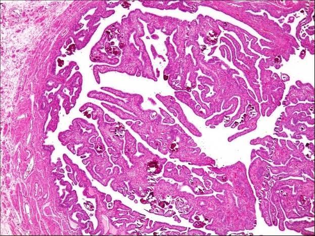
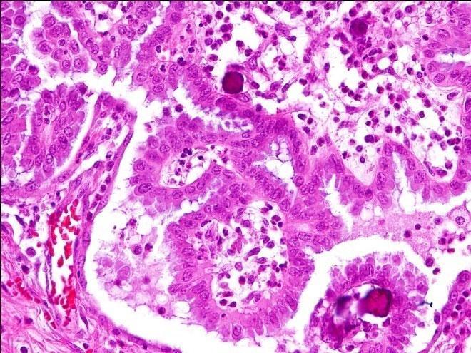
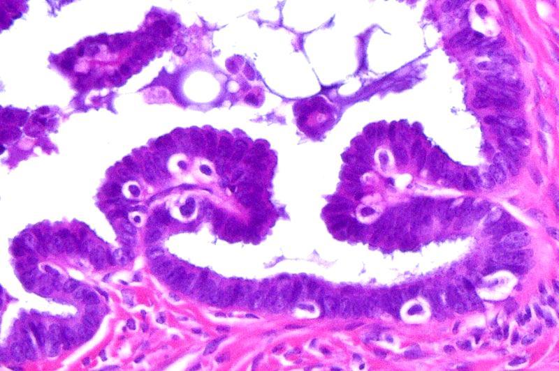
5 Definition of Tubal Hyperplasia in Current Study Papillary tufting, detached clusters of epithelial cells (secretory and ciliated) and papillae composed of bland epithelium with or without associated psammoma bodies Papillary tubal hyperplasia (PTH)
6
7 Papillary Tubal Hyperplasia
8 Findings in Danish Study Age of patients yrs; mean 42 yrs 10 yrs younger than cohort; no other differences PTH in 20 (91%) of 22 Psammoma bodies in 50% Chronic salpingitis in 36% Evidence of prior PID in 36% Noninvasive implants in omentum and/or uterine serosa in 14% Endosalpingiosis on tube or in omentum in 27%
9 Findings in JHH Study Age of patients yrs; mean 46 yrs Psammoma bodies in 57% Chronic inflammation in 42% Evidence of prior PID in 57% Noninvasive implants in 14% Endosalpingiosis in 29%

10 Proposed Evolution of Tubal Hyperplasia Focal epithelial stratification

11 Proposed Evolution of Tubal Hyperplasia Formation of tuft that expands, forming rounded papillae


12 Proposed Evolution of Tubal Hyperplasia Papillae are pinched off and extruded into lumen
13 Papillary Tubal Hyperplasia
14 Proposed Evolution of Tubal Hyperplasia Expulsion of papillae/clusters from tube with implantation on ovary and peritoneal surfaces with development of Cystadenoma and SBT Endosalpingiosis Noninvasive implants Spectrum of serous proliferations
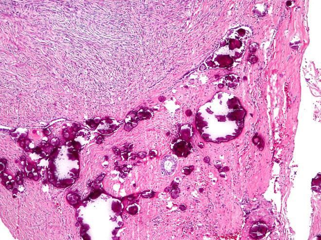
15 Proposed Evolution of Psammoma Bodies Develop in epithelium and when papillae form they are in the central core (salpingoliths) Seidman et al. Int J Gynecol Pathol 2002;21: Salpingolith
16 Proposed Evolution of Psammoma Bodies If epithelium invaginates psammoma bodies are deposited in the lamina propria
17 Proposed Evolution of Psammoma Bodies Expulsion from tube with implantation on ovary and peritoneum Degeneration of epithelium that surrounds the psammoma bodies results in naked psammoma bodies Naked psammoma bodies appear to act as an irritant in the peritoneal cavity leading to fibrosis which can in turn lead to adhesions and possibly bowel obstruction
18
19 Endosalpingiosis and Noninvasive Implants?Progression Various studies (and personal experience) suggest that endosalpingiosis and noninvasive implants can undergo malignant transformation resulting in the development of low-grade serous carcinoma
20 Three Possible Mechanisms to Explain Association of Tubal Hyperplasia and SBT (APST)s The tubal and ovarian lesions arise independently, so-called field effect Ovarian tumor is primary and tubal hyperplasia is secondary Tubal hyperplasia is primary and SBT (APST), noninvasive implants and endosalpingiosis are secondary
21 Problems with Independent Development of Tubal Hyperplasia and SBT (APST) (Field Effect) One field but different embryological origins Ovaries develop from the genital ridge not from the müllerian ducts Fallopian tubes develop from the müllerian ducts
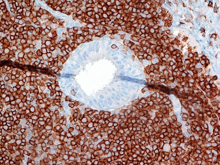
22 Problems with Independent Development of Tubal Hyperplasia and SBT (APST) (Field Effect) SBT (APST)s have a müllerian phenotype but the ovarian surface epithelium (OSE) from which it is postulated SBT (APST)s are derived from, is mesothelial
23 Problems with the Field Effect Psammoma bodies are an integral part of PTH but are only rarely associated with mesothelial proliferations Morphology, immunohistochemistry and gene expression profiles link SBT (APST)s to fallopian tube epithelium not to the OSE Marquez RT, et al. Clin Cancer Res 2005;11: SBT (APST)s, noninvasive implants and endosalpingiosis, for all practical purposes, do not occur in males
24 Origin of Some SBT (APST)s from Cortical Inclusion Cysts A distinct possibility

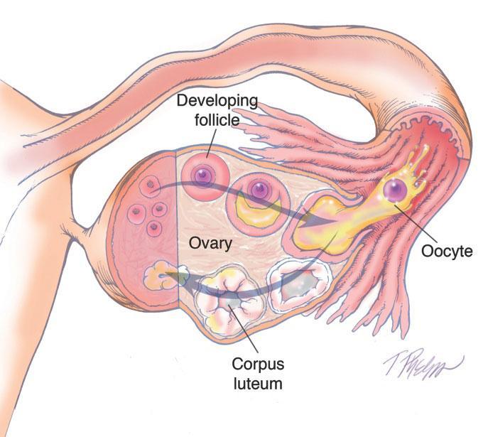
25 Tube and Ovary at Ovulation

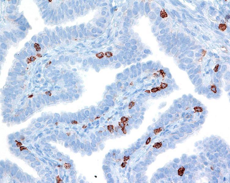
26 Formation of Cortical Inclusion Cyst


27 Implantation of FTE on Ovary OSE OSE FTE FTE CIC CIC Calretinin PAX 8
28 Two Types of Ovarian Cortical Inclusions Cysts (CICs) CICs lined by flat epithelium represent invagination of ovarian surface epithelium CICs lined by columnar epithelium are of tubal origin An interpretation also recently proposed by Li J. et al Mod Pathol 2011;24:


29 Fallopian Tube Ovary - CIC CD 45
30 CICs Cellular composition of FTE and CICs (columnar epithelial type) is identical OSE does not contain lymphocytes Aneuploidy in CICs Pothuri B, et al PLoS One 2010, 5e10358 CICs may account for the origin of some SBT (APST)s but they develop from tubal epithelium not from OSE
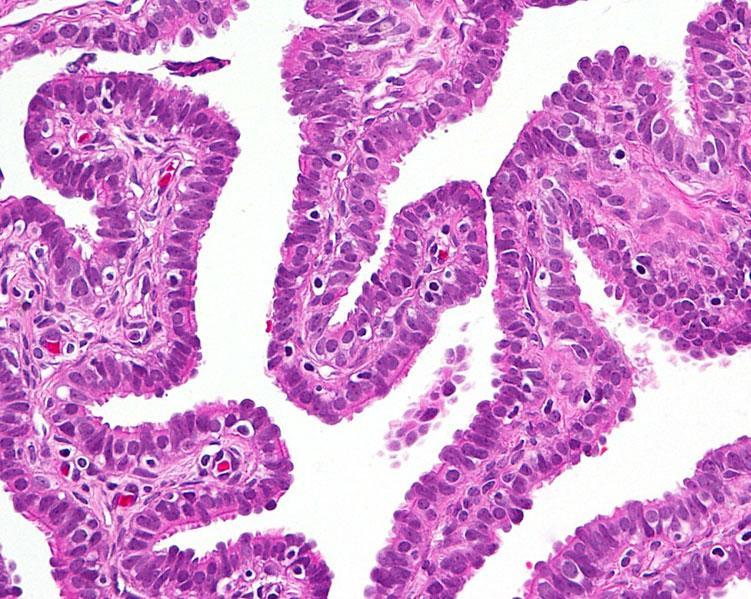
31 Papillary Tubal Hyperplasia

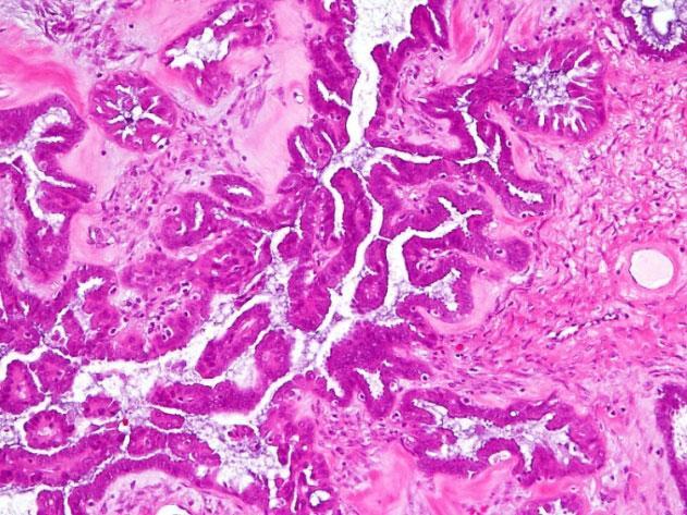
32 Papillary Tubal Hyperplasia Atypical Proliferative Tumor

33 CD3 + T and NKp44 + NK cells in Tubal Epithelium CD3=Pink NKp44=Brown Courtesy Dr W.Vermi and Dr F.Facchetti
")


34 APST (SBT) CD68R L Ardghiri et al CD8
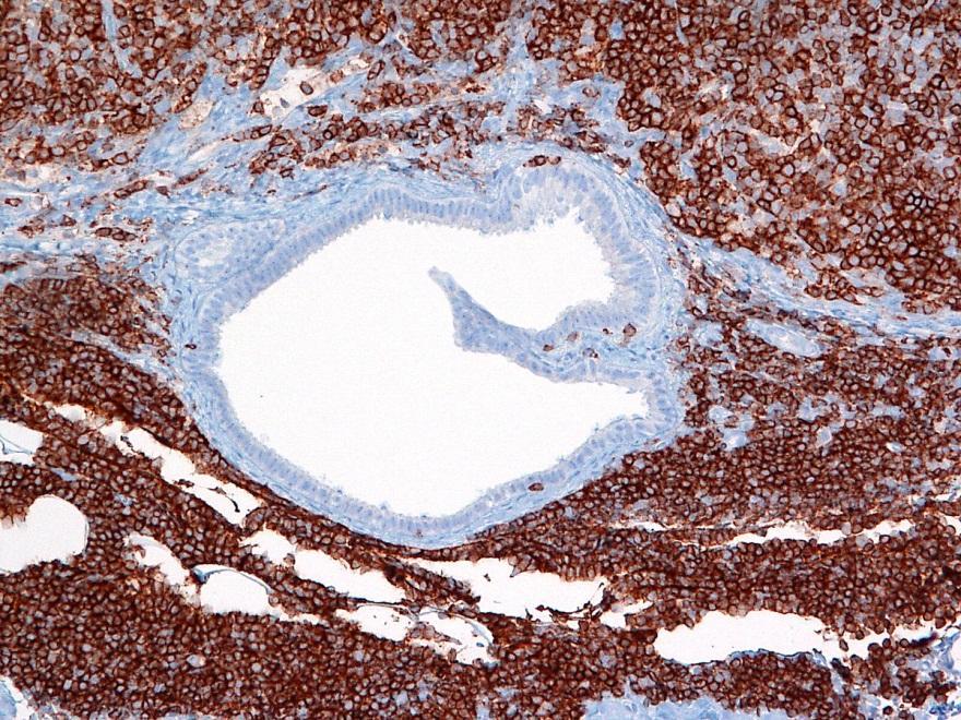
35 Endosalpingiosis in a Lymph Node CD 45 CD 45
36 Intraepithelial Leukocytes (IELs) Fallopian tube epithelium (FTE), CICs and APSTs contain the same population of cells of the immune system Most are cytotoxic T-cells (CD8+) and macrophages (CD68R+) In addition, NKp44 + NK cells have been identified in FTE. This subset of NK cells secretes IL-22, a cytokine known for providing mucosal protection Conclusions Subpopulations of leukocytes, primarily involved in immune mucosal protection, may, through inflammation, play a role in neoplastic development
37 Model for the Origin and Development of all Low-grade Pelvic Serous Proliferations Chronic inflammation induces proliferation of tubal epithelium In some cases the proliferation progresses to PTH with shedding of papillae and epithelial clusters that implant on ovarian and peritoneal surfaces On ovary lesion is a cortical inclusion cyst On peritoneum it is endosalpingiosis
38 Origin and Development of all Lowgrade Pelvic Serous Proliferations Cortical inclusion cysts (tubal type) can develop into a serous cystadenoma Mutation of KRAS or BRAF results in the development of a SBT(APST)
")


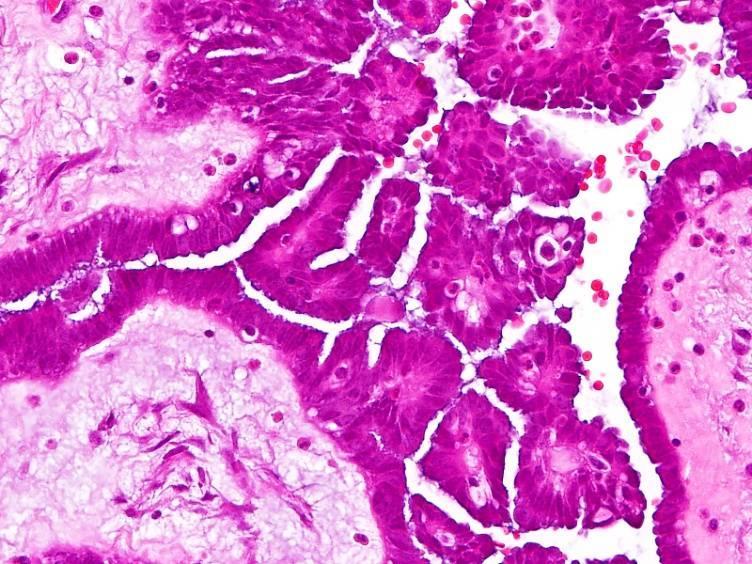
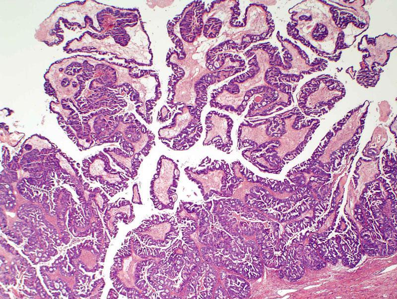
39 Small SBT(APST) arising in Cystadenoma Borderline tumor Cyst Before After Cheng EJ, et al Lab Invest 2004; 84:
 3 mm Serous cystadenoma adjacent to SBT(APST)")

40 Mutations of KRAS and BRAF Precede the Development of a SBT(APST) 3 mm Serous cystadenoma adjacent to SBT(APST) SBT(APST) in Serous cystadenoma BRAF mutation Codon 600, T1796A BRAF mutation Codon 600, T1796A Cheng EJ, et al Lab Invest 2004; 84:
41 PTH - Primary or Secondary? PTH is the primary event Seven cases of PTH in this study in the absence of an ovarian tumor PTH is morphologically identical in patients with or without an ovarian tumor
42 Origin and Development of all Lowgrade Pelvic Serous Proliferations Model not only explains development of ovarian SBT(APST)s But also primary peritoneal SBT(APST)s and noninvasive implants in the absence of an ovarian tumor In conclusion, all pelvic serous proliferations, benign and malignant, develop from the fallopian tube epithelium
43 The era of indifference to the fallopian tube has passed Woodruff JD, Pauerstein CJ The Fallopian Tube Structure, Function, Pathology and Management Williams and Wilkins Co; 1969 Finally
NIH Public Access Author Manuscript Am J Surg Pathol. Author manuscript; available in PMC 2012 November 1.
 NIH Public Access Author Manuscript Published in final edited form as: Am J Surg Pathol. 2011 November ; 35(11): 1605 1614. doi:10.1097/pas.0b013e318229449f. Papillary Tubal Hyperplasia. The Putative Precursor
Current Concept in Ovarian Carcinoma: Pathology Perspectives
 Current Concept in Ovarian Carcinoma: Pathology Perspectives Rouba Ali-Fehmi, MD Professor of Pathology The Karmanos Cancer Institute, Wayne State University School of Medicine Current Concept in Ovarian
Case # 4 Low-Grade Serous Carcinoma (Macropapillary) of the Ovary Arising in an Atypical Proliferative Serous Tumor
 of the Ovary Arising in an Atypical Proliferative Serous Tumor") Case # 4 Low-Grade Serous Carcinoma (Macropapillary) of the Ovary Arising in an Atypical Proliferative Serous Tumor Robert J Kurman, M.D. Johns Hopkins University School of Medicine Case History A 53 year
Case # 4 Low-Grade Serous Carcinoma (Macropapillary) of the Ovary Arising in an Atypical Proliferative Serous Tumor Robert J Kurman, M.D. Johns Hopkins University School of Medicine Case History A 53 year
Low-grade serous neoplasia. Robert A. Soslow, MD
 Low-grade serous neoplasia Robert A. Soslow, MD soslowr@mskcc.org Outline Orientation Ovarian tumor overview Non serous borderline tumors Serous borderline tumors Clinical summary Morphologic description
Endosalpingiosis. Case report
 Case report Endosalpingiosis Michael D. Holmes, M.D. Howard S. Levin M.D. Department of Pathology Lester A. Ballard, Jr., M.D. Department of Gynecology Endosalpingiosis, a term referring to tuballike epithelium
Case 1. Pathology of gynecological cancer. What do we need to know (Case 1) Luca Mazzucchelli Istituto cantonale di patologia Locarno
 Luca Mazzucchelli Istituto cantonale di patologia Locarno") Case 1 Pathology of gynecological cancer. What do we need to know (Case 1) Luca Mazzucchelli Istituto cantonale di patologia Locarno SAMO Interdisciplinary Workshop on Gynecological Tumors Lucern, October
Case 1 Pathology of gynecological cancer. What do we need to know (Case 1) Luca Mazzucchelli Istituto cantonale di patologia Locarno SAMO Interdisciplinary Workshop on Gynecological Tumors Lucern, October
Original contribution
 Human Pathology (2012) 43, 747 752 www.elsevier.com/locate/humpath Original contribution The presence and location of epithelial implants and implants with epithelial proliferation may predict a higher
Low-Grade Serous Ovarian Tumors Debra A. Bell, MD Mayo Clinic and Mayo Medical School Rochester, MN
 1 Low-Grade Serous Ovarian Tumors Debra A. Bell, MD Mayo Clinic and Mayo Medical School Rochester, MN It is very appropriate to discuss low-grade ovarian serous neoplasms in a symposium in honor of Dr.
of 20 to 80 and subsequently declines [2].
![of 20 to 80 and subsequently declines [2].](/thumbs/80/81450506.jpg "of 20 to 80 and subsequently declines [2].") - - According to the 2014 World Health Organization (WHO) classification and tumor morphology, primary ovarian tumors are subdivided into three categories: epithelial (60%), germ cell (30%), and sex-cord
- - According to the 2014 World Health Organization (WHO) classification and tumor morphology, primary ovarian tumors are subdivided into three categories: epithelial (60%), germ cell (30%), and sex-cord
Serous Borderline Tumors of the Ovary: Implants, Manifestations, Biology & New Insights in Progression
 Serous Borderline Tumors of the Ovary: Implants, Manifestations, Biology & New Insights in Progression Stanley J. Robboy, MD Professor of Pathology Professor of Obstetrics & Gynecology Vice Chairman for
3 cell types in the normal ovary
 Ovarian tumors 3 cell types in the normal ovary Surface (coelomic epithelium) the origin of the great majority of ovarian tumors 90% of malignant ovarian tumors Totipotent germ cells Sex cord-stromal cells
3 cell types in the normal ovary
 Ovarian tumors 3 cell types in the normal ovary Surface (coelomic epithelium) the origin of the great majority of ovarian tumors (neoplasms) 90% of malignant ovarian tumors Totipotent germ cells Sex cord-stromal
Bibliography. Serous Tumors of the Ovary. Nomenclature
 Bibliography Serous Tumors of the Ovary Nomenclature 1. Allison KH, Swisher EM, Kerkering KM, et al. Defining an appropriate threshold for the diagnosis of serous borderline tumor of the ovary: when is
Epithelial Ovarian Cancer 8/2/2013. Tu-be or Not Tu-be: Is the Fallopian Tube the Source of Ovarian Cancer?
 Tu-be or Not Tu-be: Is the Fallopian Tube the Source of Ovarian Cancer? Ann E. Smith Sehdev, MD Director, Center for Gynecologic Pathology Cascade Pathology, Portland, Oregon Ann E. Smith Sehdev has no
Mousa. Najat kayed &Renad Al-Awamleh. Nizar Alkhlaifat
 6 Mousa Najat kayed &Renad Al-Awamleh Nizar Alkhlaifat P a g e 1 This sheet written based on record 13 on website Cover slide( 95-117 ) No need to go back to slide FALLOPIAN TUBE PATHOLOGY In general fallopian
Borderline tumors. Borderline tumors. Serous borderline tumor are NOT benign. Low grade serous carcinoma: pathogenesis. Serous carcinoma: pathogenesis
 Serous borderline tumor are NOT benign Robert A. Soslow, MD Memorial Sloan-Kettering Cancer Center soslowr@mskcc.org Borderline tumors Serous BTs and seromucinous BTs are both histopathologically borderline
Mucinous Tumors of the Ovary Beirut, Lebanon. Anaís Malpica, M.D. Professor Department of Pathology
 Mucinous Tumors of the Ovary Beirut, Lebanon Anaís Malpica, M.D. Professor Department of Pathology Primary Mucinous Tumors of the Ovary Cystadenoma Borderline (Tumor of Low Malignant Potential/Atypical
International Society of Gynecological Pathologists Symposium 2007
 International Society of Gynecological Pathologists Symposium 2007 Anais Malpica, M.D. Department of Pathology The University of Texas M.D. Anderson Cancer Center Grading of Ovarian Cancer Histologic grade
Section 1. Biology of gynaecological cancers: our current understanding
 Section 1 Biology of gynaecological cancers: our current understanding Chapter 1 Morphological sub-types of ovarian carcinoma: new developments and pathogenesis W Glenn McCluggage 1 Introduction In most
Ovarian cancer: 2012 Update Srini Prasad MD Univ Texas MD Anderson Cancer Center
 Ovarian cancer: 2012 Update Srini Prasad MD Univ Texas MD Anderson Cancer Center Ovarian cancer is not a single disease Ovarian Epithelial Tumors: Histological Spectrum* Type Frequency Histology High-Grade
5/26/2016. Pelvic Serous Carcinoma: 2014 W.H.O. Update. Outline of Talk. Changes to 2014 WHO system for pelvic serous tumors
 Pelvic Serous Carcinoma: 2014 W.H.O. Update Outline of Talk Practical Implications for Pathologists Changes to 2014 WHO system for pelvic serous tumors High grade serous carcinoma versus low grade serous
Clinical History USCAP Specialty Conference. Gynecologic Pathology Case 3
 2010 USCA Specialty Conference Gynecologic athology Case Kathleen R. Cho, M.D. Department of athology Clinical History 46 yo woman presented with bilateral ovarian masses and elevated CA-125 TAH/BSO, pelvic
How to Recognize Gynecologic Cancer Cells from Pelvic Washing and Ascetic Specimens
 How to Recognize Gynecologic Cancer Cells from Pelvic Washing and Ascetic Specimens Wenxin Zheng, M.D. Professor of Pathology and Gynecology University of Arizona zhengw@email.arizona.edu http://www.zheng.gynpath.medicine.arizona.edu/index.html
Ovarian serous tumors of low malignant potential
 ORIGINAL ARTICLE Lymph Node Involvement in Ovarian Serous Tumors of Low Malignant Potential (Borderline Tumors) Pathology, Prognosis, and Proposed Classification Jesse K. McKenney, MD, Bonnie L. Balzer,
SALPINGITIS IN OVARIAN ENDOMETRIOSIS
 FERTILITY AND STERILITY Copyright 1978 The American Fertility Society Vol. 30, No. 1, July 1978 Printed in U.S.A. SALPINGITIS IN OVARIAN ENDOMETRIOSIS BERNARD CZERNOBILSKY, M.D.*t ALAN SILVERSTEIN, M.D.
Institute of Pathology First Faculty of Medicine Charles University. Ovary
 Ovary Barrett esophagus ph in vagina between 3.8 and 4.5 ph of stomach varies from 1-2 (hydrochloric acid) up to 4-5 BE probably results from upward migration of columnar cells from gastroesophageal junction
Invited Re vie W. Molecular genetics of ovarian carcinomas. Histology and Histo pathology
 Histol Histopathol (1 999) 14: 269-277 http://www.ehu.es/histol-histopathol Histology and Histo pathology Invited Re vie W Molecular genetics of ovarian carcinomas J. Diebold Pathological Institute, Ludwig-Maximilians-University
SEROUS TUMORS. Dr. Jaime Prat. Hospital de la Santa Creu i Sant Pau. Universitat Autònoma de Barcelona
 SEROUS TUMORS Dr. Jaime Prat Hospital de la Santa Creu i Sant Pau Universitat Autònoma de Barcelona Serous Borderline Tumors (SBTs) Somatic genetics Clonality studies have attempted to dilucidate whether
Ovarian Cancer is an Imported Disease: Fact or Fiction?
 Curr Obstet Gynecol Rep (2012) 1:1 9 DOI 10.1007/s13669-011-0004-1 DIAGNOSIS AND MANAGEMENT OF ADNEXAL MASS (H KATABUCHI, SECTION EDITOR) Ovarian Cancer is an Imported Disease: Fact or Fiction? Elisabetta
Concurrent Primary Peritoneal Low-Grade Serous Carcinoma and Endometrial High-Grade Serous Carcinoma
 International Journal of Gynecological Pathology 34:288 292, Lippincott Williams & Wilkins, Baltimore r 2015 International Society of Gynecological Pathologists Case Report Concurrent Primary Peritoneal
The Diagnostic Challenges of Low Grade and High Grade Tubo-Ovarian Serous Carcinomas. W Glenn McCluggage Belfast, Northern Ireland
 The Diagnostic Challenges of Low Grade and High Grade Tubo-Ovarian Serous Carcinomas W Glenn McCluggage Belfast, Northern Ireland Enterprise Interest None OVARIAN SEROUS CARCINOMA (OSC) RECENT DEVELOPMENTS
Pathology of the female genital tract
 Pathology of the female genital tract Common illnesses of the female genital tract Before menarche Developmental anomalies Tumors (ovarial teratoma) Amenorrhea Fertile years PCOS, ovarian cysts Endometriosis
Gross appearance of peritoneal cysts. They have a thin, translucent wall and contain a clear fluid.
 Gross appearance of peritoneal cysts. They have a thin, translucent wall and contain a clear fluid. So-called multicystic benign mesothelioma. A, Gross appearance. So-called multicystic benign mesothelioma.
Survival Analysis and Prognosis for Patients with Serous and Mucinous Borderline Ovarian Tumors: 14-Year Experience from a Tertiary Center in Iran
 ORIGINAL ARTICLE Survival Analysis and Prognosis for Patients with Serous and Mucinous Borderline Ovarian Tumors: 14-Year Experience from a Tertiary Center in Iran Katayoun Ziari, Ebrahim Soleymani, and
Biomarker expression in normal fimbriae: Comparison of high- and low-grade serous ovarian carcinoma
 1008 Biomarker expression in normal fimbriae: Comparison of high- and low-grade serous ovarian carcinoma ZHANG XUYIN *, DING JINGXIN *, TAO XIANG, JIA LUOQI and HUA KEQIN Department of Obstetrics and Gynecology,
Joseph Misdraji, M.D. GI pathology Unit Massachusetts General Hospital
 Joseph Misdraji, M.D. GI pathology Unit Massachusetts General Hospital jmisdraji@partners.org Low-grade appendiceal mucinous neoplasm (LAMN) High-grade appendiceal mucinous neoplasm (HAMN) Adenocarcinoma
Characterizing Adnexal Masses: Pearls and Pitfalls 20 th Annual Summer Practicum SCBT-MR Jackson Hole August 11, 2010
 Characterizing Adnexal Masses: Pearls and Pitfalls 20 th Annual Summer Practicum SCBT-MR Jackson Hole August 11, 2010 Evan S. Siegelman MD University of Pennsylvania Medical Center Adnexal Masses: Pearls
Atypical Hyperplasia/EIN
 EIN Atypical Hyperplasia/EIN Based on scientific and diagnostic advances, in 2014 the WHO moved that the precursor lesion for endometrioid carcinoma be atypical hyperplasia/ein, rather than what was previously
Patologia Molecular del Carcinoma de Ovario
 Curso de Patologia Molecular XXVI Congreso Nacional de la SEAP Cadiz Patologia Molecular del Carcinoma de Ovario Jaime Prat Barcelona Ovarian Epithelial Tumors WHO 1999 and 2003 Serous Mucinous Endometrioid
Gynaecological Pathology Reporting. Peritoneal cytology Tony Williams Birmingham
 Gynaecological Pathology Reporting Peritoneal cytology Tony Williams Birmingham Ascites Abdominopelvic peritoneal washings in gynaecological procedures Ovarian cyst aspirates Clinical guidance; RCOG &
Adenocarcinoma of Mullerian origin: review of pathogenesis, molecular biology, and emerging treatment paradigms
 Cobb et al. Gynecologic Oncology Research and Practice (2015) 2:1 DOI 10.1186/s40661-015-0008-z REVIEW Adenocarcinoma of Mullerian origin: review of pathogenesis, molecular biology, and emerging treatment
Endometrial polyp icd-10
 Endometrial polyp icd-10 Billable Medical Code for Polyp of Corpus Uteri Diagnosis Code for Reimbursement Claim: ICD-9-CM 621.0. Code will be replaced by October 2015 and relabeled as ICD-10. Free, official
6/5/2010. Outline of Talk. Endometrial Alterations That Mimic Cancer & Vice Versa: Metaplastic / reactive changes. Problems in Biopsies/Curettages
 Outline of Talk Endometrial Alterations That Mimic Cancer & Vice Versa: Problems in Biopsies/Curettages Metaplastic / reactive changes Mucinous change Microglandular hyperplasia-like change Squamous metaplasia
Urinary Bladder: WHO Classification and AJCC Staging Update 2017
 Urinary Bladder: WHO Classification and AJCC Staging Update 2017 Houston Society of Clinical Pathologists 58 th Annual Spring Symposium Houston, TX April 8, 2017 Jesse K. McKenney, MD Classification
Published Ahead of Print on September 28, 2012 as /theoncologist
 The Oncologist Gynecologic Oncology Diagnosis, Treatment, and Follow-Up of Borderline Ovarian Tumors DANIELA FISCHEROVA, a MICHAL ZIKAN, a PAVEL DUNDR, b DAVID CIBULA a a Gynecological Oncology Center,
ACCME/Disclosures. Case History 4/13/2016. USCAP GU Specialty Conference Case 3. Ann Arbor, MI
 USCAP GU Specialty Conference Case 3 March 2016 L. Priya Kunju, M.D. University of Michigan Health System Ann Arbor, MI University of Michigan Health System ACCME/Disclosures The USCAP requires that anyone
Papillary Cystadenoma of the Fallopian Tube Not Associated with von Hippel-Lindau Disease: A Case Report
 The Korean Journal of Pathology 2014; 48: 382-386 BRIEF CASE REPORT Papillary Cystadenoma of the Fallopian Tube Not Associated with von Hippel-Lindau Disease: A Case Report Jae Yeon Seok Myunghee Kang
Papillary Lesions of the Breast
 Papillary Lesions of the Breast Laura C. Collins, M.D. Associate Professor of Pathology Associate Director, Division of Anatomic Pathology Beth Israel Deaconess Medical Center and Harvard Medical School
Key Words. Borderline ovarian tumor Prognostic parameter Ultrasound Fertility Conservative surgery Recurrence
 The Oncologist Gynecologic Oncology Diagnosis, Treatment, and Follow-Up of Borderline Ovarian Tumors DANIELA FISCHEROVA, a MICHAL ZIKAN, a PAVEL DUNDR, b DAVID CIBULA a a Gynecological Oncology Center,
Cancer arising from Endometriosis and Its Clinical implications
 Cancer arising from Endometriosis and Its Clinical implications 1) Nezhat F, Cohen C, Rahaman J, Gretz H, Cole P, Kalir T. Comparative immunohistochemical studies of bcl-2 and p53 proteins in benign
Neoplasias Quisticas del Páncreas
 SEAP -Aproximación Práctica a la Patología Gastrointestinal- Madrid, 26 de mayo, 2006 Neoplasias Quisticas del Páncreas Gregory Y. Lauwers, M.D. Director, Service Massachusetts General Hospital Harvard
Cytyc Corporation - Case Presentation Archive - October 2001
 ThinPrep Pap Test History: 82 Year Old Female Specimen Type: Peritoneal Washings Case provided by Dr. Berle Stratton, Southwest Washington Medical Center, Vancouver, Washington. *The images, analysis and
Mody. AIS vs. Invasive Adenocarcinoma of the Cervix
 Common Problems in Gynecologic Pathology Michael T. Deavers, M.D. Houston Methodist Hospital, Houston, Texas Common Problems in Gynecologic Pathology Adenocarcinoma in-situ (AIS) of the Cervix vs. Invasive
Lavage of the uterine cavity as potential tool for diagnosis of epithelial ovarian cancer and its precursors
 Lavage of the uterine cavity as potential tool for diagnosis of epithelial ovarian cancer and its precursors Gynecologic Cancer Intergroup GCIG 2013 Autumn Meeting London, UK November 17 th 2013 Type II
Interpretation of p53 Immunostains. P53 Mutations are Ubiquitous in High Grade Serous Carcinoma. Diffuse strong positive nuclear staining
 Stains for Tumor Classification p53 p16 WT1 HMGA2 P53 Mutations are Ubiquitous in High Grade Serous Carcinoma Source Ahmed et al Australian Ovarian Cancer Study Cancer Genome Atlas Research Network Cases
Icd 10 ovarian stroma
 Icd 10 ovarian stroma Struma ovarii; Micrograph of a struma ovarii. Characteristic thyroid follicles are seen on the right, and ovarian stroma on the left. H&E stain. Classification and. Free, official
The relative frequency and histopathological patterns of ovarian lesions: study of 116 cases
 Original article: The relative frequency and histopathological patterns of ovarian lesions: study of 116 cases Dr Dimple Mehta*,Dr Alpesh Chavda**, Dr Hetal Patel*** *Assistant Professor, **Tutor, ***3
Bases biológicas del cáncer de ovario en el siglo XXI
 Bases biológicas del cáncer de ovario en el siglo XXI Iñigo Espinosa, M.D. Clínica Universidad de Navarra Epithelial Ovarian Tumors WHO 1973-2014 Serous Mucinous Endometrioid Clear cell Transitional Squamous
Fast Facts: Ovarian Cancer
 Fast Facts Fast Facts: Ovarian Cancer Christina Fotopoulou MD PhD Consultant Gynaecological Oncologist Queen Charlotte s and Chelsea Hospital London, UK Thomas J Herzog MD Professor of Obstetrics and Gynecology
Ovarian Clear Cell Carcinoma
 Ovarian Clear Cell Carcinoma Rouba Ali-Fehmi, MD Professor of Pathology The Karmanos Cancer Institute, Wayne State University School of Medicine 50 year old woman with chief complaint of shortness of breath
MPH Quiz. 1. How many primaries are present based on this pathology report? 2. What rule is this based on?
 MPH Quiz Case 1 Surgical Pathology from hysterectomy performed July 11, 2007 Final Diagnosis: Uterus, resection: Endometrioid adenocarcinoma, Grade 1 involving most of endometrium, myometrial invasion
Pathobiology of ovarian carcinomas
 Chinese Journal of Cancer Review Mojgan Devouassoux-Shisheboran 1 and Catherine Genestie 2 Abstract Ovarian tumors comprise a heterogeneous group of lesions, displaying distinct tumor pathology and oncogenic
Introduction. 23 rd Annual Seminar in Pathology. FLUIDS, Part 1. Pittsburgh, PA Gladwyn Leiman UVMMC, VT
 23 rd Annual Seminar in Pathology Pittsburgh, PA Gladwyn Leiman UVMMC, VT FLUIDS, Part 1 "Blue walls", Claudia Hansen, 2009 Introduction o Challenging to everyone o Almost any benign or malignant process
Triage of Ovarian Masses. Andreas Obermair Brisbane
 Triage of Ovarian Masses Andreas Obermair Brisbane Why Triage? In ovarian cancer, best outcomes for patients can be achieved when patients are treated in tertiary centres by a multidisciplinary team led
Article begins on next page
 Pseudopapillary Granulosa Cell Tumor: A Case of This Rare Subtype Rutgers University has made this article freely available. Please share how this access benefits you. Your story matters. [https://rucore.libraries.rutgers.edu/rutgers-lib/50622/story/]
ENDOMETRIOSIS WITH LYMPHATIC SPREAD P. Narmadha 1, P. Viswanathan 2, Rehana Tippoo 3, U. Manohar 4, Lavanya Kumari 5
 ENDOMETRIOSIS WITH LYMPHATIC SPREAD P. Narmadha 1, P. Viswanathan 2, Rehana Tippoo 3, U. Manohar 4, Lavanya Kumari 5 HOW TO CITE THIS ARTICLE: P. Narmadha, P. Viswanathan, Rehana Tippoo, U. Manohar, Lavanya
Histopathological analysis of neoplastic and non neoplastic lesions of ovary: A study of one hundred cases
 Orginal Article Histopathological analysis of neoplastic and non neoplastic lesions of ovary: A study of one hundred cases 2 G Prathima, Srikanth Shastry 2 Consultant Pathologist, Image Diagnostics, Kadapa,
Page # 1. Endometrium. Cellular Components. Anatomical Regions. Management of SIL Thomas C. Wright, Jr. Most common diseases:
 Endometrium Pathology of the Endometrium Thomas C. Wright Columbia University, New York, NY Most common diseases: Abnormal uterine bleeding Inflammatory conditions Benign neoplasms Endometrial cancer Anatomical
Histopathological Study of Spectrum of Lesions Seen in Surgically Resected Specimens of Fallopian Tube
 Original Article Print ISSN: 2321-6379 Online ISSN: 2321-595X DOI: 10.17354/ijss/2016/613 Histopathological Study of Spectrum of Lesions Seen in Surgically Resected Specimens of Fallopian Tube Pratima
Case Report Ovarian Seromucinous Borderline Tumor and Clear Cell Carcinoma: An Unusual Combination
 Case Reports in Obstetrics and Gynecology Volume 2015, Article ID 690891, 5 pages http://dx.doi.org/10.1155/2015/690891 Case Report Ovarian Seromucinous Borderline Tumor and Clear Cell Carcinoma: An Unusual
Ovarian carcinoma classification. Robert A. Soslow, MD
 Ovarian carcinoma classification Robert A. Soslow, MD soslowr@mskcc.org WHO classification Serous Mucinous Endometrioid Clear cell Transitional Squamous Mixed epithelial Undifferentiated Introduction Rationale
Epithelial tumors. Dr. F.F. Khuzin, PhD Dr. M.O. Mavlikeev
 Epithelial tumors Dr. F.F. Khuzin, PhD Dr. M.O. Mavlikeev Epithelial tumors Tumors from the epithelium are the most frequent among tumors. There are 2 group features of these tumors: The presence in most
ENODMETRIAL CARCINOMA: SPECIAL & NOT SO SPECIAL VARIANTS
 ENODMETRIAL CARCINOMA: SPECIAL & NOT SO SPECIAL VARIANTS Pacific Northwest Society of Pathologists Vancouver, B.C. September 26, 2015 Teri A. Longacre, M.D. longacre@stanford.edu Stanford University, Stanford,
Diseases of the vulva
 Diseases of the vulva 1. Bartholin Cyst - Infection of the Bartholin gland produces an acute inflammation within the gland (adenitis) and may result in an abscess. Bartholin duct cysts - Are relatively
Interactive Staging Bee
 Interactive Staging Bee ROBIN BILLET, MA, CTR GA/SC REGIONAL CONFERENCE NOVEMBER 6, 2018? Clinical Staging includes any information obtained about the extent of cancer obtained before initiation of treatment
Biliary tract tumors
 Short Course 2010 Annual Fall Meeting of the Korean Society for Pathologists Biliary tract tumors Joon Hyuk Choi, M.D., Ph.D. Professor, Department of Pathology, Yeungnam Univ. College of Medicine, Daegu,
Endometrial hyperplasia vs. Intraepithelial neoplasia. Martin Chang, MD PhD FRCPC Pathology Update Friday November 9, 2012
 Endometrial hyperplasia vs. Intraepithelial neoplasia Martin Chang, MD PhD FRCPC Pathology Update Friday November 9, 2012 Disclosure No relevant financial conflicts to declare. Case 1 Gland crowding Gland
Disclosure of Relevant Financial Relationships
 Squamous entities of the thyroid: Reactive to Neoplastic Michelle D. Williams Associate Professor Dept of Pathology, Head & Neck Section University of Texas MD Anderson Cancer Center Disclosure of Relevant
What s (new) and Important in Reporting of Uterine Cancers Katherine Vroobel The Royal Marsden
 and Important in Reporting of Uterine Cancers Katherine Vroobel The Royal Marsden") What s (new) and Important in Reporting of Uterine Cancers Katherine Vroobel The Royal Marsden Maastricht Pathology 2018 Wednesday 20 th June Endometrioid adenocarcinoma High grade carcinomas (common)
What s (new) and Important in Reporting of Uterine Cancers Katherine Vroobel The Royal Marsden Maastricht Pathology 2018 Wednesday 20 th June Endometrioid adenocarcinoma High grade carcinomas (common)
David Nunns on behalf of the Gynae Guidelines Group Date:
 Title of Guideline (must include the word Guideline (not protocol, policy, procedure etc) Borderline tumours of the ovary management and follow-up Author: Contact Name and Job Title Directorate & Speciality
Cytyc Corporation - Case Presentation Archive - March 2002
 FirstCyte Ductal Lavage History: 68 Year Old Female Gail Index: Unknown Clinical History: Negative Mammogram in 1995 6 yrs. later presents with bloody nipple discharge Subsequent suspicious mammogram Suspicious
ACCME/Disclosures. Case 4 USCAP Pulmonary Panel Case 4 History
 Case 4 USCAP Pulmonary Panel 2016 Andrew Churg, MD Department of Pathology Vancouver General Hospital & University of British Columbia Vancouver, BC achurg@mail.ubc.ca. ACCME/Disclosures The USCAP requires
Gynecologic Cytopathology: Glandular lesions
 Gynecologic Cytopathology: Glandular lesions Lin Wai Fung (MSc, MPH, CMIAC) 17/4/2014 Glandular lesions of the uterus Endocervix Endometrium Normal endocervical cells Sheets, strips well-preserved architecture:
CYTOMORPHOLOGY MODULE 28.1 INTRODUCTION OBJECTIVES 28.2 GENERAL GUIDELINES. Notes
 28 CYTOMORPHOLOGY 28.1 INTRODUCTION Light microscopic examination of stained cells in smears is the method of choice of diagnostic cytology. It allows classification of most normal cells as to type and
ACCME/Disclosures. Diagnosing Mesothelioma in Limited Tissue Samples. Papanicolaou Society of Cytopathology Companion Meeting March 12 th, 2016
 Diagnosing Mesothelioma in Limited Tissue Samples Papanicolaou Society of Cytopathology Companion Meeting March 12 th, 2016 Sanja Dacic, MD, PhD University of Pittsburgh ACCME/Disclosures GENERAL RULES
Borderline Brenner tumor of the ovary: a case report with immunohistochemical and molecular study
 De Cecio et al. Journal of Ovarian Research 2014, 7:101 CASE REPORT Open Access Borderline Brenner tumor of the ovary: a case report with immunohistochemical and molecular study Rossella De Cecio 1, Monica
Proliferative Epithelial lesions of the Breast. Sami Shousha, MD, FRCPath Charing Cross Hospital & Imperial College, London
 Proliferative Epithelial lesions of the Breast Sami Shousha, MD, FRCPath Charing Cross Hospital & Imperial College, London Amman, November2013 Proliferative Epithelial Lesions of the Breast Usual type
Normal endometrium: A, proliferative. B, secretory.
 Normal endometrium: A, proliferative. B, secretory. Nội mạc tử cung Nội mạc tử cung Cyclic changes in endometrium.. Approximate relationship of useful microscopic changes. Arias-Stella reaction in endometrial
Pathology of Ovarian Tumours. Dr. Jyothi Ranganathan MD ( Path) AFMC Pune PDCC (Cytopathology) PGI Chandigarh
 AFMC Pune PDCC (Cytopathology) PGI Chandigarh") Pathology of Ovarian Tumours Dr. Jyothi Ranganathan MD ( Path) AFMC Pune PDCC (Cytopathology) PGI Chandigarh Outline Incidence Risk factors Classification Pathology of tumours Tumour markers Prevention
Pathology of Ovarian Tumours Dr. Jyothi Ranganathan MD ( Path) AFMC Pune PDCC (Cytopathology) PGI Chandigarh Outline Incidence Risk factors Classification Pathology of tumours Tumour markers Prevention
Diseases of the breast (1 of 2)
") Diseases of the breast (1 of 2) Introduction A histology introduction Normal ducts and lobules of the breast are lined by two layers of cells a layer of luminal cells overlying a second layer of myoepithelial
Diseases of the breast (1 of 2) Introduction A histology introduction Normal ducts and lobules of the breast are lined by two layers of cells a layer of luminal cells overlying a second layer of myoepithelial
Flat Epithelial Atypia
 Flat Epithelial Atypia Richard Owings, M.D. University of Arkansas for Medical Sciences Department of Pathology Flat epithelial atypia can be a difficult lesion May be a subtle diagnosis Lots of changes
Select problems in cystic pancreatic lesions
 Disclosure Select problems in cystic pancreatic lesions Five Prime Therapeutics shareholder Adicet Bio shareholder Bristol-Meyer Squibb advisory board grace.kim@ucsf.edu Pancreatic cystic lesions Intraductal
Histopathological Spectrum of Lesions in Fallopian Tube
 IOSR Journal of Dental and Medical Sciences (IOSR-JDMS) e-issn: 2279-0853, p-issn: 2279-0861.Volume 16, Issue 1 Ver. III (January. 2017), PP 75-80 www.iosrjournals.org Histopathological Spectrum of Lesions
Biology Response Controversies and Advances
 Biology Response Controversies and Advances in BRCA related ovarian cancer Lessons learned and future directions Michael Friedlander The Prince of Wales Hospital and Royal Hospital for Women Sydney BREAST-CANCER
Proliferative Breast Disease: implications of core biopsy diagnosis. Proliferative Breast Disease
 Proliferative Breast Disease: implications of core biopsy diagnosis Jean F. Simpson, M.D. Breast Pathology Consultants, Inc. Nashville, TN Proliferative Breast Disease Must be interpreted in clinical and
CLINICAL SIGNIFICANCE OF BENIGN EPITHELIAL CHANGES
 Papillomas. Papillomas are composed of multiple branching fibrovascular cores, each having a connective tissue axis lined by luminal and myoepithelial cells ( Fig. 23-11 ). Growth occurs within a dilated
Human Anatomy Unit 3 REPRODUCTIVE SYSTEM
 Human Anatomy Unit 3 REPRODUCTIVE SYSTEM In Anatomy Today Male Reproductive System Gonads = testes primary organ responsible for sperm production development/maintenan ce of secondary sex characteristics
Case study 1. Rie Horii, M.D., Ph.D. Division of Pathology Cancer Institute Hospital, Japanese Foundation for Cancer Research
 NCCN/JCCNB Seminar in Japan April 15, 2012 Case study 1 Rie Horii, M.D., Ph.D. Division of Pathology Cancer Institute Hospital, Japanese Foundation for Cancer Research Present illness: A 50y.o.premenopausal
A Serous Borderline Tumor of the Fallopian Tube Detected Incidentally
 A Serous Borderline Tumor of the Fallopian Tube Detected Incidentally Imrana Tanvir, Ghania Ali, Haseeb Ahmed Khan and Ahmed Nasir Hanifi* Dept. of Histopathology, FMH College of Medicine & Dentistry,
Female Genital Tract Lab. Dr. Nisreen Abu Shahin Assistant Professor of Pathology University of Jordan
 Female Genital Tract Lab Dr. Nisreen Abu Shahin Assistant Professor of Pathology University of Jordan Ovarian Pathology A 20-year-old female presented with vague left pelvic pain. Pelvic exam revealed