1. Background Gonadoblastoma (GB) is regarded as an in situ form of germ cell tumor in dysgenetic gonads (type
|
|
|
- Melvin Kelly
- 5 years ago
- Views:
Transcription
1 Disease Markers 34 (2013) DOI /DMA IOS Press Utility of OCT3/4, TSPY and β-catenin as biological markers for gonadoblastoma formation and malignant germ cell tumor development in dysgenetic gonads Icela Palma a,b, Nayely Garibay c, Rocio Pena-Yolanda d, Alejandra Contreras e, Atlantida Raya f, Carolina Dominguez g,mirnaromero a, Gerardo Aristi h,i and Gloria Queipo c,h, a Molecular and Cellular Morphology Laboratory, Escuela Superior de Medicina, Instituto Politécnico Nacional, Mexico City, Mexico b Morphology Department, Facultad de Medicina Veterinaria y Zootecnia, UNAM, Mexico City, Mexico c Human Genetics Department, Hospital General de México, Mexico City, Mexico d Pathology Department, Hospital Infantil de México-Federico Gómez, Mexico City, Mexico e Biology Development Department, Hospital Infantil de México-Federico Gómez, Mexico City, Mexico f Urology Department, Hospital Infantil de México- Federico Gómez, Mexico City, Mexico g Endocrinology Department, Hospital Infantil de México-Federico Gómez, Mexico City, Mexico h Facultad de Medicina Universidad Nacional Autónoma de México, Mexico City, Mexico i Pathology Department, Hospital General de México, Mexico City, Mexico Abstract. BACKGROUND: Gonadoblastoma (GB) is regarded as an in situ form of germ cell tumor in dysgenetic gonads, and 30% of patients with GB develop a dysgerminoma/seminoma tumor. OBJECTIVE: Determine whether OCT3/4 and β-catenin are expressed in dysgenetic gonads before GB development and whether TSPY participates in the OCT3/4-β-catenin pathways in the malignant invasive behavior. METHODS: dysgenetic gonads of Disorders of sex differentiation (DSD) patients with mixed gonadal dysgenesis were analyzed by immunohistochemistry and immunofluorescence for comparison with GB and dysgerminoma/seminoma. RESULTS: Our results suggest that the development of GB is secondary to the interaction of OCT3/4 and TSPY, that β-catenin does not participate in this process. CONCLUSIONS: The use of this biological markers detects the potential high risk gonads. Keywords: Gonadoblastoma, OCT3/4, TSPY, β-catenin, dysgenetic gonads, mixed gonadal dysgenesis 1. Background Gonadoblastoma (GB) is regarded as an in situ form of germ cell tumor in dysgenetic gonads (type Corresponding author: Gloria Queipo, Human Genetics Department, Hospital General de Mexico, Mexico City, Mexico; Facultad de Medicina Universidad Nacional Autonoma de Mexico, Mexico City, Mexico. Dr. Balmis 142 Col, Doctores CP Mexico DF. Tel.: (1278/1279); gqueipo@nanolab.com.mx, gqueipo99@yahoo.com. II GCTs). This type of tumor is thought to be a precursor to seminoma/dysgerminoma tumors. It almost exclusively affects a subset of patients with disorders of sex differentiation (DSD) [5,7]. In 35% of GB cases, overgrowth of the germinal component leads to dysgerminoma/seminoma [8]. The TSPY gene (testisspecific protein, Y encoded) localized within the GBY locus (gonadoblastoma locus on the Y chromosome) has been shown to be involved in the multistep transformation of germ cells to GB [3,13]. However, the ISSN /13/$27.50 c 2013 IOS Press and the authors. All rights reserved
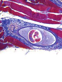
2 420 I. Palma et al. / Utility of OCT3/4, TSPY and β-catenin as biological markers for gonadoblastoma Table 1 Tissue samples histopathology, and biological markers localization Case Gonadal histopatolgy OCT3/4 TSPY B-catenin Co-localization TSPY/OCT3/4 Co-localization TSPY/B-catenin 1 UGT/SG (+) (+) ( ) (+) ( ) 2 DT (+) (+) ( ) ( ) ( ) 3 (R) SG ( ) ( ) ( ) ( ) ( ) (L) SG ( ) ( ) ( ) ( ) ( ) 4 (R) UGT/SG (+) (+) (+) (+) (+) (L) DT (+) (+) ( ) ( ) 5 DT (+) (+) (+) (+) (+) 6 (L)UGT/SG (+) (+) ( ) ( ) ( ) 7 DT (+) (+) ( ) ( ) ( ) 8 DT (+) (+) ( ) ( ) ( ) 9 UGT/SG (+) (+) ( ) (+) ( ) 10 DT (+) (+) ( ) ( ) ( ) 11 DT ( ) (+) ( ) ( ) ( ) 12 UGT/SG (+) ( ) ( ) (+) (+) 13 DT (+) (+) ( ) ( ) ( ) 14 (R)GB (L)GB (+) (+) (+) (+) (+) 15 DG (+) (+) (+/ ) UGT = tissue with burnt-out gonadoblastoma, DT = dysgenetic testis, SG = streak gonad, UGT = undifferentiated gonadal tissue, R = right, L = left, GB = gonadoblastoma, DG = dysgerminoma. precise role that TSPY plays in GB development and its involvement in the malignant transformation are not clear [15]. OCT3/4 has been implicated in the GB oncogenic process, but the molecular details of OCT3/4 deregulation are still unknown [6,12]. Analysis of OCT3/4, E-cadherin and β-catenin showed that the proliferation of immature germ cells in GB may be due to the interaction between OCT3/4 and accumulated β-catenin in the nuclei of the immature germ cells, leading to the development of invasive behavior and the progression of GB into dysgerminoma/seminoma in dysgenetic gonads [4]. In the present study, to determine whether TSPY participates in the OCT3/4-β-catenin pathway in the dysgenetic gonad and whether OCT3/4 and β-catenin are expressed in the dysgenetic gonad, we analyzed 18 dysgenetic gonads from DSD patients with mixed gonadal dysgenesis and compared them with GB and dysgerminoma/seminoma tumors. 2. Materials and methods Eighteen paraffin-embedded tissue samples from 15 pediatric patients with mixed gonadal dysgenesis or ambiguous genitalia and a 45, X/46, XY karyotype were studied. Tissue samples from two bilateral GB and one dysgenetic gonad with dysgerminoma/seminoma transformation were included. The use of the tissues was approved by the Institutional Bioethics Board. The analyses were performed using the classification of the World Health Organization. Formalin-fixed, paraffin-embedded sections were analyzed using immunohistochemistry and immunofluorescence (Table 1). The assays were performed in triplicate. Positive controls for β-catenin, TSPY and OCT3/4 were included in each experiment. The analyses were performed by an experienced pathologist, using the classification of the World Health Organization. The histological results were assessed by two scientists experienced in germ cell pathology (YRP and IP). Antigen-antibody complexes were detected using the avidin-biotin peroxidase method (KO679 LSAB+Sys/HRP kit, DakoCytomation, Carpinteria, CA) or with a secondary antibody conjugated to fluorescein isothiocyanate. The histological characteristics of the tissues revealed three of the four morphological patterns described by Martine Cools et al. [8]. Eight of the 18 samples were from dysgenetic testis (DT); germ cells in all 8 of the DT samples were confirmed by positive TSPY-staining. The second pattern, found in 5/18 samples, was streak tissue within undifferentiated gonadal tissue (UGT). UGT is characterized by germ cells that are not enclosed in seminiferous tubules or follicles organized in cord-like structures or by those without apparent organization. One of the UGTs contained a burnt-out gonadoblastoma. The third pattern observed in 2/18 cases was streak tissue. We also included one bilateral GB and one dysgerminoma as controls (Figs 1(A), (D), (G), (J)). In the rest of the samples, no GB or developing tumor was observed (Table 1).
 streak gonad with an undifferentiated gonadal tissue (UGT) organized in cord-like structures, (D) dysgenetic testis containing seminiferous tubules consistent with a")
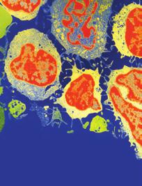
3 I. Palma et al. / Utility of OCT3/4, TSPY and β-catenin as biological markers for gonadoblastoma 421 Fig. 1. H&E light microscopy of a (A) streak gonad with an undifferentiated gonadal tissue (UGT) organized in cord-like structures, (D) dysgenetic testis containing seminiferous tubules consistent with a testicular differentiation pattern, (G) a typical gonadoblastoma nest showing a mixture of mature and immature germ cells and (J) a dysgerminoma tumor. TSPY immunohistochemistry showing positive immunoreactive signals in the immature germ cells in an UGT (B), DT (E) and (H) positive signal in immature germ cells within a gonadoblastoma nest. and dysgerminoma (K) OCT3/4 in the same expression pattern as TSPY in UGT, DT, GB and dysgerminoma (C, F, I, L). 3. Results In 14/18 of the gonadal samples, the germ cells stained positive for OCT3/4; OCT3/4 immunoreactivity was detected in the nuclei of immature germ cells and was observed exclusively in the DT, UGT, and control tumors as well as in GB and dysgerminoma tumors (Figs 1(C), (F), (I), (L)). OCT3/4 protein was not detected in mature germ cells or the streak tissue. TSPY immunostaining was positive in 14/18 gonads. TSPY protein staining was strongly positive in the nuclei of the germ cells in DT, UGT, GB and dysger- minoma tissues. Some protein was also detected as a faint stain in the germ cell cytoplasm (Figs 1(B), (E), (H), (K)). As in the case of GB, TSPY was detected in the UGT tissue containing burnt-out gonadoblastoma, suggesting that OCT3/4 and TSPY are key proteins in the development of GB. The samples that were negative for TSPY were mainly those with streak regions lacking germ cells. To determine whether TSPY and OCT3/4 were colocalized in the nuclei of immature germ cells, confocal microscopy was performed. It showed that both proteins were colocalized in the immature germ cell nuclei in one dysgenetic testis, in

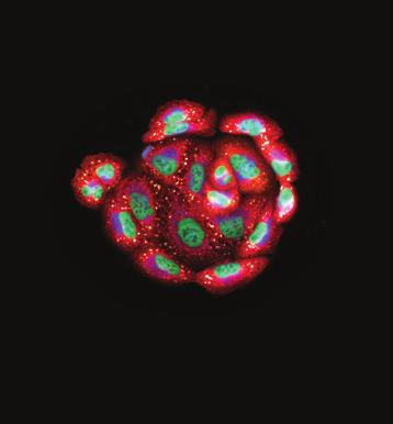
4 422 I. Palma et al. / Utility of OCT3/4, TSPY and β-catenin as biological markers for gonadoblastoma Fig. 2. Double-staining immunofluorescence and confocal analysis of dysgenetic testis positive for TSPY, OCT3/4 and β-catenin. (A) TSPY (green) localized in immature germ cells inside the seminiferous tubules, (B) OCT3/4 in the same tissues showing the same immunofluorescence pattern (red), (C) merged image and transmitted light micrograph optical transmission of the analyzed area indicating colocalization of both proteins in the nuclei of immature germ cells, (D) TSPY-positive immunofluorescence signal (green), (E) β-catenin (green) and (F) optical transmission and merged image showing colocalization of both proteins. UGT and in GB (Figs 2(A) (C)). Previous results have suggested that OCT3/4 and β-catenin participate in important steps during GB malignant transformation. B- catenin immunoreactive regions were observed only in 3/18 dysgenetic gonads. Nuclear staining in immature germ cells was observed in one DT, in UGT/GB and in GB (Figs 3(A) (J)). Interestingly, in our previous report, dysgerminoma showed diminished β-catenin expression. Gonads that were positive for β-catenin also expressed OCT3/4 and TSPY, which colocalized. All OCT3/4, TSPY positive gonads demonstrated expression of Ki67, a cell proliferative marker (Fig. 1P- S). Confocal microscopy showed colocalization of β- catenin and OCT3/4 in all the positively stained samples, as we reported previously. TSPY colocalized with β-catenin in β-catenin-positive cells (Fig. 2D-F). However, the majority of the dysgenetic gonads tested were negative for this marker; eliminating the possibility that β-catenin participates in gonadoblastoma formation. 4. Discussion Pure GB is regarded as an in situ form of germ cell tumor that affects almost exclusively a subset of DSD patients with dysgenetic gonads. GB does not behave as a malignant lesion; nevertheless, approximately 30% of all patients with gonadoblastoma develop a dysgerminoma/seminoma [1,7,8]. The age at diagnosis is variable, with approximately 94% of the cases being diagnosed during the second or third decades of life; we have demonstrated the presence of GB in infants [14]. It is important to identify a biological marker capable of detecting those dysgenetic gonads with a high potential for developing a tumor. Key proteins associated with germ cell tumor development, such as OCT3/4, β-catenin, TSPY and Ki67, were analyzed in 16 dysgenetic gonads and two germ cell tumors. GB originates from the surviving OCT3/4- positive germ cells within undifferentiated gonadal tissue in the dysgenetic gonad [8] We classified our dysgenetic tissue into three patterns (DT, UGT and streak gonad). It is important to emphasize that the streak tissue must be carefully examined to identify UGT in all the patients. In our samples, five streak tissues contained UGT (Figs 1(A) (C)). Gonadal biopsy identified as UGT or DT contained OCT3/4-positive cells, indicating a high risk for germ cell tumor formation because OCT3/4-positive cells are implicated in the GB oncogenic process. OCT3/4 is considered the most informative marker for the diagnosis of germ cell tumors [9,11]. In contrast, TSPY gene is the putative gene that predisposes dysgenetic gonads of intersex patients to develop gonadoblastomas. TSPY-positive immature germ cells
 positive staining in immature germ cells in UGT tissue, (C) negative in most of DT, (E) positive pattern in DT (G)")
5 I. Palma et al. / Utility of OCT3/4, TSPY and β-catenin as biological markers for gonadoblastoma Fig. 3. β-catenin immunohistochemistry showing the three different patterns identified: (A) positive staining in immature germ cells in UGT tissue, (C) negative in most of DT, (E) positive pattern in DT (G) strong immunoreactivity in gonadoblastoma, (I) positive but faint staining in dysgerminoma. Ki67 was used as a proliferative marker (B, D, F, H, and J). were observed in only two dysgenetic gonads, one of which was in a UGT containing a burnt-out GB. Not all OCT3/4-positive cells showed the presence of TSPYpositive nuclei, suggesting that the interaction between OCT3/4 and TSPY is an important step in GB formation. The colocalization of both proteins in the nuclei of immature germ cells, together with the Ki proliferative marker, supports the idea that the interaction of these two proteins in the nuclei of immature germ cell leads to cellular proliferation and GB development (Fig. 2). This finding confirms that the study of these proteins are a significant diagnostic marker for GB, CIS/ITGCNU and seminomatous tumors [1, 9]. The abundant expression of TSPY in both gonadoblastomas and CIS/ITGCNU tissues further supports the concept of a common origin [13] (Figs 1(E) (H)). In the same way, the OCT3/4 transcription factor plays a pivotal role as a key regulator of pluripotency in the early stages of mammalian development [12]. Our observations suggest that in the dysgenetic gonad, OCT3/4 and TSPY nuclear overexpression are the key factors in the development of GB. The ectopic germ cells and the dysgenetic tissues require the presence of both proteins to proliferate. OCT3/4 expression in germ cell tumors and cancers of somatic origins suggests that it might have a proliferative function at the cellular level when it is ectopically expressed in these cells [13]. GB is not a common tumor; therefore, an insufficient number of cases have been analyzed. Previous research on β-catenin and OCT3/4 suggests that both proteins participate in the same oncogenic pathway during germ cell tumor development. The interaction between OCT3/4 and the β-catenin that accumulated in the nuclei of immature germ cells leads to the development of invasive behavior and the progression of GB into dysgerminoma/seminoma in dysgenetic gonads [14]. Here, the data show that β-catenin is expressed only in the nuclei of immature germ cells in the dysgenetic tissues that coexpressed OCT3/4 and TSPY. The remainder of the samples did not express β-catenin. This finding suggests that β-catenin participates only after the GB is established and is not involved in dysgenetic gonad progression to GB. In our previous study, we found that β-catenin expression is diminished in dysgerminoma tumors by comparison with colon adenocarcinoma; however, its colocalization with OCT3/4 suggests that both proteins participate in the same oncogenic pathway [4]. These observations distinguish β-catenin as a malignancy marker in the germ cells in which it is expressed and in dysgenetic tissues that are OCT3/4-TSPY-positive. In conclusion, dysgenetic tissue expressing OCT3/4TSPY is associated with an extremely high risk for GB development, and both proteins are key players during GB development. The analysis of OCT4, SRY, TSPY and β-catenin expression in dysgenetic gonads may introduce modifications in the microenvironment that could contribute to a malignant transformation process.
6 424 I. Palma et al. / Utility of OCT3/4, TSPY and β-catenin as biological markers for gonadoblastoma The presence of β-catenin suggests that this protein is linked to malignant transformation (Figs 2(D) (F)) [2]. The presence of OCT3/4-TSPY in the gonadal biopsy tissues from DSD patients is an indicator of a high risk for GB, and β-catenin should be used as a marker for malignant germ cell tumors. Acknowledgments This work was performed in the Human Genetics Department at the Hospital General de México Eduardo Liceag, Facultad de Medicina UNAM. This work was supported by the Research Division of the Hospital General de México, CONACYT grant number References [1] AM Kersemaekers, et al., Identification of germ cells at risk for neoplastic transformation in gonadoblastoma: an immunohistochemical study for OCT3/4 and TSPY, Hum Pathol 36 (2005), [2] B Bianco, KC Oliveira, AD Guedes, et al., OCT4 gonadal gene expression related to the presence of Y-chromosome sequences in Turner syndrome, Fertil Steril 94 (2010), [3] F Schnieders, et al., Testis-specific protein, Y-encoded (TSPY) expression in testicular tissues, Hum Mol Genet 5 (1996), [4] I Palma, et al., Participation of OCT3/4 and beta-catenin during dysgenetic gonadal malignant transformation, Cancer Lett 263 (2008), [5] JW Oosterhuis and LH Looijenga, Testicular germ-cell tumours in a broader perspective Nat Rev Cancer 5 (2005), [6] L Cheng, et al., OCT4: Biological functions and clinical applications as a marker of germ cell neoplasia, J Pathol 211 (2007), 1-9. [7] LH ooijenga, et al., Gonadal tumours and DSD, Best Pract Res Clin Endocrinol Metab 24 (2010), [8] M Cools, et al., Morphological and immunohistochemical differences between gonadal maturation delay and early germ cell neoplasia in patients with undervirilization syndromes, J Clin Endocrinol Metab 90 (2005), [9] M Cools, et al., Gonadoblastoma arising in undifferentiated gonadal tissue within dysgenetic gonad, J Clin Endocrinol Metab 91 (2006), [10] N Liu, et al., Genome-wide gene expression profiling reveals aberrant MAPK and Wnt signaling pathways associated with early parthenogenesis, J Mol Cell Biol, 2 (2010), [11] R Hersmus, et al., New insights into type II germ cell tumor pathogenesis based on studies of patients with various forms of disorders of sex development (DSD), Mol Cell Endocrinol 291 (2008), [12] S Gidekel, et al., Oct-3/4 is a dose-dependent oncogenic fate determinant, Cancer Cell 4 (2003), [13] Y Li, ZL Tabatabai, et al., The Y-encoded TSPY protein: A significant marker potentially plays a role in the pathogenesis of testicular germ cell tumors, Hum Pathol 38 (2007), [14] Y-R Peña, K Nieto, R Alvarez, I Palma, N Nájera, L Eraña, LM, S Kofman-Alfaro, G Queipo, Distribution of Y chromosome-bearing cells in gonadoblastoma and dysgenetic testis in 45, X/46, XY infants, Mod Pathol 18 (2005), [15] YF Lau, Y Li and T Kido, Role of the Y-located putative gonadoblastoma gene in human spermatogenesis, Syst Biol Reprod Med 57 (2011),


7 MEDIATORS of INFLAMMATION The Scientific World Journal Gastroenterology Research and Practice Diabetes Research International Endocrinology Immunology Research Disease Markers Submit your manuscripts at BioMed Research International PPAR Research Obesity Ophthalmology Evidence-Based Complementary and Alternative Medicine Stem Cells International Oncology Parkinson s Disease Computational and Mathematical Methods in Medicine AIDS Behavioural Neurology Research and Treatment Oxidative Medicine and Cellular Longevity
Management of gonads in DSD
 Management of gonads in DSD Martine Cools, paediatric endocrinologist, Katja Wolffenbuttel and Piet Hoebeke, paediatric urologists, all at University Hospital Ghent, Belgium Sten Drop, paediatric endocrinologist
Case Report Primary Malignancy in a Supernumerary Testicle Presenting as a Large Pelvic Mass
 Hindawi Volume 2017, Article ID 4529853, 4 pages https://doi.org/10.1155/2017/4529853 Case Report Primary Malignancy in a Supernumerary Testicle Presenting as a Large Pelvic Mass Justin Noroozian, 1 Daniel
-The cause of testicular neoplasms remains unknown
 - In the 15- to 34-year-old age group, they are the most common tumors of men. - include: I. Germ cell tumors : (95%); all are malignant. II. Sex cord-stromal tumors: from Sertoli or Leydig cells; usually
Distribution of Y-chromosome-bearing cells in gonadoblastoma and dysgenetic testis in 45,X/46,XY infants
 & 2005 USCAP, Inc All rights reserved 0893-3952/05 $30.00 www.modernpathology.org in gonadoblastoma and dysgenetic testis in 45,X/46,XY infants Rocío Peña-Alonso 1, Karem Nieto 2, Rebeca Alvarez 2, Icela
Best Practice & Research Clinical Endocrinology & Metabolism
 Best Practice & Research Clinical Endocrinology & Metabolism 24 (2010) 291 310 Contents lists available at ScienceDirect Best Practice & Research Clinical Endocrinology & Metabolism journal homepage: www.elsevier.com/locate/beem
Note: The cause of testicular neoplasms remains unknown
 - In the 15- to 34-year-old age group, they are the most common tumors of men. - Tumors of the testis are a heterogeneous group of neoplasms that include: I. Germ cell tumors : 95%; all are malignant.
Research Article Delayed Recognition of Disorders of Sex Development (DSD): A Missed Opportunity for Early Diagnosis of Malignant Germ Cell Tumors
: A Missed Opportunity for Early Diagnosis of Malignant Germ Cell Tumors") Hindawi Publishing Corporation International Journal of Endocrinology Volume 2012, Article ID 671209, 9 pages doi:10.1155/2012/671209 Research Article Delayed Recognition of Disorders of Sex Development
Hindawi Publishing Corporation International Journal of Endocrinology Volume 2012, Article ID 671209, 9 pages doi:10.1155/2012/671209 Research Article Delayed Recognition of Disorders of Sex Development
Intratubular Germ Cell Neoplasia of the Testis
 Intratubular Germ Cell Neoplasia of the Testis KS Ngoo Department of Urology Hospital Selayang Advanced Urology Course 15 Aug 2014 MUA Office Clinical scenario A 33 years old man has bilateral testicular
Histological Value of Duodenal Biopsies
 Case Study TheScientificWorldJOURNAL (2005) 5, 396 400 ISSN 1537-744X; DOI 10.1100/tsw.2005.44 Histological Value of Duodenal Biopsies Limci Gupta and B. Hamid Countess of Chester Hospital NHS Foundation
State of the art review in gonadal dysgenesis: challenges in diagnosis and management
 McCann-Crosby et al. International Journal of Pediatric Endocrinology 2014, 2014:4 PES REVIEW State of the art review in gonadal dysgenesis: challenges in diagnosis and management Open Access Bonnie McCann-Crosby
Research Article Stromal Expression of CD10 in Invasive Breast Carcinoma and Its Correlation with ER, PR, HER2-neu, and Ki67
 SAGE-Hindawi Access to Research International Breast Cancer Volume 20, Article ID 47957, 4 pages doi:0.406/20/47957 Research Article Stromal Expression of CD0 in Invasive Breast Carcinoma and Its Correlation
A novel SRY missense mutation affecting nuclear import in a 46,XY female patient with bilateral gonadoblastoma
 (2009) 17, 1642 1649 & 2009 Macmillan Publishers Limited All rights reserved 1018-4813/09 $32.00 ARTICLE www.nature.com/ejhg A novel SRY missense mutation affecting nuclear import in a 46,XY female patient
DAX1, testes development role 7, 8 DFFRY, spermatogenesis role 49 DMRT genes, male sex differentiation role 15
 Subject Index N-Acetylcysteine, sperm quality effects 71 Ambiguous genitalia, origins 1, 2 Anti-Müllerian hormone function 13 receptors 13 Sertoli cell secretion 10, 38 Apoptosis assays in testes 73, 74
COMPLETE GONADAL DYSGENESIS WITH XY CHROMOSOMAL CONSTITUTION
 Tipar Cap coada final.qxd 1/22/2007 11:57 PM Page 465 Case report COMPLETE GONADAL DYSGENESIS WITH XY CHROMOSOMAL CONSTITUTION Dorina Stoicanescu *,1, Valerica Belengeanu 1, Dana Amzar 2, Cristina Popa
Bilateral Renal Angiomyolipomas with Invasion of the Renal Vein: A Case Report
 Case Study TheScientificWorldJOURNAL (2008) 8, 145 148 TSW Urology ISSN 1537-744X; DOI 10.1100/tsw.2008.29 Bilateral Renal Angiomyolipomas with Invasion of the Renal Vein: A Case Report C. Blick, N. Ravindranath,
Bilateral Segmental Testicular Infarction
 Case Study TheScientificWorldJOURNAL (2007) 7, 779 783 TSW Urology ISSN 1537-744X; DOI 10.1100/tsw.2007.146 Bilateral Segmental Testicular Infarction Aaron Bayne 1, Brad Koslin 2, and Siamak Daneshmand
Genetic Studies of Dysgerminoma
 Genetic Studies of Chromosome 12p abnormalities are characteristic of germ cell tumors isochromosome 12p and 12p overrepresentation Some can be detected by karyotyping FISH study of 21 dysgerminomas showed
Germ cell tumours UK SH. Ivo Leuschner. Kiel Pediatric Tumor Registry, Institute of Pathology University Hospital of Schleswig-Holstein Campus Kiel
 Germ cell tumours Ivo Leuschner Kiel Pediatric Tumor Registry, Institute of Pathology University Hospital of Schleswig-Holstein Campus Kiel UK SH Old histogenetic Concept of Germ cell tumours Pluripotent
Case Report A Rare Cutaneous Adnexal Tumor: Malignant Proliferating Trichilemmal Tumor
 Case Reports in Medicine Volume 2015, Article ID 742920, 4 pages http://dx.doi.org/10.1155/2015/742920 Case Report A Rare Cutaneous Adnexal Tumor: Malignant Proliferating Trichilemmal Tumor Omer Alici,
Testicular Germ Cell Tumors; A Simplistic Approach
 Testicular Germ Cell Tumors; A Simplistic Approach Merce Jorda, MD, PhD, MBA Professor and Vice Chair, Director of Anatomic Pathology Director of Genitourinary Pathology Service Interim Director of Cytopathology
Technique and feasibility of a dual staining method for estrogen receptors and AgNORs
 151 Technical note Technique and feasibility of a dual staining method for estrogen receptors and AgNORs Lukas Günther a, and Peter Hufnagl b a Department of Surgery, University of Heidelberg, Heidelberg,
(Epi)Genetics in normal and malignant germ cell development.
Genetics in normal and malignant germ cell development.") (Epi)Genetics in normal and malignant germ cell development. Leendert Looijenga, Department of Pathology, Lab. Exp. Patho-Oncol. Erasmus MC, Rotterdam OPTIMAL PATIENT CARE MM Basic and Transl. Oncol.,
(Epi)Genetics in normal and malignant germ cell development. Leendert Looijenga, Department of Pathology, Lab. Exp. Patho-Oncol. Erasmus MC, Rotterdam OPTIMAL PATIENT CARE MM Basic and Transl. Oncol.,
Gonadal Pathology and Tumor Risk in Relation to Clinical Characteristics in Patients with 45,X/46,XY Mosaicism
 JCEM ONLINE Advances in Genetics Endocrine Research Gonadal Pathology and Tumor Risk in Relation to Clinical Characteristics in Patients with 45,X/46,XY Mosaicism M. Cools, J. Pleskacova, H. Stoop, P.
Analysis of the Sex-determining Region of the Y Chromosome (SRY) in a Case of 46, XX True Hermaphrodite
 in a Case of 46, XX True Hermaphrodite") Clin Pediatr Endocrinol 1994; 3(2): 91-95 Copyright (C) 1994 by The Japanese Society for Pediatric Endocrinology Analysis of the Sex-determining Region of the Y Chromosome (SRY) in a Case of 46, XX True
Clin Pediatr Endocrinol 1994; 3(2): 91-95 Copyright (C) 1994 by The Japanese Society for Pediatric Endocrinology Analysis of the Sex-determining Region of the Y Chromosome (SRY) in a Case of 46, XX True
A COMPARATIVE STUDY OF GERM CELL KINETICS IN THE TESTES OF CHILDREN WITH UNILATERAL CRYPTORCHIDISM: A PRELIMINARY REPORT*
 FERTILITY AND STERILITY Copyright 1970 by the Williams & Wilkins Co. Vol. 21, No. 11, November 1970 Printed in U.S.A. A COMPARATIVE STUDY OF GERM CELL KINETICS IN THE TESTES OF CHILDREN WITH UNILATERAL
Neoplastic Potential of Germ Cells in Relation to Disturbances of Gonadal Organogenesis and Changes in Karyotype
 Journal of Andrology, Vol. 24, No. 2, March/April 23 Copyright American Society of Andrology Neoplastic Potential of Germ Cells in Relation to Disturbances of Gonadal Organogenesis and Changes in Karyotype
Conference Paper Programmed Cell Death Induced by Modulated Electrohyperthermia
 Conference Papers in Medicine, Article ID 187835, 3 pages http://dx.doi.org/10.1155/2013/187835 Conference Paper Programmed Cell Death Induced by Modulated Electrohyperthermia Meggyesházi Nóra, 1 Andócs
IN SUMMARY HST 071 NORMAL & ABNORMAL SEXUAL DIFFERENTIATION Fetal Sex Differentiation Postnatal Diagnosis and Management of Intersex Abnormalities
 Harvard-MIT Division of Health Sciences and Technology HST.071: Human Reproductive Biology Course Director: Professor Henry Klapholz IN SUMMARY HST 071 Title: Fetal Sex Differentiation Postnatal Diagnosis
Bi-potent Gonads. Sex Determination
 יצירת הגונדות Primordial Germ Cells (PGCs) Somatic cells Genital ridge Bi-potent Gonads Sex Determination Testis and Sperm Ovary and Oocyte Migration of Primordial Germ Cells in the Chick Embryo The
chapter 4. The effect of oncogenic HPV on transformation zone epithelium
 chapter 4. The effect of oncogenic HPV on transformation zone epithelium CHAPTER 1 All squamous cervical cancer (and probably all cervical adenocarcinoma) is associated with oncogenic HPV, and the absence
Coordinate Expression of Cytokeratins 7 and 20 in Prostate Adenocarcinoma and Bladder Urothelial Carcinoma
 Anatomic Pathology / CYTOKERATINS 7 AND 20 IN PROSTATE AND BLADDER CARCINOMAS Coordinate Expression of Cytokeratins 7 and 20 in Prostate Adenocarcinoma and Bladder Urothelial Carcinoma Nader H. Bassily,
Disorders of sex development (DSD) is
 is") Update on disorders of sex development Disorders of sex development: update on the genetic background, terminology and risk for the development of germ cell tumors Martine Cools, Leendert HJ Looijenga,
Update on disorders of sex development Disorders of sex development: update on the genetic background, terminology and risk for the development of germ cell tumors Martine Cools, Leendert HJ Looijenga,
Case Report PET/CT Imaging in Oncology: Exceptions That Prove the Rule
 Case Reports in Oncological Medicine Volume 2013, Article ID 865032, 4 pages http://dx.doi.org/10.1155/2013/865032 Case Report PET/CT Imaging in Oncology: Exceptions That Prove the Rule M. Casali, 1 A.
Conference Paper Oncothermia Basic Research at In Vivo Level: The First Results in Japan
 Conference Papers in Medicine, Article ID 197328, 6 pages http://dx.doi.org/.11/13/197328 Conference Paper Oncothermia Basic Research at In Vivo Level: The First Results in Japan G. Andocs, 1 Y. Okamoto,
When testes make no testosterone: Identifying a rare cause of 46, XY female phenotype in adulthood
 When testes make no testosterone: Identifying a rare cause of 46, XY female phenotype in adulthood Gardner DG, Shoback D. Greenspan's Basic & Clinical Endocrinology, 10e; 2017 Sira Korpaisarn, MD Endocrinology
(A) PCR primers (arrows) designed to distinguish wild type (P1+P2), targeted (P1+P2) and excised (P1+P3)14-
 PCR primers (arrows) designed to distinguish wild type (P1+P2), targeted (P1+P2) and excised (P1+P3)14-") 1 Supplemental Figure Legends Figure S1. Mammary tumors of ErbB2 KI mice with 14-3-3σ ablation have elevated ErbB2 transcript levels and cell proliferation (A) PCR primers (arrows) designed to distinguish
1 Supplemental Figure Legends Figure S1. Mammary tumors of ErbB2 KI mice with 14-3-3σ ablation have elevated ErbB2 transcript levels and cell proliferation (A) PCR primers (arrows) designed to distinguish
SUPPLEMENTAL INFORMATION FOR. PAX7 expression defines germline stem cells in the adult testis
 SUPPLEMENTAL INFORMATION FOR PAX7 expression defines germline stem cells in the adult testis Gina M. Aloisio, Yuji Nakada, Hatice D. Saatcioglu, Christopher G. Peña, Michael D. Baker, Edward D. Tarnawa,
A multi-exon deletion within WWOX is associated with a 46,XY Disorder of Sex
 A multi-exon deletion within WWOX is associated with a 46,XY Disorder of Sex Development. Stefan White 1,2, Jacqueline Hewitt 1,3, Erin Turbitt 1, Yvonne van der Zwan 4, Remko Hersmus 4, Sten Drop 4, Peter
Title: Synuclein Gamma Predicts Poor Clinical Outcome in Colon Cancer with Normal Levels of Carcinoembryonic Antigen
 Author's response to reviews Title: Synuclein Gamma Predicts Poor Clinical Outcome in Colon Cancer with Normal Levels of Carcinoembryonic Antigen Authors: Caiyun Liu (liucaiyun23@yahoo.com.cn) Bin Dong
Knockout TM SR : ; ; ; : R ; R : A : X(2013) , ,, B. , (Knockout TM
 , ,, B. , (Knockout TM") 33 1 Vol.33 No.1 013 1 Dec. 013 Reproduction & Contraception doi: 10.7669/j.issn.03-37X.013.1.0804 E-mail: randc_journal@163.com Knockout TM SR ; ; ; 400014 : FBS Knockout TM SRKSR : FBS KSR HE TUNEL RT-PCR
33 1 Vol.33 No.1 013 1 Dec. 013 Reproduction & Contraception doi: 10.7669/j.issn.03-37X.013.1.0804 E-mail: randc_journal@163.com Knockout TM SR ; ; ; 400014 : FBS Knockout TM SRKSR : FBS KSR HE TUNEL RT-PCR
p53 expression in invasive pancreatic adenocarcinoma and precursor lesions
 Malaysian J Pathol 2011; 33(2) : 89 94 ORIGINAL ARTICLE p53 expression in invasive pancreatic adenocarcinoma and precursor lesions NORFADZILAH MY MBBCH,* Jayalakshmi PAILOOR MPath, FRCPath,* RETNESWARI
Female with 46, XY karyotype
 Case Report Obstet Gynecol Sci 2017;60(4):378-382 https://doi.org/10.5468/ogs.2017.60.4.378 pissn 2287-8572 eissn 2287-8580 Female with 46, XY karyotype Eun Jung Jung 1, Do Hwa Im 1, Yong Hee Park 1, Jung
Case Report Five-Year Survival after Surgery for Invasive Micropapillary Carcinoma of the Stomach
 Case Reports in Surgery Volume 2013, Article ID 560712, 4 pages http://dx.doi.org/10.1155/2013/560712 Case Report Five-Year Survival after Surgery for Invasive Micropapillary Carcinoma of the Stomach Shigeo
Gastric Carcinoma with Lymphoid Stroma: Association with Epstein Virus Genome demonstrated by PCR
 Gastric Carcinoma with Lymphoid Stroma: Association with Epstein Virus Genome demonstrated by PCR Pages with reference to book, From 305 To 307 Irshad N. Soomro,Samina Noorali,Syed Abdul Aziz,Suhail Muzaffar,Shahid
Hersmus et al. BMC Medical Genetics 2012, 13:108
 Hersmus et al. BMC Medical Genetics 2012, 13:108 RESEARCH ARTICLE Open Access SRY mutation analysis by next generation (deep) sequencing in a cohort of chromosomal Disorders of Sex Development (DSD) patients
Correlation between expression and significance of δ-catenin, CD31, and VEGF of non-small cell lung cancer
 Correlation between expression and significance of δ-catenin, CD31, and VEGF of non-small cell lung cancer X.L. Liu 1, L.D. Liu 2, S.G. Zhang 1, S.D. Dai 3, W.Y. Li 1 and L. Zhang 1 1 Thoracic Surgery,
Sestrin2 and BNIP3 (Bcl-2/adenovirus E1B 19kDa-interacting. protein3) regulate autophagy and mitophagy in renal tubular cells in. acute kidney injury
 regulate autophagy and mitophagy in renal tubular cells in. acute kidney injury") Sestrin2 and BNIP3 (Bcl-2/adenovirus E1B 19kDa-interacting protein3) regulate autophagy and mitophagy in renal tubular cells in acute kidney injury by Masayuki Ishihara 1, Madoka Urushido 2, Kazu Hamada
Sestrin2 and BNIP3 (Bcl-2/adenovirus E1B 19kDa-interacting protein3) regulate autophagy and mitophagy in renal tubular cells in acute kidney injury by Masayuki Ishihara 1, Madoka Urushido 2, Kazu Hamada
R. F. Falkenstern-Ge, 1 S. Bode-Erdmann, 2 G. Ott, 2 M. Wohlleber, 1 and M. Kohlhäufl Introduction. 2. Histology
 Case Reports in Oncological Medicine Volume 2013, Article ID 167585, 4 pages http://dx.doi.org/10.1155/2013/167585 Case Report Late Lung Metastasis of a Primary Eccrine Sweat Gland Carcinoma 10 Years after
Reviewers' comments: Reviewer #1 (Remarks to the Author):
:") Reviewers' comments: Reviewer #1 (Remarks to the Author): In this study the authors analysed 18 deep penetrating nevi for oncogenic genomic changes (single nucleotide variations, insertions/deletions,
Reviewers' comments: Reviewer #1 (Remarks to the Author): In this study the authors analysed 18 deep penetrating nevi for oncogenic genomic changes (single nucleotide variations, insertions/deletions,
Case Report Atypical Presentation of Atypical Teratoid Rhabdoid Tumor in a Child
 Case Reports in Oncological Medicine Volume 2013, Article ID 815923, 4 pages http://dx.doi.org/10.1155/2013/815923 Case Report Atypical Presentation of Atypical Teratoid Rhabdoid Tumor in a Child Y. T.
Immunopathology of Lymphoma
 Immunopathology of Lymphoma Noraidah Masir MBBCh, M.Med (Pathology), D.Phil. Department of Pathology Faculty of Medicine Universiti Kebangsaan Malaysia Lymphoma classification has been challenging to pathologists.
Case Report Intracranial Capillary Hemangioma in the Posterior Fossa of an Adult Male
 Case Reports in Radiology Volume 2016, Article ID 6434623, 4 pages http://dx.doi.org/10.1155/2016/6434623 Case Report Intracranial Capillary Hemangioma in the Posterior Fossa of an Adult Male Jordan Nepute,
Case Report Renal Cell Carcinoma Metastatic to Thyroid Gland, Presenting Like Anaplastic Carcinoma of Thyroid
 Case Reports in Urology Volume 2013, Article ID 651081, 4 pages http://dx.doi.org/10.1155/2013/651081 Case Report Renal Cell Carcinoma Metastatic to Thyroid Gland, Presenting Like Anaplastic Carcinoma
Applications of IHC. Determination of the primary site in metastatic tumors of unknown origin
 Applications of IHC Determination of the primary site in metastatic tumors of unknown origin Classification of tumors that appear 'undifferentiated' by standard light microscopy Precise classification
Disordered Sex Differentiation Mixed gonadal dysgenesis Congenital adrenal hyperplasia Mixed gonadal dysgenesis
 Disordered Sex Differentiation DSD has superceded intersex in describing genital anomalies in childhood DSD results from hormonal imbalances due to (i) abnormal genetic status, (ii) enzyme defects, or
Testicular Tumors: What s New, True, Important Cristina Magi-Galluzzi, MD, PhD
 Testicular Tumors: What s New, True, Important Cristina Magi-Galluzzi, MD, PhD Director, Genitourinary Pathology R.J. Tomsich Pathology & Laboratory Medicine Institute Professor of Pathology, Lerner College
Solitary Contralateral Adrenal Metastases after Nephrectomy for Renal Cell Carcinoma
 Original Report ISSN 1537-744X; DOI 10.1100/tsw.2004.39 Solitary Contralateral Adrenal after Nephrectomy for Renal Cell Carcinoma Nikolaos Antoniou, M.D. and Demetrios Karanastasis, M.D. General Hospital
Case Report Synchronous Bilateral Solid Papillary Carcinomas of the Breast
 Case Reports in Surgery Volume 2013, Article ID 812129, 4 pages http://dx.doi.org/10.1155/2013/812129 Case Report Synchronous Bilateral Solid Papillary Carcinomas of the Breast Noriko Yoshimura, 1 Shigeru
Male Genital Cancers in the US in Frequency of Types
 Germ Cell Tumors of the Testis Pathology, Immunohistochemistry, and the Often Confusing Appearance of Their Metastases Charles Zaloudek, MD Department of Pathology UCSF Male Genital Cancers in the US in
Spermatogenesis in Man
 Spermatogenesis in Man I. Nuclear Morphology During Spermatogenesis in Man BRUNETTO CHIARELLI, PH.D., ARTHUR FALEK, PH.D., KAREN J. BACK, B.S., and C. THOMAS COWART, M.D. THE SEQUENCE of transformations
Gastric Signet-Ring Cell Carcinoma: Unilateral Lower Extremity Lymphoedema as the Presenting Feature
 Clinical Image TheScientificWorldJOURNAL (2007) 7, 1189 1192 ISSN 1537-744X; DOI 10.1100/tsw.2007.199 Gastric Signet-Ring Cell Carcinoma: Unilateral Lower Extremity Lymphoedema as the Presenting Feature
DISORDERS OF MALE GENITALS
 Wit JM, Ranke MB, Kelnar CJH (eds): ESPE classification of paediatric endocrine diagnosis. 9. Testicular disorders/disorders of male genitals. Horm Res 2007;68(suppl 2):63 66 ESPE Code Diagnosis OMIM ICD10
LIST OF ORGANS FOR HISTOPATHOLOGICAL ANALYSIS:!! Neural!!!!!!Respiratory:! Brain : Cerebrum,!!! Lungs and trachea! Olfactory, Cerebellum!!!!Other:!
 LIST OF ORGANS FOR HISTOPATHOLOGICAL ANALYSIS:!! Neural!!!!!!Respiratory:! Brain : Cerebrum,!!! Lungs and trachea! Olfactory, Cerebellum!!!!Other:! Spinal cord and peripheral nerves! Eyes, Inner ear, nasal
46 XY gonadal dysgenesis in adulthood pitfalls of late diagnosis
 Reminder of important clinical lesson 46 XY gonadal dysgenesis in adulthood pitfalls of late diagnosis Jarna Naing Hamin,1 Francis Raymond P Arkoncel,2 Frances Lina Lantion-Ang,1 Mark Anthony S Sandoval1
CD15 and CEA expression in thymic epithelial neoplasms
 Turkish Journal of Cancer Volume 8, No., 8 CD and CEA expression in thymic epithelial neoplasms AYTEKİN AKYOL, AYŞEGÜL ÜNER Hacettepe University, Department of Pathology, Ankara-Turkey ABSTRACT The aim
Table of Contents. 1. Overview. 2. Interpretation Guide. 3. Staining Gallery Cases Negative for CINtec PLUS
 Staining Atlas Table of Contents 1. Overview 1.1 Introduction 1.2 Role of p16 INK4a 1.3 Role of Ki-67 1.4 Molecular Pathogenesis 1.5 p16 INK4a Expression in Cervical Dysplasia 1.6 The Concept of CINtec
Edinburgh Research Explorer
 Edinburgh Research Explorer Intratubular germ cell neoplasia of the human testis Citation for published version: Mitchell, RT, E Camacho-Moll, M, Macdonald, J, Anderson, R, Kelnar, CJH, O'Donnell, M, Sharpe,
N-cadherin Expression in Testicular Germ Cell and Gonadal Stromal Tumors
 381 Ivyspring International Publisher Research Paper Journal of Cancer 2012; 3: 381-389. doi: 10.7150/jca.5017 N-cadherin Expression in Testicular Germ Cell and Gonadal Stromal Tumors Daniel J. Heidenberg
Case Report A Case of p63 Positive Diffuse Large B Cell Lymphoma of the Bladder
 Case Reports in Hematology Volume 2016, Article ID 4348208, 4 pages http://dx.doi.org/10.1155/2016/4348208 Case Report A Case of p63 Positive Diffuse Large B Cell Lymphoma of the Bladder Chelsey D. Deel,
Renal Pelvis Squamous Cell Carcinoma and Renal Cell Carcinoma in a Tuberculous Kidney
 Case Study TheScientificWorldJOURNAL (2004) 4, 965 968 ISSN 1537-744X; DOI 10.1100/tsw.2004.196 Renal Pelvis Squamous Cell Carcinoma and Renal Cell Carcinoma in a Tuberculous Kidney M. Al-Assiri 1, M.F.
Case Report Clear Cell Basal Cell Carcinoma
 SAGE-Hindawi Access to Research Volume 2011, Article ID 386921, 4 pages doi:10.4061/2011/386921 Case Report Clear Cell Basal Cell Carcinoma Deba P. Sarma, 1 Daniel Olson, 1 Jennifer Olivella, 1 Tracey
Characterization and significance of MUC1 and c-myc expression in elderly patients with papillary thyroid carcinoma
 Characterization and significance of MUC1 and c-myc expression in elderly patients with papillary thyroid carcinoma Y.-J. Hu 1, X.-Y. Luo 2, Y. Yang 3, C.-Y. Chen 1, Z.-Y. Zhang 4 and X. Guo 1 1 Department
Neoplasia 18 lecture 6. Dr Heyam Awad MD, FRCPath
 Neoplasia 18 lecture 6 Dr Heyam Awad MD, FRCPath ILOS 1. understand the role of TGF beta, contact inhibition and APC in tumorigenesis. 2. implement the above knowledge in understanding histopathology reports.
The Prognostic Importance of Prostate-Specific Antigen in Monitoring Patients Undergoing Maximum Androgen Blockage for Metastatic Prostate Cancer
 Research Article TheScientificWorldJOURNAL (005) 5, 8 4 ISSN 57-744X; DOI 0.00/tsw.005.9 The Prognostic Importance of Prostate-Specific Antigen in Monitoring Patients Undergoing Maximum Androgen Blockage
Case Report Seminoma Presenting as Renal Mass, Inferior Vena Caval Thrombus, and Regressed Testicular Mass
 Case Reports in Urology Volume 2015, Article ID 835962, 4 pages http://dx.doi.org/10.1155/2015/835962 Case Report Seminoma Presenting as Renal Mass, Inferior Vena Caval Thrombus, and Regressed Testicular
Done By : WESSEN ADNAN BUTHAINAH AL-MASAEED
 Done By : WESSEN ADNAN BUTHAINAH AL-MASAEED Acute Myeloid Leukemia Firstly we ll start with this introduction then enter the title of the lecture, so be ready and let s begin by the name of Allah : We
CHAPTER-VII : SUMMARY AND CONCLUSIONS
 CHAPTER-VII : SUMMARY AND CONCLUSIONS 199 SUMMARY AND CONCLUSIONS t The rapid development of human genetics during the past couple of decades and the discovery of numerous cytogenetic abnormalities have
OVOTESTIS Background Pathophysiology
 OVOTESTIS Background Ovotestis refers to the histology of a gonad that contains both ovarian follicles and testicular tubular elements. Such gonads are found exclusively in people with ovotesticular disorder
The Pathology of Germ Cell Tumours of the Ovary
 The Pathology of Germ Cell Tumours of the Ovary Professor Mike Wells University of Sheffield Amman, Jordan November 2013 Professor Francisco Paco Nogales I. Primitive germ cell tumors A. Dysgerminoma
True Hermaphroditism and Mixed Gonadal Dysgenesis in Young Children: A Clinicopathologic Study of 10 Cases
 True Hermaphroditism and Mixed Gonadal Dysgenesis in Young Children: A Clinicopathologic Study of 10 Cases Kyu-Rae Kim, M.D., Youngmee Kwon, M.D., Jae Young Joung, M.D., Kun Suk Kim, M.D., Alberto G. Ayala,
Case Report Müllerian Remnant Cyst as a Cause of Acute Abdomen in a Female Patient with Müllerian Agenesis: Radiologic and Pathologic Findings
 Volume 2016, Article ID 6581387, 4 pages http://dx.doi.org/10.1155/2016/6581387 Case Report üllerian Remnant Cyst as a Cause of Acute Abdomen in a Female Patient with üllerian Agenesis: Radiologic and
Research Article A Clinicopathological and Immunohistochemical Correlation in Cutaneous Metastases from Internal Malignancies: A Five-Year Study
 Skin Cancer, Article ID 793937, 5 pages http://dx.doi.org/10.1155/2014/793937 Research Article A Clinicopathological and Immunohistochemical Correlation in Cutaneous Metastases from Internal Malignancies:
CINtec PLUS Cytology. Interpretation training
 CINtec PLUS Cytology Interpretation training Objectives After reviewing this learning module, you will have a basic understanding of how to interpret CINtec PLUS Cytology, including: The mechanism of action
Incidental gonadal tumours at the time of gonadectomy in women with Swyer syndrome: a case series. Journal of Pediatric and Adolescent Gynecology
 Accepted Manuscript Incidental gonadal tumours at the time of gonadectomy in women with Swyer syndrome: a case series Amie JM. Hanlon, MBBS Rebecca M. Kimble, MBBS, FRANZCOG PII: S1083-3188(14)00272-1
Endoscopic Ultrasonography Assessment for Ampullary and Bile Duct Malignancy
 Diagnostic and Therapeutic Endoscopy, Vol. 3, pp. 35-40 Reprints available directly from the publisher Photocopying permitted by license only (C) 1996 OPA (Overseas Publishers Association) Amsterdam B.V.
Expression of the Tumour Suppressor Gene p53 in Odontogenic Cysts
 Turk J Med Sci 33 (2003) 243-247 TÜB TAK CLINICAL INVESTIGATIONS Expression of the Tumour Suppressor Gene p53 in Odontogenic Cysts Ayla ÖZVEREN 1, Can TUSKAN 3, Mehmet YALTIRIK 3, Belir ATALAY 3, Gülçin
Conference Paper Antithrombotic Therapy in Patients with Acute Coronary Syndromes: Biological Markers and Personalized Medicine
 Conference Papers in Medicine, Article ID 719, pages http://dx.doi.org/1.1155/13/719 Conference Paper Antithrombotic Therapy in Patients with Acute Coronary Syndromes: Biological Markers and Personalized
Case Report Uncommon Mixed Type I and II Choledochal Cyst: An Indonesian Experience
 Case Reports in Surgery Volume 2013, Article ID 821032, 4 pages http://dx.doi.org/10.1155/2013/821032 Case Report Uncommon Mixed Type I and II Choledochal Cyst: An Indonesian Experience Fransisca J. Siahaya,
Pitfalls in the diagnosis of well-differentiated hepatocellular lesions
 2013 Colorado Society of Pathology Pitfalls in the diagnosis of well-differentiated hepatocellular lesions Sanjay Kakar, MD University of California, San Francisco Outline Hepatocellular adenoma: new WHO
Correlation Between GATA-3, Ki67 and p53 Expressions to Histopathology Grading of Breast Cancer in Makassar, Indonesia
 Cancer Research Journal 2016; 4(3): 43-47 http://www.sciencepublishinggroup.com/j/crj doi: 10.11648/j.crj.20160403.11 ISSN: 2330-8192 (Print); ISSN: 2330-8214 (Online) Correlation Between GATA-3, Ki67
A CASE OF SEX REVERSAL SYNDROME WITH SEX-DETERMINING REGION (XX MALE)
") Nagoya J. Med. Sci. 58. 111-115, 1995 A CASE OF SEX REVERSAL SYNDROME WITH SEX-DETERMINING REGION (XX MALE) MASANORI YAMAMOTO, KEISUKE YOKOI, SATOSHI KATSUNO, HATSUKI HIBI and Kon MIYAKE Department of
Nagoya J. Med. Sci. 58. 111-115, 1995 A CASE OF SEX REVERSAL SYNDROME WITH SEX-DETERMINING REGION (XX MALE) MASANORI YAMAMOTO, KEISUKE YOKOI, SATOSHI KATSUNO, HATSUKI HIBI and Kon MIYAKE Department of
Morphologic Aspects Aspects
 Testicular Testicular Germ Germ Cell Cell Neoplasia: Neoplasia: Molecular Molecular Pathways Pathways & Challenging Morphologic Aspects Aspects George George J. J. Netto, Netto, M.D. M.D. Johns Johns Hopkins
AMBIGUOUS GENITALIA. Dr. HAKIMI, SpAK. Dr. MELDA DELIANA, SpAK
 AMBIGUOUS GENITALIA (DISORDERS OF SEXUAL DEVELOPMENT) Dr. HAKIMI, SpAK Dr. MELDA DELIANA, SpAK Dr. SISKA MAYASARI LUBIS, SpA Pediatric Endocrinology division USU/H. ADAM MALIK HOSPITAL 1 INTRODUCTION Normal
Characterization of morphologically benign biologically aggressive meningiomas
 Characterization of morphologically benign biologically aggressive meningiomas Original Article Shalinee Rao, N. Sadiya, Saraswathi Doraiswami, D. Prathiba Department of Pathology, Sri Ramachandra Medical
Chromosome 12p abnormalities in dysgerminoma of the ovary: a FISH analysis
 & 2006 USCAP, Inc All rights reserved 0893-3952/06 $30.00 www.modernpathology.org Chromosome 12p abnormalities in dysgerminoma of the ovary: a FISH analysis Paolo Cossu-Rocca 1,2, Shaobo Zhang 1, Lawrence
Neoplasia part I. Dr. Mohsen Dashti. Clinical Medicine & Pathology nd Lecture
 Neoplasia part I By Dr. Mohsen Dashti Clinical Medicine & Pathology 316 2 nd Lecture Lecture outline Review of structure & function. Basic definitions. Classification of neoplasms. Morphologic features.
Aberrant cell Growth. Younas Masih New Life College of Nursing Karachi. 3/4/2016 Younas Masih ( NLCON)
") Aberrant cell Growth Younas Masih New Life College of Nursing Karachi 1 Objectives By the end of this session the learners will be able to, Define the characteristics of the normal cell Describe the characteristics
Aberrant cell Growth Younas Masih New Life College of Nursing Karachi 1 Objectives By the end of this session the learners will be able to, Define the characteristics of the normal cell Describe the characteristics
W.S. O University of Hong Kong
 W.S. O University of Hong Kong Development of the Genital System 1. Sexual differentiation 2. Differentiation of the gonads a. Germ cells extragonadal in origin b. Genital ridge intermediate mesoderm consisting
Case Report Two Cases of Small Cell Cancer of the Maxillary Sinus Treated with Cisplatin plus Irinotecan and Radiotherapy
 Case Reports in Otolaryngology Volume 2013, Article ID 893638, 4 pages http://dx.doi.org/10.1155/2013/893638 Case Report Two Cases of Small Cell Cancer of the Maxillary Sinus Treated with Cisplatin plus
ARTICLE IN PRESS. Pathology Research and Practice
 Pathology Research and Practice 206 (2010) 305 309 Contents lists available at ScienceDirect Pathology Research and Practice journal homepage: www.elsevier.de/prp Original Article Immunohistochemical expression