I 1 HAS been reported8 that, by the intraarterial
|
|
|
- Lucy Elliott
- 5 years ago
- Views:
Transcription

1 EFFECTS OF INTRAARTERIAL ADMINISTRATION OF NITROGEN MUSTARD J. R. BARBERIO, M.D., C. T. KLOPP, M.D., IV. ICY. AYKES, and H. A. GROSS, M.D. M.D., I 1 HAS been reported8 that, by the intraarterial administration of methylbis(2chlorocthy1)aiiiine hydrochloride, HN2, changes are produced within regional normal and cancer tiwie that are not obtained by the intravenous administration of therapeutic amounts of the same drug. Clinical studies were limited with one exception to malignant tumors of the head and neck rcgion. The patients were placed on dosage schedules that had been somewhat arbitrarily selected. Thc present stud) was undertaken to determine by animal experimentation: (1) a method for regional intraarterial treatment of pelvic cancer, (2) the pathological effects of intraarterial HN2 on the normal pelvic structures of the dog, (3) the effect on the white and redbloodcell counts, (4) the most effective dosage schedules, and (5) possible physical methods for increasing the intensity of the local rcaction. With the information gained, niorc effective methods of therapy iiiight be outlined for intraarterial HN2 therapy in gencral and for pelvic cancer in particular. l wo clinical tiials weic made of the effcct of the intraar terial adiriinistration of IN2 on hunian patients with inoperable recurrent adenocarcinonia of rectal origin. From the Naval Medical Research Institute, the Naval Medical School, arid the United States Naval Hospital, National Naval Medical Center, Bethesda, Maryland, and the George Washington University School of Medicine, Washington, D. C. The opinions and vielts set forth are those of the authors and are not to be considered as reflecting the vimvs of the Department of thc Navy. That portion of the work conducted at the George 1V;ishington University School of Medicine was supported by grants from the IJnitetl States Public Hcalth Service aid the American Cancer Society, Inc. The methylbis(2~liloroethyl)an~ine hydrochloride iisc.d was supplied as Rlectilorcthairiiii~ through the courtesy of Merck 8i Company, Inc., Kah\vay, Nen Je1sey. The polyethylene tubing used was..\eroflex, nliiiiulactiired by Anchor Plastics Company, Inc., New York, R cw York. Received for publication, June 29, [l341] AN~MAL EXFFRIMENTS MATERIALS AND hik. rhods Technzquc. of Inlertzon. The method for intraaiterial therapy is a modification of that devised by Donovan for the intraarterial administration of heparin. In these studies, the following procedure was employed: Using areptic surgical technique and nembu tal anmthesia, a left paramedian abdominal incision was made. The left deep circuniflex iliac artery was exposed, completely freed from suriounding tissues for a distance of 1.5 to 2 cm. from its origin from the aorta, antl ligated and dia ided. Polyethj lene tubing (sile x in.) was then inserted through a slit in the wall of the proximal portion oi the divided artei y and guided through this branch and down the aorta past the openings of the external iliac branches to a point just above the bifurcation of the aorta into the two internal iliac arteries (Fig. 1). The tubing was held in position by four ligatures placed around the deep circumflex iliac artery, anchored by suture ligatures to the iascia of the pra\ertebral muscles and the abdominal wall, antl brought out through a stab wound in the flank. To obbiate the possibility of the dog chewing it, the tubing was carried siibcutaneously from the initial stab wound to a point irnmediately behind the occiput. 7 he open end of the tubing was sealed by the insertion of a wooden peg. Injections were nude into the tubing at desired intenals. Each dose of HN2 solution was diluted with saline to minimize loss of the drug within the dead space of thc tubing. Dogs ieceiling only one injection of HN2 per [la) usually received an additional injection of 5terile water at one other time during the tla) to aid in keeping the tube patent. No anticoagulants were nsed. Dogs receiling intravenous HN2 were given a daily injection into a superficial leg vein by venipuncture.

2 13421 CANCER Nouem ber days of treatment. In female dogs, vaginal biopsies were also obtained. The dogs in group 12 had only a single biopsy taken on the thirtyseventh day of treatment. On the dog that received a single intraarterial injection of 0.6 mg. per Kg., rectal biopsies were taken before treatment and at 1, 2, 3, 4, 6, 8, 10, 12, and 23 hours after the injection. The dog which received hourly injections of HN2 had biopsies of the rectum taken before treatment and at 1, 2, 3, 4, 6, 8, 10, and 24 hours after the beginning of treatment. Some biopsies were made to include the anorectal junction. Autopsies were done on all animals. All tissues were fixed in 10 per cent formalin and stained with hematoxylin and eosin. As indicated, some sections were stained with toluidine blue, with Wilder's reticulin stain, and with trichrome. FIG. 1. Exposure of the trifurcation of the aorta in the dog. Polyethylene tubing is in place, introduced through the proximal portion of the left deep circumflex iliac artery until its tip lies within the aorta, distal to the orifices of the external iliac arteries. RESULTS Relation of Daily Dose of HN2 to Length of Survival. The average survival time of the dogs in the various groups is recorded in TABLE 1 SUMMARY OF PROCEDURES, BY GROUPS Av. Dosage total Av. No. Oper. schedule dose* surv..* Gr. dogs proc. Treat. mg./kg. mg./kg. days Dosage Schedules. Fortyfour dogs were used in these experiments. Information as to group, the number of dogs in each group, the operative procedure, treatment, dosage schedule, total dose, and survival time is shown in Table 1. The injections in each dog were continued until the animal died or was sacrificed. The nitrogen mustard administered was methylbis(2chloroethy1)amine hydrochloride. The crystals were dissolved in normal saline and administered in a solution containing 1 mg. per cc. Blood Studies. On each dog in groups 1 to 9 and on representative dogs in groups 10 to 12, total redbloodcell counts, whitebloodcell counts, and differential counts were made at various intervals during treatment. Venous blood was used. Pathological Studies. In groups 1 to 11, rectal biopsies were obtained immediately prior to the initial injection and on the fourth, eighth. fourteenth. and twentvfirst 1 8 Insert. I. A. HN tubing q. 8 hr. 2 3 Insert. I. A. HN2 0.1 tubing q. 24 hr. 3 5 Insert. I. A. HN tubing q. 8 hr. 4 4 Insert. I. A. HN2 0.2 tubing q. 24 hr. 5 2 Insert. I. A. HN2 0.1 tubing q. 8 hr. 6 1 Insert. I. A. HN tubing q. 8 hr. 7 2 Insert. I. A. HN2 0.4 tubing q. 24 hr. 8 2 Insert. I. A. HN2 0.6 tubing q. 24 hr. 9 3 Insert. I. A. saline 5 cc. tubing twice daily 10 4 Insert. I. A. HN2 0.1 tubing; q. 24 hr. caval ligat. below renal veins 11 5 Insert. I. A. HN2 0.2 tubing; q. 24 hr. caval ligat. below renal veins 12 3 None I. V. HN2 0.1 q. 24 hr. Others 1 Insert. I. A. HN2 0.2 tubing per hr. x 10 1 Lapar.; I. A. HN2 0.6 Single EF2 in]. into aorta *Figures do not include those dogs that were sacrificed or " ' 1 ~ ~ those that died of operative complications.

3 ._ IQ C. 0 I 1 c : 10.Y 9 C. 8 U N6 z = * 3 % ::2 I I IN1 KAAR1 ERIAL NITROGEN hlilstard G R O U P S FIG. 2. Average survival time of dogs given intraarterial HN2 into the pelvis, according to groups. Survival time varies inversely with the daily dose of IIN2. Iiijcctions contintied up to day of death. I'nblc I 7 hc suriiial time of dogs in gioup 1 1 'ingcd fioin nineteen to thirtynine days; the iri group 2, twe1k.c to twelve and a half dajs, gioup 3, nine to twelke da)s; group 4, nine to tcn davs; gioup 5, six to eight days; gioup 6, sc\en cl'ijq; gioup 7, six to eight days; g1 oup 8, tht ec to fix c days. The three dogs in gi oup 9, 1 eceii ing saline, were sacrificed at six, twcli c,,ind eighteen days respectively. The 511~ ival times in group 10 ranged from thirteen to foi ty four days. (In the dogs in groups 10 arid I I, the posterior iena caia wcis ligated and scieicd bclow chc leiel of tlic ieiial ieins.) 411 the dogs in group 10 surii\ed longer than tlic dog5 recei\ing the same dosage ot HN2 but without caial ligation, ix., gloup 2. The akeiage increase in sur\i\al time was 100 per cent. The range of survival times in group 11 was ten to setenteen da)s. This group showed Rnrberio, Klopp, Ayes, A+ G).oss [ 1343 ail average increase in survival time of 43 per cent over the corresponding group without caval ligation (group 4). Two of the dogs in group 12 were sacrificed after fillyone days of treatment, and one, alter seventytwo days. The dog receiving hourly injections of HN2 died twentylour hours after the beginning of treatment. The dog receiving a single intraarterial injection of HN:! was sacrificed at twentythree hours. Survival times in groups 1 to 8 are shown graphically in Fig. 2. The animals receiving the lowest daily dose (groups 1 and 2) sixvived the longest; those receiving the highest daily dose (group 8) survived the shortest period of time. Of the animals that received the lower daily doses, those in which it was administered in thirds every eight hours survived longer than those that received the same total daily dose in a single injection, as shown GROUPS FIG. 3. Average total dore of HN2 (mg. per Kg.) dministered intraarterially, according to groups. The total amount administered to each group remains relatively constant irre5pective of daily do,age, except that in group 1, one dog (represented by crosshatched area) received 3.87 mg. per Kg. Injections continued up to the day of death.

4 ~ CANCER November TABLE 2. EFFECT OF INTRAARTERIAL ADMINISTRATION OF HN2 W.B.C. Group 1Dogs no. Group 2Dogs no. Group 3Dogs no. Day Lymph's Ave Ave Ave 's 's 's 18.4 lnn'q " 1000's 's 's 's 's 17.9 ~ 100's 's 's 's 's 's 's 17.0 ~ 1000's 's ~ ~ ~ _ by a comparison of group 1 with group 2, and of group 3 with group 4. This trend was not apparent in animals to which larger daily doses were administered. Relation of Total Dose to Survival. Average total amounts of HN2 that could be administered beforc death are shown in Table 1. In group 1 the total dose ranged from 1.9 to 3.9 mg. pcr Kg.; in group 2, 1.2 (two dogs in group 2 receivcd the same total dose; the third was sacrificed and therefore not included in these figures); group 3, 2.0 to 2.3; group 4, 1.9 to 2.0; group 5, 1.7 to 2.4; group 6, 2.9; group 7, 2.4 to 3.2; group 8, 1.8 to 3.0. Dogs in group 9 received saline. In group 10, the total dose of HN2 ranged from 1.3 to 4.4 mg. per Kg.; in group 11, from 2.0 to 3.4. The dogs in group 12 werc sacrificed when it was no longer technically possible to perlorm venipuncture on superficial wins. At the time of sacrifice, two of these dogs had each received a total of 5.1 mg. per Kg. and one had received 7.2 mg. per Kg. The dog receiving hourly injections of HN2 was given a total of 2.0 mg. per Kg. The results in groups 1 to 8 are shown graphically in Fig. 3. Dcspite a wide variation in daily dose of HN2, the total amount that could be administered before an animal died remained quite constant. In all except one of the animals in these groups, the total dose administered ranged from 1.2 to 3.2 mg. per Kg. In one animal in group 1 that had received mg. per Kg. every eight hours, the total dose administered was 3.9 mg. per Kg. Clinical Behavior. Generally, the animals took very little food. The dogs in groups 1, 2, and 10 ate well until the last few days of life. However, the dogs in all other groups receiving intraarterial HN2 showed much loss of appetite early in the course of their treatment as early as the second day in dogs receiving more than 0.2 mg. per Kg. pcr day. Diarrhea and vomiting occurred in some dogs. Since the dogs were not isolated in separate cages, it is not possible to state exactly which dogs exhibited these symptoms. However, since the dogs being observed at any given time were usually in the same dosage group, we can state generally that these symptoms occurred only rarely in dogs receiving 0.2 mg. per Kg. per day or less; and more frcquently in those receiving higher dosages. The stools were sometimes bloody, but the significance of this symptom is difficult to ascertain in view of the trauma associated with the periodic biopsies of the rectal mucosa. In all of the dogs except those whose deaths could be attributed to surgical complications, death was preceded by a period of one to two days during which the dogs did not eat and were inactive, apathetic, and only slightly responsive to external stimuli. The dogs in groups 1, 2, and 10 did not appear to lose weight during their course of treatment but they were not weighed at autopsy. The dogs in the higher dosage groups that were weighed at autopsy lost from 7 to 24 per cent of original body weight.

5 ~ INTRAARI'ERIAL NITROGEN MUSTARD * Barberio, Klopp, Ayres, L Chess [I345 ON TOTAL WHITEBLOODCELL AND TOTAL LYMPHOCYTE COUNTS ~~ ~ ~~ ~ ~ Group 4Dogs no Group 5Dogs no. Group 6Dog no. Group 7 Dogs no. Group 8Dogs no. ~ _ ~ _ ~, Ave. LO 21 Ave Ave Ave _ The dogs that received intraarterial saline (group 9) or intravenous HN2 (group 12) showed no abnormal clinical behavior up to [lie time of sacrifice. An interesting symptom was the occasional appearance of edema in one or both hind extremities, associated with disuse of the limb. This symptom did not occur in any animal in either of the first two groups. It was present in twelve of the twentyfive dogs in the remaining groups receiving intraarterial HN2, appearing two to eight days after the beginning of the injections. It was not seen in any of the dogs receiving intraarterial saline or intravenous HN2. The time of its appearance was not consistently related to the amount of HN2 injected in any one dose or in total arnount, nor was it constant in any one group. Moreover, it occurred more frequently unilaterally. In all dogs in which edema occurred, the catheter was found at autopsy to be within the aorta, and in only one dog had its tip bcen displaced to a point above the origin of the external iliac arteries. Hematological Eflects. In none of the groups did all of the animals show a consistent, marked alteration of the redcell count. With the exception of group 1, the reduction in red blood cells was more marked in those animals that had the longer periods of survival. Thus in group 10, there was an average fall in redcell count of 34 per cent; in group 11, of 19 per cent; in group 12, of 15 per cent. In the control group (9), there was an average fall of 6 per cent. In groups 1 to 8, the average reduction ranged from 5 to 11 per cent. There were marked changes in the total whitebloodcell and total lymphocyte counts. The data for groups 1 to 8 are shown in Table 2. The controls in group 9 that received saline showed no alteration in the whitebloodcell count. Blood counts were done on two dogs in each of the groups with caal ligations (groups 10 and 11). In group 10, there was no significant alteration of the whitebloodcell count in either of the dogs. In group 11, one dog had an initial count of 26,800; there was a progressive fall to a terminal level of 850 white cells on the fourteenth day. The other dog in this group had an initial count ot 35,100 white cells and a fall to 6,700. The dogs in group 12 were sacrificed. The one sacrificed after seventytwo days did not show a significant alteration (13,900 to 9400). One dog sacrificed after fiftyone days showed a fall from 17,100 to 3650; the other showed a fall from 18,900 to The total whitecell count remained fairly constant in groups 1, 2, and 10 (those receiving 0.1 mg. per Kg. of HN2 per day), but, in all other groups receiving HN2, it declined steadily throughout the observation period. There was a transient initial increase in groups 3, 4, and 5. With high daily dosages, however, the fall was rapid and immediate. There was a constant direct relationship between the amount of HN2 administered daily

6 13461 CANCER November " s Those dogs receiving a daily dose of HN2 administered in thirds every eight hours had a slower rate of decrease of both total whitebloodcell and total lymphocyte counts than the dogs that received the same daily dose in one injection every twentyfour hours. In addition to a slower rate of decrease, these dogs also had a higher absolute total whitebloodcell and total lymphocyte count at the time of death, as shown by a comparison of group 1 with 2, and of group 6 with 7. (Figs. 4, 5.) D.,..I Wr FIG. 4. Effect of the intraarterial administration of HN2 on the peripheral whitebloodcell count. Comparison of group 1 with group 2 and group 6 with group 7 illustrates the effect of a varied individual dose, but a constant daily dose. Comparison of groups 1 and 2 with groups 6 and 7.., illustrates the effect of varying the daily dose. 00, group 1; 00, POUP 2; 00, group 6; group 7. and the rate of fall and final level of the whitebloodcell count (Fig. 4). Total lymphocyte counts paralleled the fall of the total whitebloodcell count (Fig. 5). A definite decrease occurred in all but group 1. This index was slightly more sensitive than the total whitebloodcell count in that the fall to final low levels was slightly more rapid than that of the total whitecell counts. This can best be seen by comparing the two indices in groups 6 and 7 (Figs. 4, 5). '\. t, t :' :: :; :; e : ; ;\ ; I : \ ; :;, FIG. 6. Rectal mucosa. First perceptible changes 8 a ; t,., ; in cells lining the deeper portions of the glands. 'q, *,..i \ I, \ Note karyorrhexis and slight infiltration of poly '.,.' Y h morphonuclear leukocytes. m m 30 sa PATHOLOGICAL FINDINGS m..i I(*, Gross Findings. Few gross changes were FIG. 5. Effect of the intraarterial administration present in any of the dogs at the time of auof HN2 on the total circulating lymphocyte count. topsy or sacrifice. The only changes in the Of group with group and group pelvic organs were focal areas of hemorrhage with group 7 illustrated the effect of a varied individual dose, but a constant daily dose. Comparison in the portion Of the rectum and of groups 1 and 2 with groups 6 and 7 illustrates sionally in the vagina, uterus, bladder, and the effect of varying the daily dose. prostate. These changes were not consistent 00, group 1; 00, group 2; 00, group 6;, in any one group but were more often found group 7. in animals receiving high doses of HN2. There

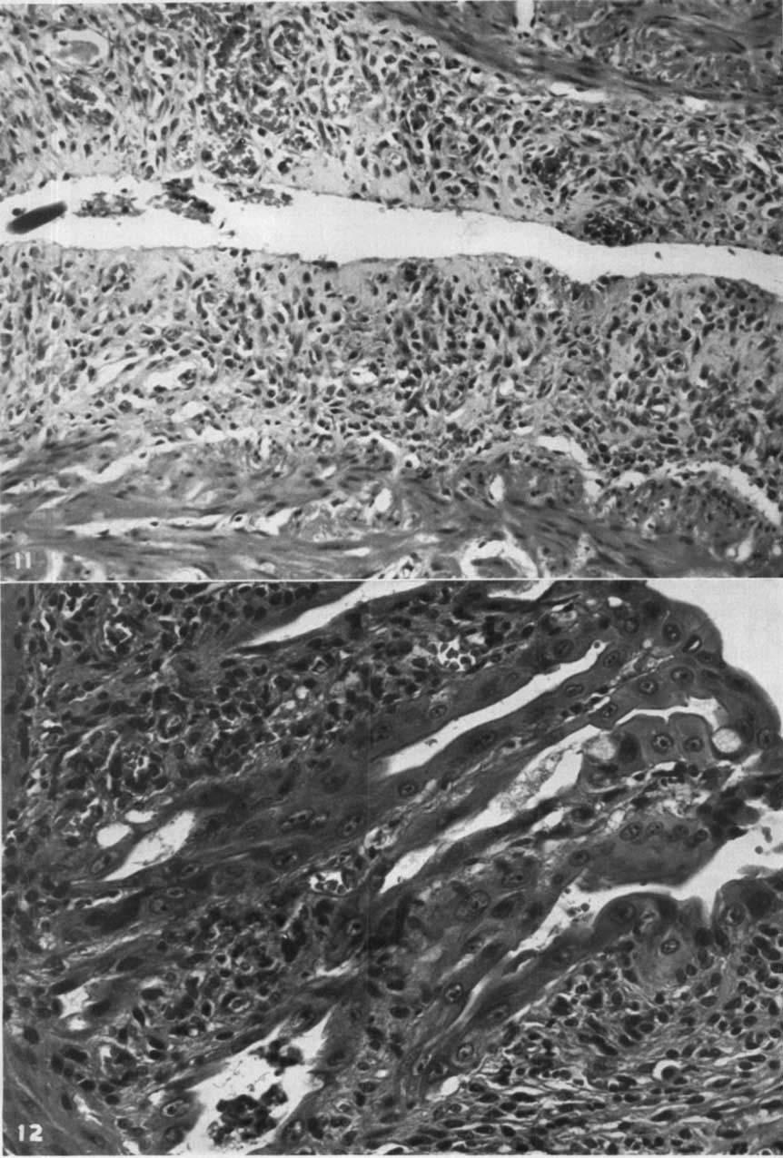
7 FIG. 7. Rectal mucosal glands showing moderate HN2 effect. Note hypochromic nuclei, distinct nuclear membrane, and large multiple irregular nucleoli. The stroma is infiltrated with plasma cells. FIG. 8. Detail of Fig. 7. was no evidence of hemorrhage into the lumen of the bowel and no infarction. There was no loss of hair. A few dogs had swelling of the hind extremities; the subcutaneous tissue of the thigh was soft, of gelatinous consistency, and the muscles were soft and boggy. One dog (no. 18) had multiple, small, hemorrhagic polyps of the mucosa and submucosa of the urinary bladder. Some dogs in group 1 exhibited edema and scar formation in the periaortic tissue at the level of the tip of the cathether. In two dogs (nos. 17, 22) the tube had slipped upward slightly and one of these (no. 17) had swelling of the hind extremity. The tube remained in place in all other dogs. consisted of karyorrhexis, associated with slight infiltration with polymorphonuclear leukocytes (Fig. 6). When the HN2 effect was more marked, the nuclei became hypochromic, exhibited a distinct nuclear membrane, and contained multiple, large, irregular nucleoli (Figs. 7, 8). The columnar cells became cuboidal, and their cytoplasm was vacuolated. Many of the glands became dilated in their basal portions (Fig. 9). The goblet cells were decreased in number. With high doses of HN2, the glands disappeared in focal areas (Figs. 10, 11). Squamous metaplasia of glandular epithelium was seen occasionally; the lining cells of the glands assumed a triangular or Microscopic Findings. PATHOLOGICAL polygonal shape and had finely granular cyto CHANGES IN Docs RECEIVING REPEATED DOSES plasm (Fig. 12). Paneth's cells occasionally per OF INTRAARTERIAL HN2 (GROUPS 1 TO 8). sisted. The architecture of the mucosa and the RECTUM. The most pronounced changes in remainder of the rectal wall was well mainthe mucosa were in the glands of Lieberkiihn. The first perceptible changes were in the cells lining the deeper portions of the glands and tained in most of the animals. The thickness of the mucosa was reduced, particularly when there was complete disappearance of glands

8

9 _ lylra AR'ILRIAI, NllROGLN hlus1ard (Figs. 10, I 1). Although the surtace epitlielium between the crypts of Liebeihuhn was lost, deep ulceration was absent, except in some animals receiving very high dosages of HN2. Even in these animals (0.6 mg. per Kg. per day), the ulceration did not extend deep to the rnuscularis mucosae. The inflammatory response, even in the presence of severe changes in the glands, was slight to moderate and consisted almost entirely of an infiltration of plasma cells. However, theie may be an early, transient infiltration of polymorphonuclear leukocytes. A polymoi phonuclearleukoc ytic response was found about ulcerations in those animals receiling high doses of HN2. Very few eosinophils and no mast cells were noted. The outer one half of the mucosa in a tcw dogs contained dilated capillaries with occasional moderate hemorrhages into the 1 unica propria. Thromboses of blood vessels %ere not noted. The submucosa was congested and edematous. The solitary lymphoid follicles showed a pronounced and early atrophy, beginning centrally with nearly complete disappearance of small lymphocytes and lymphoblasts, while the reticular cells and the reticular framework were maintained. On higher doses of HN2, there was hemorrhage into the centers of the follicles. The nuclei of the small lymphocytes 5howed karyorrhexis as the first change. With atrophy of the solitary follicles, the adjacent glands were displaced into the atrophic follicle, to lie well below the muscularis mucmae. The niusciilaris showed no changes. The serosa showed edema and congestion of the blood Lesscls. There was slight swelling of the cndothelial cells of the blood xessels. 4 ~ 5. The anal mucous membrane showed no changes in those animals reccik ing lower dosages of HN2. In the animals receiling 0.6 rng. per Kg. per day (group 8), the stratified sc~uamous epithelium exhibited hyperchromic nuclei and showed atypical downgrowths into the submucosa. ILEUM. The ileum showed changes due to HN2 similar to those of the rectum but to a Iesxr degiee. ~ * Barberio, Klopp, Ayres, & GIOSS [ 1349 URINARY BLADDER. In tlic aiiirnals in group 1 to 4 that receiictl fioiii 0.1 to 0.2 mg. per Kg. per day, the pathological changes in the urinary bladder consisted of moderate edema in the submucosa, associated with congestion of capillaries. The epitheliuni was intact and not hyperplastic. In a few dogs, vacuolization of the cytoplasm of basally placed transitional cells occurred. In groups 5 to 8 that were given 0.3 to 0.6 mg. per Kg., these changes were progressively more severe. There was often severe hemorrhage into the edematous subniucosa. In one dog, this resulted in polyp formation into the lumen of the bladder. Inflammatory reaction in the stroma was slight and consisted of the presence of plasma cells. There was a slight proliferation of fibroblasts. The epithelium occasionally was ulcerated. The muscularis showed separation of the muscle bundles by a proteinrich edema fluid, often containing fibrin strands. In the perivascular connective tissue, there was severe fibrinoid necrosis of the smaller blood vessels, as3ociated with hemorrhage. Other blood vessels showed swollen endothelial cells but no thrombi. PROSTATE. Animals receiving 0.1 to 0.3 nig. per Kg. of HN2 per day showed congested blood vessels and slight hemorrhage into the acini and stroma. In a few animals, there was focal atrophy and/or necrosis of glands. The nuclei of the acinar cells showed hypochromatism similar to that seen in the cells of the rectal glands. Animals receiving 0.4 to 0.6 mg. per Kg. per day showed more profound changes consisting of large areas oh necrosis in which only vague outlines of the glandular architecture persisted. Hemorrhage was present in these necrotic areas. Inflammatory reaction was almost completely absent but, when present, consisted of polyniorphonuclrar leukocytes. Some acini were dilated and lined by cuboidal cells containing hypochromic nuclei. The edematous stroma contained proliferating fibroblasts, many of which had doubletailed cytoplasmic processes siniilar to those seen in irradiation reaction. There wa? fibrinoid necrosis of the walls of arterioles, which were eosinophilic, homogeneous, and infiltrated by polyniorphonuclear leukocytes ~ ~~ FIL. 9. Rectal rnucosal glands showing moderate HN2 effert. Note cuboidal cells lining dilated glands. FIG. 10. Rectal mucosa showing marked HN2 effect. Note disappearance of glands in focal areas, dilation of capillaries. ~~~~
10

11 ~ ~ ~ ~ ~ ~~ IN? RAARTERIAL NITROGEN MUSTARD TESTES. The pathological findings were difficult to interpret since the ages of the animals were not known. The impression was that spermatogenesis was reduced. There were no areas of hernorihage, necrogs, or \ asculitis. VAGINA. The vagina showed changes similar to those of the bladder. Edema and congestion of the subiiiucosa occur1 ed arid there was oc( asionally niininial hemori hage into the Stroma. The changes in the epithelium were slight and consisted of edema of the basalcell la~ers and slight hyperplasia of the inner half ol the stratified syuanious epithelium, with 11) perchromic nuclei. Inflammatory cells were alncnt or sparse, and there was no ulceration. uri RCS. Theie way edema and congestion of the endometrial stronia and infiltration with hemosiderinfilled macrophages. In a few 'iniinals, the endoirieti ial glands showed hj pothromic nuclei and prominent nucleoli. Neciosis and ulceration were present in only one animal. In a few animals receiving large amounts of HN2, there was hemorrhage into the endometrial stronia and dilatation or glands. ADKI UM~. 7 he adienal glands showed \eiy ICM' pctiliologic,tl c11angcs. In mo5t cases, there wexc n~ttnerous spongioc) tes aniong the cortical cell5 indicating 110 se~ eie lipid deple Lion. One aniinal sliowed focal neciosis and hcnioi rhage in the medulla, antl another animal showed atiophj and heniorrhage into the 7ona fasciculata. OTHI K ORGANS. The heart, pancreas, and >lorilath were noiinal. The lungs %ere congestcd, antl in a few of tlie animals there was tnoclei ate pulnionai y ctlenia. The spleen diowetl set ci e a ti 0phy of inalpigliim bodies mid nuniei om henio~itlex infilled macrophages; oxtranicdullarj Iieniatopoiesis was absent. Ilic kidneys were nornial except for an al Iiuminous precipitate in Bowman's capsule louiid in a few of the dogs. The liver showed congesiion and, in a few of tlie dogs, fatty tnctarnorphosis. Two of the dogs that lived tliii tynine and twel\ e days iespectively ~liowecl riuiiicrous embolic bacterial abscesses. ~~ ~ ~ * Bniberio, Klopp, Ayres, & Gloss [I351 COMPARISON Or PATHOLOCIC41, C langks ACCORDING TO DOSE OF HN2. Pathological changes found in the 1 ec tal biopsies were compared as to groups arid as to the day of the biopsy. Similar histological changes were found in groups 1, 2, 3, 3, and 5; there was no apparent increase in the se\ eiity of pathological changes with highci daily doses of HN2, whether giten in a single injection or in di T ided doses. Considerable L ariation occui red in the se\erity of changes in iiidividu;i1 dogs within the same group and in diffexerit are'is of the colon in the same dogs. A11 dogs in groups 6, 7, and 8 showed more severe rectal chaiiges. PhlHOIOGICAL CHANCES 1Y THE RFCTUM OF A DOC AFTFR A SINGLF INrRAARTERIAL INTrC TION or HN2. In order to deteriiiine the sc (1 iience ot pa thological changes in t he rec tun1 within the fii st twentyfoul houi s after a single injection, 0.6 rng. per Kg. ol HN2 was injected into the aorta of one dog. Biopsies of the rec tum '~\'crc taheii at 7ei0, 1, 2, 3. 4, 6, 8, 10, 12, and 23 lioius after the injection, at which time the dog was sacrificed. The1 c wex e no changes in the 1)iopsies taken at /ei o, OIIC, two, and threc hoitis. The biops) at loin houi s 41iowcd itifiltr atioii 1)\ n~i~r~cr~its pol)iiiorpliotii~clc~ti Icuhc\ tes into an edematous and congested iniicosa and sulxnucos'i, c\pcciaily in and about the sul)iiiiito5al capillaries. In the niucosa wei c minute hcmorihages. A1 six hours similar but inoie seaeie changes M ei e found. At eight hours, the biopsy showed only normal anal mucosa. At ten hours, the changes just desci ilxd TYCI e nioic se\ei e, and, in addition, thei e was pronouncctl harjorihexis of the nuclei in the solitai j I\niplioid follicles. Thci e weic no changes in the glands of 1,iebci krrhn. The biopsv at ticel\ e hours showed only normal anal cosa. At twentythree hours, there was kar) orrhexis of the nuclei of the cells lining the glands of Liebcrkiihn and atrophy of the solitary follicles. The niicioscopic appearance of thc lungs, li\ er, heart, adrenals, hidnej s, pancreas, antl stomach was unremarkable. The colon showed FIG. 11. Rcrtal muro5a showing marked HN2 effect with complete disappearance of glands. 'I'hc initcosi is thin and the capillaric5 'ire dilated. The muscularis mucosae is int'ict. FIG. 12. Rect,tl niucosa showing marked HA'? effect. Note squamous metaplntia of glandular epithelium. ~

12 CANCER Nouem ber I 95 1 changes similar to those of the biopsy of the rectum taken at twentythree hours. There were focal areas of hemorrhage into the prostate. The spleen showed focal necrosis 01 the germinal centers of the malpighian bodies. were severely atrophic, and there was reduction in mitotic activity and karyorrhexis in the basal portion of the glands. The colon showed the same changes as the ileum, but these were less severe than in the rectum. In the spleen, there was atrophy and hemorrhage PATHOLOGICAL CHANCL~ IN THE KFCTUM OF A DOG RFCXIVING HOURLY INTRAARTERIAL INinto the malpighian bodies; the red pulp JECTIONS OF HN2. In order to determine the showed erythrophagocytosis by the reticulosequence of pathological changes in the first endothelial cells. twentyfour hours, one dog was given 0.2 nig. PAT nolocic:al CHANCES IN ANIMALS KECEIVper Kg. into the aorta hourly for a period of ING HN2 IN WHICH THE VENA CAVA WAS ten hours. Biopsies of the rectum were taken IJGATED AND SEVERED. In the dogs in groups at zero, 1, 2, 3, 4, 6, 8, 10, and 24 hours. The 10 and 11 that had the posterior vena cava dog died at twentyfour hours. ligated and severed, the pathological changes The first biopsy revealed normal niiicosa. in the pelvic organs were not different from There was no change in the first hour. At two those in animals that received the same dosage hours, there was an acute inflammatory re without caval ligation. action in and about blood vessels of the sub PATHOLOGICAL CHANGES mucosa, manifested by a moderate infiltration INTRAVENOUS HN2. The three dogs in group of polymorphonuclear leucocytes. There were 12 were given 0.1 mg. per Kg. daily. Two of also edema, congestion, and minute hemorthe dogs were sacrificed at fiftyone days and rhages in the submucosa. At three hours, the one at seventytwo days. Biopsies of the anomicroscopic appearance was similar to the prerectal junction on all three dogs on the thirtyceding biopsy, but, in addition, showed beseventh day revealed no changes in the muginning karyorrhexis of the lymphocytes in cosa. In one dog, there was atrophy of rectal the solitary lymphoid follicles. At four hours, lymphoid follicles. there was atrophy of the lymphoid follicles. At autopsy, there were no changes in the The acute inflammatory reaction persisted. rectal mucosa, but there was atrophy of the There was focal karyorrhexis of the nuclei in lyrnplioid follicles. The ileum in one animal the basal portion of the glands of Lieberkuhn. showed atrophic changes ascribable to HN2 At six and eight hours, the histological apin addition to atrophy of the lymphoid folpearance was similar but more severe and, at licles. The spleen showed atrophy of malpighten hours, still more pronounced. The karyorian bodies similar to that seen in the animals rhexis was confined to the basal portion of the glands of Lieberkuhn. The capillaries of that received intraarterial HN2. The other organs showed no changes. the submucosa were prominently dilated and congested. The surface epithelium of the mu CONTROL ANIMALS. Three animals in the cosa remained intact. At the end of twenty control group received 5 cc. of saline intrafour hours, the glands of Lieberkuhn showed arterially twice daily for six, twelve, and karyorrhexis, with hypochromic nuclei. The eighteen days respectively. Biopsy and autopsy acute inflammatory reaction was now minimal findings were negative. and was replaced by a chronic inflammatory ieaction manifested by the presence of lympho TREATMILNT OF ADENOCARCINOMA OF THE RECcytes. There were small areas of hemorrhage TUM WITH INTRAARTERIAL HN2: CLINICAL in the submucosa and mucosa. Focal loss of TRIALS surface epithelium of the mucosa was present. Since the animal experiments indicated that The microscopic findings in the heart, the tissues of the pelvis and pelvic organs lung, stomach, kidneys, pancreas, and adrenals tolerated periodic injections of moderate were unremarkable. The bladder showed amounts oe HN2 into the abdominal aorta edema, congestion, and minute hemorrhages and since the tissue that showed the greatest in the submucosa. The epithelium was intact, sensitivity was the rectal mucosa, initial clinand the cells showed some edema. The pros ical trials were designed to study the effect of tate showed focal necrosis and focal atrophy intraarterial HN2 on rectal adenocarcinoma. of acini. In the ileum, the lymphoid follicles Injections were given through polyethylene IN Docs RECEIVING
. Note nodularity along the periphery and at the base of the defect. FIG. 14. Perineal defect.")
13 INTRAARTERIAL NITROGEN MUSTARD Bmlberio, Klopp, Ayres, d~ Gvoss [1353 FIG. 13. Recurrence of rectal adenocarcinoma in perineal wound following abdominoperineal resection. Photograph taken at beginning of treatment with intraarterial HN2 (three days). Note nodularity along the periphery and at the base of the defect. FIG. 14. Perineal defect. Photograph taken seventeen days after onset of intraarterial HN2 therapy. Note the smooth edges and clean base of the defect. Note in the upper portion of the defect the levator ani, now free of gross tumor. tubing that had been introduced through a branch of the femoral or the external iliac artery and had been advanced in a retrograde fashion until the tip of the tube was within the abdominal aorta. Tubing inserted in this manner remained patent for four or more weeks. While the injections were made through the tubing, bloodpressure cuffs that had been placed about both thighs were inflated to above the known systolic blood pressure. The position of the tip of the tubing was determined by arteriograms obtained by injection of either thorotrast or diatrast through the tubing. The injection of diatrast produced a sensation of warmth in the region supplied by the artery into which it was injected. This served as a further check on the position of the tip of the catheter. Injection of saline, thorotrast, or HN2 produced no sensation. Two patients with recurrent adenocarcinoma of rectal origin localized to the perineum were chosen for treatment. Each had had a resection of the rectum and had an end colostomy, thus presenting no possibility of injury to normal rectal tissue. CASE REPORTS Case 1. A 44yearold man was admitted to George Washington University Hospital on August 14, Eleven months prior to admission, he had had an abdominoperineal resection of the rectum for adenocarcinoma. Five months after the initial operation, recurrent cancer was discovered in the perineal wound. Excision was attempted but was unsuccessful. A course of deep roentgenray therapy was then given, followed by a second course six weeks later and the insertion of
, he was first admitted to the hospital.")
14 I3541 CANCER Nouember 1951 radiiim needles two weeks after this. About file weeks after the removal of the radium needles (no other therapy was given in the interim), he was first admitted to the hospital. On admission, he was pale and showed some ekidence of weight loss. A functioning colostomy was present in the left lower quadrant. The only evidence of tumor was in the perineum. Here, therc was a defect measuring about 8 cm. in transverse diameter, extending from the tip of the coccyx to the base of the scrotum. The edges were nodular and the base was necrotic. The defect extended up into the hollow of the sacrum (Fig. 13). Biopsies obtained from the wound edges showed the presence of recurrent adenocarcinoma. Within twentyfour hours of admission and before any therapy had been begun, the patient developed a fecal fistula that opened externally into the perineal defect. On August 16, polyethylene tubing was inserted into the right deep epigastric artery and advanced in a retrograde manner until a 25crn. length had been introduced. Although the tip of the tubing appeared to be in the abdominal aorta, repcated arteriograms showed filling of the arteries of the right leg only. Through this tubing, 2 mg. of HN2 was administered ekery eight hours until a total of 38 nig. had been given over a six and a half day period. In order to treat tlie leit pelvis, an 18cni. length of pol) ethylene tubing was introduced through tlie left deep epigastric artery. Artei iogram5 showed good filling of the arteries of the IeLt 4de of the pelvis. Through this tubing, 2 ing. of HN2 was administered every eight hours until a total of 40 mg. had been given over a six and a half day period. During the same period (August 25 to 31), 0.5 mg. of HN2 was adininistcred into the tubing in the right pel~is aery eight hours until a total of 10 mg. had been given, making a grand total of 88 mg. The patient s clinical response I+ as difficult to ex aluate because the perineal fistula proved to be connected with the small bowel and was vei p irritating. However, within three days of the start of therapy, he was able to lie on his hack. Perineal tenderness had pi evented this for many weeks. Within one week after the start of therapy, the nodules along the edges of the perineal wound decreased in siie. The base of the defect became smooth, clean, gianular, and pink, as if the area had been clebrided (Fig. 14). The odor of tissue necrosis disappeared. The ti$sues niairitainctl this appearance during the c9urse of the1 apy. Biopsies were obtained from the perineal defect at fortyeighthour intervals during the period of therapy and about every five days thereafter. On September 14, 1950, the patient was started on a course of cortisone. His subsequent course, as well as his hematological studies, will be reported in a subsequent paper. PATHOLOGICAL FINDINGS. The pathological findings are summarized in Table 3. The sections were somewhat difficult to interpret, since the patient had previously received irradiation, and irradiation necrosis was present in the first biopsies. Although the biopsies were labeled left and right, thcy were often taken within a few centimeters of one another, and the possibility oe spread of HN2 from either side was probable. However, even when these factors are taken into consideration, it is believed that changes in the adenocarcinoma caused by HN2 can be stated with a fair degree of certainty. It is also probable that earlier changes owing to HN2 could have been determined if the masking effect of the previous irradiation had been obviated. Adding to the difficulty is the known ability of both HN2 and irradiation to produce similar microscopic pathological changes. Another complicating factor was the administration of cortisone after cessation of HN2 therapy. The observed changes in sequence are as follows: (I) karyorrhexis and cytoplasmic vacuolilation ob malignant cells; (2) dilation of the malignant glands with rupture and ingress of necrotic debris; neciosis of malignant glands; change of cells from columnar to cuboidal; hyalinimtion of interbening stroma and decrease in leukocytes; (3) cancerous tissue either absent or atrophic; malignant cells polygonal with hyperchroinic nuclei, resenibling squamous cells; hyalinimtion of the stroma and the presence of bkarie fibroblasts similar to those seen in irradiation reaction ; (4) regrowth of adenocarcinoma after cessation of treatment; evidence of return of inlection. Cusp 2. A 38yearold white woman was admitted to the United States Na\al Hospital, Rcthesda, Maryland, on April 15, 1950, because of perineal pain and ulceration. Three years previously, she had had an abdorriinoperineal resection for carcinoma of the rec tum. She was frrst admitted to this hospital in late 1949 with the complaint of perineal pain. Laparotonly revealed extensive, inoperable, recurrent carcinoma confined to the pelvis. She was discharged on November 15, 19/19. She felt fairly well and gained weight
15 ~~~ ~ Date 8/17 8/18 8/19 8 / 20 8, /23 8,124 8/25 8/26 8/27 8/ TABLE 3 SUMMARY OF PATHOLOGICAL CHANGES IN CASE 1 ~ ~ ~ ~ ~ ~ _ ~ _ Cumulative Days of dose treatment Pathology ~~ Rt. Lt. Rt. Lt Right Irradiation necrosis. No adeno Adenocarcinoma. Irradiation necrosis carcinoma Adenocarcinoma. Irradiation (Fig. 15) Irradiation necrosis. No adenocarcinoma necrosis Malignant cells show karyorrhexis, Adenocarcinoma, with no change vacuolization of cytoplasm, indicating possible early HN2 effect Necrosis; no adenocarcinoma Adenocarcinoma, with karyorrhexis of nuclei, pyssibly due to HN2 Malignant glands dilated, cells Adenocarcinoma with increased karyorcuboidal. Karyorrhexis. Necro rhexis, vacuolization of cytoplasm. sis of malignant glands. Stroma Changes comparable to those on left hyalinized. Decrease in infec on third day tion. First definite changes due to HN2 Similar to that on 5th day Similar to that on 5th day (1 inj.) No No 8 Similar to that on 5th day treat treatment ment On August 25, a new schedule of treatment was started. on the right, and 2.0 mg. was given on the left every eiqht (8) hours Left Similar to that on 5th day Insufficient tissue Similar to that on 5th day A daily total dose of 1.5 mg. of HN2 was given _ 9 Similar to that on 5th day Similar to that on 5th day Similar to that on 5th day Similar to that on 5th day Similar to that on 5th day, except Insufficient tissue stroma more hyalinized Insufficient tissue Dense fibrous stroma with atypical fibroblasts; clumps of polygonal malignant cells, indicative of severe HN2 effect Similar to that on 13th day Malignant glands dilated. Cells atrophic. Karyorrhexis. Stroma hyalinized. Decrease in infection. First definite changes due to HN2. Section similar to those on right, 5th day of treatment No change from that of 3d day of treatment (left) 8, 30 No change from that of 3d day of treatment 8/ Insufficient tissue Similar to that on the 3d day but more severe. In addition, hyalinization and fibrosis of stroma; necrosis of malignant glands (Fig. 16) 91 I No biopsy No biopsy 12 ini.) 12 ini.) _I, \ J, No further treatment was given. Biopsies continued to be taken at about 5day intervals 9;'S No thrrapy 5 days after Sections on the right and left were similar. Few isolated clumps of altered treatment 7#'9 No therapy 9 days after treatment malignant cells in dense connectivetissue stroma. Nuclei hyperchromic; cells polygonal, resembling squamous cells. Numerous atypical fibroblasts. Severe changes due to HN2 (Fig. 17) Biopsies on the left and right were similar and were similar to those taken 4 days previously, except there was less alteration in the malignant glands, and the fibrous stroma was edematous. Changes indicate possible revival of growth of malignant cells and less HN2 effect 9/14* No therapy 14 days after treat No adenocarcinoma. Fibrous stroma containing atypical Adenocarcinoma similar to that of first biopsy, indicating regrowth of maligment fibroblasts nant cells 9/19 No therapy 19 days Sections from both right and left were similar. Islands of little altered ad.eno after treat carcinoma similar to original biopsy; also adenocarcinoma showing HN2 effect ment 9/27 No thcrapy 27 days after treat Only necrotic tissue insufficient for study Covering squamous epithelium; adenocarcinoma no different from original ment biopsies 10,'3 No therapy 33 days after treat Only necrotic tissue insufficient for study Covering squamous epithelium; adenocarcinoma showing. moderate IN2 ment effect 1016 No therapy 36 days Infected granulation tissue con Similar to previous biopsy after treat taining few clumps of maligment nant cells ~... ~~~. ~ ~~ * Cortisone acetate, 50 mg./day, begun.

16 13x1 CASCER.\'mienil~er 1951 FIG. 15. Case 1. Initial biopsy from left side. Adenocarcinoma surrounded by an area of necrosis. FIG. 16. Case 1. Biopsy from left side after six days of treatment. Moderately severe changes due to HN2. Note dilated glands, karyorrhexis, and necrosis of malignant glands. until one week prior to admission. Physical examination revealed moderate emaciation. Positive physical findings were as follows: functioning colostomy in the left lower quadrant, scars of abdominoperineal resection and laparotomy, and a gaping, granulating perineal wound that had a foul fecal odor. Treatment with intraarterial nitrogen mustard was begun on April 26, A polyethylene catheter was inserted through the right superficial circumflex iliac artery into the right femoral artery, and advanced for a distance of 25 cm. An arteriogram showed the catheter to be' coiled in the right common iliac artery. HN2 in doses of 1.0 mg. was injected into this tube every eight hours for five days. Therapy was stopped for three days and then resumed for three additional days, for a total of eight days of treatment. In addition, 10 to 20 cc. of normal saline was injected immediately after the HN2. The appearance of the perineal lesion on the side of the injection changed from that of shaggy necrotic tissue into what appeared to be rather healthy granulation tissue. Biopsies taken at various intervals showed severe changes in the tumor cells with necrosis and, in some areas, replacement by a hyaline material. This necrotizing process was less severe in the stroma. In view of the good results obtained on the right side, it was decided to insert another catheter on the left. This was done on May 26, 1950, and HN2 was given for a total of 22 mg. Similar gross and microscopic changes were noted. During all of this time, the patient had a moderate anemia, and several blood transfusions were given. On June 23, a second course of nitrogen mustard was begun on the left side, 1.0 mg. being injected three times a day for six days. No response to this therapy was noted, and no biopsies were obtained. The patient followed a progessively downhill course and died quietly on July 18, BIOPSY FINDINGS. A control biopsy taken on February 16, 1950, eleven weeks prior to treatment with HN2, revealed welldifferentiated adenocarcinoma of colonic origin similar to


17 INTRAARTERI AL NITROGEN MUSTARD FIG. 17. Case 1. Biopsy from left side, four days after cessation of treatment, showing severe changes. Note isolated clump of altered malignant cells in connectivetissue stroma. The nuclei are hyperchromic and the cells polygonal, resembling squamous cells. that in case 1. However, in distinction to the previous case, considerable mucin formation was present and no changes due to irradiation. On April 28, two days after the start of therapy, when a total dose of 6 mg. of HN2 had been given, biopsies were taken from the perineal site on the left and right. The biopsy from the right revealed adenocarcinoma similar to the original biopsy. There were areas of necrotic neoplastic tissue. The cancerous cells were columnar and contained oval hyperchromic nuclei and multiple bizarre nucleoli. The cytoplasm was granular, and the cells were lined up on connectivetissue strands. There was minimal cytoplasmic vacuolization and karyorrhexis. The biopsy from the left side showed adenocarcinoma with only a few areas of necrosis. The cancerous cells were similar to those on the right and produced mucin. Karyorrhexis was present, and mitotic figures were more numerous. A biopsy from the right side after a total * Barberio, Klofip, Ayres, Q Gross [1357 dose of 8 mg. revealed adenocarcinoma similar to the previous biops), but some of the malignant glands showed lack of definition of cell membrane, nucleus, and nucleoli. There were large areas of necrotic debris, infiltrated with polymorphonuclear leukocytes. The biopsy from the lelt showed no change from the previous one. A biopsy on May 1 after five days of therapy and 14 mg. of HN2 revealed adenocarcinoma in which the glands were dilated and lined by \ acuolated, cuboidaltocolumnar cells and the karyorrliectic nuclei exhibited loss of polarity. Some of the malignant glands were dilated or ruptured, permitting the ingress of eosinophilic homogeneous material. In the intervening eosinophilic stroma were rounded clumps of one to six malignant cells. Sections taken on the left were similar to those on the right. (On May 1, after five days of treatment in which 15 mg. of HN2 had been given, therapy was stopped until May 3, at which time therapy was again begun and continued on the same dosage schedule for a period of three days [lo mg. of HNZ], giving a cumulative dose of 25 mg. On May 9, after an interval of three days without treatment, HN2 injections were again begun, and, over a period of four days, 12 mg. was administered.) On May 13, after a total dose of 37 mg., given over a period of eighteen days of intermittent therapy, biopsy showed only necrotic debris and a strip of squamous epithelium. During the next two days, 15 mg. of HN2 was given for a total cumulative dose of 52 mg. On May 26, the left superficial circumflex iliac artery was catheterized, and six doses of 1.0 mg. were given over a period of two days, at which time therapy was discontinued because of a slough on the medial aspect of thigh; this was due to the presence of the catheter in the distal portion of the femoral artery. On June 8, a catheter was inserted into the left femoral artery through its profunda branch and advanced in a retrograde fashion for a distance of 25 cm. Intraarterial HN2 therapy was given with the same daily dosage. A biopsy on the left two days later, after 6 mg. had been given, showed viable adenocarcinoma with only slight HN2 effect, surrounded by areas of necrotic debris. The intervening stroma was edematous and contained isolated clumps of malignant cells. There were

18 13581 CANCER November 1951 large areas of necrosis of malignant glands in which only vague outlines of the glands could be made out. After four days and 12 mg. of this course, biopsies revealed adenocarcinoma in which the glands were dilated and the malignant cells were cuboidal with hyperchromic nuclei. The strips of malignant epithelium were arranged in an anastomotic pattern. The intervening stronia consisted of eosinophilic laminated material with sparse polymorphonuclear leukocytes. These changes appeared to be due to HN2. This course was continued from Julie 8 to June 15, for a total of 22 mg. and a total cumulative dose of 74 nig. A biopsy was taken on June 19, four days after cessation of therapy. On the left side, this showed large areas of viable adenocarcinoina, with little or no HN2 effect. However, in some areas there were clumps of cells resembling signetring cells. A final course of treatment was begun on June 23 after eightdays cessation, and was continued for five days, until June 28. A total dose of 5 mg. was gi\eii. Thi5 made a grand total ot 79 nig. giken either lelt or right o\er a period of 5ixtythree days. The patient died on July 18, twenty dajs alter the last injection ol HN2. AUTOPSY FINDING^. At autopsy, tlie body was that of an emaciated white woman, allpearing soniewhat older than the stated age of 38 years, weighing approximately 80 lb., and measuring about 62 in. There wa.s a fairly recent, partially healed lower aldorninal incision. On the medial aspect of the upper portion of the left thigh, there was a iaised, firm, indurated area, measuring 7 x 4 cm., the superficial portion of which was ulceiatetl. There was a bluishpurple discoloration of the tips of all of the digits of tlie right foot. The entire perineum was involved in a huge ulcer that extended from the tip of the coccyx to the mons veneris. The posterior vaginal wall was absent, and this cavity extended up in the pelvis to the peritoneal reflection and wa3 lined by necrotic, yellowgray tumor tissue. Over the sacrum, there was a superficial decubitus ulcer. ABDOMlNAL CAVI ry. There weie nl1nieious fine adhesions between loops of small intestine and the tentral abdominal wall. The sigmoid colostomy stoma was patent. Most of the loops of small bowel in the pelvis were densely adherent to the peritoneal pelvic floor formed at the time of the abdominoperineal resection. In separating these loops of bowel from this situation, a fistula was found between one of tlie loops of howl and the perineal ulceration Both the bladder and the uterus were involved in the ulcerated tumor mass. The liver contained only three areas of metastatic tumor, the largest of which measured 3 C I ~. in diameter. The other abdominal organs were in their normal relationships. PELVIC ORGANS. The previously described perineal ulceration liad for its rool peritoneum that had been sutured across the true pclvis at surgery. Beneath this membrane were the uterus, adnexa, and bladder. The entire area was surrounded by partially necrotic, yellow tumor tissue. The wall of the bladder was extremely thin. The uterus, tubes, and ovaries were partially replaced by tumor tissue. I lie remaining organs were essen tially normal. hficroscoi I<: FINDINGS. PEKINEAI. DEFECT. Sections revealed adenocarcinonia in which the glands were markedly dilated and lined by either colulnnar or cuboidal epi~fieliuni. Within these dilated glands was a mucinous exudate. There were isolated clunips of malignant cells, while other cells were arranged in acini. In other areas, only the collagenow framework of the neoplasm remained, the neoplastic cells being absent. Rctween these anastoinotic strands was abundant basophilic niucin. In still other areas, there was deposition of calcium within the arcas of niucin and stroma. Very little evidence of acute inflaniniation was found within these areas of neoplastic tissue. In the dermis, there was prolilcration of rnyxotnatous tissue arid dilation of lynphatics, with only niinirnal chronic inflarnniatory reaction. The uterus showed adenocarcinoma that liad the appearance of a mucinous or colloid type in that there were isolated clumps ol malignant cells surrounded by niucin. This appearance was considerably different from that of the original biopsy in which mucin was only minimal. 7 1ie riglit and left common iliac arteries at the site of the tip of the catheter revealed duplication, necrosis, and calcification of the internal elastic membrane.

19 IN IRAARTERIAL NI1 ROGF.N AlUS I AKD o I I~KR ~RGANS. There was modcrate atroihy 01 the iiialpig1ii;tn 1)odies oc tlie spleen. In tlie red pulp were nunicrous hcniosideriiifillcd niacrophagcs. I lie skin at the sight of WN2 leakage ahowctl fat necrosis of the tcla subcutanea. 71 lic skin appcn~1;iges ~vere atrophic, but tlie cpi thcliuni was normal. The lungs showed atrophy and emphysema. I licrc was severe drpletion of lipoid in the cortcs of the adrenal gland. A mesenteric I yrripli node revealed marked depletion of with replaccnicnt by plasma cells. l liere W;IS no er)throphagocytosis. The kidneys were normal. The heart showed serous atrophy of epicardial fat and atrophic in) ocardial lilwrs. 7 he liver aliowed arcas of adenocarcinoiiia in a better state of preservation than that seen in the pei\is. In some of the areas, however, there were changes due to HN2 similar to those found in tlie adenocarcinoma present in thc pelvis. There was purulent peritonitis. Dlscussrolu The field supplied by the arterial system into which the injection was made in tlic dog was not identical with that in the human patient. In the dog, tlie internal iliac artery supplies the lower rectum, bladder, prostate, anus, vagina, uterine cervix, esternal genitalia, and the muscles of tlie perineum, but not the extremities or gonads. In the human, the lower abdoiiiinal aorta also supplies the entire pelvic bone and both extremities. Therefore, the clinical patient is not so well adapted for pelvic tlierapy as the dog. This shortcoming can bc ~oiiiewlriit overcome, as long as periodic iii.jc.ctions rather than continuous infusions ;ire used, 1)) applying arterial tourniquets ahit the thighs at tlie time of each injection. In tlic clinical patient, coinparison of iliac marrow with sternal marrow indicated that tlic action of tlie HK2 was more marked in the local pelvic area.2 However, these results c~oultl not be expected in the dog in which the iliac cixst is supplied by arterial branches that emerge above the tip of the polyethylene tube. The actual field of treatment, as described, can he outlined quite accurately in the dog by the i ti traarterial injection of methylene blue.i3 This is riot possible in the patient hecause of t lie extrenie discomfort produced by the inicc tion. I he canriulization technique has permitted. Ba?bei.io, Klopp, Ayi,es, 6 G?oss [ 1359 periodic injections over an extendcd period of time in dogs and patients. The nictliod introduced two piominelit Iiazartls, naniely local arterial daniage and bloodstream inkction. In none of the dogs was there evidence of any significant degree of local arterial tliroinbus foriliation or peripheral embolic episodes. In inany of the dogs, there was evidence of local action of HN2 on the aorta at the tip oc tlie cannula, as evidenced by the presence of bizarre fibroblasts in the adventitia and by necrosis and hemorrhage into the inuscularis. ;\rterial thrombosis can occur under certain circumstances. It has been noted following administration of other drugs in dog cxperimcnts and l ollowiiig HN2 injections in sotlie Iiiciiian cancer patients.13 Local hemorrhage can occur if the cannula is dislodged. While the latter occurred in the dog, it has riot been seen in clinical trials, wliere patient cooperation is possible. Systemic inkction, as indicated by tlie autopsy finding of embolic abscesses, has becn noted in a few dogs and is seen occasionally in clinical cases. That this is not clue primarily to the presence of the cannula alone is indicated by its absence in dogs receiving saliiic iii,jcctions. rl liat it is not due to the action of HN2 alone is indicated by its absence in tlie intravenously treated dogs. The infections would therefore seein to be due to the cornbination of the action of the HN2 and the presence of the indwelling intraarterial cannula. These observations, together with the known radiomimetic properties of HN2, suggested the possible value of concoinitant HN2 and antibiotic therapy.1 rhis is now being evaluated in ternis of aureoniycin. This factor of systemic infection is particularly important in patients, as a clinical course of intraarterial HAT2 therapy covers an extended period of time, and the dog experiments indicate that the longer the period of therapy, the higher tlie incidence and degree of systemic infection. It was apparent that the pelvic structures of the dog will tolerate fractionated intraarterial therapy with HN2 and that such therapy could be used in the treatment of clinical cases. Since the rectal niucosa was affected consistently even with smaller dosages, it is safe to assume that the rectal rnucosa can serve as an index of the upper limits of dosage for local therapy of pelvic structures, as sternal bonemarrow changes are used as an index of the upper limit in systemic therapy.

20 Fol piactical ~~~~rposcs, at least with dosagc sihetldes and physical factors utilized to date, the local iedction has not been as important as systemic toxicity. It has been postulated that intravascularly injected HN2 reacts rapidly with body constituents and is rapidly detoxified.? Thus, intraarterially injected HN2 would be detoxified in the local tissue capillary bed, thus preventing systemic reaction. This hypothesis appeared to be substantiated by the observation of Bierman that the injection of a single large dose of HAT2 into the hepatic artcry of patients produced no leukopenia. However, using peripheral blood counts as an index ot systemic toxic action in the dog, and sternal bonemarrow studies as the index in patients, it is apparent that the initial passage through the capillary bcd of the pelvis detoxified only a small amount of the HN2. The possibility still remains that other tissues, such as the liver, may detoxify larger amounts of HNP morc rapidly. Early in the study, it became evidcnt that any increase in dosage above the schedulc established in group 1 was impractical since there was a sharp drop in survival time. Consequently, a larger number of dogs were studied in the first four groups than in the higherdosage groups. The data available have consistently shown that divided doses are more desirable than a single daily injection. This is most convincingly seen in the comparison of survival time between groups 1 and 2 (recriving 0.1 mg. per Kg. per day) in Fig. 2. It has been shown that occlririon of the circulation of the small intestinc for 5 to 15 minutes6.7 and that of the femoral inairow ioi 2 to 15 minutes? afforded some dcgree ot protection to tlie involved tissues following intravenous administration. These findings suggested that HN2 excrts its effect in a relatively short period of time. Hence, if HN2 could be retained in the pelvic circulation for a longer period of time, less drug would be available to the extrapelvic circulation. In those dogs in which the posterior vena cava was ligated below the level of thc renal vcins, the survival time was increased in all instanws. The results shown in Fig. 3 may not be a true appraisal of tlie maximum tolerated dose. There is a sharp drop in the total dose tolerated between group 1 and thc groups immediately following, while in the three highest dosage groups the total amount tolerated again rises. Since the animals were mori bund duiing their lnst one to three days 01 ircdtmcnt, thc HN2 administered during this time probably did not contribute to their cled~lis. If this be true, the total dose tolerated by tlie animals in each group would be essentially the same except for group 1. The longer survival time noted in those animals that received the smallest daily ciose and the relativcly longer survival time of those animals in which the daily dosagc was given in three injections as compared to the animals that received the same daily dosc in one injcction indicate that a certain portion of each dose is detoxified in the first capillary bed. The ideal method of therapy would be that which kept the local tissue HN2 receptors saturated, but allowed little spillage into the systemic circulation. This ability to localize in a given capillary bed will vary i n dcgree according to the drug used. Certain dyes (gentian violet, methylene blue) will selectit ely color the leg of a rabbit when injected into the femoral artery while others (Evans blue, mercurochrome) show almost no selectilc coloring.13 Theoretically, one should hc able to deliver a drug to a given region at the rate at which it is absorbed and detoxified arid thus increase its local effectiveness. The pathological changcs in the rectum wcre similar to those seen in the small intestines after intravenous administration of HN These changes are very similar to those following local and total body irradiation. In both intraarterial nitrogenmustard therapy and irradiation, there is atrophy of intestinal glands, associated with hypochromasia of the nuclei, squamous metaplasia, a minimal inflammatory response, and capillai y hemorrhage. Also, in both conditions thcrc is early and rapid atrophy of lymphoid tissue. Bimrre fibroblasts, as seen in local irradiation, were found most consistently when there was permeation of HN2 into the adventitia 01 the aorta and into the serosa of the urinary bladder. Another feature in common was erythiophagocytosis, best seen in the reticulocndothelial cells of the lymph nodes. The mechanism of action of HN2 is not known, but, like irradiation, it has a pronounced effcct on dividing cells. This is demonstrated by profound changcs in the basal portions of the glands of Lieberkuhn, in which there is normally considerable mitotic activity. The action of HN2 appeared to be selective in that rapidly dividing cells, such as lymphocytes and glandular epithelium, were seriously af

21 INTRkARTERIAL NITROGEN MUSTARD fected, while cells that have less mitotic activity, as fibrocytes and smooth muscle, werc not seriously affected. Cell nuclei are particularly affected, since karyorrhexis is an early and prominent feature. Much useful information could be obtained if there were available in a large laboratory animal a malignant tumor that could be treated by fractionated intraarterial chemotherapy. In the absence of such a preparation, studies can be made of the effect of regional cytotoxic drug therapy on rapidly dividing cells 01 normal tissues or organs. Clinical experience suggested that the bone marrow or the growth of hair might be used to measure the changes resulting from regional treatment with HN2. Strangely, the growth of hair in dogs is not influenced as was the growth of hair in scalp, beard, and pubic areas in clinical patients.* However, the cells of the intestinal tract in the dogs were found to show distinct changes as a result of this mode of therapy and hence seemed preferable as an end organ for study. Of all the areas of the gastrointestinal tract, the lower rectum seems best suited to therapy, since inclusion of the upper gastrointestinal tract within the field of therapy has been associated with a decreased survival time of the animal.13 The cells of the gastrointestinal tract of the rat are replaced every 1.35 days.9 As HN2 even in small dosages appears to affect rapidly dividing cells, it is reasonable that it should show a selective affinity for rectal mucosa as compared to bladder and vaginal mucosa, skin, muscle, nerve, and connective tissue. Therefore, measurable changes within the rectal niucosa should yield information as to those cytotoxic properties of drugs that are caused by the selective action on dividing cells. As this characteristic appears to be common to most presently used cancerchemotherapeutic agents (roentgen rays, HN2, urethane, aminopterin, colchicine, etc.), information gained relative to the local selective effect on rectal mucosa as compared to that 011 surrounding pelvic tissues and thc rest of the gastrointestinal tract should be coniparable to the effect of the same agent on rapidly dividing malignant tumor cells following local therapy as compared to the effect on surrounding normal structures and distal, rapidly dividing normal cells. This concept has been used by Webber et al. who evaluated the synergistic effects of HN2 and irradiation by a study of their action on intestinal mucosa. * Rni bei io, Klopp, Ayies, L (>i 05s [lsc,l 1 he ef ec ts 01 lraction,itetl, 1 egional intraarterial HN2 therapy on epidermoid cancers have been clemonstrated to be similar to those of fractionated roentgenray therapy.b This method of HN2 therapy also produced histological, but no gloss, evidence of destruction of a fibrosarcoma that had been resistant to roentgenray therapy. Gross evidence of regression has a150 been seen lollowing regional HN2 treatment of a radiationresistant sarcoma.ll Therefore, as measured by its effects on varied types of clinical cancer, this action of HN2 appeared dissimilar to roentgenray therapy, a dissimilarity that has been strengthened by the results obtained in the treatment of the two cases of recurrent rectal adenocarcinoma, which is considered to be resistant to the effects of roentgen rays. The cause of death in the treated animals is not clear. While the animals, except in groups 1 and 2, showed marked changes in whitebloodcell counts, none died a hematological death, i.e., of massive hrmorrhage or with evidence of purpura. This confirmed the clinical obserlations niade on patients treated iegionally with HN,. The evidence of bacterial emboli in tissues from some dogs and positi\e blood cultures in a number of treated patients13 suggests that a decrease in resistance to inlection is a factor. * Ihe clinical appearance of partial loss of function of the posterior extremities of the dog was unexpected. The anatomical pattern of the arterial blood supply precludes any direct effect on the peripheral portion of the leg. Local pelvic pain might be a factoi, but the absence of any pain in patients receiving siinilar therapy makes this a rather improbable explanation. An effect on the nerve supply to the leg is the most likely possibility but this cannot be proled from our present studies, since neither the spinal cord and cauda equina nor the ph) siological characteristics of the treated nerhes were studied. The occasional clinical appearance of a peripheral nerve pa1 alysis suggests that a more detailed study of the effects of high concentrations of HN2 on nerve structures should be made. The absence of histological changes would not preclude action on the neir c, since HN2 is known to have an effect on the acetylcliolinecholinesterase systems,l, 12 and the clinical symptoms could readily be explained by an agent that interferes with this reaction. In single microscopic fields, changes in the glands of Lieberkiihn simulated malignancy.

22 13621 CANCER N oc~ernber There wcre loss of polarity of the cells, variation in \taining qualities of the nuclei, and multiple, bizarre nucleoli. Thc glads of Lieberkuhn weie irregular. There was no tlisruption of the glandular basement membrane, but when the atypical glandular epithelium was apparently drawn into an atrophied, solitary lymphoid follicle, invasion was simulated. Only in highdosage groups did the squamous epithelium of the anus and vagina show atypical proliferation of cells. There was a difference in the pathological changes in the glandular epithelium and stratified epithelium in respome to HN2. The glandulcir epithelium showed niodcratc to sevcrc changes with small amounts of HN2, while the stratified epithelium was only minimally affected eken with large doses of HN2. One of the constant pathological findings was the maintenance of the architecture of the rectum and other organs. This can be explained by the relative refractoriness of collagen and reticulin to HN2 in contrast to thc effect of HN2 on glandular epithelium and lymphoid tissue. Ulceration of the rectum occurred only with high doses of HN2, and then ulceration did not extend below the muscularis mucosa and was not apparent grossly. The effect on blood vessels varied. There was nearly always dilation and congestion of small blood vessels. In the rectum, uterus, prostate, bladder, and vagina, there were often small hemorrhages into the stroma, indicating damage to the bloodvessel wall. In the serosa covering the bladder occurred the most severe change, consisting ol fibrinoid necrosis of the bloodvessel wall with homogeneous eosinophilic staining of the wall. Similar fibrinoid necrosis was found in the perivascular tissue. There was transient peri Iascular infiltration with polymorphonuclear leukocytes. These vascular changes indicate that HN2 diffuses through the walls of the blood vessels and, in high concentration, results in necrosis. No massive hemorrhages were noted in spite of the damage to blood vessels. The aorta showed acellular necrosis and degeneration of collagen at the level of the tip of the catheter. Also, the adventitia about the aorta in some dogs showed hernorrhage, some necrosis, and proliferation of bizarreshaped fibroblasts, indicative of diffusion of HN2 through the wall of the aorta. The inflammatory response to HN2 in the rectum consisted chiefly of an infiltration of plasma cells. Leukocytes were found when ulceration occurred, when there were foci of necrosis in a pelvic organ, or a5 an early transient reaction. The action of HN2 on lyiiiphocytes and the glands of Lieberkiihn is rapid, since, with repeated hourly injections of HN2, the lymphocytes showed karyorrhexis at three hours, and the glands of Lieberkuhn, at four hours. However, even with the large single dose (0.6 mg. per Kg.) given to one dog in this study, the changes seen at twentyfour hours were not nearly so marked as those often seen in dogs receiving repeated daily irijcctions of smaller doses. Thus, at least with schedules presently employed, the HN2 atlniinistcred probably has a cumulative effect. SUMMAKY AND CONCLUSIONS 1. A method for regional intraarterial treatment of pelvic structures in dogs and clinical patients is described. 2. The survival time of dogs given intraarterial HN2 was inversely proportional to the sile of the daily dose administered. 3. The survival time of dogs givcn divided doses of intraarterial HN2 was increased over that of dogs given the same amount in a single daily injection. 4. Ligation of the posterior vena cava resulted in increased survival time in dogs given intraarterial HN2. 5. The pathological changes after administration of intraarterial HN2 are described. 6. The pathological changes after intraarterial HN2 were similar to those of irradiation, but the action of HN2 appeared to be more rapid and less sustained than that of irradiation. 7. The cause of death in most of the dogs after intraarterial administration of HN2 could not be explained. In two of the dogs, death was attributed to septicemia with numerous embolic bacterial abscesses. 8. The dogs given HN2 did not have an associated hemorrhagic diathesis. 9. The pathological changes in two patients with adenocarcinoma of the rectum after intraarterial HN2 were similar to those occurring in the rectum of dogs given HN2 intraarterially. 10. The administration of intraarterial HN2 resulted in regression but not eradication of adenocarcinoma of the rectum in two patients.
SESSION 1: GENERAL (BASIC) PATHOLOGY CONCEPTS Thursday, October 16, :30am - 11:30am FACULTY COPY
 PATHOLOGY CONCEPTS Thursday, October 16, :30am - 11:30am FACULTY COPY") SESSION 1: GENERAL (BASIC) PATHOLOGY CONCEPTS Thursday, October 16, 2008 9:30am - 11:30am FACULTY COPY GOAL: Describe the basic morphologic (structural) changes which occur in various pathologic conditions.
SESSION 1: GENERAL (BASIC) PATHOLOGY CONCEPTS Thursday, October 16, 2008 9:30am - 11:30am FACULTY COPY GOAL: Describe the basic morphologic (structural) changes which occur in various pathologic conditions.
Epithelia will be discussed according to the following scheme: Type Number of layers Shape Line drawing. Squamous Cuboidal Columnar
 Epithelia Epithelia will be discussed according to the following scheme: Type Number of layers Shape Line drawing Simple Squamous Cuboidal Columnar Covering and Lining epithelium Pseudostratified Stratified
2015 Descriptive Vet Path Course. Histo Exam #3 KEY
 2015 Descriptive Vet Path Course Histo Exam #3 KEY Test 3, Slide 1 Tissue from a guinea pig. MORPHOLOGIC DIAGNOSIS: Heart: Multifocally and randomly (1 pt), within the left and right ventricular myocardium
Kidney Case 1 SURGICAL PATHOLOGY REPORT
 Kidney Case 1 Surgical Pathology Report February 9, 2007 Clinical History: This 45 year old woman was found to have a left renal mass. CT urography with reconstruction revealed a 2 cm medial mass which
WSC , Conference 9, Case 1. Tissue from a nyala.
 WSC 2009-2010, Conference 9, Case 1. Tissue from a nyala. MICROSCOPIC DESCRIPTION: Heart, atrium (1 pt.): Approximately 40% of the atrial myocardium is replaced by areas of fibrous connective tissue (1
General Structure of Digestive Tract
 Dr. Nabil Khouri General Structure of Digestive Tract Common Characteristics: Hollow tube composed of a lumen whose diameter varies. Surrounded by a wall made up of 4 principal layers: Mucosa Epithelial
EXPERIMENTAL THERMAL BURNS I. A study of the immediate and delayed histopathological changes of the skin.
 EXPERIMENTAL THERMAL BURNS I A study of the immediate and delayed histopathological changes of the skin. RJ Brennan, M.D. and B. Rovatti M.D. The purpose of this study was to determine the progressive
Lab activity manual - Histology of the digestive system. Lab activity 1: esophagus stomach - small intestines
 Lab activity manual - Histology of the digestive system Jeanne Adiwinata Pawitan Prerequisite: Histology of the 4 basic tissues In this module we learn about the histology of the digestive system, from
DIGESTIVE TRACT ESOPHAGUS
 DIGESTIVE TRACT From the lower esophagus to the lower rectum four fundamental layers comprise the wall of the digestive tube: mucosa, submucosa, muscularis propria (externa), and adventitia or serosa (see
HISTOLOGY VIRTUAL LABORATORY GASTROINTESTINAL SYSTEM
 HISTOLOGY VIRTUAL LABORATORY GASTROINTESTINAL SYSTEM LIP (Slides GI 1, 2) Identify the outer portion lined by stratified squamous (keratinized) epithelium. Note the hair follicles and sebaceous glands
Small intestine. Small intestine
 General features Tubular organ longest part; 5-6 m most of chemical digestion absorption of nutrients reabsorption of H2O occurs. Two structural features; maximize the lumenal surface area villi microvilli
Slide 154: Pancreas, H&E
 Slide 154: Pancreas, H&E the pancreas, located adjacent to the duodenum, is a mixed exocrine and endocrine gland; it is usually readily identifiable by the presence of the interspersed endocrine pancreatic
The peripheral (secondary) lymphoid tissues
 lymphoid tissues") The peripheral (secondary) lymphoid tissues The peripheral (secondary) lymphoid tissues : are the lymph nodes, spleen, Mucosal associated lymphoid tissue (MALT). All secondary lymphoid organs have one
The peripheral (secondary) lymphoid tissues The peripheral (secondary) lymphoid tissues : are the lymph nodes, spleen, Mucosal associated lymphoid tissue (MALT). All secondary lymphoid organs have one
Histopathology: skin pathology
 Histopathology: skin pathology These presentations are to help you identify, and to test yourself on identifying, basic histopathological features. They do not contain the additional factual information
Disorders of Cell Growth & Neoplasia. Histopathology Lab
 Disorders of Cell Growth & Neoplasia Histopathology Lab Paul Hanna April 2010 Case #84 Clinical History: 5 yr-old, West Highland White terrier. skin mass from axillary region. has been present for the
Epithelial tumors. Dr. F.F. Khuzin, PhD Dr. M.O. Mavlikeev
 Epithelial tumors Dr. F.F. Khuzin, PhD Dr. M.O. Mavlikeev Epithelial tumors Tumors from the epithelium are the most frequent among tumors. There are 2 group features of these tumors: The presence in most
PLATES 24 TO 26. (Received for publication, December 4, 1935)
") Published Online: 1 March, 1936 Supp Info: http://doi.org/10.1084/jem.63.3.303 Downloaded from jem.rupress.org on January 19, 2019 THE VISCERAL LESIONS PRODUCED IN MICE BY THE SALIVARY GLAND VIRUS OF MICE*
Published Online: 1 March, 1936 Supp Info: http://doi.org/10.1084/jem.63.3.303 Downloaded from jem.rupress.org on January 19, 2019 THE VISCERAL LESIONS PRODUCED IN MICE BY THE SALIVARY GLAND VIRUS OF MICE*
Case year female. Routine Pap smear
 Case 1 57 year female Routine Pap smear Diagnosis? 1. Atypical glandular cells of unknown significance (AGUS) 2. Endocervical AIS 3. Endocervical adenocarcinoma 4. Endometrial adenocarcinoma 5. Adenocarcinoma
Bladder Case 1 SURGICAL PATHOLOGY REPORT. Procedure: Cystoscopy, transurethral resection of bladder tumor (TURBT)
") Bladder Case 1 February 17, 2007 Specimen (s) received: Bladder Tumor Pre-operative Diagnosis: Bladder Cancer Post operative Diagnosis: Bladder Cancer Procedure: Cystoscopy, transurethral resection of
Bladder Case 1 February 17, 2007 Specimen (s) received: Bladder Tumor Pre-operative Diagnosis: Bladder Cancer Post operative Diagnosis: Bladder Cancer Procedure: Cystoscopy, transurethral resection of
Epithelium. Four primary tissue types:
 Epithelium Four primary tissue types: Epithelial (covering) Connective (support) Nervous (control) Muscular (movement) Smooth muscle Cardiac muscle Skeletal muscle 1 Epithelial Tissue Features Epithelial
Prelab #4 BLOOD; BONE MARROW; RESPIRATORY; INTEGUEMENT Page 1
 Prelab #4 BLOOD; BONE MARROW; RESPIRATORY; INTEGUEMENT Page 1 Blood Slide 101 This a classic slide of blood cells using a Wright stain. Inspect red blood cells and their appearance. Note the approximate
H I S T O L O G Y O F T H E U R I N A R Y S Y S T E M
 SCPA 602- Anatomical Basis For Pathological Study H I S T O L O G Y O F T H E U R I N A R Y S Y S T E M S O M P H O N G N A R K P I N I T, M. D. D E P A R T M E N T O F P A T H O B I O L O G Y F A C U
Mammary Nodular Hyperplasia in Intact R hesus Monkeys
 Vet. Path. 10: 130-134 (1973) Mammary Nodular Hyperplasia in Intact R hesus Monkeys L. W NELSON and L. D. SHOTT Department of Pathology and Toxicology, Mead Johnson Research Center, Evansville, Ind., and
SUGGESTIVE FINDINGS REVEALED AT AUTOPSY IN PATIENTS TREATED BY RADIATION *
 SUGGESTIVE FINDINGS REVEALED AT AUTOPSY IN PATIENTS TREATED BY RADIATION * A. A. THIBAUDEAU (From the New York State Institute jor the Study of Malignant Disease. Burton T. Simpson, Director) The studies
Inflammation Laboratory 3 Emphasis: Chronic inflammation and healing. Shannon Martinson: VPM 152: April 2013
 Inflammation Laboratory 3 Emphasis: Chronic inflammation and healing Shannon Martinson: http://people.upei.ca/smartinson VPM 152: April 2013 Example A Reproductive tract and colon/rectum from a sheep Previous
Case 3 - GYN. History: 66 year old, routine Pap test. Dr. Stelow
 Case 3 - GYN History: 66 year old, routine Pap test Dr. Stelow Case 3 66 year year old woman Routine Pap Test Cytologic Features 3 dimensional clusters of cells with small to moderate amount of
8. The polyp in the illustration can be described as (circle all that apply) a. Exophytic b. Pedunculated c. Sessile d. Frank
 a. Exophytic b. Pedunculated c. Sessile d. Frank") Quiz 1 Overview 1. Beginning with the cecum, which is the correct sequence of colon subsites? a. Cecum, ascending, splenic flexure, transverse, hepatic flexure, descending, sigmoid. b. Cecum, ascending,
Quiz 1 Overview 1. Beginning with the cecum, which is the correct sequence of colon subsites? a. Cecum, ascending, splenic flexure, transverse, hepatic flexure, descending, sigmoid. b. Cecum, ascending,
Basic Urinary Tract Anatomy and Histology
 Basic Urinary Tract Anatomy and Histology The two kidneys are located in the retroperitoneum on either side of the vertebral bladder and the contraction of the detrusor muscle. Any mechanical barrier,
Copyright 2003 Pearson Education, Inc. publishing as Benjamin Cummings. Dr. Nabil Khouri
 Dr. Nabil Khouri Objectives: General objectives: - to identify the kidney s structures, function and location - to analyze the relationship between microscopic structure and function Specific objectives:
Diseases of the breast (1 of 2)
") Diseases of the breast (1 of 2) Introduction A histology introduction Normal ducts and lobules of the breast are lined by two layers of cells a layer of luminal cells overlying a second layer of myoepithelial
Diseases of the breast (1 of 2) Introduction A histology introduction Normal ducts and lobules of the breast are lined by two layers of cells a layer of luminal cells overlying a second layer of myoepithelial
HISTOLOGY OF THE RESPIRATORY SYSTEM I. Introduction A. The respiratory system provides for gas exchange between the environment and the blood. B.
 HISTOLOGY OF THE RESPIRATORY SYSTEM I. Introduction A. The respiratory system provides for gas exchange between the environment and the blood. B. The human respiratory system may be subdivided into two
Normal Morphology. Anatomic Considerations. Normal Urothelial Histology and Cytology
 1 Normal Morphology Anatomic Considerations The urinary tract can be divided into three regions: the kidney; the calyces, pelves and ureters (upper collecting system or upper tract); and the bladder and
Observations on the Pathology of Lesions Associated with Stephanofilaria dinniki Round, 1964 from the Black Rhinoceros (Diceros bicornis)
") Journal of Helminthology, ~ol. XXXVIII, Nos. 1/2, 1964, pp. 171-174. Observations on the Pathology of Lesions Associated with Stephanofilaria dinniki Round, 1964 from the Black Rhinoceros (Diceros bicornis)
Journal of Helminthology, ~ol. XXXVIII, Nos. 1/2, 1964, pp. 171-174. Observations on the Pathology of Lesions Associated with Stephanofilaria dinniki Round, 1964 from the Black Rhinoceros (Diceros bicornis)
Sinusoids and venous sinuses
 LYMPHOID SYSTEM General aspects Consists of organs that are made of lymphoid tissue; Immune defense Breakdown of red blood cells. 1 Sinusoids In place of capillaries Endothelium; often fenestrated More
Basic Tissue Types and Functions
 Tissues Histology Basic Tissue Types and Functions 1) Epithelial tissue covering 2) Connective tissue support 3) Muscle tissue movement 4) Nervous tissue control Epithelial Tissue 1) Covers a body surface
Pathology Slides. [Pathology]
![Pathology Slides. [Pathology]](/thumbs/94/120604575.jpg "Pathology Slides. [Pathology]") Pathology Slides MedicoNotes provides real laboratory pathological slides to aid you to differentiate between different pathological structures under microscope. www.mediconotes.com Histology slides example
Pathology Slides MedicoNotes provides real laboratory pathological slides to aid you to differentiate between different pathological structures under microscope. www.mediconotes.com Histology slides example
Small Intestine, Large Intestine and anal cannel
 Small Intestine, Large Intestine and anal cannel 32409 Small intestine Large intestine Small intestine General Structure of the Digestive Tract rat 32409 Epithelium with goblet cells and absorptive cells
Ischaemia It means local anemia, it is characterized by a decrease amount of blood in an organ or region. Causes of Ischemia: *1.
 المرحلة الثالثة م. هالة عباس ناجي Ischaemia It means local anemia, it is characterized by a decrease amount of blood in an organ or region. Causes of Ischemia: *1.External pressure upon an artery e.g:
PROBABLE HODGKIN'S DISEASE IN A DOG: REPORT OF A CASE 1
 PROBABLE HODGKIN'S DISEASE IN A DOG: REPORT OF A CASE 1 LEONARD K. STALKER, M.D. Fellow in Surgery, The Mayo Foundation CARL F. SCHLOTTHAUER, D.V.M. AND WILLIAM H. FELDMAN, D.V.M., M.S. Division of Experimental
LYMPHOBLASTOMA OF THE SPLEEN
 LYMPHOBLASTOMA OF THE SPLEEN By ELLIS KELLERT, M.D. (From the Ellis Hospital Laboratory, Schenectady, N. Y.) Several recently reported cases have stimulated interest in a newly described form of the ever
6/5/2010. Renal vein invasion & Capsule Penetration (T3a) Adrenal Gland involvement (T4 vs. M1) Beyond Gerota s Fascia? (?T4).
 Adrenal Gland involvement (T4 vs. M1) Beyond Gerota s Fascia? (?T4).") GU Cancer Staging: Updates and Challenging Areas 13 th Current Issues in Surgical Pathology San Francisco, CA June 5, 2010 Jeffry P. Simko, PhD, MD Associate Professor Departments of Urology and Anatomic
GU Cancer Staging: Updates and Challenging Areas 13 th Current Issues in Surgical Pathology San Francisco, CA June 5, 2010 Jeffry P. Simko, PhD, MD Associate Professor Departments of Urology and Anatomic
Gross appearance of nodular hyperplasia in material obtained from suprapubic prostatectomy. Note the multinodular appearance and the admixture of
 Tiền liệt tuyến Tiền liệt tuyến Gross appearance of nodular hyperplasia in material obtained from suprapubic prostatectomy. Note the multinodular appearance and the admixture of solid and microcystic areas.
PATHOLOGY Intracellular Degeneration LAB 1
 PATHOLOGY Intracellular Degeneration LAB 1 Cellular swelling Liver Organ :- Liver Lesion :- 1. Narrowing of hepatic sinusoids due to the swelling of hepatocyte. 2. The cytoplasm of affected hepatocyte
Lab Activity 31. Anatomy of the Urinary System. Portland Community College BI 233
 Lab Activity 31 Anatomy of the Urinary System Portland Community College BI 233 Urinary System Organs Kidneys Urinary bladder: provides a temporary storage reservoir for urine Paired ureters: transport
Tissues. tissue = many cells w/ same structure and function. cell shape aids its function tissue shape aids its function
 Tissues tissue = many cells w/ same structure and function cell shape aids its function tissue shape aids its function Histology = study of tissues 4 types of tissues Epithelial coverings contact openings
Experimental Recanalization of the Fallopian Tubes in the Macacus Rhesus Monkey
 Experimental Recanalization of the Fallopian Tubes in the Macacus Rhesus Monkey Mario A. Castallo, M.D. with the technical assistance of JoHN M. STACK, M.D., AND AMos S. WAINER, M.D. THis PAPER REPORTS
Biology Human Anatomy Abdominal and Pelvic Cavities
 Biology 351 - Human Anatomy Abdominal and Pelvic Cavities Please place your name and I.D. number on the back of the last page of this exam. You must answer all questions on this exam. Because statistics
Unit I Problem 9 Histology: Basic Tissues of The Body
 Unit I Problem 9 Histology: Basic Tissues of The Body - What is the difference between cytology and histology? Cytology: it is the study of the structure and functions of cells and their contents. Histology:
Alimentary Canal (I)
") Alimentary Canal (I) Esophagus and Stomach (Objectives) By the end of this lecture, the student should be able to discuss the microscopic structure in correlation with the function of the following organs:
Alimentary Canal (I) Esophagus and Stomach (Objectives) By the end of this lecture, the student should be able to discuss the microscopic structure in correlation with the function of the following organs:
Case Scenario 1: Thyroid
 Case Scenario 1: Thyroid History and Physical Patient is an otherwise healthy 80 year old female with the complaint of a neck mass first noticed two weeks ago. The mass has increased in size and is palpable.
Lymphoid Organs. Dr. Sami Zaqout. Dr. Sami Zaqout IUG Faculty of Medicine
 Lymphoid Organs Dr. Sami Zaqout Cells of the Immune System Lymphocytes Plasma cells Mast cells Neutrophils Eosinophils Cells of the mononuclear phagocyte system Distribution of cells of the immune system
URINARY SYSTEM ANATOMY
 URINARY SYSTEM ANATOMY Adapted from Human Anatomy & Physiology Marieb and Hoehn (9 th ed.) OVERVIEW Metabolism of nutrients by the body produces wastes that must be removed from the body. Although excretory
(b) Stomach s function 1. Dilution of food materials 2. Acidification of food (absorption of dietary Fe in small intestine) 3. Partial chemical digest
 Stomach s function 1. Dilution of food materials 2. Acidification of food (absorption of dietary Fe in small intestine) 3. Partial chemical digest") (1) General features a) Stomach is widened portion of gut-tube: between tubular and spherical; Note arranged of smooth muscle tissue in muscularis externa. 1 (b) Stomach s function 1. Dilution of food
(1) General features a) Stomach is widened portion of gut-tube: between tubular and spherical; Note arranged of smooth muscle tissue in muscularis externa. 1 (b) Stomach s function 1. Dilution of food
Basaloid carcinoma of the anal canal
 J. clin. Path. (1967), 0, 18 Basaloid carcinoma of the anal canal LILLIAN S. C. PANG AND B. C. MORSON From the Research Department, St. Mark's Hospital, London SYNOPSIS The pathology and results of treatment
Tissues. tissue = many cells w/ same structure and function. cell shape aids function tissue shape aids function. Histology = study of tissues
 Tissues tissue = many cells w/ same structure and function cell shape aids function tissue shape aids function Histology = study of tissues 4 types of tissues Epithelial coverings contact openings Connective
5/21/2018. Prostate Adenocarcinoma vs. Urothelial Carcinoma. Common Differential Diagnoses in Urological Pathology. Jonathan I.
 Common Differential Diagnoses in Urological Pathology Jonathan I. Epstein Prostate Adenocarcinoma vs. Urothelial Carcinoma 1 2 NKX3.1 NKX3.1 3 4 5 6 Proposed ISUP Recommendations Option to use PSA as a
Fig. 59 Malignant phaeochromocytoma, hepatic metastasis.
 Fig. 59 Malignant phaeochromocytoma, hepatic metastasis. X 120 Hyperte nsion Fig. 60 Malignant sympathetic paraganglioma, lymph node metastasis Primary in bladder. x 1 20 Hypertension Fig. 61 Malignant
Tongue In the buccal cavity of the digestive system
 Tongue In the buccal cavity of the digestive system same layers as those of tubular organs Mucosa, submucosa, and muscularis muscularis = the muscularis externa no muscularis mucosa 1 Tongue ling = tongue
Histology Urinary system
 Histology Urinary system Urinary system Composed of two kidneys, two ureters, the urinary bladder, and the urethra, the urinary system plays a critical role in: 1- Blood filtration,(filtration of cellular
AN OBSERVATION OF PRIMARY TUMORS OF THE PITUITARY, OVARIES, AND MAMMARY GLANDS IN A MOUSE
 AN OBSERVATION OF PRIMARY TUMORS OF THE PITUITARY, OVARIES, AND MAMMARY GLANDS IN A MOUSE W. U. GARDNER,' L. C. STRONG,' AND G. M. SMITH (Front the Department of Anatomy, Yale Univerdy School of Medicine)
CINtec p16 INK4a Staining Atlas
 CINtec p16 INK4a Staining Atlas Rating Rating Positive The rating positive will be assigned if the p16 INK4a -stained slide shows a continuous staining of cells of the basal and parabasal cell layers of
Tissue: The Living Fabric: Part A
 PowerPoint Lecture Slides prepared by Janice Meeking, Mount Royal College C H A P T E R 4 Tissue: The Living Fabric: Part A Tissues Groups of cells similar in structure and function Types of tissues Epithelial
Schistosome life cycle.
 Schistosomiasis infects approximately 200 million persons and kills approximately 280,000 annually. Most of the mortality comes from hepatic granulomas and fibrosis Schistosoma japonicum and Schistosoma
Lecture Overview. Chapter 4 Epithelial Tissues Lecture 9. Introduction to Tissues. Epithelial Tissues. Glandular Epithelium
 Visual Anatomy & Physiology First Edition Martini & Ober Chapter 4 Lecture 9 Lecture Overview Introduction to Tissues Location General characteristics Functions Classification Glandular Epithelium 2 Where
Flow Cytometry. Hanan Jafar (2017)
") 1 Flow Cytometry Flow cytometry is a popular laser-based technology to analyze the characteristics of cells or particles. It is predominantly used to measure fluorescence intensity produced by fluorescent-labeled
1 Flow Cytometry Flow cytometry is a popular laser-based technology to analyze the characteristics of cells or particles. It is predominantly used to measure fluorescence intensity produced by fluorescent-labeled
From the Plastic and Jaw Department, United She3~eM Hospitals.
 THE " BLUE FLAP ": A METHOD OF TREATMENT By WILFRED HYNES, F.R.C.S. From the Plastic and Jaw Department, United She3~eM Hospitals. THE " blue flap," one of the most difficult problems in surgery, is apt
LYMPH GLAND. By : Group 1
 LYMPH GLAND By : Group 1 ANATOMY LYMPH NODE Lymphatic Organs Red bone marrow Thymus gland Lymph nodes Lymph nodules Spleen Primary organs Secondary organs Lymph Nodes Firm, smooth-surfaced, bean-shaped
Melanoma Case Scenario 1
 Melanoma Case Scenario 1 History and physical 11/5/16 Patient is a single, 48-year-old male in good health who presented to his primary physician for a yearly physical exam during which a 3.4 x 2.8 x 1.5
BREAST PATHOLOGY. Fibrocystic Changes
 BREAST PATHOLOGY Lesions of the breast are very common, and they present as palpable, sometimes painful, nodules or masses. Most of these lesions are benign. Breast cancer is the 2 nd most common cause
Epithelial Lecture Test Questions
 Epithelial Lecture Test Questions 1. Which of the following free surfaces lack(s) epithelia: a. lung alveoli (air sacs) b. hard palate c. joint cavities d. abdominal cavity e. salivary gland ducts 2. Which
BOSNIAN-TURKISH CYTOPATHOLOGY SCHOOL June 18-19, 2016 Sarajevo. Case Discussions. 60 year old woman Routine gynecologic control LBC
 BOSNIAN-TURKISH CYTOPATHOLOGY SCHOOL June 18-19, 2016 Sarajevo Case Discussions Prof Dr Sıtkı Tuzlalı Tuzlalı Pathology Laboratory 60 year old woman Routine gynecologic control LBC 1 2 Endometrial thickening
AN EXPERIMENTAL TUBE PEDICLE LINED WITH SMALL BOWEL. By J. H. GOLDIN, F.R.C.S.(Edin.) Plastic Surgery Unit, St Thomas' Hospital, London
 Plastic Surgery Unit, St Thomas' Hospital, London") British Journal of Plastic Surgery (I972), 25, 388-39z AN EXPERIMENTAL TUBE PEDICLE LINED WITH SMALL BOWEL By J. H. GOLDIN, F.R.C.S.(Edin.) Plastic Surgery Unit, St Thomas' Hospital, London ONE of the
British Journal of Plastic Surgery (I972), 25, 388-39z AN EXPERIMENTAL TUBE PEDICLE LINED WITH SMALL BOWEL By J. H. GOLDIN, F.R.C.S.(Edin.) Plastic Surgery Unit, St Thomas' Hospital, London ONE of the
Histology Review Can you identify the Cell Structures? Can you identify the Stain? Can you identify the Cell type?
 Histology Review Can you identify the Cell Structures? Can you identify the Stain? Can you identify the Cell type? 2.01 Border of Epithelia (fluorescence) M A: lamina basalis B: epithelium C: other tissues
Basic Histology. By Mrs. Bailey
 Basic Histology By Mrs. Bailey Primary Tissues 1. Epithelial Tissue 2. Connective Tissue 3. Muscle Tissue 4. Nervous Tissue Very cellular Supported by underlying connective tissue Epithelial & connective
Histopathology: healing
 Histopathology: healing These presentations are to help you identify, and to test yourself on identifying, basic histopathological features. They do not contain the additional factual information that
Melanoma Case Scenario 1
 Melanoma Case Scenario 1 History and physical 11/5/16 Patient is a single, 48-year-old male in good health who presented to his primary physician for a yearly physical exam during which a 3.4 x 2.8 x 1.5
The Lymphoid System Pearson Education, Inc.
 23 The Lymphoid System Introduction The lymphoid system consists of: Lymph Lymphatic vessels Lymphoid organs An Overview of the Lymphoid System Lymph consists of: Interstitial fluid Lymphocytes Macrophages
What s your diagnosis? Malori Marotz. Squirt, an 8month old mix breed puppy. History:
 What s your diagnosis? Malori Marotz Squirt, an 8month old mix breed puppy History: The owner obtained squirt at 12 weeks of age. The owner reported that Squirt was passing soft stools lately and he is
Pathology of Hypertension
 2016-03-07 Pathology of Hypertension Honghe Zhang honghezhang@zju.edu.cn Tel:88208199 Department of Pathology ❶ Genetic predisposition ❷ Dietary factors ❸ Environmental factors ❹ Others Definition and
Salivary Glands 3/7/2017
 Salivary Glands 3/7/2017 Goals and objectives Focus on the entities unique to H&N Common board type facts Information for your future practice Salivary Glands Salivary Glands Major gland. Paratid. Submandibular.
Urinary system. Urinary system
 INTRODUCTION. Several organs system Produce urine and excrete it from the body Maintenance of homeostasis. Components. two kidneys, produce urine; two ureters, carry urine to single urinary bladder for
Human Anatomy Unit 3 URINARY SYSTEM
 Human Anatomy Unit 3 URINARY SYSTEM In Anatomy Today Components Kidneys Ureters Urinary bladder Urethra Functions Storage of urine Bladder stores up to 1 L of urine Excretion of urine Transport of urine
Perineum. done by : zaid al-ghnaneem
 Perineum done by : zaid al-ghnaneem Hello everyone, this sheet will talk about 2 nd Lecture which is Perineum but there are some slides and info from 1 st Lecture. Everything included Slides + Pics Let
Urinary System Laboratory
 Urinary System Laboratory 1 Adrenal gland Organs of The Urinary System Renal artery and vein Kidney Ureter Urinary bladder Figure 26.1 2 Urethra Functions of the urinary system organs: Urethra expels urine
A215- Urinary bladder cancer tissues
 A215- Urinary bladder cancer tissues (formalin fixed) For research use only Specifications: No. of cases: 45 Tissue type: Urinary bladder cancer tissues No. of spots: 2 spots from each cancer case (90
Gynecologic Cytopathology: Glandular lesions
 Gynecologic Cytopathology: Glandular lesions Lin Wai Fung (MSc, MPH, CMIAC) 17/4/2014 Glandular lesions of the uterus Endocervix Endometrium Normal endocervical cells Sheets, strips well-preserved architecture:
Papillary Lesions of the breast
 Papillary Lesions of the breast Emad Rakha Professor of Breast Pathology The University of Nottingham Papillary lesions of the breast are a heterogeneous group of disease, which are characterised by neoplastic
URINARY SYSTEM ANATOMY PART
 URINARY SYSTEM ANATOMY PART 1 DANIL HAMMOUDI.MD Urinary System Composed of kidneys, ureters, urinary bladder, and urethra Eliminates nitrogenous wastes from the body Regulates water, electrolyte, and ph
SECTION 2 CELL INJURY
 Adapted myocyte Normal myocyte Reversibly-injured myocyte SECTION 2 CELL INJURY Cell death 5/4/2014 1 5/4/2014 2 Reversible Degeneration Irreversible Cellular Swelling Fatty Change Hyaline Change Amyloid
Kidney Functions Removal of toxins, metabolic wastes, and excess ions from the blood Regulation of blood volume, chemical composition, and ph
 The Urinary System Urinary System Organs Kidneys are major excretory organs Urinary bladder is the temporary storage reservoir for urine Ureters transport urine from the kidneys to the bladder Urethra
Some prostatic diseases
 Some prostatic diseases Benign Prostatic Hyperplasia (Nodular Hyperplasia) Extremely common Present in a significant number of men by the age of 40 & its frequency rises progressively with age, reaching
General Human Histology. The Urinary System
 General Human Histology Lecture 8 Assist. Prof. Ahmed Anwar Albir The Urinary System Collecting Tubules & Ducts Urine passes from the distal convoluted tubules to collecting tubules that join each other
Pathological Classification of Hepatocellular Carcinoma
 3 rd APASL Single Topic Conference: HCC in 3D Pathological Classification of Hepatocellular Carcinoma Glenda Lyn Y. Pua, M.D. HCC Primary liver cancer is the 2 nd most common cancer in Asia HCC is the
Lecture Overview. Marieb s Human Anatomy and Physiology. Chapter 4 Tissues: The Living Fabric Epithelial Tissues Lecture 9. Introduction to Tissues
 Marieb s Human Anatomy and Physiology Marieb Hoehn Chapter 4 Tissues: The Living Fabric Epithelial Tissues Lecture 9 Lecture Overview Introduction to Tissues Epithelial Tissues Location General characteristics
S1.04 Principal clinician. G1.01 Comments. G2.01 *Specimen dimensions (prostate) S2.02 *Seminal vesicles
 S2.02 *Seminal vesicles") Prostate Cancer Histopathology Reporting Proforma (Radical Prostatectomy) Includes the International Collaboration on Cancer reporting dataset denoted by * Family name Given name(s) Date of birth Sex Male
Prostate Cancer Histopathology Reporting Proforma (Radical Prostatectomy) Includes the International Collaboration on Cancer reporting dataset denoted by * Family name Given name(s) Date of birth Sex Male
A916: rectum: adenocarcinoma
 General facts of colorectal cancer The colon has cecum, ascending, transverse, descending and sigmoid colon sections. Cancer can start in any of the r sections or in the rectum. The wall of each of these
ULAR LESIONS CAUSED BY CROTALUS VENOM.*
 AN EXPERIMENTAL STUDY OF THE LATE GLOMER- ULAR LESIONS CAUSED BY CROTALUS VENOM.* BY RICHARD M. PEARCE, M.D. (From the John Herr Musser Department of Research Medicine of the University of Pennsylvania,
Urinary system. Urinary system
 Distal convoluted tubule (DCT) Highly coiled, ~ 5 mm in length Last part of the nephron. Wall; simple cuboidal epithelium Less metabolically active than the PCT no brush border light eosinophilic cytoplasm
Gastro system. Examination
 Gastro system Examination 1. INSPECTION: Skin lesions- scars Blood vessels: ABDOMEN Nine regions Inf vena cava Obstruction shows veins in flanks and emptying from distal to proximal SVC Portal vein Obstruction
EXPERIMENTAL SHIGELLA INFECTIONS: CHARACTERISTICS OF A FATAL
 EXPERIMENTAL SHIGELLA INFECTIONS: CHARACTERISTICS OF A FATAL INFECTION PRODUCED IN GUINEA PIGS' SAMUEL B. FORMAL, GUSTAVE J. DAMMIN, E. H. LABREC, AND H. SCHNEIDER Walter Reed Army Institute of Research,