Development of Urinary System
|
|
|
- Elisabeth Blair
- 5 years ago
- Views:
Transcription

1 Jordan University Faculty Of Medicine Development of Urinary System Dr. Ahmed Salman Assistant professor of anatomy & embryology
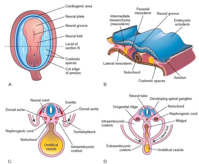
2 Development of the upper urinary system It is developed from the intraembryonic intermediate mesoderm. - After folding of the embryo, this mesoderm lies behind the intraembryonic coelom on each side of the descending aorta. - The kidney development passes in three successive stages : 1. Pronephros. 2. Mesonephros. 3. Metanephros. Dr Ahmed Salman
3
4
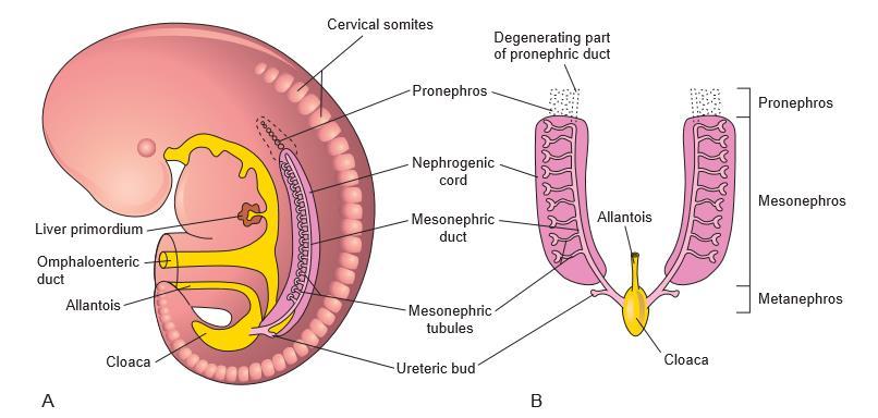
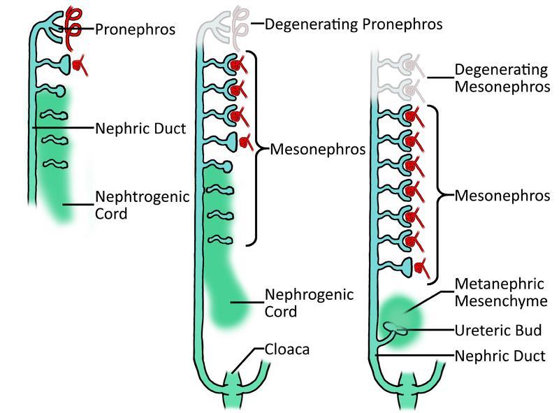
5 The Pronephros It develops from the intermediate mesoderm of the cervical region the embryo at 4th week - The intermediate mesoderm is segmented into 7 cell clusters called nephrotomes. - The nephrotomes elongate and become canalized to form Pronephros tubules. - Each tubule has two ends: Medial end receives a capillary plexus from the adjacent aorta, forming an internal glomerulus Lateral end grows in a caudal direction and unites with the succeed tubules to form the pronephric duct, which descends to open in cloaca. Fate of the pronephros: - The pronephric tubules degenerate. - The pronephric duct is transformed into the mesonephric duct, serves the second kidney
6 The mesonephros It develops from the intermediate mesoderm of the thoracic and upper lumbar regions. Development: - The intermediate mesoderm is segmented into about 70 clusters. - These clusters elongate and become canalized to form S- shaped mesonephric tubules. - Each tubule has two ends: Medial end is invaginated by a capillary plexus to form a primitive glomerulus. Around the glomerulus the tubules form Bowman s capsule, and together these structures constitute a renal corpuscle Lateral end joins the mesonephric duct or wolffian duct
7
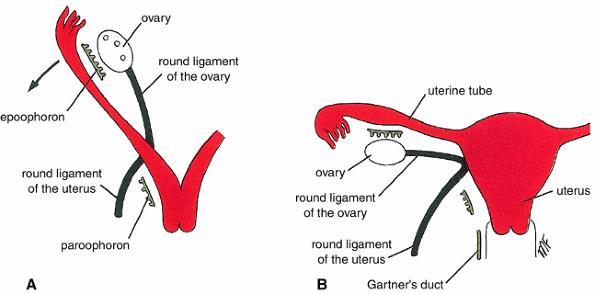
8 Fate of the mesonephros: - The mesonephros degenerates and is replaced by the metanephros (permanent kidney). - However, parts of the mesonephors persist to form urogenital structure which differ in male and female. 1. The mesonephric tubules form : Male Efferent ductules of the testis Paradidymis Female Epoophorn paroophoron
9 2. Mesonephric ducts In the male form Genital structures - Body and tail of epididymis and its appendix - Vas deferens - Seminal vesicle. - Ejaculatory duct Urinary structures - Ureteric bud and its derivatives (ureter, renal pelvis, calyces and collecting tubules) - Trigone of the urinary bladder - Posterior wall of the supra collicular part of the prostatic urethra 2. Mesonephric ducts In the Female form - Duct of epoophorn. Genital structures Urinary structures - Ureteric bud and its derivatives (ureter, - Gartner's duct. renal pelvis, calyces and collecting tubules). - Trigone of the urinary bladder. - The whole dorsal wall of the female urethra.
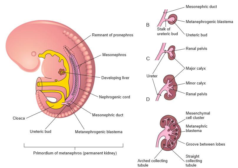
10 Metanephros Site: in the sacral region at 5 th month of development It develops from two mesodermal structures, ureteric bud and Metanephric cap. A. The ureteric bud. It arises as a diverticulum from the lower part of the mesonephric duct near the cloaca. The bud gives rise to the collecting system of urine: Ureter from its stem. Renal pelvis from its cranial end which divides to form 2 calyces which in turn divide to form 7-11 minor calyces. Collecting tubules.
11 B. Metanephric cap - It is the caudal part of the intermediate mesoderm. - This mesoderm (is induced by the ureteric bud) to divide into thousands of cell clusters which lie close to the collecting tubules of the ureteric bud. - The cell clusters elongate and become canalized to form renal vesicle which give rise to nephrons, which are the active excretory units of the kidney. - Each nephron gives rise to: Bowman's capsule which receives an afferent arteriole to form glomerulus. The capsule and the glomerulus constitute together a renal corpuscle. Proximal convoluted tubule. Loop of Henle. Distal convoluted tubule, which joins a nearby collecting tubule to form a complete functional unit.
12
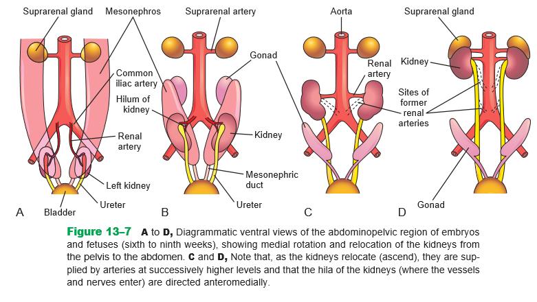
13 Postnatal changes in the metanephros : 1. Change in shape: the fetal kidney is lobulated with irregular surface. Lobulation disappears during early infancy. 2. Change in position and blood supply: At first it is a pelvic organ, which receives its blood supply from the median sacral artery. As it ascends into the abdomen, it changes its blood supply to be derived from the common iliac artery and finally from the abdominal aorta. 3. Change in direction: originally, the hilum of the kidney is directed anteriorly but with its ascent, the kidneys rotate medially almost 90 degrees the hilum rotates to face medially.
14
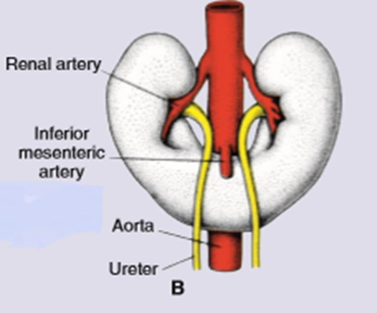
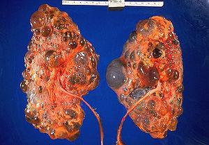
15 Congenital anomalies 1- Renal agenesis with absence of one or the two kidneys respectively. In this case the ureteric bud fails to induce the metanephric cap to divide 2. Congenital polycystic kidney Cysts form from collecting ducts kidney shows many cysts filled with urine 3. Ectopic kidney, in which case it fails to ascend. 4. Horse - shoe kidney Two kidneys are fused at their lower poles. Ascent of the kidneys is prevented by the origin of the inferior mesenteric artery.
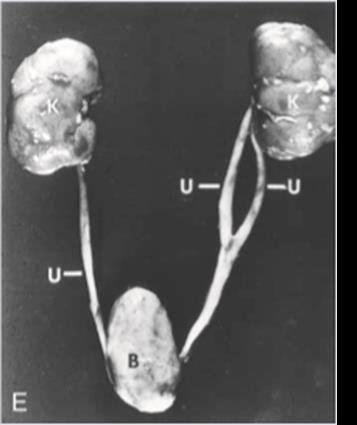
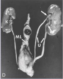
16 5. Accessory renal artery: an additional artery may enter the upper or lower pole of the kidney. 6. Bifid ureter is due to the bifurcation of the upper end of the ureteric bud with double renal pelvis. 7.Double ureter duplication of the urinary tract Occurs when the ureteric bud prematurely divides before penetrating the metanephric cup Results in either a double kidney or duplicated ureter and renal pelvis


17 Bifid ureter Horseshoe kidney duplication of the urinary tract Congenital polycystic kidney Ectopic kidney
18 Development of the urinary bladder Dr Ahmed Salman
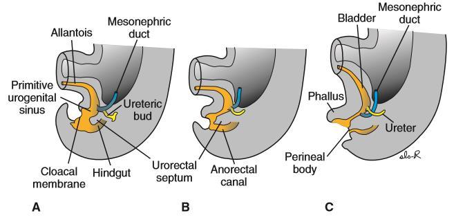
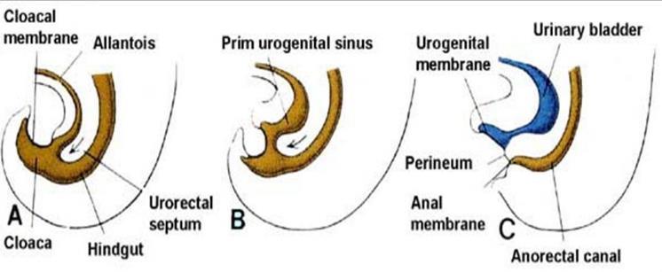
19 A. Development of the cloaca: - The cloaca is a dilatation lined by endoderm at the terminal part of the Hindgut. - The cloaca is Ventrally it is continuous with the allantois. Its sides receive the mesonephric ducts. Caudally it is closed by cloacal membrane. - A mesodermal urorectal septum descends between the allantois and hindgut to reach the cloacal membrane.
20 The cloaca is divided into two parts: Ventral part called the primitive urogenital sinus, which is continuous with the allantois and still receives the right and left mesonephric ducts. Dorsal part called anorectal canal, which is continuous with the hindgut and gives rise to the rectum and the upper part of the anal canal. the cloacal membrane is also divided into two parts. Ventral part called the urogenital membrane closes the caudal end of tie primitive urogenital sinus. Dorsal part called the anal membrane closes the caudal end of the anorectal canal.
21
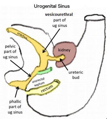
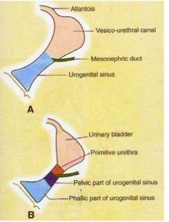
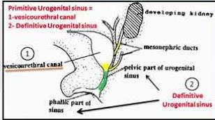
22 A-Urogenital sinus is subdivided by the openings of mesonephric ducts into two parts. Cranial part (the vesico-urethral canal), whose apex is continuous with the allantois. Caudal part called the definitive urogenital sinus, which is further subdivided, into three parts: vesical, pelvic and phallic parts. B- Allantois - It constricts to form a fibrous cord called the urachus that is continuous with the apex of the urinary bladder. - After birth, the urachus is transformed into the median umbilical ligament. C- Caudal parts of the mesonephric ducts - Below the ureteric buds, the caudal parts of the mesonephric ducts are absorbed into the wall of the urinary bladder forming its trigone.
23
 is formed by the lower absorbed parts of the mesonephric ducts.")
24 The urinary bladder develops from : 1. Its major part develops from the vesical part of the primitive urogenital snus (endodermal). 2. The trigone (mesodermal) is formed by the lower absorbed parts of the mesonephric ducts. 3- The coats of the urinary bladder are derived from the splanchnic mesoderm.


25 Congenital anomalies 1. Ectopia vesicae in which the mucosa of the posterior wall of the bladder is exposed to the outside due to defective formation of the infraumbilical of the anterior abdominal wall. It is usually associated with Epispadias.

26 2. Anomalies of the urachus Patent urachus Communication between the bladder and umbilicus through a urachus Urachal cyst :a fluid-filled dilatation of the mid urachus Urachal sinus :blind focal dilatation of the umbilical end of the urachus
27 Dr Ahmed Salman Development of the urethra
28 A. Male urethra 1. Prostatic urethra. - It is divided by the seminal colliculus into: Supracollicular part develops from the vesical part (endodermal) of the primitive urogenital sinus except its dorsal wall which develops from the absorbed lower parts of the mesonephric ducts (mesodermal). Infracollicular part develops from the pelvic part of the primitive urogenital sinus. 2. Membranous urethra: develops also from the pelvic part of the primitive urogenital sinus.
29 3. Penile (spongy) urethra: Develops from the phallic part of the primitive urogenital sinus (due to fusion of the two urethral folds) except its terminal part within the glans penis, which develops from an ectodermal ingrowths. The glandular plate becomes canalized to form the navicular fossa N.B. The male urethra develops from endoderm except two parts. The dorsal wall of the supracollicular part of the prostatic urethra (mesodermal). The terminal part within the glans penis (ectodermal).
30
31 B. Female urethra It develops from the vesical part of the primitive urogenital sinus (endodermal) except its dorsal wall, which is mesodermal in origin, being derived from the absorbed lower parts of the mesonephric ducts.
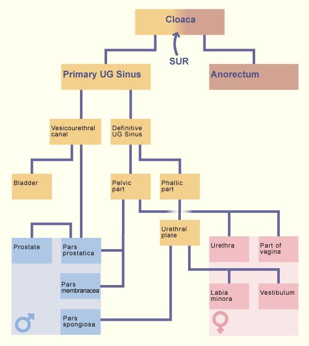
32 Derivatives of the three parts of the primitive urogenital sinus Male Female 1.Vesical part - The urinary bladder except its trigone, which is mesodermal in origin. - The supracollicular part of the prostatic - The urinary bladder except its trigone, which is mesodermal in origin. urethra except its dorsal wall which is - The whole urethra except its mesodermal in origin dorsal wall, which is mesodermal in origin 2.Pelvic part - The infracollicular part of the prostatic - urethra. The pelvic and the phallic parts form: 3.Phallic part - Membranous urethra - The penile urethra except its terminal part in the glans penis, which is ectodermal in origin. a) Lower 2/3 of the vagina. b) Vestibule of the vagina
33
34 Development of the gonads Dr Ahmed Salman
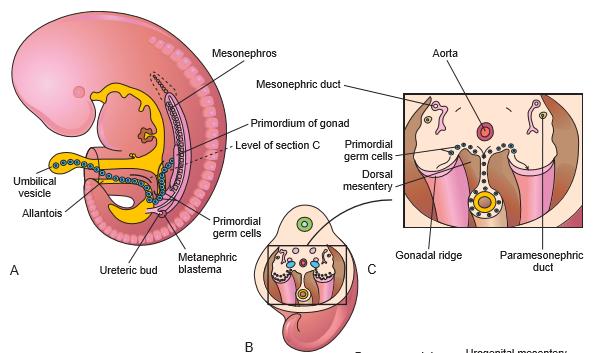
35 The gonads develop form three sources (the first two are mesodermal, the third one is endodermal. 1.Proliferating coelomic epithelium on the medial side of the mesonephros. 2. Adjacent mesenchyme dorsal to the proliferating coelomic epithelium. 3. Primordial germ cells (endodermal), which develop in the wall of the yolk sac and migrate along the dorsal mesentery to reach the developing gonad.
36 The indifferent stage of the developing gonads - The coelomic epithelium (on either side of the aorta) proliferates and becomes multi layered and forms a longitudinal projection into the coelomic cavity called the genital ridge. - The genital ridge forms a number of epithelial cords called the primary sex cords that invade the underlying mesenchyme, which separate the cords from each other. - Up to the 6th or 7th week, the developing gonad cannot be differentiated into testis or ovary.
37

38 Development of the testis and its descent Under the effect of the testis determining factor (T.D.F) present on the short arm of Y - chromosome, the undifferentiated gonad is switched to form a testis. 1. The coelomic epithelium. - The primary sex cords elongate to form testis cords (future seminiferous tubules) which undergo three important events : Ventrally, they lose contact with the surface epithelium by the developing tunica albuginae. Dorsally, they communicate with each other to form rete testis. Internally, they are invaded by the primitive germ cells.
, which affects the development of the genital ducts. Primitive germ cells (endodermal) from the wall of the yolk sac, they give rise to spermatogonia 2-The subjacent mesenchyme.")
39 The testis cords become lined by two types of cells: Sertoli supporting cells (mesoderml) from the coelomic epithelium. They synthesize mullarian inhibitory factor (M.I.F), which affects the development of the genital ducts. Primitive germ cells (endodermal) from the wall of the yolk sac, they give rise to spermatogonia 2-The subjacent mesenchyme. It forms tunica albuginae that surrounds the testis. It forms the interstitial cells of Leydig, which secrete testosterone. 3. The primitive germ cells. They reach the genital ridge and give rise to spermatogonia, which (at puberty) differentiate to form spermatozoa.


40 Descent of the testis : - The testis develops in the posterior abdominal wall opposite the 2 nd lumbar vertebra. Here, it receives its testicular artery from the abdominal aorta. - The genital mesentery of the testis is divided into three parts: Its cranial part forms the suspensory ligament of the testis. It soon degenerates. Its middle part forms the mesorchium, which forms the site at which blood vessels, and lymphatics enter and leave the testis. Its caudal part is transformed into a fibromuscular structure called gubernaculum of the testis. - It extends between the caudal end of the testis to the developing scrotum. - It is aiding its descent into the scrotum.


41 The testis undergoes two steps of descent: 1.Internal descent: occurs from the 4 th to the 6 th month of development. The testis descends into the iliac fossa close to the deep inguinal ring. 2.External descent: occurs from the 7 th to the 9 th month of development At 7th month, it traverses the deep inguinal ring. At 8th month, it traverses the inguinal canal. At 9th month, it begins to traverse the superficial inguinal ring. -

42 Before descent of the testis, a peritoneal diverticulum called processus vaginalis creates and traverses the inguinal canal down to the scrotum. After descent of the testis the tunica vaginalis is divided into three parts: Proximal part forms the vestige of processus vaginalis at the deep inguinal ring. Intermediate part is obliterated. Distal part (in the scrotum) persists and forms the tunica vaginalis Factors helping descent of the testis: 1. Shortening of the gubernaculum. 2. Hormones as androgens and gonadotrophins. 3. Increased intra - abdominal pressure.
43 Congenital anomalies: 1.Cryptorchism (undescended testis):in which the testis may remain in the iliac fossa or in any part of the inguinal canal. Undescended testis is susceptible to damage of the process of spermatogenesis and occurrence of malignancy. 2. Ectopic testis (maldescended testis) in which, the testis descends in the inguinal canal but is located outside the scrotum at root of penis or in the upper part in the front of the thigh. 3.Congenital oblique inguinal hernia, in which a loop of intestine descends via unobliterated tunica vaginalis.


44 4-Hydrocele : the abdominal end of the processus vaginalis remains open but is too small to permit herniation of intestine. Peritoneal fluid passes into the patent processus vaginalis and forms a scrotal hydrocele. If only the middle part of the processus vaginalis remains open, fluid may accumulate and give rise to a hydrocele of the spermatic cord

45 Development of the ovary - In the absence of T.D.F, the undifferentiated gonad is switched on to form an ovary. 1. Coelomic epithelium: - The primary sex cords invade into the subjacent mesenchyme to form medullary sex cords. - It replaced by fibromuscular stroma, forming the medulla of the ovary. - The coelomic epithelium proliferates to form a second generation of sex cords called the secondary (cortical) sex cords, which remain near the coelomic epithelium, forming the cortex of the ovary. - The cortical sex cords break down to form cell clusters which form perimordial ovarian follicles
46 2. Subjacent mesenchyme: it forms: The stroma of the ovary. Very thin tunica albuginae, which intervenes between the ovary and the surface epithelium. 3. Primitive germ cells: - They invade the primordial follicles and proliferate by mitosis to form Primary oocytes. - At 12 lh week of the intrauterine life, the primary oocytes enter the first meiotic division and are arrested (at 20th week) in its prophase till puberty. Descent of the ovary : - The ovary developed (like the testis) in the posterior abdominal wall opposite at 2nd lumbar vertebra, where it is suspended by a genital mesentery. - It reaches the greater pelvis at 3 rd month of gestation - Then reach the lesser pelvis shortly after birth
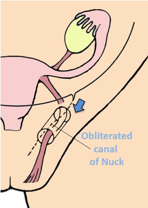
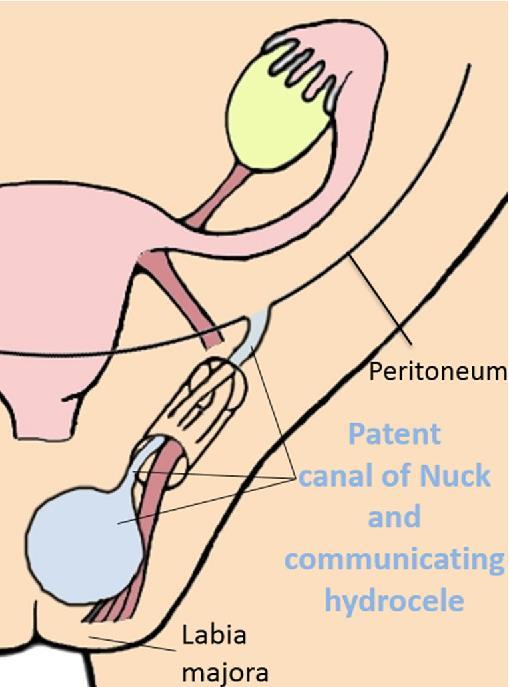
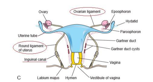
47 The genital mesentery of the ovary is divided into three parts: Cranial part forms the suspensory ligament of the ovary Middle part forms the mesovarium. Caudal part is transformed into the gubernaculum of the ovary, which extends between the lower end of the ovary and the developing labium majora. - The middle of: the gubernaculum is attached to the lateral angle of the developing uterus and thus gives rise to two ligaments. Ligament of the ovary, between the ovary and uterus. Round ligament of the uterus, between the uterus and labium majora. Persistence of small processus vaginalis, gives rise to canal of Nuck. Congenital anomalies 1. Ovarian agenesis. 2. Congenital inguinal hernia The ovary may undergo external descent via the inguinal canal when the gubernaculum is not attached to the angle of the developing uterus may occur in a persistent canal of Nuck.


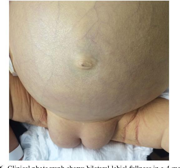
48 bilateral labial fullness in a 4-monthold girl
49
50
51 Dr Ahmed Salman Development of the genital ducts
52 - In either the male or female, there are two genital ducts on each side: Mesonephric (Wollfian) duct and a laterally located paramesonephric (Mullerian) duct All are mesodermal in origin. - In the male, under the effect of antimullerian factor (A.M.F) synthesized by Sertoli cells of the testis, mesonephric ducts will develop. - Paramesonephric ducts will regress, leaving vestigial structures. - In the female, in the absence of A.M.F, paramesonephric ducts will develop. - The mesonephric ducts will regress leaving vestigial structures.
53 1-Indifferent stage of genital ducts - Up to the 6 th week of development, male or female embryos have two pairs of genital ducts. Two (right and left) mesonephric ducts. Two (right and left) paramesonephric ducts. - The paramesonephric duct develops in the coelomic epithelium lateral to the cranial end of the mesonephric duct and continues to grow caudally lateral to that duct - Then crosses ventral to it and then descends medial to it. - The upper end of each paramesonephric duct opens by an abdominal ostium into the coelomic cavity (future peritoneal cavity). - Their lower parts fuse to form a Y - shaped uterovaginal canal project into dorsal wall of the urogenital sinus to form Mullerian tubercle.
54 A. Paramesonepbric ducts in the male - They regress under the effect of M.I.F synthesized by Sertoli cells of the testis. Its cranial end forms appendix of the testis. Their caudal fused parts form utricle inside the prostate. Mullarien tubercle gives rise to seminal colliculus in the posterior wall of the prostatic urethra
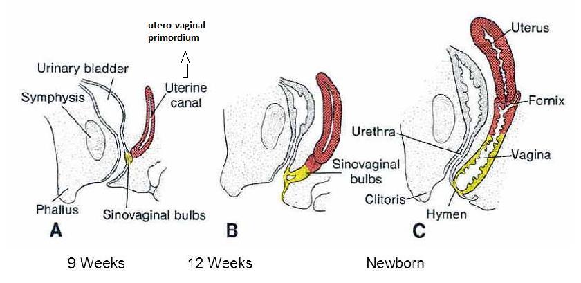
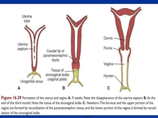
55 A. Paramesonephric ducts in the female - The cranial and middle parts of each duct form the uterine tube, which opens in the coelomic cavity close to the ovary - Their caudal vertical parts (utrovaginal canal) form the uterus and upper 1/3 of the vagina (mesodermal}.! The lower 2/3 of the vagina are endodermal in origin, Formed from the dorsal wall of the definitive urogenital sinus which is induced to form two solid invaginations called sino-vaginal bulbs which unite to form a single vaginal plate. The vaginal plate is canalized to form the lower 2/3 of the vagina. At the lower end of the vagina, a thin membrane remains to form the hymen that lies at the original site of the Mullarien tubercle. - So, the vagina is formed as follows: Upper 1/3 (mesodemral) develops from the lower part of the utrovaginal canal. Lower 2/3 (endodermal)develops from the vaginal plate derived from the dorsal wall of the definitive urogenital sinus.
56
57

58 As the middle parts of the paramesonephric ducts cross medially to reach the midline, they drag with them transverse folds of peritoneum, which will form the broad ligaments of the uterus
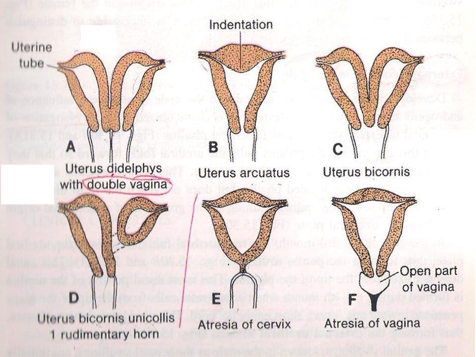
59 Congenital anomalies 1. Bipartite uterus in which the uterus is partially subdivided into two parts by a small septum. 2. Septate uterus in which the uterus is completely subdivided into two parts by a large median septum. 3. Uterus bicornis unicollis (collis = cervix), the uterus has two horns, which open into a single vagina. 4. Uterus bicornis bicollis, the uterus is divided completely into two horns and each has a separate cervix. 5. Uterus unicornis unicollis, the uterus is formed only of a single horn and the other horn is rudimentary. 6. Septate uterus and septate vagina. 7. Septate vagina, in which only the vagina is divided into two parts by a median septum. 8. Atresia of the vagina due to failure of canalization of the vaginal plate. 9. Imperforate hymen. 10. Congenital rectovaginal fistula due to incomplete development of urorectal septum.
60
61 Development of external genitalla Dr Ahmed Salman

62 Indifferent stage of the external genitalla From the 4 th to the 7 th week of development, the external genitalia can not be differentiated into male or female. In the 4 th week, the mesenchyme around the urogenital membrane proliferates to produce five elevations, all are covered by ectoderm. 1- A single genital tubercle at the cranial end of the urogenital membrane elongates to form the phallus. 2-Right and left genital (urethral) folds on the sides of the urogenital membrane. 3-Right and left genital (labio - scrotal) swellings on the sides of the genital folds.
63 Development of the male external genitalia Due to the secretion of testosterone by the developing testis, the undifferentiated external genitalia are switched to form male type external genitalia. 1. Genital tubercle: it elongates to form the phallus, whose mesenchyme forms two corpora cavernosa. 2. The genital (urethral folds): - Rupture of the urogenital membrane - A longitudinal urethral groove appears on the ventral aspect of the developing penis. The sides of that groove are bordered by the urethral folds. - The floor and sides of the urethral groove become lined with an endodermal urethral plate. - The edges of that plate are continuous with those of the urethral folds. - The edges of the endodermal urethral plate are fused with each other to form the penile urethra except its terminal part within the glands penis.
64 The edges of the mesenchyme within the urethral folds fuse around the penile urethra and form the single corpus spongiosum. The two genital swellings: fuse in the midline to form the scrotum


65 Development of the penis: Its dorsal and lateral aspects are formed by the mesenchyme of the phallus, whose mesenchyme forms the two corpora cavernosa. Its ventral aspect is formed by the mesenchyme of the urethral folds, whose mesenchyme forms the single corpus spongiosum. Congenital anomalies: 1. Hypospadius: the urethral orifice is present in the ventral aspect of the penis due to incomplete fusion of the two urethral folds. 2. Epispadius: an abnormal orifice is present on the dorsal aspect of the penis, usually associated with ectopia vesicae.
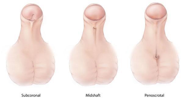
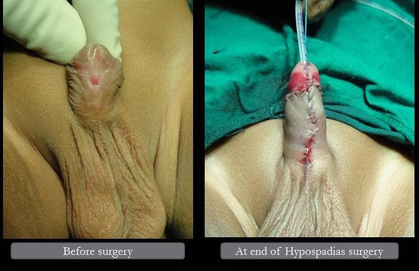
66 Hypospadius
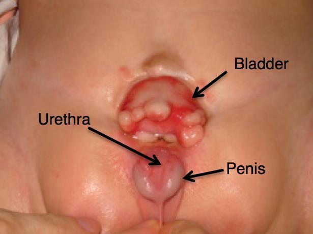
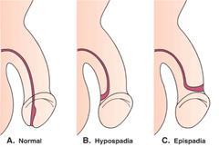
67 Epispadius
. 2.")
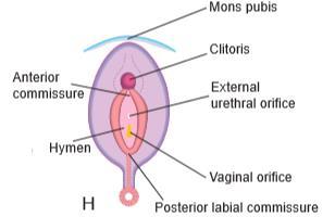
68 Development of the female external genttalla - Under the affects of the maternal and placental oestrogens, the external genitalia are switched to Form Female type of genitalia. 1. Genital tubercle: it elongates to form the clitoris, whose mesenchymef Forms its two corpora cavernosa (note that, the clitoris has no corpus spongiosum). 2. Genital (urethral folds): they remain separate to form the two labia minora. 3. Genital swellings: they remain separate to form the two labia majora. 4. The vaginal vestibule: is formed when primitive urogenital sinus are shortened to form the vestibule between the two labia minora.
69
Development of Urinary System
 Jordan University Faculty Of Medicine Development of Urinary System DR. AHMED SALMAN ASSISTANT PROFESSOR OF ANATOMY & EMBRYOLOGY Development of The kidney Dr Ahmed Salman Development of the upper urinary
Urinary system development. Male ( ) and Female ( ) Reproductive Systems Development
 and Female ( ) Reproductive Systems Development") Urinary system development Male ( ) and Female ( ) Reproductive Systems Development Urogenital system develops from mesodermal uro-genital ridge (intermediate mesoderm) development of male and female genital
Urinary system development Male ( ) and Female ( ) Reproductive Systems Development Urogenital system develops from mesodermal uro-genital ridge (intermediate mesoderm) development of male and female genital
Development of the Genital System
 Development of the Genital System Professor Alfred Cuschieri Department of Anatomy University of Malta The mesonephros develops primitive nephrotomes draining into a mesonephric duct nephrotome mesonephric
Development of the urogenital system
 Development of the urogenital system Location of the pronephros, mesonephros and metanephros Differentiation of the intermedierm mesoderm into nephrotome and mesonephric tubules Connection between aorta
under its influence, male development occurs; in its absence, female development is established.
 Sex differentiation is a complex process that involves many genes, including some that are autosomal. The key to sexual dimorphism is the Y chromosome, which contains the testis determining gene called
Urogenital System. PUMC Dept. of Anat. Hist. & Embry. 钱晓菁 XIAO-JING QIAN Dept. of Anatomy, Histology & Embryology Peking Union Medical College
 Urogenital System 钱晓菁 XIAO-JING QIAN Dept. of Anatomy, Histology & Embryology Peking Union Medical College intermediate mesoderm urogenital ridge mesonephric ridge genital ridge I. Urinary System 1. kidney
Development of the urinary system
 Development of the urinary system WSO School of Biomedical Sciences, University of Hong Kong. 3 sets of kidneys developing in succession (temporally and spatially) : Pronephros ] Mesonephros ]- Intermediate
Development of the Urinary System
 Development of the Urinary System Lecture Objectives Understand the development of the kidney and related organs of the urinary system. Define the pronephrons, mesonephrons and metanephrons. Understand
Development of the Urinary System. 3 Distinct Embryonic Kidney Structures
 Development of the Urinary System Excretory portion of urinary system derived from intermediate mesoderm Week 4: 1 st nephrons/renal corpuscles form Nephrotomes form and develop hollow lumens to form nephric
W.S. O University of Hong Kong
 W.S. O University of Hong Kong Development of the Genital System 1. Sexual differentiation 2. Differentiation of the gonads a. Germ cells extragonadal in origin b. Genital ridge intermediate mesoderm consisting
10. Development of genital system. Gonads. Genital ducts. External genitalia.
 10. Development of genital system. Gonads. Genital ducts. External genitalia. Gonads, genital ducts and the external genital organs initially pass through an indifferent period of development, which is
Urinary System. J. H. Lue. intermediate mesoderm cloaca coelomic epithelium
 Urinary System J. H. Lue Primordium: intermediate mesoderm cloaca coelomic epithelium 1 3w(18d) 3 w 4w (24d) 4w (26d) 2 Intermediate mesoderm 3 Intermediate mesoderm intermediate mesoderm urogenital ridge
Urogenital system - Development. Aleš Hampl
 Urogenital system - Development Aleš Hampl cloaca Urogenital system Overall picture Urogenital system Reminder Urogenital system Intermediate mesoderm Urogenital system Early forms of kidneys - Pronephros
Sexual differentiation is sequential process:
 Genital lsystem J. H. Lue Sexual differentiation is sequential process:.genetic (chromosomal) sex -- determined at fertilization.gonad sex -- is differentiated after 7th week.phenotypic sex -- under normal
Midgut. Over its entire length the midgut is supplied by the superior mesenteric artery
 Gi Embryology 3 Midgut the midgut is suspended from the dorsal abdominal wall by a short mesentery and communicates with the yolk sac by way of the vitelline duct or yolk stalk Over its entire length the
Embryology Relevant to Ultrasound Imaging of the Male Genitalia
 Embryology Relevant to Ultrasound Imaging of the Male Genitalia Gideon Richards and Bruce R. Gilbert A basic understanding of the embryologic development of the male genitalia and the male genital blood
Urinary System. ectoderm. notochord. mesonephric tubules. Nephrogenic Cord (left)
") Urinary System NOTE: Urine proion requires an increased capillary surface area (glomeruli), epithelial s to collect plasma filtrate and extract desirable constituents, and a system to convey urine away
Urinary System NOTE: Urine proion requires an increased capillary surface area (glomeruli), epithelial s to collect plasma filtrate and extract desirable constituents, and a system to convey urine away
The functional anatomy of the urinary system. Human Anatomy Department Dr. Anastasia Bendelic
 The functional anatomy of the urinary system Human Anatomy Department Dr. Anastasia Bendelic Plan Development of the kidneys and their abnormalities Development of the urinary ways and their abnormalities
Human Anatomy Unit 3 REPRODUCTIVE SYSTEM
 Human Anatomy Unit 3 REPRODUCTIVE SYSTEM In Anatomy Today Male Reproductive System Gonads = testes primary organ responsible for sperm production development/maintenan ce of secondary sex characteristics
Embryology of the Female Reproductive Tract
 Embryology of the Female Reproductive Tract Andrew Healey Contents 1 Introduction... 21 2 Embryology of the Female Genitourinary Tract... 22 2.1 Development of the Gonads... 22 2.2 Relationship Between
Embryology /organogenesis/ Week 4 Development and teratology of reproductive system.
 Embryology /organogenesis/ Week 4 Development and teratology of reproductive system. Male or female sex is determined by spermatozoon Y in the moment of fertilization SRY gene, on the short arm of the
Biology 224 Human Anatomy and Physiology II Week 9; Lecture 2; Wednesday Stuart Sumida. Development and Structure, of the Reproductive System
 Biology 224 Human Anatomy and Physiology II Week 9; Lecture 2; Wednesday Stuart Sumida Development and Structure, of the Reproductive System Don t forget the relationships of the structures of the layers
Development of the Urinary System
 Development of the Urinary System ## every things is written in this sheet so u don t have to go back to the slide. ## I write everything and I hope everything will be clear,, if some note doesn t clear
Pelvis MCQs. Block 1. B. Reproductive organs. C. The liver. D. Urinary bladder. 1. The pelvic diaphragm includes the following muscles: E.
 Pelvis MCQs Block 1 1. The pelvic diaphragm includes the following muscles: A. The obturator internus B. The levator ani C. The coccygeus D. The external urethral sphincter E. The internal urethral sphincter
DEVELOPMENT OF THE ADRENAL GLAND; TESTES AND MESONEPHRIC DUCT. Reading Assignment: The Developing Human, Clinically Oriented Embryology pp
 Developmental Anatomy Steven M. Hill, Ph.D. 9/16/13 DEVELOPMENT OF THE ADRENAL GLAND; TESTES AND MESONEPHRIC DUCT Reading Assignment: The Developing Human, Clinically Oriented Embryology pp. 264-273. Objectives:
Obstetrics Content Outline Obstetrics - Fetal Abnormalities
 Obstetrics Content Outline Obstetrics - Fetal Abnormalities Effective February 2007 10 16% renal agenesis complete absence of the kidneys occurs when ureteric buds fail to develop Or degenerate before
Genitourinary System Imaging- Based Overview of Anatomy and Embryology. Sameer Ahmed, MS IV 9/8/11
 Genitourinary System Imaging- Based Overview of Anatomy and Embryology Sameer Ahmed, MS IV 9/8/11 ObjecDves Review very high- yield concepts Anatomy test + USMLE Step 1 GU Embryology How it relates directly
Lab #9: Kidney: Gross Anatomy & Histology
 Name Date Lab #9: Kidney: Gross Anatomy & Histology Lab #10: Male Reproductive System: Human Models & Histology Lab #11: Female Reproductive System: Human Models & Histology Stuff to Know Dr. L. Bacha
DISSECTION 8: URINARY AND REPRODUCTIVE SYSTEMS
 8546d_c01_1-42 6/25/02 4:32 PM Page 38 mac48 Mac 48: 420_kec: 38 Cat Dissection DISSECTION 8: URINARY AND REPRODUCTIVE SYSTEMS Typically, the urinary and reproductive systems are studied together, because
Development of the Digestive System. W.S. O The University of Hong Kong
 Development of the Digestive System W.S. O The University of Hong Kong Plan for the GI system Then GI system in the abdomen first develops as a tube suspended by dorsal and ventral mesenteries. Blood
MALE REPRODUCTIVE SYSTEM
 MALE REPRODUCTIVE SYSTEM 1. The male reproductive system is made up of the following structures, EXCEPT: a. prostate; b. testicle; c. spermatic ducts; d. vestibular bulbs; e. seminal vesicles. 2.The testicle:
PELVIS II: FUNCTION TABOOS (THE VISCERA) Defecation Urination Ejaculation Conception
 Defecation Urination Ejaculation Conception") PELVIS II: FUNCTION TABOOS (THE VISCERA) Defecation Urination Ejaculation Conception REVIEW OF PELVIS I Pelvic brim, inlet Pelvic outlet True pelvis-- --viscera Tilt forward Mid-sagital views-- --how the
PELVIS II: FUNCTION TABOOS (THE VISCERA) Defecation Urination Ejaculation Conception REVIEW OF PELVIS I Pelvic brim, inlet Pelvic outlet True pelvis-- --viscera Tilt forward Mid-sagital views-- --how the
Animal Science 434 Reproductive Physiology"
 Animal Science 434 Reproductive Physiology" Embryogenesis of the Pituitary and Sexual Development: Part A Development of the Pituitary Gland" Infundibulum" Brain" Rathke s Pouch" Stomodeum" Germ Cell Migration"
The Urinary System Pearson Education, Inc.
 26 The Urinary System Introduction The urinary system does more than just get rid of liquid waste. It also: Regulates plasma ion concentrations Regulates blood volume and blood pressure Stabilizes blood
Development of the female Reproductive System. Dr. Susheela Rani
 Development of the female Reproductive System Dr. Susheela Rani Genital System Gonads Internal genitals External genitals Determining sex chronology of events Genetic sex Determined at fertilization Gonadal
Development of the Digestive System. W.S. O School of Biomedical Sciences, University of Hong Kong.
 Development of the Digestive System W.S. O School of Biomedical Sciences, University of Hong Kong. Organization of the GI tract: Foregut (abdominal part) supplied by coeliac trunk; derivatives include
Pelvis Perineum MCQs. Block 1.1. A. Urinary bladder. B. Rectum. C. Reproductive organs. D. The thigh
 Pelvis Perineum MCQs Block 1.1 1. The pelvic diaphragm includes the following muscles: A. The coccygeus B. The levator ani C. The external urethral sphincter D. The internal urethral sphincter E. The obturator
Sexual differentiation:
 Abnormal Development of Female Genitalia Dr. Maryam Fetal development of gonads, external genitalia, Mullerian ducts and Wolffian ducts can be disrupted at a variety of points, leading to a wide range
FORMS OF EMBRYONIC PRIMORDIA
 FORMS OF EMBRYONIC PRIMORDIA BY PROF. ANTHONY OBIOMA NWAOPARA UNIVERSITY OF MEDICAL SCIENCES ONDO CITY, ONDO STATE LEARNING OBJECTIVES To recognise the different forms of embryonic primordia. To recognise
M. Al-Mohtaseb. Tala Saleh. Faisal Nimri
 4 5 M. Al-Mohtaseb Tala Saleh Faisal Nimri Inguinal Hernia - An abdominal hernia is the protrusion of part of the abdominal content beyond the normal confines of the abdominal wall through weak points
Animal Science 434 Reproductive Physiology
 Animal Science 434 Reproductive Physiology Development of the Pituitary Gland Lec 5: Embryogenesis of the Pituitary and Sexual Development Stomodeum Brain Infundibulum Rathke s Pouch Germ Cell Migration
Chapter 26: Reproductive Systems. Male 11/29/2015. Male reproductive system is composed of... BIO 218 Fall Gonads (testes)
") Chapter 26: Reproductive Systems BIO 218 Fall 2015 Male Male reproductive system is composed of... Gonads (testes) Duct system (epididymis, ductus deferens, ejaculatory ducts, urethra) Accessory sex glands
Chapter 26: Reproductive Systems BIO 218 Fall 2015 Male Male reproductive system is composed of... Gonads (testes) Duct system (epididymis, ductus deferens, ejaculatory ducts, urethra) Accessory sex glands
Male Reproductive System. Dr Maan Al-Abbasi PhD, MSc, MBChB, MD
 Male Reproductive System Dr Maan Al-Abbasi PhD, MSc, MBChB, MD Learning Objectives 1. Describe the General Anatomy of the Male Reproductive System 2. Identify the structures that are related to the prostate.
Fig General Structural Features of a kidney
 Chapter 11 Development of the Mesodermal Organs in Vertebrates 11.1. Subdivisions of the Mesoderm There are 4 subdivisions of the mesoderm: 1. The Notochord develops into the centrum of the vertebral column.
Embryology - GIT - Lecture 2
 Embryology - GIT - Lecture 2 Last time we talked about embryology of the GIT. We said that the development of the stomach is accompanied with the development of the duodenum and the pancreas. Also we talked
Primary sex organs (gonads): testes and ovaries. Accessory reproductive organs: ducts, glands, and external genitalia
: testes and ovaries. Accessory reproductive organs: ducts, glands, and external genitalia") Male Reproductive System Primary sex organs (gonads): testes and ovaries Produce sex cells (gametes) Secrete steroid sex hormones Androgens (males) Estrogens and progesterone (females) Accessory reproductive
Male Reproductive System Primary sex organs (gonads): testes and ovaries Produce sex cells (gametes) Secrete steroid sex hormones Androgens (males) Estrogens and progesterone (females) Accessory reproductive
Gross Anatomy of the Urinary System
 Gross Anatomy of the Urinary System Lecture Objectives Overview of the urinary system. Describe the external and internal anatomical structure of the kidney. Describe the anatomical structure of the ureter
- production of two types of gametes -- fused at fertilization to form zygote
 Male reproductive system I. Sexual reproduction -- overview - production of two types of gametes -- fused at fertilization to form zygote - promotes genetic variety among members of a species -- each offspring
Lab Schedule for Rest of Semester
 Laboratory 9 Cat Dissection II Respiratory Urinary/Reproductive Systems Lab Schedule for Rest of Semester Cat dissection labs Dissection II (today) Respiratory (Ex. 57 in Hole) Human Reproductive Systems
Yes, cranially with ovarian, caudally with vaginal. Yes, with uterine artery (collateral circulation between abdominal +pelvic source)
") Blood supply to internal female genitalia: uterine Internal iliac Sup. large branch: uterus, inf. Small branch: cervix+ sup. Vagina Yes, cranially with ovarian, caudally with vaginal Medially in base of
Blood supply to internal female genitalia: uterine Internal iliac Sup. large branch: uterus, inf. Small branch: cervix+ sup. Vagina Yes, cranially with ovarian, caudally with vaginal Medially in base of
Male Anatomy. testes, genetically determined in mammals - testis releases hormones that then control the development of secondary sex characteristics
 Male Anatomy Male Anatomy Primary Organ testes, genetically determined in mammals - testis releases hormones that then control the development of secondary sex characteristics 1) Secondary Organs internal
Male Reproductive System Dr. Gary Mumaugh
 Male Reproductive System Dr. Gary Mumaugh Reproductive System Basics Primary sex organs (gonads) testes in males, ovaries in females Gonads produce sex cells called gametes (gametes means spouses) and
حسام أبو عوض. -Dr. Mohammad Muhtasib. 1 P a g e
 5 حسام أبو عوض - -Dr. Mohammad Muhtasib 1 P a g e There are two types of inguinal hernia: direct and indirect. Hernia: protrusion of the small intestine or the greater omentum of the intra-abdominal organs
Lab Activity 31. Anatomy of the Urinary System. Portland Community College BI 233
 Lab Activity 31 Anatomy of the Urinary System Portland Community College BI 233 Urinary System Organs Kidneys Urinary bladder: provides a temporary storage reservoir for urine Paired ureters: transport
SUBJECTS 2nd year, 1st semester I. 1. Primitive gut - limits, derivatives 2. Foregut -limits, evolution, derivatives 3. Midgut -limits, evolution,
 SUBJECTS 2nd year, 1st semester I. 1. Primitive gut - limits, derivatives 2. Foregut -limits, evolution, derivatives 3. Midgut -limits, evolution, derivatives 4. Hindgut- limits, evolution, derivatives
Urinary System Chapter 16
 Urinary System Chapter 16 1 Urology- the branch of medicine that treats male and female urinary systems as well as the male reproductive system. Nephrology- the scientific study of the anatomy, physiology,
Normal and Abnormal Development of the Genital Tract. Dr.Raghad Abdul-Halim
 Normal and Abnormal Development of the Genital Tract Dr.Raghad Abdul-Halim objectives: Revision of embryology. Clinical presentation, investigations and clinical significance of most common developmental
أحمد رواجبة- محمود الحربي- أحمد السالمان-
 -6 أحمد رواجبة- محمود الحربي- أحمد السالمان- 1 P a g e The Male Reproductive System The male genital system structures are divided into: Internal structures: 1- Prostate 3-Ejaculatory ducts External structures:
11. SEXUAL DIFFERENTIATION. Germinal cells, gonocytes. Indifferent stage INDIFFERENT STAGE
 11. SEXUAL DIFFERENTIATION INDIFFERENT STAGE Early in pregnancy, (within 10-15 % of the pregnancy s expected length) a genital ridge is formed in the sides of the embryonic tissue, ventral to the mesonephros
Sex Determination and Development of Reproductive Organs
 Sex Determination and Development of Reproductive Organs Sex determination The SRY + gene is necessary and probably sufficient for testis development The earliest sexual difference appears in the gonad
Reproductive System. Where it all begins
 Reproductive System Where it all begins When it comes the reproductive anatomy of my gender, I would rate my knowledge (1 very poor, 10 excellent) When it comes the reproductive anatomy of the opposite
The Male and Female Internal Genitalia. Dr Oluwadiya Kehinde
 The Male and Female Internal Genitalia Dr Oluwadiya Kehinde www.oluwadiya.com Overview The reproductive role of the male is to produce sperm, deliver them to the female Primary sex organs are the gonads
Anatomy Lecture Notes Chapter 24
 primary sex organs = gonads produce gametes secrete hormones that control reproduction secondary sex organs = accessory structures Development and Differentiation A. gonads develop from mesoderm starting
REPRODUCTIVE SYSTEM By Dr.Ahmed Salman
 The University Of Jordan Faculty Of Medicine Anatomy Department REPRODUCTIVE SYSTEM By Dr.Ahmed Salman Assistant Professor of Anatomy &embryology Perineum It is the diamond-shaped lower end of the trunk
To General Embryology Dr: Azza Zaki
 Introduction To General Embryology The Human Development is a continuous process that begins when an ovum from a female is fertilized by a sperm from a male. Cell division, growth and differentiation transform
Formation of Urine: Formation of Urine
 The Urinary outflow tract: monitors and regulates extra-cellular fluids excretes harmful substances in urine, including nitrogenous wastes (urea) returns useful substances to bloodstream maintain balance
URINARY SYSTEM I. Kidneys II. Nephron Unit and Urine Formation
 URINARY SYSTEM I. Kidneys A. Location and Structure 1. Retroperitoneal 2. Between T12 and L3 3. Rt. kidney slightly lower 4. Two bean shaped organs 5. Adrenal gland 6. Internal construction a. Renal cortex
LESSON ASSIGNMENT. After completing this lesson, you should be able to: 8-1. Define urogenital systems.
 LESSON ASSIGNMENT LESSON 8 The Human Urogenital Systems. TEXT ASSIGNMENT Paragraphs 8-1 through 8-16. LESSON OBJECTIVES After completing this lesson, you should be able to: 8-1. Define urogenital systems.
Development of pancreas and Small Intestine. ANATOMY DEPARTMENT DR.SANAA AL-AlSHAARAWY DR.ESSAM Eldin Salama
 Development of pancreas and Small Intestine ANATOMY DEPARTMENT DR.SANAA AL-AlSHAARAWY DR.ESSAM Eldin Salama OBJECTIVES At the end of the lecture, the students should be able to : Describe the development
LECTURE 4. Anatomy of the urinary and genital organs.
 LECTURE 4 Anatomy of the urinary and genital organs. SKELETOTOPY OF THE KIDNEYS Left kidney upper pole of the kidney - ThXI lower pole of the kidney LIII Right kidney is located on the half vertebra
Embryology of the Midgut and Hind gut
 Embryology of the Midgut and Hind gut Prof. Abdulameer Al-Nuaimi E-mail: a.al-nuaimi@sheffield.ac.uk E-mail: abdulameerh@yahoo.com Abdominal organs www.google.co.uk/search? Development of Duodenum The
4.05 Remember the structures of the reproductive system
 4.05 Remember the structures of the reproductive system 4.05 Remember the structures of the reproductive system Essential question What are the structures of the reproductive system? 2 Structures of the
PHYSIOLOGY AND PATHOLOGY OF SEXUAL DIFFERENTIATION
 PHYSIOLOGY AND PATHOLOGY OF SEXUAL DIFFERENTIATION Prof. Dr med. Jolanta Słowikowska-Hilczer Department of Andrology and Reproductive Endocrinology Medical University of Łódź, Poland Sexual determination
ESUR SCROTAL AND PENILE IMAGING WORKING GROUP MULTIMODALITY IMAGING APPROACH TO SCROTAL AND PENILE PATHOLOGIES 2ND ESUR TEACHING COURSE
 ESUR SCROTAL AND PENILE IMAGING WORKING GROUP MULTIMODALITY IMAGING APPROACH TO SCROTAL AND PENILE PATHOLOGIES 2ND ESUR TEACHING COURSE NORMAL ANATOMY OF THE SCROTUM MICHAEL NOMIKOS M.D. F.E.B.U. UROLOGICAL
Objectives: 1. Review male & female reproductive anatomy 2. Gametogenesis & steroidogenesis 3. Reproductive problems
 CH. 15 - REPRODUCTIVE SYSTEM Objectives: 1. Review male & female reproductive anatomy 2. Gametogenesis & steroidogenesis 3. Reproductive problems 3. Male Reproductive anatomy and physiology. Testes = paired
URINARY SYSTEM ANATOMY
 URINARY SYSTEM ANATOMY Adapted from Human Anatomy & Physiology Marieb and Hoehn (9 th ed.) OVERVIEW Metabolism of nutrients by the body produces wastes that must be removed from the body. Although excretory
Copyright 2003 Pearson Education, Inc. publishing as Benjamin Cummings. Dr. Nabil Khouri
 Dr. Nabil Khouri Objectives: General objectives: - to identify the kidney s structures, function and location - to analyze the relationship between microscopic structure and function Specific objectives:
Urinary System. Chapter 17 7/19/11. Introduction
 7/19/11 Chapter 17 Urinary System Introduction A. The urinary system consists of two kidneys that filter the blood, two ureters, a urinary bladder, and a urethra to convey waste substances to the outside.
Urinary Bladder. Prof. Imran Qureshi
 Urinary Bladder Prof. Imran Qureshi Urinary Bladder It develops from the upper end of the urogenital sinus, which is continuous with the allantois. The allantois degenerates and forms a fibrous cord in
The Reproductive System
 Essentials of Human Anatomy & Physiology Elaine N. Marieb Seventh Edition Chapter 16 The Reproductive System Slides 16.1 16.20 Lecture Slides in PowerPoint by Jerry L. Cook The Reproductive System Gonads
Aniko Szabo Hill 1 of 12
 Common Function: produce offsprings endocrine glands: sex hormone production Primary organs or gonads: sex cell or gametes production Secondary or accessory organs: glands nourish gametes ducts transport
The Reproductive System
 16 PART A The Reproductive System PowerPoint Lecture Slide Presentation by Jerry L. Cook, Sam Houston University ESSENTIALS OF HUMAN ANATOMY & PHYSIOLOGY EIGHTH EDITION ELAINE N. MARIEB The Reproductive
Human Sexuality - Ch. 2 Sexual Anatomy (Hock)
") Human Sexuality - Ch. 2 Sexual Anatomy (Hock) penis penile glans corona frenulum penile shaft erection foreskin circumcision corpora cavernosa corpus spongiosum urethra scrotum spermatic cords testicles
Human Sexuality - Ch. 2 Sexual Anatomy (Hock) penis penile glans corona frenulum penile shaft erection foreskin circumcision corpora cavernosa corpus spongiosum urethra scrotum spermatic cords testicles
Inguinal Canal. It is an oblique passage through the lower part of the anterior abdominal wall. Present in both sexes
 Inguinal canal Inguinal Canal It is an oblique passage through the lower part of the anterior abdominal wall Present in both sexes It allows structures to pass to and from the testis to the abdomen in
Histotopography of nephron
 THE URINARY SYSTEM Microscopic structure and development of urinary systém Kidney: nephron structure and function Urinary passages: ureter, urinary bladder, urethra Development of urinary system THE URINARY
4.05 Remember the structures of the reproductive system
 4.05 Remember the structures of the reproductive system Scrub In The external area between the vulva and the anus is the : a. Cervix b. Endometrium c. Perineum d. Vagina What structure connects the testes
Male Reproductive System
 Male Reproductive System Please view our Editing File before studying this lecture to check for any changes. Color Code Important Doctors Notes Notes/Extra explanation Objectives At the end of the lecture,
The Reproductive System
 PowerPoint Lecture Slide Presentation by Patty Bostwick-Taylor, Florence-Darlington Technical College The Reproductive System 16PART A The Reproductive System Gonads primary sex organs Testes in males
Comparative Anatomy Urogenital System. Note Set 11 Chapter 15
 Comparative Anatomy Urogenital System Note Set 11 Chapter 15 Urogenital System Ducts of excretory and reproductive systems are intimately associated Figure 14.1: Embryonic and evolutionary development
18 Urinary system. 19 Male reproductive system. Female reproductive system. Blok 11: Genital and Urinary Tract Diseases
 Blok 11: Genital and Urinary Tract Diseases 18 Urinary System 19 Male Genital System 20 Female Genital System 18 Urinary system You should be able to: 1. Describe the structures and associated functions
Outline. Male Reproductive System Testes and Sperm Hormonal Regulation
 Outline Male Reproductive System Testes and Sperm Hormonal Regulation Female Reproductive System Genital Tract Hormonal Levels Uterine Cycle Fertilization and Pregnancy Control of Reproduction Infertility
STRUCTURAL BASIS OF MEDICAL PRACTICE EXAMINATION 3. October 16, 2015
 STRUCTURAL BASIS OF MEDICAL PRACTICE EXAMINATION 3 October 16, 2015 PART l. Answer in the space provided. (12 pts) 1. Identify the structures. (2 pts) A. B. A B C. D. C D 2. Identify the structures. (2
The Male Reproductive System
 The Male Reproductive System The male reproductive system Testes Genital ducts Accessory sex glands: seminal vesicles prostate bulbourethral glands External genitalia: penis Structure of the Testis Tunica
ANATOMY AND PHYSIOLOGY HOMEWORK CHAPTER 15 AND 16
 ANATOMY AND PHYSIOLOGY HOMEWORK CHAPTER 15 AND 16 Name Identify the following: 1) The ureter is indicated by letter 2) The renal pyramid is indicated by letter 3) The fibrous capsule is indicated by letter
Chapter 22 Reproductive Systems. Male Reproductive Organs. Male Reproductive Organs. Specialized to produce, maintain the male sex cells (sperm)
") Chapter 22 Reproductive Systems Male reproductive organs 1 Male Reproductive Organs posterior view 2 Male Reproductive Organs Specialized to produce, maintain the male sex cells (sperm) Transport these
Chapter 22 Reproductive Systems Male reproductive organs 1 Male Reproductive Organs posterior view 2 Male Reproductive Organs Specialized to produce, maintain the male sex cells (sperm) Transport these
The Repr duct ve System. Function: producing offspring
 The Repr duct ve System Function: producing offspring Anatomy of male reproductive system Location: The reproductive organs are classified as external and internal genitalia. The external genitalia are
Rama Nada. - Ensherah Mokheemer. - Ahmed salman. 1 P a g e
 - 5 - Rama Nada - Ensherah Mokheemer - Ahmed salman 1 P a g e We will continue talking about the urinary bladder The ligaments of the bladder: 1-Median umbilical ligament: Continuous with apex of the bladder
The Scrotum & Testes Prof. Dr. Imran Qureshi
 The Scrotum & Testes Prof. Dr. Imran Qureshi The Scrotum It is a cutaneous pouch of the anterior abdominal wall. Most layers of the abdominal wall are represented in its structure. It contains the testes
Urogenital System Objectives - see handout or website
 Urogenital System Objectives - see handout or website Urogenital System shared ducts due to evolutionary legacy and development Urinary or Excretory System blood filtration and excretion of salts and nitrogenous
Biology 340 Comparative Embryology Lecture 10 Dr. Stuart Sumida. Further Development of the Mesoderm (and Endoderm)
") Biology 340 Comparative Embryology Lecture 10 Dr. Stuart Sumida Further Development of the Mesoderm (and Endoderm) Further Development: Digestive System Foregut, Midgut, Hindgut Heart and Aortic Arches
Biology 340 Comparative Embryology Lecture 10 Dr. Stuart Sumida Further Development of the Mesoderm (and Endoderm) Further Development: Digestive System Foregut, Midgut, Hindgut Heart and Aortic Arches