بسم هللا الرحمن الرحيم. Prof soha Talaat
|
|
|
- Francine Fowler
- 5 years ago
- Views:
Transcription


1 بسم هللا الرحمن الرحيم


2

3 Ovarian tumors The leading indication for gynecologic surgery. Preoperative characterization of complex solid and cystic adnexal masses is crucial for informing patients about possible surgical strategies.

4 Ovarian cancer It is a leading cause of death among women It is the second most common gynecological cancer after cancer cervix It is the fifth most common cancer in women after (lung, breast, colorectal, and pancreatic cancers)
5 Ovarian cancer Approximately 75% 80% of ovarian cancers are diagnosed at stages II IV. Patients with stage IA or IB disease have a 90% 95% 5-year survival rate following surgery alone whereas patients with stage IV disease have a dismal prognosis, with a 10% 5-year survival rate despite aggressive multimodality treatment.
6 Borderline ovarian tumors Comprise up to 15 20% of ovarian epithelial neoplasms. Borderline ovarian tumors are histologically characterized as epithelial tumors with a stratified growth pattern but without destructive stromal invasion. Serous and mucinous neoplasms constitute the majority of borderline tumors and occur mostly in women of reproductive age Acs G. Serous and mucinous borderline (low malignant potential) tumors of the ovary. Am J Clin Pathol2005 ; 123[suppl]:S13 S57
7 Proper management depends on: Clinical examination OECs are notoriously difficult to diagnose in the early stages. Even patients with advanced disease may present with nonspecific abdomino-pelvic symptoms. Laboratory tests Although CA 125 is a useful biomarker, elevated CA-125 levels are seen in only 50% 60% of mucinous or clear cell variants and early-stage cancers. Radiological assessment


8 Pelvic & TVS with CFM The initial diagnostic modality of choice.


9 TVS Colour Doppler & 3D Combining morphologic assessment with TVS with color Doppler features has allowed accurate assessments, the overall vascularity was classified as high, low, or intermediate, rather than determining vascular indices. Improves the detection of morphologic abnormalities indicative of neoplastic ovarian masses. In particular, small papillary projections or focal wall (mural) irregularities.
10 Role of Cross-sectional Imaging Diagnosis, Characterization, and Surveillance Assessment of primary tumours. The pattern of extra-ovarian spread may be (a) extraovarian intrapelvic (stage II). (b) extrapelvic intraabdominal (stage III). (c) Intra-abdominal with intrahepatic parenchymal deposits or extra-abdominal with distant metastasis (stage IV).
11 The emerging role of functional imaging techniques Radioimmunoscintigraphy, PET/CT, diffusionweighted MRI, dynamic contrast-enhanced MRI, and magnetic resonance spectroscopy in staging ovarian cancer and assessing treatment response. Diffusion MRI The combination of functional information with conventional anatomical visualization holds promise to accurately characterize peritoneal disease, and provides noninvasive biomarkers of therapeutic performance and patient prognosis.
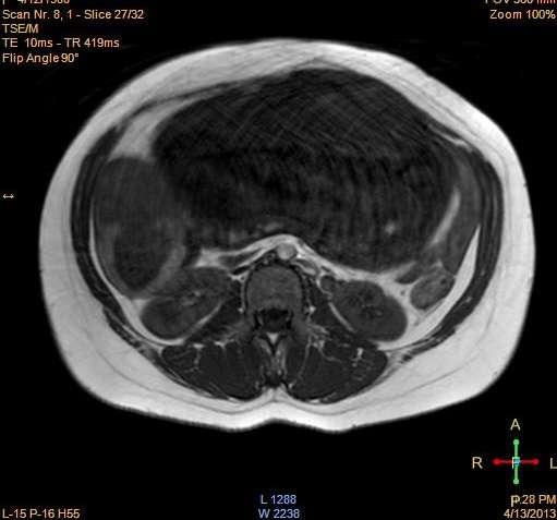
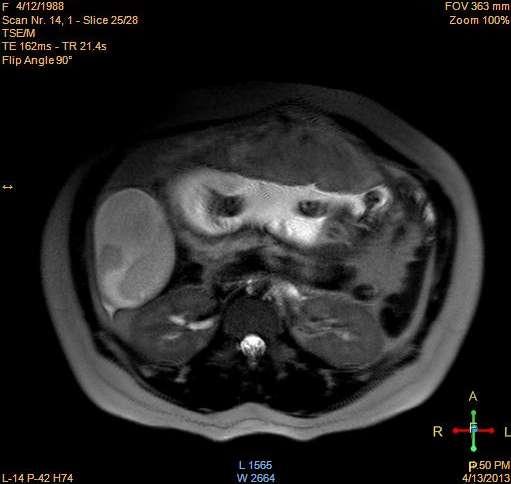
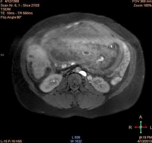
12 Conventional MRI : Contrast-enhanced MRI may be helpful in cases of complex ultrasound findings MR Imaging protocol: Sagittal T2 Axial T2 Axial T1 Axial T1 SPAIR DWI (b: 0, 500, 1000, and 1500) Coronal T2 Axial T1 post contrast Coronal T1 post contrast
Conventional images Appearance")


13 Interpretation of images: A)Conventional images Appearance of the tumor ; whether cystic, solid or mixed. Involvement of one or both ovaries Signal intensity Enhancement Wall thickness Presence of vegetations Ascites Lymph nodes Other pelvic organs Peritoneal and omental deposit
14 One of the new functional MRI techniques is DWI Tissue microstructures affect the random motion of water molecules
15 Tissues with low cellularity free water diffusion (Low signal) Tissues with high cellularity restriction of water diffusion (high signal)
16 B) DWI Qualitatively, regarding the signal Restricted diffusion = DW+ ADC Quantitatively, regarding the ADC values ADC maps generated from different b values ROI measured manually, over the largest possible area for the solid and cystic tumors ADC value 1.25 x 10⁻³ mm2/s may be an optimal cutoff value (Li et al, 2011)


17 What are the different clinical applications? Characterization of primary lesion
18 Peritoneal deposits

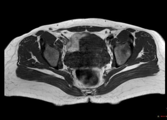
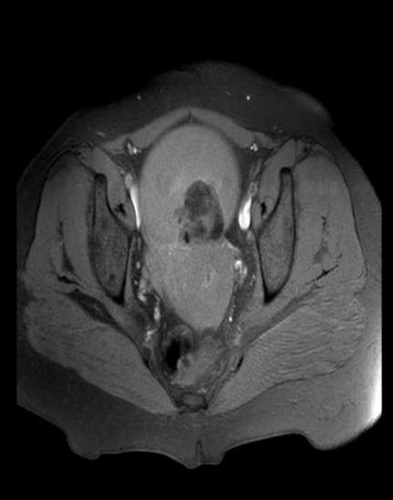
19 Lymph node assessment
20 Differentiating residual and Recurrent Disease from Post operative Change
21 A study was performed on ovarian tumors (Soha Talaat,Safaa saif, sahar mansour,2011) ADC for cystic component Minimum Maximum Mean +/-SD p-value ADC ( x 10-3 ) in malignant tumors (± 0.37) ADC ( x 10-3 )in benign tumors (± 0.56)
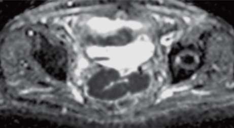
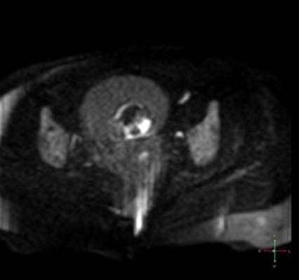
22 ADC for solid component Minimum Maximum Mean +/-SD p-value ADC ( x 10-3 ) in malignant tumors (± 0.11) ADC ( x 10-3 )in benign tumors (± 0.67)

23 100% 90% 80% 100% 83.30% 85.70% 85% 80% 78.60% 70% 60% 50% 40% MRI DW 30% 20% 10% 0% SENSITIVITY SPECIFICITY ACCURACY


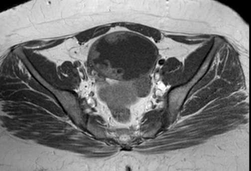
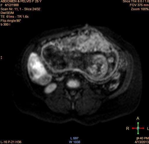
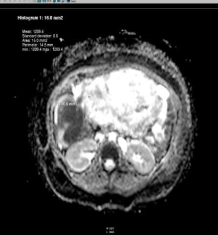
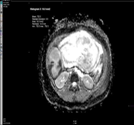
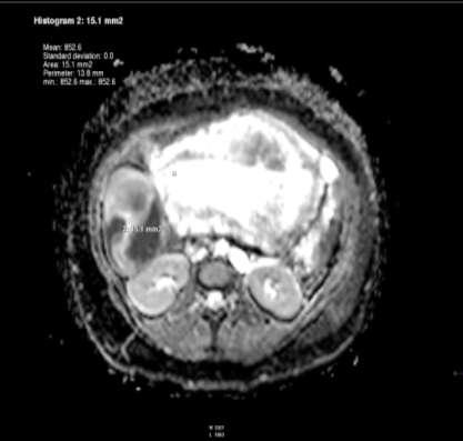
24 case 1 Female patient 21 year old complaining of abdominal enlargement.us showed solid pelvi-abdominal mass with increased vascularity on Doppler examination
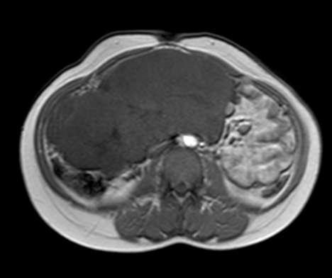
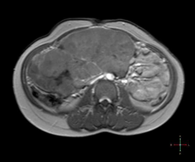
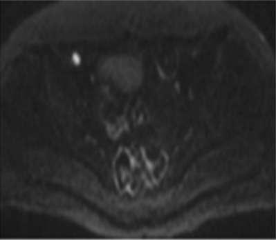
25 Case 1


26 case 1 Pathological diagnosis Granulosa cell tumor DWI High signal on DWI Low signal on ADC maps ADC value 0.68 x 10⁻³mm2/s


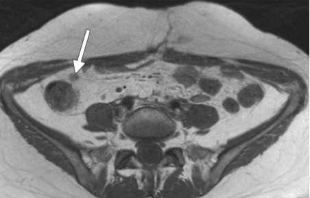
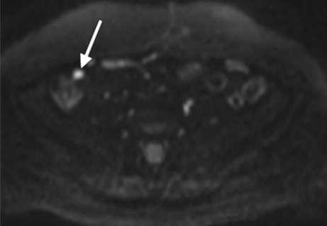
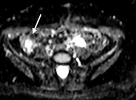
27 case 2 47 year old female patient came complaining of abdominal pain, US showed multilocular adnexal mass
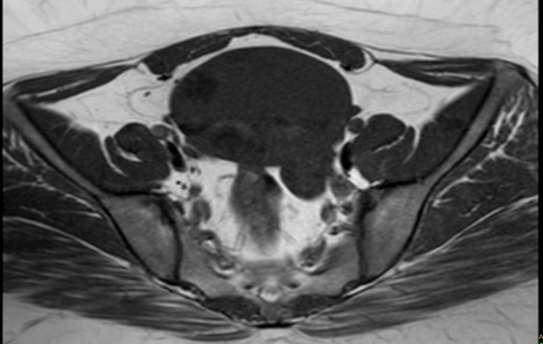
28 Case 2 Conventional MRI Benign looking, likely serous cystadenoma Minimal free pelvic ascites


29 Case 2 DWI High Signal On DWI(T2 shine through) High signal on ADC maps ADC value 2.01 x 10⁻³mm2/s Pathological diagnosis Serous cystadenoma


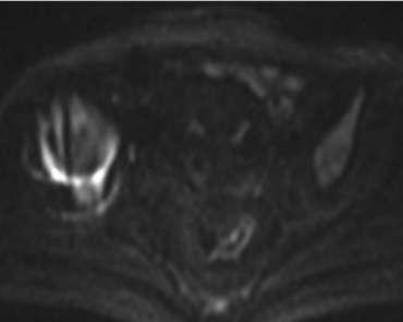
30 case 3 Female patient 35 year old complained of abdominal pain, TVUS showed a deeply seated rounded right solid ovarian mass, Marked vascularity on Doppler examination

31 case 4


32 case 4 No abnormal high signal on DWI High signal on ADC map ADC value 0.98 x 10⁻³mm2/s Pathological diagnosis Ovarian Fibroma


33 case 4 Female patient 34 year old complaining of abdominal discomfort, US revealed complex adnexal mass with multiple papillary projections
34 case 5


35 case 4 DWI Papillary projections moderate high signal Low signal on ADC maps ADC values 1.03 x 10⁻³mm2/s Pathological diagnosis Borderline papillary serous cystadenoma

 with")
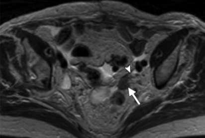
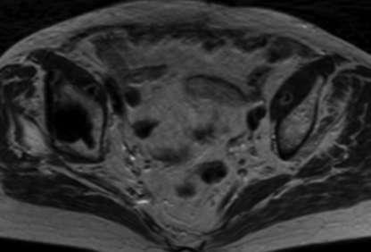
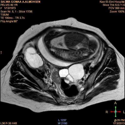
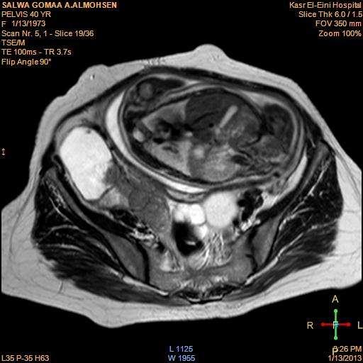
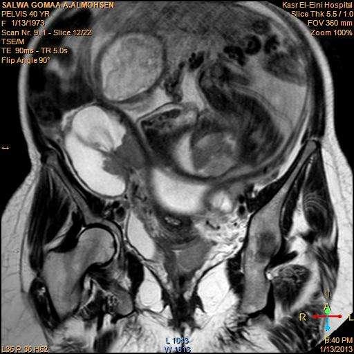
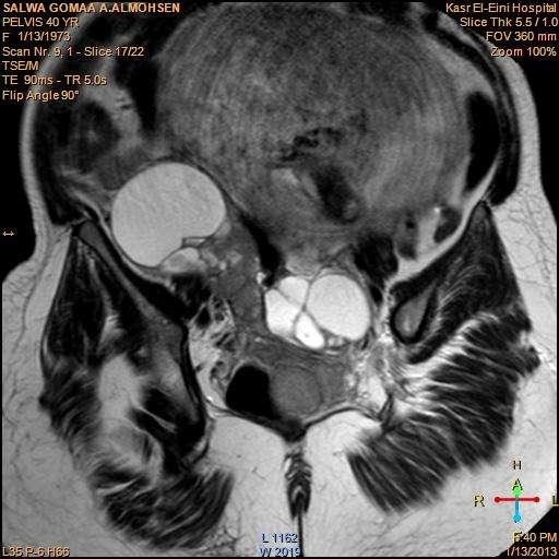
36 case 5 Female patient 42 year old came complaining of dull aching pelvic pain,us revealed two masses(pelvic and left lumbar) with echogenic foci
37 6 case
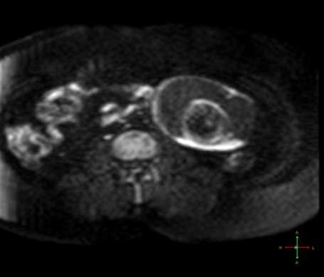
38 case 6
 Pathological diagnosis Bilateral mature cystic teratomas Mature cystic teratomas showed restriction may be attributed to keratinoid")
39 case 5 DWI Nodular part showed foci of restricted diffusion Marked low ADC values for both masses (0.39 x 10⁻³ mm2/s) and(0.64 x 10⁻³ mm2/s) Pathological diagnosis Bilateral mature cystic teratomas Mature cystic teratomas showed restriction may be attributed to keratinoid substance and Rokitansky protuberance

40 case 6 Pregnant Female 25 year old came complaining of dull aching right hypochondrial pain
41
42
43
,complaining of")

44 case 7 Female patient, 40 years old, pregnant (32 wks),complaining of severe diffuse pelvi-abdominal pain and intermittent attacks of vaginal bleeding
45
46
47
48


49 Cervical& upper vaginalmass Mass Diffusion ADC
50 Limitations DWI interpretation should be in conjunction with other morphological criteria in conventional MRI sequences Recommendations Study with large number of cases with special concern for BOTs Cases of mature cystic teratomas better to be excluded as their DWI findings are misleading and can give false results
51 DWI is one of the new functional MRI techniques When properly indicated, it can help increasing the specificity of MRI DWI implies using: Completely noninvasive technique No radiation exposure Might be an alternative for contrast (pregnancy) Increases the radiologist s confidence in image interpretation DWI interpretation should be done in conjugation with the conventional MRI


52 Ovarian lesions detected by US Functional anechoic cyst less than or equal 5 cm Complex cystic (with no solid parts) Complex cystic and solid Purely solid repeated follow up by US MRI for characterization (T1, T2, fat suppression) MRI for staging (T1, T2, fat suppression, post contrast, DWI ) MRI for staging (T1, T2, fat suppression, post contrast, DWI ) complicat ed or increased in size low signal in T2, and DWI in favor of benignity High signal in T2, and DWI in favor of malignanc y MRI
53
Terminology Estimate the risk of malignancy in adnexal masses - Overview
 Understanding the IOTA (International Ovarian Tumor Analysis) terminology & Classification Using the IOTA simple rules to estimate the risk of malignancy in women with adnexal masses Elisabeth Epstein,
JMSCR Vol 05 Issue 06 Page June 2017
 www.jmscr.igmpublication.org Impact Factor 5.84 Index Copernicus Value: 83.27 ISSN (e)-2347-176x ISSN (p) 2455-0450 DOI: https://dx.doi.org/10.18535/jmscr/v5i6.29 MRI in Clinically Suspected Uterine and
The Adnexal Mass. Handout NCUS 3/18/2017 Suzanne Dixon, MD
 The Adnexal Mass Handout NCUS 3/18/2017 Suzanne Dixon, MD Objectives: Pelvic mass differential Characteristics of the normal ovary Standard terminology for ovarian masses Benign vs. malignant features
Researcher 2018;10(8)
") Value of Magnetic Resonance Imaging (MRI) and Diffusion Weighted (DWI) MR in Diagnosis of Ovarian Lesions Enas ahmed a, Ikram Hamed b, Noha Abdel Shafy b, Ahmed Abdel Fattah a,* a Faculty of Medicine,
Value of Magnetic Resonance Imaging (MRI) and Diffusion Weighted (DWI) MR in Diagnosis of Ovarian Lesions Enas ahmed a, Ikram Hamed b, Noha Abdel Shafy b, Ahmed Abdel Fattah a,* a Faculty of Medicine,
Case 1. Gynaecology Case Presentation. Objectives. Disclosures 22/10/ year old female Clinical history: Assess right ovarian cyst
 Gynaecology Case Presentation Organ Imaging 2016 University of Toronto Sarah Johnson 39 year old female Clinical history: Assess right ovarian cyst Clinically diagnosed endometriosis Started fertility
Adnexal Masses and Problem Solving Pelvic MRI
 28th Congress of the Hungarian Society of Radiologists RCR Session Budapest June 2016 Adnexal Masses and Problem Solving Pelvic MRI DrSarah Swift St James s University Hospital Leeds, UK Objectives Characterisation
Diagnostic accuracy of ultrasonography with color doppler imaging techniques in adnexal masses and correlation with histopathological analysis
 Original Article Diagnostic accuracy of ultrasonography with color doppler imaging techniques in adnexal masses and correlation with histopathological analysis Neha Gupta 1*, Poonam Gupta 2, Omvati Gupta
Ovarian Lesion Benign vs Malignant?
 Ovarian Lesion Benign vs Malignant? Michele Keenan 1,2 Bernice Dunne 2 Mary Moran 1 Therese Herlihy 1 1. Radiography and Diagnostic Imaging, School of Medicine, University College Dublin, Ireland 2. Midland
Assessment of adnexal masses. Ultrasound workup of adnexal masses. symptoms. symptoms. Age. Serum tumor markers 10/1/2018
 Assessment of adnexal masses Ultrasound workup of adnexal masses Kevin Robinson, DO Department of Radiology Michigan State University October 4, 2018 Patients symptoms Age Menstrual status Serum tumor
IOTA and Models for Screening for Ovarian Cancer
 IOTA and Models for Screening for Ovarian Cancer Hennie Botha MARCH 2017 T H IG PY R O C F O SP EA KE R Silent Killer to Whispering Disease Listening to your body.. new, persistent, and increases in severity
2/24/19. Ovarian pathology: IOTA ADNEXAL MASSES. Content. IOTA terms for description of an adnexal mass. IOTA terms for description of an adnexal mass
 Content Ovarian pathology: IOTA ADNEXAL MASSES X SIMPLE COMPLEX Dr DESCRIBE WHAT YOU SEE FRANZCOG, MPH, DDU, COGU Sonologist Clinically useful Benign Malignant Communication between clinicians/research
Triage of Ovarian Masses. Andreas Obermair Brisbane
 Triage of Ovarian Masses Andreas Obermair Brisbane Why Triage? In ovarian cancer, best outcomes for patients can be achieved when patients are treated in tertiary centres by a multidisciplinary team led
Top Tips for Gynaecological Ultrasound. Catherine Kirkpatrick Consultant Sonographer Dublin Oct 2018
 Top Tips for Gynaecological Ultrasound Catherine Kirkpatrick Consultant Sonographer Dublin Oct 2018 We can all scan a pelvis so what can we do to improve? Uterus, endometrium and ovaries, got it covered!
A Practical Approach to Adnexal Masses
 A Practical Approach to Adnexal Masses Darcy J. Wolfman, MD Section Chief of Genitourinary Imaging American Institute for Radiologic Pathology Clinical Associate Johns Hopkins Community Radiology Division
MR diagnostics of adnexal masses
 MR diagnostics of adnexal masses Poster No.: C-1499 Congress: ECR 2017 Type: Educational Exhibit Authors: O. Nikolic, J. Ostojic, M. Basta Nikolic, A. Spasic, D. Donat, S. Stojanovic; Novi Sad/RS Keywords:
Essentials of Clinical MR, 2 nd edition. 73. Urinary Bladder and Male Pelvis
 73. Urinary Bladder and Male Pelvis Urinary bladder carcinoma is best locally staged with MRI. It is important however to note that a thickened wall (> 5 mm) is a non-specific finding seen in an underfilled
Gynecologic Ultrasound. Sujata Ghate, MD Associate Professor of Radiology Duke University Medical Center
 Gynecologic Ultrasound Sujata Ghate, MD Associate Professor of Radiology Duke University Medical Center Objectives Understand work-up of endometrial abnormalities Show examples of uterine and endometrial
MRI features of primary and metastatic mucinous ovarian tumors
 MRI features of primary and metastatic mucinous ovarian tumors Poster No.: C-0551 Congress: ECR 2014 Type: Authors: Keywords: DOI: Educational Exhibit P.-E. LAURENT, J. Thomassin-Piana, A. JALAGUIER; Marseille/
Risk of Malignancy Index in the Preoperative Evaluation of Patients with Adnexal Masses among Women of Perimenopausal and Postmenopausal Age Group
 IOSR Journal of Dental and Medical Sciences (IOSR-JDMS) e-issn: 2279-0853, p-issn: 2279-0861.Volume 17, Issue 9 Ver. 8 (September. 2018), PP 20-25 www.iosrjournals.org Risk of Malignancy Index in the Preoperative
Can diffusion weighted imaging distinguish between benign and malignant solid or predominantly solid gynecological adnexal masses?
 The Egyptian Journal of Radiology and Nuclear Medicine (2013) 44, 113 119 Egyptian Society of Radiology and Nuclear Medicine The Egyptian Journal of Radiology and Nuclear Medicine www.elsevier.com/locate/ejrnm
Characterizing Adnexal Masses: Pearls and Pitfalls 20 th Annual Summer Practicum SCBT-MR Jackson Hole August 11, 2010
 Characterizing Adnexal Masses: Pearls and Pitfalls 20 th Annual Summer Practicum SCBT-MR Jackson Hole August 11, 2010 Evan S. Siegelman MD University of Pennsylvania Medical Center Adnexal Masses: Pearls
Adnexal Masses in Menopausal Women Surgery or Surveillance?
 Adnexal Masses in Menopausal Women Surgery or Surveillance? FREDTALK IDEASWORTHSPREADING Disclosure I am a member of Vermillion s Speakers Bureau I am NOT a paid consultant for Vermillion Inc. nor do I
Imaging evaluation of ovarian masses.
 Imaging evaluation of ovarian masses. Poster No.: C-0988 Congress: ECR 2012 Type: Educational Exhibit Authors: M. Forment Navarro, C. La Parra Casado, A. Vera, C. Martínez 1 2 2 2 2 2 1 Rubio, M. Mazón
MR Imaging of the Adnexal Masses: A Review
 Page54 Review of Literature NJR 2011;1(1):54 60; Available online at www.nranepal.org MR Imaging of the Adnexal Masses: A Review I Ahmad 1, S Kirmani 1, M Rashid 2, K Ahmad 3 1 Department of Radiodiagnosis,
Case 9551 Primary ovarian Burkitt lymphoma
 Case 9551 Primary ovarian Burkitt lymphoma Monteiro V, Cunha TM, Saldanha T Section: Genital (Female) Imaging Published: 2011, Nov. 20 Patient: 23 year(s), female Authors' Institution V Monteiro 1, TM
ISUOG Basic Training Typical Ultrasound Appearances of Common Pathologies in the Adnexae
 ISUOG Basic Training Typical Ultrasound Appearances of Common Pathologies in the Adnexae Learning objectives At the end of the lecture series you will be able to: Compare the differences between typical
Value of MRI in Characterizing Adnexal Masses
 The Journal of Obstetrics and Gynecology of India (July August 2015) 65(4):259 266 DOI 10.1007/s13224-015-0730-9 PHOTO ESSAY Value of MRI in Characterizing Adnexal Masses Alpana Karnik 1 Raina Anil Tembey
L/O/G/O. Ovarian Tumor. Xiaoyu Niu Obstetrics and Gynecology Department Sichuan University West China Second Hospital
 L/O/G/O Ovarian Tumor Xiaoyu Niu Obstetrics and Gynecology Department Sichuan University West China Second Hospital Essentials classification of ovarian tumor clinical manifestation of ovarian tumor metastatic
H&E, IHC anti- Cytokeratin
 Cat No: OVC2281 - Ovary cancer tissue array Lot# Cores Size Cut Format QA/QC OVC228101 228 1.1mm 4um 12X19 H&E, IHC anti- Cytokeratin Recommended applications: For Research use only. RNA or protein ovary
objectives Pitfalls and Pearls in PET/CT imaging Kevin Robinson, DO Assistant Professor Department of Radiology Michigan State University
 objectives Pitfalls and Pearls in PET/CT imaging Kevin Robinson, DO Assistant Professor Department of Radiology Michigan State University To determine the regions of physiologic activity To understand
Role of Diffusion weighted MR Imaging in the Evaluation of Ovarian Tumors
 Role of Diffusion weighted MR Imaging in the Evaluation of Ovarian Tumors Thesis Submitted For Partial Fulfillment of the M.Sc. Degree In Radio diagnosis By Safaa Ibrahim Saif El Nasr Supervised by Dr.
11/10/2015. Prostate cancer in the U.S. Multi-parametric MRI of Prostate Diagnosis and Treatment Planning. NIH estimates for 2015.
 Multi-parametric MRI of Prostate Diagnosis and Treatment Planning Temel Tirkes, M.D. Associate Professor of Radiology Director, Genitourinary Radiology Indiana University School of Medicine Department
Clinical summary. Male 30 year-old with past history of non-seminomous germ cell tumour. Presents with retroperitoneal lymphadenopathy on CT.
 Clinical summary Male 30 year-old with past history of non-seminomous germ cell tumour. Presents with retroperitoneal lymphadenopathy on CT. For restaging PET/CT. PET/CT findings No significant FDG uptake
CAN TV U/S REDUCE THE
 CAN TV U/S REDUCE THE NEED FOR SURGERY IN GYNECOLOGY? Steven R. Goldstein, M.D. Professor of Obstetrics & Gynecology New York University it School of fmedicine i Director of Gynecologic Ultrasound Co-Director
MR imaging of FIGO stage I uterine cervical cancer: The diagnostic impact of 3T-MRI
 MR imaging of FIGO stage I uterine cervical cancer: The diagnostic impact of 3T-MRI Poster No.: C-1191 Congress: ECR 2010 Type: Educational Exhibit Topic: Genitourinary Authors: M. Takeuchi, K. Matsuzaki,
3 cell types in the normal ovary
 Ovarian tumors 3 cell types in the normal ovary Surface (coelomic epithelium) the origin of the great majority of ovarian tumors 90% of malignant ovarian tumors Totipotent germ cells Sex cord-stromal cells
Histopathological analysis of neoplastic and non neoplastic lesions of ovary: A study of one hundred cases
 Orginal Article Histopathological analysis of neoplastic and non neoplastic lesions of ovary: A study of one hundred cases 2 G Prathima, Srikanth Shastry 2 Consultant Pathologist, Image Diagnostics, Kadapa,
Prof. Dr. NAGUI M. ABDELWAHAB,M.D.; MARYSE Y. AWADALLAH, M.D. AYA M. BASSAM, Ms.C.
 Role of Whole-body Diffusion MR in Detection of Metastatic lesions Prof. Dr. NAGUI M. ABDELWAHAB,M.D.; MARYSE Y. AWADALLAH, M.D. AYA M. BASSAM, Ms.C. Cancer is a potentially life-threatening disease,
3 cell types in the normal ovary
 Ovarian tumors 3 cell types in the normal ovary Surface (coelomic epithelium) the origin of the great majority of ovarian tumors (neoplasms) 90% of malignant ovarian tumors Totipotent germ cells Sex cord-stromal
Female Genital Tract Lab. Dr. Nisreen Abu Shahin Assistant Professor of Pathology University of Jordan
 Female Genital Tract Lab Dr. Nisreen Abu Shahin Assistant Professor of Pathology University of Jordan Ovarian Pathology A 20-year-old female presented with vague left pelvic pain. Pelvic exam revealed
Disclosure. Acknowledgement. What is the Best Workup for Rectal Cancer Staging: US/MRI/PET? Rectal cancer imaging. None
 What is the Best Workup for Rectal Cancer Staging: US/MRI/PET? Zhen Jane Wang, MD Assistant Professor in Residence UC SF Department of Radiology Disclosure None Acknowledgement Hueylan Chern, MD, Department
American Journal of Oral Medicine and Radiology
 American Journal of Oral Medicine and Radiology e - ISSN - XXXX-XXXX ISSN - 2394-7721 Journal homepage: www.mcmed.us/journal/ajomr ULTRASONOGRAPHIC EVALUATION OF ADNEXAL MASSES Nageswar Rao* Professor,
Dr Claire Smith, Consultant Radiologist St James University Hospital Leeds
 Dr Claire Smith, Consultant Radiologist St James University Hospital Leeds Imaging in jaundice and 2ww pathway Image protocol Staging Limitations Pancreatic cancer 1.2.4 Refer people using a suspected
OVARIES. MLS Basic histological diagnosis MLS HIST 422 Semester 8- batch 7 L13 Dr: Ali Eltayb.
 OVARIES MLS Basic histological diagnosis MLS HIST 422 Semester 8- batch 7 L13 Dr: Ali Eltayb. OBJECTIVES Recognize different disease of ovaries Classify ovarian cyst Describe the pathogenesis, morphology
 INTRAUTERINE DEVICE = IUD INTRAUTERINE DEVICE = IUD CONGENITAL DISORDERS Pyometra = pyometrea is a uterine infection, it is accumulation of purulent material in the uterine cavity. Ultrasound is usually
EDUCATIONAL COMMENTARY CA 125. Learning Outcomes
 EDUCATIONAL COMMENTARY CA 125 Learning Outcomes Upon completion of this exercise, participants will be able to: discuss the use of CA 125 levels in monitoring patients undergoing treatment for ovarian
One of the commonest gynecological cancers,especially in white Americans.
 Gynaecology Dr. Rozhan Lecture 6 CARCINOMA OF THE ENDOMETRIUM One of the commonest gynecological cancers,especially in white Americans. It is a disease of postmenopausal women with a peak incidence in
Staging and Treatment Update for Gynecologic Malignancies
 Staging and Treatment Update for Gynecologic Malignancies Bunja Rungruang, MD Medical College of Georgia No disclosures 4 th most common new cases of cancer in women 5 th and 6 th leading cancer deaths
Institute of Pathology First Faculty of Medicine Charles University. Ovary
 Ovary Barrett esophagus ph in vagina between 3.8 and 4.5 ph of stomach varies from 1-2 (hydrochloric acid) up to 4-5 BE probably results from upward migration of columnar cells from gastroesophageal junction
Serous borderline tumor icd 10
 Note. May be used as an additional code to identify functional activity associated with a carcinoid tumor. Free, official information about 2012 (and also 2013-2015) ICD -9-CM diagnosis code 220, including
Category Term Definition Comments 1 Major Categories 1a
 Working Lexicon Categories, Terms & Definitions Category Term Definition Comments 1 Major Categories 1a Physiologic Category (consistent with normal ovarian physiology) Follicle Simple 3 cm in premenopausal
Pathology of Ovarian Tumours. Dr. Jyothi Ranganathan MD ( Path) AFMC Pune PDCC (Cytopathology) PGI Chandigarh
 AFMC Pune PDCC (Cytopathology) PGI Chandigarh") Pathology of Ovarian Tumours Dr. Jyothi Ranganathan MD ( Path) AFMC Pune PDCC (Cytopathology) PGI Chandigarh Outline Incidence Risk factors Classification Pathology of tumours Tumour markers Prevention
Pathology of Ovarian Tumours Dr. Jyothi Ranganathan MD ( Path) AFMC Pune PDCC (Cytopathology) PGI Chandigarh Outline Incidence Risk factors Classification Pathology of tumours Tumour markers Prevention
General history. Basic Data : Age :62y/o Date of admitted: Married status : Married
 General history Basic Data : Age :62y/o Date of admitted:940510 Married status : Married General history Chief Complain : bilateral ovarian cyst incidentally being found out during pap smear. Present Illness
See the latest estimates for new cases of ovarian cancer and deaths in the US and what research is currently being done.
 About Ovarian Cancer Overview and Types If you have been diagnosed with ovarian cancer or are worried about it, you likely have a lot of questions. Learning some basics is a good place to start. What Is
Role of imaging in RCC. Ultrasonography. Solid lesion. Cystic RCC. Solid RCC 31/08/60. From Diagnosis to Treatment: the Radiologist Perspective
 Role of imaging in RCC From Diagnosis to Treatment: the Radiologist Perspective Diagnosis Staging Follow up Imaging modalities Limitations and pitfalls Duangkamon Prapruttam, MD Department of Therapeutic
Diane DeFriend Derriford Hospital, Plymouth
 Diane DeFriend Derriford Hospital, Plymouth Ultrasound US remains primary imaging modality for investigation of an adnexal mass Aim to characterise Benign Malignant Indeterminate 90% adnexal masses characterised
Pre-operative Ultrasound of Lymph Nodes in Thyroid Cancer
 Pre-operative Ultrasound of Lymph Nodes in Thyroid Cancer AACE - Advances in Medical and Surgical Management of Thyroid Cancer - 2018 Robert A. Levine, MD, FACE, ECNU Thyroid Center of New Hampshire Geisel
IN THE NAME OF GOD POV: CYSTIC OVARIAN LESION
 IN THE NAME OF GOD POV: CYSTIC OVARIAN LESION CASE 1 20 years old girl with AUB and pelvic pain from 2 weeks ago Impression :Simple unilocular 6 cm ovarian cyst Next step? Almost certainly benign so FU
Joseph Misdraji, M.D. GI pathology Unit Massachusetts General Hospital
 Joseph Misdraji, M.D. GI pathology Unit Massachusetts General Hospital jmisdraji@partners.org Low-grade appendiceal mucinous neoplasm (LAMN) High-grade appendiceal mucinous neoplasm (HAMN) Adenocarcinoma
FDG-PET/CT in Gynaecologic Cancers
 Friday, August 31, 2012 Session 6, 9:00-9:30 FDG-PET/CT in Gynaecologic Cancers (Uterine) cervical cancer Endometrial cancer & Uterine sarcomas Ovarian cancer Little mermaid (Edvard Eriksen 1913) honoring
Approach to imaging of the ovaries
 First encounter Approach to imaging of the ovaries Mariam Moshiri MD Associate professor Body Imaging Most common first encounter is via ultrasound Many of clinicians order US imaging for various female
Low-grade serous neoplasia. Robert A. Soslow, MD
 Low-grade serous neoplasia Robert A. Soslow, MD soslowr@mskcc.org Outline Orientation Ovarian tumor overview Non serous borderline tumors Serous borderline tumors Clinical summary Morphologic description
Endometrioma With Calcification Simulating a Dermoid on Sonography
 Case Report Endometrioma With Calcification Simulating a Dermoid on Sonography Kiran A. Jain, MD Several investigators have explored the sonographic diagnostic criteria of endometriomas. Endometriomas
Gynecologic Oncologist. Surgery Chemotherapy Radiation Therapy Hormonal Therapy Immunotherapy. Cervical cancer
 Gynecologic Oncology Pre invasive vulvar, vaginal, & cervical disease Vulvar Cervical Endometrial Uterine Sarcoma Fallopian Tube Ovarian GTD Gynecologic Oncologist Surgery Chemotherapy Radiation Therapy
Erratum to: Fertility-sparing for young patients with gynecologic cancer: How MRI can guide patient selection prior to conservative management
 Abdominal Radiology ª Springer Science+Business Media, LLC 2017 Published online: 11 November 2017 Abdom Radiol (2017) 42:2966 2973 DOI: 10.1007/s00261-017-1205-5 Erratum to: Fertility-sparing for young
What Radiologists do?
 Multimodality Imaging in Oncology 2018 March 5 th 9th Diagnostic Imaging in Oncology What Radiologists do? Chikako Suzuki, MD, PhD Department of Diagnostic Radiology, KS Solna Department of Molecular Medicine
Case Fibrothecoma of the ovary
 Case 10646 Fibrothecoma of the ovary Elisa Melo Abreu, Teresa Margarida Cunha Section: Genital (Female) Imaging Published: 2015, Jan. 2 Patient: 70 year(s), female Authors' Institution Department of Radiology,
Clinicopathological and Histological Features of Ovarian Tumour- A Study
 IOSR Journal of Dental and Medical Sciences (IOSR-JDMS) e-issn: 2279-0853, p-issn: 2279-0861.Volume 16, Issue 9 Ver. IX (September. 2017), PP 56-60 www.iosrjournals.org Clinicopathological and Histological
A NEW QUANTITATIVE METHOD TO EVALUATE ADNEXAL TUMORS
 ORIGINAL ARTICLE A NEW QUANTITATIVE METHOD TO EVALUATE ADNEXAL TUMORS Chung-Yuan Lee, Ching-Cheng Tseng, Chen-Bin Wang, Yu-Hsiang Lin, Chun-Hung Chen, Ting-Hung Wun, Ying-Lun Sun, Chih-Jen Tseng* Department
Borderline Ovarian Tumours:MRI features to aid a challenging diagnosis
 Borderline Ovarian Tumours:MRI features to aid a challenging diagnosis Poster No.: R-0095 Congress: RANZCR ASM 2013 Type: Scientific Paper Authors: C. Shadbolt, S. Kouloyan-Ilic, A. Dobrotwir; Melbourne/AU
Staging Colorectal Cancer
 Staging Colorectal Cancer CT is recommended as the initial staging scan for colorectal cancer to assess local extent of the disease and to look for metastases to the liver and/or lung Further imaging for
Current staging of endometrial carcinoma with MR imaging
 Current staging of endometrial carcinoma with MR imaging Poster No.: C-1436 Congress: ECR 2015 Type: Educational Exhibit Authors: M. Magalhaes, H. Donato, C. B. Marques, P. Gomes, F. Caseiro Alves; Coimbra/PT
Sarah Burton. Lead Gynae Oncology Nurse Specialist Cancer Care Cymru
 Sarah Burton Lead Gynae Oncology Nurse Specialist Cancer Care Cymru Gynaecological Cancers Cervical Cancers Risk factors Presentation Early sexual activity Multiple sexual partners Smoking Human Papiloma
Index. mri.theclinics.com. Note: Page numbers of article titles are in boldface type.
 Index Note: Page numbers of article titles are in boldface type. A Angiogenesis, and cancer of prostate, 689 690 Angiography, MR. See MR angiography. Apoptosis, MR imaging of, 637 Apparent diffusion coefficient,
Ovarian pathology: A practical approach to imaging diagnosis and management
 Ovarian pathology: A practical approach to imaging diagnosis and management Award: Certificate of Merit Poster No.: C-1865 Congress: ECR 2015 Type: Educational Exhibit Authors: A. Castan, A. Mir Torres,
Cervical Cancer: 2018 FIGO Staging
 Cervical Cancer: 2018 FIGO Staging Jonathan S. Berek, MD, MMS Laurie Kraus Lacob Professor Stanford University School of Medicine Director, Stanford Women s Cancer Center Senior Scientific Advisor, Stanford
Pathology of the female genital tract
 Pathology of the female genital tract Common illnesses of the female genital tract Before menarche Developmental anomalies Tumors (ovarial teratoma) Amenorrhea Fertile years PCOS, ovarian cysts Endometriosis
Appendix 5. EFSUMB Newsletter. Gastroenterological Ultrasound
 EFSUMB Newsletter 87 Examinations should encompass the full range of pathological conditions listed below A log book listing the types of examinations undertaken should be kept Training should usually
Case Scenario 1. Pathology report Specimen from mediastinoscopy Final Diagnosis : Metastatic small cell carcinoma with residual lymphatic tissue
 Case Scenario 1 Oncology Consult: Patient is a 51-year-old male with history of T4N3 squamous cell carcinoma of tonsil status post concurrent chemoradiation finished in October two years ago. He was hospitalized
Evaluation of serum level of CA-125 and HE4 in patients with an adnexal mass for the discrimination of benign from malignant cases
 Evaluation of serum level of CA-125 and HE4 in patients with an adnexal mass for the discrimination of benign from malignant cases Ahmet Göçmen, Fatih Şanlıkan, Yüksel Sayın, Fatma Seda Öztürk, Abdülhamid
Malignant Transformation of Endometriosis: Magnetic Resonance Imaging Aspects
 Malignant Transformation of Endometriosis: Magnetic Resonance Imaging Aspects Poster No.: C-0084 Congress: ECR 2014 Type: Scientific Exhibit Authors: E. A. Yukhno, I. Trofimenko, G. Trufanov; St. Petersburg/RU
Malignant Transformation of Endometriosis: Magnetic Resonance Imaging Aspects
 Malignant Transformation of Endometriosis: Magnetic Resonance Imaging Aspects Poster No.: C-0084 Congress: ECR 2014 Type: Scientific Exhibit Authors: E. A. Yukhno, I. Trofimenko, G. Trufanov; St. Petersburg/RU
performed to help sway the clinician in what the appropriate diagnosis is, which can substantially alter the treatment of management.
 Hello, I am Maura Polansky at the University of Texas MD Anderson Cancer Center. I am a Physician Assistant in the Department of Gastrointestinal Medical Oncology and the Program Director for Physician
Utility of ADC Measurements in the Discrimination between Benign and Lymphomatous Abdomino-Pelvic Lymph Nodes
 Med. J. Cairo Univ., Vol. 84, No. 2, September: 1-7, 2016 www.medicaljournalofcairouniversity.net Utility of ADC Measurements in the Discrimination between Benign and Lymphomatous Abdomino-Pelvic Lymph
The relative frequency and histopathological patterns of ovarian lesions: study of 116 cases
 Original article: The relative frequency and histopathological patterns of ovarian lesions: study of 116 cases Dr Dimple Mehta*,Dr Alpesh Chavda**, Dr Hetal Patel*** *Assistant Professor, **Tutor, ***3
PET/CT in Gynaecological Cancers. Stroobants Sigrid, MD, PhD Departement of Nuclear Medicine University Hospital,Antwerp
 PET/CT in Gynaecological Cancers Stroobants Sigrid, MD, PhD Departement of Nuclear Medicine University Hospital,Antwerp Cervix cancer Outline of this talk Initial staging Treatment monitoring/guidance
Ubol Saeng-Anan, Tawiwan Pantasri, Vithida Neeyalavira, Theera Tongsong*
 DOI:http://dx.doi.org/10.7314/APJCP.2013.14.9.5409 RESEARCH ARTICLE Sonographic Pattern Recognition of Endometriomas Mimicking Ovarian Cancer Ubol Saeng-Anan, Tawiwan Pantasri, Vithida Neeyalavira, Theera
The new FIGO classification in endometrial carcinoma
 The new FIGO classification in endometrial carcinoma Poster No.: C-1073 Congress: ECR 2012 Type: Educational Exhibit Authors: A. IGLESIAS CASTAÑON, M. Arias Gonzales, J. Mañas Uxó, 1 2 1 2 2 2 B. NIETO
Adnexal Masses in Menopausal Women
 Adnexal Masses in Menopausal Women Surgery or Surveillance? Disclosure Frederick R. Ueland, MD Professor and Director Division of Gynecologic Oncology University of Kentucky I have no financial disclosures
JMSCR Vol 3 Issue 9 Page September 2015
 www.jmscr.igmpublication.org Impact Factor 3.79 ISSN (e)-2347-176x DOI: http://dx.doi.org/10.18535/jmscr/v3i9.09 A Study of Histopathological Pattern of Ovarian Neoplasms and their Age Distribution in
Chapter 2: Initial treatment for endometrial cancer (including histologic variant type)
") Chapter 2: Initial treatment for endometrial cancer (including histologic variant type) CQ01 Which surgical techniques for hysterectomy are recommended for patients considered to be stage I preoperatively?
Chapter 2: Initial treatment for endometrial cancer (including histologic variant type) CQ01 Which surgical techniques for hysterectomy are recommended for patients considered to be stage I preoperatively?
GENERAL DATA. Sex : female Age : 40 years old Marriage status : married
 GENERAL DATA Sex : female Age : 40 years old Marriage status : married CHIEF COMPLAINT Bilateral ovarian tumors discovered by sonography accidentally PRESENT ILLNESS 2003-06-26 :bilateral ovarian tumors
Please complete prior to the webinar. HOSPITAL REGISTRY WEBINAR FEMALE REPRODUCTIVE SYSTEM EXERCISES CASE 1: FEMALE REPRODUCTIVE
 Please complete prior to the webinar. HOSPITAL REGISTRY WEBINAR FEMALE REPRODUCTIVE SYSTEM EXERCISES PHYSICAL EXAMINATION CASE 1: FEMALE REPRODUCTIVE 3/5 Patient presents through the emergency room with
3 Summary of clinical applications and limitations of measurements
 CA125 (serum) 1 Name and description of analyte 1.1 Name of analyte Cancer Antigen 125 (CA125) 1.2 Alternative names Mucin-16 1.3 NLMC code To follow 1.4 Description of analyte CA125 is an antigenic determinant
Staging. Carcinoma confined to the corpus. Carcinoma confined to the endometrium. Less than ½ myometrial invasion. Greater than ½ myometrial invasion
 5 th of June 2009 Background Most common gynaecological carcinoma in developed countries Most cases are post-menopausal Increasing incidence in certain age groups Increasing death rates in the USA 5-year
How to Recognize Gynecologic Cancer Cells from Pelvic Washing and Ascetic Specimens
 How to Recognize Gynecologic Cancer Cells from Pelvic Washing and Ascetic Specimens Wenxin Zheng, M.D. Professor of Pathology and Gynecology University of Arizona zhengw@email.arizona.edu http://www.zheng.gynpath.medicine.arizona.edu/index.html
Neoplasia literally means "new growth.
 NEOPLASIA Neoplasia literally means "new growth. A neoplasm, defined as "an abnormal mass of tissue the growth of which exceeds and is uncoordinated with that of the normal tissues and persists in the
Imaging in gastric cancer
 Imaging in gastric cancer Gastric cancer remains a deadly disease because of late diagnosis. Adenocarcinoma represents 90% of malignant tumors. Diagnosis is based on endoscopic examination with biopsies.
The key contribution of MRI in adnexal mass evaluation is in: 1. Identifying benign features. 2. Identifying malignant features.
 19 th Annual Women s Imaging Conference University of Toronto - 2016 Disclosures : None phyllis.glanc@sunnybrook.ca Sunnybrook Health Science Centre University of Toronto, Dept Medical Imaging, Obstetrics
Is It Time To Implement Ovarian Cancer Screening?
 Is It Time To Implement Ovarian Cancer Screening? Prof Dr Samet Topuz Istanbul Medıcal Faculty Department Of Obstetrics and Gynecology ESGO Prevention in Gynaecological Malignancies September 08 2016 Antalya