Case 5385 Tubular ectasia of the rete testis: a benign testicular entity diagnosed on imaging
|
|
|
- Audra Stevenson
- 6 years ago
- Views:
Transcription

1 Case 5385 Tubular ectasia of the rete testis: a benign testicular entity diagnosed on imaging A. C. Tsili 1, C. Tsampoulas 1, D. Giannakis 2, A. Chaidou 1, N. Sofikitis 2, S. C. Efremidis 1 1 Department of Clinical Radiology 2Department of Urology University Hospital of Ioannina, GREECE. University Hospital of Ioannina Section: Uroradiology & Genital Male Imaging Published: 2006, Nov. 30 Patient: 58 year(s), male Clinical History Dilatation or tubular ectasia of the rete testis (TERT) is a well-known benign intratesticular entity, for which the clinical, as well as the imaging findings may unable a correct diagnosis and the avoidance of invasive tests, as unnecessary biopsy or orchiectomy. Imaging Findings A 58-year old man was referred to the Urology department with a six-month history of painless swelling of the right hemiscrotum. On physical examination a firm mass was palpable in the head of the right epididymis and a normal ipsilateral testis was separated from the extratesticular mass. Sonographic examination of the scrotum revealed the presence of a large right cystic mass involving the area of the head of the epididymis. An ovoid cluster of small anechoic structures within the mediastinum of the right testis, measuring was also detected (Fig. 1a). Doppler sonography showed absence of blood flow within the intratesticular lesion (Fig. 1b). Scrotal MR imaging examination was performed using fast spin-echo T2-weighted images and spin-echo unenhanced and contrast-enhanced T1-weighted images. A large right spermatocele, a moderate hydrocele and a multilocular intratesticular lesion, were detected (Fig. 2). A smaller lesion involving the apex of the left testis was also revealed, the same lesion not detected on sonography. The intratesticular masses were located in the region of the mediastinum and had signal intensity similar to that of water. Their signal intensity was identical to that of the spermatocele on all pulse
2 sequences. After the administration of gadopentetate dimeglumine (Fig. 2d), none of the above lesions enhanced. Based on the clinical and imaging findings, suggestive for the diagnosis of tubular ectasia of the rete testis, spermatocelectomy was performed. Follow-up sonogram, two years after the initial presentation, revealed no change of the imaging findings. Discussion The majority of intratesticular masses are malignant; therefore orchiectomy is mandatory to rule out malignancy [1]. However, differentiating benign from malignant intratesticular lesions is critical, since the correct preoperative characterization may obviate an unwarranted biopsy or surgery [1, 2]. Dilatation or tubular ectasia of the rete testis (TERT) is a well-known benign intratesticular entity [1, 2-6]. The incidence of TERT is unknown, although is reported in up to 4.3% of the routine scrotal sonographic examinations [3]. The etiology is believed to be obstructive, since the majority of patients with TERT have a history of possible obstruction of the spermatic ducts, such as chronic epididymitis, spermatoceles, as in this case, trauma, scrotal or inguinal interventions [3-11]. The sonographic findings of this entity are typical and allow a confident diagnosis in the majority of cases [3-8]. A group of small anechoic tubular structures, involving the area of the mediastinum, with so solid components and no mass effect, devoid of blood flow, as in our case, represent the typical findings of TERT. Tartar et al [9] first described the characteristic MR appearance of TERT in six patients, reporting signal homogeneity, similar to that of the coexisting spermatoceles, hypointensity on T1 and proton density-weighted images and no lesion discrimination from the normal testicular parenchyma on spin-echo T2-weighted images. In this case, we used fast-spin echo T2-weighted images, and the signal intensity was higher than that of the normal testicles on these sequences. Our case was typical of a bilateral TERT, demonstrating signal intensity similar to that of the coexistent spermatocele on all pulse sequences and no enhancement after contrast material administration [3, 9-11]. MR imaging examination of the scrotum is best recommended in cases in which the sonographic findings are equivocal or nondiagnostic, to avoid unnecessary surgical explorations. TERT is easily differentiated from testicular tumors due to its unique localization in the region of the mediastinum testis, its characteristic imaging features, both on sonography and MR examination, the absence of palpable intratesticular mass, the frequent coexistence of spermatoceles ant its occurrence in greater age, when compared with testicular malignancies [3, 4]. However, dilatation of the rete testis due to a tumor occlusion has also been described [3] and should always be evaluated in the differential diagnosis. The papillary adenocarcinoma of the rete testis may manifest as a multilocular cystic lesion [12], but in this case the mass is usually palpable and is associated with the presence of solid elements on imaging evaluation.differential diagnosis should also include the intratesticular varicocele and the cystic dysplasia of the testis. Intratesticular varicocele is an extremely rare condition, easily diagnosed by the presence of blood flow during rest or the Valsalva maneuver [13]. The cystic dysplasia of the testis is another rare, nonneoplastic entity, which may have similar sonographic and histologic appearance with TERT [14]. However, this entity is mainly diagnosed in children and often associated with renal or excretory duct malformations [15]. Final Diagnosis Tubular ectasia of the rete testis
, located in the region of the mediastinum, representing a typical form of dilatation of")

3 Figures Figure 1 Figure 1a Sagittal sonogram of the right testis depicts an ovoid cluster of small anechoic structures (cursors), located in the region of the mediastinum, representing a typical form of dilatation of the rete testis. There is also a large spermatocele (asterisk) and a small ipsilateral hydrocele (arrow). Doppler sonography demonstrates the absence of vascularity within the intratesticular lesion. Figure 2 Figure 2a
. All the lesions have similar signal intensity, higher than that of the testicular parenchyma (T).")
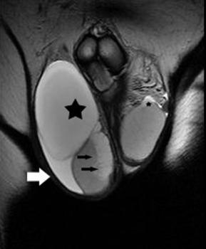
4 Coronal FSE T2-weighted images (a, b) depict the presence of a large right spermatocele (asterisk) and a multilocular ipsilateral intratesticular lesion located in the mediastinum (arrows). Another smaller lesion is seen in the left mediastinum testis (small asterisk). All the lesions have similar signal intensity, higher than that of the testicular parenchyma (T). There is also a moderate right hydrocele (arrow). Coronal FSE T2-weighted images (a, b) depict the presence of a large right spermatocele (asterisk) and a multilocular ipsilateral intratesticular lesion located in the mediastinum (arrows). Another smaller lesion is seen in the left mediastinum testis (small asterisk). All the lesions have similar signal intensity, higher than that of the testicular parenchyma (T). There is also a moderate right hydrocele (arrow).

![contrast material administration. contrast material administration. MeSH Testicular Diseases [C12.294.829]](/docs-images/72/67032429/images/5-1.jpg)
5 Transverse precontrast (c) and postcontrast (d) T1-weighted images. Both intratesticular and extratesticular lesions have low signal intensity on T1-weighted images and do not enhance after contrast material administration. Transverse precontrast (c) and postcontrast (d) T1-weighted images. Both intratesticular and extratesticular lesions have low signal intensity on T1-weighted images and do not enhance after contrast material administration. MeSH Testicular Diseases [C ]
6 Testicular Neoplasms [C ] Tumors or cancer of the TESTIS. Germ cell tumors (GERMINOMA) of the testis constitute 95% of all testicular neoplasms. Genital Diseases, Male [C12.294] References [1] Woodward PJ, Sohaey r, ODonoghue MJ, Green DE. From the Archives of the AFIP: tumors and tumorlike lesions of the testis: radiologic-pathologic correlation. Radiographics 2002; 22 (1): [2] Rubenstein RA, Dogra VS, Resnik AD, Martin I. Benign intrascrotal lesions. J Urol 2004; 171 (5): [3] Jimenez-Lopez M, Ramirez-Garrido F, Lopez-Gonzalez Garrido JD, et al. Dilatation of the rete testis: ultrasound study. Eur Radiol 1999; 9: [4] Rouviere O, Bouvier R, Pangaud C, Jeune M, Dawahra M, Lyonnet D. Tubular ectasia of the rete testis: a potencial pitfall in scrotal imaging. Eur Radiol 1999; 9: [5] Older RA, Watson LR. Tubular ectasia of the rete testis: a benign condition with a sonographic appearance that may be misinterpreted as malignant. J Urol 1994; 152: [6] Burrus JK, Lockhart ME, Kenney PJ, Kolettis PN. Cystic ectasia of the rete testis: clinical and radiographic features. J Urol 2002; 168: [7] Brown DL, Benson CB, Doherty FJ, et al. Cystic testicular mass caused by dilated rete testis: Sonographic Findings in 31 Cases. AJR 1992; 158: [8] Colangelo SM, Fried K, Hyacinthe LM, Fracchia JA. Tubular ectasia of the rete testis: an ultrasound diagnosis. Urology 1995; 45 (3): [9] Tartar VM, Trambert MA, Balsara ZN, Mattrey RF. Tubular ectasia of the testicle: Sonographic and MR imaging appearance. AJR 1993; 160: [10] Monette RJ, Woodward PJ. MR appearance of dilated rete testis. AJR 1994; 163: 482. [11] Meyer DR, Huppe T, Lock U, Hodek E, Friedrich M. Pronounced cystic transformation of the rete testis: MRI appearance. Invest Radiol 1999; 34: [12] Stein JP, Freeman JA, Esrig D, Chandrasoma PT, Skinner DG. Papillary adenocarcinoma of the rete testis: a case report and review of the literature. Urology 1994; 44: [13] Weiss AJ, Kellman GM, Middleton WD, Kirkemo A. Intratesticular varicocele: sonographic findings in two patients. AJR 1992; 158: [14] Cho CS, Kosek J. Cystic dysplasia of the testis: sonographic and pathologic findings. Radiology 1985; 156: 777. Citation A. C. Tsili 1, C. Tsampoulas 1, D. Giannakis 2, A. Chaidou 1, N. Sofikitis 2, S. C. Efremidis 1 1
7 Department of Clinical Radiology Department of Urology University Hospital of Ioannina, 2 GREECE. (2006, Nov. 30) Tubular ectasia of the rete testis: a benign testicular entity diagnosed on imaging {Online} URL:
MRI IN THE CHARACTERIZATION OF SEMINOMATOUS AND NONSEMINOMATOUS GERM CELL TUMORS OF THE TESTIS
 MRI IN THE CHARACTERIZATION OF SEMINOMATOUS AND NONSEMINOMATOUS GERM CELL TUMORS OF THE TESTIS Ambesh Deshar *, Gyanendra KC and Zhang Lopsang *Department of Medical Imaging and Nuclear Medicine, First
CLINICAL PRESENTATION AND RADIOLOGY QUIZ QUESTION
 Donald L. Renfrew, MD Radiology Associates of the Fox Valley, 333 N. Commercial Street, Suite 100, Neenah, WI 54956 2/19/2011 Radiology Quiz of the Week # 8 Page 1 CLINICAL PRESENTATION AND RADIOLOGY QUIZ
MRI in the Characterization and Local Staging of Testicular Neoplasms
 Genitourinary Imaging Original Research Tsili et al. MRI of Testicular Neoplasms Genitourinary Imaging Original Research thina. Tsili 1 Maria I. rgyropoulou 1 Dimitrios Giannakis 2 Nikolaos Sofikitis 2
Painless palpable scrotal mass
 Clinical Case - Test Yourself Urogenital Painless palpable scrotal mass Charis Anastasiadis, Georgia Kyriakopoulou, Charikleia Triantopoulou Radiology Department, Konstantopoulio General Hospital of Nea
MRI in the Histologic Characterization of. testicular neoplasms.
 Tsili et al. MRI of Testicular Neoplasms Genitourinary Imaging Original Research 12_07_2267_Tsili.fm 11/9/07 thina C. Tsili 1 Constantine Tsampoulas 1 Xenofon Giannakopoulos 2 Dimitrios Stefanou 3 Yiannis
MRI of Patients With Suspected Scrotal or Testicular Lesions: Diagnostic Value in Daily Practice
 Genitourinary Imaging Original Research Mohrs et al. Scrotal and Testicular MRI Genitourinary Imaging Original Research Oliver K. Mohrs 1,2 Henrik Thoms 1 Tobias Egner 3 Anne Brunier 1 Michael Eiers 2
Intraparenchymatous adenomatoid tumor dependent on the rete testis: A case report and review of literature
 Intraparenchymatous adenomatoid tumor dependent on the rete testis: A case report and review of literature A. Jiménez Pacheco, J. L. Martínez Torres, F. Valle Díaz de la Guardia, M. A. Arrabal Polo, and
Testicular ultrasound in acute scrotal pain - beyond testicular torsion
 Testicular ultrasound in acute scrotal pain - beyond testicular torsion Poster No.: C-1284 Congress: ECR 2015 Type: Educational Exhibit Authors: I. Rolla, M. Nogueira, M. J. Aguiar, D. S. Garrido, J. A.
COLOR DOPPLER ULTRASOUND IN EVALUATION OF SCROTAL LESIONS
 COLOR DOPPLER ULTRASOUND IN EVALUATION OF SCROTAL LESIONS Desai Sanjay D Associate Professor, Department of Radiology, RCSM Govt. Medical College, Kolhapur. ABSTRACT: Color Doppler ultrasound is a non-invasive,
Role of Colour Doppler Ultrasonography in evaluation of scrotal pain and swelling
 Original Research Article Role of Colour Doppler Ultrasonography in evaluation of scrotal pain and swelling Assistant Professor, Department of Radiodiagnosis, Government Medical College, Rajnandgaon Chattisghar,
Case Report Testicular Arteriovenous Malformation: Gray-Scale and Color Doppler Ultrasonography Features
 Volume 2011, Article ID 876206, 4 pages doi:10.11/2011/876206 Case Report Testicular Arteriovenous Malformation: Gray-Scale and Color Doppler Ultrasonography Features Fatih Gulsen, 1 Ismail Mihmanli, 1
The Acute Scrotum: Sonographic Findings
 The Acute Scrotum: Sonographic Findings 가천의대길병원방사선과 양달모 Gachon Medical School Introduction Many diseases presenting as acute scrotal pain DDx is important for determining the appropriate treatment US with
 TitleBenign cystic lesion of the tunica Author(s) Tanaka, Kazushi; Kamidono, Sadao; Y Keiichi Citation 泌尿器科紀要 (2004), 50(1): 45-48 Issue Date 2004-01 URL http://hdl.handle.net/2433/113289 Right Type Departmental
MRI OF TESTICULAR MALIGNANCIES
 ATHENS 4-6 October 2018 European Society of Urogenital Radiology MRI OF TESTICULAR MALIGNANCIES Effrosyni I. Styliara, Athina C. Tsili, Alexia Psichou, Nikolaos Sofikitis, Maria I. Argyropoulou Department
Ultrasound of malignant testicular lesions. Arne Hørlyck Department of Radiology Aarhus University Hospital, Skejby
 Ultrasound of malignant testicular lesions Arne Hørlyck Department of Radiology Aarhus University Hospital, Skejby Testis Ultrasound is fantastic!! Scrotum Extratesticular mass: Benign Intratesticular
Sonography of the scrotum: still the best!
 Sonography of the scrotum: still the best! Poster No.: C-1110 Congress: ECR 2013 Type: Educational Exhibit Authors: W. Mnari, A. Zrig, M. Maatouk, B. Hmida, R. Salem, W. HarzallahHizem, M. Golli; Monastir/TN
Magnetic resonance imaging findings of cellular angiofibroma of the tunica vaginalis of the testis: a case report
 Ntorkou et al. Journal of Medical Case Reports (2016) 10:71 DOI 10.1186/s13256-016-0861-3 CASE REPORT Magnetic resonance imaging findings of cellular angiofibroma of the tunica vaginalis of the testis:
AIUM Practice Guideline for the Performance of Scrotal Ultrasound Examinations
 AIUM Practice Guideline for the Performance of Scrotal Ultrasound Examinations Guideline developed in collaboration with the American College of Radiology and the Society of Radiologists in Ultrasound.
Associations of Ultrasonographic Features with Scrotal Pain after Vasectomy
 www.kjurology.org http://dx.doi.org/10.4111/kju.2011.52.11.782 Infection/Inflammation Associations of Ultrasonographic Features with Scrotal Pain after Vasectomy Seung Hoon Cho, Seung Ki Min, Seung Tae
Scrotum Kacey Morrison Amanda Baxter Sabrina Tucker July 18, 2006 SCROTUM
 Scrotum Kacey Morrison Amanda Baxter Sabrina Tucker July 18, 2006 SCROTUM 1) Other Names: Scrotum None Testicles Testes (Curry Tempkin, p. 236, 2/3/2) Ductus deferens spermatic cord (Tempkin, p. 279, Anatomy
Triorchidism: A case report and review of the literature Jan Hendrickx*; Bart Claikens; Willem Roelandt; Pieter Mattelaer; Johan de Mey
 Open Journal of Clinical & Medical Case Reports Volume 3 (2017) Issue 16 Triorchidism: A case report and review of the literature Jan Hendrickx*; Bart Claikens; Willem Roelandt; Pieter Mattelaer; Johan
Genitourinary Radiology In-Training Test Questions for Diagnostic Radiology Residents
 Genitourinary Radiology In-Training Test Questions for Diagnostic Radiology Residents March, 2013 Sponsored by: Commission on Education Committee on Residency Training in Diagnostic Radiology 2013 by American
TitleUltrasonic evaluation of scrotal sw. Author(s) MISAKI, Toshimitsu; HISAZUMI, Haruo. Citation 泌尿器科紀要 (1985), 31(7):
 MISAKI, Toshimitsu; HISAZUMI, Haruo. Citation 泌尿器科紀要 (1985), 31(7):") TitleUltrasonic evaluation of scrotal sw Author(s) MISAKI, Toshimitsu; HISAZUMI, Haruo Citation 泌尿器科紀要 (1985), 31(7): 1151-1158 Issue Date 1985-07 URL http://hdl.handle.net/2433/118548 Right Type Departmental
TitleUltrasonic evaluation of scrotal sw Author(s) MISAKI, Toshimitsu; HISAZUMI, Haruo Citation 泌尿器科紀要 (1985), 31(7): 1151-1158 Issue Date 1985-07 URL http://hdl.handle.net/2433/118548 Right Type Departmental
Role of US in acute scrotal pain
 World J Urol (2011) 29:639 643 DOI 10.1007/s00345-011-0698-8 TOPIC PAPER Role of US in acute scrotal pain G. Liguori S. Bucci A. Zordani S. Benvenuto G. Ollandini G. Mazzon M. Bertolotto F. Cacciato S.
VI E Article Clinics in diagnostic imaging (114)
") 264 Medical Education Singapore Med.1 2007, 48 (3) : VI E Article Clinics in diagnostic imaging (114) Muttarak M, Thinyu S, Lojanapiwat B Fig. I Clinical photograph of the scrotum. T t o T Fig. 2a Longitudinal
264 Medical Education Singapore Med.1 2007, 48 (3) : VI E Article Clinics in diagnostic imaging (114) Muttarak M, Thinyu S, Lojanapiwat B Fig. I Clinical photograph of the scrotum. T t o T Fig. 2a Longitudinal
US features of scrotal disorders: A pictorial essay
 US features of scrotal disorders: A pictorial essay Poster No.: C-0463 Congress: ECR 2014 Type: Educational Exhibit Authors: J. SAAD, F. Marrakchi ; nejran, sa/sa, Monastir/TN Keywords: Genital / Reproductive
Encysted Spermatic Cord Hydroceles: A Report of Three Cases in Adults and a Review of the Literature
 Acta Radiologica ISSN: 0284-1851 (Print) 1600-0455 (Online) Journal homepage: https://www.tandfonline.com/loi/iard20 Encysted Spermatic Cord Hydroceles: A Report of Three Cases in Adults and a Review of
Role of Ultrasound and Colour Doppler in Scrotal Pain
 Role of Ultrasound and Colour Doppler in Scrotal Pain Dr. Vikram Patil 1, Dr.SM Chandrashekar Shetty 2 1 Radiologist, Department of Radiology, JSS Medical College, JSS University, Mysuru, Karnataka, India
Differential Diagnosis of Focal Epididymal Lesions With Gray Scale Sonographic, Color Doppler Sonographic, and Clinical Features
 Article Differential Diagnosis of Focal Epididymal Lesions With Gray Scale Sonographic, Color Doppler Sonographic, and Clinical Features Dal Mo Yang, MD, Sun Ho Kim, MD, Ha Na Kim, MD, Jee Hee Kang, MD,
Sonography of the Scrotum: Case-Based Review
 AJR Integrative Imaging LIFELONG LEARNING FOR RADIOLOGY Sonography of the Scrotum: Case-Based Review Joseph W. Stengel 1,2 and Erick M. Remer 1 ABSTRACT Objective We discuss five scenarios in which sonography
Imaging of Male Infertility: Pictorial Review
 JR Integrative Imaging LIFELONG LERNING FOR RDIOLOGY Imaging of Male Infertility: Pictorial Review William L. Simpson, Jr. 1 and Dana R. Rausch STRCT Objective This article will review the workup of infertility
BENIGN & MALIGNANT TESTIS DISEASES. Gary J. Faerber, M.D. Associate Professor, Dept of Urology March 2009 OBJECTIVES
 BENIGN & MALIGNANT TESTIS DISEASES Gary J. Faerber, M.D. Associate Professor, Dept of Urology March 2009 OBJECTIVES 1. Become familiar with the scrotal contents and their anatomical relationship with each
Evaluation of Testicular Disease using Ultrasound
 Evaluation of Testicular Disease using Ultrasound Abdullah Hamdan 1, Alsafi Abdulla 2, Mohamed Yousef 3 1 Radiologic Technology Department, College of Applied Medical Science, Qassim University, Buraduh,
Bilateral Segmental Testicular Infarction
 Case Study TheScientificWorldJOURNAL (2007) 7, 779 783 TSW Urology ISSN 1537-744X; DOI 10.1100/tsw.2007.146 Bilateral Segmental Testicular Infarction Aaron Bayne 1, Brad Koslin 2, and Siamak Daneshmand
Smooth Muscle Hyperplasia of the Epididymis
 The Korean Journal of Pathology 2009; 43: 177-81 DOI: 10.4132/KoreanJPathol.2009.43.2.177 Smooth Muscle Hyperplasia of the Epididymis - Report of A Case and Review of the Literature - Hyun-Soo Kim Ji-Youn
Varicoceles : co-relation of clinical examination with Color Doppler Sonograpghy at a tertiary care hospital
 Original article: Varicoceles : co-relation of clinical examination with Color Doppler Sonograpghy at a tertiary care hospital 1Dr. Neeraj Prajapati, 2 Dr. S.K.Ratogi, 3 Dr. Vijay Kulshrestha, 4 Dr. Abhinav
Acute scrotum. Acute Epididymo-orchitis. Phyllis Yan, APDR (QEH)
") Acute scrotum Acute Epididymo-orchitis Phyllis Yan, APDR (QEH) Conditions leading to acute pain Torsion Acute Epididymitis / Epididymoorchitis Scrotal trauma Inguinal hernias Testicular tumors Epididymitis/epididymo
Acute scrotum Acute Epididymo-orchitis Phyllis Yan, APDR (QEH) Conditions leading to acute pain Torsion Acute Epididymitis / Epididymoorchitis Scrotal trauma Inguinal hernias Testicular tumors Epididymitis/epididymo
Bilateral Epididymal Cyst in 65 Years Old Man A Case Report Thorat Nilesh Suresh 1 *, Raut Subhash Y 2 and Kedar Nita M 3
 Int J Ayu Pharm Chem CASE STUDY www.ijapc.com e-issn 2350-0204 Bilateral Epididymal Cyst in 65 Years Old Man A Case Report Thorat Nilesh Suresh 1 *, Raut Subhash Y 2 and Kedar Nita M 3 1-3 Shalyatantra
For more information about how to cite these materials visit
 Author(s): Gary Faerber, M.D., 2011 License: Unless otherwise noted, this material is made available under the terms of the Creative Commons Attribution Share Alike 3.0 License: http://creativecommons.org/licenses/by-sa/3.0/
Sonography of the Pediatric Scrotum: Emphasis on the Ts Torsion, Trauma, and Tumors
 Pediatric Imaging Clinical Perspective Sung et al. Ultrasound of Pediatric Scrotum Pediatric Imaging Clinical Perspective Downloaded from www.ajronline.org by 37.44.201.148 on 01/21/18 from IP address
Ultrasonography of the scrotum in adults
 University of Massachusetts Medical School escholarship@umms Radiology Publications and Presentations Radiology 7-1-2016 Ultrasonography of the scrotum in adults nna L. Kuhn University of Massachusetts
Papillary adenocarcinoma of the rete testis with adjacent hyperplasia: a case report
 CASE REPORT Papillary adenocarcinoma of the rete testis with adjacent hyperplasia: a case report Carolina Polanco 1, Cooley G. Pantazis 2, Rolando Prieto 2, Kenneth A. Iczkowski 1 1. Medical College of
Vikram Dogra, M.D. Professor of Radiology, Urology & BME Department of Imaging Sciences University Of Rochester Medical Center
 Ultrasound of the Scrotum Vikram Dogra, M.D. Professor of Radiology, Urology & BME Department of Imaging Sciences University Of Rochester Medical Center Etiologies of Acute Scrotal Pain Epididymitis/Orchitis
Original Research Article
 EVALUATION OF SCROTAL PATHOLOGY BY HIGH RESOLUTION ULTRASOUND AND COLOUR DOPPLER Pintu Biswas 1, Asim De 2 1Senior Resident, Department of Radiodiagnosis, Agartala Government Medical College and G. B.
Testicular tumors; Ultrasonographic and Pathologic correlation
 Testicular tumors; Ultrasonographic and Pathologic correlation Poster No.: C-0106 Congress: ECR 2014 Type: Educational Exhibit Authors: Y. Kim, S. W. Shin, E. T. Kim, M. Y. Kim ; Kuri City/KR, 1 1 2 1
Differential Diagnosis of The Acute Scrotum in The Pediatric Population.
 Differential Diagnosis of The Acute Scrotum in The Pediatric Population. Poster No.: C-1736 Congress: ECR 2013 Type: Educational Exhibit Authors: D. M. Castaño Palacios, J. C. Rayón-Aledo, I. Solis Muniz,
The Inguinal Canal. Imaging of Common and Uncommon Pathology
 The Inguinal Canal. Imaging of Common and Uncommon Pathology Poster No.: C-0632 Congress: ECR 2014 Type: Educational Exhibit Authors: M. Pire, B. Díaz Barroso, M. Parrón Pajares; Madrid/ES Keywords: Abdomen,
Role of high frequency ultrasound and color Doppler examination in scrotal lesions
 Original article: Role of high frequency ultrasound and color Doppler examination in scrotal lesions 1Sanjeev Sharma, 2 Monika Jindal, 3 Sakshi Sharma, 4 Rekha Goyal, 5 Subhash Goyal 1Assistant Professor
Role of ultrasound and colour doppler in assessment of adult scrotal pathologies
 Original Research Article Role of ultrasound and colour doppler in assessment of adult scrotal pathologies Ajay Vare 1, Dayanand Kawade 2*, Varsha Rote Kakinalkar 3, Prashant Titare 4, Samruddhi Sonawane
cysts is possible if imaging findings are correlated with appropriate clinical findings [1]. The
![cysts is possible if imaging findings are correlated with appropriate clinical findings [1]. The](/thumbs/73/68677649.jpg "cysts is possible if imaging findings are correlated with appropriate clinical findings [1]. The") Pictorial Essay Imaging of Peritoneal Inclusion Cysts Kiran. Jain1 lthough fairly common, peritoneal inclusion cysts are less well-recognized entities on imaging of the female pelvis. Peritoneal inclusion
Pictorial Essay Imaging of Peritoneal Inclusion Cysts Kiran. Jain1 lthough fairly common, peritoneal inclusion cysts are less well-recognized entities on imaging of the female pelvis. Peritoneal inclusion
Infertility Sensitivity and Specificity of Ultrasonography in Predicting Etiology of Azoospermia
 Infertility Sensitivity and Specificity of Ultrasonography in Predicting Etiology of Saad R. Abdulwahed, Essam-Eldeen M. Mohamed, Emad A. Taha, Medhat A. Saleh, Yaser M. Abdelsalam, and Ehab O. ElGanainy
D. KOJIĆ, 1 VINKA VUKOTIĆ, 1 I. BORIČIĆ, 2 U. BABIĆ, 1 SONJA KAPETANOVIĆ, 3 and T. STAVRIĆ 4
 Arch. Biol. Sci., Belgrade, 66 (3), 1041-1045, 2014 DOI:10.2298/ABS1403041K Evaluation of Indolent EPIDIDYMAL Mass Adenomatoid Tumor of the Epididymis D. KOJIĆ, 1 VINKA VUKOTIĆ, 1 I. BORIČIĆ, 2 U. BABIĆ,
Exercise. Discharge Summary
 Exercise Discharge Summary A 32-year-old Brazilian male presented with a 6 month history of right-sided scrotal swelling. Backache was present for 2 months and a history of right epididymitis was present
MAGNETIC RESONANCE IMAGING OF ACUTE SCROTUM
 Scandinavian Journal of Surgery 100: 196 201, 2011 MAGNETIC RESONANCE IMAGING OF ACUTE SCROTUM E. Mäkelä 1, T. Lahdes-Vasama 1, P. Ryymin 2, V. Kähärä 2, J. Suvanto 2, M. Kangasniemi 3, A. Kaipia 4 1 Paediatric
Ultrasonographic Evaluation of Scrotal Swelling
 Research Article Ultrasonographic Evaluation of Scrotal Swelling Pravakar BahiniPati 1, Sitansu kumar Panda 2, Ranjan Kumar Sahoo 1, Subrat Kumar Samantara 3, Priyambada Panda 4, Nabakishore Nayak 5, Rabindra
The role of ultrasonography with colour Doppler in the acute scrotum
 The role of ultrasonography with colour Doppler in the acute scrotum Poster No.: C-1509 Congress: ECR 2016 Type: Educational Exhibit Authors: M. S. R. O. Faustino, A. L. Amado Costa, J. J. B. Leitão, I.
Department of Medical Imaging, The Ottawa Hospital. Satheesh Krishna Sabarish Narayanasamy Wael Shabana Adnan Sheikh
 Department of Medical Imaging, The Ottawa Hospital. Satheesh Krishna Sabarish Narayanasamy Wael Shabana Adnan Sheikh Nothing to disclose Common and unusual presentations and manifestations of testicular
The Sonographic Pattern of Diseases Presenting with Scrotal Pain at Mulago Hospital, Kampala, Uganda
 http://www.bioline.org.br/js 68 The Sonographic Pattern of Diseases Presenting with Scrotal Pain at Mulago Hospital, Kampala, Uganda J. Opio 1, R.K. Byanyima 2, E. Kiguli-Malwadde 2, S. Kaggwa 3, M. Kawooya
Chapter 4 Varicocele Classification
 Chapter 4 Varicocele Classification In this chapter, we examine the several classification modes have been used to diagnose and grade varicocele, including physical exam, venographic examination, color
Ultrasonography of intratesticular lesions: its role in
 The Ulster Medical Journal, Volume 68, No. 2, pp. 54-8, November 1999. Ultrasonography of intratesticular lesions: its role in clinical management P T Kennedy, J M Elliott, P F Rice, B E Kelly Accepted
Scrotal Sonographic Findings in Equestrians
 Article Scrotal Sonographic Findings in Equestrians Ahmet Tuncay Turgut, MD, Ugur Kosar, MD, Pinar Kosar, MD, Ayhan Karabulut, MD Objective. Sports-related injuries are among the major causes of testicular
Role of Spectral Doppler Sonography in the Evaluation of Partial Testicular Torsion
 ase Series Role of Spectral Doppler Sonography in the Evaluation of Partial Testicular Torsion Scott assar, MD, Shweta hatt, MD, Harriet J. Paltiel, MD, Vikram S. Dogra, MD Objective. The purpose of this
Applied scrotal ultrasound anatomy and pathology:a pictorial review
 Applied scrotal ultrasound anatomy and pathology:a pictorial review Poster No.: C-1166 Congress: ECR 2014 Type: Educational Exhibit Authors: S. Santos Magadán, D. Gómez-Santos, J. García-Yavar, 1 1 2 1
Clinical study on cystic swellings of the scrotum in adults in a tertiary care hospital
 International Surgery Journal Kemparaj T et al. Int Surg J. 2017 Apr;4(4):1364-1370 http://www.ijsurgery.com pissn2349-3305 eissn 2349-2902 Original Research Article DOI: http://dx.doi.org/10.18203/2349-2902.isj20171143
Quiz 1. Assign Race 1, Race 2 and Spanish Hispanic Origin to the following scenarios.
 Quiz 1 Assign Race 1, Race 2 and Spanish Hispanic Origin to the following scenarios. 1. 62 year old Brazilian female Race 1 Race 2 Spanish/Hispanic Origin 2. 43 year old Asian male born in Japan Race 1
Prepubertal Testicular Teratomas and Epidermoid Cysts
 ORIGINAL RESEARCH Prepubertal Testicular Teratomas and Epidermoid Cysts Comparison of Clinical and Sonographic Features Min-Yung Chang, MD, Hyun Joo Shin, MD, Hyun Gi Kim, MD, Myung-Joon Kim, MD, PhD,
Solid Extratesticular Masses in Children: Radiographic and Pathologic Correlation
 Extratesticular Masses in hildren Pediatric Imaging Pictorial Essay Downloaded from www.ajronline.org by 37.44.201.88 on 11/19/17 from IP address 37.44.201.88. opyright RRS. For personal use only; all
Miss Rashmi Singh Consultant urological Surgeon. Men s Health Seminar Parkside Hospital November 2016
 Miss Rashmi Singh Consultant urological Surgeon Men s Health Seminar Parkside Hospital November 2016 Hernia Hydrocele Varicocele Infections Epididymal cyst Testicular Ca Miscellaneous Phimosis Paraphimosis
International Journal of Case Studies in Clinical Research
 Case Report International Journal of Case Studies in Clinical Research Open Access Acute Vasitis Presenting as an Inguinoscrotal Swelling: A Diagnostic Dilemma 1 Akanji Akinwunmi O, 2 Akinola Oluwaseun
Scrotal Lipoma - A Case Study Manjunath S. Naregal, 1 Prashanth K, 2 Ramachandra K. R. 3
 Scrotal Lipoma - A Case Study Manjunath S. Naregal, 1 Prashanth K, 2 Ramachandra K. R. 3 1 Final Year Post Graduate Scholar, 2 Associate Professor, 3 Professor and HOD, Department of Shalyatantra, S.D.M.
US of Acute Scrotal Trauma: Optimal Technique, Imaging Findings, and Management 1
 Note: This copy is for your personal non-commercial use only. To order presentation-ready copies for distribution to your colleagues or clients, contact us at www.rsna.org/rsnarights. EDUCATION EXHIBIT
Testicular Infarction and Rupture After Blunt Trauma Use of Diagnostic Ultrasound
 Short Review TheScientificWorldJOURNAL (2004) 4, 437 441 ISSN 1537-744X; DOI 10.1100/tsw.2004.101 Testicular Infarction and Rupture After Blunt Trauma Use of Diagnostic Ultrasound Alistair Pace 1, * and
Intracystic papillary carcinoma of the breast
 Intracystic papillary carcinoma of the breast Poster No.: C-1932 Congress: ECR 2011 Type: Educational Exhibit Authors: V. Dimarelos, F. TZIKOS, N. Kotziamani, G. Rodokalakis, 1 2 3 1 1 1 2 T. MALKOTSI
TitleSolid testicular mass in a 44-year-
 TitleSolid testicular mass in a 44-year- Murakami, Kaoru; Kobayashi, Takashi Author(s) Okubo, Kazutoshi; Kamba, Tomomi; Yo Osamu Citation Urology (2013), 82(6): 1204-1206 Issue Date 2013-12 URL http://hdl.handle.net/2433/179779
We are IntechOpen, the world s leading publisher of Open Access books Built by scientists, for scientists. International authors and editors
 We are IntechOpen, the world s leading publisher of Open Access books Built by scientists, for scientists 3,700 108,500 1.7 M Open access books available International authors and editors Downloads Our
Testicular Cancer: radiopathological correlation of testicular tumors in adulthood population. A review of 32 cases.
 Testicular Cancer: radiopathological correlation of testicular tumors in adulthood population. A review of 32 cases. Poster No.: C-1085 Congress: ECR 2015 Type: Educational Exhibit Authors: R. Miranda;
Case Scenario 1 Discharge Summary Pathology Report Final Diagnosis: Oncology Consult
 Case Scenario 1 Discharge Summary A 31-year-old Brazilian male presented with a 6 month history of right-sided scrotal swelling. Backache was present for 2 months and a history of right epididymitis was
Case Fibrothecoma of the ovary
 Case 10646 Fibrothecoma of the ovary Elisa Melo Abreu, Teresa Margarida Cunha Section: Genital (Female) Imaging Published: 2015, Jan. 2 Patient: 70 year(s), female Authors' Institution Department of Radiology,
Minimal Hydrocelectomy with the aid of scrotoscope: a ten-year experience
 ORIGINAL ARTICLE Vol. 40 (3): 384-389, May - June, 2014 doi: 10.1590/S1677-5538.IBJU.2014.03.13 Minimal Hydrocelectomy with the aid of scrotoscope: a ten-year experience Yan Bin, Wei Yong-bao, Yin Zhuo,Yang
We are IntechOpen, the world s leading publisher of Open Access books Built by scientists, for scientists. International authors and editors
 We are IntechOpen, the world s leading publisher of Open Access books Built by scientists, for scientists 3,500 108,500 1.7 M Open access books available International authors and editors Downloads Our
Global Testicular Infarction in the Presence of Epididymitis
 CASE SERIES Global Testicular Infarction in the Presence of Epididymitis Clinical Features, Appearances on Grayscale, Color Doppler, and Contrast-Enhanced Sonography, and Histologic Correlation Gibran
Prevalence of testicular microlithiasis in boys aged 0 to 19 years referred for scrotal pathology
 Chapter 2.3 Prevalence of testicular microlithiasis in boys aged 0 to 19 years referred for scrotal pathology J Goede HA Hofman AM Wagenvoort FH Pierik WWM Hack Nephro-Urol Mon, in press 59 Chapter 2.3
Renal Aplastic Dysplasia and Ipsilateral Ectopic Ureter Obstructing the Seminal Via: A Possible Cause of Male Infertility
 european urology 52 (2007) 268 272 available at www.sciencedirect.com journal homepage: www.europeanurology.com Case Study of the Month Renal Aplastic Dysplasia and Ipsilateral Ectopic Ureter Obstructing
Case Scenario 1 Discharge Summary Pathology Report Final Diagnosis: Oncology Consult
 Case Scenario 1 Discharge Summary A 31-year-old Brazilian male presented with a 6 month history of right-sided scrotal swelling. Backache was present for 2 months and a history of right epididymitis was
SONOGRAPHIC FINDINGS IN NON-NEOPLASTIC TESTICULAR LESIONS*
 Iconographic Essay Sonographic findings in non-neoplastic testicular lesions SONOGRAPHIC FINDINGS IN NON-NEOPLASTIC TESTICULAR LESIONS* Ricardo Jorge Vital 1, Leandro Accardo de Mattos 1, Luís Ronan Marquez
Title cell carcinoma syndrome (Gorlin syn. Mikami, Yoshiki; Kamoto, Toshiyuki; Citation Journal of pediatric surgery (2010)
") Title Testicular thecoma in an 11-year-ol cell carcinoma syndrome (Gorlin syn Ueda, Masakatsu; Kanematsu, Akihiro Author(s) Yoshimura, Koji; Watanabe, Kenichir Mikami, Yoshiki; Kamoto, Toshiyuki; Citation
Title Testicular thecoma in an 11-year-ol cell carcinoma syndrome (Gorlin syn Ueda, Masakatsu; Kanematsu, Akihiro Author(s) Yoshimura, Koji; Watanabe, Kenichir Mikami, Yoshiki; Kamoto, Toshiyuki; Citation
Pelvic tumor in childhood Classification, imaging approach and radiological findings
 Pelvic tumor in childhood Classification, imaging approach and radiological findings M. Mearadji International Foundation for Pediatric Imaging Aid Rotterdam, The Netherlands Solid pelvic masses in childhood
The paratesticular area includes a variety of anatomic structures,
 PICTORIAL ESSAY Imaging Features of Paratesticular Masses Mustafa Secil, MD, Michele Bertolotto, MD, Laurence Rocher, MD, Gokhan Pekindil, MD, Tiziano Stocca, MD, Jonathan Richenberg, MD, Parvati Ramchandani,
Alpha-fetoprotein
 Other Names/Abbreviations AFP 190.25 - Alpha-fetoprotein Alpha-fetoprotein (AFP) is a polysaccharide found in some carcinomas. It is effective as a biochemical marker for monitoring the response of certain
Testicular Tuberculosis Without Epididymitis Simulating Neoplasm
 Radiology Case Reports Volume 3, Issue 3, 2008 Testicular Tuberculosis Without Epididymitis Simulating Neoplasm Alin Chirindel, M.D., Felipe Martinez, M.D., Joseph A. Gagliardi, M.D., and Milton F. Armm,
ESUR SCROTAL AND PENILE IMAGING WORKING GROUP MULTIMODALITY IMAGING APPROACH TO SCROTAL AND PENILE PATHOLOGIES 2ND ESUR TEACHING COURSE
 ESUR SCROTAL AND PENILE IMAGING WORKING GROUP MULTIMODALITY IMAGING APPROACH TO SCROTAL AND PENILE PATHOLOGIES 2ND ESUR TEACHING COURSE NORMAL ANATOMY OF THE SCROTUM MICHAEL NOMIKOS M.D. F.E.B.U. UROLOGICAL
Scrotal pain and Swelling
 Scrotal pain and Swelling Color index : Important Further explanation Done By: Nada Alamri Editing link Acute Scrotal Pain DDx: 1) Testicular torsion : Twisting and strangulation of the testicle on the
Role of ultrasound and color Doppler in the evaluation of acute scrotal pain
 Bang Med J Khulna 2017; 50 : 26-30 ORIGINAL ARTICLE Abstract Role of ultrasound and color Doppler in the evaluation of acute scrotal pain G Salahuddin 1, SMZ Nayeem 2, SM Hossain 3, S Parvin 4, MM Hossain
Scrotal Swellings. Dr John Nash GPwSI Urology
 Scrotal Swellings Dr John Nash GPwSI Urology Mode of Presentation Acute Pain Elective Non-acute Pain Acute Painful Presentation Testicular Torsion Torsion of Testicular Appendage ( Hydatid of Morgagni)
ISSN (o): High frequency & color doppler ultrasound evaluation of scrotal and testicular pathologies
: High frequency & color doppler ultrasound evaluation of scrotal and testicular pathologies") Original article ISSN (o):2321 7251 High frequency & color doppler ultrasound evaluation of scrotal and testicular pathologies Neeraj Prajapati 1, Rajneesh Madhok 2, Chaitanya Tapasvi 3, Umakant Prasad
Original article ISSN (o):2321 7251 High frequency & color doppler ultrasound evaluation of scrotal and testicular pathologies Neeraj Prajapati 1, Rajneesh Madhok 2, Chaitanya Tapasvi 3, Umakant Prasad
Chapter 11 Guidelines and Best Practice Statements for the Evaluation and Management of Infertile Adult and Adolescent Males with Varicocele
 Chapter 11 Guidelines and Best Practice Statements for the Evaluation and Management of Infertile Adult and Adolescent Males with Varicocele With the continuous growth of medical knowledge and the need
Bilateral meconium hydrocele An uncommon case
 Bilateral meconium hydrocele An uncommon case Didem Turcan 1, Evrim Yılmaz 1, Deniz Arık 1, Baran Tokar 2 1 Department of Pathology, 2 Department of Pediatric Surgery, Introduction Meconium hydrocele is
JMSCR Vol 05 Issue 06 Page June 2017
 www.jmscr.igmpublication.org Impact Factor 5.84 Index Copernicus Value: 83.27 ISSN (e)-2347-176x ISSN (p) 2455-0450 DOI: https://dx.doi.org/10.18535/jmscr/v5i6.29 MRI in Clinically Suspected Uterine and
Stephen M. Larsen,*, Jonas S. Benson and Laurence A. Levine
 Microdenervation of the Spermatic Cord for Chronic Scrotal Content Pain: Single Institution Review Analyzing Success Rate After Prior Attempts at Surgical Correction Stephen M. Larsen,*, Jonas S. Benson
Scrotal emergencies Subramaniyan Ramanathan
 Scrotal emergencies Subramaniyan Ramanathan Weill Cornell Medicine, New York (Qatar campus) Hamad medical corporation, Doha, Qatar Disclosure Statement I have no relationships with commercial interests