General A&P Histology Labs Guide - Tissue Background Information and Identification Lab Exercises
|
|
|
- Marjory Allen
- 6 years ago
- Views:
Transcription
1 General A&P Histology Labs Guide - Tissue Background Information and Identification Lab Exercises 1 Have someone in your group read the following out loud, while the others read along: In this "Lab Guide", we will be looking at tissues. YOU WILL NEED THE IMAGES IN YOUR TEXTBOOK! Our lecture book has an excellent section on the tissues, including summary tables. Of course, there are usually extra textbooks in lab, but there are not enough for everyone! Take a look at the Wordlist for this lab. Pretty detailed, isn't it? I also have some links on the website to some online resources that may help, but these are not mandatory. PLEASE NOTE: Your group will be needing a microscope at the workstation. All of the steps in this guide are designed to be done at the workstation. DO NOT use a microscope that is already set up in the room, being used as a demo. Instead, get a new one from the microscope storage, get out a power cord, plug the cord into the microscope, and use the microscope at your station! The Steps found in this "Lab Guide" should be done in the order they are found.
2 2 Read Me #1 Read Me Step 1. Remembering important concepts from the epithelial videos Have someone in your group read the following out loud, while the others read along: This step assumes you have either read the book, or watched the Online Lab Videos covering histology. If not, do so before taking this step. You can come ack to it later! Let's talk about the lab practical, and what everyone has to do to get ready. For each tissue type, there are 4 things you should know, or be able to do. 1. ID the tissues. 2. Explain what the tissue does in a general sense. 3. Give some examples of where you might find them. Students do not have to name every place in the body; just be aware of the instructor's examples. 4. Identify any special cells that is associated with the tissue, or other special structures or characteristics. Terms like "goblet cell" and "osteon" should be noted. Different instructors want different things in a tissue lab. Students should make sure it is clear what the instructor wants them to know. The next few questions help define this for this lab. If the answer is "none" or "our instructor doesn't want this", we should write that down. #2 Now, answer the questions on the next page.
3 Q 1. What are the 4 basic tissue types? Next to each, write a short description of what role they play in the body, taking especial note of anything the instructor has pointed out Q2. There are 3 basic cell types found in epithelial tissues. Name them, and draw one below each name: Q3. Describe the difference between these 2 terms: Simple & Stratified? Make a drawing to help explain.
4 Q4. What are the epithelial tissue types that our instructor wants us to know? Cross out any on the list below that your instructor doesn't need you to know. Next to any that are left, write a short description of what role they play in the body. 4 Simple squamous Stratified squamous Simple cuboidal Stratified cuboidal Simple columnar Stratified columnar Pseudostratified columnar Transitional Q5. Where are these tissues found? Go back to Q4. Under each, write down a short list of where you might find each of the tissues, concentrating on anything your instructor told you. Use more paper if needed.
5 Step 2. Remembering important concepts from the connective tissues videos 5 Read Me Have someone in your group read the following out loud, while the others read along: This step assumes you have either read the book, or watched the Online Lab Videos covering histology. If not, do so before taking this step. You can come ack to it later! #1 Let's talk about the lab practical, and what everyone has to do to get ready. For each tissue type, there are 4 things you should know, or be able to do. 1. ID the tissues. 2. Explain what the tissue does in a general sense. 3. Give some examples of where you might find them. Students do not have to name every place in the body; just be aware of the instructor's examples. 4. Identify any special cells that is associated with the tissue, or other special structures or characteristics. Terms like "goblet cell" and "osteon" should be noted. Different instructors want different things in a tissue lab. Students should make sure it is clear what the instructor wants them to know. The next few questions help define this for this lab. If the answer is "none" or "our instructor doesn't want this", we should write that down. #2 Now, answer the questions on the next page.
6 From here, we will abbreviate Connective Tissues as "CTs" 6 Q 6. What are the general functions of connective tissues? (there is more than one answer, and more than one way to say it. Make sure you are focusing on what your instructor wants you to know) Q 7. Understanding basic CT concepts by "answering a question with a question"! Have someone in the group read the following directions aloud, while the others read along: Read Me The following 8 questions build on each other. The answer to a question may be in a later question, or the answer may have been in a proceeding question. Here is a overly-simple example of how this works: #1. Which of these is a farm animal? Horse Bat Fish #2. Horses all have legs. #3. Horses, pigs and chickens are all: Ungulates Beasts of burden Farm animals Read Me Now, answer the following 8 questions regarding CTs (beginning on next page). Move quickly, and don't get bogged down on one question, realizing you will see all the connections once you are done! The questions begin on the next page. If you can't answer something quickly, move to the next question, looking for the answer.

7 7 1 There are 2 basic components in connective tissues. Name them, labeling the drawing below: Generalized Connective Tissue 2 We define the cells found within CT as "mature" versus "immature". What are the suffixes we use to distinguish the two? Immature cell ends in suffix Mature cell ends in suffix 3 What are the 2 main components common to the "Extracellular Matrix" of all CTs? Circle the two from the list of options below. Be careful...some of the "wrong" answers might also be found in the matrix, but they are not one of the "2 main components"!!! Cytoplasm Electrolytes Serum Chromatin Proteins Calcium salts Organelles Hormones Histamine Immune cells Antibodies Ground substance 4 The "Ground substance" of CTs extracellular matrix often contains a lot of (choose one): Collagen Adhesive glycoproteins Reticulin Elastin
8 8 5 There are many different proteins found in the matrix of CTs. But, 3 of them are "fibrous" proteins, helping to give the matrix its structure. Name them: Fat is the common term for tissue 7 MATCH the cell type with the description: Put the number of the descriptive term next to the cell name. For example, the "fat storage cell" should have a "1" next to it! Cell Name Fibrocyte Osteocyte Chondroblast Hematocytoblast Adipocytes Description: 1 - fat storage cell 2 - mature bone cell 3 - blood tissue stem cell 4 - immature cartilage cell 5 - mature cell found in areolar tissue, dense regular tissue, and many others 8 Mast cells release a chemical called which causes blood vessels to dilate and edema to occur
9 Q8. What are the connective tissue types that our instructor wants us to know? Cross out any on the list below that your instructor doesn't need you to know. You are going to need the spaces below each term later! 9 Areolar (Loose Regular) Adipose Blood Dense Regular (White, Collagenous, Fibrous) Dense Irregular Cartilage Compact Bone Spongy Bone List others not found on above list that you have to know:
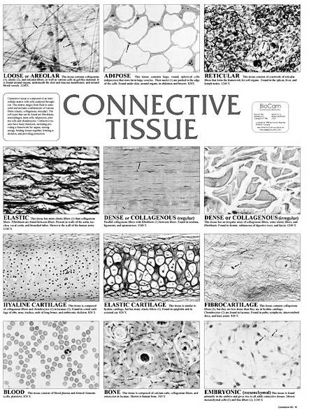
10 10 Q9. Where are the CTs tissues found? Go back to Q8. Under each, write down a short list of where you might find each of the tissues, Include the role they play. Concentrate on anything your instructor told you. Your book will have examples, also. Below is an example for "adipose" (Compare this with your instructor wants for adipose, before you write it down on Q3!!!!) Adipose - Fat (energy) storage, and protection. Found under skin (subcutaneous fat) and surrounding most visceral organs. Q10. Name the cells of Connective Tissue. Go back to Q8, and under each tissue named, write the names of any cells that makes the tissue. Limit yourself to the name of the "mature" cells (the names that end in "cyte"). Below is an example for "adipose" (Compare this with your instructor wants for adipose, before you write it down on Q3!!!!) Adipocytes: fat-storage cells Q11. ID the cells and matrix of Connective Tissue. Read all the instructions below before proceeding! There is a wall chart in the lab entitled "Connective Tissues". If you are in the lab, go to the wall chart and, for each tissue present, identify the cells and the matrix (just by pointing). We have included a photo of this wall chart on the next page of this document for you to make notes, or use if the wall chart is tied up! Read Me FIRST For some of the tissues, this will be difficult. Start with blood, which should be fairly easy. Then, try the cartilages, bone, and areolar. If you can't do it for reticular, dense regular, and elastic, do not worry; just ask your instructor for help later. If you cannot identify the cells and matrix on one or more, ask your instructor for help in lab!!!!! Please skip "embryonic" WARNING: Be careful identifying the matrix of adipose tissue!
11 11
12 Step 3. Remembering important concepts from the muscle and nervous tissues videos 12 Q 12. What are the general functions of muscle tissues? (there is more than one answer, and more than one way to say it. Make sure you are focusing on what your instructor wants you to know) Q 13. What are the general functions of nervous tissues? (there is more than one answer, and more than one way to say it. Make sure you are focusing on what your instructor wants you to know) Q 14. Which types of muscle tissues do you need to ID on the microscope?? Q 15. What do we call muscle cells?? Q 16. Choose from the list below. Both Skeletal Muscle and Cardiac Muscle are muscle tissue. Visceral Voluntary Non-contractile Striated Multi-nucleated with many nuclei Q 17. Choose from the list below. Since they are both striated, I need to look for on cardiac tissue to tell them apart. Q 18. Peristalsis is caused by which tissue? Nuclei Intercalated Discs Myofibrils Z-lines
13 13 Q 19. What do we call the nervous cells that transmit signals (or information)?? Q 20. Neurons have a lot of long projections coming off their body. These are called (choose one): Dendrites Soma Myofibrils Satellite cells

14 Step 4. Putting tissues in context: tissues versus tissue layers 14 Read Me #1 Read Me This may be done later to save time in lab! If you are past the first hour, skip this until later, and go to STEP 5. Have someone in your group read the following out loud, while the others read along: Opening Paragraph (WE WILL BE REFERING TO THIS LATER): The term 'Visceral organs' is not an exact term. It usually refers to the abdominopelvic and thoracic organs involved in Digestion, Reproduction, Respiration, Excretion. Most visceral organs can be thought of as a tube. As a substance is moved through the tube, the tube either extracts stuff from the tube (absorption), or it puts substances into the tube (secretion, or excretion. We'll talk about the differences between those 2 terms later!). The open area inside the tube is called the 'lumen'. It will be white...or at least very light in color. Sometimes there will be some debris in the lumen. Surrounding the lumen are 'tissue layers', which are a collection of tissues, with a specific task. The exact nature of each 'tissue layer" depends on the organ itself. Many (but not all) visceral organs have the tissue layers seen on the image below. Look at the layers in the image. Layers: LUMEN Read Me Not all visceral organs have these 4, but they are common. We'll see them in a lot of organs. As mentioned, each 'tissue layer' has more than 1 tissue, and has a very important function. For example, if an organ has a mucosa, it is a wet layer that helps the organ absorb from the lumen & secrete into the lumen. The submucosa often contains blood vessels, so absorbed substances can be carried to the rest of the body, and glands for secreting. The muscularis helps move substances through the tube. The serosa covers the organ, helping to protect it and attaching the organ to other structures.
15 #2 Now answer the following questions: 15 Q21. What is the difference between saying "organ" and "visceral organ" Which of the following are a "visceral organs"? Put a "check mark" next to the ones that are visceral organs. (HINT: the "Opening Paragraph" told you to consider which ORGAN SYSTEMS are involved): Small intestines Trachea and Lungs Esophagus & Stomach Bladder & Urethra Uterus & Vagina Q22. Discuss in your group: Relate what we learned in the "opening paragraph" to the organs you checked above. Can they all be described "as a tube"? Do they move things? Do they secrete and or absorbed something? If you have questions about this, ask your instructor. Q23. The opening paragraph mentioned 4 commonly seen tissue layers found in many visceral organs. Name them, moving from the lumen outward, and write down their functions next to their name: Q24. Answer these: Circle all of these that the small intestines might absorb into the bloodstream: Bacteria Nutrients in food Water Pollen Circle something the stomach might secrete into its lumen, using glands: Digestive enzymes Blood Urine Circle something the uterus might move through its lumen, using muscular action: Mucus Baby Urine Circle something the uterus might secrete into its lumen, using glands: Mucus Baby Sperm
 16 #3 Read Me We just got done looking at epithelial tissues in lab. Where might we find them? Well, all the tissue layers may have some epithelial tissues.")
16 Q25. Which can be thought of as a "mini-organ", a tissue or a tissue layer? WHY?? (HINT: the "Opening Paragraph" told you the difference between the two!) 16 #3 Read Me We just got done looking at epithelial tissues in lab. Where might we find them? Well, all the tissue layers may have some epithelial tissues. But they are especially visually obvious in the mucosal layer, where it is often stratified squamous (for protection) or columnar (for absorption & secretion), and the submucosa, where we'll see a lot of glands. Look at the image below, going through the numbered steps (first "1", then "2"): Places on a typical visceral organ, where epithelial tissues are often visually easy to see: #4 Now answer the following questions:


17 17 Q26. Label the following on the image below. While doing this, indicate "a layer" by highlighting its boundaries: Lumen Mucosa Submucosa Muscularis Serosa Q27. Below is a crude drawing of what we saw in the last question. Label the following on the image below: Lumen Mucosa Submucosa Muscularis Serosa Apical surface (draw a line over it...more or less) Basement Membrane (draw a line over it...more or less) Q28. On the drawing, indicate where we might find the following, by placing the number in the correct layer: 1. Columnar cells that are absorbing 2. Goblet cells secreting mucus 3. Glands secreting into the lumen 4. Stratified squamosal cells protecting against bacteria in the lumen 5. Muscle tissue moving substances through the lumen
 Basement Membrane (draw a line over it.")
18 18 Q29. Below is a close up of the same organ. Compare it to the images on the previous 2 pages. Label the following on the image below. While doing this, indicate "a layer" by highlighting its boundaries: Lumen Mucosa Submucosa Muscularis Serosa Apical surface (draw a line over it...more or less) Basement Membrane (draw a line over it...more or less) ALSO: On the drawing, indicate where we might find the following, by placing the number in the correct layer: 1. Columnar cells that are absorbing 2. Goblet cells secreting mucus 3. Glands secreting into the lumen 4. Stratified squamosal cells protecting against bacteria in the lumen 5. Muscle tissue moving substances through the lumen


19 19 Q30. The images in the boxes labeled "A through H" are close-ups of the" visceral organ" photo near the bottom. They are at various powers. Can you determine where they come from, MORE OR LESS, on the photo of the visceral tube? Try drawing a box on the tube where you think they might go. For example, this image : A would go in the box marked "A" on the photo. B C D E A F G Visceral organ H
20 Step 5. Tissue ID Microscope Stations 20 Read Me Aloud!! There are several microscopes set up in the room. Each has a tissue slide. You do not have to do the stations in order; if someone is at a station, go to another. PLEASE NOTE: some of the stations are meant to be studied together!! For each, make a drawing of the tissue, note which power you are looking at. Come up with a descriptor term for each ("it reminds me of donuts", "it looks like clouds", etc.) Make a note regarding location. You can use the answers you made to earlier questions. This way, you'll have a study guide for the Lab Practical in one place! Jot down any important info your instructor wants you to know, including cell names.
21 Stations 1 & 2: these 2 tissues should be studied together! 21 Station 1 Station 2 Tissue Type General Tissue Classification: Power: Descriptor Term: Write down some representative locations for this tissue Write down any extra information your instructor wants you to know, including cell names special proteins, other key words, etc. Drawing box SPECIAL QUESTION: name the structures where both these tissues are found. What do these cells do (absorb, secrete, transmit info, cause movement, connect, protect, store energy, other??)
22 Stations 3 & 4 - look at 2 tissues that are in different classes 22 Station 3 Station 4 Tissue Type General Tissue Classification: Power: Descriptor Term: Write down some representative locations for this tissue Write down any extra information your instructor wants you to know, including cell names, special proteins, other key words, etc. Drawing box SPECIAL QUESTION: What sort of connective tissue is station 4? Loose, or dense?
23 Stations 5 & 6 - compare 2 tissues that are sometimes confused 23 Station 5 Station 6 Tissue Type General Tissue Classification: Power: Descriptor Term: Write down some representative locations for this tissue Write down any extra information your instructor wants you to know, including cell names, special proteins, other key words, etc. Drawing box SPECIAL QUESTION: Go to a higher power on both slides (go back to original power for the next group). Can you see the cell nuclei on station 5? Now go back to the lower power, and re-examine.
24 Stations 7 & 8 - compare 2 tissues that are sometimes confused 24 Station 7 Station 8 Tissue Type General Tissue Classification: Power: Descriptor Term: Write down some representative locations for this tissue Write down any extra information your instructor wants you to know, including cell names, special proteins, other key words, etc. Drawing box SPECIAL QUESTION: pseudostratified columnar will have on the apical surface. We often see a border on simple columnar's apical surface. Both types may exhibit cells dispersed throughout.
25 Stations 9 & 10 - compare 2 tissues that are sometimes confused 25 You will also be using # 10 on the next page! Station 9 Station 10 Tissue Type General Tissue Classification: Power: Descriptor Term: Write down some representative locations for this tissue Write down any extra information your instructor wants you to know, including cell names, special proteins, other key words, etc. Drawing box SPECIAL QUESTION: Both these tissue types have visible in the cytoplasm. But cardiac tissue also has discs.
26 Stations 10 & 11 - compare 2 tissues that are sometimes confused 26 Notice that the slide at station 10 looks similar to that on Station 11. In fact, the slide at Station 11 has BOTH tissues on. Find a place on the slide where there are both. Can you see striations? Station 11 Tissue Type General Tissue Classification: Power: Descriptor Term: Write down some representative locations for this tissue Write down any extra information your instructor wants you to know, including cell names, special proteins, other key words, etc. Drawing box SPECIAL QUESTION: Why does this slide have both tissues? What organ is it taken from that would have both?
27 Stations 12 & 13 - look at 2 tissues that are in different classes 27 The pointer on Station #13 indicated the tissue you are interested in. What other tissue types are visible on slide 13? Label them in your drawing below. Station 12 Station 13 Tissue Type General Tissue Classification: Power: Descriptor Term: Write down some representative locations for this tissue Write down any extra information your instructor wants you to know, including cell names, special proteins, other key words, etc. Drawing box SPECIAL QUESTION: To tell connective and epithelial tissues apart, it is always good to look for the lumen. Can you find the lumen on both slides? Label it on your drawings.
28 28 Stations 14 & 15 - look at 2 tissues that cannot be confused with any other Station 14 Station 15 Tissue Type General Tissue Classification: Power: Descriptor Term: Write down some representative locations for this tissue Write down any extra information your instructor wants you to know, including cell names, special proteins, other key words, etc. Drawing box SPECIAL QUESTION: Make sure you name all the parts of the bone slide your instructor wants you to know!
29 Demo Station at the front of the room - 29 There are 5 slides. First slide: Small low power Find the lumen, mucosa, submucosa, muscularis externa, and serosa Second slide: Small intestine What special cell is indicated by the pointer? Third slide: Cardiac muscle What special structure is indicated by the pointer? Fourth and Fifth slide: hyaline cartilage & compact bone. What special structure is indicated by the pointer?
30 Step 6. Muscle Tissue and Neuron Models 30 Read Me There are 4 models in the room. We will be looking at all 3 in more detail in later labs. For right now: 1. be able to ID them. 2. Find the striations on cardiac and skeletal muscle models. 3. Find the intercalated discs on the cardiac model. 4. Find the body, dendrites, and axon on the neuron model. The End!
A&P 1 Histology Labs Guide #7- Putting Epithelial Tissues In Context: Tissue Layers Lab Exercises
 1 &P 1 Histology Labs Guide #7- Putting Epithelial Tissues In Context: Tissue Layers Lab Exercises Have someone in your group read the following out loud, while the others read along: In this "Walk bout",
A&P 1 Histology Labs #12 - Post-Lab Exercises: Putting Connective, Muscle and Nervous Tissues In Context: Re-visit Tissue Layers
 1 A&P 1 Histology Labs #12 - Post-Lab Exercises: Putting Connective, Muscle and Nervous Tissues In Context: Re-visit Tissue Layers Have someone in your group read the following out loud, while the others
Histology 101! !! Name:! Block: Identify and describe the functions of major tissue types including their subclasses and varieties!
 Histology 101 Identify and describe the functions of major tissue types including their subclasses and varieties Name: Block: "1 Introduction to Tissues Histology Notes Tissue (living fabric) : groups
Epithelial Tissue. Simple Cuboidal Function: secretion and absorption. Simple Squamous
 Epithelial Tissue General Functions: Lines and covers organs Absorbs / secretes substances Gas exchange Protection Special Characteristics: - have an apical surface on top - have a basement membrane below
Lab 1 ANIMAL TISSUES
 Lab 1 ANIMAL TISSUES Levels of Organization Animals are multicellular heterotrophs whose cells lack cell walls. Most animals exhibit a hierarchical level of organization: Cells are organized into tissues
Basic Histology. By Mrs. Bailey
 Basic Histology By Mrs. Bailey Primary Tissues 1. Epithelial Tissue 2. Connective Tissue 3. Muscle Tissue 4. Nervous Tissue Very cellular Supported by underlying connective tissue Epithelial & connective
Anatomy and Physiology Tissue Review
 Anatomy and Physiology Tissue Review OVERVIEW Histology practicals can be rough, especially when access to slides is limited to the lab period. This resource provides an opportunity to learn or review
Classification of Tissues
 6 R e v i e w S h e e t Exercise Classification of Tissues NAME LAB TIME/DATE Tissue Structure and Function General Review 1. Define tissue. A group of cells similar to one another in structure that perform
TISSUES. Objectives. Tissues
 TISSUES Objectives Introduce the four major types of tissues Describe the general characteristics and functions of epithelial & connective tissue Name the major types of epithelial & connective tissues
Tissues (Histology) Ch. 3 Human Anatomy lecture
 Ch. 3 Human Anatomy lecture") I. Histology the study of tissues A. 4 basic tissue types epithelial connective muscle nervous Tissues (Histology) Ch. 3 Human Anatomy lecture B. Usually found in combinations to form organs. C. As you
I. Histology the study of tissues A. 4 basic tissue types epithelial connective muscle nervous Tissues (Histology) Ch. 3 Human Anatomy lecture B. Usually found in combinations to form organs. C. As you
Unit II: Tissues and Integumentary System
 Unit II: Tissues and Integumentary System 2.1 - Tissues Chapter 4 Written Response #1 1. What is a tissue? 2. What are four major types of tissues? Tissue Definition: a group or mass of similar cells working
Histology. Study of body tissues
 Histology Study of body tissues 2 Introduction to Body Tissues 1. Composed of specialized cells of similar structure and perform a common function 2. Four major types (4 Cs) a. Epithelial - Cover b. Connective
Epithelia of Coverings and Linings. Tissues. Tissue
 Tissue Tissues Chapter 3 Definition an aggregation of cells in which each cooperates with all others in the performance of a given function Examples of general functions Movement Protection Support Production
Lab Animal Tissue. LEARNING OBJECTIVES: To understand the relationship between the structure and function of different animal tissues
 Name: Bio A.P. PURPOSE: HYPOTHESIS: NONE Lab Animal Tissue BACKGROUND: In animals, groups of closely related cells specialized to perform the same function are called tissues. There are four general classes
HISTOLOGY. Simple squamal lungs
 HISTOLOGY Lab Objectives: Students should be able to... 1. Visually identify each class of tissue and examples within each class 2. Indicate the location (in the human body and/or organ) and function of
Classification of Tissues
 M06_MARI0000_00_SE_CH06.qxd 3/28/11 4:37 PM Page 35 NAME LAB TIME/DATE R E V I E W S H E E T EXERCISE 6 Classification of Tissues Tissue Structure and Function General Review 1. Define tissue. A group
Section B: Epithelial Tissue 1. Where are epithelial tissues found within the body? 2. What are the functions of the epithelial tissues?
 Tissue worksheet Name Section A: Intro to Histology Cells are the smallest units of life. In complex organisms, cells group together with one another based on similar structure and function to form tissues.
Tissues and Structures to Know for the Lab Practical
 Ch. 3 - Cells and Tissues Tissues and Structures to Know for the Lab Practical Miss School, Miss Out! Simple squamous epithelium line and cover; site of diffusion Simple squamous epithelium apical surface
The Tissue Level of Organization
 Tissue The Tissue Level of Organization Chapter 3 Definition an aggregation of cells in which each cooperates with all others in the performance of a given function Examples of general functions Movement
Tissue = groups of cells that are similar in structure and function
 Tissue = groups of cells that are similar in structure and function Types Epithelial - covering Connective - support Muscle - movement Nervous - control Membranes line body cavities and hold organs together
Outline. Bio 105: Tissues Laboratory. Organization of the Human Body. Tissue - Epithelium. Tissues 3/2/ Copyright 2009 Pearson Education, Inc
 Outline Bio 105: Tissues Laboratory Laboratory 5 Reading: Chapter 4 I. Cell to cell contact II. Body Cavities III. Membranes IV. Homeostasis V. Integumentary System I. Includes skin, hair and nails 1 2
Tissues 10/21/2016. Epithelial Tissue
 Tissues This is a generalized cell diagram. It shows the anatomy of a cell, but most cells do not actually look like this. Cells can have a wide variety of shapes and sizes, depending on their function.
Chapter 4 Histology: The study of body tissues
 Chapter 4 Histology: The study of body tissues https://www.youtube.com/watch?v=zwxm2a0tfxm Body Tissues Cells are specialized for particular functions Tissues = groups of cells with similar structure and
Anatomy &- Physiology Histology Worksheet
 Anatomy &- Physiology Histology Worksheet 1. The four primary tissue types found in the human body are a) squamous, cuboidal, columnar, glandular b) adipose, elastic, reticular, cartilage c) skeletal,
Body Tissues Pearson Education, Inc.
 Body Tissues Tissues Groups of cells with similar structure and function Four primary types: Epithelial tissue (epithelium).1 Connective tissue.2 Muscle tissue.3 Nervous tissue.4 Epithelial Tissues Locations:
Cell and Tissue Types. Epithelial, Connective, Muscle, Nerve
 Cell and Tissue Types Epithelial, Connective, Muscle, Nerve Objectives Explain the major stages of the cell cycle and cellular division (mitosis). Describe specific events occurring in each of the phases
Pick a cell that isn t yours!
 Pick a cell that isn t yours! Quiz 1: Introduction and Cells Module 2: Histology The study of tissues This module is very visual! Know these images! Introduction www.quizlet.com is a very useful tool for
Anatomy Chapter 4 Tissues
 4 Principle Tissue Types Epithelial tissue Covering and lining Glandular Connective tissue Highly variable Most abundant tissue type Muscular tissue 3 major types Produce force through contraction Nervous
BIOL 2457 CHAPTER 4 Part 2 SI All connective tissues arise from, an embryonic tissue.
 BIOL 2457 CHAPTER 4 Part 2 SI 1 1. All connective tissues arise from, an embryonic tissue. 2. Describe the vascularity of connective tissues, which are very diverse. 3. Describe the innervation of connective
Tissues. Group of cells that are similar in structure and function. 4 primary types. Epithelium (covering) Connective (support) Nervous(control)
 Connective (support) Nervous(control)") Tissues Tissues Group of cells that are similar in structure and function 4 primary types Epithelium (covering) Connective (support) Nervous(control) Epithelial tissue (epithelium) Lining, covering, and
Tissues Tissues Group of cells that are similar in structure and function 4 primary types Epithelium (covering) Connective (support) Nervous(control) Epithelial tissue (epithelium) Lining, covering, and
Epithelial Tissue lining, covering, glandular tissue > Function protect, absorption, filtration, secretion, excretion
 Chapter 4: TISSUES IX. Tissues Intro Epithelial Tissue lining, covering, glandular tissue > Function protect, absorption, filtration, secretion, excretion Connective Tissue most widespread tissue type
TISSUE. A group of cells that perform a similar function within an organism. Epithelium Connective Muscle Nervous CREDITS
 TISSUE A group of cells that perform a similar function within an organism. Epithelium Connective Muscle Nervous CREDITS Epithelium Connective Muscle Nervous Epithelium Composed of a layer of cells. Lines
Name: Test Date: Chapter 4- Tissues. Use the choices to identify the major tissue types found below:
 Name: Test Date: Chapter 4- Tissues Use the choices to identify the major tissue types found below: A. Connective B. Epithelium C. Muscle D. Nervous 1. B Lines body cavities and covers the body s external
Tissues Chapter 5...Tissue - a group or mass of similar cells working together to perform certain common functions
 Tissues Chapter 5...Tissue - a group or mass of similar cells working together to perform certain common functions There are 4 major types of tissue Epithelial Connective Muscle Nervous 1. Epithelial Tissue
Air sacs of lungs and the lining of the heart, blood vessels, and lymphatic vessels
 Cells Location Function Simple squamous epithelium Air sacs of lungs and the lining of the heart, blood vessels, and lymphatic vessels Allows materials to pass through by diffusion and filtration, and
Basic Tissue Types and Functions
 Tissues Histology Basic Tissue Types and Functions 1) Epithelial tissue covering 2) Connective tissue support 3) Muscle tissue movement 4) Nervous tissue control Epithelial Tissue 1) Covers a body surface
B. Classification of epithelium: by number of cell layers present and by shape of the superficial cell layers.
 I. Introduction - tissue: group of cells that are closely associated, similar in structure and function, and perform a common or related function. - four primary tissues: epithelial tissue, connective
Epithelium. Four primary tissue types:
 Epithelium Four primary tissue types: Epithelial (covering) Connective (support) Nervous (control) Muscular (movement) Smooth muscle Cardiac muscle Skeletal muscle 1 Epithelial Tissue Features Epithelial
Chapter 5. Tissues. 4 Types of Body Tissues. Tissues
 Chapter 5 Tissues Tissues Tissues - groups of cells that are similar in structure & function RBC, WBC, & platelets are a group of cells working together to form BLOOD tissue Histology Pathohistology study
Tissues. groups of cells similar in structure and function 4 types. epithelium connective muscle nervous
 Tissues groups of cells similar in structure and function 4 types epithelium connective muscle nervous Epithelial Tissue lining covering glandular Functions protection absorption filtration secretion Epithelium
What is histology? HISTOLOGY
 Introduction to Histology What is histology? HISTOLOGY histo = tissue ogy = study So HISTOLOGY = the study of tissues! What is a TISSUE? Tissues are groups of cells with specialized structural and functional
8/30/2017. Tissue: The Living Fabric. 4.3 Connective Tissue
 Chapter 4 Part B Tissue: The Living Fabric Annie Leibovitz/Contact Press Images PowerPoint Lecture Slides prepared by Karen Dunbar Kareiva Ivy Tech Community College 4.3 Connective Tissue Connective tissue
Use for reference if needed:
 A- 2.5 Describe how structure and function are related in terms of cell and tissue types. I can recognize different types of body tissue. I can explain how different tissue structures affect their functions.
Study of different tissues Abnormal cells and tissues can be compared to normal tissues to identify disease, such as cancer Being able to know and
 CHAPTER 4 Study of different tissues Abnormal cells and tissues can be compared to normal tissues to identify disease, such as cancer Being able to know and recognize normal tissues under the microscope
NOTES: CH 40 Introduction to Human Anatomy & Physiology
 NOTES: CH 40 Introduction to Human Anatomy & Physiology THE HUMAN BODY Anatomy Physiology (= structures) (= functions or processes) Characteristics of LIFE: 1) Made up of 1 or more CELLS. 2) Obtain and
THE TISSUE LEVEL OF ORGANIZATION PART I: EPITHELIAL TISSUE
 THE TISSUE LEVEL OF ORGANIZATION PART I: EPITHELIAL TISSUE 4 Main Tissue Types Epithelium Covers surfaces, lines cavities, forms glands Connective Tissue Support and protects body Muscular Tissue Movement
A. cells that perform related functions and are similar in structure. B. extracellular material - made by cells and secreted into interstitial space
 I. tissue components A. cells that perform related functions and are similar in structure B. extracellular material - made by cells and secreted into interstitial space II. tissue types A. epithelium (e.)
Chapter 1: Cells and Tissues
 Chapter 1: Cells and Tissues Cells and Tissues Carry out all chemical activities needed to sustain life Cells are the building blocks of all living things Tissues are groups of cells that are similar in
Epithelial Tissues. Types of Epithelial Tissues: Lining of Kidney
 Epithelial Tissues Covers the entire body surface and most of the body s inner cavities Outer epidermis (skin) protects from injury and drying out Inner epidermal tissue (on internal surfaces) often serves
Unit I Problem 9 Histology: Basic Tissues of The Body
 Unit I Problem 9 Histology: Basic Tissues of The Body - What is the difference between cytology and histology? Cytology: it is the study of the structure and functions of cells and their contents. Histology:
Tissues. How do cells form tissues?
 Tissues How do cells form tissues? Using cell junctions Tissues Epithelial tissue Connective tissue Muscle tissue Nervous tissue Epithelial Tissue Closely packed cells in continuous sheets connected by
Body Tissues. Cells are specialized for particular functions Tissues - groups of cells with similar structure. and function Four primary tissue types:
 Chapter 3 Tissues Body Tissues Cells are specialized for particular functions Tissues - groups of cells with similar structure and function Four primary tissue types: Epithelium Connective tissue Nervous
Human anatomy Unit III. Tissue
 Human anatomy Unit III Tissue Definition of Tissues Biological tissue is a collection of interconnected cells that perform a similar function within an organism. In other words, it is a group of cells
d SIMPLE EPITHELIA Top view Side view
 Chapter Two I UPLANd I 23 Cells, Tissues, and Integument me lea SIMPLE EPITHELIA There are four types of tissues in humans and these make up all of the organs and binding material in the body. Epithelial
Tissues. Tissues. Four basic tissues. A collection of cells with a common function. 1. Epithelial 2. Connective 3. Muscular 4.
 Tissues Tissues A collection of cells with a common function Four basic tissues 1. Epithelial 2. Connective 3. Muscular 4. Nervous Epithelia: cells in layers Types of epithelia 1) lining Layers of cells
UNIT 4 T I S S U E S
 UNIT 4 T I S S U E S WHAT IS A TISSUE Group of cells that work together to do a function Cells are similar Extracellular fluid around them is similar Histology EPITHELIAL TISSUE Also called epithelium
Tissues and Membranes
 I. In the Beginning a. Egg + sperm! Tissues and Membranes b. 1 cell divides to make 2, 2 divide to make 4, 4 divide to make 8, and then? c. d. e. Totipotent: f. Pluripotent: II. III. Tissues a. Tissues
HOLE S ANATOMY CHAPTER 5, PART II Lecture notes
 HOLE S ANATOMY CHAPTER 5, PART II Lecture notes I. Connective Tissue A. Structure 1. have few cells that are spaced apart and can divide; two categories: a. fixed cells cells that are present in tissue
Hole s Human Anatomy and Physiology
 Hole s Human Anatomy and Physiology 1 Chapter 5 Tissues Four major tissue types 1. Epithelial 2. Connective 3. Muscle 4. Nervous 2 Epithelial Tissues General characteristics - cover organs and the body
TISSUE, INFLAMMATION AND REPAIR
 TISSUE, INFLAMMATION AND REPAIR TISSUE DEFINITION A group of cells with similar function/s and structure/morphology and similar extracellular substance HISTOLOGY The study of normal tissue HISTOPATHOLOGY
Chapter 05. Review. Copyright The McGraw-Hill Companies, Inc. Permission required for reproduction or display.
 Chapter 05 Review 5.1: Introduction Similar cells with a common function are called tissues. The study of tissues is called histology. There are four (4) primary or major tissue types: 1. Epithelial Tissue
Histology. There are four basic tissue types in the body are :-
 Histology Lab.I There are four basic tissue types in the body are :- 1- Epithelial tissues (Epithelium) 2- Connective tissues 3- Muscular tissues 4- Nervous tissues 1-Epithelial tissues epithelial tissues
TISSUES. Dr. Gary Mumaugh
 TISSUES Dr. Gary Mumaugh Tissues Tissues - Groups of cells similar in structure and function and perform a common function Histology The study of tissues The four types of tissues Epithelial Connective
Tissues, Glands, and Membranes. Chapter Five Mrs. Hornacek
 Tissues, Glands, and Membranes Chapter Five Mrs. Hornacek Objectives 1. Name the four main groups of tissues and give the location and general characteristics of each. 2. Differentiate between voluntary
They cells can not function death.
 Jenna Hellack Jan 2001 Tissues What do you think happens when the cells use up their food and oxygen before there is time to replenish it? They cells can not function death. Blood Cell Cancer cell Plant
Histology= the study of tissues
 Unit 3-Histology Histology= the study of tissues A tissue is a group of cells that have a similar shape and function. Different types of tissues can be found in different organs. In humans, there are four
BIOLOGY. Chapter 33 Animal Body: Histology Portion Pearson Education, Inc.
 BIOLOGY Chapter 33 Animal Body: Histology Portion Tissues: groups of cells with common function Tissue Category Epithelial (covers & lines) Simple squamous Simple cuboidal Simple columnar Tissues to know:
Histology. Becoming familiar with tissues of the Human Body. structure determines function
 Histology Becoming familiar with tissues of the Human Body structure determines function Histology is the study of the microscopic structure of tissues. Familiarity with tissue structure is essential to
What is a tissue? Points to ponder. Tissues Connective Tissue. 1. Connective tissue 2/23/2019. Organization and Regulation of Body Systems
 Organization and Regulation of Body Systems Chapter 04 Lecture Outline See separate PowerPoint slides for all figures and tables preinserted into PowerPoint without notes. Copyright 2016 McGraw-Hill Education.
Tissue Outline (chapter 4) Tissues group of cells that perform structural and roles. List the 4 types:
 Tissues group of cells that perform structural and roles. List the 4 types:") Tissue Outline (chapter 4) Tissues group of cells that perform structural and roles. List the 4 types: 1. 2. 3. 4. I. Epithelial Tissue covers all the surfaces, inside & out. Are the major tissues of,
Tissue Outline (chapter 4) Tissues group of cells that perform structural and roles. List the 4 types: 1. 2. 3. 4. I. Epithelial Tissue covers all the surfaces, inside & out. Are the major tissues of,
Connexons: hollow connective tubes
 Chapter 3 1. tight junctions: like a zipper, these junctions hold the cells tightly together making them impermeable to the extracellular fluid that surrounds them. 2. desmosomes: like buttons, these
PRACTICAL HISTOLOGY LAB
 PRACTICAL HISTOLOGY LAB.1 ----------------------------------------------------------------------------- INTRODUCTION Cells are the smallest units of life, and are named according to their function. Cells
Tissues. Tissues - Overview. Bio211 Laboratory 2. Epithelial and Connective Tissues
 Bio211 Laboratory 2 Epithelial and Connective Tissues 1 Tissues Tissues to be examined under the microscope Epithelial Tissue (p. 79 Lab Manual) [TODAY] Connective Tissue (p. 93 Lab Manual) [TODAY] Muscle/Nervous
Biology 325 Fall 2003
 Name: MULTIPLE CHOICE. Choose the one alternative that best completes the statement or answers the question. 1) Which of the following is not one of the primary tissue types? A) germinative tissue B) muscle
Brief Overview of Tissues STUDENT NOTES Date: 1. Tissue 2. Connective Tissue. 3. Tissue 4. Nervous Tissue
 Levels of Structural Organization: Brief Overview of Tissues STUDENT NOTES Date: Recall The four major tissue types include: 1. Tissue 2. Connective Tissue 3. Tissue 4. Nervous Tissue Epithelial Tissue
Essentials of Anatomy and Physiology, 9e (Marieb) Chapter 3 Cells and Tissues. Short Answer. Figure 3.1
 Chapter 3 Cells and Tissues. Short Answer. Figure 3.1") Essentials of Anatomy and Physiology, 9e (Marieb) Chapter 3 Cells and Tissues Short Answer Figure 3.1 Using Figure 3.1, match the following: 1) The illustration of simple cuboidal epithelium is. Answer:
Essentials of Anatomy and Physiology, 9e (Marieb) Chapter 3 Cells and Tissues Short Answer Figure 3.1 Using Figure 3.1, match the following: 1) The illustration of simple cuboidal epithelium is. Answer:
ACTIVITY 2: HISTOLOGY AND INTEGUMENT
 ACTIVITY 2: HISTOLOGY AND INTEGUMENT Objectives: 1) How to get ready: Read Chapter 4 and 5, McKinley et al., Human Anatomy, 4e. All text references are for this textbook. 2) Identify each tissue (26 tissues)
Anatomy & Homeostasis. Unit 5
 Anatomy & Homeostasis Unit 5 Main Ideas discuss with a buddy 2 What is Homeostasis? How is homeostasis different in single-celled organisms vs. multicellular organisms? What unique challenges to maintaining
Chapter 20 UNIFYING CONCEPTS OF ANIMAL STRUCTURE AND FUNCTION
 Chapter 20 UNIFYING CONCEPTS OF ANIMAL STRUCTURE AND FUNCTION I. Life is based on many structural levels Levels of animal structure: Atoms and molecules Cells Tissues Organs Organ systems Organism: May
Tissues Description Function(s) Locations Miscellaneous. avascular -thelium = covering
 Locations Miscellaneous. avascular -thelium = covering") Epithelial Tissue Simple Squamous flattened cells diffusion and Kidney glomeruli disc-shaped central filtration air sacs of lung Simple = Single layer nuclei secretes lubricating lining of heart, blood
Epithelial Tissue Simple Squamous flattened cells diffusion and Kidney glomeruli disc-shaped central filtration air sacs of lung Simple = Single layer nuclei secretes lubricating lining of heart, blood
Study of Tissues Dr. A. Ebneshahidi
 Study of Tissues Dr. A. Ebneshahidi Tissues Tissues are composed of cells similar in structure and specialized to perform a specific function for the body. The human body is made of four general types
Tissues. Student Learning Objectives:
 Tissues Student Learning Objectives: Distinguish between the different varieties of tissue: epithelium, connective tissue, muscle, and nervous tissue. Types of tissues: Epithelium: Simple Simple squamous
Anatomy and Physiology 1 Chapter 4 Outline Tissues and Membranes
 Anatomy and Physiology 1 Chapter 4 Outline Tissues and Membranes 1 Tissue group of cells with similar structure and function o 4 major groups epithelial, connective, muscle, nerve Epithelial tissue (Fig
Simple Squamous Epithelium
 Histology Simple Squamous Epithelium One layer of flattened cells. Protective characteristics are diminished because of this. Examples: Alveoli in the lungs Capillaries where diffusion of nutrients and
Dr. Heba Kalbouneh. Dr. Heba Kalbouneh. Dr. Heba Kalbouneh
 Dr. Heba Kalbouneh Dr. Heba Kalbouneh Dr. Heba Kalbouneh Tissue: is a group of cells that serve the same function, they are surrounded by extra cellular matrix. The 4 basic types of tissue: 1. epithelial
DEPARTMENT OF PHYSICAL EDUCATION
 DEPARTMENT OF PHYSICAL EDUCATION Subject:- Anatomy, Physiology and Health Education Class:- B.P. Ed. Semester- I Presented By:- Dr. Mahesh Singh Dhapola Cell Theory All living things are made up of cells.
Tissue: The Living Fabric: Part A
 PowerPoint Lecture Slides prepared by Janice Meeking, Mount Royal College C H A P T E R 4 Tissue: The Living Fabric: Part A Tissues Groups of cells similar in structure and function Types of tissues Epithelial
Lesson 9A Tissues in Animals
 Lesson 9A Tissues in Animals Levels of Organization in the Human Body Similar types of cells Different types of tissues Different organs Many organ systems cell tissue organ organ system organism Levels
Histology review. Histology. Slides. Epithelial tissue. Another example - kidney. Simple cuboidal epithelium. What to look for
 Histology review Histology What to look for Histology Practical = 50 pts Some slides set up on scopes (~10) Some Powerpoint pictures on the projector Questions I will ask: What kind of tissue? General
Introduction to Types of Body Tissue Putting it All Together. Packet #12
 Introduction to Types of Body Tissue Putting it All Together Packet #12 Introduction Body Tissues Tissues Groups of cells with similar structure and function Four primary types Epithelial tissue (epithelium)
Histology= the study of tissues
 Histology 2014 Histology= the study of tissues A tissue is a group of cells that have a similar shape and function. Different types of tissues can be found in different organs. In humans, there are four
Histology Notes -Part 1: Epithelial Tissues
 Introduction Group of cells w/ similar structure & function = TISSUE Four Basic Tissue Types 1. Epithelial-covers 2. Connective-supports 3. Muscular*-produces movement (will discuss in the muscular system
Chapter 4 :Organization & Regulation of Body Systems
 Chapter 4 :Organization & Regulation of Body Systems 4.1 Types of tissues What is a tissue? A collection of cells of the same type that perform a common function There are 4 major tissue types in the body:
Tissues. tissue = many cells w/ same structure and function. cell shape aids function tissue shape aids function. Histology = study of tissues
 Tissues tissue = many cells w/ same structure and function cell shape aids function tissue shape aids function Histology = study of tissues 4 types of tissues Epithelial coverings contact openings Connective
Cells are specialized for particular functions Tissues
 Histology Body Tissues Cells are specialized for particular functions Tissues Groups of cells with similar structure and function Extracellular Matrix cell glue between cells Histology study of tissue
Tissues are: group of similar or identical cells that share a common function. used to build organs
 Tissues: Four classes Epithelium Connective Muscle Nervous Tissues are: group of similar or identical cells that share a common function. used to build organs Overview: Epithelial o Line body cavities
ACTIVITY 2: HISTOLOGY AND INTEGUMENT
 ACTIVITY 2: HISTOLOGY AND INTEGUMENT Objectives: 1) How to get ready: Read Chapter 4 and 5, McKinley et al., Human Anatomy, 5e. All text references are for this textbook. 2) Identify each tissue (26 tissues)
A. Incorrect! Axons covey messages from the cell body of the neuron. D. Correct! Dendrites convey messages to the cell body of the neuron.
 CLEP Biology - Problem Drill 14: Animal Form No. 1 of 10 1. The branches of a neuron receiving information from another cell and which transmit the message to the cell body are called? (A) (B) (C) (D)
Histology. The study of tissues.
 Histology The study of tissues. Body Tissues Cells are specialized for particular functions Tissues Groups of cells with similar structure and function Four primary types Epithelium Connective tissue Nervous