HISTOLOGY. Simple squamal lungs
|
|
|
- Barbra Williams
- 6 years ago
- Views:
Transcription
 and function of each example. 3. Identify parts associated with specific examples of particular tissues 4.")
1 HISTOLOGY Lab Objectives: Students should be able to Visually identify each class of tissue and examples within each class 2. Indicate the location (in the human body and/or organ) and function of each example. 3. Identify parts associated with specific examples of particular tissues 4. Identify the various tissues and structures associated with the integumentary system I. Overview Tissue - groups of cells with similar structure and function Four principle human tissue types: Epithelium (covering) Connective (support) Muscle (movement) Nervous (control) II. Epithelium Epithelia are classified according to their shape and arrangement. Shape: squamal cuboidal, columnar Arrangement: simple (pseudostratified) or stratified Types of epithelium Simple squamal, simple cuboidal, simple columnar, pseudostratified columnar, stratified squamal, stratified cuboidal, stratified columnar, and transitional Know all types of epithelium and their description, function, and location Epithelium that will be observed in the lab: Simple squamal lungs Key terms to know: alveoli and alveolar space In this image you see round spaces made up of individual flattened cells (simple squamal epithelium) linked together. Each round space is called an alveolus.


 tubules. Inside each circle is space and is referred to as the lumen.")
2 Simple squamal kidneys Key terms to know: Bowmans (Glomerular) capsule, renal corpuscle, renal tubule (nephron) In this image you see a round structure that contains a mass of cells insides. Under the microscope you will see that surrounding this round structure is a single layer of flat cells. The single layer in this tissue is called the Bowman s capsule and the inner mass of cells are called the Glomerulus Simple cuboidal kidneys Key terms to know: renal tubule (nephron) In this image you see circles composed of one layer of cube-shaped cells (simple cuboidal epithelium). The circles are kidney (renal) tubules. Inside each circle is space and is referred to as the lumen. Simple columnar intestine (and/or observe this tissue in the gall bladder) Key terms to know: goblet cells and cilia In this image you see tissue lined with a single layer of column-shaped cells (simple columnar epithelium)


3 Pseudostratified -(ciliated columnar) lungs Key terms to know: goblet cells and cilia In this image you see (on the left hand side) column-shaped cells that are staggered to give you the impression that there is more than one layer of cells. However, there is only one layer! In addition, the cells should have long wispy cell extensions called cilia. Stratified squamal tongue Key terms to know: keratinized and non-keratinized In this image you see a rounded structure called a papilla. On the top of this papilla are many layers of flat cells (stratified squamal epithelium). Also notice that there are clusters of lighter cells on either side of the papilla. These clusters are called taste buds. Stratified squamal skin Key terms to know: keratinized and non-keratinized In this image you see many layers of flat cells (stratified squamal epithelium)


4 Transitional urinary bladder In this image you see many layers of cells but do not see only one type of cell shape. Rather, you can see some that are all three epithelial cells shapes and even some round or triangular looking cells! III. Connective Tissue Connective tissue is classified based upon the presence of a cell and the type of extra-cellular matrix (ground substance and fibers) Classes of connective tissue: loose, dense, cartilage, and other Loose: areolar, adipose, and reticular Dense: dense regular and dense irregular Cartilage: hyaline, elastic, and fibrocartilage Other: bone and blood Know all types of connective tissue and their description, function, and location Connective tissue that will be observed in lab: Areolar - gell-like matrix contains all three fiber (collagen, elastin, and reticular); cell type = fibroblast; widely distributed under epithelium; and forms lamina propria and wraps and cushions organs.


, tendons (connecting bone to muscle), and aponeurosis (connecting muscle")
5 Adipose - matrix contains sparse amount of all three fibers; adipocytes have a flattened nucleus as a result of fat droplets contained inside of cell; used as fuel resource. Reticular - reticular fibers w/ reticular cells in loose ground substance; found in lymph nodes, bone marrow, and spleen; fibers form internal skeleton that supports other cell types Dense regular - fibroblasts with collagen fibers; forms ligaments (connecting bone to bone), tendons (connecting bone to muscle), and aponeurosis (connecting muscle to muscle).


6 Dense irregular irregularly arranged collagen fibers with fibroblast; found in the dermis of the skin and fibrous joint capsules; withstands tension and provides strength. Hyaline - chondroblasts produce matrix with few collagen fibers, chrondrocytes located within lacunae (spaces); found in ribs, nose, trachea, and the larynx; supports and reinforces. Elastic - chondroblasts produce matrix with elastic fibers; supports external ear; maintains shape and structure.


, Leukocytes (WBCs) and cell fragments called Thrombocytes (Platelets); located in")
7 Fibrocartilage - matrix similar to hyaline but less firm and also contains more and thicker collagen fibers; component of intervertebral discs; gives tensile strength. (slides may not be available) Bone (osseous) - hard, calcified matrix containing many collagen fibers; osteocytes lie in lacunae; high vascularized; forms skeletal system; provides support and protection as well as blood production. Obvious haversian (central) canal with many lamellae and canaliculi present. Blood - Erythrocytes (RBCs), Leukocytes (WBCs) and cell fragments called Thrombocytes (Platelets); located in blood vessels; transports respiratory gases, wastes, nutrients, etc...
.")
; attached to skeleton and skin; under")
; located in the walls of the heart; is under")
8 IV. Muscular Tissue Highly vascularized muscular tissue is comprised of elongated cells (called fibers) containing myofilaments (actin and myosin proteins). There three types of muscular tissue: skeletal, cardiac, and smooth Muscle tissue that will be observed in lab: Skeletal - long, cylindrical, multinucleate cells with striations (specific arrangement of actin and myosin); attached to skeleton and skin; under voluntary control; provides movement. Cardiac - branching, uninucleate, striated cells with junctions (intercalated discs); located in the walls of the heart; is under involuntary control; propels blood (circulation). Smooth - spindle-shaped, uninucleate cells without striations; found in the walls of hollow organs; under involuntary control; propels substances.

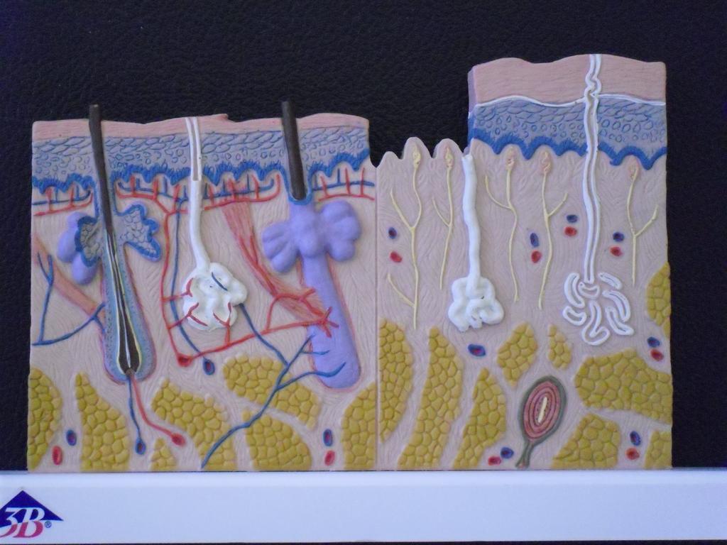
9 V. Nervous Tissue Nervous tissue (nervous system) conducts electrical impulses and has integrative functions. Nervous tissue is composed of neurons which are branching cells located in the brain, spinal cord, and nerves. Neurons transmit electrochemical signals from sensory receptors to effectors. Neuroglia are cells surrounding neurons and help to feed, support, and protect. Neurons are composed of dendrites, a cell body, and an axon. Nervous tissue that will be observed in lab: Nervous Tissue In this view you see a neuron with its cell extensions. What you cannot tell is whether or not the cells extensions are dendrites or an axon. VI. Use of the Integumentary System Skin is one component of the integumentary system Human skin is composed of many tissue types. Using slides provided, identify the various tissue types: stratifed squamous epithelium, dense irregular connective, adipose, and stratified cuboidal. In addition, please identify the two layers of the dermis (epidermis and dermis), the hypodermis (subcutaneous), and the following accessory structures associated with the skin: hair follicle and shaft, sebaceous gland, stratum corneum, stratum basale (if visible). Using the skin models provided, identify the following: epidermis, dermis, hypodermis, stratum corneum and basale, hair shaft and follcile, sebaceous and eccrine (sudoriferous) gland, Pacinian corpuscle, Meissner's corpuscle, dermal papillae, arrector pili, arteries and veins, and lymph vessels. Models used in lab:

10
Basic Histology. By Mrs. Bailey
 Basic Histology By Mrs. Bailey Primary Tissues 1. Epithelial Tissue 2. Connective Tissue 3. Muscle Tissue 4. Nervous Tissue Very cellular Supported by underlying connective tissue Epithelial & connective
Lab 1 ANIMAL TISSUES
 Lab 1 ANIMAL TISSUES Levels of Organization Animals are multicellular heterotrophs whose cells lack cell walls. Most animals exhibit a hierarchical level of organization: Cells are organized into tissues
Body Tissues Pearson Education, Inc.
 Body Tissues Tissues Groups of cells with similar structure and function Four primary types: Epithelial tissue (epithelium).1 Connective tissue.2 Muscle tissue.3 Nervous tissue.4 Epithelial Tissues Locations:
ACTIVITY 2: HISTOLOGY AND INTEGUMENT
 ACTIVITY 2: HISTOLOGY AND INTEGUMENT Objectives: 1) How to get ready: Read Chapter 4 and 5, McKinley et al., Human Anatomy, 5e. All text references are for this textbook. 2) Identify each tissue (26 tissues)
Outline. Bio 105: Tissues Laboratory. Organization of the Human Body. Tissue - Epithelium. Tissues 3/2/ Copyright 2009 Pearson Education, Inc
 Outline Bio 105: Tissues Laboratory Laboratory 5 Reading: Chapter 4 I. Cell to cell contact II. Body Cavities III. Membranes IV. Homeostasis V. Integumentary System I. Includes skin, hair and nails 1 2
10/3/2012. Tissue: The Living Fabric: Part B. Extracellular matrix Ground substance Fibers Collagen fiber Elastic fiber Reticular fiber.
 PowerPoint Lecture Slides prepared by Janice Meeking, Mount Royal College C H A P T E R 4 Tissue: The Living Fabric: Part B Copyright 2010 Pearson Education, Inc. Copyright 2010 Pearson Education, Inc.
Histology 101! !! Name:! Block: Identify and describe the functions of major tissue types including their subclasses and varieties!
 Histology 101 Identify and describe the functions of major tissue types including their subclasses and varieties Name: Block: "1 Introduction to Tissues Histology Notes Tissue (living fabric) : groups
What is a tissue? Points to ponder. Tissues Connective Tissue. 1. Connective tissue 2/23/2019. Organization and Regulation of Body Systems
 Organization and Regulation of Body Systems Chapter 04 Lecture Outline See separate PowerPoint slides for all figures and tables preinserted into PowerPoint without notes. Copyright 2016 McGraw-Hill Education.
Anatomy and Physiology Tissue Review
 Anatomy and Physiology Tissue Review OVERVIEW Histology practicals can be rough, especially when access to slides is limited to the lab period. This resource provides an opportunity to learn or review
ACTIVITY 2: HISTOLOGY AND INTEGUMENT
 ACTIVITY 2: HISTOLOGY AND INTEGUMENT Objectives: 1) How to get ready: Read Chapter 4 and 5, McKinley et al., Human Anatomy, 4e. All text references are for this textbook. 2) Identify each tissue (26 tissues)
THE TISSUE LEVEL OF ORGANIZATION PART I: EPITHELIAL TISSUE
 THE TISSUE LEVEL OF ORGANIZATION PART I: EPITHELIAL TISSUE 4 Main Tissue Types Epithelium Covers surfaces, lines cavities, forms glands Connective Tissue Support and protects body Muscular Tissue Movement
TISSUE. A group of cells that perform a similar function within an organism. Epithelium Connective Muscle Nervous CREDITS
 TISSUE A group of cells that perform a similar function within an organism. Epithelium Connective Muscle Nervous CREDITS Epithelium Connective Muscle Nervous Epithelium Composed of a layer of cells. Lines
Mitosis Models 3-5. Chromosome. #1 Prophase. #2 Prophase. 2n = 4 4 Chromosomes 8 Chromatids. 2n = 4
 MITOSIS Mitosis Models 3-5 Chromosome #1 Prophase 2n = 4 4 Chromosomes 8 Chromatids #2 Prophase 2n = 4 4 Chromosomes 8 Chromatids Mitosis Models 3-5 Astral Rays Chromosomes Chromosome Chromosome Spindle
Tissues 10/21/2016. Epithelial Tissue
 Tissues This is a generalized cell diagram. It shows the anatomy of a cell, but most cells do not actually look like this. Cells can have a wide variety of shapes and sizes, depending on their function.
The Tissue Level of Organization
 Tissue The Tissue Level of Organization Chapter 3 Definition an aggregation of cells in which each cooperates with all others in the performance of a given function Examples of general functions Movement
Epithelial Tissue. Simple Cuboidal Function: secretion and absorption. Simple Squamous
 Epithelial Tissue General Functions: Lines and covers organs Absorbs / secretes substances Gas exchange Protection Special Characteristics: - have an apical surface on top - have a basement membrane below
Histology review. Histology. Slides. Epithelial tissue. Another example - kidney. Simple cuboidal epithelium. What to look for
 Histology review Histology What to look for Histology Practical = 50 pts Some slides set up on scopes (~10) Some Powerpoint pictures on the projector Questions I will ask: What kind of tissue? General
Chapter 5. Tissues. 4 Types of Body Tissues. Tissues
 Chapter 5 Tissues Tissues Tissues - groups of cells that are similar in structure & function RBC, WBC, & platelets are a group of cells working together to form BLOOD tissue Histology Pathohistology study
Histology. Study of body tissues
 Histology Study of body tissues 2 Introduction to Body Tissues 1. Composed of specialized cells of similar structure and perform a common function 2. Four major types (4 Cs) a. Epithelial - Cover b. Connective
A. cells that perform related functions and are similar in structure. B. extracellular material - made by cells and secreted into interstitial space
 I. tissue components A. cells that perform related functions and are similar in structure B. extracellular material - made by cells and secreted into interstitial space II. tissue types A. epithelium (e.)
Epithelia of Coverings and Linings. Tissues. Tissue
 Tissue Tissues Chapter 3 Definition an aggregation of cells in which each cooperates with all others in the performance of a given function Examples of general functions Movement Protection Support Production
Tissues. Cells work together in functionally related groups called tissues Types of tissues: 1. Epithelial lining and covering. 2. Connective support
 Histology Tissues Cells work together in functionally related groups called tissues Types of tissues: 1. Epithelial lining and covering 2. Connective support 3. Muscle movement 4. Nervous control Epithelial
Tissues and Structures to Know for the Lab Practical
 Ch. 3 - Cells and Tissues Tissues and Structures to Know for the Lab Practical Miss School, Miss Out! Simple squamous epithelium line and cover; site of diffusion Simple squamous epithelium apical surface
Lab Animal Tissue. LEARNING OBJECTIVES: To understand the relationship between the structure and function of different animal tissues
 Name: Bio A.P. PURPOSE: HYPOTHESIS: NONE Lab Animal Tissue BACKGROUND: In animals, groups of closely related cells specialized to perform the same function are called tissues. There are four general classes
Function: Provides reserve food fuel; Copyright 2011 Pearson Education, Inc. Copyright 2011 Pearson Education, Inc. White blood cell (lymphocyte)
") Adipose Tissue Closely packed adipocytes Have nucleus pushed to one side by fat droplet Richly vascularized Provides reserve food fuel Insulates against heat loss Supports and protects organs Under skin
Adipose Tissue Closely packed adipocytes Have nucleus pushed to one side by fat droplet Richly vascularized Provides reserve food fuel Insulates against heat loss Supports and protects organs Under skin
Name: Test Date: Chapter 4- Tissues. Use the choices to identify the major tissue types found below:
 Name: Test Date: Chapter 4- Tissues Use the choices to identify the major tissue types found below: A. Connective B. Epithelium C. Muscle D. Nervous 1. B Lines body cavities and covers the body s external
Tissues Description Function(s) Locations Miscellaneous. avascular -thelium = covering
 Locations Miscellaneous. avascular -thelium = covering") Epithelial Tissue Simple Squamous flattened cells diffusion and Kidney glomeruli disc-shaped central filtration air sacs of lung Simple = Single layer nuclei secretes lubricating lining of heart, blood
Epithelial Tissue Simple Squamous flattened cells diffusion and Kidney glomeruli disc-shaped central filtration air sacs of lung Simple = Single layer nuclei secretes lubricating lining of heart, blood
Anatomy &- Physiology Histology Worksheet
 Anatomy &- Physiology Histology Worksheet 1. The four primary tissue types found in the human body are a) squamous, cuboidal, columnar, glandular b) adipose, elastic, reticular, cartilage c) skeletal,
Tissues Chapter 5...Tissue - a group or mass of similar cells working together to perform certain common functions
 Tissues Chapter 5...Tissue - a group or mass of similar cells working together to perform certain common functions There are 4 major types of tissue Epithelial Connective Muscle Nervous 1. Epithelial Tissue
Body Tissues. Cells are specialized for particular functions Tissues - groups of cells with similar structure. and function Four primary tissue types:
 Chapter 3 Tissues Body Tissues Cells are specialized for particular functions Tissues - groups of cells with similar structure and function Four primary tissue types: Epithelium Connective tissue Nervous
Introduction to Types of Body Tissue Putting it All Together. Packet #12
 Introduction to Types of Body Tissue Putting it All Together Packet #12 Introduction Body Tissues Tissues Groups of cells with similar structure and function Four primary types Epithelial tissue (epithelium)
Human anatomy Unit III. Tissue
 Human anatomy Unit III Tissue Definition of Tissues Biological tissue is a collection of interconnected cells that perform a similar function within an organism. In other words, it is a group of cells
Tissues are: group of similar or identical cells that share a common function. used to build organs
 Tissues: Four classes Epithelium Connective Muscle Nervous Tissues are: group of similar or identical cells that share a common function. used to build organs Overview: Epithelial o Line body cavities
Section B: Epithelial Tissue 1. Where are epithelial tissues found within the body? 2. What are the functions of the epithelial tissues?
 Tissue worksheet Name Section A: Intro to Histology Cells are the smallest units of life. In complex organisms, cells group together with one another based on similar structure and function to form tissues.
TISSUES. Dr. Gary Mumaugh
 TISSUES Dr. Gary Mumaugh Tissues Tissues - Groups of cells similar in structure and function and perform a common function Histology The study of tissues The four types of tissues Epithelial Connective
Chapter 05. Review. Copyright The McGraw-Hill Companies, Inc. Permission required for reproduction or display.
 Chapter 05 Review 5.1: Introduction Similar cells with a common function are called tissues. The study of tissues is called histology. There are four (4) primary or major tissue types: 1. Epithelial Tissue
Epithelial Tissue lining, covering, glandular tissue > Function protect, absorption, filtration, secretion, excretion
 Chapter 4: TISSUES IX. Tissues Intro Epithelial Tissue lining, covering, glandular tissue > Function protect, absorption, filtration, secretion, excretion Connective Tissue most widespread tissue type
Hole s Human Anatomy and Physiology
 Hole s Human Anatomy and Physiology 1 Chapter 5 Tissues Four major tissue types 1. Epithelial 2. Connective 3. Muscle 4. Nervous 2 Epithelial Tissues General characteristics - cover organs and the body
8/30/2017. Tissue: The Living Fabric. 4.3 Connective Tissue
 Chapter 4 Part B Tissue: The Living Fabric Annie Leibovitz/Contact Press Images PowerPoint Lecture Slides prepared by Karen Dunbar Kareiva Ivy Tech Community College 4.3 Connective Tissue Connective tissue
Tissue = groups of cells that are similar in structure and function
 Tissue = groups of cells that are similar in structure and function Types Epithelial - covering Connective - support Muscle - movement Nervous - control Membranes line body cavities and hold organs together
Basic Tissue Types and Functions
 Tissues Histology Basic Tissue Types and Functions 1) Epithelial tissue covering 2) Connective tissue support 3) Muscle tissue movement 4) Nervous tissue control Epithelial Tissue 1) Covers a body surface
B. Classification of epithelium: by number of cell layers present and by shape of the superficial cell layers.
 I. Introduction - tissue: group of cells that are closely associated, similar in structure and function, and perform a common or related function. - four primary tissues: epithelial tissue, connective
Classification of Tissues
 6 R e v i e w S h e e t Exercise Classification of Tissues NAME LAB TIME/DATE Tissue Structure and Function General Review 1. Define tissue. A group of cells similar to one another in structure that perform
Tissues are groups of cells with a common structure (form) and function (job).
 and function (job).") Dr Narmeen S. Ahmad Tissues are groups of cells with a common structure (form) and function (job). There are (4) types of tissue: 1. Epithelial 2. Connective 3. Muscle 4. Nervous Epithelial cells Epithelium
Dr Narmeen S. Ahmad Tissues are groups of cells with a common structure (form) and function (job). There are (4) types of tissue: 1. Epithelial 2. Connective 3. Muscle 4. Nervous Epithelial cells Epithelium
Air sacs of lungs and the lining of the heart, blood vessels, and lymphatic vessels
 Cells Location Function Simple squamous epithelium Air sacs of lungs and the lining of the heart, blood vessels, and lymphatic vessels Allows materials to pass through by diffusion and filtration, and
Tissues. groups of cells similar in structure and function 4 types. epithelium connective muscle nervous
 Tissues groups of cells similar in structure and function 4 types epithelium connective muscle nervous Epithelial Tissue lining covering glandular Functions protection absorption filtration secretion Epithelium
Chapter 4 :Organization & Regulation of Body Systems
 Chapter 4 :Organization & Regulation of Body Systems 4.1 Types of tissues What is a tissue? A collection of cells of the same type that perform a common function There are 4 major tissue types in the body:
Tissues, Glands, and Membranes. Chapter Five Mrs. Hornacek
 Tissues, Glands, and Membranes Chapter Five Mrs. Hornacek Objectives 1. Name the four main groups of tissues and give the location and general characteristics of each. 2. Differentiate between voluntary
What is histology? HISTOLOGY
 Introduction to Histology What is histology? HISTOLOGY histo = tissue ogy = study So HISTOLOGY = the study of tissues! What is a TISSUE? Tissues are groups of cells with specialized structural and functional
Unit I Problem 9 Histology: Basic Tissues of The Body
 Unit I Problem 9 Histology: Basic Tissues of The Body - What is the difference between cytology and histology? Cytology: it is the study of the structure and functions of cells and their contents. Histology:
Tissues. Student Learning Objectives:
 Tissues Student Learning Objectives: Distinguish between the different varieties of tissue: epithelium, connective tissue, muscle, and nervous tissue. Types of tissues: Epithelium: Simple Simple squamous
Histology. The study of tissues.
 Histology The study of tissues. Body Tissues Cells are specialized for particular functions Tissues Groups of cells with similar structure and function Four primary types Epithelium Connective tissue Nervous
Classification of Tissues
 M06_MARI0000_00_SE_CH06.qxd 3/28/11 4:37 PM Page 35 NAME LAB TIME/DATE R E V I E W S H E E T EXERCISE 6 Classification of Tissues Tissue Structure and Function General Review 1. Define tissue. A group
Lesson 9A Tissues in Animals
 Lesson 9A Tissues in Animals Levels of Organization in the Human Body Similar types of cells Different types of tissues Different organs Many organ systems cell tissue organ organ system organism Levels
Chapter 1: Cells and Tissues
 Chapter 1: Cells and Tissues Cells and Tissues Carry out all chemical activities needed to sustain life Cells are the building blocks of all living things Tissues are groups of cells that are similar in
Unit II: Tissues and Integumentary System
 Unit II: Tissues and Integumentary System 2.1 - Tissues Chapter 4 Written Response #1 1. What is a tissue? 2. What are four major types of tissues? Tissue Definition: a group or mass of similar cells working
Histology= the study of tissues
 Unit 3-Histology Histology= the study of tissues A tissue is a group of cells that have a similar shape and function. Different types of tissues can be found in different organs. In humans, there are four
Study of different tissues Abnormal cells and tissues can be compared to normal tissues to identify disease, such as cancer Being able to know and
 CHAPTER 4 Study of different tissues Abnormal cells and tissues can be compared to normal tissues to identify disease, such as cancer Being able to know and recognize normal tissues under the microscope
Study of Tissues Dr. A. Ebneshahidi
 Study of Tissues Dr. A. Ebneshahidi Tissues Tissues are composed of cells similar in structure and specialized to perform a specific function for the body. The human body is made of four general types
Chapter 3. Cells and Tissues. Lecture Presentation by Patty Bostwick-Taylor Florence-Darlington Technical College Pearson Education, Inc.
 Chapter 3 Cells and Tissues Lecture Presentation by Patty Bostwick-Taylor Florence-Darlington Technical College Body Tissues Tissues Groups of cells with similar structure and function Four primary types:
Connexons: hollow connective tubes
 Chapter 3 1. tight junctions: like a zipper, these junctions hold the cells tightly together making them impermeable to the extracellular fluid that surrounds them. 2. desmosomes: like buttons, these
Connective Tissue. Found everywhere in the body. Most abundant and widely distributed. Never exposed to the outside environment.
 Connective Tissue Found everywhere in the body. Most abundant and widely distributed. Never exposed to the outside environment. Connective Tissue Functions Binding and support Protection Insulation Transportation
Use for reference if needed:
 A- 2.5 Describe how structure and function are related in terms of cell and tissue types. I can recognize different types of body tissue. I can explain how different tissue structures affect their functions.
Cells and Tissues 3PART D. PowerPoint Lecture Slide Presentation by Patty Bostwick-Taylor, Florence-Darlington Technical College
 PowerPoint Lecture Slide Presentation by Patty Bostwick-Taylor, Florence-Darlington Technical College Cells and Tissues 3PART D Connective Tissue Found everywhere in the body Includes the most abundant
Occurs in the body as: Covering, lining, glandular epithelium Functions include: Protection, absorption, filtration,secretion.
 Complements study of gross anatomy Tissues are groups of cells w/common and related functions. Primary tissue types: Epithelial(covering),Connective(support), Muscle(movement), Neural(control). Occurs
TISSUES. Objectives. Tissues
 TISSUES Objectives Introduce the four major types of tissues Describe the general characteristics and functions of epithelial & connective tissue Name the major types of epithelial & connective tissues
Tissues. Group of cells that are similar in structure and function. 4 primary types. Epithelium (covering) Connective (support) Nervous(control)
 Connective (support) Nervous(control)") Tissues Tissues Group of cells that are similar in structure and function 4 primary types Epithelium (covering) Connective (support) Nervous(control) Epithelial tissue (epithelium) Lining, covering, and
Tissues Tissues Group of cells that are similar in structure and function 4 primary types Epithelium (covering) Connective (support) Nervous(control) Epithelial tissue (epithelium) Lining, covering, and
Cells are specialized for particular functions Tissues
 Histology Body Tissues Cells are specialized for particular functions Tissues Groups of cells with similar structure and function Extracellular Matrix cell glue between cells Histology study of tissue
Epithelial Tissues. Types of Epithelial Tissues: Lining of Kidney
 Epithelial Tissues Covers the entire body surface and most of the body s inner cavities Outer epidermis (skin) protects from injury and drying out Inner epidermal tissue (on internal surfaces) often serves
NOTES: CH 40 Introduction to Human Anatomy & Physiology
 NOTES: CH 40 Introduction to Human Anatomy & Physiology THE HUMAN BODY Anatomy Physiology (= structures) (= functions or processes) Characteristics of LIFE: 1) Made up of 1 or more CELLS. 2) Obtain and
ALL PHOTOS ARE IDENTIFIED IN THE LOWER RIGHT CORNER WITH THE MAGNIFICATION POWER THAT THE PHOTO WAS TAKEN WITH. SCAN - THIS IS A VERY LOW POWER IMAGE
 ALL PHOTOS ARE IDENTIFIED IN THE LOWER RIGHT CORNER WITH THE MAGNIFICATION POWER THAT THE PHOTO WAS TAKEN WITH. SCAN - THIS IS A VERY LOW POWER IMAGE THAT WE USE WHEN A SAMPLE IS SO BIG THAT YOU CAN T
Histology. There are four basic tissue types in the body are :-
 Histology Lab.I There are four basic tissue types in the body are :- 1- Epithelial tissues (Epithelium) 2- Connective tissues 3- Muscular tissues 4- Nervous tissues 1-Epithelial tissues epithelial tissues
Simple Squamous Epithelium
 Histology Simple Squamous Epithelium One layer of flattened cells. Protective characteristics are diminished because of this. Examples: Alveoli in the lungs Capillaries where diffusion of nutrients and
They cells can not function death.
 Jenna Hellack Jan 2001 Tissues What do you think happens when the cells use up their food and oxygen before there is time to replenish it? They cells can not function death. Blood Cell Cancer cell Plant
Tissues- of cells with similar and
 Tissues- of cells with similar and. Four types of tissues 1. 2. 3. 4. Characteristics of Epithelial Tissue -Highly Cellular -Special contacts -Polar (apical and basal surfaces) -Supported by connective
I. Introduction. Unit One. Tendons of the hand. The white glistening appearance results from the collagen of which tendons are composed.
 5 Tendons of the hand tendons The white glistening appearance results from the collagen of which tendons are composed. Chapter 5 Karen Webb Smith Unit One I. Introduction A. Cells are arranged in tissues
Lab Exercise 6a-2. Classification of connective tissues. Connective Tissue. Connective tissues. Areolar. Areolar tissue
 Classification of connective tissues Lab Exercise 6a-2 Connective Tissue Nervous Muscle Connective Tissue Connective tissues Connective tissue proper Fluid connective tissue Supportive connecting tissue
Anatomy & Homeostasis. Unit 5
 Anatomy & Homeostasis Unit 5 Main Ideas discuss with a buddy 2 What is Homeostasis? How is homeostasis different in single-celled organisms vs. multicellular organisms? What unique challenges to maintaining
d SIMPLE EPITHELIA Top view Side view
 Chapter Two I UPLANd I 23 Cells, Tissues, and Integument me lea SIMPLE EPITHELIA There are four types of tissues in humans and these make up all of the organs and binding material in the body. Epithelial
Tissues organs system organism. pg151
 Histology is the study of tissues A TISSUE is a group of cells, usually of one kind, & their intercellular substance (e.g. intercellular matrix in animal) which are linked together & perform a particular
A. Incorrect! Axons covey messages from the cell body of the neuron. D. Correct! Dendrites convey messages to the cell body of the neuron.
 CLEP Biology - Problem Drill 14: Animal Form No. 1 of 10 1. The branches of a neuron receiving information from another cell and which transmit the message to the cell body are called? (A) (B) (C) (D)
HOLE S ANATOMY CHAPTER 5, PART II Lecture notes
 HOLE S ANATOMY CHAPTER 5, PART II Lecture notes I. Connective Tissue A. Structure 1. have few cells that are spaced apart and can divide; two categories: a. fixed cells cells that are present in tissue
Anatomy and Physiology 1 Chapter 4 Outline Tissues and Membranes
 Anatomy and Physiology 1 Chapter 4 Outline Tissues and Membranes 1 Tissue group of cells with similar structure and function o 4 major groups epithelial, connective, muscle, nerve Epithelial tissue (Fig
Connective Tissue Nervous Muscle. Classification of connective tissues
 Connective Tissue Nervous Muscle Lab Exercise 6a-2 Classification of connective tissues 1 Connective Tissue Connective tissue proper Fluid connective tissue Supportive connecting tissue Connective tissues
Chapter 04 Lecture Outline
 Chapter 04 Lecture Outline See separate PowerPoint slides for all figures and tables preinserted into PowerPoint without notes. Copyright 2016 McGraw-Hill Education. Permission required for reproduction
Chapter 4 Histology: The study of body tissues
 Chapter 4 Histology: The study of body tissues https://www.youtube.com/watch?v=zwxm2a0tfxm Body Tissues Cells are specialized for particular functions Tissues = groups of cells with similar structure and
Tissues. Tissues. Four basic tissues. A collection of cells with a common function. 1. Epithelial 2. Connective 3. Muscular 4.
 Tissues Tissues A collection of cells with a common function Four basic tissues 1. Epithelial 2. Connective 3. Muscular 4. Nervous Epithelia: cells in layers Types of epithelia 1) lining Layers of cells
Tissues (Histology) Ch. 3 Human Anatomy lecture
 Ch. 3 Human Anatomy lecture") I. Histology the study of tissues A. 4 basic tissue types epithelial connective muscle nervous Tissues (Histology) Ch. 3 Human Anatomy lecture B. Usually found in combinations to form organs. C. As you
I. Histology the study of tissues A. 4 basic tissue types epithelial connective muscle nervous Tissues (Histology) Ch. 3 Human Anatomy lecture B. Usually found in combinations to form organs. C. As you
Cell and Tissue Types. Epithelial, Connective, Muscle, Nerve
 Cell and Tissue Types Epithelial, Connective, Muscle, Nerve Objectives Explain the major stages of the cell cycle and cellular division (mitosis). Describe specific events occurring in each of the phases
Connec<ve Tissue. Major Func<ons of Connec<ve Tissue 9/8/14. Most and widely distributed <ssue type Four classes. Tissue: The Living Fabric: Part B
 Connec
Connec<ve Tissue 9/8/14. Major Func<ons of Connec<ve Tissue. of Connec<ve Tissue. Characteris<cs of Connec<ve Tissue
 Connec
Most abundant and widely distributed tissues in the body Binds, support, and strengthen body tissues, protect and insulate internal organ, serve as
 Connective tissue Most abundant and widely distributed tissues in the body Binds, support, and strengthen body tissues, protect and insulate internal organ, serve as major transport system, compartmentalizes
Tissue Outline (chapter 4) Tissues group of cells that perform structural and roles. List the 4 types:
 Tissues group of cells that perform structural and roles. List the 4 types:") Tissue Outline (chapter 4) Tissues group of cells that perform structural and roles. List the 4 types: 1. 2. 3. 4. I. Epithelial Tissue covers all the surfaces, inside & out. Are the major tissues of,
Tissue Outline (chapter 4) Tissues group of cells that perform structural and roles. List the 4 types: 1. 2. 3. 4. I. Epithelial Tissue covers all the surfaces, inside & out. Are the major tissues of,
BIOLOGY. Chapter 33 Animal Body: Histology Portion Pearson Education, Inc.
 BIOLOGY Chapter 33 Animal Body: Histology Portion Tissues: groups of cells with common function Tissue Category Epithelial (covers & lines) Simple squamous Simple cuboidal Simple columnar Tissues to know:
Collin College. BIOL Chapter 4. Tissue Levels CONNECTIVE TISSUE. C.T. derives from Mesenchyme embryonic tissue.
 Collin College BIOL. 2401 Chapter 4 Tissue Levels. CONNECTIVE TISSUE C.T. derives from Mesenchyme embryonic tissue. Depending on the stimuli, mesenchyme develops into specific cells that give rise to the
Epithelium. Four primary tissue types:
 Epithelium Four primary tissue types: Epithelial (covering) Connective (support) Nervous (control) Muscular (movement) Smooth muscle Cardiac muscle Skeletal muscle 1 Epithelial Tissue Features Epithelial
ANIMAL ORGANIZATION, HOMEOSTASIS, AND THE INTEGUMENTARY SYSTEM. Chapter 31
 ANIMAL ORGANIZATION, HOMEOSTASIS, AND THE INTEGUMENTARY SYSTEM Chapter 31 Tissue Tissues are groups of similar cells performing similar functions Organs are groups of tissues performing a specialized function
3. Dense connective tissue is found in skin, & surrounding blood vessels, nerves, and organs.
 Ch.4&5 Group Quiz True/False Indicate whether the statement is true or false. 1. There are 4 basic types of tissue in the human body. 2. Cartilage is also known as osseous tissue. 3. Dense connective tissue
Connective tissue binds organs together, provides protection, fills spaces, produces blood support and cells, and stores fat. As a rule, connective
 Connective tissue binds organs together, provides protection, fills spaces, produces blood support and cells, and stores fat. As a rule, connective tissue cells are widely separated by a matrix, consisting