Abdominal Ultrasound. Diane Hallinen, MD. Bloodroot
|
|
|
- Henry Tucker
- 6 years ago
- Views:
Transcription

1 Abdominal Ultrasound Diane Hallinen, MD Bloodroot


2 Abdominal Ultrasound Vasculature Hepatobiliary Spleen Kidney Bladder Bowel

3 Where to put the probe?
4 Vasculature We are going to talk about Celiac Trunk Superior Mesenteric Artery Portal Veins Hepatic Veins

5 Celiac, SMA

6 Celiac Trunk and SMA

7 Transverse Celiac

8 Transverse SMA- Bull s Eye


9 Biliary Scanning - Longitudinal For the gallbladder start with the probe marker pointing to the head, in the mid-clavicular line. Move the probe toward the midline until you find a gallbladder looking structure. Rotate the probe so you get a long-axis view.


10 Transverse Approach from Superior to Inferior Hepatic veins Portal vein Then locate the most anterior cystic structure the gallbladder.
11 Some questions Who are you going to scan? What are you going to do with the information? What if you get no information? When are you going to do it? Before you order labs? After everything comes back? Does everybody need a formal exam? What are you going to say to the patient?
12 My Answers I haul the machine in if the triage note makes me think gallbladder. I do it right away since I may order lots or little based on what I see in conjunction with the H & P. I ll order a formal exam on everybody who needs to see a surgeon, or if I m not sure. I tell patients this is a focused exam, and they may need more stuff done later.
13 Biliary Colic Workup

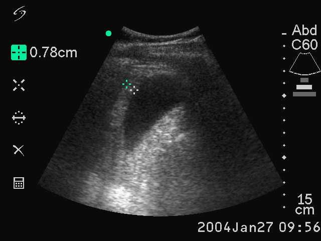
14 Thickened Gallbladder Wall Measure the anterior wall. Normal less than 2mm. Need to be fasting. Thick wall seen in jaundice, hepatitis, ascites, etc.
15 Thick Gallbladder Wall

16 Pericholecystic Fluid and other findings

17 Posterior Shadow Stones send back all the sound waves to the transducer, thus they make a shadow posterior shadowing. Stones also move if they are not stuck.

18

19 A Layer of a Whole Bunch of There is a multiple tiny stones producing this posterior shadowing. Small Stones

20 SONOGRAPHIC MURPHY See liver, or bowel, or whatever. Press on it. Does it hurt? See gallbladder. Press on gallbladder. See it move. Does that hurt?
21 Sonographic Murphy s Sign, A positive sonographic Murphy s sign, defined as the presence of maximal tenderness elicited by direct pressure of the transducer over a sonographically localized gallbladder, is present in most patients with acute cholecystitis. The positive predictive value of sonographic Murphy s sign combined with the presence of gallstones is reported to be 77% to 92%.

22 Negative Sonographic Murphys


23 Happens when the gallbladder is full of stones, or when it has one big one. Wall Echo Sign

24 Wall Echo Sign-Multiple Stones Note the hypoechoic (black) stripe between the wall and stones. This represents bile. Important to know about since it can be confused with gas filled bowel.

25 Portal Vein

26 CBD anterior to Portal Vein
27 Portal Vein Visualize the portal vein in a long axis view. Anterior to the PV, (towards the skin) you should see the bile duct. Portal vein has brighter (more echogenic) walls compared to hepatic veins.

28 Find that CBD Follow the gallbladder neck to the portal triad. The main lobar fissure should take you there (present in about 70% of people).

29 Main Lobar Fissure

30

31 Porta Hepatis=Portal Triad Rotating the probe on the portal vein should bring the common bile duct, hepatic artery and portal vein into view. The probe marker should be pointing to the patient s right.


32 Mickey Mouse If the CBD is dilated than the rodent has a swollen right ear. If you forget which ear is which, you can doppler the big one.

33 Dilated Common Bile Duct
34 CBD- Measure the Internal Diameter. Mean diameter is 4 mm, more than 7 mm is dilated. CBD diameter increases with age, up to 10 mm in the elderly. In general it should be 1/10th the patient s age.

35 Acute Cholecystitis The solid arrows are pointing to the thickened gallbladder wall. What are the open arrows pointing to?


36 Polyps Non-mobile No posterior shadowing

37 Gallbladder Sludge Mobile Nonshadowing Dependent


38 Phrygian Cap Transverse congenital septum in the fundus
39 Pitfalls in Gallbladder Imaging Misidentification of the gallbladder. A loop of bowel or the IVC can look like a gallbladder. Gut does peristalsis, IVC has flow on doppler. If possible you should show that the gallbladder communicates with main portal triad via the major lobar fissure. Bowel gas, try left lateral decubitus position. Contracted gallbladder in non-fasting patient. Lack of power to penetrate adipose.

40 The Liver Interesting findings can be seen in many patients including portal hypertension cirrhosis fatty liver bile duct obstruction cysts tumors


41 Vena Cava and Hepatic Veins There are three main hepatic veins, right, left and middle. They have thin, hypoechoic walls, and converge on the vena cava. Remember that the portal vein has hyperechoic walls.


42 Bunny Sign The rabbit s head is the vena cava, the ears are the middle and left hepatic veins. Use m-mode to observe the change in diameter of the vena cava with respiration

43 Transducer Parallel to the Costal Margin


44 Portal Hypertension Usually seen in the setting of cirrhosis. The portal vein is dilated to more than 15 mm. Often seen with ascites and splenomegaly. Note the portal vein s hyperechoic walls.

45 Cirrhosis-Early Stage Loss of peripheral hepatic vessels Increased echogenic wall of the portal vein. Hepatomegaly

46 Cirrhosis Advanced Stage Nodular liver contour. Contracted liver. Usually ascites
.")

47 Fatty Liver Results in increased echogenicity (brighter). Best compared to right kidney cortex. Focal Fatty Liver. Diffuse Fatty Liver


48 Bile Duct Obstruction

49 Liver Cysts-Benign To be called a cyst it must have echo free content spherical shape smooth outline distal acoustic enhancement

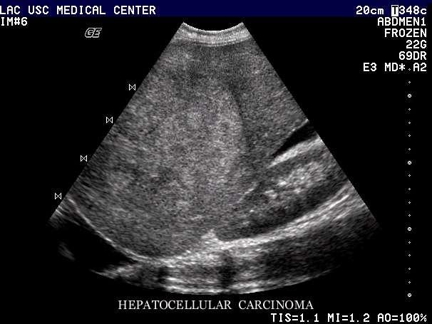
50 Liver Tumors The liver is a favorite site for metastatic tumors. GI tract, breast, lung, and the esophagus are the usual primaries. Note the hypoechoic halo.
51

52 Renal Ultrasound Hydronephrosis Cysts including polycystic kidney disease Stones Wilm s tumor Measuring Bladder Volume

53 The Normal Kidney The echogenicity (brightness) of the renal cortex should be less than that of the liver or spleen. In other words the kidney should be a tad darker than the liver.

54 Kidney Size Size is best evaluated as length, which should be 10 to 12 cm in adults. Each kidney should be within 2 cm of each other in length.

55 Approach Use the liver as your window on the right. The left can be harder. Try prone. Breath holding helps.

56 Hydronephrosis Grades - Mild, I spraying II bear claw III severe, loss of renal parenchyma Moderate, Severe This is grade 2.


57 Bear Claw- Hydronephrosis

58 Simple Cyst Seen while doing a FAST


59 Polycystic Kidney

60 Nephrolithiasis CT is better, but there is a place for doing a focused ED U.S. Someone with known stones to look for obstruction. Pregnancy. When AAA is in the differential.

61 Stones are rarely seen in the ureter, but they are visible in the kidney, bladder and UVJ. Note distal shadowing Seeing the Stone

62 Ureteral Jets Color Doppler probe is used to find urine squirting from the ureter into the bladder. If the jet is present then there is no obstruction on that side.

63 Wilm s tumor Younger than 6. Have a flank mass and hematuria. Mass is solid on U.S.

64 Urinary Bladder Female patient (note the uterus). Top image is a longitudinal view. Bottom is transverse.

65 Measure urine volume Why only one up down measurement and two side to side?

66 Measure Urine Volume Bladder volume is estimated by measuring maximal anteroposterior, coronal, and craniocaudal measurements and multiplying by pi/6 (0.52)

67 Spleen Viewing Position Best seen from the a posteriolateral approach, with the probe position in between the ribs.

68 Spleen A spleen looks like a crescent moon. Usually best seen halfway between full inspiration and expiration.

69 Splenomegaly The normal spleen measures less than 12 cm long, 5 cm thick and 7 cm in breadth. It can be hard to get the whole spleen on the screen if your probe s footprint isn t big enough.

70 Bowel Obstruction/Ileus I mention this because you will run into while looking for a AAA. Usually bowel isn t seen since gas disperses the beam. But in obstruction the bowel has a lot of fluid in it.

71 Bowel Obstruction

72 Bowel Obstruction


73 Appendicitis Greater than 6 mm in diameter

74 The image on the right is obtained by pressing down on the probe. Note the appendix does not compress. Press Down


75 Wall is greater than 3 mm thick. Appendicitis

76 Appendicolith Note the posterior shadowing. Exam is done with a high frequency probe.

77 How to use this skill clinically. 17 year old girl C/O RLQ pain. HCG Appendicitis versus tuboovarian abscess?

78 Mesenteric Adenitis Often seen in children after a viral illness. Often confused with appendicitis on clinical exam.
79 References References: Abdominal Ultrasound CD by Mark Deutchman (great resource) Emergency Ultrasound by Ma and Mateer Ultrasound in Emergency and Ambulatory Medicine by Simon and Snoey Ultrasound Teaching Manual by Hofer
80 Web Pages /us/education/msucmehd.html
My Patient Has Abdominal Pain PoCUS of the Biliary Tract and the Urinary Tract
 My Patient Has Abdominal Pain PoCUS of the Biliary Tract and the Urinary Tract Objectives PoCUS for Biliary Disease PoCUS for Renal Colic PoCUS for Urinary Retention Biliary Disease A patient presents
Hepatobiliary Ultrasound Rimon Bengiamin, MD, RDMS Assistant Clinical Professor Director of Emergency Ultrasound UCSF Fresno. Objectives. Why?
 Hepatobiliary Ultrasound Rimon Bengiamin, MD, RDMS Assistant Clinical Professor Director of Emergency Ultrasound UCSF Fresno Objectives Discuss the goals of point-of-care biliary ultrasound Review the
Abdominal ultrasound:
 Abdominal ultrasound: Non-traumatic acute abdomen Wittanee Na-ChiangMai, MD Department of Radiology ChiangMai University 26/04/2017 Contents Technique of examination Normal anatomy Emergency conditions
Biliary Ultrasonography Kathleen O Brien MD MPH RDMS Kaiser Permanente South Sacramento
 Biliary Ultrasonography Kathleen O Brien MD MPH RDMS Kaiser Permanente South Sacramento https://www.google.com/search?sa=g&hl=en&q=public+disclosure&tbm=isch&tbs=simg:caqsigeahwelekju2aqaaawlelcmpwgaygpgcamskpib_1qnza7ai
Abdomen and Retroperitoneum Ultrasound Protocols
 Abdomen and Retroperitoneum Ultrasound Protocols Reviewed By: Anna Ellermeier, MD Last Reviewed: March 2018 Contact: (866) 761-4200, Option 1 **NOTE for all examinations: 1. If documenting possible flow
Guidelines, Policies and Statements D5 Statement on Abdominal Scanning
 Guidelines, Policies and Statements D5 Statement on Abdominal Scanning Disclaimer and Copyright The ASUM Standards of Practice Board have made every effort to ensure that this Guideline/Policy/Statement
Normal Sonographic Anatomy
 hapter 2:The Liver DUNSTAN ABRAHAM Normal Sonographic Anatomy Homogeneous, echogenic texture (Figure 2-1) Measures approximately 15 cm in length and 10 12.5 cm anterior to posterior; measurement taken
US in non-traumatic acute abdomen. Lalita, M.D. Radiologist Department of radiology Faculty of Medicine ChiangMai university
 US in non-traumatic acute abdomen Lalita, M.D. Radiologist Department of radiology Faculty of Medicine ChiangMai university Sagittal Orientation Transverse (Axial) Orientation Coronal Orientation Intercostal
Objectives. Hepatobiliary Ultrasound: Anatomy, Technique, Pathology. RUQ: Normal Anatomy. Emergency Ultrasound: Gallbladder Location
 Hepatobiliary Ultrasound: Anatomy, Technique, Pathology Laleh Gharahbaghian, MD FAAEM Associate Director, EM Ultrasound Co-Director, EM Ultrasound Fellowship Stanford University Medical Center Seric Cusick,
Abdominal Ultrasound : Aorta, Kidneys, Bladder
 Abdominal Ultrasound : Aorta, Kidneys, Bladder Nilam J. Soni, MD, MSc Associate Professor of Medicine Divisions of Hospital Medicine and Pulmonary/Critical Care Medicine Department of Medicine University
Basic Abdominal Sonography
 24S Basic Abdominal Sonography Procedural Overview JOHN FATCHETT II, RDMS is provided. Patient preparation (i.e., fasting) scanning techniques, spleen, transducer. evaluation of abdominal anatomy in the
FHS Appendicitis US Protocol
 FHS Appendicitis US Protocol Reviewed By: Shireen Khan, MD; Sarah Farley, MD; Anna Ellermeier, MD Last Reviewed: May 2018 Contact: (866) 761-4200 **NOTE for all examinations: 1. If documenting possible
Radiology of hepatobiliary diseases
 GI cycle - Lecture 14 436 Teams Radiology of hepatobiliary diseases Objectives 1. To Interpret plan x-ray radiograph of abdomen with common pathologies. 2. To know the common pathologies presentation.
L o o k L i s t e n F e e l S c a n. Your Pocus Cards For Your Every Day Scanning.
 L o o k L i s t e n F e e l S c a n Your Pocus Cards For Your Every Day Scanning E-FAST Extended Focused Assessment by Sonography in Trauma Subcostal Heart View Pleural Sliding on M-mode (Sea-shore sign)
Anatomy Jessica Ferguson Ashley Dobos May 31, 2006 LIVER
 Anatomy Jessica Ferguson Ashley Dobos May 31, 2006 LIVER 1) Other Names: Reidel s Lobe normal anatomic variant; projection of the right lobe that can extend as far as the iliac crest (Tempkin, p.54, Anatomy).
Policies, Standards, and Guidelines. Guidelines for Abdominal Ultrasound Examination
 Policies, Standards, and Guidelines Guidelines for Abdominal Ultrasound Examination Approved by Council Feb 2018 Disclaimer and Copyright The ASUM Standards of Practice Board have made every effort to
Abdominal Ultrasonography
 Abdominal Ultrasonography David A. Masneri, DO, FACEP, FAAEM Assistant Professor of Emergency Medicine Assistant Director, Emergency Medicine Residency Medical Director, Operational Medicine Division Center
Bedside RUQ Ultrasound. Replace Formal ULS? Why Bedside ULS RUQ? RUQ Ultrasound. Bedside ULS is Limited, Goal-Directed
 Bedside RUQ Ultrasound RUQ Ultrasound Why do it How to do it Elizabeth Kwan UCSF Emergency Ultrasound Fellow Why Bedside ULS RUQ? Dx or Rule Out Acute Cholecystitis Cholelithiasis, Choledocolithiasis Earlier
IT 의료융합 1 차임상세미나 복부질환초음파 이재영
 IT 의료융합 1 차임상세미나 2013-4-3 복부질환초음파 이재영 나는오늘누구를위하여 종을울리나? 전통적의료 의사 공학설계자 의사 최첨단진단장비들 USG, CT, MRI 환자 환자 현대의료 사용자중심의사고 US in the Abdomen Detection DDx Look Behavior Response by external stimuli Guiding Tool
Certificate in Clinician Performed Ultrasound (CCPU) Syllabus. Biliary
 Syllabus. Biliary") Certificate in Clinician Performed Ultrasound (CCPU) Syllabus Biliary Page 1 of 6 12/18 Biliary Syllabus Purpose: This unit is designed to cover the theoretical and practical curriculum for basic ultrasound
Certificate in Clinician Performed Ultrasound (CCPU) Syllabus Biliary Page 1 of 6 12/18 Biliary Syllabus Purpose: This unit is designed to cover the theoretical and practical curriculum for basic ultrasound
Elastography in the. technically difficult patient. EPIQ ultrasound system. Ultrasound
 Ultrasound Elastography in the technically difficult patient EPIQ ultrasound system Chairman Department of Diagnostic Radiology Allegheny General Hospital Pittsburgh, PA, USA You can offer more information
US Applications. Case Based Wrap-Up 1. Case 1 E-FAST. Case presentations E-FAST Abdominal. Pearls for each indication
 Case Based Wrap-Up 1 Stephanie J. Doniger MD RDMS FAAP FACEP Associate Director, Pediatric Emergency Ultrasound Stanford University Medical Center US Applications Case presentations E-FAST Abdominal Aorta
Imaging of common diseases of hepatobiliary and GI system
 Imaging of common diseases of hepatobiliary and GI system Natthaporn Tanpowpong, M.D. Diagnostic radiology Faculty of Medicine, Chulalongkorn University Normal plain radiograph A = Common bile duct
Basic of Ultrasound Physics E FAST & Renal Examination. Dr Muhammad Umer Ihsan MBBS,MD, DCH CCPU,DDU1,FACEM
 Basic of Ultrasound Physics E FAST & Renal Examination Dr Muhammad Umer Ihsan MBBS,MD, DCH CCPU,DDU1,FACEM What is Sound? Sound is Mechanical pressure waves What is Ultrasound? Ultrasounds are sound waves
Q129. Which of the following is NOT true about lymph node?
 Q129. Which of the following is NOT true about lymph node? (1). Normal lymph node is not seen in the ultrasound image (2). It is general that high frequency probe is used due to normal lymph node is located
Appendix 5. EFSUMB Newsletter. Gastroenterological Ultrasound
 EFSUMB Newsletter 87 Examinations should encompass the full range of pathological conditions listed below A log book listing the types of examinations undertaken should be kept Training should usually
Abdominal Examination Benchmarks
 Abdominal Examination Benchmarks Preparation and Positioning: Stand on the right side of the patient. The patient should be supine and double draped so only the abdomen is exposed o To relax the abdominal
Point-of-Care Ultrasound Guide for Landmarks, Recording, and Report Content. TJUH/MHD EM Ultrasound Division 2012
 Point-of-Care Ultrasound Guide for Landmarks, Recording, and Report Content TJUH/MHD EM Ultrasound Division 2012 Table of Contents 1 - Objectives 2 - Procedural 3 - AAA 4 - Abdominal OB 5 - Transvaginal
1. Long images of aorta (prox, mid, and dist) with AP measurements. 2. Trans images of aorta (prox, mid, and dist) with R/L measurements.
 with AP measurements. 2. Trans images of aorta (prox, mid, and dist) with R/L measurements.") Aorta 1. Long images of aorta (prox, mid, and dist) with AP measurements. 2. Trans images of aorta (prox, mid, and dist) with R/L measurements. 3. Long images of R/L common iliac arteries with AP measurements.
Aorta 1. Long images of aorta (prox, mid, and dist) with AP measurements. 2. Trans images of aorta (prox, mid, and dist) with R/L measurements. 3. Long images of R/L common iliac arteries with AP measurements.
Pediatric Hepatobiliary, Pancreatic & Splenic US
 Pediatric Hepatobiliary, Pancreatic & Splenic US Susan J. Back, MD Department of Radiology, The Children s Hospital of Philadelphia No Disclosures Objectives Normal Abnormal: cases and US advances Objectives
4/9/2018 OBJECTIVES PANCREAOTO BILIARY ULTRASOUND: BEYOND CHOLECYSTITIS
 PANCREAOTO BILIARY ULTRASOUND: BEYOND CHOLECYSTITIS Jean Yves Sewah Kaiser Permanente West Los Angeles 1 OBJECTIVES Discuss the role of ultrasound in the evaluation of the gallbladder, biliary tree and
Abdominal Ultrasound
 Abdominal Ultrasound Imaging Control Buttons Depth The organ imaged should take up 3/4 of the screen Frequency = Penetration Use high frequencies (harmonics) for fluid filled and superficial structures
Hepatic Imaging: What Every Practitioner Should Know
 Hepatic Imaging: What Every Practitioner Should Know Shuchi K. Rodgers, MD Section Chief, Abdominal Imaging Director of Ultrasound Department of Radiology Einstein Medical Center rodgerss@einstein.edu
Biliary Tree Ultrasound - In a nutshell. Pamela Parker Lead Sonographer
 Biliary Tree Ultrasound - In a nutshell Pamela Parker Lead Sonographer Aims Review what we know about the biliary system Common pathologies Pitfalls Reporting tips The Nutshell Background Biliary examinations
US LI-RADS v2017 CORE
 US LI-RADS v2017 CORE Screening or surveillance US in patient at high risk for HCC US category US-1 US-2 US-3 Negative Subthreshold Positive Category Concept Definition US-1 Negative US-2 Subthreshold
Case Study: #3: Gallbladder Carcinoma?
 Case Study: #3: Gallbladder Carcinoma? By: Megan Wyatt K. SON Wyatt 225 2B1 RDMS, RVT Patient: Male 85 YOA Caucasian Indication: Elevated Alkaline Phosphatase History Annual physical showed elevated alkaline
Lab Monitor Images Dissection of the Abdominal Vasculature + Lower Digestive System
 Lab Monitor Images Dissection of the Abdominal Vasculature + Lower Digestive System Stomach & Duodenum Frontal (AP) View Nasogastric tube 2 1 3 4 Stomach Pylorus Duodenum 1 Duodenum 2 Duodenum 3 Duodenum
Learning Objectives. Ultrasound for the Primary Care Provider. Portable Ultrasound: Laptops, Tablets, Plug-in Probes, and Pocket devices
 Learning Objectives Ultrasound for the Primary Care Provider Richard Hoppmann, MD, FACP University of South Carolina School of Medicine Assess the main components and functions of a portable ultrasound
Emergency Ultrasound Standard Reporting Guidelines
 Emergency Ultrasound Standard Reporting Guidelines October 2011 *heterogenous corrected to heterogeneous on pages 9 & 11, January 2016 Emergency Ultrasound Standard Reporting Guidelines: Introduction and
Urinary system Ultrasound (Renal & Urinary bladder)
") Urinary system Ultrasound (Renal & Urinary bladder) Edited & Presented by ; Hussien A.B ALI DINAR. Msc.Phd ISRRT Associate Member Lecturer (National university) Reporting Sonographer (PHC) Objective By
Urinary system Ultrasound (Renal & Urinary bladder) Edited & Presented by ; Hussien A.B ALI DINAR. Msc.Phd ISRRT Associate Member Lecturer (National university) Reporting Sonographer (PHC) Objective By
ASSESSING THE PLAIN ABDOMINAL RADIOGRAPH M A A M E F O S U A A M P O F O
 ASSESSING THE PLAIN ABDOMINAL RADIOGRAPH M A A M E F O S U A A M P O F O Introduction The abdomen (less formally called the belly, stomach, is that part of the body between the thorax (chest) and pelvis,
Appendix 9: Endoscopic Ultrasound in Gastroenterology
 Appendix 9: Endoscopic Ultrasound in Gastroenterology This curriculum is intended for clinicians who perform endoscopic ultrasonography (EUS) in gastroenterology. It includes standards for theoretical
Biliary Tree Ultrasound - In a nutshell. Pamela Parker Lead Sonographer
 Biliary Tree Ultrasound - In a nutshell Pamela Parker Lead Sonographer Aims Review what we know about the biliary system Common pathologies Pitfalls Reporting tips The Nutshell Background Biliary examinations
Alice Fung, MD Oregon Health and Science University
 Alice Fung, MD Oregon Health and Science University Disclosure Comments The speaker Alice Fung, MD Has relevant financial relationships to disclose. Received honorarium from (Guerbet). This individual
Body MRI from the Liver to the Bladder
 Body MRI from the Liver to the Bladder I Want You! Audience Participation Methodist Hospital Continuing Education Seminar Jordan Swensson, MD November 7, 2015 Objectives Observe the uses of MRI for organs
Diagnostic Ultrasound. Sutiporn Khampunnip, M.D.
 Diagnostic Ultrasound Sutiporn Khampunnip, M.D. Definition of Ultrasound Ultrasound is simply sound waves, like audible sound. High-frequency sound and refers to mechanical vibrations above 20 khz. Human
Abdomen Sonography Examination Content Outline
 Abdomen Sonography Examination Content Outline (Outline Summary) # Domain Subdomain Percentage 1 2 3 Anatomy, Perfusion, and Function Pathology, Vascular Abnormalities, Trauma, and Postoperative Anatomy
Terminology Tissue Appearance
 By Marc Nielsen, MD Advantages/Disadvantages Generation of Image Ultrasound Machine/Transducer selection Modes of Ultrasound Terminology Tissue Appearance Scanning Technique Real-time Portable No ionizing
WELCOME! Introduction to Bedside Ultrasound
 WELCOME! Introduction to Bedside Ultrasound TEACHERS University of California-Irvine School of Medicine Nathan Molina nathan.d.molina@gmail.com Trevor Plescia taplescia90@gmail.com Jack Silva jpsilva42@gmail.com
Gallbladder & Pancreas Ultrasonography
 복부초음파 : 담낭과췌장 Gallbladder & Pancreas Ultrasonography 김정훈 Department of Radiology 1 Interaction of sound with matter (1) 반사 (Reflection) (2) 굴절 (Refraction) (3) 흡수 (Absorption) (4) 산란 (Scattering) 음향저항
Category Term Definition Comments 1 Major Categories 1a
 Working Lexicon Categories, Terms & Definitions Category Term Definition Comments 1 Major Categories 1a Physiologic Category (consistent with normal ovarian physiology) Follicle Simple 3 cm in premenopausal
Focused Assessment Sonography of Trauma (FAST) Scanning Protocol
 Scanning Protocol") Focused Assessment Sonography of Trauma (FAST) Scanning Protocol Romolo Gaspari CHAPTER 3 GOAL OF THE FAST EXAM Demonstrate free fluid in abdomen, pleural space, or pericardial space. EMERGENCY ULTRASOUND
Focused Assessment Sonography of Trauma (FAST) Scanning Protocol Romolo Gaspari CHAPTER 3 GOAL OF THE FAST EXAM Demonstrate free fluid in abdomen, pleural space, or pericardial space. EMERGENCY ULTRASOUND
Guide to Small Animal Vascular Imaging using the Vevo 770 Micro-Ultrasound System
 Guide to Small Animal Vascular Imaging using the Vevo 770 Micro-Ultrasound System January 2007 Objectives: After completion of this module, the participant will be able to accomplish the following: Understand
ULTRASOUND NOMENCLATURE
 Chapter 1: Ultrasound Nomenclature, Image Orientation, and Basic Instrumentation CYNTHIA SIKOWSKI Ultrasound waves are sound waves that have a frequency exceeding 20,000 Hz. When sound waves are transmitted
Job Task Analysis for ARDMS Abdomen Data Collected: June 30, 2011
 Job Task Analysis for ARDMS Abdomen Data Collected: June 30, 2011 Reported: Analysis Summary for: Abdomen Examination Survey Dates 06/13/2011-06/26/2011 Invited Respondents 6,000 Surveys with Demographics
Children's (Pediatric) Ultrasound - Abdomen
 Ultrasound - Abdomen") Scan for mobile link. Children's (Pediatric) Ultrasound - Abdomen Children s (pediatric) ultrasound imaging of the abdomen is a safe, noninvasive test that uses sound waves to produce a clear picture of
Scan for mobile link. Children's (Pediatric) Ultrasound - Abdomen Children s (pediatric) ultrasound imaging of the abdomen is a safe, noninvasive test that uses sound waves to produce a clear picture of
Nasogastric tube. Stomach. Pylorus. Duodenum 1. Duodenum 2. Duodenum 3. Duodenum 4
 Esophagus Barium Swallow Stomach and Duodenum 4 year old Upper GI Nasogastric tube Stomach and Duodenum 4 year old Upper GI Nasogastric tube Stomach Pylorus Duodenum 1 Duodenum 2 Duodenum 3 Duodenum 4
Certificate in Clinician Performed Ultrasound (CCPU) Syllabus. Renal Hydronephrosis & Calculi
 Syllabus. Renal Hydronephrosis & Calculi") Certificate in Clinician Performed Ultrasound (CCPU) Syllabus Renal Hydronephrosis & Calculi Page 1 of 6 01/17 Renal Hydronephrosis and Calculi Syllabus Purpose: This unit is designed to cover the theoretical
Certificate in Clinician Performed Ultrasound (CCPU) Syllabus Renal Hydronephrosis & Calculi Page 1 of 6 01/17 Renal Hydronephrosis and Calculi Syllabus Purpose: This unit is designed to cover the theoretical
Introduction & Physics of ED Ultrasound. Objectives. What? - Limited Studies. Who? - ED Docs
 Introduction & Physics of ED Ultrasound Martine Sargent, MD Ultrasound Director, Assistant Professor UCSF Department of Emergency Medicine San Francisco General Hospital & Trauma Center Objectives Who?
Diagnostic Imaging
 www.fisiokinesiterapia.biz Diagnostic Imaging Diagnostic Imaging is no longer limited to radiography. Major technological advancements have lead to the use of new and improved imaging technologies. The
Duodenum retroperitoneal
 Duodenum retroperitoneal C shaped Initial region out of stomach into small intestine RETROperitoneal viscus Superior 1 st part duodenal cap ; moves upwards and backwards to lie on the R crura medial to
Outline. Introduction to imaging modalities of the urinary system. Case base learning of common diseases in urinary tract
 Outline Introduction to imaging modalities of the urinary system Case base learning of common diseases in urinary tract Outline Introduction to imaging modalities of the urinary system Case base learning
Outline. Introduction to imaging modalities of the urinary system. Case base learning of common diseases in urinary tract
 Outline Introduction to imaging modalities of the urinary system Case base learning of common diseases in urinary tract Diagnostic Investigations in Urinary System PLAIN KUB EXCRETORY UROGRAPHY RETROGRADE
Abdominal Doppler Mastering the next level of vascular anatomy in the belly. Cindy A. Owen, RDMS, RVT
 Abdominal Doppler Mastering the next level of vascular anatomy in the belly Cindy A. Owen, RDMS, RVT Introduction Abdominal Doppler is a tough exam Success is dependent on: Patient body habitus Patient
Evolution of Clinical Anatomy with Ultrasonography Past Present and Future
 College of Medicine Evolution of Clinical Anatomy with Ultrasonography Past Present and Future Andrew F. Payer, Ph.D. Human Body Structure and Function Module Director Christine Bellew, M.D., Caridad Hernandez,
Medical application of transabdominal ultrasound in gastrointestinal diseases
 Medical application of transabdominal ultrasound in gastrointestinal diseases Hsiu-Po Wang Department of Emergency Medicine National Taiwan University Hospital Real-time ultrasound has become a standard
Image optimization for critical care US
 Image optimization for critical care US 1 Although we assume you are already familiar with focused US in the ED, it might not hurt to revise the basics: Machines & transducers US appearance of normal tissues
What is Ultrasound? What is Ultrasound? B A. Basic Principles of Ultrasound. Basic Principles of Ultrasound. Basic Principles of Ultrasound
 Introduction to Ultrasound Principles Mani Montazemi, RDMS Baylor College of Medicine Division of Maternal-Fetal Medicine Department of Obstetrics and Gynecology Manager, Maternal Fetal Center Imaging
Abdominal radiology 腹部放射線學
 Abdominal radiology 腹部放射線學 台北醫學大學 - 市立萬芳醫院 留偉順 laowilson@hotmail.com The Normal Abdominal Series Chest Supine abdomen Erect abdomen Left lateral decubitus abdomen Learning objectives Understanding normal
Imaging of liver and pancreas
 Imaging of liver and pancreas.. Disease of the liver Focal liver disease Diffusion liver disease Focal liver disease Benign Cyst Abscess Hemangioma FNH Hepatic adenoma HCC Malignant Fibrolamellar carcinoma
Ultrasound Applied Physics
 Ultrasound Applied Physics University of Toronto Department of Medical Imaging Applied Physics Mini-Course #3 2016 Ultrasound Laboratory Manual and Examination Booklet 1/21/2016 Ultrasound Applied Physics
Learning Objectives. Frequency: resolution and depth. The Evolution of Ultrasound Technology. Systems are smaller and portable
 9:45 10:45am Ultrasound for the PCP SPEAKER Richard Hoppmann, MD, FACP Presenter Disclosure Information The following relationships exist related to this presentation: Richard Hoppmann, MD, FACP, has no
Intrahepatic Cholangiocarcinoma (ICC) Detected by Sonography
 Detected by Sonography") 661245JDMXXX10.1177/8756479316661245Journal of Diagnostic Medical SonographyHamer research-article2016 Case Study Intrahepatic Cholangiocarcinoma (ICC) Detected by Sonography Journal of Diagnostic Medical
661245JDMXXX10.1177/8756479316661245Journal of Diagnostic Medical SonographyHamer research-article2016 Case Study Intrahepatic Cholangiocarcinoma (ICC) Detected by Sonography Journal of Diagnostic Medical
Accessory Glands of Digestive System
 Accessory Glands of Digestive System The liver The liver is soft and pliable and occupies the upper part of the abdominal cavity just beneath the diaphragm. The greater part of the liver is situated under
ULTRASOUND AND ABDOMINAL MASSES
 Med. J. Malaysia Vol. 37 No. I March 1982. ULTRASOUND AND ABDOMINAL MASSES AHMAD KAMAL BIN MD ALIF INTRODUCTION It is approximately 30 years since ultrasound was first introduced into the field of medicine,
Gastrointestinal Tract. Anatomy of GI Tract. Anatomy of GI Tract. (Effective February 2007) (1%-5%)
 (1%-5%)") Gastrointestinal Tract (Effective February 2007) (1%-5%) Anatomy of GI Tract Esophagus bulls-eye or target EG junction seen on sagittal scan posterior to left lobe of liver and anterior to aorta Anatomy
Gastrointestinal Tract (Effective February 2007) (1%-5%) Anatomy of GI Tract Esophagus bulls-eye or target EG junction seen on sagittal scan posterior to left lobe of liver and anterior to aorta Anatomy
In any operation. Indications. Anaesthesia. Position of the patient. Incision. Steps of the operation. Complications.
 In any operation Indications. Anaesthesia. Position of the patient. Incision. Steps of the operation. Complications. Abdominal operation I position for operation Supine Abdominal operation I position for
Summary and conclusions
 Summary and conclusions 7 Chapter 7 68 Summary and conclusions Chapter 1 provides a general introduction to this thesis focused on the use of ultrasound (US) in children with abdominal problems. The literature
The 2 nd Cambridge Advanced Emergency Ultrasound Course
 The 2 nd Cambridge Advanced Emergency Ultrasound Course Addenbrooke s Hospital Cambridge Sept 2008 1 2 Faculty! UK! USA! Australia! Toshiba! Emergency Medicine! Radiology 3 Programme! Day 1 Introduction
Sonography of Gall Bladder
 Sonography of Gall Bladder Vikram Dogra,MD Professor of Radiology, Urology and BME Director of Ultrasound Associate Chair of Education and Research University of Rochester, NY Objectives Describe the Congenital
Abdominal Sonography Review
 L I F E I S A T E S T... P A S S I T! 1-2-3 Step Ultrasound Education & Test Preparation Step 1 Review text Step 2 Mock examination Step 3 Q&A memory skills flashcard drill Abdominal Sonography Review
To describe the liver. To list main structures in porta hepatis.
 GI anatomy Lecture: 6 د. عصام طارق Objectives: To describe the liver. To list main structures in porta hepatis. To define portal system & portosystemic anastomosis. To list parts of biliary system. To
The abdominal Esophagus, Stomach and the Duodenum. Prof. Oluwadiya KS
 The abdominal Esophagus, Stomach and the Duodenum Prof. Oluwadiya KS www.oluwadiya.com Viscera of the abdomen Abdominal esophagus: Terminal part of the esophagus The stomach Intestines: Small and Large
Obstetrics Content Outline Obstetrics - Fetal Abnormalities
 Obstetrics Content Outline Obstetrics - Fetal Abnormalities Effective February 2007 10 16% renal agenesis complete absence of the kidneys occurs when ureteric buds fail to develop Or degenerate before
Hilar cholangiocarcinoma. Frank Wessels, Maarten van Leeuwen, UMCU utrecht
 Hilar cholangiocarcinoma Frank Wessels, Maarten van Leeuwen, UMCU utrecht Content Anatomy Biliary strictures (Hilar) Cholangiocarcinoom Staging Biliary tract 1 st order Ductus hepatica dextra Ductus hepaticus
Abdominal Ultrasound
 Abdominal Ultrasound What is Ultrasound Imaging of the Abdomen? What are some common uses of the procedure? How should I prepare? What does the equipment look like? How does the procedure work? How is
Principles of Ultrasound. Cara C. Prideaux, M.D. University of Utah PM&R Sports Medicine Fellow March 14, 2012
 Principles of Ultrasound Cara C. Prideaux, M.D. University of Utah PM&R Sports Medicine Fellow March 14, 2012 None Disclosures Outline Introduction Benefits and Limitations of US Ultrasound (US) Physics
CT abdomen and pelvis
 CT abdomen and pelvis General indications: Assessment of vague abdominal symptoms (pain, colics,distenstion,...) Varifecation of a lesion discovered by other diagnostic modalities as US, barium,ivp, Staging
Acute flank pain in children: Imaging considerations
 Acute flank pain in children: Imaging considerations Carlos J. Sivit MD Rainbow Babies and Children s Hospital Case Western Reserve School of Medicine Flank pain Results from distention of ureter or renal
Emergency Ultrasound Educational Objectives
 Emergency Ultrasound Educational Objectives 1) Basics of Technique A) Machine Operation Administrative Demonstrate basic workflow ordering, performing, reporting studies Explain how to enter pt info if
Excretory urography (EU) or IVP US CT & radionuclide imaging
 or IVP US CT & radionuclide imaging") Excretory urography (EU) or IVP US CT & radionuclide imaging MRI arteriography studies requiring catherization or direct puncture of collecting system EU & to a lesser extent CT provide both functional
Excretory urography (EU) or IVP US CT & radionuclide imaging MRI arteriography studies requiring catherization or direct puncture of collecting system EU & to a lesser extent CT provide both functional
Roaa M.Hussein Professor, Department of Physics, College of Science, Ramadi, Iraq. Abstract:
 Abstract: Ultrasound Waves Employment in the Medical Diagnostic for Lıver and Gallbladder Faik H. Antar Professor, Department of Physics, College of Science, AL- Anbar University, Ramadi, Iraq. Ultrasound
Imaging Guided Biopsy. Edited & Presented by ; Hussien A.B ALI DINAR. Msc Lecturer,Reporting Sonographer
 Imaging Guided Biopsy Edited & Presented by ; Hussien A.B ALI DINAR. Msc Lecturer,Reporting Sonographer Objective By the End of this lessons you should : Define what biopsy Justify Aim to perform biopsy
DIAGNOSTIC IMAGING: LIVER DISEASE
 Vet Times The website for the veterinary profession https://www.vettimes.co.uk DIAGNOSTIC IMAGING: LIVER DISEASE Author : Abby Caine Categories : Vets Date : February 1, 2010 ABBY CAINE reviews both established
EFSUMB EUROPEAN FEDERATION OF SOCIETIES FOR ULTRASOUND IN MEDICINE AND BIOLOGY Building a European Ultrasound Community
 MINIMUM TRAINING REQUIREMENTS FOR THE PRACTICE OF MEDICAL ULTRASOUND IN EUROPE Appendix 9: Endoscopic Ultrasound in Gastroenterology This curriculum is intended for clinicians who perform endoscopic ultrasonography
Reporting Initiative: Motivations and Approach
 The RSNA Structured Reporting Initiative: Motivations and Approach Curtis P. Langlotz, MD, PhD Chair, RSNA Structured Reporting Committee Vice Chair for Informatics, Department of Radiology Professor of
Rad Lab 4 Unknowns: Genitourinary!
 Rad Lab 4 Unknowns: Genitourinary! Peter Clarke MD! Don Di Salvo, MD! Clerkship Directors for Radiology! Harvard Medical School! Brigham and Women s Hospital! Dana Farber Cancer Institute! Case 1: 69 year
Chapter 3. Sonographic Image Interpretation
 Chapter 3 Sonographic Image Interpretation Sonograms are two-dimensional gray-scale images that allow assessment and diagnosis of many anatomic and pathologic changes that can occur in the human body.
Chest X-ray Interpretation
 Chest X-ray Interpretation Introduction Routinely obtained Pulmonary specialist consultation Inherent physical exam limitations Chest x-ray limitations Physical exam and chest x-ray provide compliment
Basic Training Programme. 16 Februrary 2018, ROTTERDAM. Pre and Post-Course Test Answers
 Basic Training Programme 16 Februrary 2018, ROTTERDAM Pre and Post-Course Test Answers Your details: Name: Conference registration number/ BT delegate number: Email address: Are you already performing