Case Report Testicular Arteriovenous Malformation: Gray-Scale and Color Doppler Ultrasonography Features
|
|
|
- Elmer Robinson
- 6 years ago
- Views:
Transcription
1 Volume 2011, Article ID , 4 pages doi:10.11/2011/ Case Report Testicular Arteriovenous Malformation: Gray-Scale and Color Doppler Ultrasonography Features Fatih Gulsen, 1 Ismail Mihmanli, 1 Fatih Kantarci, 1 Abdulkadir Eren, 1 and Suleyman Onder Ataus 2 1 Department of Radiology, Cerrahpasa Medical Faculty, Istanbul University, Kocamustafapasa, Istanbul, Turkey 2 Department of Urology, Cerrahpasa Medical Faculty, Istanbul University, Kocamustafapasa, Istanbul, Turkey Correspondence should be addressed to Fatih Gulsen, drfgulsen@yahoo.com Received 29 April 2011; Accepted 21 May 2011 Academic Editor: Michael N. Varras Copyright 2011 Fatih Gulsen et al. This is an open access article distributed under the Creative Commons Attribution License, which permits unrestricted use, distribution, and reproduction in any medium, provided the original work is properly cited. Intratesticular arteriovenous malformations (AVMs) are extremely rare benign incidental lesions of the testis. Ultrasonography (US) generally reveals a hypoechoic solid mass within the testicular parenchyma. We describe a patient with intratesticular AVM which was found incidentally during workup for infertility. The gray-scale and Doppler US appearance of an intratesticular AVM and the differential diagnosis have been presented. Based on the gray-scale, US appearance differentiation from malignant testicular tumors is difficult. Doppler US examination aids in the diagnosis by demonstrating the vascular nature of the tumor. 1. Introduction Testicular cancer is the most common cancer in men aged years and accounts for 1% of all malignancies in men. Gray-scale ultrasound (US) is the standard imaging technique used to identify testicular carcinoma and nearly sensitive for detection of testicular tumors [1]. One of the many benefits of US examination in the diagnosis of testicular cancer is the differentiation of intratesticular from extratesticular lesions. The majority of extratesticular masses are benign and intratesticular masses are more likely to be malignant [1]. There are a variety of benign intratesticular processes, such as hematoma, orchitis, abscess, infarction, and granuloma, which mimic testicular malignancy and must therefore be considered in the differential diagnosis. It is important to be familiar with their US appearance and to closely correlate US findings and patient history to avoid unnecessary interventions. Color Doppler and power Doppler US demonstrate increased vascularity in the majority of malignant tumors and help to better define testicular involvement [2]. We present the gray-scale and color Doppler US findings in a case of intratesticular arteriovenous malformation (AVM) which was incidentally diagnosed during work-up for infertility. 2. Case Presentation A 26-year-old man was admitted to our department for sonographic evaluation of the scrotum due to infertility. On physical examination, both testes were intrascrotal, normal sized, there was no palpable lesion, and no clinical evidence of varicocele. A sperm count revealed an oligospermia (sperm count: /mL). FSH level was in the upper limit of normal. The α-fetoprotein and human chorionic gonadotropin levels were within normal limits. Gray-scale images showed a well circumscribed,.7 mm sized hypoechoic round lesion within the left testis (Figure 1). Color Doppler US demonstrated that the lesion consisted almost entirely of multiple tortuous enlarged vessels (Figure 2). There were 2 dilated vessels on both sides of the vascular mass. One of them was arterial in nature and displayed high flow velocities with a peak systolic velocity of 20.3 and end diastolic velocity of 13.6 (Figure 3(a)). The resistive index was 0.33 and pulsatility index was The vessel on the other end showed pulsatile venous flow (Figure 3(b)) on spectral Doppler US. Based on the gray-scale and color Doppler US examinations, an arteriovenous malformation of the testis was diagnosed. US findings were not correlated to the patient s fertility status. The patient refused surgery


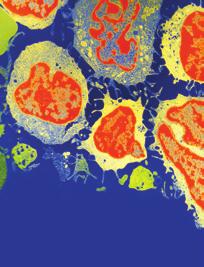
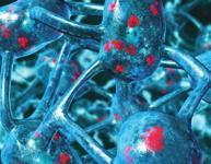
![lesions such as lipomas, hemangiomas, and AVMs can be observed [1, 4, ]. Intratesticular AVM is a rare mass lesion of the testis and it can be confused with the other intratesticular lesions.](/docs-images/76/73931779/images/2-5.jpg "There are sporadic case reports with hypoechoic and solid appearance on gray-scale US [4, 6, 7]. They are generally less than 1 cm in diameter.")
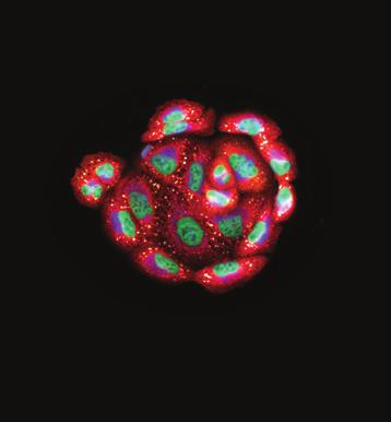
![Of the cystic lesions, testicular cysts appear with an anechoic center, through-transmission, and without a perceptible wall [1]. There is no vascular signal on color Doppler US.](/docs-images/76/73931779/images/2-6.jpg "Intratesticular varicocele is seen as straight and serpentine hypoechoic tubular structures within testicular parenchyma. Color Doppler US reveals venous flow within the tubular structures.")
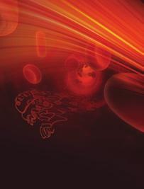
![The main differential of intratesticular varicocele is tubular ectasia of the rete testis in which flow is absent within the cystic structures [8].](/docs-images/76/73931779/images/2-7.jpg "These cystic lesions can readily be differentiated from malignant neoplasms at US. However, benign but solid-appearing lesions at US make the differentiation complicated.")
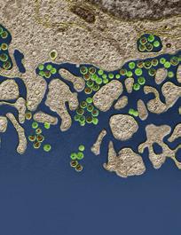
![Intratesticular lipomas are almost always solitary, and may appear hyper- or hypoechoic avascular lesions on US [9].](/docs-images/76/73931779/images/2-8.jpg "Testicular hemangiomas usually are not associated with hemangiomas at other locations and categorized as capillary, cavernous, and epitheloid [10].")

![have slower flow or lesser degrees of vascular pooling. Another reported color Doppler US feature is the presence of a low resistance pattern probably representing arteriovenous shunting [10].](/docs-images/76/73931779/images/2-10.jpg "Intratesticular AVMs may easily be confused with hemangioma on US.")
2 2 MI: 1.1 Figure 1: Gray-scale ultrasound image reveals a hypoechoic solid mass within the left testis. TIS: 1.3 TIB: 1.3 Figure 2: Color Doppler ultrasound demonstrates prominent vessels that entirely involve the mass. for the mass. Because of the small size of the vascular mass, invasiveness, and risk of radiation, no angiographic imaging and endovascular interventional procedure were performed. During the followup period, there were no changes in patient s clinical and radiological findings. 3. Discussion AVMs involving the lower urinary tract are uncommon as opposed to AVMs located in the Central Nervous System. Arteriovenous malformations of the spermatic cord and testis are benign lesions consisting of complex tangles of enlarged dilated arteries and veins without intervening capillaries. They can present as either painless paratesticular masses or as incidental findings during evaluation for infertility or as combination of both infertility and scrotal swelling and rarely as recurrent acute scrotal pain [3]. Most intratesticular masses are malignant and ultrasonography is the method of choice in the determination of the mass nature (cystic or solid), size, and vasculature. Besides primary testicular tumors a number of cystic appearing benign lesions such as testicular cysts, tubular ectasia of the rete testis, intratesticular varicocele, and solid-appearing benign lesions such as lipomas, hemangiomas, and AVMs can be observed [1, 4, ]. Intratesticular AVM is a rare mass lesion of the testis and it can be confused with the other intratesticular lesions. There are sporadic case reports with hypoechoic and solid appearance on gray-scale US [4, 6, 7]. They are generally less than 1 cm in diameter. Of the cystic lesions, testicular cysts appear with an anechoic center, through-transmission, and without a perceptible wall [1]. There is no vascular signal on color Doppler US. Intratesticular varicocele is seen as straight and serpentine hypoechoic tubular structures within testicular parenchyma. Color Doppler US reveals venous flow within the tubular structures. The main differential of intratesticular varicocele is tubular ectasia of the rete testis in which flow is absent within the cystic structures [8]. These cystic lesions can readily be differentiated from malignant neoplasms at US. However, benign but solid-appearing lesions at US make the differentiation complicated. Intratesticular lipomas are almost always solitary, and may appear hyper- or hypoechoic avascular lesions on US [9]. Testicular hemangiomas usually are not associated with hemangiomas at other locations and categorized as capillary, cavernous, and epitheloid [10]. Hemangiomas are usually characterized by testicular enlargement with or without tenderness. Gray scale US generally shows a focal, well-defined hypoechoic mass with calcifications, and color Doppler US patterns may show variations among different types of hemangiomas, because some of them have slower flow or lesser degrees of vascular pooling. Another reported color Doppler US feature is the presence of a low resistance pattern probably representing arteriovenous shunting [10]. Intratesticular AVMs may easily be confused with hemangioma on US. Presence of extensive vascularity within the lesion with high peak systolic, enddiastolic velocities, and a low resistance flow can be seen in both AVM and hemangioma. However, demonstration of a draining vein is characteristic for an AVM as was in our case [6]. Use of color and power Doppler US in malignant tumors of the testis help to better define testicular involvement [1]. The presence of hypervascularity is not specific enough for a diagnosis of malignancy, and it may be difficult to demonstrate increased blood flow in small tumors. On the other hand, color Doppler US is an extremely useful method in differentiating benign lesions. Intratesticular AVM is one of the lesions that color Doppler US aids in the diagnosis, since the gray-scale US findings closely resemble a malignant tumor. Magnetic resonance (MR) imaging is an important imaging technique in the evaluation of scrotal masses, providing a useful adjunct to ultrasonography (US). MR imaging allows tissue characterization, with its signal intensity properties allowing detection of fat, blood products, granulomatous tissue, and fibrosis. MR imaging performed after intravenous administration of gadolinium-based contrast material allows more accurate assessment of the vascularity of testicular lesions than color Doppler US does. The pattern of enhancement of scrotal lesions can also be evaluated. MR can also help localize the lesion as intra- or extratesticular and can clearly identify an undescended testis. The main differential diagnosis of an AVM, if all other possibilities have been
 TIB: 1.1 6 mm 60 1 10 (b) Figure 3: Spectral Doppler ultrasound depicts arterial flow in the feeding pedicle (a) and pulsatile venous flow in the drainage vessel (b).")
3 3 TIB: 1 10 mm 14 L/Testis Detail 2D THI/H14 MHz 1 db/dr70 SC 2 Map A/ST3 C CDV/7. MHz 3 db/flow Gen PRF 867/F2 D PW/6.2 MHz 62 db/dr PRF 163/F PS = 20.3 ED = 13.6 TAV = 16.6 PI = 0.4 RI = 0.33 S/D = 1.49 (a) TIB: mm (b) Figure 3: Spectral Doppler ultrasound depicts arterial flow in the feeding pedicle (a) and pulsatile venous flow in the drainage vessel (b). excluded, is that of a hemangioma. The MRI appearance of hemangioma is poorly documented. Essig et al. reported the MRI findings of a capillary hemangioma in a 26-yearold man [11]. The tumor demonstrated homogenous low signal intensity on proton density and T2-weighted images compared to normal testicular tissue. On T1-weighted images, the mass appeared almost isointense to the rest of the testis and could not be delineated. In conclusion, intratesticular AVMs are extremely rare lesions of the testis with solid and hypoechoic appearance on gray-scale US resembling a malignant neoplasm. Color Doppler US aids in the diagnosis by showing the vascular nature of the lesion and the prominent venous drainage. References [1] V. S. Dogra, R. H. Gottlieb, M. Oka, and D. J. Rubens, Sonography of the scrotum, Radiology, vol. 227, no. 1, pp , [2] W. G. Horstman, G. L. Melson, W. D. Middleton, and G. L. Andriole, Testicular tumors: findings with color Doppler US, Radiology, vol. 18, no. 3, pp , [3] P. Sountoulides, A. Bantis, I. Asouhidou, and H. Aggelonidou, Arteriovenous malformation of the spermatic cord as the cause of acute scrotal pain: a case report, Medical Case Reports, vol. 1, article 110, [4] S. Bhatt, D. J. Rubens, and V. S. Dogra, Sonography of benign intrascrotal lesions, Ultrasound Quarterly, vol. 22, no. 2, pp , 2006.
4 4 [] I. Mihmanli and F. Kantarci, Sonography of scrotal abnormalities in adults: an update, Diagnostic and Interventional Radiology, vol. 1, no. 1, pp , [6] R. Kutlu, A. Alkan, A. Soylu, A. Sigirci, and A. Dusak, Intratesticular arteriovenous malformation. Color doppler sonographic findings, Ultrasound in Medicine, vol. 22, no. 3, pp , [7] V. Skiadas, A. Antoniou, H. Primetis, L. Moulopoulos, and L. Vlahos, Intratesticular arteriovenous malformation. Clinical course, ultrasound and MRI findings of an extremely rare lesion on a 7 year follow-up basis, International Urology and Nephrology, vol. 38, no. 1, pp , [8] I. Mihmanli, F. Kantarci, and M. Ozbayrak, Intratesticular varicocele: a rare cause of testicular pain, Ultraschall in der Medizin, vol. 28, no., pp , [9] M.Harper,M.Arya,J.L.Peters,S.Buckingham,A.Freeman, and E. P. O Donoghue, Intratesticular lipoma, Scandinavian Urology and Nephrology, vol. 36, no. 3, pp , [10] Z. Ricci, M. Koenigsberg, and K. Whitney, Sonography of an arteriovenous-type hemangioma of the testis, American Roentgenology, vol. 174, no. 6, pp , [11] M. Essig, M. V. Knopp, H. Hawighorst, and G. van Kaick, MRI of capillary hemangioma of the testis, Computer Assisted Tomography, vol. 21, no. 3, pp , 1997.


5 MEDIATORS of INFLAMMATION The Scientific World Journal Gastroenterology Research and Practice Diabetes Research International Endocrinology Immunology Research Disease Markers Submit your manuscripts at BioMed Research International PPAR Research Obesity Ophthalmology Evidence-Based Complementary and Alternative Medicine Stem Cells International Oncology Parkinson s Disease Computational and Mathematical Methods in Medicine AIDS Behavioural Neurology Research and Treatment Oxidative Medicine and Cellular Longevity
COLOR DOPPLER ULTRASOUND IN EVALUATION OF SCROTAL LESIONS
 COLOR DOPPLER ULTRASOUND IN EVALUATION OF SCROTAL LESIONS Desai Sanjay D Associate Professor, Department of Radiology, RCSM Govt. Medical College, Kolhapur. ABSTRACT: Color Doppler ultrasound is a non-invasive,
Bilateral Segmental Testicular Infarction
 Case Study TheScientificWorldJOURNAL (2007) 7, 779 783 TSW Urology ISSN 1537-744X; DOI 10.1100/tsw.2007.146 Bilateral Segmental Testicular Infarction Aaron Bayne 1, Brad Koslin 2, and Siamak Daneshmand
Role of Colour Doppler Ultrasonography in evaluation of scrotal pain and swelling
 Original Research Article Role of Colour Doppler Ultrasonography in evaluation of scrotal pain and swelling Assistant Professor, Department of Radiodiagnosis, Government Medical College, Rajnandgaon Chattisghar,
Testicular ultrasound in acute scrotal pain - beyond testicular torsion
 Testicular ultrasound in acute scrotal pain - beyond testicular torsion Poster No.: C-1284 Congress: ECR 2015 Type: Educational Exhibit Authors: I. Rolla, M. Nogueira, M. J. Aguiar, D. S. Garrido, J. A.
Acute scrotum. Acute Epididymo-orchitis. Phyllis Yan, APDR (QEH)
") Acute scrotum Acute Epididymo-orchitis Phyllis Yan, APDR (QEH) Conditions leading to acute pain Torsion Acute Epididymitis / Epididymoorchitis Scrotal trauma Inguinal hernias Testicular tumors Epididymitis/epididymo
Acute scrotum Acute Epididymo-orchitis Phyllis Yan, APDR (QEH) Conditions leading to acute pain Torsion Acute Epididymitis / Epididymoorchitis Scrotal trauma Inguinal hernias Testicular tumors Epididymitis/epididymo
Sonography of the scrotum: still the best!
 Sonography of the scrotum: still the best! Poster No.: C-1110 Congress: ECR 2013 Type: Educational Exhibit Authors: W. Mnari, A. Zrig, M. Maatouk, B. Hmida, R. Salem, W. HarzallahHizem, M. Golli; Monastir/TN
Role of US in acute scrotal pain
 World J Urol (2011) 29:639 643 DOI 10.1007/s00345-011-0698-8 TOPIC PAPER Role of US in acute scrotal pain G. Liguori S. Bucci A. Zordani S. Benvenuto G. Ollandini G. Mazzon M. Bertolotto F. Cacciato S.
Vikram Dogra, M.D. Professor of Radiology, Urology & BME Department of Imaging Sciences University Of Rochester Medical Center
 Ultrasound of the Scrotum Vikram Dogra, M.D. Professor of Radiology, Urology & BME Department of Imaging Sciences University Of Rochester Medical Center Etiologies of Acute Scrotal Pain Epididymitis/Orchitis
Sonography of soft-tissue vascular lesions
 Sonography of soft-tissue vascular lesions Oscar M. Navarro Associate Professor, University of Toronto Dept. of Diagnostic Imaging, The Hospital for Sick Children Toronto, Canada Declaration of Disclosure
Case 5385 Tubular ectasia of the rete testis: a benign testicular entity diagnosed on imaging
 Case 5385 Tubular ectasia of the rete testis: a benign testicular entity diagnosed on imaging A. C. Tsili 1, C. Tsampoulas 1, D. Giannakis 2, A. Chaidou 1, N. Sofikitis 2, S. C. Efremidis 1 1 Department
The Acute Scrotum: Sonographic Findings
 The Acute Scrotum: Sonographic Findings 가천의대길병원방사선과 양달모 Gachon Medical School Introduction Many diseases presenting as acute scrotal pain DDx is important for determining the appropriate treatment US with
Case Report Intracranial Capillary Hemangioma in the Posterior Fossa of an Adult Male
 Case Reports in Radiology Volume 2016, Article ID 6434623, 4 pages http://dx.doi.org/10.1155/2016/6434623 Case Report Intracranial Capillary Hemangioma in the Posterior Fossa of an Adult Male Jordan Nepute,
Devendra V. Kulkarni, Rahul G. Hegde, Ankit Balani, and Anagha R. Joshi. 2. Case Report. 1. Introduction
 Case Reports in Radiology, Article ID 614647, 4 pages http://dx.doi.org/10.1155/2014/614647 Case Report A Rare Case of Pulmonary Atresia with Ventricular Septal Defect with a Right Sided Aortic Arch and
Case Report Internal Jugular Vein Thrombosis in Isolated Tuberculous Cervical Lymphadenopathy
 Volume 2016, Article ID 5184196, 4 pages http://dx.doi.org/10.1155/2016/5184196 Case Report Internal Jugular Vein Thrombosis in Isolated Tuberculous Cervical Lymphadenopathy Sanjay Khaladkar, Avadhesh
CLINICAL PRESENTATION AND RADIOLOGY QUIZ QUESTION
 Donald L. Renfrew, MD Radiology Associates of the Fox Valley, 333 N. Commercial Street, Suite 100, Neenah, WI 54956 2/19/2011 Radiology Quiz of the Week # 8 Page 1 CLINICAL PRESENTATION AND RADIOLOGY QUIZ
Varicoceles : co-relation of clinical examination with Color Doppler Sonograpghy at a tertiary care hospital
 Original article: Varicoceles : co-relation of clinical examination with Color Doppler Sonograpghy at a tertiary care hospital 1Dr. Neeraj Prajapati, 2 Dr. S.K.Ratogi, 3 Dr. Vijay Kulshrestha, 4 Dr. Abhinav
Case Report Uncommon Mixed Type I and II Choledochal Cyst: An Indonesian Experience
 Case Reports in Surgery Volume 2013, Article ID 821032, 4 pages http://dx.doi.org/10.1155/2013/821032 Case Report Uncommon Mixed Type I and II Choledochal Cyst: An Indonesian Experience Fransisca J. Siahaya,
Intraparenchymatous adenomatoid tumor dependent on the rete testis: A case report and review of literature
 Intraparenchymatous adenomatoid tumor dependent on the rete testis: A case report and review of literature A. Jiménez Pacheco, J. L. Martínez Torres, F. Valle Díaz de la Guardia, M. A. Arrabal Polo, and
Case Report Crossed Renal Ectopia without Fusion An Unusual Cause of Acute Abdominal Pain: A Case Report
 Case Reports in Urology Volume 2012, Article ID 728531, 4 pages doi:10.1155/2012/728531 Case Report Crossed Renal Ectopia without Fusion An Unusual Cause of Acute Abdominal Pain: A Case Report D. P. Ramaema,
Painless palpable scrotal mass
 Clinical Case - Test Yourself Urogenital Painless palpable scrotal mass Charis Anastasiadis, Georgia Kyriakopoulou, Charikleia Triantopoulou Radiology Department, Konstantopoulio General Hospital of Nea
US features of scrotal disorders: A pictorial essay
 US features of scrotal disorders: A pictorial essay Poster No.: C-0463 Congress: ECR 2014 Type: Educational Exhibit Authors: J. SAAD, F. Marrakchi ; nejran, sa/sa, Monastir/TN Keywords: Genital / Reproductive
Testicular Infarction and Rupture After Blunt Trauma Use of Diagnostic Ultrasound
 Short Review TheScientificWorldJOURNAL (2004) 4, 437 441 ISSN 1537-744X; DOI 10.1100/tsw.2004.101 Testicular Infarction and Rupture After Blunt Trauma Use of Diagnostic Ultrasound Alistair Pace 1, * and
Differential Diagnosis of Focal Epididymal Lesions With Gray Scale Sonographic, Color Doppler Sonographic, and Clinical Features
 Article Differential Diagnosis of Focal Epididymal Lesions With Gray Scale Sonographic, Color Doppler Sonographic, and Clinical Features Dal Mo Yang, MD, Sun Ho Kim, MD, Ha Na Kim, MD, Jee Hee Kang, MD,
Ultrasound of malignant testicular lesions. Arne Hørlyck Department of Radiology Aarhus University Hospital, Skejby
 Ultrasound of malignant testicular lesions Arne Hørlyck Department of Radiology Aarhus University Hospital, Skejby Testis Ultrasound is fantastic!! Scrotum Extratesticular mass: Benign Intratesticular
Case Report Contrast Enhanced Ultrasound of a Gallbladder Lesion in a Patient with a History of Renal Cell and Rectal Cancer
 Case Reports in Gastrointestinal Medicine Volume 2013, Article ID 538534, 4 pages http://dx.doi.org/10.1155/2013/538534 Case Report Contrast Enhanced Ultrasound of a Gallbladder Lesion in a Patient with
Case Report Overlap of Acute Cholecystitis with Gallstones and Squamous Cell Carcinoma of the Gallbladder in an Elderly Patient
 Case Reports in Surgery Volume 2015, Article ID 767196, 4 pages http://dx.doi.org/10.1155/2015/767196 Case Report Overlap of Acute Cholecystitis with Gallstones and Squamous Cell Carcinoma of the Gallbladder
VI E Article Clinics in diagnostic imaging (114)
") 264 Medical Education Singapore Med.1 2007, 48 (3) : VI E Article Clinics in diagnostic imaging (114) Muttarak M, Thinyu S, Lojanapiwat B Fig. I Clinical photograph of the scrotum. T t o T Fig. 2a Longitudinal
264 Medical Education Singapore Med.1 2007, 48 (3) : VI E Article Clinics in diagnostic imaging (114) Muttarak M, Thinyu S, Lojanapiwat B Fig. I Clinical photograph of the scrotum. T t o T Fig. 2a Longitudinal
The role of ultrasonography with colour Doppler in the acute scrotum
 The role of ultrasonography with colour Doppler in the acute scrotum Poster No.: C-1509 Congress: ECR 2016 Type: Educational Exhibit Authors: M. S. R. O. Faustino, A. L. Amado Costa, J. J. B. Leitão, I.
Ultrasound of soft-tissue vascular anomalies
 Ultrasound of soft-tissue vascular anomalies Oscar M. Navarro Associate Professor, University of Toronto Dept. of Diagnostic Imaging, The Hospital for Sick Children Toronto, Canada Declaration of Disclosure
Genitourinary Radiology In-Training Test Questions for Diagnostic Radiology Residents
 Genitourinary Radiology In-Training Test Questions for Diagnostic Radiology Residents March, 2013 Sponsored by: Commission on Education Committee on Residency Training in Diagnostic Radiology 2013 by American
Solitary Contralateral Adrenal Metastases after Nephrectomy for Renal Cell Carcinoma
 Original Report ISSN 1537-744X; DOI 10.1100/tsw.2004.39 Solitary Contralateral Adrenal after Nephrectomy for Renal Cell Carcinoma Nikolaos Antoniou, M.D. and Demetrios Karanastasis, M.D. General Hospital
Case Report Formation of a Tunnel under the Major Hepatic Vein Mouths during Removal of IVC Tumor Thrombus
 Case Reports in Urology Volume 2013, Article ID 129632, 4 pages http://dx.doi.org/10.1155/2013/129632 Case Report Formation of a Tunnel under the Major Hepatic Vein Mouths during Removal of IVC Tumor Thrombus
Bilateral Renal Angiomyolipomas with Invasion of the Renal Vein: A Case Report
 Case Study TheScientificWorldJOURNAL (2008) 8, 145 148 TSW Urology ISSN 1537-744X; DOI 10.1100/tsw.2008.29 Bilateral Renal Angiomyolipomas with Invasion of the Renal Vein: A Case Report C. Blick, N. Ravindranath,
Scrotum Kacey Morrison Amanda Baxter Sabrina Tucker July 18, 2006 SCROTUM
 Scrotum Kacey Morrison Amanda Baxter Sabrina Tucker July 18, 2006 SCROTUM 1) Other Names: Scrotum None Testicles Testes (Curry Tempkin, p. 236, 2/3/2) Ductus deferens spermatic cord (Tempkin, p. 279, Anatomy
Splenogonadal Fusion Diagnosed by Doppler Ultrasonography
 Original Articles TheScientificWorldJOURNAL (2004) 4 (S1), 253 257 ISSN 1537-744X; DOI 10.1100/tsw.2004.73 Splenogonadal Fusion Diagnosed by Doppler Ultrasonography José Murillo B. Netto, MD, Luis M. Pérez,
Case Report Asymptomatic Pulmonary Vein Stenosis: Hemodynamic Adaptation and Successful Ablation
 Case Reports in Cardiology Volume 2016, Article ID 4979182, 4 pages http://dx.doi.org/10.1155/2016/4979182 Case Report Asymptomatic Pulmonary Vein Stenosis: Hemodynamic Adaptation and Successful Ablation
Case Report Renal Cell Carcinoma Metastatic to Thyroid Gland, Presenting Like Anaplastic Carcinoma of Thyroid
 Case Reports in Urology Volume 2013, Article ID 651081, 4 pages http://dx.doi.org/10.1155/2013/651081 Case Report Renal Cell Carcinoma Metastatic to Thyroid Gland, Presenting Like Anaplastic Carcinoma
Multifocal testicular capillary hemangioma
 www.edoriumjournals.com case Report open ACCESS Multifocal testicular capillary hemangioma Shikha Singhal, Waseem Akhtar, Mabel Das Thyveetil ABSTRACT Introduction: Capillary hemangioma of testis is extremely
Mandana Moosavi 1 and Stuart Kreisman Background
 Case Reports in Endocrinology Volume 2016, Article ID 6471081, 4 pages http://dx.doi.org/10.1155/2016/6471081 Case Report A Case Report of Dramatically Increased Thyroglobulin after Lymph Node Biopsy in
Case Report Three-Dimensional Dual-Energy Computed Tomography for Enhancing Stone/Stent Contrasting and Stone Visualization in Urolithiasis
 Case Reports in Urology Volume 2013, Article ID 646087, 4 pages http://dx.doi.org/10.1155/2013/646087 Case Report Three-Dimensional Dual-Energy Computed Tomography for Enhancing Stone/Stent Contrasting
Case Report Müllerian Remnant Cyst as a Cause of Acute Abdomen in a Female Patient with Müllerian Agenesis: Radiologic and Pathologic Findings
 Volume 2016, Article ID 6581387, 4 pages http://dx.doi.org/10.1155/2016/6581387 Case Report üllerian Remnant Cyst as a Cause of Acute Abdomen in a Female Patient with üllerian Agenesis: Radiologic and
Baris Beytullah Koc, 1 Martijn Schotanus, 1 Bob Jong, 2 and Pieter Tilman Introduction. 2. Case Presentation
 Case Reports in Orthopedics Volume 2016, Article ID 7898090, 4 pages http://dx.doi.org/10.1155/2016/7898090 Case Report The Role of Dynamic Contrast-Enhanced MRI in a Child with Sport-Induced Avascular
Case Report Complex Form Variant of Dysembryoplastic Neuroepithelial Tumor of the Cerebellum
 Case Reports in Pathology Volume 2012, Article ID 718651, 4 pages doi:10.1155/2012/718651 Case Report Complex Form Variant of Dysembryoplastic Neuroepithelial Tumor of the Cerebellum Jesús Vaquero, 1,
Sonography of the Scrotum: Case-Based Review
 AJR Integrative Imaging LIFELONG LEARNING FOR RADIOLOGY Sonography of the Scrotum: Case-Based Review Joseph W. Stengel 1,2 and Erick M. Remer 1 ABSTRACT Objective We discuss five scenarios in which sonography
Case Report Sinus Venosus Atrial Septal Defect as a Cause of Palpitations and Dyspnea in an Adult: A Diagnostic Imaging Challenge
 Case Reports in Medicine Volume 2015, Article ID 128462, 4 pages http://dx.doi.org/10.1155/2015/128462 Case Report Sinus Venosus Atrial Septal Defect as a Cause of Palpitations and Dyspnea in an Adult:
Endocrine University, 2016 AACE-ACE-MAYO CLINIC
 Endocrine University, 2016 AACE-ACE-MAYO CLINIC Dev Abraham MD, MRCP (UK), ECNU Professor of Medicine (clinical), Division of Endocrinology Adjunct Professor of Surgery and Pathology Medical Director,
Original Research Article
 EVALUATION OF SCROTAL PATHOLOGY BY HIGH RESOLUTION ULTRASOUND AND COLOUR DOPPLER Pintu Biswas 1, Asim De 2 1Senior Resident, Department of Radiodiagnosis, Agartala Government Medical College and G. B.
Research Article Comparison of Colour Duplex Ultrasound with Computed Tomography to Measure the Maximum Abdominal Aortic Aneurysmal Diameter
 International Vascular Medicine, Article ID 574762, 4 pages http://dx.doi.org/10.1155/2014/574762 Research Article Comparison of Colour Duplex Ultrasound with Computed Tomography to Measure the Maximum
Case Report Multiple Giant Cell Tumors of Tendon Sheath Found within a Single Digit of a 9-Year-Old
 Case Reports in Orthopedics Volume 2016, Article ID 1834740, 4 pages http://dx.doi.org/10.1155/2016/1834740 Case Report Multiple Giant Cell Tumors of Tendon Sheath Found within a Single Digit of a 9-Year-Old
Ultrasound imaging of vascular anomalies: pearls and pitfalls
 Ultrasound imaging of vascular anomalies: pearls and pitfalls Oscar Navarro, MD Dept. of Medical Imaging, University of Toronto Dept. of Diagnostic Imaging, The Hospital for Sick Children Declaration of
Case Report Pulmonary Embolism Originating from a Hepatic Hydatid Cyst Ruptured into the Inferior Vena Cava: CT and MRI Findings
 Case Reports in Radiology Volume 2016, Article ID 3589812, 4 pages http://dx.doi.org/10.1155/2016/3589812 Case Report Pulmonary Embolism Originating from a Hepatic Hydatid Cyst Ruptured into the Inferior
Case Report A Case of Cystic Basal Cell Carcinoma Which Shows a Homogenous Blue/Black Area under Dermatoscopy
 Volume 20, Article ID 450472, 4 pages doi:0.55/20/450472 Case Report A Case of Cystic Basal Cell Carcinoma Which Shows a Homogenous Blue/Black Area under Dermatoscopy Akihiro Yoneta, Kohei Horimoto, Keiko
Case Report Features of the Atrophic Corpus Mucosa in Three Cases of Autoimmune Gastritis Revealed by Magnifying Endoscopy
 Volume 2012, Article ID 368160, 4 pages doi:10.1155/2012/368160 Case Report Features of the Atrophic Corpus Mucosa in Three Cases of Autoimmune Gastritis Revealed by Magnifying Endoscopy Kazuyoshi Yagi,
Role of ultrasound and colour doppler in assessment of adult scrotal pathologies
 Original Research Article Role of ultrasound and colour doppler in assessment of adult scrotal pathologies Ajay Vare 1, Dayanand Kawade 2*, Varsha Rote Kakinalkar 3, Prashant Titare 4, Samruddhi Sonawane
Triorchidism: A case report and review of the literature Jan Hendrickx*; Bart Claikens; Willem Roelandt; Pieter Mattelaer; Johan de Mey
 Open Journal of Clinical & Medical Case Reports Volume 3 (2017) Issue 16 Triorchidism: A case report and review of the literature Jan Hendrickx*; Bart Claikens; Willem Roelandt; Pieter Mattelaer; Johan
Management of an Unusual Iliac Fossa Venous Plexus
 Management of an Unusual Iliac Fossa Venous Plexus Irwin M Best, Emory University Journal Title: Case Reports in Vascular Medicine Volume: Volume 2011, Number 2011 Publisher: 2011-11-22, Pages 1-4 Type
Case Report Pediatric Transepiphyseal Seperation and Dislocation of the Femoral Head
 Case Reports in Orthopedics Volume 2013, Article ID 703850, 4 pages http://dx.doi.org/10.1155/2013/703850 Case Report Pediatric Transepiphyseal Seperation and Dislocation of the Femoral Head Mehmet Elmadag,
What effects will proximal or distal disease have on a waveform?
 Spectral Doppler Interpretation Director of Ultrasound Education & Quality Assurance Baylor College of Medicine Division of Maternal-Fetal Medicine Maternal Fetal Center Imaging Manager Texas Children
Case Report Tubular Carcinoma of the Breast: Advantages and Limitations of Breast Tomosynthesis
 Case Reports in Radiology Volume 2016, Article ID 3906195, 4 pages http://dx.doi.org/10.1155/2016/3906195 Case Report Tubular Carcinoma of the Breast: Advantages and Limitations of Breast Tomosynthesis
Case Report Late Type 3b Endoleak with an Endurant Endograft
 Case Reports in Radiology Volume 2015, Article ID 783468, 4 pages http://dx.doi.org/10.1155/2015/783468 Case Report Late Type 3b Endoleak with an Endurant Endograft Mehmet Barburoglu, 1 Bulent Acunas,
What effects will proximal or distal disease have on an waveform?
 Spectral Doppler Interpretation Director Director of of Ultrasound Ultrasound Education Education & & Quality Quality Assurance Assurance Baylor Baylor College College of of Medicine Medicine Division
Role of high frequency ultrasound and color Doppler examination in scrotal lesions
 Original article: Role of high frequency ultrasound and color Doppler examination in scrotal lesions 1Sanjeev Sharma, 2 Monika Jindal, 3 Sakshi Sharma, 4 Rekha Goyal, 5 Subhash Goyal 1Assistant Professor
Case Report A Case of Primary Submandibular Gland Oncocytic Carcinoma
 Case Reports in Otolaryngology Volume 2013, Article ID 384238, 4 pages http://dx.doi.org/10.1155/2013/384238 Case Report A Case of Primary Submandibular Gland Oncocytic Carcinoma Kunihiko Tokashiki, Kiyoaki
Case Report Osteolysis of the Greater Trochanter Caused by a Foreign Body Granuloma Associated with the Ethibond Suture after Total Hip Arthroplasty
 Hindawi Volume 2017, Article ID 6082302, 4 pages https://doi.org/10.1155/2017/6082302 Case Report Osteolysis of the Greater Trochanter Caused by a Foreign Body Granuloma Associated with the Ethibond Suture
Research Article Relationship between Pain and Medial Meniscal Extrusion in Knee Osteoarthritis
 Advances in Orthopedics Volume 2015, Article ID 210972, 4 pages http://dx.doi.org/10.1155/2015/210972 Research Article Relationship between Pain and Medial Meniscal Extrusion in Knee Osteoarthritis Hiroaki
Case Report Spontaneous Pelvic Rupture as a Result of Renal Colic in a Patient with Klinefelter Syndrome
 Volume 2013, Article ID 374973, 4 pages http://dx.doi.org/10.1155/2013/374973 Case Report Spontaneous Pelvic Rupture as a Result of Renal Colic in a Patient with Klinefelter Syndrome Sergey Reva and Yuri
Endoscopic Ultrasonography Assessment for Ampullary and Bile Duct Malignancy
 Diagnostic and Therapeutic Endoscopy, Vol. 3, pp. 35-40 Reprints available directly from the publisher Photocopying permitted by license only (C) 1996 OPA (Overseas Publishers Association) Amsterdam B.V.
Role of imaging in RCC. Ultrasonography. Solid lesion. Cystic RCC. Solid RCC 31/08/60. From Diagnosis to Treatment: the Radiologist Perspective
 Role of imaging in RCC From Diagnosis to Treatment: the Radiologist Perspective Diagnosis Staging Follow up Imaging modalities Limitations and pitfalls Duangkamon Prapruttam, MD Department of Therapeutic
BENIGN & MALIGNANT TESTIS DISEASES. Gary J. Faerber, M.D. Associate Professor, Dept of Urology March 2009 OBJECTIVES
 BENIGN & MALIGNANT TESTIS DISEASES Gary J. Faerber, M.D. Associate Professor, Dept of Urology March 2009 OBJECTIVES 1. Become familiar with the scrotal contents and their anatomical relationship with each
PUBLISHED VERSION.
 PUBLISHED VERSION Shay Keren, Gad Dotan, Leah Leibovitch, Dinesh Selva, and Igal Leibovitch Indocyanine green assisted removal of orbital lacrimal duct cysts in children Ophthalmology, 2015; 2015:130215-1-130215-5
Case Report Two Cases of Small Cell Cancer of the Maxillary Sinus Treated with Cisplatin plus Irinotecan and Radiotherapy
 Case Reports in Otolaryngology Volume 2013, Article ID 893638, 4 pages http://dx.doi.org/10.1155/2013/893638 Case Report Two Cases of Small Cell Cancer of the Maxillary Sinus Treated with Cisplatin plus
Case Report PET/CT Imaging in Oncology: Exceptions That Prove the Rule
 Case Reports in Oncological Medicine Volume 2013, Article ID 865032, 4 pages http://dx.doi.org/10.1155/2013/865032 Case Report PET/CT Imaging in Oncology: Exceptions That Prove the Rule M. Casali, 1 A.
Sonography and Magnetic Resonance Imaging Characteristics of Testicular Adrenal Rest Tumors
 Sonography and Magnetic Resonance Imaging Characteristics of Testicular Adrenal Rest Tumors The Harvard community has made this article openly available. Please share how this access benefits you. Your
Evaluation of Testicular Disease using Ultrasound
 Evaluation of Testicular Disease using Ultrasound Abdullah Hamdan 1, Alsafi Abdulla 2, Mohamed Yousef 3 1 Radiologic Technology Department, College of Applied Medical Science, Qassim University, Buraduh,
Scrotal Sonographic Findings in Equestrians
 Article Scrotal Sonographic Findings in Equestrians Ahmet Tuncay Turgut, MD, Ugur Kosar, MD, Pinar Kosar, MD, Ayhan Karabulut, MD Objective. Sports-related injuries are among the major causes of testicular
Case Report Sacral Emphysematous Osteomyelitis Caused by Escherichia coli after Arthroscopy of the Knee
 Case Reports in Orthopedics Volume 2016, Article ID 1961287, 4 pages http://dx.doi.org/10.1155/2016/1961287 Case Report Sacral Emphysematous Osteomyelitis Caused by Escherichia coli after Arthroscopy of
Role of Ultrasound and Colour Doppler in Scrotal Pain
 Role of Ultrasound and Colour Doppler in Scrotal Pain Dr. Vikram Patil 1, Dr.SM Chandrashekar Shetty 2 1 Radiologist, Department of Radiology, JSS Medical College, JSS University, Mysuru, Karnataka, India
Case Report A Rare Case of Near Complete Regression of a Large Cervical Disc Herniation without Any Intervention Demonstrated on MRI
 Case Reports in Radiology, Article ID 832765, 4 pages http://dx.doi.org/10.1155/2014/832765 Case Report A Rare Case of Near Complete Regression of a Large Cervical Disc Herniation without Any Intervention
Ultrasound of the Breast BASICS FOR THE ORDERING CLINICIAN
 Ultrasound of the Breast BASICS FOR THE ORDERING CLINICIAN Breast Ultrasound Anatomy Skin Breast Parenchyma Pectoralis Fascia Pectoralis Breast Ultrasound Anatomy Indications for Breast Ultrasound Palpable
Alex Lam, HMS III. September The Acute Scrotum. Alex Lam, Harvard Medical School Year III Gillian Lieberman, MD. Gillian Lieberman, MD
 September 2002 The Acute Scrotum Alex Lam, Harvard Medical School Year III DDx: : Acute Scrotal Pain & Enlargement PAIN Inflammatory disorder Testicular torsion Testicular infarction Testicular abscess
Case Report Tortuous Common Carotid Artery: A Report of Four Cases Observed in Cadaveric Dissections
 Case Reports in Otolaryngology Volume 2016, Article ID 2028402, 4 pages http://dx.doi.org/10.1155/2016/2028402 Case Report Tortuous Common Carotid Artery: A Report of Four Cases Observed in Cadaveric Dissections
Case Report A Rare Cutaneous Adnexal Tumor: Malignant Proliferating Trichilemmal Tumor
 Case Reports in Medicine Volume 2015, Article ID 742920, 4 pages http://dx.doi.org/10.1155/2015/742920 Case Report A Rare Cutaneous Adnexal Tumor: Malignant Proliferating Trichilemmal Tumor Omer Alici,
AIUM Practice Guideline for the Performance of Scrotal Ultrasound Examinations
 AIUM Practice Guideline for the Performance of Scrotal Ultrasound Examinations Guideline developed in collaboration with the American College of Radiology and the Society of Radiologists in Ultrasound.
Case Report Successful Implantation of a Coronary Stent Graft in a Peripheral Vessel
 Case Reports in Vascular Medicine Volume 2015, Article ID 725168, 4 pages http://dx.doi.org/10.1155/2015/725168 Case Report Successful Implantation of a Coronary Stent Graft in a Peripheral Vessel Alexander
Evaluation of Liver Mass Lesions. American College of Gastroenterology 2013 Regional Postgraduate Course
 Evaluation of Liver Mass Lesions American College of Gastroenterology 2013 Regional Postgraduate Course Lewis R. Roberts, MB ChB, PhD Division of Gastroenterology and Hepatology Mayo Clinic College of
Testicular tumors; Ultrasonographic and Pathologic correlation
 Testicular tumors; Ultrasonographic and Pathologic correlation Poster No.: C-0106 Congress: ECR 2014 Type: Educational Exhibit Authors: Y. Kim, S. W. Shin, E. T. Kim, M. Y. Kim ; Kuri City/KR, 1 1 2 1
HEPATO-BILIARY IMAGING
 HEPATO-BILIARY IMAGING BY MAMDOUH MAHFOUZ MD PROF.OF RADIOLOGY CAIRO UNIVERSITY mamdouh.m5@gmail.com www.ssregypt.com CT ABDOMEN Indications Patient preparation Patient position Scanogram Fasting 4-6 hours
Eisuke Nomura, Hisatada Hiraoka, and Hiroya Sakai. 1. Introduction. 2. Case Report
 Case Reports in Orthopedics Volume 2016, Article ID 1026861, 5 pages http://dx.doi.org/10.1155/2016/1026861 Case Report Spontaneous Recurrent Hemarthrosis of the Knee: A Report of Two Cases with a Source
Case Report Denosumab Chemotherapy for Recurrent Giant-Cell Tumor of Bone: A Case Report of Neoadjuvant Use Enabling Complete Surgical Resection
 Case Reports in Oncological Medicine Volume 2013, Article ID 496351, 4 pages http://dx.doi.org/10.1155/2013/496351 Case Report Denosumab Chemotherapy for Recurrent Giant-Cell Tumor of Bone: A Case Report
Case Report Postoperative Megarectum in an Adult Patient with Imperforate Anus and Rectourethral Fistula
 Case Reports in Gastrointestinal Medicine Volume 2015, Article ID 613926, 4 pages http://dx.doi.org/10.1155/2015/613926 Case Report Postoperative Megarectum in an Adult Patient with Imperforate Anus and
Osman Ilkay Ozdamar, 1 Gul Ozbilen Acar, 1 Cigdem Kafkasli, 1 M. Tayyar Kalcioglu, 1 Tulay Zenginkinet, 2 and H. Gonca Tamer 3. 1.
 Case Reports in Otolaryngology Volume 2015, Article ID 79658, 4 pages http://dx.doi.org/10.1155/2015/79658 Case Report Papillary Thyroid Microcarcinoma with a Large Cystic Dilated Lymph Node Metastasis
Chapter 4 Varicocele Classification
 Chapter 4 Varicocele Classification In this chapter, we examine the several classification modes have been used to diagnose and grade varicocele, including physical exam, venographic examination, color
Doppler Ultrasound of the Kidney
 Doppler Ultrasound of the Kidney Chang-Kyu Sung Department of Radiology, Seoul National University College of Medicine, SNU-SMG Boramae Medical Center, Seoul, Korea Doppler ultrasound (US) is a very useful
Radiology of hepatobiliary diseases
 GI cycle - Lecture 14 436 Teams Radiology of hepatobiliary diseases Objectives 1. To Interpret plan x-ray radiograph of abdomen with common pathologies. 2. To know the common pathologies presentation.
Case Report Coronary Artery Perforation and Regrowth of a Side Branch Occluded by a Polytetrafluoroethylene-Covered Stent Implantation
 International Scholarly Research Network Volume 2011, Article ID 212851, 4 pages doi:10.5402/2011/212851 Case Report Coronary Artery Perforation and Regrowth of a Side Branch Occluded by a Polytetrafluoroethylene-Covered
Case Report Computed Tomography Angiography Successfully Used to Diagnose Postoperative Systemic-Pulmonary Artery Shunt Narrowing
 Case Reports in Cardiology Volume 2011, Article ID 802643, 4 pages doi:10.1155/2011/802643 Case Report Computed Tomography Angiography Successfully Used to Diagnose Postoperative Systemic-Pulmonary Artery
Clinical Study The Incidence and Management of Pleural Injuries Occurring during Open Nephrectomy
 Advances in Urology Volume 2009, Article ID 948906, 4 pages doi:10.1155/2009/948906 Clinical Study The Incidence and Management of Pleural Injuries Occurring during Open Nephrectomy Ali Fuat Atmaca, Abdullah
Job Task Analysis for ARDMS Abdomen Data Collected: June 30, 2011
 Job Task Analysis for ARDMS Abdomen Data Collected: June 30, 2011 Reported: Analysis Summary for: Abdomen Examination Survey Dates 06/13/2011-06/26/2011 Invited Respondents 6,000 Surveys with Demographics
HIGH-FLOW ARTERIOVENOUS MALFORMATION WİTHİN ENLARGED FETAL LEG (Congenital Hemangioma vs Parkes Weber Syndrome)
") HIGH-FLOW ARTERIOVENOUS MALFORMATION WİTHİN ENLARGED FETAL LEG (Congenital Hemangioma vs Parkes Weber Syndrome) DORUK CEVDI KATLAN, MD Department of Obstetrics and Gynecology / Perinatology Suleymaniye
HIGH-FLOW ARTERIOVENOUS MALFORMATION WİTHİN ENLARGED FETAL LEG (Congenital Hemangioma vs Parkes Weber Syndrome) DORUK CEVDI KATLAN, MD Department of Obstetrics and Gynecology / Perinatology Suleymaniye
Angiographic features of rapidly involuting congenital hemangioma (RICH)
") Pediatr Radiol (2003) 33: 15 19 DOI 10.1007/s00247-002-0726-3 CASE REPORT Orhan Konez Patricia E. Burrows John B. Mulliken Steven J. Fishman Harry P.W. Kozakewich Angiographic features of rapidly involuting
Pediatr Radiol (2003) 33: 15 19 DOI 10.1007/s00247-002-0726-3 CASE REPORT Orhan Konez Patricia E. Burrows John B. Mulliken Steven J. Fishman Harry P.W. Kozakewich Angiographic features of rapidly involuting