|
|
|
- Sybil O’Connor’
- 6 years ago
- Views:
Transcription
1
2
3
4

5

6
7
8
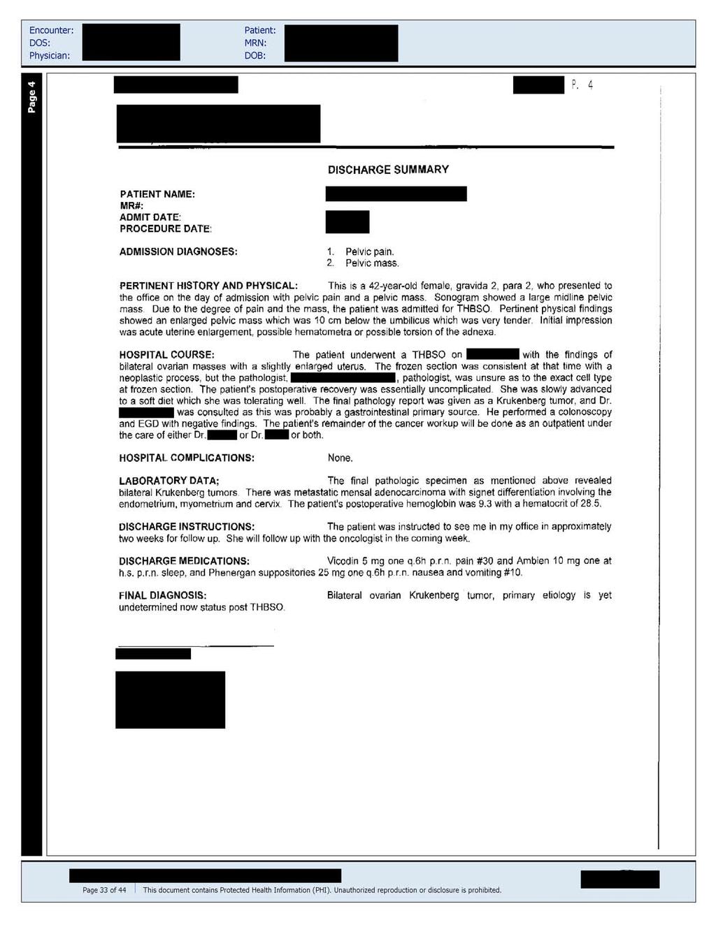
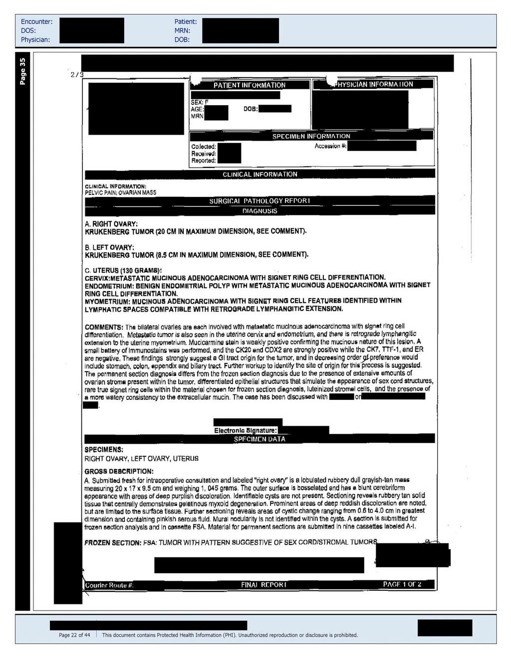
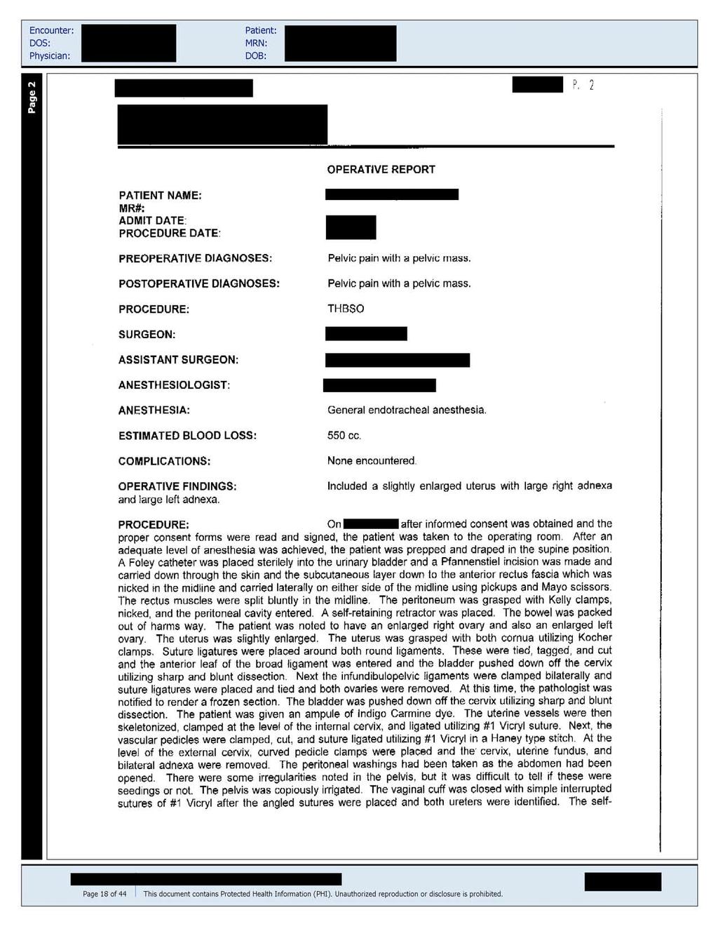
9 DIAGNOSIS A. RIGHT OVARY: Krukenberg tumor (20 cm in maximum dimension, see comment). B. LEFT OVARY: Krukenberg tumor (8.5 cm in maximum dimension, see comment). C. UTERUS (130 Grams): Cervix: Metastatic mucinous adenocarcinoma with signet ring cell differentiation. Endometrium: Benign endometrial polyp with metastatic mucinous adenocarcinoma with signet ring cell differentiation. Myometrium: Mucinous adenocarcinoma with signet ring cell features identified within lymphatic spaces compatible with retrograde lymphangitic extension. COMMENTS: The bilateral ovaries are each involved with metastatic mucinous adenocarcinoma with signet ring cell differentiation. Metastatic tumor is also seen in the uterine cervix and endometrium, and there is retrograde lymphangitic extension to the uterine myometrium. Mucicarmine stain is weakly positive confirming the mucinous nature of this lesion. A small battery of immunostains was performed, and the CK20 and CDX2 are strongly positive while the CK7, TTF-1, and ER are negative. These findings strongly suggest a GI tract origin for the tumor, and in decreasing order of preference would include stomach, colon, appendix and biliary tract. Further workup to identify the site of origin for this process is suggested. The permanent section diagnosis differs from the frozen section diagnosis due to the presence of extensive amounts of ovarian troma present within the tumor, differentiated epithelial structures that simulate the appearance of sex cord structures, rare true signet ring cells within the material chosen for frozen section diagnosis, luteinized stromal cells, and the presence of a more watery consistency to the extracellular mucin. The case has been discussed with [NAME] on [DATE]. SPECIMEN DATA SPECIMENS: Right ovary, Left ovary, uterus. GROSS DESCRIPTION: A. Submitted fresh for intraoperative consultation and labeled right ovary is a lobulated rubbery dull grayish-tan mass measuring 20x17x9.5 cm and weighing grams. The outer surface is bosselated and has a blunt cerebriform appearance with areas of deep purplish discoloration. Identifiable cysts are not present. Sectioning reveals rubbery tan solid tissue that centrally demonstrates gelatinous myxoid degeneration. Prominent areas of deep reddish discoloration are noted, but are limited to the surface tissue. Further sectioning reveals areas of cystic change ranging from 0.6 to 4.0 cm in greatest dimension and containing pinkish serous fluid. Mural nodularity is not identified within the cysts. A section is submitted for frozen section analysis and in cassette FSA. Material for permanent sections are submitted in nine cassettes labeled A-I. FROZEN SECTION: FSA: Tumor with patter suggestive of sex cord/stromal tumors. B. Submitted fresh for intraoperative consultation and labeled left ovary is a lobulated reddish-tan kidneyshaped mass measuring 8.5x6.0x3.5 cm. The outer surface is bosselated and imparts a convoluted appearance. Sectioning reveals solid grayish-tan tissue towards the periphery intermixed with central areas of cystic change measuring up to 3.0 cm in greatest dimension. One of these cysts contains a mural nodule measuring 1.1 cm in greatest dimension. Areas of degenerative and myxoid change are not identified. A representative section is
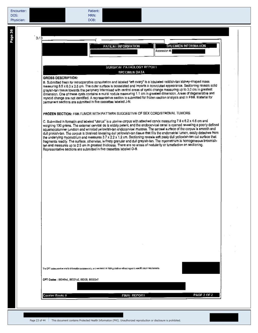
10 submitted for frozen section analysis and in FSB. Material for permanent sections are submitted in five cassettes labeled J-N. FROZEN SECTION: FSB: Tumor with pattern suggestive of sex cord/stromal tumors. C. Submitted in (formatin) and labeled uterus is a uterine corpus with attached cervix measuring 7.8x6.5x4.6 cm and weighing 130 grams. The external cervical (os) is widely patent, and the endocervical canal is opened revealing a poorly defined squamocolumnar junction and wrinkled yellowish-tan endocervical mucosa. The serosal surface of the corpus is smooth and dull pinkish-tan. The corpus is bivalved revealing dull yellowish-tan tissue that fills the endometrial lumen, easily detaches from the underlying myometrium and measures 3.7x2.2x1.2 cm. Sectioning reveals soft pasty dull yellowish-tan cut surface that fragments readily. The surface, otherwise, is finely granular and dull grayish-tan. The myometrium is homogeneous brownish-tan and measure up to 2.5 cm in greatest thickness. There are no areas of nodularity or turnefaction on sectioning. Representative sections are submitted in five cassettes labeled O-S.
11
12
13
14
15
16
17
18
19
20
21
22
23
24
25
26
27
28
29
30
31
Bladder Case 1 SURGICAL PATHOLOGY REPORT. Procedure: Cystoscopy, transurethral resection of bladder tumor (TURBT)
") Bladder Case 1 February 17, 2007 Specimen (s) received: Bladder Tumor Pre-operative Diagnosis: Bladder Cancer Post operative Diagnosis: Bladder Cancer Procedure: Cystoscopy, transurethral resection of
Bladder Case 1 February 17, 2007 Specimen (s) received: Bladder Tumor Pre-operative Diagnosis: Bladder Cancer Post operative Diagnosis: Bladder Cancer Procedure: Cystoscopy, transurethral resection of
MPH Quiz. 1. How many primaries are present based on this pathology report? 2. What rule is this based on?
 MPH Quiz Case 1 Surgical Pathology from hysterectomy performed July 11, 2007 Final Diagnosis: Uterus, resection: Endometrioid adenocarcinoma, Grade 1 involving most of endometrium, myometrial invasion
How to Recognize Gynecologic Cancer Cells from Pelvic Washing and Ascetic Specimens
 How to Recognize Gynecologic Cancer Cells from Pelvic Washing and Ascetic Specimens Wenxin Zheng, M.D. Professor of Pathology and Gynecology University of Arizona zhengw@email.arizona.edu http://www.zheng.gynpath.medicine.arizona.edu/index.html
Kidney Case 1 SURGICAL PATHOLOGY REPORT
 Kidney Case 1 Surgical Pathology Report February 9, 2007 Clinical History: This 45 year old woman was found to have a left renal mass. CT urography with reconstruction revealed a 2 cm medial mass which
Circulation: X Case number: 501 Number of responses: 84 Date: 4 MAY 12
 Circulation: X Case number: 500 Number of responses: 81 Date: 4 MAY 12 Female, aged 65 TAH and BSO for G1 endometrioid adenocarcinoma. Tumour positive with inhibin, vimentin, CD56 and SMA. Negative with
UTERINE SARCOMA EXAMPLE OF A UTERINE SARCOMA USING PROPOSED TEMPLATE
 UTERINE SARCOMA EXAMPLE OF A UTERINE SARCOMA USING PROPOSED TEMPLATE Case: Adenosarcoma with heterologous elements and stromal overgrowth o TAH, BSO, omentectomy, staging biopsies of cul-de-sac, bladder
Papillary adenocarcinoma gallbladder with simultaneously detected bilateral ovarian metastases: A Case Report
 ISPUB.COM The Internet Journal of Gynecology and Obstetrics Volume 9 Number 1 Papillary adenocarcinoma gallbladder with simultaneously detected bilateral ovarian metastases: A Case K Majumdar, D Singh,
Atypical Hyperplasia/EIN
 EIN Atypical Hyperplasia/EIN Based on scientific and diagnostic advances, in 2014 the WHO moved that the precursor lesion for endometrioid carcinoma be atypical hyperplasia/ein, rather than what was previously
CELL AND TISSUE INJURY COURSE-II PATHOLOGY LABORATORY. PATHOLOGY of MASS LESIONS and TISSUE DEFECTS -MACROSCOPY Assoc. Professor Rengin Ahıskalı
 CELL AND TISSUE INJURY COURSE-II PATHOLOGY LABORATORY PATHOLOGY of MASS LESIONS and TISSUE DEFECTS -MACROSCOPY Assoc. Professor Rengin Ahıskalı M1 - RENAL TUBERCULOSIS cavitary areas caseous necrosis fibrous
This peer-reviewed article can be downloaded, printed and distributed freely for any purposes (see copyright notice below).
.") Diagnostic Pathology This Provisional PDF corresponds to the article as it appeared upon acceptance. Fully formatted PDF and full text (HTML) versions will be made available soon. Endometrioid adenocarcinoma
Diagnostic Pathology This Provisional PDF corresponds to the article as it appeared upon acceptance. Fully formatted PDF and full text (HTML) versions will be made available soon. Endometrioid adenocarcinoma
Endometrial polyp icd-10
 Endometrial polyp icd-10 Maternity Dx (12-55 years) Female Dx. Applicable To. Placental polyp. O90.89. ) Codes. N84 Polyp of female genital tract. N84.0 Polyp of corpus uteri; N84.1 Polyp of cervix uteri;
Normal endometrium: A, proliferative. B, secretory.
 Normal endometrium: A, proliferative. B, secretory. Nội mạc tử cung Nội mạc tử cung Cyclic changes in endometrium.. Approximate relationship of useful microscopic changes. Arias-Stella reaction in endometrial
Endometrial Stromal Sarcoma
 May 26, 2011 By Sushila Ladumor, MD [1] Endometrial stromal sarcoma (ESS) is a rare malignant tumor of the endometrium, occurring in the age group of 40-50 years. History The 50-year-old, female patient
General history. Basic Data : Age :62y/o Date of admitted: Married status : Married
 General history Basic Data : Age :62y/o Date of admitted:940510 Married status : Married General history Chief Complain : bilateral ovarian cyst incidentally being found out during pap smear. Present Illness
Pathology of the female genital tract
 Pathology of the female genital tract Common illnesses of the female genital tract Before menarche Developmental anomalies Tumors (ovarial teratoma) Amenorrhea Fertile years PCOS, ovarian cysts Endometriosis
Gynaecological Malignancies
 Gynaecological Malignancies Dr Rodney Itaki Lecturer Anatomical Pathology Discipline University of Papua New Guinea Division of Pathology School of Medicine & Health Sciences Overview Genital tract tumors
Mody. AIS vs. Invasive Adenocarcinoma of the Cervix
 Common Problems in Gynecologic Pathology Michael T. Deavers, M.D. Houston Methodist Hospital, Houston, Texas Common Problems in Gynecologic Pathology Adenocarcinoma in-situ (AIS) of the Cervix vs. Invasive
Page # 1. Endometrium. Cellular Components. Anatomical Regions. Management of SIL Thomas C. Wright, Jr. Most common diseases:
 Endometrium Pathology of the Endometrium Thomas C. Wright Columbia University, New York, NY Most common diseases: Abnormal uterine bleeding Inflammatory conditions Benign neoplasms Endometrial cancer Anatomical
ENDOMETRIOSIS WITH LYMPHATIC SPREAD P. Narmadha 1, P. Viswanathan 2, Rehana Tippoo 3, U. Manohar 4, Lavanya Kumari 5
 ENDOMETRIOSIS WITH LYMPHATIC SPREAD P. Narmadha 1, P. Viswanathan 2, Rehana Tippoo 3, U. Manohar 4, Lavanya Kumari 5 HOW TO CITE THIS ARTICLE: P. Narmadha, P. Viswanathan, Rehana Tippoo, U. Manohar, Lavanya
2009 USCAP Gyn Pathology Evening Session Case #3. Richard J. Zaino, MD Hershey Medical Center Penn State University Hershey, PA
 2009 USCAP Gyn Pathology Evening Session Case #3 Richard J. Zaino, MD Hershey Medical Center Penn State University Hershey, PA rzaino@psu.edu Clinical history Middle aged woman with an exophytic mass of
Please complete prior to the webinar. HOSPITAL REGISTRY WEBINAR FEMALE REPRODUCTIVE SYSTEM EXERCISES CASE 1: FEMALE REPRODUCTIVE
 Please complete prior to the webinar. HOSPITAL REGISTRY WEBINAR FEMALE REPRODUCTIVE SYSTEM EXERCISES PHYSICAL EXAMINATION CASE 1: FEMALE REPRODUCTIVE 3/5 Patient presents through the emergency room with
Dr Sanjiv Manek Oxford. Oxford Pathology Course 2010 for FRCPath Illustration-Cellular Pathology. Oxford Radcliffe NHS Trust
 Dr Sanjiv Manek Oxford Oxford Pathology Course 2010 for FRCPath Illustration-Cellular Pathology. Oxford Radcliffe NHS Trust Ovarian Endometrial Vulvo-vaginal Cervical Illustration-Cellular Pathology. Oxford
Endometrial polyp icd-10
 Endometrial polyp icd-10 Billable Medical Code for Polyp of Corpus Uteri Diagnosis Code for Reimbursement Claim: ICD-9-CM 621.0. Code will be replaced by October 2015 and relabeled as ICD-10. Free, official
Mucinous Adenocarcinoma Involving the Ovary: Comparative Evaluation of the Classification Algorithms using Tumor Size and Laterality
 J Korean Med Sci 2010; 25: 220-5 ISSN 1011-8934 DOI: 10.3346/jkms.2010.25.2.220 Mucinous Adenocarcinoma Involving the Ovary: Comparative Evaluation of the Classification Algorithms using Tumor Size and
Disclosure. Case. Mixed Tumors of the Uterine Corpus and Cervix. I have nothing to disclose
 Mixed Tumors of the Uterine Corpus and Cervix Marisa R. Nucci, M.D. Division of Women s and Perinatal Pathology Department of Pathology Brigham and Women s Hospital Boston, MA UCSF Current Issues in Anatomic
PRIMARY ADENOCARCINOMA OF THE FALLOPIAN TUBE - A CASE REPORT
 PRIMARY ADENOCARCINOMA OF THE FALLOPIAN TUBE - A CASE REPORT MANDAKINI BT, HAKEEM A, RAJASHREE P, SHAGUFTA R, PATTANKAR VL DEPARTMENT OF PATHOLOGY & OBSTETRICS AND GYNECOLOGY KHAJA BANDANAWAZ INSTITUTE
Endometrial Stromal Tumors
 Endometrial Stromal Tumors WHO Categories: Endometrial Stromal Nodule (ESN) Endometrial Stromal Sarcoma, low grade (LGESS) Endometrial Stromal Sarcoma, high grade (HGESS) Undifferentiated Uterine Sarcoma
Case 3 - GYN. History: 66 year old, routine Pap test. Dr. Stelow
 Case 3 - GYN History: 66 year old, routine Pap test Dr. Stelow Case 3 66 year year old woman Routine Pap Test Cytologic Features 3 dimensional clusters of cells with small to moderate amount of
Cytology and Surgical Pathology of Gynecologic Neoplasms
 Cytology and Surgical Pathology of Gynecologic Neoplasms Current Clinical Pathology ANTONIO GIORDANO, MD, PHD SERIES EDITOR For further titles published in this series, go to http://www.springer.com/springer/series/7632
JMSCR Vol 05 Issue 06 Page June 2017
 www.jmscr.igmpublication.org Impact Factor 5.84 Index Copernicus Value: 83.27 ISSN (e)-2347-176x ISSN (p) 2455-0450 DOI: https://dx.doi.org/10.18535/jmscr/v5i6.29 MRI in Clinically Suspected Uterine and
Category Term Definition Comments 1 Major Categories 1a
 Working Lexicon Categories, Terms & Definitions Category Term Definition Comments 1 Major Categories 1a Physiologic Category (consistent with normal ovarian physiology) Follicle Simple 3 cm in premenopausal
Value of MRI in Characterizing Adnexal Masses
 The Journal of Obstetrics and Gynecology of India (July August 2015) 65(4):259 266 DOI 10.1007/s13224-015-0730-9 PHOTO ESSAY Value of MRI in Characterizing Adnexal Masses Alpana Karnik 1 Raina Anil Tembey
Case Scenario 1. 1/2/13 History: 64-year-old white female presented with right leg swelling and redness, abdominal pain.
 Case Scenario 1 1/2/13 History: 64-year-old white female presented with right leg swelling and redness, abdominal pain. 1/02/13 CT Abdomen/Pelvis: Abnormal area of nodular mesenteric and left anterior
Female Genital Tract Lab. Dr. Nisreen Abu Shahin Assistant Professor of Pathology University of Jordan
 Female Genital Tract Lab Dr. Nisreen Abu Shahin Assistant Professor of Pathology University of Jordan Ovarian Pathology A 20-year-old female presented with vague left pelvic pain. Pelvic exam revealed
Pathology Slides. [Pathology]
![Pathology Slides. [Pathology]](/thumbs/94/120604575.jpg "Pathology Slides. [Pathology]") Pathology Slides MedicoNotes provides real laboratory pathological slides to aid you to differentiate between different pathological structures under microscope. www.mediconotes.com Histology slides example
Pathology Slides MedicoNotes provides real laboratory pathological slides to aid you to differentiate between different pathological structures under microscope. www.mediconotes.com Histology slides example
List of Available TMAs in the PRN
 TMA RPCI_BrainCa01 RPCI_BrCa03 RPCI_BrCa04 RPCI_BrCa05 RPCI_BrCa0 RPCI_BrCa07 RPCI_BrCa08 RPCI_BrCa15 RPCI_BrCa1 RPCI_BrCa17 RPCI_BrCa18 RPCI_BrCa19 RPCI_BrCa20 RPCI_BrCa21 RPCI_BrCa24 RPCI_BrCa25 RPCI_BrCa2
6/5/2010. Outline of Talk. Endometrial Alterations That Mimic Cancer & Vice Versa: Metaplastic / reactive changes. Problems in Biopsies/Curettages
 Outline of Talk Endometrial Alterations That Mimic Cancer & Vice Versa: Problems in Biopsies/Curettages Metaplastic / reactive changes Mucinous change Microglandular hyperplasia-like change Squamous metaplasia
Anatomy & Physiology Revealed Instructions. 1. From the Module dropdown menu, chose the 12. Digestive system.
 #10 - Objectives: Examine the histology of selected body organs using Anatomy & Physiology Revealed software and microscope slides. Be able to identify each organ and the specific structures indicated
3 cell types in the normal ovary
 Ovarian tumors 3 cell types in the normal ovary Surface (coelomic epithelium) the origin of the great majority of ovarian tumors (neoplasms) 90% of malignant ovarian tumors Totipotent germ cells Sex cord-stromal
Abstracting Upper GI Cancer Incidence and Treatment Data Quiz 1 Multiple Primary and Histologies Case 1 Final Pathology:
 Abstracting Upper GI Cancer Incidence and Treatment Data Quiz 1 Multiple Primary and Histologies Case 1 A 74 year old male with a history of GERD presents complaining of dysphagia. An esophagogastroduodenoscopy
Hyperchromatic Crowded Groups: What is Your Diagnosis? Session 3000
 Hyperchromatic Crowded Groups: What is Your Diagnosis? Session 3000 Thomas A. Bonfiglio, M.D. Professor Emeritus, Pathology and Laboratory Medicine University of Rochester Disclosures In the past 12 months,
Endometrial adenocarcinoma icd 10 code
 P ford residence southampton, ny Endometrial adenocarcinoma icd 10 code Jun 24, 2014. Billable Medical Code for Malignant Neoplasm of Corpus Uteri, Except Isthmus Diagnosis Code for Reimbursement Claim:
Dr Agata T Kochman Wishaw General Hospital
 Dr Agata T Kochman Wishaw General Hospital Case E1 84 year old male Symptoms: R shoulder pain CT = thymic mass and (R) LL nodules + (L) lung nodule Clinically metastatic lesions in lung with primary thymic
C ORPUS UTERI C ARCINOMA STAGING FORM (Carcinosarcomas should be staged as carcinomas)
") C ORPUS UTERI C ARCINOMA STAGING FORM CLINICAL Extent of disease before any treatment y clinical staging completed after neoadjuvant therapy but before subsequent surgery Tis * T1 I T1a IA NX N0 N1 N2
C ORPUS UTERI C ARCINOMA STAGING FORM CLINICAL Extent of disease before any treatment y clinical staging completed after neoadjuvant therapy but before subsequent surgery Tis * T1 I T1a IA NX N0 N1 N2
Case year female. Routine Pap smear
 Case 1 57 year female Routine Pap smear Diagnosis? 1. Atypical glandular cells of unknown significance (AGUS) 2. Endocervical AIS 3. Endocervical adenocarcinoma 4. Endometrial adenocarcinoma 5. Adenocarcinoma
A 53 year-old woman with a lung mass, right hilar mass and mediastinal adenopathy.
 November 2015 Case of the Month A 53 year-old woman with a lung mass, right hilar mass and mediastinal adenopathy. Contributed by: Rasha Salama, M.D., IU Department of Pathology and Laboratory Medicine
Cutaneous metastases. Thaddeus Mully. University of California, San Francisco Professor, Departments of Pathology and Dermatology
 Cutaneous metastases Thaddeus Mully University of California, San Francisco Professor, Departments of Pathology and Dermatology DISCLOSURE OF RELATIONSHIPS WITH INDUSTRY Thaddeus Mully Course C005 Essential
Case Scenario 1. 1/2/13 History: 64-year-old white female presented with right leg swelling and redness, abdominal pain.
 Case Scenario 1 1/2/13 History: 64-year-old white female presented with right leg swelling and redness, abdominal pain. 1/02/13 CT Abdomen/Pelvis: Abnormal area of nodular mesenteric and left anterior
Gynecologic Cytopathology: Glandular lesions
 Gynecologic Cytopathology: Glandular lesions Lin Wai Fung (MSc, MPH, CMIAC) 17/4/2014 Glandular lesions of the uterus Endocervix Endometrium Normal endocervical cells Sheets, strips well-preserved architecture:
Presentation material is for education purposes only. All rights reserved URMC Radiology Page 1 of 98
 Presentation material is for education purposes only. All rights reserved. 2011 URMC Radiology Page 1 of 98 Radiology / Pathology Conference February 2011 Brooke Koltz, Cytopathology Resident Presentation
CONSULTATION DURING SURGERY / NOT A FINAL DIAGNOSIS. FROZEN SECTION DIAGNOSIS: - A. High grade sarcoma. Wait for paraffin sections results.
 Pathology Report Date: 3/5/02 A, B. Biopsy right distal femur- high grade spindle cell sarcoma Immunohistochemistry studies are pending to further classify the nature of the tumor. CONSULTATION DURING
Endometrial adenocarcinoma icd 10 code
 Endometrial adenocarcinoma icd 10 code Gogamz Menu Cancer of the endometrium, adenocarcinoma ;. (mucous membrane that lines the endometrial cavity). ICD - 10 -CM C54.1 is grouped within. ICD-10 -CM Diagnosis
Kieran Sultan, PGY4 Penrose St. Francis Hospital
 Kieran Sultan, PGY4 Penrose St. Francis Hospital 67 G3, P3 female with no routine medical care and PMH of DM-2. Presented to the ED 10 days after a road trip c/o SOB, intermittent nonproductive cough and
The many faces of Endometriosis
 The many faces of Endometriosis Beryl Benacerraf M.D Harvard Medical School What is Endometriosis? Endometriosis is defined as the presence of normal endometrial tissue occurring outside of the endometrial
PRE TEST CERVICAL SCREENING MANAGEMENT COLPOSCOPY PATHOLOGIC DIAGNOSIS AND TREATMENT
 PRE TEST CERVICAL SCREENING MANAGEMENT COLPOSCOPY PATHOLOGIC DIAGNOSIS AND TREATMENT QUESTION #1 WHICH OF THE FOLLOWING IS NOT A RISK FACTOR FOR CERVICAL CANCER? A. HIGH RISK HPV B. CIGARETTE SMOKING C.
UTERINE LESIONS ASSOCIATED WITH FIBROMYOMA*
 UTERINE LESIONS ASSOCIATED WITH FIBROMYOMA* F. W. LIGHT, JR. From the Department of Pathology, St. John's Hospital, Springfield, Illinois Fibromyoma of the uterus is recognized as one of the commonest
05/07/2018. Types of challenges. Challenging cases in uterine pathology. Case 1 ` 65 year old female Post menopausal bleeding Uterine Polyp
 Types of challenges Challenging cases in uterine pathology Nafisa Wilkinson Gynaecological Pathologist UCLH London Lack of complete history often, NO clinical history at all! Cases from other centres often
Mousa. Najat kayed &Renad Al-Awamleh. Nizar Alkhlaifat
 6 Mousa Najat kayed &Renad Al-Awamleh Nizar Alkhlaifat P a g e 1 This sheet written based on record 13 on website Cover slide( 95-117 ) No need to go back to slide FALLOPIAN TUBE PATHOLOGY In general fallopian
ADENOMYOSIS CHRONIC PELVIC PAIN IN WOMEN IMAGING CHRONIC PELVIC PAIN IN WOMEN CHRONIC PELVIC PAIN IN WOMEN ADENOMYOSIS: PATHOLOGY ADENOMYOSIS
 CHRONIC PELVIC PAIN IN WOMEN IMAGING CHRONIC PELVIC PAIN IN WOMEN MOSTAFA ATRI, MD Dipl. Epid. UNIVERSITY OF TORONTO Non-menstrual pain of 6 months Prevalence 15%: 18-50 years of age 10-40% of gynecology
Case 1. Gynaecology Case Presentation. Objectives. Disclosures 22/10/ year old female Clinical history: Assess right ovarian cyst
 Gynaecology Case Presentation Organ Imaging 2016 University of Toronto Sarah Johnson 39 year old female Clinical history: Assess right ovarian cyst Clinically diagnosed endometriosis Started fertility
Gross appearance of peritoneal cysts. They have a thin, translucent wall and contain a clear fluid.
 Gross appearance of peritoneal cysts. They have a thin, translucent wall and contain a clear fluid. So-called multicystic benign mesothelioma. A, Gross appearance. So-called multicystic benign mesothelioma.
Female Reproductive System
 Female Reproductive System (Part A-1) Module 10 -Chapter 12 Overview Female reproductive organs Ovaries Fallopian tubes Uterus and vagina Mammary glands Menstrual cycle Pregnancy Labor and childbirth Menopause
The SUM Program for Medical Transcription Training Career Development Series: Interpreting Anatomic Pathology Dictation
 The SUM Program for Medical Transcription Training Career Development Series: Interpreting Anatomic Pathology Dictation Table of Contents INTRODUCTION Anatomic Pathology by John H. Dirckx, M.D. Exercises
performed to help sway the clinician in what the appropriate diagnosis is, which can substantially alter the treatment of management.
 Hello, I am Maura Polansky at the University of Texas MD Anderson Cancer Center. I am a Physician Assistant in the Department of Gastrointestinal Medical Oncology and the Program Director for Physician
The relative frequency and histopathological patterns of ovarian lesions: study of 116 cases
 Original article: The relative frequency and histopathological patterns of ovarian lesions: study of 116 cases Dr Dimple Mehta*,Dr Alpesh Chavda**, Dr Hetal Patel*** *Assistant Professor, **Tutor, ***3
Icd 10 code metastatic adenocarcinoma endometrial
 Icd 10 code metastatic adenocarcinoma endometrial 1-10-2017 Free, official coding info for 2018 ICD-10-CM D07.0 - includes detailed rules, notes, synonyms, ICD-9-CM conversion,. 2018 ICD-10-CM Diagnosis
Synonyms. Nephrogenic metaplasia Mesonephric adenoma
 Nephrogenic Adenoma Synonyms Nephrogenic metaplasia Mesonephric adenoma Definition Benign epithelial lesion of urinary tract with tubular, glandular, papillary growth pattern Most frequently in the urinary
A215- Urinary bladder cancer tissues
 A215- Urinary bladder cancer tissues (formalin fixed) For research use only Specifications: No. of cases: 45 Tissue type: Urinary bladder cancer tissues No. of spots: 2 spots from each cancer case (90
Institute of Pathology First Faculty of Medicine Charles University. Ovary
 Ovary Barrett esophagus ph in vagina between 3.8 and 4.5 ph of stomach varies from 1-2 (hydrochloric acid) up to 4-5 BE probably results from upward migration of columnar cells from gastroesophageal junction
uterine cancer endometrial cancer
 2018 ICD-10-CM Diagnosis Code. Adenocarcinoma of endometrium ; Cancer of the. (mucous membrane that lines the endometrial cavity). ICD-10-CM C54.1 is grouped. Home ICD 9 Codes Endometrial Cancer ICD 9
Fig. 59 Malignant phaeochromocytoma, hepatic metastasis.
 Fig. 59 Malignant phaeochromocytoma, hepatic metastasis. X 120 Hyperte nsion Fig. 60 Malignant sympathetic paraganglioma, lymph node metastasis Primary in bladder. x 1 20 Hypertension Fig. 61 Malignant
Unexpected Gynecologic Findings at Laparotomy. Susan A. Davidson, MD University of Colorado, Denver School of Medicine
 Unexpected Gynecologic Findings at Laparotomy Susan A. Davidson, MD University of Colorado, Denver School of Medicine Adnexal Mass: Gyn Etiologies Uterine Leiomyomas Pregnancy Malignancy Tubal Pregnancy
Mu ath M.A. Rjoub Supervised by: Dr. Huda Zahawi, FRCPath. King Abdullah University Hospital )KAUH(
KAUH(") Mu ath M.A. Rjoub Supervised by: Dr. Huda Zahawi, FRCPath. King Abdullah University Hospital )KAUH( Clinical History A 56 year old single female, presented complaining of postmenopausal bleeding. She underwent
Mu ath M.A. Rjoub Supervised by: Dr. Huda Zahawi, FRCPath. King Abdullah University Hospital )KAUH( Clinical History A 56 year old single female, presented complaining of postmenopausal bleeding. She underwent
CT EVALUATION OF GASTRIC LESIONS:
 CT EVALUATION OF GASTRIC LESIONS: Pictural essay Hasni Bouraoui I, Kahloun A, Jemni H, Elouni F, Moulahi H, Daadoucha A, Ben Ali A, Sriha B, Tlili Graies K Departments of Radiology, Gastro enterology,
Right adnexal cystic lesion icd 10
 Search Search Right adnexal cystic lesion icd 10 Adnexal Cyst Causes of Mass in Adnexa and Ovary.. 2013 at 10:55 am. Hello! Within the right adnexal. 3.7 cm Rt adnexal cystic lesion with internal. 08/02/2017
SHN-1 Human Digestive Panel Test results
 SHN-1 Human Digestive Panel Test results HN-30 tongue HN-24 salivary gland HN-12 larynx HN-28 esophagus HN-29 stomach HN-20 pancreas HN-13 liver HN-14 gall bladder HN-27-1 duodenum HN-27-2 ileum HN-27-3
Muco-epidermoid tumours of the anal canal
 J. clin. Path. (1963), 16, 200 Muco-epidermoid tumours of the anal canal B. C. MORSON AND H. VOLKSTADT From the Research Department, St. Mark's Hospital, London SYNOPSIS The pathology of 21 cases of muco-epidermoid
Adenocarcinoma of the Cervix
 Question 1. Each of the following statements about cervical adenocarcinoma is true except: Adenocarcinoma of the Cervix SAMS a) A majority of women with cervical adenocarcinoma have stage I tumors at diagnosis.
GOBLET CELL CARCINOID. Hanlin L. Wang, MD, PhD University of California Los Angeles
 GOBLET CELL CARCINOID Hanlin L. Wang, MD, PhD University of California Los Angeles Disclosure of Relevant Financial Relationships USCAP requires that all planners (Education Committee) in a position to
GOBLET CELL CARCINOID
 GOBLET CELL CARCINOID Hanlin L. Wang, MD, PhD University of California Los Angeles Disclosure of Relevant Financial Relationships USCAP requires that all planners (Education Committee) in a position to
Endosalpingiosis. Case report
 Case report Endosalpingiosis Michael D. Holmes, M.D. Howard S. Levin M.D. Department of Pathology Lester A. Ballard, Jr., M.D. Department of Gynecology Endosalpingiosis, a term referring to tuballike epithelium
Pitfalls in thyroid tumor pathology. Prof.Valdi Pešutić-Pisac MD, PhD
 Pitfalls in thyroid tumor pathology Prof.Valdi Pešutić-Pisac MD, PhD Too many or... Tumour herniation through a torn capsule simulating capsular invasion fibrous capsule with a sharp discontinuity, suggestive
64 YO lady THBSO for prolapse At gross : A 3 cm endometrial polyp in the fundus
 Case 6 64 YO lady THBSO for prolapse At gross : A 3 cm endometrial polyp in the fundus Numerous irregular, large glands with leaf-like pattern Large glands with broad-based papillary infolding into the
Ovarian Clear Cell Carcinoma
 Ovarian Clear Cell Carcinoma Rouba Ali-Fehmi, MD Professor of Pathology The Karmanos Cancer Institute, Wayne State University School of Medicine 50 year old woman with chief complaint of shortness of breath
Prepared By Jocelyn Palao and Layla Faqih
 Prepared By Jocelyn Palao and Layla Faqih The structure of the suspected atypical cell should always be compared to the structure of other similar, benign, cells which are present in the smears. The diagnosis
2 to 3% of All New Visceral Cancers Peak Incidence is 6th Decade M:F = 2:1 Grossly is a Bright Yellow, Necrotic Mass with a Pseudocapsule
 GENITOURINARY PATHOLOGY Kathleen M. O Toole, M.D. Renal Cell Carcinoma 2 to 3% of All New Visceral Cancers Peak Incidence is 6th Decade M:F = 2:1 Grossly is a Bright Yellow Necrotic Mass Grossly is a Bright
Krukenberg tumor in a pregnant patient with severe preeclampsia
 1476 Krukenberg tumor in a pregnant patient with severe preeclampsia JIE ZHANG 1*, XINYU CHENG 2*, CHA HAN 3, ZENGYAN LI 3, MIN WANG 2 and YUE ZHU 2 Departments of 1 General Surgery, 2 Tianjin Medical
C ORPUS UTERI C ARCINOMA STAGING FORM (Carcinosarcomas should be staged as carcinomas)
") CLINICAL C ORPUS UTERI C ARCINOMA STAGING FORM PATHOLOGIC Extent of disease before S TAGE C ATEGORY D EFINITIONS Extent of disease through any treatment completion of definitive surgery y clinical staging
CLINICAL C ORPUS UTERI C ARCINOMA STAGING FORM PATHOLOGIC Extent of disease before S TAGE C ATEGORY D EFINITIONS Extent of disease through any treatment completion of definitive surgery y clinical staging
Pathology of Ovarian Tumours. Dr. Jyothi Ranganathan MD ( Path) AFMC Pune PDCC (Cytopathology) PGI Chandigarh
 AFMC Pune PDCC (Cytopathology) PGI Chandigarh") Pathology of Ovarian Tumours Dr. Jyothi Ranganathan MD ( Path) AFMC Pune PDCC (Cytopathology) PGI Chandigarh Outline Incidence Risk factors Classification Pathology of tumours Tumour markers Prevention
Pathology of Ovarian Tumours Dr. Jyothi Ranganathan MD ( Path) AFMC Pune PDCC (Cytopathology) PGI Chandigarh Outline Incidence Risk factors Classification Pathology of tumours Tumour markers Prevention
Int. J. Curr. Res. Med. Sci. (2017). 3(1): International Journal of Current Research in Medical Sciences
. 3(1): International Journal of Current Research in Medical Sciences") International Journal of Current Research in Medical Sciences ISSN: 2454-5716 www.ijcrims.com Volume 3, Issue 1-2017 Case Report DOI: http://dx.doi.org/10.22192/ijcrms.2017.03.01.006 A rare case report
International Journal of Current Research in Medical Sciences ISSN: 2454-5716 www.ijcrims.com Volume 3, Issue 1-2017 Case Report DOI: http://dx.doi.org/10.22192/ijcrms.2017.03.01.006 A rare case report
Oppgave: MED5600_OPPGAVE04_V18_ORD
 Side 23 av 63 Oppgave: MED5600_OPPGAVE04_V18_ORD Del 1: Sofie, 38 years, para1, comes to your office complaining about dyspareunia and spotting she has recently observed on several occasions, unrelated
The new FIGO classification in endometrial carcinoma
 The new FIGO classification in endometrial carcinoma Poster No.: C-1073 Congress: ECR 2012 Type: Educational Exhibit Authors: A. IGLESIAS CASTAÑON, M. Arias Gonzales, J. Mañas Uxó, 1 2 1 2 2 2 B. NIETO
Pacific Northwest Society of Pathologists Fall Meeting September 2015 Intraoperative Consultation in Gynecological Pathology: The Adnexal Mass
 Pacific Northwest Society of Pathologists Fall Meeting September 2015 Intraoperative Consultation in Gynecological Pathology: The Adnexal Mass Julie Irving, MD Department of Pathology, University of British
Trophoblastic tumors
 Trophoblastic tumors Uterus tumor course Oslo, 21-22/1/16 Prof. Ben Davidson, MD PhD Department of Pathology, Norwegian Radium Hospital, Oslo University Hospital, Oslo, Norway Cases 45 38 39 4 Case 45
Supplementary Online Content
 Supplementary Online Content Chacón MR, Enrico DH, Burton J, Waisberg FD, Videla VM. Incidence of placebo adverse events in randomized clinical trials of targeted and immunotherapy cancer drugs in the
Ascitic Fluid and Use of Immunocytochemistry. Mercè Jordà, University of Miami
 Ascitic Fluid and Use of Immunocytochemistry Mercè Jordà, University of Miami Is It Malignant? Yes? No Ascitic Fluid Cytomorphologic Useful Findings Tight clusters with smooth borders Cellular and nuclear
BOSNIAN-TURKISH CYTOPATHOLOGY SCHOOL June 18-19, 2016 Sarajevo. Case Discussions. 60 year old woman Routine gynecologic control LBC
 BOSNIAN-TURKISH CYTOPATHOLOGY SCHOOL June 18-19, 2016 Sarajevo Case Discussions Prof Dr Sıtkı Tuzlalı Tuzlalı Pathology Laboratory 60 year old woman Routine gynecologic control LBC 1 2 Endometrial thickening
CLINICAL PRESENTATION AND RADIOLOGY QUIZ QUESTION
 Donald L. Renfrew, MD Radiology Associates of the Fox Valley, 333 N. Commercial Street, Suite 100, Neenah, WI 54956 8/20/2011 Radiology Quiz of the Week # 34 Page 1 CLINICAL PRESENTATION AND RADIOLOGY
Immunohistochemistry on Fluid Specimens: Technical Considerations
 Immunohistochemistry on Fluid Specimens: Technical Considerations Blake Gilks Dept of Pathology University of British Columbia, Vancouver, BC, Canada Disclosures None Learning Objectives At the end of
Appendix cancer mimicking ovarian cancer
 Int J Gynecol Cancer 2002, 12, 768 772 CORRESPONDENCE AND BRIEF REPORTS Appendix cancer mimicking ovarian cancer P. A. GEHRIG *, J. F. BOGGESS*, D. W. OLLILA, P. A. GROBEN & L. VAN LE* *Division of Gynecologic