I. SCLEROSING LESIONS OF THE MEDIASTINUM
|
|
|
- Emil Gray
- 6 years ago
- Views:
Transcription
1 I. SCLEROSING LESIONS OF THE MEDIASTINUM Mark R. Wick, MD Division of Surgical Pathology University of Virginia Health System Charlottesville, VA, USA SCLEROSING MEDIASTINITIS A slowly-evolving tumefactive fibroinflammatory process in the anterior & middle mediastinum that may present as a discrete mass or an infiltrative lesion that entraps great vessels, thymus, and lung tissue May be associated with signs & symptoms of superior vena cava syndrome SCLEROSING MEDIASTINITIS: Other Clinical Data Most often seen in Caucasian females (F:M ratio 3:1) below the age of 30 yrs. Roughly 40% of patients are asymptomatic and have the condition detected radiographically The remainder present with cough, short of breath, chest pain, wheezing, dysphagia, or hemoptysis Diffuse effacement of mediastinal architecture may be seen radiographically, or a discrete mass can be present with or without calcifications Compression of pulmonary artery branches may cause secondary pulmonary infarction


2 Potential Etiological Factors for Fibrosing Mediastinitis Fungal Infections Histoplasmosis Aspergillosis Zygomycosis Cryptococcosis Mycobacterial Infections Tuberculous Non-tuberculous Other Bacterial Infections Nocardiosis Actinomycosis Autoimmune Conditions Behcet syndrome IgG4-related fibrosclerosing disease Sarcoidosis Rheumatic Fever Prior Trauma Selected Drugs (Methysergide) Idiopathic


 The four principal subsets of the")
 - Autoimmune disease (e.g.,")

3 GMS ZN WHAT IS THE RELATIONSHIP BETWEEN SCLEROSING MEDIASTINITIS & IgG4-RELATED FIBROSCLEROSIS? This question is still being examined, but the best hypothesis is that all forms of sclerosing mediastinitis represent a type IV hypersensitivity response that shares similar histologic manifestations, regardless of the inciting factor(s) The four principal subsets of the disease are: - Infection-related (20-25%) - IgG4-related (~30%) - Autoimmune disease (e.g., Sjogren syndrome, primary sclerosing cholangitis, primary biliary cirrhosis, inflammatory bowel disease)- related (~20%) - Idiopathic (25-30%)


4 IgG4 SCLEROSING MEDIASTINITIS: Outcomes Most cases pursue a slowly-evolving, self-limited course that may last for several years Administration of antifungal or antimycobacterial drugs empirically does not appear to alter outcomes Symptomatic patients may benefit from placement of vascular stents, balloon angioplasty, or surgical reconstructive procedures Only ~3% of patients die of cardiorespiratory failure Because of the potential simulation of sclerosing mediastinitis by other fibrosing conditions, the former diagnosis is one of ultimate exclusion and close correlation with clinical findings
 thymoma Sclerosing thymic carcinoid Sclerosing paraganglioma Calcifying fibrous pseudotumor Solitary fibrous")

5 FIBROSING NEOPLASMS THAT MAY SIMULATE SCLEROSING MEDIASTINITIS IN SMALL BIOPSIES Obliterative subtotal sclerosis -type Hodgkin lymphoma Sclerosing non-hodgkin lymphoma (large-cell type) Sclerosing seminoma Desmoplastic mesothelioma presenting in the mediastinum Sclerotic ( ancient ) thymoma Sclerosing thymic carcinoid Sclerosing paraganglioma Calcifying fibrous pseudotumor Solitary fibrous tumor Peripheral nerve sheath tumors Selected metastatic carcinomas Obliterative Subtotal Sclerosis -Type Hodgkin Lymphoma (HL) This terminology was used by Rappaport in the 2 nd series AFIP fascicle on hematopoietic tumors in 1966, to refer to a subtype of nodular sclerosis HL cases in which a densely-fibrotic stroma dominated the microscopic image of the lesion The OSS variant of HL is not well-recognized by general pathologists, but several publications have described its ability to imitate non-neoplastic fibrosclerosing conditions such as Oulmont s and Ormond s diseases in the mediastinum and retroperitoneum


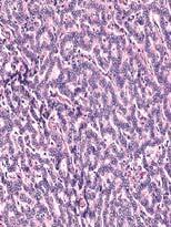
6 LARGE-CELL NON-HODGKIN LYMPHOMA OF THE THYMIC REGION, SCLEROSING B-CELL TYPE Recognized in the late 1970s & early 1980s as a distinctive intrathoracic neoplasm that could be associated with the superior vena cava syndrome Now well-characterized as a B-cell proliferation that is centered in the thymus, with singular cytogenetic & molecular characteristics


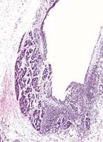
7 SCLEROSING SEMINOMA OF THE MEDIASTINUM A rare variant of seminoma, previously reported only in the testis; the speaker also has observed 3 such cases in the anterior mediastinum Tumor cells are scant in number and obscured by stromal fibrosis and chronic inflammation Immunostains for PLAP, CD117, OCT ¾, and SALL4 are usually necessary to document the presence of neoplastic germ cells in such tumors
 An uncommon histologic")


 serosal plaque")

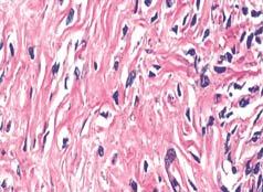
8 OCT 3/4 DESMOPLASTIC MESOTHELIOMA OF THE MEDIASTINUM (DMM) An uncommon histologic variant of an uncommon neoplasm in an uncommon anatomical location DMM may be surprisingly hypocellular, with a predominance of hyalinizing stroma which superficially resembles the histologic appearance of a fibrohyaline (asbestosrelated) serosal plaque At least modest nuclear atypia and growth into fat are necessary microscopic diagnostic elements Neoplastic cells are pankeratin-positive


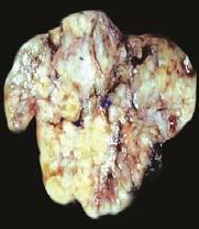
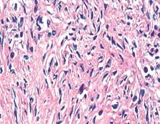
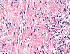
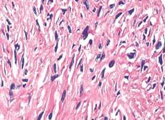
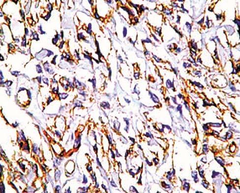
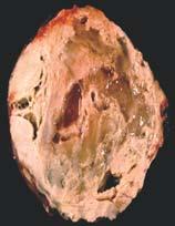
9 Pankeratin ANCIENT (SCLEROTIC) THYMOMA An unusual thymoma variant reported by Moran & Suster in 2004 Only 10% of patients had myasthenia gravis; the remainder presented with nondescript signs & symptoms or were entirely asymptomatic Densely-fibrotic stroma accounted for 85% to 90% of the tumor masses in each case Thorough sampling was necessary to document the presence of epithelial neoplastic cell groups


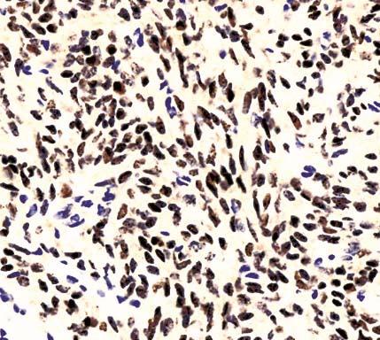
10 SCLEROTIC THYMIC CARCINOID Extensive sclerosis in neuroendocrine carcinomas of the lung was described by Kalhor et al. in 2010; the speaker has encountered 2 primary thymic neoplasms with similar changes Dense stromal fibrosis may obscure the diagnosis in small biopsies and also interfere with grading of the tumors Lesional cells are immunoreactive for pankeratin, as well as markers of neuroendocrine differentiation Marked sclerosis does not appear to affect behavior or prognosis


11 Pankeratin SCLEROSING PARAGANGLIOMAS Described by Plaza & colleagues in 2006 A pathologic variant of paraganglioma that tends to be overrepresented in middle-aged women Can be encountered in both the anterior and posterior mediastinal compartments Dominated by mature fibrous stroma, compressing tumor cell groups into irregular, pseudo-infiltrative profiles that can be confused with those of an invasive carcinoma or thymoma Non-immunoreactive for pankeratin
 OF THE MEDIASTINUM Analogous to")


12 Chromogranin CALCIFYING (PSEUDO)TUMOR (CPT) OF THE MEDIASTINUM Analogous to other lesions of the soft tissue, serosal surfaces, lungs, esophagus, liver, & spine Typically presents as a discrete mass, rather than an infiltrative process such as fibrosing mediastinitis Nondescript radiographic & gross appearance, except for the presence of multifocal calcifications Paucicellular background fibroblastic proliferation Appears to be a singular entity distinct from inflammatory myofibroblastic tumor and solitary fibrous tumor


13 SOLITARY FIBROUS TUMOR OF THE MEDIASTINUM Identical pathologically to lesions that are prototypically seen in the pleura Approximately 80% arise in the anterior mediastinum, with the remainder being in the middle & posterior mediastinal compartments May demonstrate well-demarcated or infiltrative contours radiographically Range of histological patterns, including patternless, epithelioid, and sclerosinghypocellular variants Potential immunoreactivity for CD34, CD99, bcl-2 protein, and STAT6

14 CD34 STAT6


 or perineurioma Potential immunoreactivity for")
15 SCLEROTIC PERIPHERAL NERVE SHEATH TUMORS Typically present as well-delimited masses in the posterior mediastinum Often asymptomatic; may cause neuralcompressive symptoms & signs or back pain Predominantly benign in nature; malignant nerve sheath tumors are rare in the mediastinum May be represented histologically by neurofibroma, neurilemmoma (schwannoma) or perineurioma Potential immunoreactivity for S100 protein, CD34, CD56, & CD57 S100


16 SCLEROSING METASTATIC CARCINOMA IN THE MEDIASTINUM May or may not be lymph node-based, and can be present in all 3 mediastinal compartments Metastatic lobular breast carcinoma and signet ring-cell gastric carcinomas are principally represented Linear single-file arrays or small nests of neoplastic cells embedded in a desmoplastic or mature fibrous stromal background Pankeratin stains are helpful to delineate the distribution of the tumor cells Mammaglobin
17 4/12/2018 II. CYSTIC LESIONS OF THE MEDIASTINUM Mark R. Wick, MD Division of Surgical Pathology University of Virginia Health System Charlottesville, VA, USA Cystic Mediastinal Lesions Account for 10-15% of intrathoracic masses found by radiographic imaging Several tissue types are represented, including pericardial, thymic, enteric, and bronchogenic elements Represent a mixture of developmental and acquired lesions Developmental (Congenital) Mediastinal Cysts 1


18 4/12/2018 Pericardial Cysts Usually seen in the basal portion of the mediastinum, abutting the heart shadow, as a rounded mass of variable density on plain films CT scans demonstrate a fluid-filled, thinwalled cyst in continuity with the pericardium Microscopy shows a mesothelial-lined fibrous cyst the hernia sac of the mediastinum 2


19 4/12/2018 Calretinin Unilocular Thymic Cysts May be present in the anterior or middle mediastinum, as an irregular or rounded density on plain films of the chest CT scans demonstrate a cyst with variable dense contents and an irregular wall; multiloculation may be present Microscopy shows a squamous lining with thymic tissue sometimes incorporated into the wall of the cyst; cholesterol clefts and calcification are common 3


20 4/12/2018 Unilocular Thymic Cyst Bronchogenic Cysts Rounded masses, usually in the middle mediastinum Patients may or may not complain of cough and expectoration of foul-tasting material ( motor oil ), depending on whether the cyst connects to a major bronchus CT scans may show calcified cartilaginous tissue in bronchogenic cysts Microscopy demonstrates the presence of cartilage, smooth muscle, and ciliated bronchialtype epithelium 4

 may be present Mullerian")

21 4/12/2018 Enteric Duplication (Gastroenteric) Mediastinal Cysts Probably derived from misplaced foregut rests Typically seen in children < 15 years old, who present with dysphagia, cough, or vomiting Characteristically present in the posterior mediastinum as spheroid masses that may show internal loculation Specialized gastric-mucosal, squamoid, or simple columnar epithelial linings (or mixtures thereof) may be present Mullerian (Hattori) Cysts of the Posterior Mediastinum Paravertebral in location, in women Unilocular, with an epithelial lining resembling that of endosalpingiosis Immunoreactive for CA-125, ERP, PRP, PAX8, and WT1 Simple excision is curative 5


22 4/12/2018 PAX8 ERP Acquired Non-Neoplastic Mediastinal Cyst Multilocular Thymic Cyst Multilocular Thymic Cysts Usually present in the anterior mediastinum as irregular or rounded densities in radiographic studies CT scans demonstrate variably dense contents and internal multiloculation Cholesterol clefts are inconspicuous, and mural lymphoid tissue is abundant Multilocular thymic cysts may rarely undergo malignant transformation 6


23 4/12/2018 Proliferating Thymic Cysts Unusual examples of multilocular thymic cyst in which the squamoid lining epithelium proliferates irregularly into the cyst wall, yielding an image which simulates that of squamous carcinoma Probably represents pseudoepitheliomatous hyperplasia of the lining epithelium, with an unknown cause 7


24 4/12/2018 Neoplastic & Paraneoplastic Cystic Lesions of the Mediastinum Mediastinal Lymphangiomas May be seen in all 3 mediastinal compartments, as unilocular or multilocular masses on imaging studies Predominate in children Interanastomosing vascular channels, associated with infiltrates of lymphocytes, & sometimes containing internal micropapillations 8


25 4/12/2018 Thymic Cysts in Hodgkin or Non-Hodgkin Lymphoma Usually seen after therapy of some kind (radiation; chemotherapy) but may occur as a spontaneous tumor-related phenomenon as well A central cystic cavity is surrounded by atypical lymphoid tissue containing diagnostic Reed cells or, alternatively, non-hodgkin lymphoma Other Potentially-Cystic Neoplasms of the Anterior Mediastinum Teratoma Thymoma Carcinoma ex thymic cyst Cystic de novo thymic carcinoma Seminoma Thymic carcinoid tumor 9


26 4/12/2018 Intrathymic Cystic Teratomas Predominantly seen in children and young adults, typically presenting with nondescript symptoms & signs or as lesions found incidentally on chest radiographs Eggshell calcification of the mass is possibly seen in plain-film radiographs At least 2 of 3 germ layers must be represented in the lesional tissue Immature neuroepithelial components are not prognostically important before the age of 15 years 10


27 4/12/2018 Cystic Thymoma Spontaneous cystic change may be so marked in thymoma that the initial pathologic impression is that of thymic cyst Thorough sampling of the lesional wall may well be necessary to identify neoplastic tissue Occasional tumors manifest a striking degree of hemorrhage and necrosis, but these findings do not influence prognosis 11


28 4/12/2018 Carcinomas & Thymic Cysts A rare event that appears to be associated only with the multilocular form of thymic cyst Enlarging mural nodules are visible in the cyst radiographically Histotypes of the carcinomas in this setting include keratinizing & non-keratinizing squamous carcinoma; basaloid carcinoma; mucoepidermoid carcinoma; papillary carcinoma; & sarcomatoid carcinoma De novo thymic carcinomas also may become cystic 12


29 4/12/
SCLEROSING LESIONS OF THE MEDIASTINUM
 SCLEROSING LESIONS OF THE MEDIASTINUM Mark R. Wick, MD Division of Surgical Pathology University of Virginia Health System Charlottesville, VA, USA SCLEROSING MEDIASTINITIS A slowly-evolving tumefactive
Pathology of Mediastinal Tumors
 SAMO Meeting Lucerne 2009 Pathology of Mediastinal Tumors Alex Soltermann Most common lesions (adults) Clinical presentation 50% of the patients are asymptomatic, lesion discovered incidentally Symptoms
Gross appearance of peritoneal cysts. They have a thin, translucent wall and contain a clear fluid.
 Gross appearance of peritoneal cysts. They have a thin, translucent wall and contain a clear fluid. So-called multicystic benign mesothelioma. A, Gross appearance. So-called multicystic benign mesothelioma.
Mediastinal Tumors: Imaging
 Mediastinal Tumors: Imaging References Imaging in Oncology, Husband and Reznek Computed Tomography and Magnetic Resonance of the thorax, Naidich, Zerhouni, Siegelman, Mediastinal compartments Anterior:
THYMIC CARCINOMAS AN UPDATE
 THYMIC CARCINOMAS AN UPDATE Mark R. Wick, M.D. University of Virginia Medical Center Charlottesville, VA CARCINOMA OF THE THYMUS General Clinical Features No apparent gender predilection Age range of 35-75
Case of the Day Chest
 Case of the Day Chest Darin White MDCM FRCPC Department of Radiology, Mayo Clinic 76 th Annual Scientific Meeting Canadian Association of Radiologists Montreal, QC April 26, 2013 2013 MFMER slide-1 Disclosures
SESSION 1: GENERAL (BASIC) PATHOLOGY CONCEPTS Thursday, October 16, :30am - 11:30am FACULTY COPY
 PATHOLOGY CONCEPTS Thursday, October 16, :30am - 11:30am FACULTY COPY") SESSION 1: GENERAL (BASIC) PATHOLOGY CONCEPTS Thursday, October 16, 2008 9:30am - 11:30am FACULTY COPY GOAL: Describe the basic morphologic (structural) changes which occur in various pathologic conditions.
SESSION 1: GENERAL (BASIC) PATHOLOGY CONCEPTS Thursday, October 16, 2008 9:30am - 11:30am FACULTY COPY GOAL: Describe the basic morphologic (structural) changes which occur in various pathologic conditions.
Basic Data. Sex:Male 31 years old Occupation: 搬家工人
 Basic Data Sex:Male 31 years old Occupation: 搬家工人 Chief Complaint Intermittent chest pain with shortness of breath for 2-3 months. Present Illness 4 months ago, he started having occasional chest pain
Mesothelioma: diagnostic challenges from a pathological perspective. Naseema Vorajee August 2016
 Mesothelioma: diagnostic challenges from a pathological perspective Naseema Vorajee August 2016 Naseema.vorajee@nhls.ac.za Pleural diseases (whether neoplastic, reactive or infective) may have similar
Salivary Glands 3/7/2017
 Salivary Glands 3/7/2017 Goals and objectives Focus on the entities unique to H&N Common board type facts Information for your future practice Salivary Glands Salivary Glands Major gland. Paratid. Submandibular.
Presentation material is for education purposes only. All rights reserved URMC Radiology Page 1 of 98
 Presentation material is for education purposes only. All rights reserved. 2011 URMC Radiology Page 1 of 98 Radiology / Pathology Conference February 2011 Brooke Koltz, Cytopathology Resident Presentation
Papillary Lesions of the Breast A Practical Approach to Diagnosis. (Arch Pathol Lab Med. 2016;140: ; doi: /arpa.
 Papillary Lesions of the Breast A Practical Approach to Diagnosis (Arch Pathol Lab Med. 2016;140:1052 1059; doi: 10.5858/arpa.2016-0219-RA) Papillary lesions of the breast Span the spectrum of benign,
Biopsy Interpretation of Spindle cell proliferations of the Serosa
 Biopsy Interpretation of Spindle cell proliferations of the Serosa Richard Attanoos, Cardiff. U.K. Disclosure of Relevant Financial Relationships USCAP requires that all planners (Education Committee)
PATHOLOGICAL COMPARATIVE ASSESSMENT OF TWO CASES OF THYMIC CYST AND CYSTIC THYMOMA AND REVIEW OF THE LITERATURE
 Rev. Med. Chir. Soc. Med. Nat., Iaşi 2012 vol. 116, no. 3 INTERNAL MEDICINE - PEDIATRICS CASE REPORTS PATHOLOGICAL COMPARATIVE ASSESSMENT OF TWO CASES OF THYMIC CYST AND CYSTIC THYMOMA AND REVIEW OF THE
Diseases of the breast (1 of 2)
") Diseases of the breast (1 of 2) Introduction A histology introduction Normal ducts and lobules of the breast are lined by two layers of cells a layer of luminal cells overlying a second layer of myoepithelial
Diseases of the breast (1 of 2) Introduction A histology introduction Normal ducts and lobules of the breast are lined by two layers of cells a layer of luminal cells overlying a second layer of myoepithelial
Sectional Anatomy Quiz II
 Sectional Anatomy II Rashid Hashmi Rural Clinical School, University of New South Wales, Wagga Wagga, New South Wales, Australia A R T I C L E I N F O Article type: Article history: Received: 3 Aug 2017
New lung lesion in a 55 year-old male treated with chemoradiation for non-small cell lung carcinoma
 July 2016 New lung lesion in a 55 year-old male treated with chemoradiation for non-small cell lung carcinoma Contributed by: Laurel Rose, MD, Resident Physician, Indiana University School of Medicine,
TUMOR AND TUMOR-LIKE CONDITIONS OF THE PERITONEUM AND OMENTUM/MESENTERY 40 th. Annual Meeting SCBTMR September 9-13, 2017, Nashville, Tennessee
 TUMOR AND TUMOR-LIKE CONDITIONS OF THE PERITONEUM AND OMENTUM/MESENTERY 40 th. Annual Meeting SCBTMR September 9-13, 2017, Nashville, Tennessee Isaac R Francis University of Michigan Department of Radiology
Chest Radiology Interpretation: Findings of Tuberculosis
 Chest Radiology Interpretation: Findings of Tuberculosis Get out your laptops, smart phones or other devices pollev.com/chestradiology Case #1 1 Plombage Pneumonia Cancer 2 Reading the TB CXR Be systematic!
Note: The cause of testicular neoplasms remains unknown
 - In the 15- to 34-year-old age group, they are the most common tumors of men. - Tumors of the testis are a heterogeneous group of neoplasms that include: I. Germ cell tumors : 95%; all are malignant.
04/10/2018. Intraductal Papillary Neoplasms Of Breast INTRADUCTAL PAPILLOMA
 Intraductal Papillary Neoplasms Of Breast Savitri Krishnamurthy MD Professor of Pathology Deputy Division Head The University of Texas MD Anderson Cancer Center 25 th Annual Seminar in Pathology Pittsburgh,
Normal endometrium: A, proliferative. B, secretory.
 Normal endometrium: A, proliferative. B, secretory. Nội mạc tử cung Nội mạc tử cung Cyclic changes in endometrium.. Approximate relationship of useful microscopic changes. Arias-Stella reaction in endometrial
Basal cell carcinoma 5/28/2011
 Goal of this Presentation A practical approach to the diagnosis of cutaneous carcinomas and their mimics Thaddeus Mully, MD University of California San Francisco To review common non-melanoma skin cancers
Breast pathology. 2nd Department of Pathology Semmelweis University
 Breast pathology 2nd Department of Pathology Semmelweis University Breast pathology - Summary - Benign lesions - Acute mastitis - Plasma cell mastitis / duct ectasia - Fat necrosis - Fibrocystic change/
Gross appearance of nodular hyperplasia in material obtained from suprapubic prostatectomy. Note the multinodular appearance and the admixture of
 Tiền liệt tuyến Tiền liệt tuyến Gross appearance of nodular hyperplasia in material obtained from suprapubic prostatectomy. Note the multinodular appearance and the admixture of solid and microcystic areas.
Kidney Case 1 SURGICAL PATHOLOGY REPORT
 Kidney Case 1 Surgical Pathology Report February 9, 2007 Clinical History: This 45 year old woman was found to have a left renal mass. CT urography with reconstruction revealed a 2 cm medial mass which
BREAST PATHOLOGY. Fibrocystic Changes
 BREAST PATHOLOGY Lesions of the breast are very common, and they present as palpable, sometimes painful, nodules or masses. Most of these lesions are benign. Breast cancer is the 2 nd most common cause
Normal thyroid tissue
 Thyroid Pathology Overview Normal thyroid tissue Normal thyroid tissue with follicles filled with colloid. Thyroid cells form follicles, spheres of epithelial cells (always single layered in health, usually
Desmoplastic Melanoma R/O BCC. Clinical Information. 74 y.o. man with lesion on left side of neck r/o BCC
 R/O BCC Sabine Kohler, M.D. Professor of Pathology and Dermatology Dermatopathology Service Stanford University School of Medicine Clinical Information 74 y.o. man with lesion on left side of neck r/o
3/27/2017. Pulmonary Pathology Specialty Conference. Disclosure of Relevant Financial Relationships. Clinical History:
 Pulmonary Pathology Specialty Conference Saul Suster, M.D. Medical College of Wisconsin Disclosure of Relevant Financial Relationships USCAP requires that all planners (Education Committee) in a position
Anatomy of the biliary tract
 Harvard-MIT Division of Health Sciences and Technology HST.121: Gastroenterology, Fall 2005 Instructors: Dr. Jonathan Glickman Anatomy of the biliary tract Figure removed due to copyright reasons. Biliary
Thymic Tumors. Feiran Lou MD. MS. Kings County Hospital Department of Surgery
 Thymic Tumors Feiran Lou MD. MS. Kings County Hospital Department of Surgery Case HPI 53 yo man referred from OSH for anterior mediastinal mass. Initially presented with leg weakness and back pain for
Diagnostically Challenging Cases in Gynecologic Pathology
 Diagnostically Challenging Cases in Gynecologic Pathology Eric C. Huang, M.D., Ph.D. Department of Pathology and Laboratory Medicine University of California, Davis Medical Center Case 1 Presentation 38
Karoline Nowillo, MD. February 1, 2008
 Case Presentation Karoline Nowillo, MD SUNY Downstate t February 1, 2008 Case Presentation Chief complaint enlarging goiter x 8 months History of present illness shortness of breath, heaviness in chest
Diplomate of the American Board of Pathology in Anatomic and Clinical Pathology
 A 33-year-old male with a left lower leg mass. Contributed by Shaoxiong Chen, MD, PhD Assistant Professor Indiana University School of Medicine/ IU Health Partners Department of Pathology and Laboratory
A Case of Pediatric Plasma Cell Granuloma
 August 2001 A Case of Pediatric Plasma Cell Granuloma Nii Tetteh, Harvard Medical School Year IV Our Patient 8 year old male with history of recurrent left lower lobe and lingular pneumonias since 1994.
CHRONIC PANCREATITIS OR DUCTAL ADENOCARCINOMA? N. Volkan Adsay, \ MD
 CHRONIC PANCREATITIS OR DUCTAL ADENOCARCINOMA? N. Volkan Adsay, \ MD Case for discussion 67 y/o male Back pain and weight loss CT: 4.5 cm ill-defined, solid lesion in the head FNA/Core bx: Inconclusive
CYSTIC TUMORS OF THE KIDNEY JOHN N. EBLE, M.D. CYSTIC NEPHROMA
 Page 1 CYSTIC TUMORS OF THE KIDNEY JOHN N. EBLE, M.D. Department of Pathology & Laboratory Medicine Phone (317) 274-4806 Medical Science A-128 FAX: (317) 278-2018 635 Barnhill Drive jeble @iupui.edu Indianapolis,
LYMPHATIC DRAINAGE AXILLARY (MOSTLY) INTERNAL MAMMARY SUPRACLAVICULAR
 INTERNAL MAMMARY SUPRACLAVICULAR") BREAST LYMPHATIC DRAINAGE AXILLARY (MOSTLY) INTERNAL MAMMARY SUPRACLAVICULAR HISTOLOGY LOBE: (10 in whole breast) LOBULE: (many per lobe) ACINUS/I, aka ALVEOLUS/I: (many per lobule) DUCT(S): INTRA- or
BREAST LYMPHATIC DRAINAGE AXILLARY (MOSTLY) INTERNAL MAMMARY SUPRACLAVICULAR HISTOLOGY LOBE: (10 in whole breast) LOBULE: (many per lobe) ACINUS/I, aka ALVEOLUS/I: (many per lobule) DUCT(S): INTRA- or
Spindle Cell Lesions Of The Breast. Emad Rakha Professor of Breast Pathology and Consultant Pathologist
 Spindle Cell Lesions Of The Breast Emad Rakha Professor of Breast Pathology and Consultant Pathologist * SCLs comprise a wide spectrum of diseases, ranging from reactive processes to aggressive malignant
Lách
 Lách Lách Lách Lách Splenogonadal fusion. Splenic tissue is attached to testicular tissue. Pseudocyst (false or secondary cyst). A, Outer aspect. Pseudocyst (false or secondary cyst). B, Inner surface.
The Relevance of Cytologic Atypia in Cutaneous Neural Tumors
 The Relevance of Cytologic Atypia in Cutaneous Neural Tumors Recent Findings - New Developments New Problems Zsolt B. Argenyi, M.D. Professor of Pathology & Dermatology Director of Dermatopathology Department
Tinh hoàn
 Tinh hoàn Tinh hoàn Tinh hoàn Tiền liệt tuyến Tiền liệt tuyến Mào tinh hoàn Mào tinh hoàn Túi tinh Túi tinh Túi tinh Túi tinh So-called cystadenoma of seminal vesicle. Gross appearance of granulomatous
Mody. AIS vs. Invasive Adenocarcinoma of the Cervix
 Common Problems in Gynecologic Pathology Michael T. Deavers, M.D. Houston Methodist Hospital, Houston, Texas Common Problems in Gynecologic Pathology Adenocarcinoma in-situ (AIS) of the Cervix vs. Invasive
Respiratory Interactive Session. Elaine Borg
 Respiratory Interactive Session Elaine Borg Case 1 Respiratory Cytology 55 year old gentleman Anterior mediastinal mass EBUS FNA Case 1 Respiratory Cytology 55 year old gentleman with anterior mediastinal
Diseases of the vulva
 Diseases of the vulva 1. Bartholin Cyst - Infection of the Bartholin gland produces an acute inflammation within the gland (adenitis) and may result in an abscess. Bartholin duct cysts - Are relatively
CPC 4 Breast Cancer. Rochelle Harwood, a 35 year old sales assistant, presents to her GP because she has noticed a painless lump in her left breast.
 CPC 4 Breast Cancer Rochelle Harwood, a 35 year old sales assistant, presents to her GP because she has noticed a painless lump in her left breast. 1. What are the most likely diagnoses of this lump? Fibroadenoma
Benign and malignant epithelial lesions: Seborrheic keratosis: A common benign pigmented epidermal tumor occur in middle-aged or older persons more
 Benign and malignant epithelial lesions: Seborrheic keratosis: A common benign pigmented epidermal tumor occur in middle-aged or older persons more common on the trunk; but extremities, head and neck are
Anterior Mediastinal Masses: The 4 T s
 May 2001 Anterior Mediastinal Masses: The 4 T s Rachel Van Sambeek, Harvard Medical School, Year III 1 Mediastinal Compartments 3 arbitrary divisions that do not correlate with anatomic planes: Anterior
Neoplasia part I. Dr. Mohsen Dashti. Clinical Medicine & Pathology nd Lecture
 Neoplasia part I By Dr. Mohsen Dashti Clinical Medicine & Pathology 316 2 nd Lecture Lecture outline Review of structure & function. Basic definitions. Classification of neoplasms. Morphologic features.
encapsulated thyroid nodule with a follicular architecture and some form of atypia. The problem is when to diagnose
 Histological Spectrum of Papillary Carcinoma of Thyroid A Two Years Study Gomathi Srinivasan 1, M. Vennila 2 1 Associate Professor Pathology, Government Medical College, Omandurar Estate, Chennai 600 002
Neoplasia literally means "new growth.
 NEOPLASIA Neoplasia literally means "new growth. A neoplasm, defined as "an abnormal mass of tissue the growth of which exceeds and is uncoordinated with that of the normal tissues and persists in the
CNS TUMORS. D r. Ali Eltayb ( U. of Omdurman. I ). M. Path (U. of Alexandria)
. M. Path (U. of Alexandria)") CNS TUMORS D r. Ali Eltayb ( U. of Omdurman. I ). M. Path (U. of Alexandria) CNS TUMORS The annual incidence of intracranial tumors of the CNS ISmore than intraspinal tumors May be Primary or Secondary
CNS TUMORS D r. Ali Eltayb ( U. of Omdurman. I ). M. Path (U. of Alexandria) CNS TUMORS The annual incidence of intracranial tumors of the CNS ISmore than intraspinal tumors May be Primary or Secondary
Case year old Chinese female. Radiological echo-distortion in the right breast at o clock. Core biopsy of the o clock lesion.
 Case 3 64 year old Chinese female. Radiological echo-distortion in the right breast at 10-12 o clock. Core biopsy of the 11-12 o clock lesion. Division of Pathology Courtesty of Dr Lester Leong ill-defined,
3 cell types in the normal ovary
 Ovarian tumors 3 cell types in the normal ovary Surface (coelomic epithelium) the origin of the great majority of ovarian tumors (neoplasms) 90% of malignant ovarian tumors Totipotent germ cells Sex cord-stromal
Patient Information. Age: 8 y/o Sex: Female. Date of Admission: Date of Discharge:
 Patient Information Age: 8 y/o Sex: Female Date of Admission: 92-10-08 Date of Discharge: 92-10-18 Chief Complaint Severe admominal pain and vomiting with dysuria since last afternoon Present Illness Lower
Disclosures. Parathyroid Pathology. Objectives. The normal parathyroid 11/10/2012
 Disclosures Parathyroid Pathology I have nothing to disclose Annemieke van Zante MD/PhD Assistant Professor of Clinical Pathology Associate Chief of Cytopathology Objectives 1. Review the pathologic features
Case 5 15-year-old male
 Case 5 15-year-old male Present illness: Six months ago, abnormality of ECG was incidentally detected by annual health check. His blood level of γ-gtp, HbA1c and norepinephrine were elevated; however,
objectives Pitfalls and Pearls in PET/CT imaging Kevin Robinson, DO Assistant Professor Department of Radiology Michigan State University
 objectives Pitfalls and Pearls in PET/CT imaging Kevin Robinson, DO Assistant Professor Department of Radiology Michigan State University To determine the regions of physiologic activity To understand
Interesting Cases from Liver Tumor Board. Jeffrey C. Weinreb, M.D.,FACR Yale University School of Medicine
 Interesting Cases from Liver Tumor Board Jeffrey C. Weinreb, M.D.,FACR Yale University School of Medicine jeffrey.weinreb@yale.edu Common Liver Diseases Hemangioma Cyst FNH Focal Fat/Sparing THID Non-Cirrhotic
Primary mediastinal tumours
 Primary mediastinal tumours Thorax (1974), 29, 475. YOUSF D. AL-NAAMAN, MOHAMAD S. AL-AN, and MUAYYAD M. AL-OMER Department of Thoracic and Cardiovascular Surgery, College of Medicine, University of Baghdad,
أملس عضلي غرن = Leiomyosarcoma. Leiomyosarcoma 1 / 5
 Leiomyosarcoma 1 / 5 EPIDEMIOLOGY Exact incidence is unknown, but older studies suggest that leiomyosarcomas comprise approximately 3 percent of soft-tissue sarcomas. Superficial leiomyosarcoma occurs
Primary pulmonary chondrosarcoma and a fast-growing mass that accidentally mimicked teratoma
 Case Report Primary pulmonary chondrosarcoma and a fast-growing mass that accidentally mimicked teratoma Jingjin Jiang 1, Qian Shen 2, Wei Ding 3, Jianying Zhou 2 1 Department of VIP, 2 Department of Respiratory
Case Report A case report of sclerosing thymoma of the anterior mediastinum: an exceedingly rare morphology
 Int J Clin Exp Pathol 2015;8(4):4233-4237 www.ijcep.com /ISSN:1936-2625/IJCEP0006183 Case Report A case report of sclerosing thymoma of the anterior mediastinum: an exceedingly rare morphology Shogo Tajima
Imaging in breast cancer. Mammography and Ultrasound Donya Farrokh.MD Radiologist Mashhad University of Medical Since
 Imaging in breast cancer Mammography and Ultrasound Donya Farrokh.MD Radiologist Mashhad University of Medical Since A mammogram report is a key component of the breast cancer diagnostic process. A mammogram
Alpha-fetoprotein
 Other Names/Abbreviations AFP 190.25 - Alpha-fetoprotein Alpha-fetoprotein (AFP) is a polysaccharide found in some carcinomas. It is effective as a biochemical marker for monitoring the response of certain
Salivary Gland Cytology
 Salivary Gland Cytology Diagnostic challenges and potential pitfalls Tarik M. Elsheikh, MD Professor and Medical Director Anatomic Pathology Cleveland Clinic FNA Salivary Gland Lesions Indications Distinguish
Radiology Pathology Conference
 Radiology Pathology Conference Sharlin Johnykutty,, MD, Cytopathology Fellow Sara Majewski, MD, Radiology Resident Friday, August 28, 2009 Presentation material is for education purposes only. All rights
How to Analyse Difficult Chest CT
 How to Analyse Difficult Chest CT Complex diseases are:- - Large lesion - Unusual or atypical pattern - Multiple discordant findings Diffuse diseases are:- - Numerous findings in both sides 3 basic steps
CODING TUMOUR MORPHOLOGY. Otto Visser
 CODING TUMOUR MORPHOLOGY Otto Visser INTRODUCTION The morphology describes the tissue of the tumour closest to normal tissue Well differentiated tumours are closest to normal Undifferentiated tumours show
Objectives. Salivary Gland FNA: The Milan System. Role of Salivary Gland FNA 04/26/2018
 Salivary Gland FNA: The Milan System Dr. Jennifer Brainard Section Head Cytopathology Cleveland Clinic Objectives Introduce the Milan System for reporting salivary gland cytopathology Define cytologic
Alpha-fetoprotein
 Other Names/Abbreviations AFP 190.25 - Alpha-fetoprotein Alpha-fetoprotein (AFP) is a polysaccharide found in some carcinomas. It is effective as a biochemical marker for monitoring the response of certain
Alpha-fetoprotein
 Other Names/Abbreviations AFP 190.25 - Alpha-fetoprotein Alpha-fetoprotein (AFP) is a polysaccharide found in some carcinomas. It is effective as a biochemical marker for monitoring the response of certain
Breast Pathology. Breast Development
 Breast Pathology Lecturer: Hanina Hibshoosh, M.D. Reading: Kumar, Cotran, Robbins, Basic Pathology, 6th Edition, pages 623-635 Breast Development 5th week - thickening of the epidermis - milk line 5th
Malignant Peripheral Nerve Sheath Tumor
 C H A P T E R 120 Malignant Peripheral Nerve Sheath Tumor Currently, malignant peripheral nerve sheath tumor (MPNST) is the most commonly used generic name for the neoplasms known in the past as neurosarcoma,
Protocol for the Examination of Specimens From Patients With Thymic Tumors
 Protocol for the Examination of Specimens From Patients With Thymic Tumors Version: Protocol Posting Date: June 2017 Includes ptnm requirements from the 8 th Edition, AJCC Staging Manual For accreditation
Dr Sanjiv Manek Oxford. Oxford Pathology Course 2010 for FRCPath Illustration-Cellular Pathology. Oxford Radcliffe NHS Trust
 Dr Sanjiv Manek Oxford Oxford Pathology Course 2010 for FRCPath Illustration-Cellular Pathology. Oxford Radcliffe NHS Trust Ovarian Endometrial Vulvo-vaginal Cervical Illustration-Cellular Pathology. Oxford
Pulmonary Sarcoidosis - Radiological Evaluation
 Original Research Article Pulmonary Sarcoidosis - Radiological Evaluation Jayesh Shah 1, Darshan Shah 2*, C. Raychaudhuri 3 1 Associate Professor, 2 1 st Year Resident, 3 Professor and HOD Radiology Department,
Pitfalls in thyroid tumor pathology. Prof.Valdi Pešutić-Pisac MD, PhD
 Pitfalls in thyroid tumor pathology Prof.Valdi Pešutić-Pisac MD, PhD Too many or... Tumour herniation through a torn capsule simulating capsular invasion fibrous capsule with a sharp discontinuity, suggestive
Lymphoma: What You Need to Know. Richard van der Jagt MD, FRCPC
 Lymphoma: What You Need to Know Richard van der Jagt MD, FRCPC Overview Concepts, classification, biology Epidemiology Clinical presentation Diagnosis Staging Three important types of lymphoma Conceptualizing
ARRO Case Thymoma. Jordan Kharofa, MD Elizabeth Gore, MD Medical College of Wisconsin
 ARRO Case Thymoma Jordan Kharofa, MD Elizabeth Gore, MD Medical College of Wisconsin History HPI 54 yo male who presented to PCP with complaining subacute shortness of breath and chest pain. Pain increased
Histological Typing Of Cancer And Precancer Of The Oral Mucosa
 Histological Typing Of Cancer And Precancer Of The Oral Mucosa 1 / 7 2 / 7 3 / 7 Histological Typing Of Cancer And Within the last decade, histologic grading has become widely accepted as a powerful indicator
An Introduction to Radiology for TB Nurses
 An Introduction to Radiology for TB Nurses Garold O. Minns, MD September 14, 2017 TB Nurse Case Management September 12 14, 2017 EXCELLENCE EXPERTISE INNOVATION Garold O. Minns, MD has the following disclosures
57th Annual HSCP Spring Symposium 4/16/2016
 An Unusual Malignant Spindle Cell Lesion to Involve the Breast Erinn Downs-Kelly, D.O. Associate Professor of Pathology University of Utah & ARUP Laboratories No disclosures Case 39 y/o female with no
Oncocytic-Appearing Salivary Gland Tumors. Oncocytic, Cystic, Mucinous, and High Grade Salivary Gland Tumors SALIVARY GLAND FNA: PART II
 William C. Faquin, MD, PhD Professor of Pathology Harvard Medical School Director of Head and Neck Pathology Massachusetts Eye and Ear Massachusetts General Hospital SALIVARY GLAND FNA: PART II Oncocytic,
Differential Diagnosis of Oral Masses. Palatal Lesions
 Differential Diagnosis of Oral Masses Palatal Lesions Palatal Masses Periapical Abscess Torus Palatinus Mucocele Lymphoid Hyperplasia Adenomatous Hyperplasia Benign Salivary Neoplasms Malignant Salivary
Case E1. Female aged 63 years Right Nephrectomy Two separate tumours Section of each tumour
 Case E1 Female aged 63 years Right Nephrectomy Two separate tumours Section of each tumour Tumour 1 Upper pole tumour 28mm macro diameter Circumscribed Friable cut surface Tumour 2 Middle pole Part solid
Non Small Cell Lung Cancer Histopathology ד"ר יהודית זנדבנק
 Non Small Cell Lung Cancer Histopathology ד"ר יהודית זנדבנק 26.06.09 Lecture outlines WHO histological classification Macro/Micro assessment Early diagnosis Minimal pathology Main subtypes SCC, AdCa, LCLC
Testicular Germ Cell Tumors; A Simplistic Approach
 Testicular Germ Cell Tumors; A Simplistic Approach Merce Jorda, MD, PhD, MBA Professor and Vice Chair, Director of Anatomic Pathology Director of Genitourinary Pathology Service Interim Director of Cytopathology
Case Scenario 1: Thyroid
 Case Scenario 1: Thyroid History and Physical Patient is an otherwise healthy 80 year old female with the complaint of a neck mass first noticed two weeks ago. The mass has increased in size and is palpable.
ONCOLOGY. Csaba Bödör. Department of Pathology and Experimental Cancer Research november 19., ÁOK, III.
 ONCOLOGY Csaba Bödör Department of Pathology and Experimental Cancer Research 2018. november 19., ÁOK, III. bodor.csaba1@med.semmelweis-univ.hu ONCOLOGY Characteristics of Benign and Malignant Neoplasms
GUT-C 11/30/2017. Debasmita Das, M.D. PGY-1 Danbury Hospital
 GUT-C 11/30/2017 Debasmita Das, M.D. PGY-1 Danbury Hospital CLINICAL SUMMARY 8/2017 59 year old female Presented to the ED with 1 month history of general malaise, fever and weight loss PMH: Significant
Surveys and Anatomic Pathology Education Programs
 Surveys and Anatomic Pathology Education Programs Performance Improvement Program in Surgical Pathology PIP/PIPW-B 2018 Participant Summary 2018 College of American Pathologists. The College does not permit
Problem diagnoses. Current issues in Anatomic pathology. Problem Diagnoses in Tumors of the Oral Cavity 5/29/2009
 Current issues in Anatomic pathology Problem Diagnoses in Tumors of the Oral Cavity Richard Jordan DDS PhD FRCPath Professor of Oral Pathology & Pathology Director, UCSF Oral Pathology Diagnostic Laboratory
Tumors of the Thvmus and Thee
 Tumors of the Thvmus Thee Region: 111. Clinic&pathological Skdies on Teratornas Tumors of Germ Cell Type N. P. Bergh, M.D., P. Gatzinsky, M.D., S. Larsson, M.D., P. Lundin, M.D., B. Ridell, M.D. ABSTRACT
Lesion Imaging Characteristics Mass, Favoring Benign Circumscribed Margins Intramammary Lymph Node
 Lesion Imaging Characteristics Mass, Favoring Benign Circumscribed Margins Intramammary Lymph Node Oil Cyst Mass, Intermediate Concern Microlobulated Margins Obscured Margins Mass, Favoring Malignant Indistinct
Case Report A Rare Cutaneous Adnexal Tumor: Malignant Proliferating Trichilemmal Tumor
 Case Reports in Medicine Volume 2015, Article ID 742920, 4 pages http://dx.doi.org/10.1155/2015/742920 Case Report A Rare Cutaneous Adnexal Tumor: Malignant Proliferating Trichilemmal Tumor Omer Alici,
Neoplasms of the Canine, Feline and Lemur Liver:
 Neoplasms of the Canine, Feline and Lemur Liver: Classification and Prognosis Annual Seminar of the French Society of Veterinary Pathology John M. Cullen VMD PhD DACVP North Carolina State University Primary
Case Scenario 1. The patient agreed to a CT guided biopsy of the left upper lobe mass. This was performed and confirmed non-small cell carcinoma.
 Case Scenario 1 An 89 year old male patient presented with a progressive cough for approximately six weeks for which he received approximately three rounds of antibiotic therapy without response. A chest