(b) Stomach s function 1. Dilution of food materials 2. Acidification of food (absorption of dietary Fe in small intestine) 3. Partial chemical digest
|
|
|
- Lionel McCarthy
- 6 years ago
- Views:
Transcription
1 (1) General features a) Stomach is widened portion of gut-tube: between tubular and spherical; Note arranged of smooth muscle tissue in muscularis externa. 1
2 (b) Stomach s function 1. Dilution of food materials 2. Acidification of food (absorption of dietary Fe in small intestine) 3. Partial chemical digestion of proteins continued digestion of carbohydrates 2
3 4. Little to no absorption under normal conditions a. Can absorb glucose b. Absorbs ethanol c. Absorbs Na + and K +, but only if levels of these ions are subnormal d. Absorbs water when body dehydrated and osmotic pressure abnormally high and blood volume and pressure unusually low. 5. Formation of acidic chyme: Mixture of acidic gastric juice and food 3


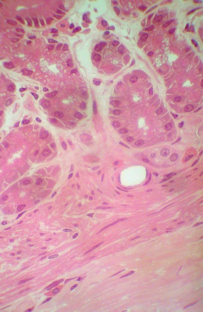
4 (c) Mucosa: very folded; folds longitudinally oriented and called rugae. 1. Lumenal epithelium: simple columnar epithelium: secretes mucus; cytoplasm tends to be acidophilic; cells lack microvilli. 2. Lumenal surface has abundant pits: funnel-like depressions that lead to the mucosal glands; pits vary in steepness and depth from one region of organ to another. 4
 cells endocrine cells (G cells).")
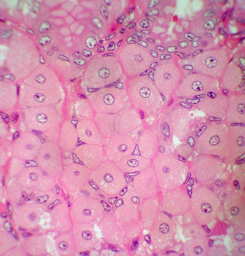
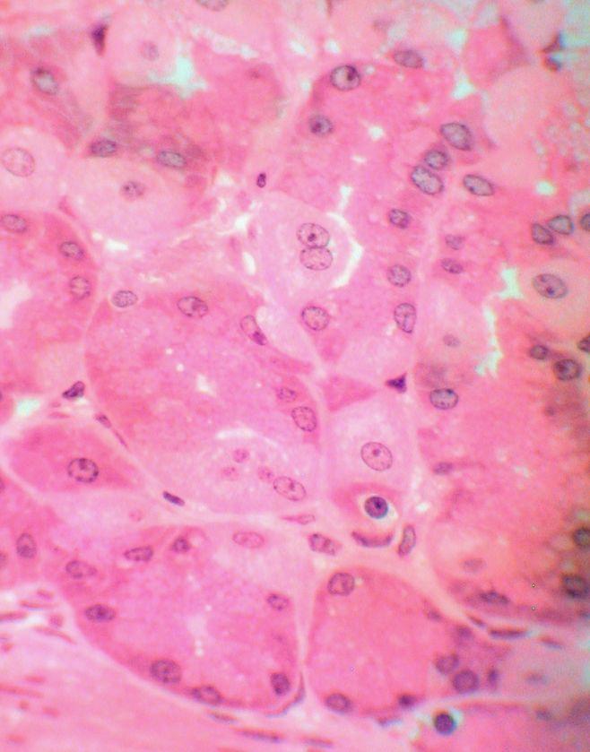
5 3. Mucosal glands common compound (branched) tubular glands extending into deeper parts of mucosa, from pits; not acinar wall of gland one cell thick 4 possible cell types mucous cells parietal cells chief (principal; peptic) cells endocrine cells (G cells). 5
6 a. Except for endocrine cells, restricted to one region of stomach, glands are exocrine, secreting fluid into lumen of stomach b. All cells of wall of gland are glandular cells. c. Uppermost part of gland, continuous with epithelium of pit; neck of gland neck composed of low columnar mucus cells - not as eosinophilic as lumenal epithelial cells mucus neck cells. f. Stomach. 6
 Parietal cell: large, spherical, eosinophilic, sometimes granular cytoplasm; secretes HCl:")
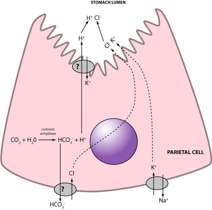
7 d. Below neck of gland, cells include the other 3 types. (1) Parietal cell: large, spherical, eosinophilic, sometimes granular cytoplasm; secretes HCl: acidification of the food; often found in upper part of gland. 7
8 8
9 9
 Chief cells therefore are zymogenic cells.")
10 (2) Chief (principal; peptic) cell tends to be in lower part, narrower than parietal cell cytoplasm basophilic nucleus toward basal side of cell very active in protein synthesis: produces and secretes pepsinogen zymogen of pepsin: enzyme that breaks protein molecules into chains of several to many amino acid residues. (a) Chief cells therefore are zymogenic cells. (b) Pepsinogen converted to pepsin in lumen of gland and in lumen of organ. 10
11 11

12 (3) Endocrine cells: specifically gastrin cells, for the hormone they produce and release restricted to deepest parts of glands of one region of stomach. A subset of enteroendocrine cells; >12 types gastrin, histamine, endorphins, serotonin, cholecystokinin, somatostatin 12
13 4. Muscularis mucosae present, composed of smooth muscle tissue, often in 3 layers; has numerous thin extensions up into lamina propria. 5. Lamina propria composed of unusually cellular dense C.T. with capillaries, arterioles, venules, lymphatic capillaries, small nerves; all of these but capillaries very difficult to detect MM 13

14 (d) Submucosa 1. Extends up into rugae. 2. Mostly dense C.T small muscular arteries, small veins, smaller blood vessels, small lymphatic vessels, small masses of neural tissue and sometimes clumps of adipocytes areas of loose C.T. may be present also. f. Stomach Mucosa Submucosa MM Muscularis externa 14
15 (e) Muscularis externa of stomach 1. Normal Inner-circular, outer-longitudinal organization partially lost: smooth muscle tissue is in large slabs oriented in a variety of directions, some remnant of inner-circ., outer-long pattern usually detectable. Inner circular Outer longitudinal 15
16 2. Myenteric plexus usually detectable; its location indicates the original interface between inner-circ, outer-long layers. 3. This organization of smooth muscle tissue relates to the organ's shape. 4. Muscularis is, overall, relatively thick and powerful. Myenteric plexus 16
17 (f) Tunica adventitia 1. T. adventitia of stomach is distinct layer with an outer, mesothelium, covering. 2. T. adventitia composed of dense C.T. and loose C.T. and contains larger blood vessels and nerves than those found in submucosa. T.adventitia 17
18 g) Stomach regions divided into 4 regions; in most cases these regions differ in mucosal structure only. 1. Cardiac region: small region adjacent to cardiac sphincter. 2. Fundic region (fund: root meaning deep): one side of upper part of organ. 3. Body: most of organ. 4. Pyloric region: lower end, adjacent to pyloric sphincter. 18
19 2) Special features of region of stomach (a) Cardiac region 1. pits; intermediate depth and cardiac glands are almost entirely mucus cells 2. Glands sometimes not as densely packed as in other regions. 19
20 20

21 (b) Fundic region and body (no histological differences between them) 1. Pits relatively shallow (because fluid secreted by glands is nonviscous) 2. Gastric glands densely packed; C.T. of lamina propria not obvious. Neck of gland composed of mucus neck cells; other cells of gland parietal cells and chief cells 21

22 (c) Pyloric region 1. Pits deep half the depth of the mucosa Gland composed of mucus cells ;look like mucus neck cells diameter of gland's lumen high, due to high viscosity of secreted fluid Called pyloric glands. f. Stomach 22


23 23
24 a. Pyloric region and cardiac region; major function; mucus production; mucus coating of the lumenal epithelium protection from acidity and proteolytic action of gastric juice. If mucus layer becomes thin or lost, the pepsin and HCl in the lumen will destroy the lumenal epithelium of that area and perhaps begin destroying the glands and C.T. of the lamina propria Called a peptic ulcer. 24
25 b. Lower parts of the pyloric glands ; cells are mainly endocrine; with H&E staining they are not distinguishable from mucus cells gastrin endocrine cells produce the hormone gastrin, controls gastric glands gastrin is released to the capillaries of the lamina propria rather than to the lumen of the gland. c. Overall length of glands relatively great 25

26 3. Mucosa unusually thick overall; staining relatively pale. 4. Pyloric region ends at pyloric sphincter; between stomach and duodenum sphincter itself is a thickened region of inner, circular layer of muscularis externa. 26
 Mucus enteroendocrine cells (not visible w H&E) Function Protection of cardiac region and distal esophagus")
27 ` Regions Cardiac Fundus/ body Pyloric Pits Intermediate depth Shallow Deep, at least 1/2 of mucosa with wide lumen Glands Not densely packed obvious lumen Densely packed Not densely packed obvious lumen Gland cells Mucus Mucus, parietal, chief, enteroendocrine cells (not visible w H&E) Mucus enteroendocrine cells (not visible w H&E) Function Protection of cardiac region and distal esophagus Production of gastric juice; Hormone regulation Protection of pyloric region Hormone regulation 27
Alimentary Canal (I)
") Alimentary Canal (I) Esophagus and Stomach (Objectives) By the end of this lecture, the student should be able to discuss the microscopic structure in correlation with the function of the following organs:
Alimentary Canal (I) Esophagus and Stomach (Objectives) By the end of this lecture, the student should be able to discuss the microscopic structure in correlation with the function of the following organs:
Digestive system L 2. Lecturer Dr. Firdous M. Jaafar Department of Anatomy/Histology section
 Digestive system L 2 Lecturer Dr. Firdous M. Jaafar Department of Anatomy/Histology section objectives 1-Describe the general structure of digestive tract: a-mucosa. b-submucosa. c-muscularis externa d-adventitia
HISTOLOGY. GIT Block 432 Histology Team. Lecture 1: Alimentary Canal (1) (Esophagus & Stomach) Done by: Ethar Alqarni Reviewed by: Ibrahim Alfuraih
 (Esophagus & Stomach) Done by: Ethar Alqarni Reviewed by: Ibrahim Alfuraih") HISTOLOGY Lecture 1: Alimentary Canal (1) (Esophagus & Stomach) Done by: Ethar Alqarni Reviewed by: Ibrahim Alfuraih Color Guide: Black: Slides. Red: Important. Green: Doctor s notes. Blue: Explanation.
HISTOLOGY Lecture 1: Alimentary Canal (1) (Esophagus & Stomach) Done by: Ethar Alqarni Reviewed by: Ibrahim Alfuraih Color Guide: Black: Slides. Red: Important. Green: Doctor s notes. Blue: Explanation.
Small intestine. Small intestine
 General features Tubular organ longest part; 5-6 m most of chemical digestion absorption of nutrients reabsorption of H2O occurs. Two structural features; maximize the lumenal surface area villi microvilli
Dana Alrafaiah. Dareen Abu Shalbak. Mohammad Almuhtaseb. 1 P a g e
 2 Dana Alrafaiah Dareen Abu Shalbak Mohammad Almuhtaseb 1 P a g e Esophagus: A muscular tube that is 25 cm long, but if measured from the incisors it would be 45cm long. Extends from C6 of cervical vertebra,
General Structure of Digestive Tract
 Dr. Nabil Khouri General Structure of Digestive Tract Common Characteristics: Hollow tube composed of a lumen whose diameter varies. Surrounded by a wall made up of 4 principal layers: Mucosa Epithelial
The doctor mentioned a few things about the esophagus from the previous lecture:
 السالم عليكم [HISOLOGY 2] April 27, 2014 The doctor mentioned a few things about the esophagus from the previous lecture: Esophagus - It is about 25 cm in length (from the incisor it is 45 cm) Histological
Dr Nadine Gravett School of Anatomical Sciences Room 2B10B
 Dr Nadine Gravett School of Anatomical Sciences Room 2B10B Nadine.Gravett@wits.ac.za Oral cavity Mechanical breakdown Formation of bolus Oesophagus Conduit from mouth to stomach Stomach Digestion Temporary
DIGESTIVE TRACT ESOPHAGUS
 DIGESTIVE TRACT From the lower esophagus to the lower rectum four fundamental layers comprise the wall of the digestive tube: mucosa, submucosa, muscularis propria (externa), and adventitia or serosa (see
Digestive System Module 4: The Stomach *
 OpenStax-CNX module: m49286 1 Digestive System Module 4: The * Donna Browne Based on The by OpenStax This work is produced by OpenStax-CNX and licensed under the Creative Commons Attribution License 4.0
Gastrointestinal Anatomy and Physiology. Bio 219 Napa Valley College Dr. Adam Ross
 Gastrointestinal Anatomy and Physiology Bio 219 Napa Valley College Dr. Adam Ross Functions of digestive system Digestion Breakdown of food (chemically) using enzymes, acid, and water Absorption Nutrients,
Tongue In the buccal cavity of the digestive system
 Tongue In the buccal cavity of the digestive system same layers as those of tubular organs Mucosa, submucosa, and muscularis muscularis = the muscularis externa no muscularis mucosa 1 Tongue ling = tongue
Esophagus. Transport is achieved by peristaltic contractions and relaxation of the esophageal sphincters (upper and lower)
") GI Histology 2 Esophagus is a muscular tube whose function is to transport foodstuffs from the mouth to the stomach and to prevent the retrograde flow of gastric contents Transport is achieved by peristaltic
GI Histology 2 Esophagus is a muscular tube whose function is to transport foodstuffs from the mouth to the stomach and to prevent the retrograde flow of gastric contents Transport is achieved by peristaltic
Urinary system. Urinary system
 Distal convoluted tubule (DCT) Highly coiled, ~ 5 mm in length Last part of the nephron. Wall; simple cuboidal epithelium Less metabolically active than the PCT no brush border light eosinophilic cytoplasm
The Digestive System and Body Metabolism
 14 PART B The Digestive System and Body Metabolism PowerPoint Lecture Slide Presentation by Jerry L. Cook, Sam Houston University ESSENTIALS OF HUMAN ANATOMY & PHYSIOLOGY EIGHTH EDITION ELAINE N. MARIEB
الله الر ح م ن الر ح يم مسب
 بسم رلا هللارلا هللا This is the second histology lecture in the GI system. In this lecture, we will discuss the histology of the esophagus, stomach, and small intestine so prepare yourself.this sheet
Anatomy & Histology of The Small intestine
 Anatomy & Histology of The Small intestine Prof. Abdulameer Al-Nuaimi E-mail: a.al-nuaimi@sheffield.ac.uk E. mail: abdulameerh@yahoo.com Jejunum Ileum Histology: Duodenum, jejunum, and ileum
Digestive System 7/15/2015. Outline Digestive System. Digestive System
 Digestive System Biology 105 Lecture 18 Chapter 15 Outline Digestive System I. Functions II. Layers of the GI tract III. Major parts: mouth, pharynx, esophagus, stomach, small intestine, large intestine,
The Digestive System. What is the advantage of a one-way gut? If you swallow something, is it really inside you?
 The Digestive System What is the advantage of a one-way gut?! If you swallow something, is it really inside you? Functions and Processes of the Digestive System: Move nutrients, water, electrolytes from
Stomach. Stomach. Nerve supply. Blood supply. Sympathe0c and parasympathe0c fibers of the autonomic nervous system
 Stomach Nerve supply Sympathe0c and parasympathe0c fibers of the autonomic nervous system Blood supply Celiac trunk, and corresponding veins (part of the hepa0c portal system) Stomach Figure 23.14a Chapter
Chapter 14: The Digestive System
 Chapter 14: The Digestive System Digestive system consists of Muscular tube (digestive tract) alimentary canal Accessory organs teeth, tongue, glandular organs 6 essential activities 1. 2. 3. 4. 5. 6.
Digestive System. - Food is ingested
 11 V. Digestive Processes in the Mouth - Food is ingested - Mechanical digestion begins (chewing) - Salivary amylase begins chemical breakdown of starch - Propulsion is initiated by Deglutition (Swallowing)
MICROSTRUCTURES LIPS TOOTH TONGUE OESOPHAGUS STOMACH, CARDIAC, PYLORIC FUNDIC GLANDS
 MICROSTRUCTURES LIPS TOOTH TONGUE OESOPHAGUS STOMACH, CARDIAC, PYLORIC FUNDIC GLANDS HUMAN ANATOMY: MICROSTRUCTURES CLASSIFICATION: LOCATION AND BOUNDARIES, FORM, FUNCTION, MICROSCOPIC STRUCTURE: A hollow
University of Buea. Faculty of Health Sciences. Programme in Medicine
 Faculty of Health Sciences University of Buea Wednesday, 28 th January 2009 Time: 8 00-10 00 Programme in Medicine MED 303 (Gastrointestinal Physiology) EXAMS (2008-2009) Identify the letter of the choice
Slide 154: Pancreas, H&E
 Slide 154: Pancreas, H&E the pancreas, located adjacent to the duodenum, is a mixed exocrine and endocrine gland; it is usually readily identifiable by the presence of the interspersed endocrine pancreatic
/30/17 Ch 8: Muscular System 1. Table of Contents # Date Title Page # 03/13/17 Ch 10: Somatic and Special Senses 53
 Table of Contents # Date Title Page # 1. 01/30/17 Ch 8: Muscular System 1 2. 3. 4. 5. 6. 7. 02/14/17 Ch 9: Nervous System 12 03/13/17 Ch 10: Somatic and Special Senses 53 03/27/17 Ch 11: Endocrine System
The Digestive System. Chapter 25
 The Digestive System Chapter 25 Introduction Structure of the digestive system A tube that extends from mouth to anus Accessory organs are attached Functions include Ingestion Movement Digestion Absorption
Small Intestine, Large Intestine and anal cannel
 Small Intestine, Large Intestine and anal cannel 32409 Small intestine Large intestine Small intestine General Structure of the Digestive Tract rat 32409 Epithelium with goblet cells and absorptive cells
HISTOLOGY VIRTUAL LABORATORY GASTROINTESTINAL SYSTEM
 HISTOLOGY VIRTUAL LABORATORY GASTROINTESTINAL SYSTEM LIP (Slides GI 1, 2) Identify the outer portion lined by stratified squamous (keratinized) epithelium. Note the hair follicles and sebaceous glands
The Digestive System Laboratory
 The Digestive System Laboratory 1 The Digestive Tract The alimentary canal is a continuous tube stretching from the mouth to the anus. Liver Gallbladder Small intestine Anus Parotid, sublingual, and submaxillary
Includes mouth, pharynx, esophagus, stomach, small intestine, large intestine, rectum, anus. Salivary glands, liver, gallbladder, pancreas
 Chapter 14 The Digestive System and Nutrition Digestive System Brings Nutrients Into the Body The digestive system includes Gastrointestinal (GI) tract (hollow tube) Lumen: space within this tube Includes
Anatomy of the liver and pancreas
 Anatomy of the liver and pancreas Prof. Abdulameer Al-Nuaimi E-mail: a.al-nuaimi@sheffield.ac.uk abdulameerh@yahoo.com Liver Aorta Pulm. Trunk Rt. At, Duct. Art. Lt. Ven. Rt. Ven. Internal Posterior
Lab activity manual - Histology of the digestive system. Lab activity 1: esophagus stomach - small intestines
 Lab activity manual - Histology of the digestive system Jeanne Adiwinata Pawitan Prerequisite: Histology of the 4 basic tissues In this module we learn about the histology of the digestive system, from
Two main groups Alimentary canal continuous coiled hollow tube Accessory digestive organs
 Digestion Breakdown of ingested food Absorption of nutrients into the blood Metabolism Production of cellular energy (ATP) Constructive and degradative cellular activities Two main groups Alimentary canal
Chapter 26 The Digestive System
 Chapter 26 The Digestive System Digestive System Gastroenterology is the study of the stomach and intestine. Digestion Catabolism Absorption Anabolism The actions of the digestive system are controlled
Week 12 - Outline. Outline. Digestive System I Major Organs. Overview of Digestive System
 Outline Week 12 - Digestive System I Major Organs Copyright The McGraw-Hill Companies, Inc. Permission required for reproduction or display. Digestive Tract Function GI Tract Structure Regulation of the
The Stomach. Bởi: OpenStaxCollege
 Bởi: OpenStaxCollege Although a minimal amount of carbohydrate digestion occurs in the mouth, chemical digestion really gets underway in the stomach. An expansion of the alimentary canal that lies immediately
Connective tissue The Digestive System
 Connective tissue The Digestive System Part 1 Structure of digestive system Functions Basic Structure of the Alimentary Canal Wall Tube is made up of four layers: 1. Mucosa 2. Submucosa 3. Muscularis externa
consists of: Muscular, hollow tube (= digestive tract ) + Various accessory organs
 + Various accessory organs") DIGESTIVE SYSTEM consists of: Muscular, hollow tube (= digestive tract ) + Various accessory organs FUNCTION Individual parts function in: ingestion mechanical digestion chemical and enzymatic digestion
DIGESTIVE SYSTEM consists of: Muscular, hollow tube (= digestive tract ) + Various accessory organs FUNCTION Individual parts function in: ingestion mechanical digestion chemical and enzymatic digestion
DIGESTIVE. CHAPTER 17 Lecture: Part 1 Part 2 BIO 212: ANATOMY & PHYSIOLOGY II
 BIO 212: ANATOMY & PHYSIOLOGY II 1 CHAPTER 17 Lecture: DIGESTIVE Part 1 Part 2 Dr. Lawrence G. Altman www.lawrencegaltman.com Some illustrations are courtesy of McGraw-Hill. Processes of DIGESTION Mechanical
The stomach is formed of three parts: -
 The stomach is formed of three parts: - (a) CARDIAC STOMACH: - It receives the oesophagus through Cardiac aperture guarded by a cardiac sphincter which prevents regurgitation of food. (b) FUNDIC PART:
Soft palate elevates, closing off the nasopharynx. Hard palate Tongue Bolus Epiglottis. Glottis Larynx moves up and forward.
 The Cephalic Phase Chemical and mechanical digestion begins in the mouth Saliva is an exocrine secretion Salivary secretion is under autonomic control Softens and lubricates food Chemical digestion: salivary
Energy, Chemical Reactions and Enzymes
 Phosphorylation Hydrolysis Energy, Chemical Reactions and Enzymes Chapter 2 (selections) What is Energy? Energy is the capacity to do work Potential Energy Kinetic Energy Chemical Bond Energy Like a rechargeable
PANCREATIC BETA CELLS PRODUCE AND SECRETE
 15 March, 2018 PANCREATIC BETA CELLS PRODUCE AND SECRETE Document Filetype: PDF 374.06 KB 0 PANCREATIC BETA CELLS PRODUCE AND SECRETE Among the oldest and cheapest drugs for diabetes are the drugs that
DIGESTIVE SYSTEM ALIMENTARY CANAL / GI TRACT & ACCESSORY ORGANS. Mar 16 10:34 PM
 DIGESTIVE SYSTEM ALIMENTARY CANAL / GI TRACT & ACCESSORY ORGANS Mar 16 10:34 PM 1 I. Digestive System Functions > Ingestion the taking in of food > Propulsion movement caused by force > Digestion breakdown
Urinary system. Kidney anatomy Renal cortex Renal. Nephrons
 Urinary system Aids homeostasis by removing cellular wastes and foreign compounds, and maintains salt and water balance of plasma Kidney anatomy Renal cortex Renal pelvis Renal medulla Cortex Ureter Medulla
by authors and SVSBT.
 The Indian Journal of Veterinary Sciences & Biotechnology (2018) Volume 13, Issue 4, 20-25 ISSN (Print) : 2394-0247 : ISSN (Print and online) : 2395-1176, abbreviated as IJVSBT 10.21887/ijvsbt.v13i4.11553
BIO 132 Anatomy and Physiology II Spring, 2016 Exam 1 Name: BIO 132 ID Number. Section 1 Answer questions 1 40 on the scan sheet.
 BIO 132 Anatomy and Physiology II Spring, 2016 Exam 1 Name: BIO 132 ID Number Section 1 Answer questions 1 40 on the scan sheet. 1. The homeostatic value of a particular controlled variable in the body
MCAT Biology Problem Drill 20: The Digestive System
 MCAT Biology Problem Drill 20: The Digestive System Question No. 1 of 10 Question 1. During the oral phase of swallowing,. Question #01 A. Initially, the food bolus is moved to the back of the tongue and
The Digestive System and Body Metabolism Premedical Biology
 The Digestive System and Body Metabolism Premedical Biology Copyright 2003 Pearson Education, Inc. publishing as Benjamin Cummings The Digestive System and Body Digestion Metabolism Breakdown of ingested
(A) Diarrhea. (B) Stomach cramps. (C) Dehydration due to excess fluid loss. (D) A, B, and C are correct. (E) Only answer B is correct.
 Diarrhea. (B) Stomach cramps. (C) Dehydration due to excess fluid loss. (D) A, B, and C are correct. (E) Only answer B is correct.") Human Anatomy - Problem Drill 21: The Digestive System Question No. 1 of 10 1. A 26-year-old male is treated in the emergency department for severe gastrointestinal disturbance. Which of the following
Human Anatomy - Problem Drill 21: The Digestive System Question No. 1 of 10 1. A 26-year-old male is treated in the emergency department for severe gastrointestinal disturbance. Which of the following
DIGESTIVE SYSTEM CLASS NOTES. tube along with several
 DIGESTIVE SYSTEM CLASS NOTES Digestion Breakdown of food and the of nutrients in the bloodstream. Metabolism Production of for and cellular activities. The digestive system is composed of the canal which
Figure Nutrition: omnivore, herbivore, carnivore
 Figure 41.1 Nutrition: omnivore, herbivore, carnivore Essential Nutrients: Amino acids Fatty acids Vitamins Minerals Figure 41.2 Complete vs incomplete Omnivore vs herbivore (vegetarian) Table 41.1 Table
January 07, ANIMALS Digestive System Stomach.notebook. The Stomach. (cardiac sphincter) bbbbbbbbbbbbbbbbbb
 bbbbbbbbbbbbbbbbbb") (cardiac sphincter) bbbbbbbbbbbbbbbbbb 1 Location: thoracic cavity Physical description: a "J" shaped organ with muscular walls lined with folds it is the widest part of the digestive tract has 2 muscular
(cardiac sphincter) bbbbbbbbbbbbbbbbbb 1 Location: thoracic cavity Physical description: a "J" shaped organ with muscular walls lined with folds it is the widest part of the digestive tract has 2 muscular
Practical Histology. Cardiovascular System. Dr Narmeen S. Ahmad
 Practical Histology Cardiovascular System Dr Narmeen S. Ahmad The Cardiovascular System A closed system of the heart and blood vessels Functions of cardiovascular system: Transport nutrients, hormones
Understandings, Applications & Skills
 D.2 Digestion Understandings, Applications & Skills Statement D.2.U1 Nervous and hormonal mechanisms control the secretion of digestive juices. D.2.U2 Exocrine glands secrete to the surface of the body
Epithelia will be discussed according to the following scheme: Type Number of layers Shape Line drawing. Squamous Cuboidal Columnar
 Epithelia Epithelia will be discussed according to the following scheme: Type Number of layers Shape Line drawing Simple Squamous Cuboidal Columnar Covering and Lining epithelium Pseudostratified Stratified
Nutrition. Autotrophs. plants, some protists & bacteria producers
 Nutrition Autotrophs plants, some protists & bacteria producers Nutrition Heterotrophs animals, fungi, some protists & bacteria consumers Animal Nutrition Most obtain food by ingestion take in their food
Tissues. tissue = many cells w/ same structure and function. cell shape aids its function tissue shape aids its function
 Tissues tissue = many cells w/ same structure and function cell shape aids its function tissue shape aids its function Histology = study of tissues 4 types of tissues Epithelial coverings contact openings
Human Structure and Function GI Tract Exercises
 GI Tract Exercises Study Exercises. Review of the Elements of the Alimentary Tube. On the following two pages is a chart or matrix of blank spaces. Each space is the intersection of a horizontal row and
Chapter 9. The digestive system. Glossary. Louise McErlean
 Chapter 9 The digestive system Louise McErlean Glossary Absorption Process whereby the products of digestion move into the blood or lymph fluid. Acini glands Produce pancreatic juice. Amylase Carbohydrate
Practical Histology o
 Practical Histology o 1.. Contents: Histology of the : Stomach Esophagus Small intestine Large intestine Liver Gallbladder Exocrine pancreas Spleen GNT Block Things you need to know before the exam : o
MALE REPRODUCTIVE SYSTEM
 MALE REPRODUCTIVE SYSTEM The male reproductive system consists of primary sex organs (testes) and secondary or accessory sex organs. The secondary organs consist of a series of genital ducts (ductules
HUMAN NUTRITION: ABSORPTION & ASSIMILATION 14 MAY 2014
 HUMAN NUTRITION: ABSORPTION & ASSIMILATION 14 MAY 2014 In this lesson, we: Absorption Lesson Description Examine and understand absorption Define absorption and describe where it occurs Study the structure
Alimentary Canal (I) Salivatory Glands. (Esophagus and Stomach) Color index: Slides.. Important..Notes..Extra..
 Salivatory Glands. (Esophagus and Stomach) Color index: Slides.. Important..Notes..Extra..") Alimentary Canal (I) (Esophagus and Stomach) Salivatory Glands Color index: Slides.. Important..Notes..Extra.. Objectives: 1. By the end of this lecture, the student should be able to discuss the microscopic
Alimentary Canal (I) (Esophagus and Stomach) Salivatory Glands Color index: Slides.. Important..Notes..Extra.. Objectives: 1. By the end of this lecture, the student should be able to discuss the microscopic
The Digestive System
 The Digestive System Identify the Structure and Function. Mesentery of the Large Intestine The mesentery functions to connect the visceral organs to the abdominal wall. Identify the Structure. Nasal Cavity
An overview of the digestive system. mouth pharynx esophagus stomach small intestine large intestine rectum anus
 An overview of the digestive system mouth pharynx esophagus stomach small intestine large intestine rectum anus Why GIT? What are the main steps in the digestive process? Ingestion intake of food via the
Tissues and organs PART 1
 Tissues and organs PART 1 Animals and plants are multicellular (made of many cells). Cells become specialised according to their function Tissues: Many cells that perform one or several functions; they
A deep groove encircles the body of the circumvallate papilla. Serous (von Ebner s) glands (serous) drain into the base of this groove.
 glands (serous) drain into the base of this groove.") By Dr. Raja Ali A deep groove encircles the body of the circumvallate papilla. Serous (von Ebner s) glands (serous) drain into the base of this groove. The flow of fluid from these glands serves to wash
By Dr. Raja Ali A deep groove encircles the body of the circumvallate papilla. Serous (von Ebner s) glands (serous) drain into the base of this groove. The flow of fluid from these glands serves to wash
Tissues. tissue = many cells w/ same structure and function. cell shape aids function tissue shape aids function. Histology = study of tissues
 Tissues tissue = many cells w/ same structure and function cell shape aids function tissue shape aids function Histology = study of tissues 4 types of tissues Epithelial coverings contact openings Connective
HISTOLOGY OF THE RESPIRATORY SYSTEM I. Introduction A. The respiratory system provides for gas exchange between the environment and the blood. B.
 HISTOLOGY OF THE RESPIRATORY SYSTEM I. Introduction A. The respiratory system provides for gas exchange between the environment and the blood. B. The human respiratory system may be subdivided into two
Lab 8: Digestive System
 BIOL 221 A&P II Lab 8: Digestive System Become familiar with the gross anatomy of the digestive system (Exercise 38) using the models, Fig. 38.1 (Activity 1), and the rat. Recognize and know the functions
The Digestive System. Basic process of digestion. Mouth and Teeth 10/30/2016
 The Digestive System Basic process of digestion 1. Ingestion: animal eats food. 2. Digestion: animal body breaks food down. Mechanical digestion: chewing (mastication). Chemical digestion: enzymes and
DIGESTIVE. CHAPTER 17 Lecture: Part 1 Part 2 BIO 212: ANATOMY & PHYSIOLOGY II
 BIO 212: ANATOMY & PHYSIOLOGY II CHAPTER 17 Lecture: DIGESTIVE Part 1 Part 2 Dr. Lawrence G. Altman www.lawrencegaltman.com Some illustrations are courtesy of McGraw-Hill. SMALL INTESTINE DUODENUM > JEJUNUM
Class XI Chapter 16 Digestion and Absorption Biology
 Question 1: Choose the correct answer among the following: (a) Gastric juice contains (i) pepsin, lipase and rennin (ii) trypsin lipase and rennin (iii) trypsin, pepsin and lipase (iv) trypsin, pepsin
Digestive Lecture Test Questions Set 4
 Digestive Lecture Test Questions Set 4 1. Which of the following is not associated directly with the small intestine: a. villi b. circular folds c. microvilli d. haustrae e. secretin 2. The largest (longest)
Copyright 2010 Pearson Education, Inc. Blood Vessel Structure
 Blood Vessel Structure Structure of Blood Vessel Walls Arteries and veins Tunica intima, tunica media, and tunica externa Lumen Central blood-containing space Capillaries Endothelium with sparse basal
Section 1.1: What is the function of digestion?
 Section 1.1: What is the function of digestion? When you have completed this section, you should be able to: Describe the overall function of the GI tract. Describe the processes involved in digestion.
 Question 1: Choose the correct answer among the following: (a) Gastric juice contains (i) pepsin, lipase and rennin (ii) trypsin lipase and rennin (iii) trypsin, pepsin and lipase (iv) trypsin, pepsin
Epithelium. Four primary tissue types:
 Epithelium Four primary tissue types: Epithelial (covering) Connective (support) Nervous (control) Muscular (movement) Smooth muscle Cardiac muscle Skeletal muscle 1 Epithelial Tissue Features Epithelial
Digestion and Absorption. Food:
 Digestion and Absorption Food: Food is a basic requirement of all living beings. Food provides energy for different activities in the body. Food also provides organic materials for growth and repair. Carbohydrates,
The Digestive System. Chapter 16. Introduction. Overview of Digestive System. Histological Organization. Movement and Mixing of Digestive Materials
 The Digestive System Chapter 16 Introduction Structure of the digestive system A tube that extends from mouth to anus Accessory organs are attached Functions include Ingestion Movement Digestion Absorption
AFTER mechanical digestion, the pieces of food are still to be used by broken down. the cells. They MUST be EVEN MORE!!!!!!
 Chemical Digestion Name Period Date AFTER mechanical digestion, the pieces of food are still to be used by broken down the cells. They MUST be EVEN MORE!!!!!! Special
Topic 6: Human Physiology
 Topic 6: Human Physiology 6.1 Digestion and Absorption D.1 Human Nutrition D.2 Digestion Essential Understandings: The structure of the digestive system allows it to move, digest, and absorb food. A balanced
Histology Lab. looking at microscopic pictures of tissues, for more information use Junqueira book and you can use BlueHistolgy website
 Done By: Aseel Twaijer & Laith Sorour Histology Lab *These notes help in differentiating tissues and you must read them while looking at microscopic pictures of tissues, for more information use Junqueira
Physiology Unit 4 DIGESTIVE PHYSIOLOGY
 Physiology Unit 4 DIGESTIVE PHYSIOLOGY In Physiology Today Functions Motility Ingestion Mastication Deglutition Peristalsis Secretion 7 liters/day! Exocrine/endocrine Digestion Absorption Digestion of
Gastrointestinal System!
 Gastrointestinal System! Assoc. Prof. Prasit Suwannalert, Ph.D. (Email: prasit.suw@mahidol.ac.th)! Objectives: After learning, student should be able to describe and discuss in topics of! 1. Anatomical
Physiological processes in the GI tract:
 Gastrointestinal physiology for medical students General principal of gastrointestinal function Motility, nervous control and blood circulation Physiological processes in the GI tract: Motility Secretion
I. The Alimentary Canal (GI track)
") A. About 9 meters long B. Passes through the ventral cavity. C.Movements of the Tube 1. Mixing movements- smooth muscles contract rhythmically. 2. Propelling movements- a wavelike motion called peristalsis.
A. About 9 meters long B. Passes through the ventral cavity. C.Movements of the Tube 1. Mixing movements- smooth muscles contract rhythmically. 2. Propelling movements- a wavelike motion called peristalsis.
Digestive System II - Lower tract Revised
 ANAT D502 Basic Histology Digestive System II - Lower tract Revised 10.12.12 Outline: I. Small intestine II. Enterocyte digestion II. Hepatic portal system IV. Large intestine V. Enteric nervous system
Overview of digestion or, gut reactions - to food
 Key concepts in Digestion. Indigestion module Overview of digestion or, gut reactions - to food Prof. Barry Campbell Gastroenterology Cellular & Molecular Physiology e-mail: bjcampbl@liv.ac.uk http://pcwww.liv.ac.uk/~bjcampbl
DIGESTIVE SYSTEM II ACCESSORY DIGESTIVE ORGANS
 DIGESTIVE SYSTEM II ACCESSORY DIGESTIVE ORGANS Dr. Larry Johnson Texas A& M University Objectives Distinguish between the parotid and submandibular salivary glands. Understand and identify the structural
Gastrointestinal Tract
 CTO Lab #5 GI TRACT & GLANDS; ENDOCRINE SYSTEM Page 1 Gastrointestinal Tract Slide 126 This section through the esophagus shows the characteristic layers of the gastrointestinal tract. Examine the non-keratinized
GASTROINTESTINAL PHYSIOLOGY PHYSIOLOGY DEPARTMENT KAMPALA INTERNATIONAL UNIVERSITY DAR ES SALAAM TANZANIA
 GASTROINTESTINAL PHYSIOLOGY PHYSIOLOGY DEPARTMENT KAMPALA INTERNATIONAL UNIVERSITY DAR ES SALAAM TANZANIA Anatomy of the GI Tract The GI tract is essentially a hollow tube connecting the mouth to the anus.
Integumentary System. Integumentary System
 1. General aspects a. The integumentary system consists of several organs major organ of the system is the skin other organs are relatively small and they can be considered as specialized structures of
Done by: Dina Sawadha & Mohammad Abukabeer
 Done by: Dina Sawadha & Mohammad Abukabeer The stomach *the stomach is a dilated part of the gastro intestinal tract, it's "J" shape. *the lower surface of the stomach ( the greater curvature ) reaches
General principles of gastrointestinal motility
 General principles of gastrointestinal motility OBJECTIVES Physiological anatomy General Principles Circulation of blood through the GIT organs Control of all GIT functions by local, nervous, and hormonal
Urinary System. Dr. Ahmed Maher Dr. Ahmed Manhal
 Urinary System Dr. Ahmed Maher Dr. Ahmed Manhal Presentation Map Kidney (cortex & medulla). Nephron. Duct system. Juxtaglomerular apparatus. Ureter, bladder & urethra. Definition & General Structure The
Digestive System. Presented by: Dr M. Arianmanesh PhD in Reproductive and Developmental Biology Dept. of Anatomical Sciences
 Digestive System Presented by: Dr M. Arianmanesh PhD in Reproductive and Developmental Biology Dept. of Anatomical Sciences Today we will discuss: Histological layers of alimentary canal Oral cavity Lip