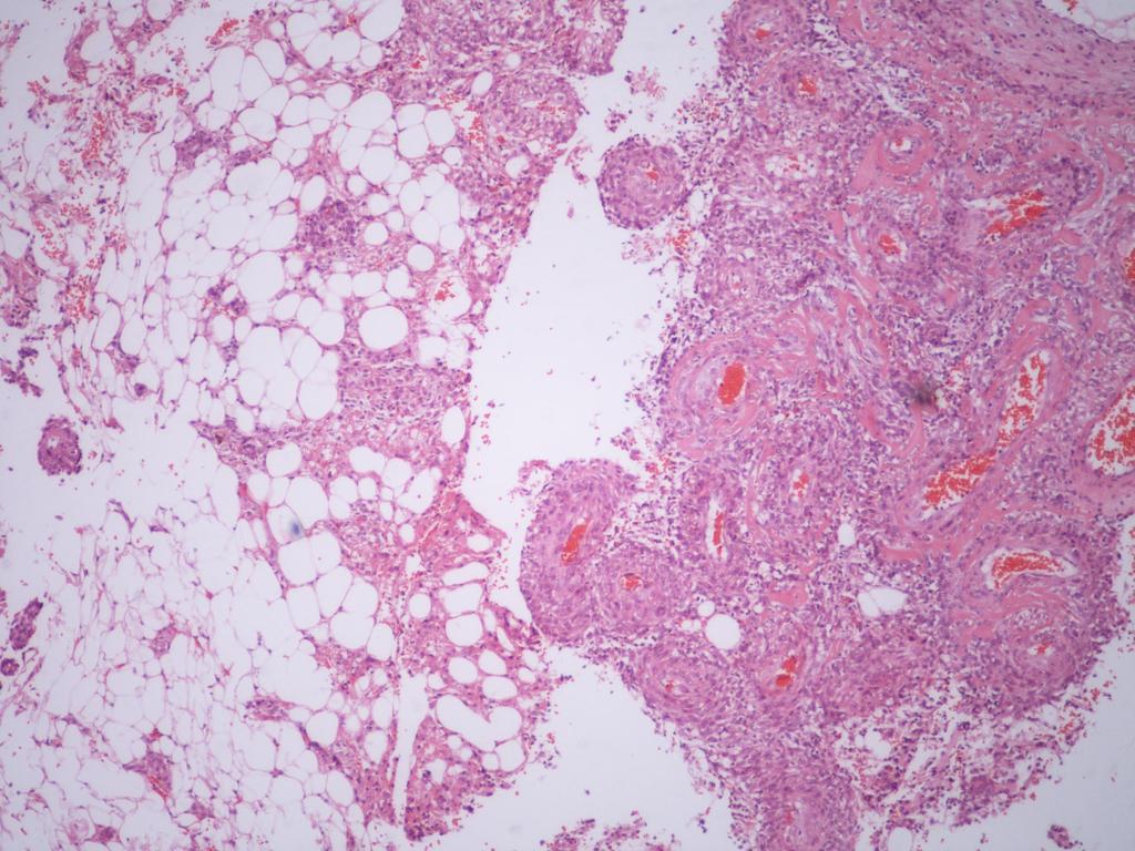
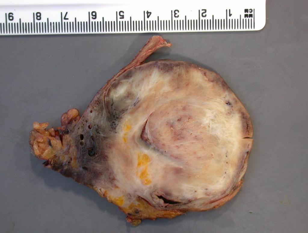
Case E1. Female aged 63 years Right Nephrectomy Two separate tumours Section of each tumour
|
|
|
- Henry Bridges
- 6 years ago
- Views:
Transcription
1 Case E1 Female aged 63 years Right Nephrectomy Two separate tumours Section of each tumour
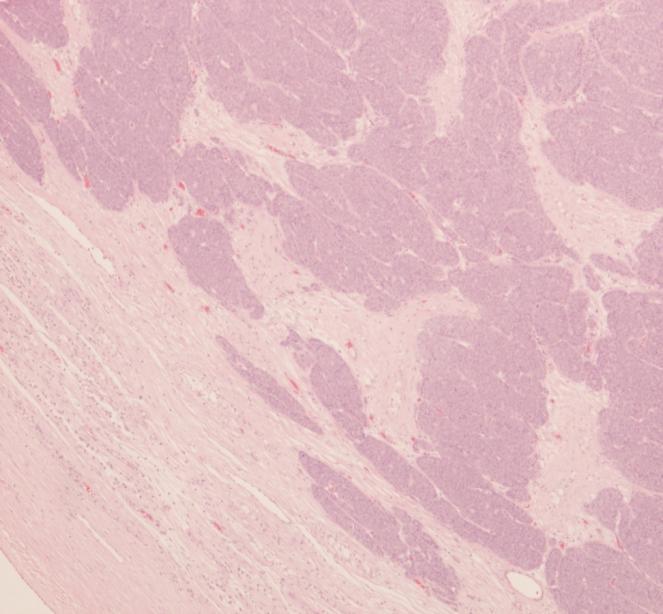
2 Tumour 1 Upper pole tumour 28mm macro diameter Circumscribed Friable cut surface
3
4
5 Tumour 2 Middle pole Part solid and part cystic 30mm macro diameter Several small cortical cysts were also noted
6
7
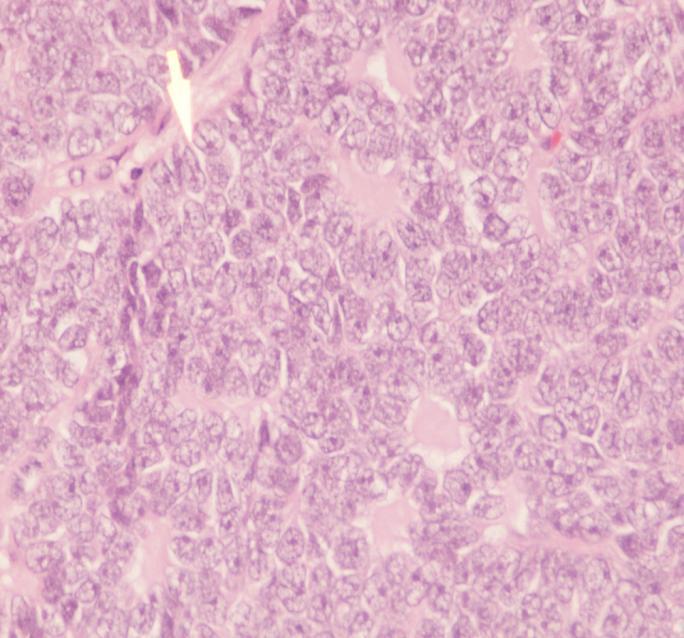
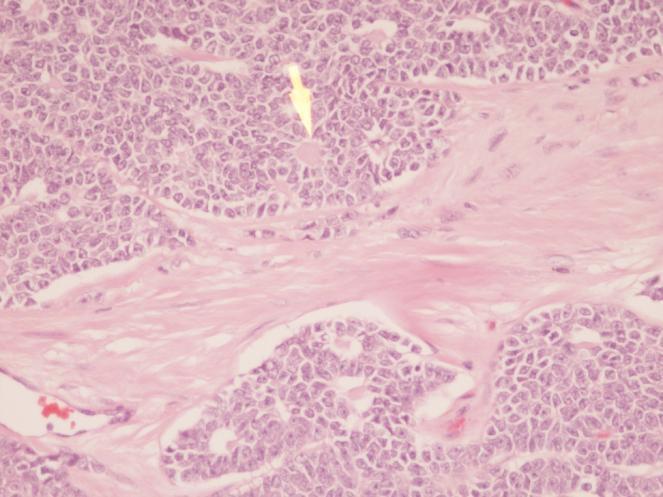
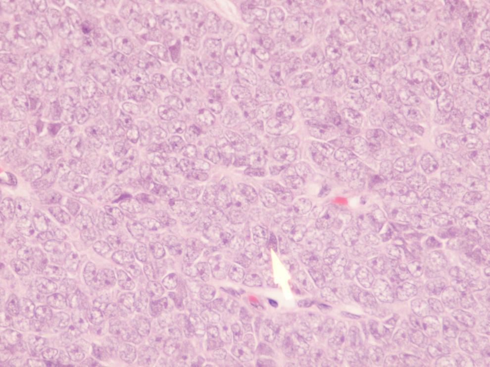
8 100 Answers returned Angiomyolipoma and Mutilocular Clear cell RCC 63 Angiomyolipoma and Clear cell RCC 19 Angiomyolipoma 5 Cystic RCC 3 Angiomyolipoma and cystic nephroma 1 Angiomyolipoma and epithelial cysts 2 Angiomyolipoma and cavernous haemangioma 1
9 Answers - continued RCC sarcomatoid type with cyst 2 Papillary RCC (type2?) and multilocular RCC 1 Papillary RCC with cysts 1 Pseudopapillary tumour with cystic lymphangioma 1 Blank answer 1 Five asked if the patient had known VHL or Tuberous Sclerosis
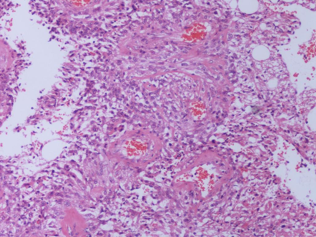
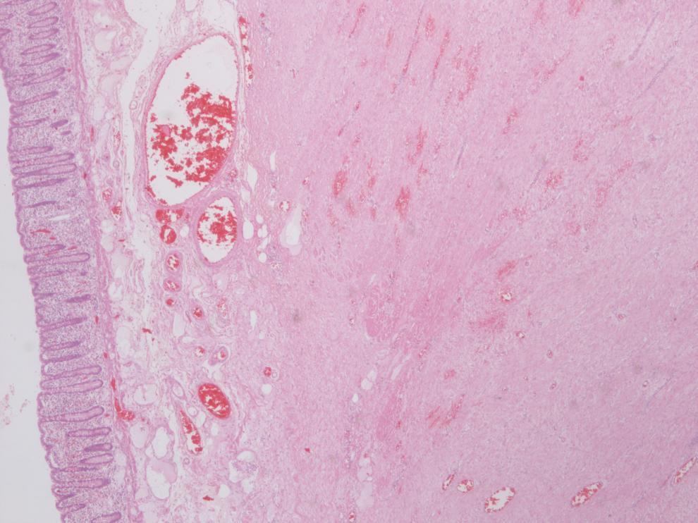
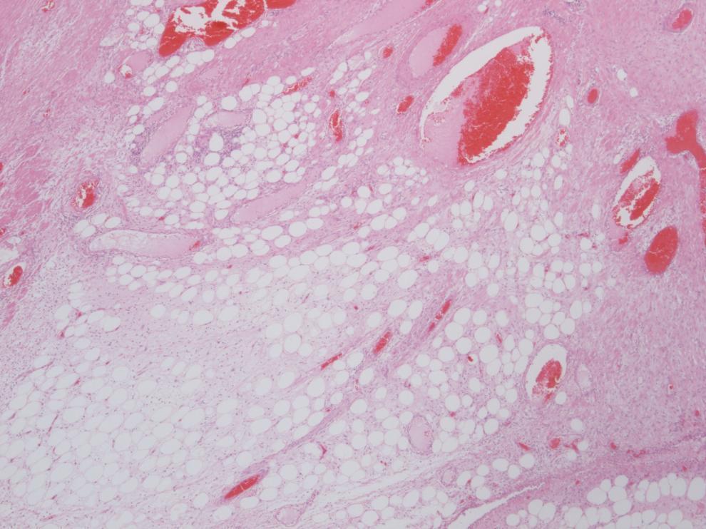
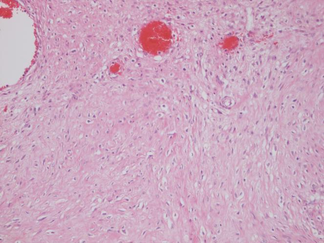
10 Diagnosis Angiomyolipoma and Multilocular cystic renal cell carcinoma (Grade 1, pt1) With further isolated cysts
11 Expert opinion As it was an unusual case it was referred to Professor Fleming in Dundee for his expert opinion. He agreed this diagnosis. Adding that finding of multiple tumour with cystic change raised the possibility of inherited syndrome of which tuberous sclerosis would be the most likely After liaising with the urologist it was established that there were no clinical or family history to suggest TS, so mutational analysis was not pursued
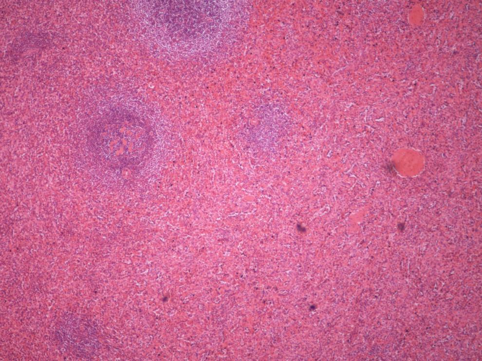
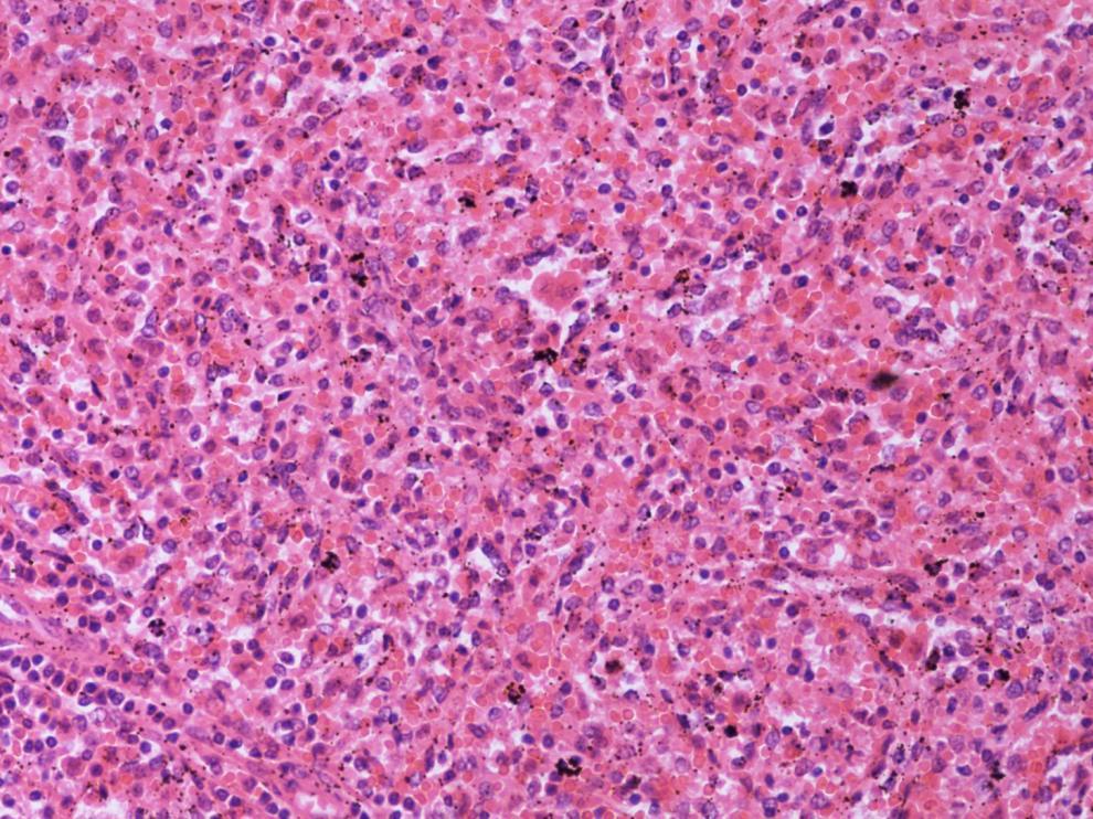
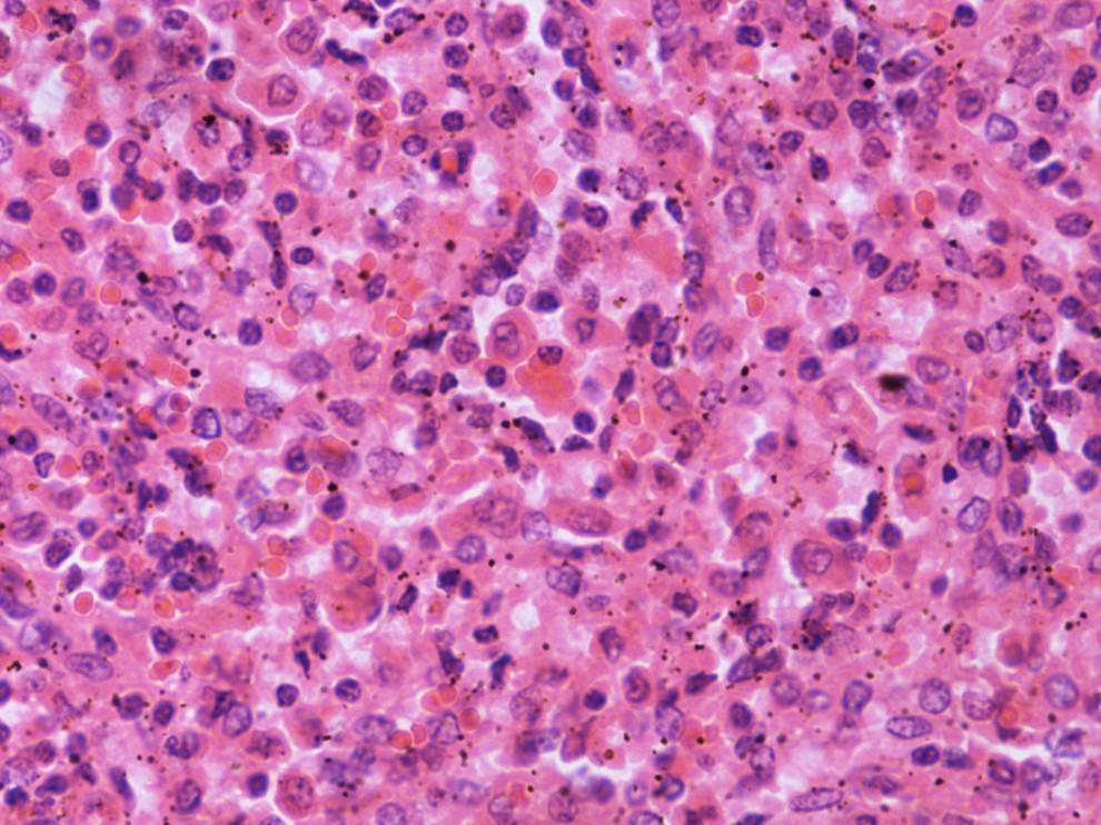
12 Case E2 Female 22 years Laparoscopic Splenectomy performed Spleen received piece-meal weighing 400g
13
14
15
16 EQA opinions 100 participants submitted a response There was a wide range of diagnoses submitted: Congestion 22 (many with? Re Hx) Hereditary spherocyosis 22 (many? Needing Hx or saying haemolytic anaemia?hs) Hairy Cell Leukaemia 9 Lymphoma or Leukaemia - 5
17 Hypersplenism 5 Too much red pulp 9 (including?congested,?haemolytic anaemia,?lymphoma or leukaemia, extramedullary haemopoesis) Angiomatosis 2 Littoral Cell Angioma 5 Haemangioma - 2
18 Other offerings Normal 1 White pulp reaction post pneumococcal vaccination 1 Haemophagocytic syndrome 1 Splenic Castleman s disease 1 Enlarged spleen -?EBV 1 Bacillary angiomatosis 1 Histeocytic infiltrate 1 Do not do haematopathology 1 Blank answer (including some which said no idea ) - 10
19 Pathological features Splenic weight 400g (normal g) Expansion of red pulp due to congestion by abnormal red blood cells, including spherical forms Erythrophagocytosis Some haemosiderin deposition No extramedullar haemopoiesis
20 Diagnosis Hereditary Spherocytosis Clinical information given was Hereditary spherocytosis laparoscopic splenectomy Appearances entirely in keeping with this

21
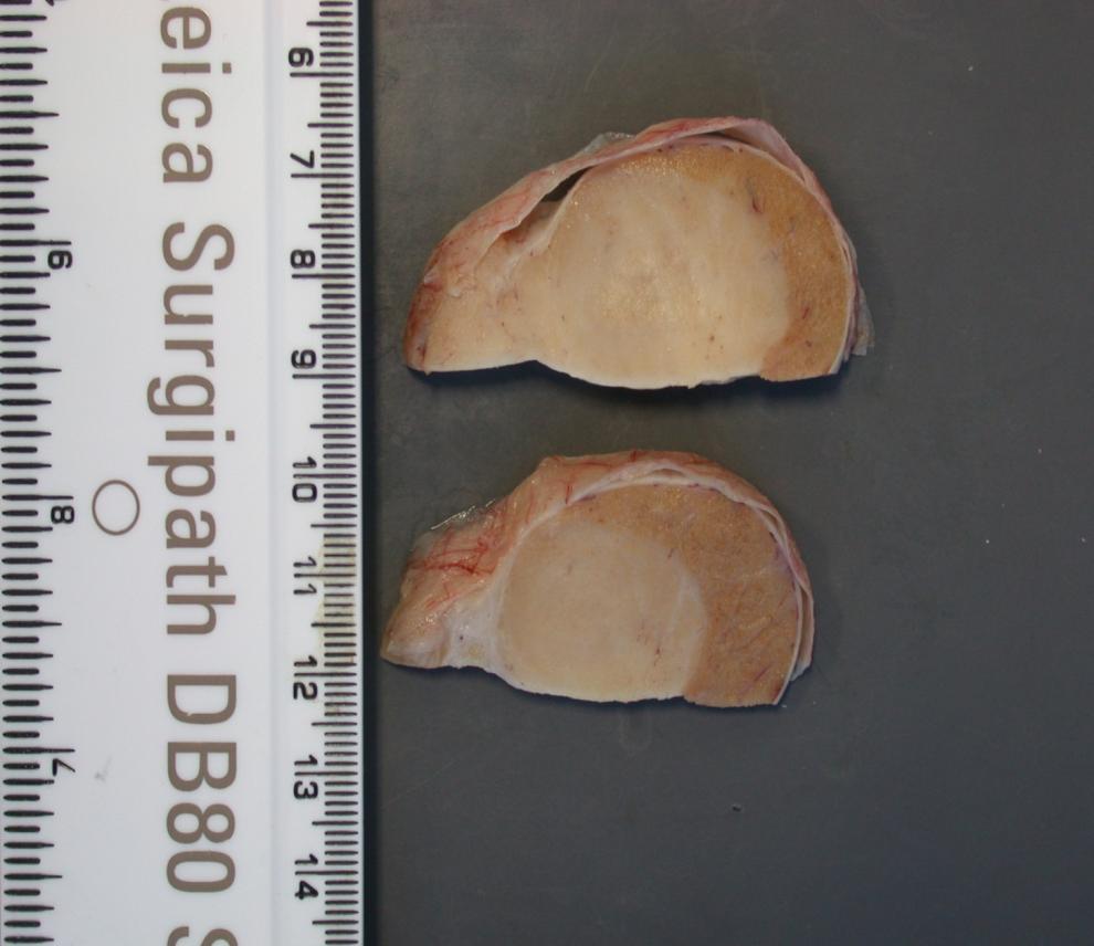
22 E3 M 68. Orchidectomy for 5cm testicular tumour.
23
24
25
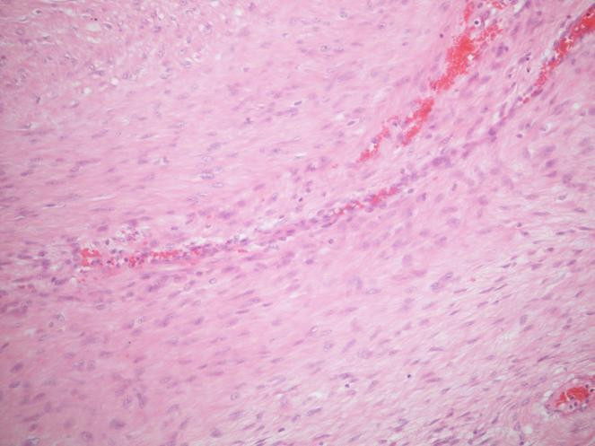
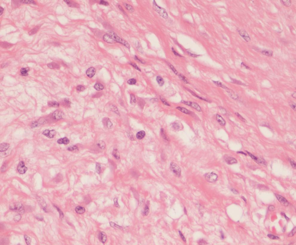
26
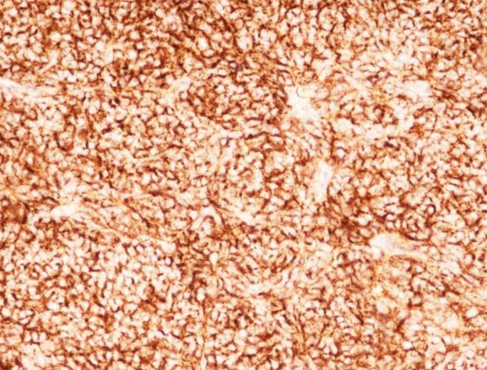
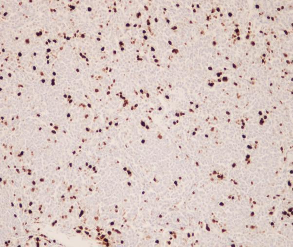
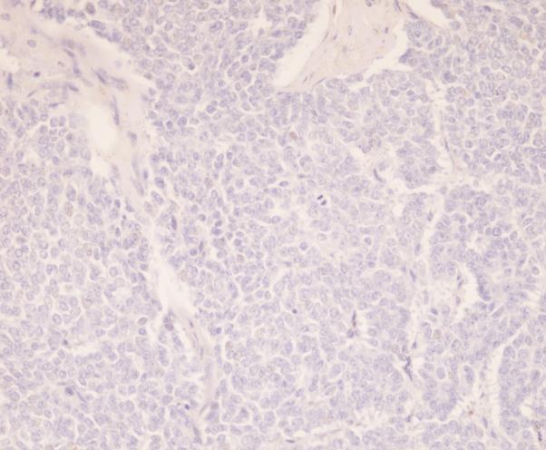
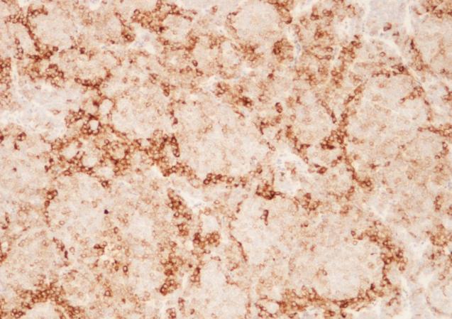
27 E 3 Granulosa cell tumour 40 Sex cord stromal tumour probably adult granulosa cell tumour 1? Sex Cord stromal tumour/germ-cell tumour/metastatic prostate cancer 1? Sertoli cell tumour immuno required 1 Sertoli cell tumour 11 Sex cord stromal tumour 2 Carcinoid tumour 6 Carcinoid or neuroendocrine tumour plus more poorly differentiated area exclude background teratoma 2 Strumal carcinoid (overgrowth in teratoma) or granulosa cell tumour or PNET 1 Malignant non-germ-cell tumour (IHC) (carcinoid, lymphoma, metastatic carcinoma) 1? Spermatocytic seminoma with sarcomatous dedifferentiation 1? Metastasis such as prostate? Granulosa cell tumour 2 Neuroendocrine tumour; exclude metastasis 1? Granulosa cell +/- neuroendocrine tumour 3? Sertoli Leydig cell tumour malignant. Rule out neuroendocrine tumour plus PNET 1 NSGCT with yolk sac elements 1 Metastatic poorly differentiated carcinoma 1 Neuroendocrine carcinoma with ITGCN/DDX solid YST with ITGCN 1 Neuroendocrine carcinoma -2. Neuroendocrine carcinoma vs PNET-1 Sertoli cell tumour/ spermatocytic seminoma/ immuno to exclude lymphoma -1 Sex cord ST/SRBCT/Sertoli/Myeloid sarcoma/mtu/ anaplastic seminoma -1 Embryonal carcinoma 3 Lymphoma 1


28 PLAP Oct3/4 CD30 AFP

29 AE1/3 ChromograninA Synaptophysin CD56

30 inhibin Inhibin WT1 calretinin CD99
31 Immunohistochemistry of SCST Leydig cell tumour Sertoli cell tumour Granulosa cell tumour Inhibin Calretinin Keratin Vimentin S100 SMA MelanA + + -/+ + -/ /- +/ /- -/ /+ + +/- -/+ - SCST, NOS +/- +/- -/ /-
32 Adult type granulosa cell tumour of testis
33 E4 Female 69. Emergency admission with abdominal pain. CT shows a caecal tumour with no distant spread. Section is from caecal lesion.
34
35
36
37
38
39 Case E4 Desmoid fibromatosis 33 Favour fibromatosis; cannot exclude Gist needs immuno 5 Bland spindle cells with no mitoses, possibly fibromatosis 1 Fibroma or fibromatosis 1 Gist 22 Spindle cell tumour in muscularis/serosa? Gist? Other (ICC) 2? Gist/? Leiomyoma/? Leiomyosarcoma (ICC) 1 Probable Gant or Gist (ICC) 1 Gist versus leiomyosarcoma 1 Spindle cell lesion? Gist? Schwannoma? MPNST? Leiomyoma? Fibromatosis ( immuno) -1 Spindle cell lesion fibromatosis/gist/leiomyoma/scar from previous operation/neural e.g. schwannoma 1 PECOMA 1 Neurofibroma 1 Sclerosing Mesenteritis 1 Spindle cell neuroendocrine tumour 19? Solitary fibrous tumour 1 Spindle cell tumour? Smooth muscle? Gist? Neural ( ICC) 1 Carcinosarcoma? Other 1 Gist/leiomyoma/peritoneal fibromatosis/spindle cell tumour 1 Spindle cell tumour? nature 1 (Angio) leiomyoma arising from outer muscle layer 2 Endometriosis 1


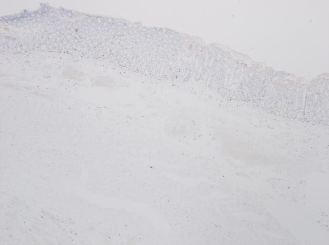

40 Desmin Caldesmon CD117 dog1
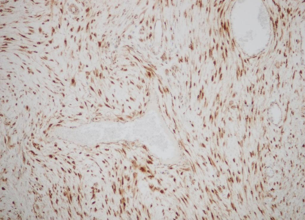
41 Β catenin
42 Dx Intra-abdominal (colonic) desmoid tumour Sporadic In association with FAP Aetiology unknown Association with abdominal surgery, trauma and oestrogen therapy
 Sporadic colonic desmoid- mutation of β catenin gene (CTNNB1) causing accumulation of nuclear β")
43 Mutational analysis FAP associated cases germline mutation of APC gene (30% FAP patients develop desmoids) Sporadic colonic desmoid- mutation of β catenin gene (CTNNB1) causing accumulation of nuclear β catenin.
Circulation: X Case number: 501 Number of responses: 84 Date: 4 MAY 12
 Circulation: X Case number: 500 Number of responses: 81 Date: 4 MAY 12 Female, aged 65 TAH and BSO for G1 endometrioid adenocarcinoma. Tumour positive with inhibin, vimentin, CD56 and SMA. Negative with
Dr Sanjiv Manek Oxford. Oxford Pathology Course 2010 for FRCPath Illustration-Cellular Pathology. Oxford Radcliffe NHS Trust
 Dr Sanjiv Manek Oxford Oxford Pathology Course 2010 for FRCPath Illustration-Cellular Pathology. Oxford Radcliffe NHS Trust Ovarian Endometrial Vulvo-vaginal Cervical Illustration-Cellular Pathology. Oxford
Male Genital Cancers in the US in Frequency of Types
 Germ Cell Tumors of the Testis Pathology, Immunohistochemistry, and the Often Confusing Appearance of Their Metastases Charles Zaloudek, MD Department of Pathology UCSF Male Genital Cancers in the US in
Testicular Tumors: What s New, True, Important Cristina Magi-Galluzzi, MD, PhD
 Testicular Tumors: What s New, True, Important Cristina Magi-Galluzzi, MD, PhD Director, Genitourinary Pathology R.J. Tomsich Pathology & Laboratory Medicine Institute Professor of Pathology, Lerner College
Tinh hoàn
 Tinh hoàn Tinh hoàn Tinh hoàn Tiền liệt tuyến Tiền liệt tuyến Mào tinh hoàn Mào tinh hoàn Túi tinh Túi tinh Túi tinh Túi tinh So-called cystadenoma of seminal vesicle. Gross appearance of granulomatous
Leydig cell tumour. Testis: non-germ cell tumours. Testis: sex cord-stromal tumours. Differential diagnosis of Leydig cell tumour TTAGS
 Non-germ cell s of the testis Dr Jonathan H Shanks The Christie NHS Foundation Trust, Manchester, UK Testis: non-germ cell s Sex cord-stromal s Haemolymphoid neoplasms Other neoplasms Tumour-like conditions
S2199 S2200. * Speaker's diagnosis 78
 98 21 2 14 13:30 * Speaker's diagnosis 78 S2199 Meningioma 48 Papillary meningioma * 30 Angiomatous meningioma 15 Ependymoma 12 Papillary ependymoma 6 Anaplastic ependymoma 2 Cellular ependymoma 1 Hemangioblastoma
Gross appearance of peritoneal cysts. They have a thin, translucent wall and contain a clear fluid.
 Gross appearance of peritoneal cysts. They have a thin, translucent wall and contain a clear fluid. So-called multicystic benign mesothelioma. A, Gross appearance. So-called multicystic benign mesothelioma.
CASE year old male with a PET avid nodule in the left adrenal gland
 CASE 1 55 year old male with a PET avid nodule in the left adrenal gland Case 1 Adrenal gland parenchyma partly replaced by a spindle cell tumour with mild nuclear pleomorphism Atypical mitoses present
South East England General Histopathology EQA Scheme
 South East England General Round g Final Case Analyses Cases 707 to 718 Circulated January February 2018 135 responses (85.44%) Prepared April 2018 Authorised by: Prof J Schofield Date: 23 rd April 2018
EDUCATIONAL CASES E1 & E2. Natasha Inglis 20/03/15
 EDUCATIONAL CASES E1 & E2 Natasha Inglis 20/03/15 CASE E1 79 year old female Rectum. Altemeier operation Histology Superficial erosions and mucosal congestion volcano lesion and pseudomembrane formation
Urological Tumours 1 Kidney tumours 2 Bladder tumours
 Urological Tumours 1 Kidney tumours 2 Bladder tumours Tim Bracey SpR Histopathology Derriford Hospital Kidney tumours What are we going to talk about?! Anatomy of urinary tract! Types of kidney tumours!
Transitional Cell Papilloma 2-3% Inverted Papilloma Rare
 BLADDER TUMORS Benign Transitional Cell Papilloma 2-3% Inverted Papilloma Rare Malignant Transitional (Urothelial) Carcinoma90% Carcinoma In-Situ (By Itself) 5-10% Squamous Cell Carcinoma 3-7% Adenocarcinoma
Testicular Tumors Including Secondary and Unusual Tumors of the Testis
 Testicular Tumors Including Secondary and Unusual Tumors of the Testis Milton W. Datta Partner, Hospital Pathology Associates University of Minnesota Minneapolis, MN Topics Review Features of Germ Cell
59 yo male with past medical history of prostate carcinoma, presented with upper abdominal pain
 December 2016 59 yo male with past medical history of prostate carcinoma, presented with upper abdominal pain Contributed by: Divya Sharma, MD. Fellow, Gastrointestinal Pathology, Department of Pathology
Testicular Germ Cell Tumors; A Simplistic Approach
 Testicular Germ Cell Tumors; A Simplistic Approach Merce Jorda, MD, PhD, MBA Professor and Vice Chair, Director of Anatomic Pathology Director of Genitourinary Pathology Service Interim Director of Cytopathology
Mesenchymal neoplasms of the gastrointestinal tract what s new? Newton ACS Wong Department of Histopathology Bristol Royal Infirmary
 Mesenchymal neoplasms of the gastrointestinal tract what s new? Newton ACS Wong Department of Histopathology Bristol Royal Infirmary Talk plan Summary from 2010 talk. What s happened since 2010. GISTs
Cardiff MRCS OSCE Courses Testicular Cancer
 Testicular Cancer Scenario: A 40-year-old male presents to the surgical out-patient clinic with a 6-8 week history of a painless lump in his left scrotum. He however complains of a dull ache in the scrotum
GUT-C 11/30/2017. Debasmita Das, M.D. PGY-1 Danbury Hospital
 GUT-C 11/30/2017 Debasmita Das, M.D. PGY-1 Danbury Hospital CLINICAL SUMMARY 8/2017 59 year old female Presented to the ED with 1 month history of general malaise, fever and weight loss PMH: Significant
Disclosure of Relevant Financial Relationships
 Evening Specialty Conference - Genitourinary Pathology Case 2 Disclosure of Relevant Financial Relationships Sean R Williamson, MD Henry Ford Health System, Detroit, MI @Williamson_SR USCAP requires that
Pathology of Ovarian Tumours. Dr. Jyothi Ranganathan MD ( Path) AFMC Pune PDCC (Cytopathology) PGI Chandigarh
 AFMC Pune PDCC (Cytopathology) PGI Chandigarh") Pathology of Ovarian Tumours Dr. Jyothi Ranganathan MD ( Path) AFMC Pune PDCC (Cytopathology) PGI Chandigarh Outline Incidence Risk factors Classification Pathology of tumours Tumour markers Prevention
Pathology of Ovarian Tumours Dr. Jyothi Ranganathan MD ( Path) AFMC Pune PDCC (Cytopathology) PGI Chandigarh Outline Incidence Risk factors Classification Pathology of tumours Tumour markers Prevention
Classification (1) Classification (3) Classification (2) Spindle cell lesions. Spindle cell lesions of bladder (Mills et al.
 Classification (3) Classification (2) Spindle cell lesions. Spindle cell lesions of bladder (Mills et al.") Non-epithelial tumours and nonepithelial tumour-like lesions of the bladder Dr Jonathan H Shanks The Christie NHS Foundation Trust, Manchester, UK Classification (1) Myofibroblastic proliferations and
Non-epithelial tumours and nonepithelial tumour-like lesions of the bladder Dr Jonathan H Shanks The Christie NHS Foundation Trust, Manchester, UK Classification (1) Myofibroblastic proliferations and
Disclosure. Relevant Financial Relationship(s) None. Off Label Usage None MFMER slide-1
 None. Off Label Usage None MFMER slide-1") Disclosure Relevant Financial Relationship(s) None Off Label Usage None 2013 MFMER slide-1 Case Presentation A 43 year old male, with partial nephrectomy for a right kidney mass 2013 MFMER slide-2 2013
Disclosure Relevant Financial Relationship(s) None Off Label Usage None 2013 MFMER slide-1 Case Presentation A 43 year old male, with partial nephrectomy for a right kidney mass 2013 MFMER slide-2 2013
2% of all malignancies Male predominance Patients usually more than 60 years old
 Benign Bladder Tumors Transitional Cell Papilloma 2-3% Inverted Papilloma Rare Malignant Transitional (Urothelial) Carcinoma 90% Carcinoma In-Situ (By Itself) 5-10% Squamous Cell Carcinoma 3-7% Adenocarcinoma
Breast - ductal carcinoma CK7 ER PR GATA3 Mammaglobin (50-70%) GCDFP-15 (50-70%) E-cadherin HMWCK CK20 PAX2 ER/PR/HER2 on all newly diagnosed cases
 GCDFP-15 (50-70%) E-cadherin HMWCK CK20 PAX2 ER/PR/HER2 on all newly diagnosed cases") Adrenal cortical carcinoma Inhibin Synap Melan-A Calretinin Vimentin Chromogr CK7 CK20 Breast - ductal carcinoma CK7 ER PR GATA3 Mammaglobin (50-70%) GCDFP-15 (50-70%) E-cadherin HMWCK CK20 PAX2 ER/PR/HER2
Adrenal cortical carcinoma Inhibin Synap Melan-A Calretinin Vimentin Chromogr CK7 CK20 Breast - ductal carcinoma CK7 ER PR GATA3 Mammaglobin (50-70%) GCDFP-15 (50-70%) E-cadherin HMWCK CK20 PAX2 ER/PR/HER2
Spindle Cell Lesions Of The Breast. Emad Rakha Professor of Breast Pathology and Consultant Pathologist
 Spindle Cell Lesions Of The Breast Emad Rakha Professor of Breast Pathology and Consultant Pathologist * SCLs comprise a wide spectrum of diseases, ranging from reactive processes to aggressive malignant
Recently, there has been an increasing incidence among women and younger persons
 Bladder Tumors Benign Transitional Cell Papilloma 2-3% Inverted Papilloma Rare Malignant Transitional (Urothelial) Carcinoma 90% Carcinoma In-Situ (By Itself) 5-10% Squamous Cell Carcinoma 3-7% Adenocarcinoma
Interesting Cases in Gynecologic Pathology. Michael Ward, MD Surgical Pathology Fellow University of Utah Health Sciences Center Salt Lake City, UT
 Interesting Cases in Gynecologic Pathology Michael Ward, MD Surgical Pathology Fellow University of Utah Health Sciences Center Salt Lake City, UT Case 1 History: 50 year old woman with a uterine mass
Lách
 Lách Lách Lách Lách Splenogonadal fusion. Splenic tissue is attached to testicular tissue. Pseudocyst (false or secondary cyst). A, Outer aspect. Pseudocyst (false or secondary cyst). B, Inner surface.
the urinary system pathology Dr. Fairoz A Eltorgman
 the urinary system pathology Dr. Fairoz A Eltorgman Tumors of the renal pelvis & kidney Benign tumors of the renal pelvis: Hemangioma Leiomyoma Malignant tumors: Transitional cell carcinoma Squamous cell
3 cell types in the normal ovary
 Ovarian tumors 3 cell types in the normal ovary Surface (coelomic epithelium) the origin of the great majority of ovarian tumors (neoplasms) 90% of malignant ovarian tumors Totipotent germ cells Sex cord-stromal
Pathology of Mediastinal Tumors
 SAMO Meeting Lucerne 2009 Pathology of Mediastinal Tumors Alex Soltermann Most common lesions (adults) Clinical presentation 50% of the patients are asymptomatic, lesion discovered incidentally Symptoms
Lung Tumor Cases: Common Problems and Helpful Hints
 Lung Tumor Cases: Common Problems and Helpful Hints Brandon T. Larsen, MD, PhD Senior Associate Consultant Department of Laboratory Medicine and Pathology Mayo Clinic Arizona Arizona Society of Pathologists
Various hereditary, acquired and neoplastic conditions can lead to cyst formation in the kidney.
 Dr. Fatima AlAl-Hashimi Hashimi,, MD, FRCPath Salmaniya Medical Complex, Bahrain Various hereditary, acquired and neoplastic conditions can lead to cyst formation in the kidney. The most frequently encountered
ONCOLOGY. Csaba Bödör. Department of Pathology and Experimental Cancer Research november 19., ÁOK, III.
 ONCOLOGY Csaba Bödör Department of Pathology and Experimental Cancer Research 2018. november 19., ÁOK, III. bodor.csaba1@med.semmelweis-univ.hu ONCOLOGY Characteristics of Benign and Malignant Neoplasms
3 cell types in the normal ovary
 Ovarian tumors 3 cell types in the normal ovary Surface (coelomic epithelium) the origin of the great majority of ovarian tumors 90% of malignant ovarian tumors Totipotent germ cells Sex cord-stromal cells
Disclosures. An update on ancillary techniques in the diagnosis of soft tissue tumors. Ancillary techniques. Introduction
 Disclosures An update on ancillary techniques in the diagnosis of soft tissue tumors. I have nothing to disclose. Andrew Horvai, MD, PhD Clinical Professor, Pathology Introduction Ancillary techniques
Gastrointestinal stromal tumor
 Gastrointestinal stromal tumor 영남의대병리학교실 최준혁 Classification of gastrointestinal mesenchymal tumor Gastrointestinal stromal tumor(gist) Smooth muscle tumors : leiomyoma, leiomyosarcoma Neurogenic tumors
New Immunohistochemical Markers in the Evaluation of Primary Non-Glial Central Nervous System Tumors
 New Immunohistochemical Markers in the Evaluation of Primary Non-Glial Central Nervous System Tumors Suzanne Z. Powell, M.D. The Methodist Hospital Houston, TX The Methodist Hospital Research Institute
A 9cm mass was excised from the jejunal wall and mesentery of a 33 year old woman.
 A Few Observations on Gastrointestinal Stromal Tumors and Their Differential Diagnosis E. Montgomery A 9cm mass was excised from the jejunal wall and mesentery of a 33 year old woman. 1 2 3 CD117/c-kit
Histopathological diagnosis of CUP
 Histopathological diagnosis of CUP Dr Karin Oien karin.oien@glasgow.ac.uk Disclosure slide Dr Karin Oien has no financial interests in any company mentioned in this presentation. Dr Karin Oien is conducting
Disclosures. An update on ancillary techniques in the diagnosis of soft tissue tumors. Ancillary techniques. Introduction
 Disclosures An update on ancillary techniques in the diagnosis of soft tissue tumors. I have nothing to disclose. Andrew Horvai, MD, PhD Clinical Professor, Pathology Introduction Ancillary techniques
Presenter: Yeh-Han Wang M.D.
 Korea-Taiwan-Japan Joint Meeting for Gynecological Pathology Mini-lecture Female Adnexal Tumor of Probable Wolffian Origin (FATWO) in Taiwan: A Small Case Series and Literature Review Presenter: Yeh-Han
Ovarian Clear Cell Carcinoma
 Ovarian Clear Cell Carcinoma Rouba Ali-Fehmi, MD Professor of Pathology The Karmanos Cancer Institute, Wayne State University School of Medicine 50 year old woman with chief complaint of shortness of breath
SMOOTH MUSCLE TUMOURS
 SMOOTH MUSCLE TUMOURS NORMAL SMOOTH MUSCLE Cytology Immunohistochemistry Ultrastructure Masson Trichrome Smooth Muscle Ultrastructure Many myofilaments running parallel to the long axis of the smooth
Updates in Urologic Pathology WHO Made Those Changes?! Peyman Tavassoli Pathology Department BC Cancer Agency
 Updates in Urologic Pathology WHO Made Those Changes?! Peyman Tavassoli Pathology Department BC Cancer Agency World Health Organization Available in Feb 2016 Frame work for reporting Major contributing
Surveys and Anatomic Pathology Education Programs
 Surveys and Anatomic Pathology Education Programs Performance Improvement Program in Surgical Pathology PIP/PIPW-B 2017 Participant Summary 2017 College of American Pathologists. The College does not permit
Sex: 女 Age: 51 Occupation: 無 Admission date:92/07/22
 Sex: 女 Age: 51 Occupation: 無 Admission date:92/07/22 Chief complaint Unknown fever for one month Hand tremor and left huge renal tumor was noted Present illness Suffered from fever for one month, hand
Uncommon secondary tumour of the stomach
 Uncommon secondary tumour of the stomach B. Bancel, Hôpital CROIX ROUSSE LYON Bucharest Nov 2013 Case report 33-year old man Profound mental retardation and motor disturbances (sequelae of neonatal meningeal
South East England General Histopathology EQA Scheme
 South East England General Round b Final Case Analyses Cases 647 to 658 Circulated May-June 2016 130 responses (90.28%) Prepared August 2016 Authorised by: Prof J Schofield Date: 15/08/16 With thanks to
STAGING AND FOLLOW-UP STRATEGIES
 ATHENS 4-6 October 2018 European Society of Urogenital Radiology STAGING AND FOLLOW-UP STRATEGIES Ahmet Tuncay Turgut, MD Professor of Radiology Hacettepe University, Faculty of Medicine Ankara 2nd ESUR
Note: The cause of testicular neoplasms remains unknown
 - In the 15- to 34-year-old age group, they are the most common tumors of men. - Tumors of the testis are a heterogeneous group of neoplasms that include: I. Germ cell tumors : 95%; all are malignant.
Leukaemia 35% Lymphoma 14%
 Distribution ib ti of Cancers in Children under 15 years Leukaemia 35% Lymphoma 14% Neuroblastoma 9% Other 5% Liver 1% Retinoblastoma 3% Bone and STS 15% CNS 20% Wilms' 8% 30-40% Mortality Germ Cell Tumours
Hereditary Leiomyomatosis and Renal Cell Carcinoma Variant of Reed s Syndrome - A Rare Case Report
 American Research Journal of Urology Volume 1, Issue 1, pp:26-30 Case Hereditary Leiomyomatosis and Renal Cell Carcinoma Variant of Reed s Syndrome - A Rare Case Manas Babu, Devesh Bansal, Sony Mehta,
The role of immunohistochemistry in surgical pathology of the uterine corpus and cervix
 The role of immunohistochemistry in surgical pathology of the uterine corpus and cervix Prof. Ben Davidson, MD PhD Department of Pathology, Norwegian Radium Hospital, Oslo University Hospital, Oslo, Norway
Various kinds of cystic tumor or tumor-like lesions in the kidney :radiologic-pathologic correlation.
 Various kinds of cystic tumor or tumor-like lesions in the kidney :radiologic-pathologic correlation. Poster No.: C-0299 Congress: ECR 2014 Type: Educational Exhibit Authors: S.-J. Lee, J.-H. Yoon, Y.
5/10. Pathology Soft tissue tumors. Farah Bhani. Mohammed Alorjani
 5/10 Pathology Soft tissue tumors Mohammed Alorjani Farah Bhani Slides are included in this sheet. Objectives: Soft tissue tumors 1. Describe soft tissue tumors. 2. Understand the classification of soft
ICD-O Morphology code. R=Rare Tier Tumour ICD-O Topography code C30.0, C31
 R=Rare Tier Tumour ICD-O Topography code ICD-O Morphology code EPITHELIAL TUMOURS OF NASAL CAVITY AND SINUSES R 2 Squamous cell carcinoma with variants of nasal cavity and sinuses C30.0, C3 C30.0, C3 8000,
Appendix E SEER Program Coding and Staging Manual DRAFT Reportable Examples
 Appendix E1 2018 SEER Program Coding and Staging Manual DRAFT Reportable Examples Malignant # Diagnosis/Condition Notes 1 Atypical fibroxanthoma (superficial malignant fibrous histiocytoma) 2 Positive
Kidney Case 1 SURGICAL PATHOLOGY REPORT
 Kidney Case 1 Surgical Pathology Report February 9, 2007 Clinical History: This 45 year old woman was found to have a left renal mass. CT urography with reconstruction revealed a 2 cm medial mass which
Female Reproduc.ve System. Kris.ne Kra7s, M.D.
 Female Reproduc.ve System Kris.ne Kra7s, M.D. Female Reproduc.ve System Outline Cervix Uterus Ovaries Breast Cervical Carcinoma Once the most common cancer in women now not even in top 10. Decrease due
Protocol for the Examination of Lymphadenectomy Specimens From Patients With Malignant Germ Cell and Sex Cord-Stromal Tumors of the Testis
 Protocol for the Examination of Specimens From Patients With Malignant Germ Cell and Sex Cord-Stromal Tumors of the Testis Version: Testis 4.0.1.1 Protocol Posting Date: February 2019 Accreditation Requirements
Applications of IHC. Determination of the primary site in metastatic tumors of unknown origin
 Applications of IHC Determination of the primary site in metastatic tumors of unknown origin Classification of tumors that appear 'undifferentiated' by standard light microscopy Precise classification
Pathology Mystery and Surprise
 Pathology Mystery and Surprise Tim Smith, MD Director Anatomic Pathology Medical University of South Carolina Disclosures No conflicts to declare Some problem cases Kidney tumor Scalp tumor Bladder tumor
Five Views of Transitional Cell Carcinoma: One Man s Journey
 September 2006 Five Views of Transitional Cell Carcinoma: One Man s Journey Amsalu Dabela, Harvard Medical School III Outline Overview: Renal Anatomy Our Patient s Story Diagnostic Imaging Studies Appearance
Pediatric Retroperitoneal Masses Radiologic-Pathologic Correlation
 Acta Radiológica Portuguesa, Vol.XVIII, nº 70, pág. 61-70, Abr.-Jun., 2006 Pediatric Retroperitoneal Masses Radiologic-Pathologic Correlation Marilyn J. Siegel Mallinckrodt Institute of Radiology, Washington
Reporting of carcinoma of unknown primary tumour (CUP)
") Reporting of carcinoma of unknown primary tumour (CUP) Prof John Schofield Kent Oncology Centre with grateful thanks to Dr Karin Oien University of Glasgow Royal College of Pathologists Cancer datasets
Reporting of carcinoma of unknown primary tumour (CUP) Prof John Schofield Kent Oncology Centre with grateful thanks to Dr Karin Oien University of Glasgow Royal College of Pathologists Cancer datasets
Renal tumours: use of immunohistochemistry & molecular pathology. Dr Lisa Browning John Radcliffe Hospital Oxford
 Renal tumours: use of immunohistochemistry & molecular pathology Dr Lisa Browning John Radcliffe Hospital Oxford Renal tumours: the use of immunohistochemistry & molecular pathology Classification of RCC
Spectrum of Preneoplastic and Neoplastic Cystic Lesions of the Kidney in Adult. by dr. Banan Burhan Mohammed Lecturer in Pathology Department
 Spectrum of Preneoplastic and Neoplastic Cystic Lesions of the Kidney in Adult by dr. Banan Burhan Mohammed Lecturer in Pathology Department Various hereditary, acquired, and neoplastic conditions can
Case Report A Rare Case with a Solitary Fibrous Tumour of the Colon and an Epithelioid Angiomyolipoma of the Kidney
 Volume 2013, Article ID 324538, 7 pages http://dx.doi.org/10.1155/2013/324538 Case Report A Rare Case with a Solitary Fibrous Tumour of the Colon and an Epithelioid Angiomyolipoma of the Kidney Thong Quang
BEST PRACTICES IN THE APPLICATION OF IMMUNOHISTOCHEMISTRY TO DIAGNOSTIC UROLOGIC PATHOLOGY: LESSONS FROM USES & ABUSES
 BEST PRACTICES IN THE APPLICATION OF IMMUNOHISTOCHEMISTRY TO DIAGNOSTIC UROLOGIC PATHOLOGY: LESSONS FROM USES & ABUSES Mahul B. Amin Professor & Chairman Department of Pathology & Laboratory Medicine Cedars-Sinai
Tumors of kidney and urinary bladder
 Tumors of kidney and urinary bladder Overview of kidney tumors Benign and malignant Of the benign: papillary adenoma -cortical -small (0.5cm) -in 40% of population -clinically insignificant The most common
Diagnostic IHC in lung and pleura pathology
 Diagnostic IHC in lung and pleura pathology Mogens Vyberg Professor of Clinical Pathology Director of NordiQC Aalborg University Hospital, Aalborg, Denmark WHO 2004 and Web Malignant mesothelioma Epithelioid
Desmoplastic Melanoma R/O BCC. Clinical Information. 74 y.o. man with lesion on left side of neck r/o BCC
 R/O BCC Sabine Kohler, M.D. Professor of Pathology and Dermatology Dermatopathology Service Stanford University School of Medicine Clinical Information 74 y.o. man with lesion on left side of neck r/o
Differential diagnosis of HCC
 Hepatocellular Carcinoma Quest for an Ideal Immunohistochemical Panel Sanjay Kakar, MD UCSF Differential diagnosis of HCC Hepatocellular lesions Adenoma, FNH, HG dysplasia Adenocarcinoma CholangioCA, metastasis
Tumour Markers. For these reasons, only a handful of tumour markers are commonly used by most doctors.
 Tumour Markers What are Tumour Markers? Tumour markers are substances that can be found in the body when cancer is present. They are usually found in the blood or urine. They can be products of cancer
GUIDELINES ON TESTICULAR CANCER
 38 (Text updated March 2005) P. Albers (chairman), W. Albrecht, F. Algaba, C. Bokemeyer, G. Cohn-Cedermark, A. Horwich, O. Klepp, M.P. Laguna, G. Pizzocaro Introduction Compared with other types of cancer
CODING TUMOUR MORPHOLOGY. Otto Visser
 CODING TUMOUR MORPHOLOGY Otto Visser INTRODUCTION The morphology describes the tissue of the tumour closest to normal tissue Well differentiated tumours are closest to normal Undifferentiated tumours show
I have nothing to disclose
 A 47 year old female with multiple lung nodules Disclosure of Relevant Financial Relationships Tamar Giorgadze, MD, PhD Professor of Pathology Medical College of Wisconsin Milwaukee, Wisconsin USCAP requires
Review of the AP Part II Practical Examination. Dr David Clift Co Chief Examiner
 Review of the AP Part II Practical Examination Dr David Clift Co Chief Examiner General Remarks The part II practical examination involved 15 cases which were presented with sufficient clinical data to
Case Based Learning Program
 Case Based Learning Program The Department of Urology Glickman Urological & Kidney Institute Cleveland Clinic Case Number 5 CBULP 2010 001 Case Based Urology Learning Program Editor: Associate Editor:
Effective January 1, 2018 ICD O 3 codes, behaviors and terms are site specific
 Effective January 1, 2018 codes, behaviors and terms are site specific /N 8551/3 Acinar adenocarcinoma (C34. _) Lung primaries diagnosed prior to 1/1/2018 use code 8550/3 For prostate (all years) see 8140/3
Effective January 1, 2018 ICD O 3 codes, behaviors and terms are site specific
 Effective January 1, 2018 codes, behaviors and terms are site specific Status /N 8010/3 Urachal carcinoma (C65.9, C66.9, C67. _, C68._) 8013/3 Combined large cell neuroendocrine carcinoma (C34. _, C37.9)
2 to 3% of All New Visceral Cancers Peak Incidence is 6th Decade M:F = 2:1 Grossly is a Bright Yellow, Necrotic Mass with a Pseudocapsule
 GENITOURINARY PATHOLOGY Kathleen M. O Toole, M.D. Renal Cell Carcinoma 2 to 3% of All New Visceral Cancers Peak Incidence is 6th Decade M:F = 2:1 Grossly is a Bright Yellow Necrotic Mass Grossly is a Bright
Institute of Pathology First Faculty of Medicine Charles University. Ovary
 Ovary Barrett esophagus ph in vagina between 3.8 and 4.5 ph of stomach varies from 1-2 (hydrochloric acid) up to 4-5 BE probably results from upward migration of columnar cells from gastroesophageal junction
Tumour Structure and Nomenclature. Paul Edwards. Department of Pathology and Cancer Research UK Cambridge Institute, University of Cambridge
 Tumour Structure and Nomenclature Paul Edwards Department of Pathology and Cancer Research UK Cambridge Institute, University of Cambridge Malignant Metastasis Core idea of cancer Normal Cell Slightly
Biopsy Interpretation of Spindle cell proliferations of the Serosa
 Biopsy Interpretation of Spindle cell proliferations of the Serosa Richard Attanoos, Cardiff. U.K. Disclosure of Relevant Financial Relationships USCAP requires that all planners (Education Committee)
An Overview of Genital Stromal Tumors
 An Overview of Genital Stromal Tumors By Konstantinos Linos MD, FCAP, FASDP Bone, Soft Tissue and Dermatopathology Assistant Professor of Pathology Dartmouth-Hitchcock Medical Center Geisel School of Medicine
Normal endometrium: A, proliferative. B, secretory.
 Normal endometrium: A, proliferative. B, secretory. Nội mạc tử cung Nội mạc tử cung Cyclic changes in endometrium.. Approximate relationship of useful microscopic changes. Arias-Stella reaction in endometrial
3/27/2017. Disclosure of Relevant Financial Relationships
 Ophthalmic Pathology Evening Specialty Conference USCAP 2017 5 th March, 2017 Mukul K. Divatia, MD Assistant Professor Department of Pathology & Genomic Medicine Weill Cornell Medical College Houston Methodist
The FRCPath Exam. How to pass the short surgicals on the first go By someone who didn t. Plus some tips on frozen sections!
 The FRCPath Exam How to pass the short surgicals on the first go By someone who didn t Plus some tips on frozen sections! Dr Paul Bennett Overview Reminder of the exam format How to format your answer
57th Annual HSCP Spring Symposium 4/16/2016
 An Unusual Malignant Spindle Cell Lesion to Involve the Breast Erinn Downs-Kelly, D.O. Associate Professor of Pathology University of Utah & ARUP Laboratories No disclosures Case 39 y/o female with no
Pediatric Abdominal Masses. Andrew Phelps MD Assistant Professor of Pediatric Radiology UCSF Benioff Children's Hospital
 Pediatric Abdominal Masses Andrew Phelps MD Assistant Professor of Pediatric Radiology UCSF Benioff Children's Hospital No Disclosures Take Home Message All you need to remember are the 5 common masses
Germ cell tumours UK SH. Ivo Leuschner. Kiel Pediatric Tumor Registry, Institute of Pathology University Hospital of Schleswig-Holstein Campus Kiel
 Germ cell tumours Ivo Leuschner Kiel Pediatric Tumor Registry, Institute of Pathology University Hospital of Schleswig-Holstein Campus Kiel UK SH Old histogenetic Concept of Germ cell tumours Pluripotent
Genetic Studies of Dysgerminoma
 Genetic Studies of Chromosome 12p abnormalities are characteristic of germ cell tumors isochromosome 12p and 12p overrepresentation Some can be detected by karyotyping FISH study of 21 dysgerminomas showed
Urology An introduction to cut up DR J R GOEPEL
 Urology An introduction to cut up DR J R GOEPEL Overview Principles Individual organs Small pieces Partial resections Whole organs Data recording and data sets Principles You are working for the patient
Pathology of the female genital tract
 Pathology of the female genital tract Common illnesses of the female genital tract Before menarche Developmental anomalies Tumors (ovarial teratoma) Amenorrhea Fertile years PCOS, ovarian cysts Endometriosis