GENERAL ABDOMINAL IMAGING PERITONEAL SPACE, PANCREAS, & SPLEEN
|
|
|
- Kerrie Riley
- 6 years ago
- Views:
Transcription


1 GENERAL ABDOMINAL IMAGING PERITONEAL SPACE, PANCREAS, & SPLEEN VMB 960 March 25, 2013 REFERENCE Chapters Pages Chapter 37 Pages Chapter 3 Pages OBJECTIVES Radiography and Ultrasound of the Abdomen Peritoneal & Retroperitoneal Spaces Specific Organ Evaluation Pancreas Spleen Patterns of Disease Mass Lesions 1


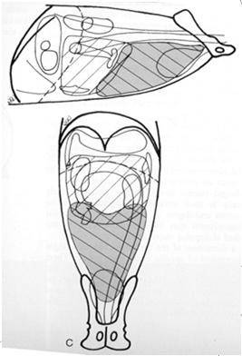
2 NORMAL ABDOMEN NORMALLY NOT SEEN GB Adrenals Adrenals Pancreas Ureter Ureter GB *Don t forget lymph nodes!* Uterus Abnormal enlargement Mass effect on adjacent organs NORMAL CAT Same organ pattern Spleen smaller Often better serosal detail Increased fat 2


3 ULTRASOUND Pros (compared to Radiographs) Great for evaluating soft tissue changes Real-time imaging Cons Does not provide global picture Highly dependent on user The two modalities complement each other ULTRASOUND PHYSICS US probe emits sound waves into tissue US probe also listens for echoes Echo VOLUME (how loud?) Echo TIMING (how long? ULTRASOUND PHYSICS Acoustic Impedance Inherent property of a substance Determined by Physical Density Speed of Sound in the tissue Two tissues with large difference in AI Creates interface US waves are reflected (an echo) 3


4 ULTRASOUND PHYSICS Frequency Measured in MHz Range 1-18 MHz Lower Frequency Better Penetration Worse Resolution Higher Frequency Better Resolution Worse Penetration ULTRASOUND PHYSICS Multifrequency probes For canine abdomen Average size dog Use a 5-8 MHz probe Deep-chested/Large dog Use a 3-5 MHz probe For feline abdomen Can use MHz probe ULTRASOUND TERMINOLOGY Hyperechoic Hypoechoic Anechoic Isoechoic Relative comparison 4


 Peritonitis")

5 SPACES OF THE ABDOMEN Retroperitoneal Dorsal to colon Kidneys, ureters, adrenal glands, lymph nodes Continuous with the mediastinum Peritoneal Surrounds visceral organs A potential space PERITONEAL SPACE Serosal detail Dependent on normal abdominal fat Poor serosal detail Poor radiographic technique Young animal Peritoneal fluid Emaciation (no fat) Peritonitis Carcinomatosis POOR SEROSAL DETAIL Young animals Less fat Brown fat Lack of fat Emaciation Chronic cachexia 5
 Fluid and soft")

 Capillary permeability (vasculitis)")
6 POOR SEROSAL DETAIL Disease of the peritoneum Peritonitis Carcinomatosis POOR SEROSAL DETAIL Peritoneal effusion Often accompanies other problems (peritonitis) Fluid and soft tissue are the same opacity Radiographs less sensitive to detection vs. US US good to determine cause for loss of detail Fluid type cannot be specified For radiographs or US PERITONEAL FLUID Effusions Heart failure ( hydrostatic pressure) Hypoproteinemia ( colloid oncotic pressure) Capillary permeability (vasculitis) Inflammation Neoplasia Hemorrhage Urine Bile 6


7 PERITONEAL FLUID Wispy ST opacity in falciform fat POOR SEROSAL DETAIL Emaciation or fluid? CHOLESTEROL GRANULOMA Bates body Dystrophic calcification of necrotic mesenteric fat More common in cats 7

 SURGICAL EMERGENCY!")


8 PNEUMOPERITONEUM Gas opacity in the peritoneal space Abdominal wall penetration Surgery Can persist days post-sx Trauma (puncture) Rupture of a hollow viscus (GI) SURGICAL EMERGENCY! PNEUMOPERITONEUM Sensitivity of ultrasound variable Tend to push superficial gas out of the way May detect if trapped Radiographs Large volumes outline the organs Look at liver and diaphragm/body wall PNEUMOPERITONEUM 8


9 POSITIONAL RADIOGRAPHY Remember Gas rises Manipulate the patient Manipulate the x-ray beam HORIZONTAL BEAM VD Right Left lateral recumbency to avoid gas in fundus Normal Right RETROPERITONEAL SPACE Pneumoretroperitoneum Retroperitoneal space is continuous with the mediastinum Rupture of trachea (or esophagus) 9


10 RETROPERITONEAL SPACE Dorsal to the colon Fluid, mass displaces colon ventrally RETROPERITONEAL MASS COLON POSITION Colon and SI subject to displacement with mass lesions However, they are also variable in normal position Must see a mass lesion also (increase in soft tissue opacity) 10


11 PANCREAS Dog Cat NORMAL PANCREAS - ULTRASOUND PANCREATIC ENLARGEMENT Caudal deviation of colon Lateral and/or ventral deviation of duodenum Silhouetting of greater curvature of stomach Evans. Miller s Anatomy of the Dog, 3 rd ed. Pancreatitis Peritonitis/fluid Persistent gas distension of the duodenum Often difficult to diagnose radiographically! 11

12 Normal Normal 12
")


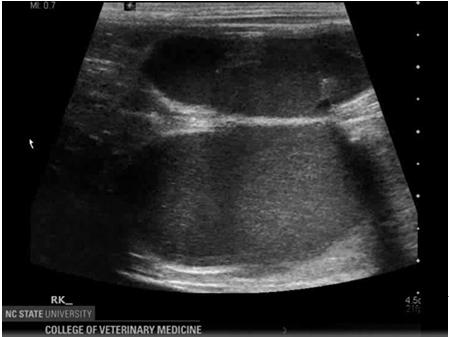
13 PANCREATITIS - ULTRASOUND PANCREATITIS - ULTRASOUND CLASSIC APPEARANCE: Hypoechoic, enlarged pancreas Hyperechoic mesentery (inflamed fat) Peritoneal effusion Dilated pancreatic duct, bile duct obstruction SENSITIVITY: Pancreas can appear normal with pancreatitis Dogs US is a more reliable test, especially when acute About 2/3 cases detected Cats Less reliable, more often normal in appearance About 1/4 to 1/3 cases detected 13


14 SPLEEN Typically surrounded by fat Usually easily seen on rads Head on VD Tail on Lateral Not in cats Sedation effects Phenothiazine tranquilizers Anesthetic gases & barbituates SPLEEN Tail on lateral Head on VD SPLEEN Tail can be variable in position Head less mobile 14

15 SPLEEN Breed variants German Shepherd Dog, Retrievers, Sighthounds SPLENIC PATHOLOGY DIFFUSE (Splenomegaly) or FOCAL (Mass) Radiographs Diffuse changes and large focal disease Ultrasound Can better identify small parenchymal lesions Specificity of imaging changes are often nonspecific DIFFUSE SPLENOMEGALY Neoplasia Lymphoma Mast Cell Congestion Sedation Right heart failure Splenic Torsion IMHA Inflammation Infarction Hyperplasia Extramedullary hematopoiesis 15

 Nodular")

16 FOCAL SPLENOMEGALY Neoplasia Hemangiosarcoma Hemangioma Lymphoma (less common appearance) Nodular Lymphoid Hyperplasia Hematoma Abscess FOCAL SPLENOMEGALY Head Tail 16

17 FOCAL SPLENIC MASS QUESTIONS? 17


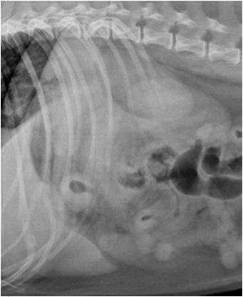
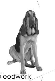
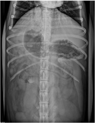
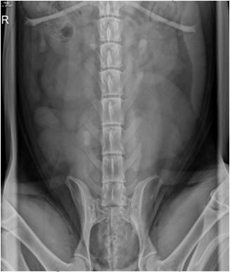
18 General Abdominal Cases VMB 960 March 26, 2013 Additional Resources Index of NORMAL abdominal (and thoracic) radiographs logy Small animal ultrasound videos 7 Year old, MC, Golden Retriever Lethargy for 2 weeks Inappetant ADR Palpable mass in caudal abdomen Bloodwork normal 1


19 Mass Lesions Identify location At least 2 views Normal organs in that location Mass effect How are the organs around the mass affected? Are there clinical signs that can help narrow the differentials? 2


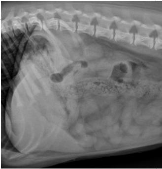
20 Retroperitoneal Mass Kidney Adrenal Ureter Lymph Nodes Other structures? Mass Lesions Common Differentials Cyst Hematoma Abscess Neoplasia Granuloma E Can all appear same on radiographs Ultrasound may help distinguish Fluid content Vascularity Margin Ultrasound Better assess character of the mass 3


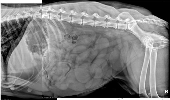
21 Ultrasound May reveal associated lesions that would be missed with rads alone Normal 5 Year old, F, Bloodhound Working dog Presented for entropion repair Polyuria/Polydypsia Febrile Leukocytosis on preoperative bloodwork 4


22 Uterine enlargement Pyometra Mucometra Hydrometra Neoplasia Pregnancy Ultrasound 5


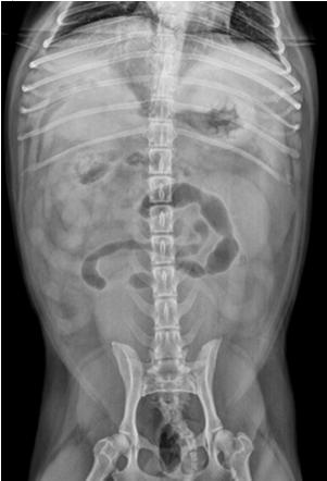
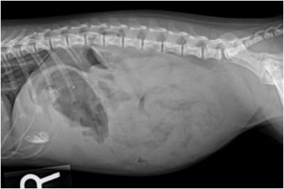
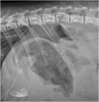
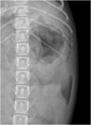
23 9 Year old, FS Labrador Retriever Acutely lethargic Panting Abdomen feels distended Pale mucous membranes 6


24 Peritoneal Fluid Effusions Transudates & Exudates Hemorrhage Urine Bile Radiographic Limitations Loss of serosal detail may obscure lesions Ultrasound What next? Surgery Consider your differentials Neoplasia? Metastatic disease evaluation Thoracic radiographs 7


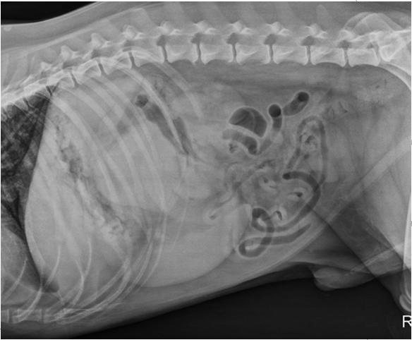
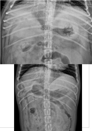
25 Thoracic Radiographs 8 Year old, FS, Miniature Schnauzer Previous bouts of pancreatitis Now febrile, leukocytosis 8


26 Right Cranial Abdomen Liver Pancreas Stomach Duodenum Colon/Cecum Lymph nodes Evans. Miller s Anatomy of the Dog, 3 rd ed. Pancreatic Abscess A sequela to pancreatitis More common in dogs than cats Can be indistinguishable from a cyst More echogenic fluid content Regional inflammation Clinical signs 9


27 Pancreatic Abscess Pancreatic Cysts Pseudocysts Results from pancreatitis Secretions into areas of necrosis Retention Cysts Smaller Due to blockage of small ducts Congenital Cysts Often large, do not change over time Often associated with polycystic disease 10


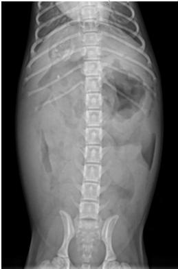
28 11 Year old, FS Mixed breed dog FS, Mixed breed dog Suspect abdominal mass Inguinal region feels firm 11


29 Sublumbar Mass Medial iliac lymph nodes Periosteal reaction along caudal lumbar vertebrae increase suspicion of neoplasia Rectal exam! 7 Year-old, MC Sheltie PU/PD for 1 week Lethargic and anorexic Hypercalcemia on bloodwork 12


30 Splenic Ultrasound Normal spleen Hyperechoic Fine texture Uniform appearance Splenic lymphoma Can be variable in appearance 6 Month old, FS Maltese Lethargy & vomiting 3 days Slightly tender on abdominal palpation Bloodwork reveals leukopenia 13


31 14


")


32 Pneumoperitoneum Surgery Penetrating abdominal trauma Gastrointestinal rupture Surgical emergency Case Management Was barium necessary? What test(s) might be more appropriate? Complications associated with barium in the peritoneal space? 15


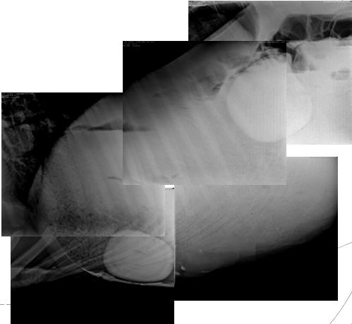
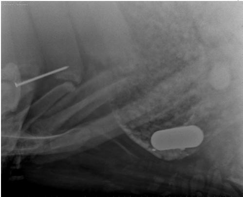
33 Large Animal Can image horses, cows, etc. Radiographs made while patient standing Creates horizontal gas-fluid interfaces in bowel Often looking for abnormal opacity Mineral, metal Peritoneal gas 16

34 17
GENERAL ABDOMINAL IMAGING PERITONEAL SPACE, PANCREAS, & SPLEEN. VMB 960 March 25, 2013
 GENERAL ABDOMINAL IMAGING PERITONEAL SPACE, PANCREAS, & SPLEEN VMB 960 March 25, 2013 REFERENCE Chapters 35-36 Pages 650-678 Chapter 37 Pages 694-701 Chapter 3 Pages 38-49 OBJECTIVES Radiography and Ultrasound
General Abdominal Radiography
 General Abdominal Radiography Tony Pease, DVM, MS Assistant Professor of Radiology North Carolina State University Objectives Acquisition of radiographs Abdominal radiographic anatomy Radiographic patterns
Close window to return to IVIS. in collaborazione con RICHIESTO ACCREDITAMENTO. organizzato da certificata ISO 9001:2000
 in collaborazione con Close window to return to IVIS RICHIESTO ACCREDITAMENTO SOCIETÀ CULTURALE ITALIANA VETERINARI PER ANIMALI DA COMPAGNIA SOCIETÀ FEDERATA ANMVI organizzato da certificata ISO 9001:2000
What s Your Diagnosis? Signalment: Species: Canine Breed: Golden Retriever Sex: Female (spayed) Date of Birth: 04/01/99
 Date of Birth: 04/01/99") What s Your Diagnosis? Signalment: Species: Canine Breed: Golden Retriever Sex: Female (spayed) Date of Birth: 04/01/99 Presenting Complaint: Acute onset of lethargy Vomited twice (partially digested food)
What s Your Diagnosis? Signalment: Species: Canine Breed: Golden Retriever Sex: Female (spayed) Date of Birth: 04/01/99 Presenting Complaint: Acute onset of lethargy Vomited twice (partially digested food)
What s Your Diagnosis?
 What s Your Diagnosis? Signalment: 5 year old MC Belgian Malinois Presenting Complaint: Perineal hernia as well as not eating or defecating History: The patient presented to the KSU VHC on 7/28/2018 for
What s Your Diagnosis?
 What s Your Diagnosis? Courtney S. Wait Signalment: 11 year old FS Labrador Retriever Presenting Complaint/History: The patient presented to the referring DVM for inappetance, vomiting, lethargy, and anorexia.
Ultrasonography of Peritoneal and Retroperitoneal Spaces and Abdominal Lymph Nodes
 IMAGING Ultrasonography of Peritoneal and Retroperitoneal Spaces and Abdominal Lymph Nodes Clifford R. Berry, DVM, DACVR; Elizabeth Huyhn, DVM; and Danielle Mauragis, CVT University of Florida Welcome
What s Your Diagnosis??? Renée Fahrenholz, Class of 2012
 Renée Fahrenholz, Class of 2012 What s Your Diagnosis??? Signalment Emma, a 9 year old, Female, Spayed, Domestic Short Haired Feline Presenting Complaint Weight loss, vomited the morning of her visit,
Proceedings of the 34th World Small Animal Veterinary Congress WSAVA 2009
 www.ivis.org Proceedings of the 34th World Small Animal Veterinary Congress WSAVA 2009 São Paulo, Brazil - 2009 Next WSAVA Congress : Reprinted in IVIS with the permission of the Congress Organizers IMAGING
ABDOMINAL RADIOLOGY UNDERSTANDING
 ABDOMINAL RADIOLOGY UNDERSTANDING CACVT 2017 SPRING CONFERENCE - GREENWOOD VILLAGE, CO Amy Newfield, CVT, VTS (ECC) BluePearl Massachusetts - Waltham, MA INTRODUCTION As a technician you will likely be
Abdominal Ultrasound
 Abdominal Ultrasound Imaging Control Buttons Depth The organ imaged should take up 3/4 of the screen Frequency = Penetration Use high frequencies (harmonics) for fluid filled and superficial structures
DIAGNOSTIC IMAGING: LIVER DISEASE
 Vet Times The website for the veterinary profession https://www.vettimes.co.uk DIAGNOSTIC IMAGING: LIVER DISEASE Author : Abby Caine Categories : Vets Date : February 1, 2010 ABBY CAINE reviews both established
What is Your Diagnosis?
 What is Your Diagnosis? Izabela Ragan, Class of 2014 Signalment Species: Canine Breed: English Bulldog Sex: Male castrated Date of birth: 04/14/11 Presenting Complaint Dog was presented for vomiting and
What s Your Diagnosis? Sara Alves, Class of Signalment: 9-year-7-month old female spay American Miniature Eskimo dog
 What s Your Diagnosis? Sara Alves, Class of 2018 Signalment: 9-year-7-month old female spay American Miniature Eskimo dog Presenting Complaint: The patient presented on 5/30/17 with signs of lethargy and
Gastrointestinal Tract Imaging. Objectives. Reference. VMB 960 April 6, Stomach Small Intestine Colon. Radiography & Ultrasound
 Gastrointestinal Tract Imaging VMB 960 April 6, 2009 Stomach Small Intestine Colon Objectives Radiography & Ultrasound Contrast Examination of the Small Intestine Reference Chapters 45 47 Pages 750 805
What s your diagnosis?
 What s your diagnosis? Signalment: 9 year old MC 2.7 kg Papillion Presenting Complaint: Presented for work up of anorexia and vomiting History: He had presented to cardiology for work up of a grad IV/VI
Radiographic Positioning. Small Animal Abdominal Radiography. Lecture Outline. Matthew Paek, VMD, MS, DACVR
 Small Animal Abdominal Radiography Matthew Paek, VMD, MS, DACVR Email: Matthew.Paek@SynergyVIP.com 7/30/2018 1 Lecture Outline Radiographic technique Introduction to systematic review and principles of
Appendix 5. EFSUMB Newsletter. Gastroenterological Ultrasound
 EFSUMB Newsletter 87 Examinations should encompass the full range of pathological conditions listed below A log book listing the types of examinations undertaken should be kept Training should usually
Signalment: Gidget, 12 year old, female spayed, Scottish Terrier, 10.7 kg
 Signalment: Gidget, 12 year old, female spayed, Scottish Terrier, 10.7 kg Presenting Complaint: Gidget presented after having elevated liver enzymes, patchy alopecia and PU/PD. History: Gidget had been
Abdomen Sonography Examination Content Outline
 Abdomen Sonography Examination Content Outline (Outline Summary) # Domain Subdomain Percentage 1 2 3 Anatomy, Perfusion, and Function Pathology, Vascular Abnormalities, Trauma, and Postoperative Anatomy
GENERAL DIAGNOSTIC IMAGING IN SMALL ANIMAL ONCOLOGY
 GENERAL DIAGNOSTIC IMAGING IN SMALL ANIMAL ONCOLOGY Jantra Ngosuwan Suran, DVM, Dipl. ACVR, Cert Clin Res University of Pennsylvania, School of Veterinary Medicine 3900 Delancey St, Philadelphia, PA 19104
DIAGNOSTIC ULTRASOUND D R. E R I C A J O H N S O N
 DIAGNOSTIC ULTRASOUND D R. E R I C A J O H N S O N ULTRASOUND BASICS Medical ultrasound machines generate and receive ultrasound waves Ultrasound waves are emitted from the peizolectric crystals of the
Radiography of the Acute Abdomen
 The 3 rd Annual Vet Education Online Veterinary Conference - July 2012 Radiography of the Acute Abdomen Dr Angela Hartman DVM Dipl. ACVR Massy University, New Zealand Vet Education Pty Ltd Vet Education
US in non-traumatic acute abdomen. Lalita, M.D. Radiologist Department of radiology Faculty of Medicine ChiangMai university
 US in non-traumatic acute abdomen Lalita, M.D. Radiologist Department of radiology Faculty of Medicine ChiangMai university Sagittal Orientation Transverse (Axial) Orientation Coronal Orientation Intercostal
What s Your Diagnosis? Jessica Eisenbarth. Signalment: Jazz is a female intact 2 year old German Shorthaired Pointer.
 What s Your Diagnosis? Jessica Eisenbarth Signalment: Jazz is a female intact 2 year old German Shorthaired Pointer. Presenting complaint: Jazz was presented to the K-State emergency service on August
Appendix 9: Endoscopic Ultrasound in Gastroenterology
 Appendix 9: Endoscopic Ultrasound in Gastroenterology This curriculum is intended for clinicians who perform endoscopic ultrasonography (EUS) in gastroenterology. It includes standards for theoretical
Radiology of hepatobiliary diseases
 GI cycle - Lecture 14 436 Teams Radiology of hepatobiliary diseases Objectives 1. To Interpret plan x-ray radiograph of abdomen with common pathologies. 2. To know the common pathologies presentation.
Plain abdomen The standard films are supine & erect AP views (alternative to erect, lateral decubitus film is used in ill patients).
.") Plain abdomen The standard films are supine & erect AP views (alternative to erect, lateral decubitus film is used in ill patients). The stomach can be readily identified by its location, gastric rugae
Plain abdomen The standard films are supine & erect AP views (alternative to erect, lateral decubitus film is used in ill patients). The stomach can be readily identified by its location, gastric rugae
Concepts in Small Animal Thoracic Radiology Thoracic Radiology
 Concepts in Small Animal Thoracic Radiology + Radiology of the Pleural Space VMB 960 2/21/2011 Optimizing Image Quality Inherent subject contrast Thorax has high inherent subject contrast c/f abdomen Primarily
What s Your Diagnosis?
 Claire Legallet What s Your Diagnosis? Signalment: Species: Canine Breed: Catahoula Sex: Female Intact Age at presentation: 6 months Presenting Complaint: Chronic intermittent bloody diarrhea and vomiting
Contents. Basic Ultrasound Principles and Terminology. Ultrasound Nodule Characteristics
 Contents Basic Ultrasound Principles and Terminology Basic Ultrasound Principles... 1 Ultrasound System... 2 Linear Transducer for Superficial Images and Ultrasound-Guided FNA... 3 Scanning Planes... 4
What s Your Diagnosis? Allison Crow, Class of 2014
 What s Your Diagnosis? Allison Crow, Class of 2014 Signalment: 13 year old male castrated mixed breed dog History: The patient presented to the rdvm for pain in the hind end, weakness and neck stretching
Chapter Overview. Chapter 1. Anatomy. Physiology
 Chapter Overview Chapter 1 An Introduction to the Human Body Define Anatomy and Physiology Levels of Organization Characteristics of Living Things Homeostasis Anatomical Terminology 1 2 Anatomy Describes
Proceedings of the American Association of Equine Practitioners - Focus Meeting. Focus on Colic. Indianapolis, IN, USA 2011
 www.ivis.org Proceedings of the American Association of Equine Practitioners - Focus Meeting Focus on Colic Indianapolis, IN, USA 2011 Next Focus Meetings: July 22-24, 2012 - Focus on Hind Limb Lameness
Proceedings of the 34th World Small Animal Veterinary Congress WSAVA 2009
 www.ivis.org Proceedings of the 34th World Small Animal Veterinary Congress WSAVA 2009 São Paulo, Brazil - 2009 Next WSAVA Congress : Reprinted in IVIS with the permission of the Congress Organizers MANAGEMENT
EFSUMB EUROPEAN FEDERATION OF SOCIETIES FOR ULTRASOUND IN MEDICINE AND BIOLOGY Building a European Ultrasound Community
 MINIMUM TRAINING REQUIREMENTS FOR THE PRACTICE OF MEDICAL ULTRASOUND IN EUROPE Appendix 9: Endoscopic Ultrasound in Gastroenterology This curriculum is intended for clinicians who perform endoscopic ultrasonography
Pediatric Hepatobiliary, Pancreatic & Splenic US
 Pediatric Hepatobiliary, Pancreatic & Splenic US Susan J. Back, MD Department of Radiology, The Children s Hospital of Philadelphia No Disclosures Objectives Normal Abnormal: cases and US advances Objectives
Pathology of the Alimentary Tract
 Pathology of the Alimentary Tract Lab 2: Lower alimentary tract SI, LI, cecum, and peritoneum GIST in the cecum of a dog Shannon Martinson: http://people.upei.ca/smartinson VPM 221: November, 2011 3 year
Life is hard. Question? Abdominal Imaging: The practical Utility of Radiography and Ultrasound 2/16/2012
 Abdominal Imaging: The practical Utility of Radiography and Ultrasound Jonathan Hayles, DVM, MS, Dipl ACVR Life is hard Internist Surgeon Oncologist Neurologist Dermatologist Radiologist!!! The digital
Sex: 女 Age: 51 Occupation: 無 Admission date:92/07/22
 Sex: 女 Age: 51 Occupation: 無 Admission date:92/07/22 Chief complaint Unknown fever for one month Hand tremor and left huge renal tumor was noted Present illness Suffered from fever for one month, hand
Welcome to ANAT 10A! What is Anatomy? Different levels of Anatomy The Language of Anatomy Pearson Education, Inc.
 Welcome to ANAT 10A! What is Anatomy? Different levels of Anatomy The Language of Anatomy Introduction Anatomy means to dissect: (ANAT 10A) The study of internal & external body structures The study of
Surgical Treatment of special Tumours. Winnie Achilles Tierklinik Hollabrunn Lastenstrasse Hollabrunn
 Surgical Treatment of special Tumours Winnie Achilles Tierklinik Hollabrunn Lastenstrasse 2 2020 Hollabrunn boexi@gmx.de Hepatocellular Tumours Hepatocellular Carcinoma, hepatocellular adenoma, and hepatoblastoma
Table of Contents. Preface xi. Acknowledgments xiii. Part I Overview of the Diagnostic Process 1. 1 Overview of Grading and Staging 3
 Table of Contents Preface xi Acknowledgments xiii Part I Overview of the Diagnostic Process 1 1 Overview of Grading and Staging 3 Identification of the process 3 Identification of tumor types 5 Grading
Normal Sonographic Anatomy
 hapter 2:The Liver DUNSTAN ABRAHAM Normal Sonographic Anatomy Homogeneous, echogenic texture (Figure 2-1) Measures approximately 15 cm in length and 10 12.5 cm anterior to posterior; measurement taken
Imaging the pancreas CONTINUING EDUCATION
 Imaging the pancreas The diagnosis of pancreatic disease can be challenging but has been improved in recent years with the introduction and advancement of various diagnostic tests. Tim Trevail BVetMed
Abdominal radiology 腹部放射線學
 Abdominal radiology 腹部放射線學 台北醫學大學 - 市立萬芳醫院 留偉順 laowilson@hotmail.com The Normal Abdominal Series Chest Supine abdomen Erect abdomen Left lateral decubitus abdomen Learning objectives Understanding normal
Imaging the Urinary Tract
 Imaging the Urinary Tract Laura Armbrust, DVM, DACVR Gregory F. Grauer, DVM, MS, DACVIM Kansas State University Radiographic and ultrasound imaging in addition to history, physical examination, and clinicopathologic
Job Task Analysis for ARDMS Abdomen Data Collected: June 30, 2011
 Job Task Analysis for ARDMS Abdomen Data Collected: June 30, 2011 Reported: Analysis Summary for: Abdomen Examination Survey Dates 06/13/2011-06/26/2011 Invited Respondents 6,000 Surveys with Demographics
My Patient Has Abdominal Pain PoCUS of the Biliary Tract and the Urinary Tract
 My Patient Has Abdominal Pain PoCUS of the Biliary Tract and the Urinary Tract Objectives PoCUS for Biliary Disease PoCUS for Renal Colic PoCUS for Urinary Retention Biliary Disease A patient presents
Muscle spasm Diminished bowel sounds Nausea/vomiting
 3 4 5 6 7 8 9 0 Chapter 8: Abdomen and Genitalia Injuries Abdominal Injuries Abdomen is major body cavity extending from to pelvis. Contains organs that make up digestive, urinary, and genitourinary systems.
Radiology of the abdomen Lecture -1-
 Radiology of the abdomen Lecture -1- Objectives To know radiology modalities used in abdomen imaging mainly GI tract. To know advantages and disadvantages of each modality. To know indications and contraindications
Case Discussion Splenic Abscess
 Case Discussion Splenic Abscess Personal Data Gender: male Birth Date: 1928/Mar/06th Allergy: Mefenamic Smoking: 0.5 PPD for 55 years Alcohol: negative (?) 4 Months Ago Abdominal pain: epigastric area
Endocrine Lab. Heather Fenton VPM 222 November
 Endocrine Lab Heather Fenton VPM 222 November 27 2012 Case 1: Nursery pig Case 1: Nursery pig Description: There are multifocal round (approximately 1cm diameter) firm lesions within the adrenal gland
Calvin 9 year old NM DLH. Dr. Norman Ackerman Memorial Radiography Case Challenge
 September 2014 Dr. Norman Ackerman served the University of Florida, College of Veterinary Medicine with distinction as Professor of Radiology from 1979 to 1994. A concerned teacher of veterinary students
Anatomy Jessica Ferguson Ashley Dobos May 31, 2006 LIVER
 Anatomy Jessica Ferguson Ashley Dobos May 31, 2006 LIVER 1) Other Names: Reidel s Lobe normal anatomic variant; projection of the right lobe that can extend as far as the iliac crest (Tempkin, p.54, Anatomy).
Imaging the Urogenital System
 maging the Urogenital System Tony Pease, DVM, MS, DACVR Assistant Professor of Radiology North Carolina State University Reading Thrall Chapters 42-46 Prostate Gland Not visible radiographically in normal
CT EVALUATION OF GASTRIC LESIONS:
 CT EVALUATION OF GASTRIC LESIONS: Pictural essay Hasni Bouraoui I, Kahloun A, Jemni H, Elouni F, Moulahi H, Daadoucha A, Ben Ali A, Sriha B, Tlili Graies K Departments of Radiology, Gastro enterology,
Proceedings of the 34th World Small Animal Veterinary Congress WSAVA 2009
 www.ivis.org Proceedings of the 34th World Small Animal Veterinary Congress WSAVA 2009 São Paulo, Brazil - 2009 Next WSAVA Congress : Reprinted in IVIS with the permission of the Congress Organizers DIFFUSE
Basic of Ultrasound Physics E FAST & Renal Examination. Dr Muhammad Umer Ihsan MBBS,MD, DCH CCPU,DDU1,FACEM
 Basic of Ultrasound Physics E FAST & Renal Examination Dr Muhammad Umer Ihsan MBBS,MD, DCH CCPU,DDU1,FACEM What is Sound? Sound is Mechanical pressure waves What is Ultrasound? Ultrasounds are sound waves
Gallbladder & Pancreas Ultrasonography
 복부초음파 : 담낭과췌장 Gallbladder & Pancreas Ultrasonography 김정훈 Department of Radiology 1 Interaction of sound with matter (1) 반사 (Reflection) (2) 굴절 (Refraction) (3) 흡수 (Absorption) (4) 산란 (Scattering) 음향저항
Ultrasonographic and Clinical Studies on Benign Prostatic Hyperplasia in Dogs
 Theriogenology Insight: 6(1): 67-72, April, 2016 DOI Number: 10.5958/2277-3371.2016.00009.7 Ultrasonographic and Clinical Studies on Benign Prostatic Hyperplasia in Dogs K. Rajkumar* and C. Ansarkamran
Guidelines, Policies and Statements D5 Statement on Abdominal Scanning
 Guidelines, Policies and Statements D5 Statement on Abdominal Scanning Disclaimer and Copyright The ASUM Standards of Practice Board have made every effort to ensure that this Guideline/Policy/Statement
Abdomen and Genitalia Injuries. Chapter 28
 Abdomen and Genitalia Injuries Chapter 28 Hollow Organs in the Abdominal Cavity Signs of Peritonitis Abdominal pain Tenderness Muscle spasm Diminished bowel sounds Nausea/vomiting Distention Solid Organs
EUROPEAN ASSOCIATION OF VETERINARY DIAGNOSTIC IMAGING EUROPEAN COLLEGE OF VETERINARY DIAGNOSTIC IMAGING
 EISAGOGIKO EUROPEAN ASSOCIATION OF VETERINARY DIAGNOSTIC IMAGING EUROPEAN COLLEGE OF VETERINARY DIAGNOSTIC IMAGING ARISTOTLE UNIVERSITY OF THESSALONIKI SCHOOL OF VETERINARY MEDICINE SECTION OF RADIOLOGY
IT 의료융합 1 차임상세미나 복부질환초음파 이재영
 IT 의료융합 1 차임상세미나 2013-4-3 복부질환초음파 이재영 나는오늘누구를위하여 종을울리나? 전통적의료 의사 공학설계자 의사 최첨단진단장비들 USG, CT, MRI 환자 환자 현대의료 사용자중심의사고 US in the Abdomen Detection DDx Look Behavior Response by external stimuli Guiding Tool
Abdominal Assessment
 Abdominal Assessment Mary Marian, MS,RD,CSO University of AZ, Tucson, AZ Neha Parekh, MS,RD,LD,CNSC Cleveland Clinic, Cleveland, OH Objectives: 1. Outline the steps in performing an abdominal examination.
Radiological Investigations of Abdominal Trauma
 76 77 Investigations of Abdominal Trauma Introduction: Trauma to abdominal organs is a common cause of patient morbidity and mortality among trauma patients. Causes of abdominal trauma include blunt injuries,
Imaging in gastric cancer
 Imaging in gastric cancer Gastric cancer remains a deadly disease because of late diagnosis. Adenocarcinoma represents 90% of malignant tumors. Diagnosis is based on endoscopic examination with biopsies.
4/7/2017. Ultrasound of the Urinary Bladder. Indications for Bladder Ultrasound. Patient Preparation. Transition Adjustments.
 Indications for Bladder Ultrasound Ultrasound of the Urinary Bladder Hematuria, pyuria, or other UA abnormality Abnormal transitional cells Pollakuria, dysuria, stranguria, periuria History of urinary
for the Veterinary Technician
 An Overview of Abdominal Ultrasound for the Veterinary Technician Valerie Gates, CVT, VTS (ECC) Learning Objective: The reader should gain a basic understanding of ultrasound, including physics, terminology,
Ultrasonography of the Neck as an Adjunct to FNA. Nicole Massoll M.D.
 Ultrasonography of the Neck as an Adjunct to FNA Nicole Massoll M.D. Basic Features of Head and Neck Ultrasound and Anatomy Nicole Massoll M.D. University of Arkansas for Medical Sciences, Little Rock
Pathology of the Hematopoietic System. Case studies
 Pathology of the Hematopoietic System Case studies Shannon Martinson, September 2015 Signalment: 9 yr-old MC cat Case Study 1 History: Cat had been anorexic and developed bleeding in the eyes Physical
FAST Focused Assessment with Sonography in Trauma
 FAST Focused Assessment with Sonography in Trauma Wilma Rodriguez Mojica,MD,FACR Professor of Radiology UPR School of Medicine Ultrasound Section - Radiological Sciences Department OBJECTIVES Understand
Principles of Ultrasound. Cara C. Prideaux, M.D. University of Utah PM&R Sports Medicine Fellow March 14, 2012
 Principles of Ultrasound Cara C. Prideaux, M.D. University of Utah PM&R Sports Medicine Fellow March 14, 2012 None Disclosures Outline Introduction Benefits and Limitations of US Ultrasound (US) Physics
Focused Assessment Sonography of Trauma (FAST) Scanning Protocol
 Scanning Protocol") Focused Assessment Sonography of Trauma (FAST) Scanning Protocol Romolo Gaspari CHAPTER 3 GOAL OF THE FAST EXAM Demonstrate free fluid in abdomen, pleural space, or pericardial space. EMERGENCY ULTRASOUND
Focused Assessment Sonography of Trauma (FAST) Scanning Protocol Romolo Gaspari CHAPTER 3 GOAL OF THE FAST EXAM Demonstrate free fluid in abdomen, pleural space, or pericardial space. EMERGENCY ULTRASOUND
ASSESSING THE PLAIN ABDOMINAL RADIOGRAPH M A A M E F O S U A A M P O F O
 ASSESSING THE PLAIN ABDOMINAL RADIOGRAPH M A A M E F O S U A A M P O F O Introduction The abdomen (less formally called the belly, stomach, is that part of the body between the thorax (chest) and pelvis,
Imaging Guided Biopsy. Edited & Presented by ; Hussien A.B ALI DINAR. Msc Lecturer,Reporting Sonographer
 Imaging Guided Biopsy Edited & Presented by ; Hussien A.B ALI DINAR. Msc Lecturer,Reporting Sonographer Objective By the End of this lessons you should : Define what biopsy Justify Aim to perform biopsy
Chapter 3. Sonographic Image Interpretation
 Chapter 3 Sonographic Image Interpretation Sonograms are two-dimensional gray-scale images that allow assessment and diagnosis of many anatomic and pathologic changes that can occur in the human body.
Abdominal ultrasound:
 Abdominal ultrasound: Non-traumatic acute abdomen Wittanee Na-ChiangMai, MD Department of Radiology ChiangMai University 26/04/2017 Contents Technique of examination Normal anatomy Emergency conditions
FHS Appendicitis US Protocol
 FHS Appendicitis US Protocol Reviewed By: Shireen Khan, MD; Sarah Farley, MD; Anna Ellermeier, MD Last Reviewed: May 2018 Contact: (866) 761-4200 **NOTE for all examinations: 1. If documenting possible
Chief Complaint. Retroperitoneal cystic mass incidentally found at health examination center.
 Personal Information Age: 34 y/o Sex: female Past history: major systemic medical history(-) surgical history(-), family history(-) Denied food or drug allergy Chief Complaint Retroperitoneal cystic mass
GASTROINTESTINAL IMAGING STUDY GUIDE
 GASTROINTESTINAL IMAGING STUDY GUIDE Pharynx Diverticula Foreign bodies Trauma o Motility Disorders Esophagus Diverticula Trauma Esophagitis Barrett esophagus Rings, webs, and strictures Varices Benign
The Spleen. Dr Fahad Ullah
 The Spleen BY Dr Fahad Ullah Spleen The spleen is an largest lymphoid organ shaped like a shoe that lies relative to the 9th and 11th ribs and is located in the left hypochondrium. Thus, the spleen is
Extrahepatic Bile Duct Ostruction (Blockage of the Extrahepatic or Common Bile Duct) Basics
 Basics") Extrahepatic Bile Duct Ostruction (Blockage of the Extrahepatic or Common Bile Duct) Basics OVERVIEW The liver is the largest gland in the body; it has many functions, including production of bile (a fluid
Extrahepatic Bile Duct Ostruction (Blockage of the Extrahepatic or Common Bile Duct) Basics OVERVIEW The liver is the largest gland in the body; it has many functions, including production of bile (a fluid
What s your diagnosis? Malori Marotz. Squirt, an 8month old mix breed puppy. History:
 What s your diagnosis? Malori Marotz Squirt, an 8month old mix breed puppy History: The owner obtained squirt at 12 weeks of age. The owner reported that Squirt was passing soft stools lately and he is
CT 101 :Pancreas and Spleen
 CT 101 :Pancreas and Spleen Shikha Khullar,, MD, MPH Division of Radiology University of South Alabama The Pancreas Normal Pancreas 3 Phase Pancreatic CT Non contrast Arterial phase : 30-35 35 second
Case # nd Annual SEVPAC May 17, Kathy-Anne Clarke
 Case # 10 42 nd Annual SEVPAC May 17, 2014 Kathy-Anne Clarke Google images Babu Babu is 10 year old spayed female French Bulldog Chronic weight loss over 4 months Febrile and lethargic at the referring
Introduction to Abdominal Ultrasound
 Introduction to Abdominal Ultrasound Debra M. Beard, DVM, MS, DACVR Clinical Associate Professor, Radiology Auburn University College of Veterinary Medicine Why use Ultrasound in your practice? US is a
REVIEW OF NORMAL EQUINE GASTROINTESTINAL ULTRASONOGRAPHY. Michelle Henry Barton DVM, PhD, DACVIM University of Georgia, Athens, GA, 30602
 REVIEW OF NORMAL EQUINE GASTROINTESTINAL ULTRASONOGRAPHY Michelle Henry Barton DVM, PhD, DACVIM University of Georgia, Athens, GA, 30602 INTRODUCTION The recent introduction of more affordable and portable
Anatomical Considerations for Lab Practical II
 Anatomical Considerations for Lab Practical II For each of the following please be prepared to provide: Identification System Organ(s) or ducts to Function(s) location which it is attached Use your lecture
Heme Database Exercise Use the Hematopoietic Database to answer the following questions
 Heme Database Exercise Use the Hematopoietic Database to answer the following questions http://seer.cancer.gov/seertools/hemelymph/ 1. Assign a topography and histology code to polycythemia vera 2. List
Imaging of common diseases of hepatobiliary and GI system
 Imaging of common diseases of hepatobiliary and GI system Natthaporn Tanpowpong, M.D. Diagnostic radiology Faculty of Medicine, Chulalongkorn University Normal plain radiograph A = Common bile duct
Multi modality Imaging in Acute Pancreatitis. Marsha Lynch, HMS III Gillian Lieberman, MD BIDMC Core Clerkship in Radiology March 2009
 Multi modality Imaging in Acute Pancreatitis Marsha Lynch, HMS III Gillian Lieberman, MD BIDMC Core Clerkship in Radiology March 2009 Our Patient R: Introduction 52M with 10d history of nausea, vomiting
Introduction and Definitions
 Bowel obstruction Introduction and Definitions Accounts for 5% of all acute surgical admissions Patients are often extremely ill requiring prompt assessment, resuscitation and intensive monitoring Obstruction
The Role of Ultrasound in the Assessment of Inflammatory Bowel Disease
 The Role of Ultrasound in the Assessment of Inflammatory Bowel Disease Dr. Richard A. Beable Consultant Gastrointestinal Radiologist Queen Alexandra Hospital Portsmouth Hospitals NHS Trust Topics for Discussion
Proceedings of the World Small Animal Veterinary Association Sydney, Australia 2007
 Proceedings of the World Small Animal Sydney, Australia 2007 Hosted by: Next WSAVA Congress THE LAST GASP II: LUNGS AND THORAX David Holt, BVSc, Diplomate ACVS University of Pennsylvania School of Veterinary
Chylothorax Basics OVERVIEW GENETICS SIGNALMENT/DESCRIPTION OF PET
 Chylothorax Basics OVERVIEW Chylo- refers to chyle; thorax refers to the chest Chyle is a milky to slightly yellow fluid composed of lymph and fats (rich in triglycerides) taken up from the intestines
The abdominal Esophagus, Stomach and the Duodenum. Prof. Oluwadiya KS
 The abdominal Esophagus, Stomach and the Duodenum Prof. Oluwadiya KS www.oluwadiya.com Viscera of the abdomen Abdominal esophagus: Terminal part of the esophagus The stomach Intestines: Small and Large
د. عصام طارق. Objectives:
 GI anatomy Lecture: 5 د. عصام طارق Objectives: To describe anatomy of stomach, duodenum & pancreas. To list their main relations. To define their blood & nerve supply. To list their lymph drainage. To
Children's (Pediatric) Ultrasound - Abdomen
 Ultrasound - Abdomen") Scan for mobile link. Children's (Pediatric) Ultrasound - Abdomen Children s (pediatric) ultrasound imaging of the abdomen is a safe, noninvasive test that uses sound waves to produce a clear picture of
Scan for mobile link. Children's (Pediatric) Ultrasound - Abdomen Children s (pediatric) ultrasound imaging of the abdomen is a safe, noninvasive test that uses sound waves to produce a clear picture of