Lecture 01 Internal surface of anterolateral abdominal wall. BY Dr Farooq Khan Aurakzai
|
|
|
- Brett Allen
- 6 years ago
- Views:
Transcription


1 Lecture 01 Internal surface of anterolateral abdominal wall BY Dr Farooq Khan Aurakzai Dated:

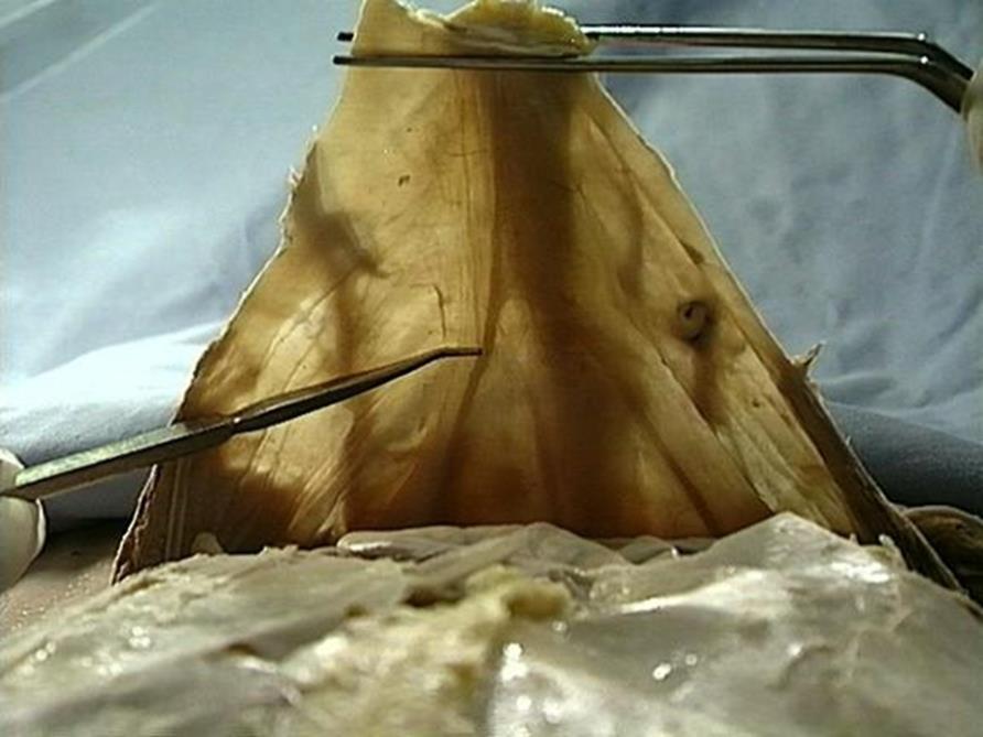
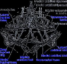
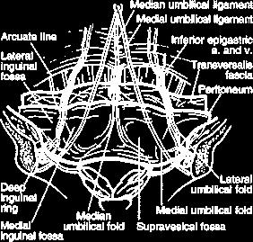
2 Internal surface of the anterolateral abdominal wall The internal ( posterior ) surface of the anterolateral abdominal wall is covered with: Transversalis fascia A variable amount of extraperitoneal fat And parietal peritoneum. 2

3 Internal surface of the anterolateral abdominal wall The internal surface of the anterolateral wall present two parts: Infra umbilical part and Supra umbilical part. INFerior to the umbilicus: The infra umblical part of this surface exhibits five umbilical peritoneal folds passing toward the umbilicus : One in the median plane Two on each side. 3

4 Internal surface of the anterolateral abdominal wall 1. Median Umbilical folds In the midline, there are elevations of peritoneum with free edges, called folds. INFERIOR to the umbilicus: The median umbilical fold extends from the apex of the urinary bladder to the umbilicus and covers the median umbilical ligament. It is a fibrous remnant of the urachus that joined the apex of the fetal bladder. 4
5 5
6 2. Medial Umbilical Folds Two medial umbilical folds, lateral to the median umbilical fold, cover the medial umbilical ligaments. It is formed by occluded parts of the umbilical arteries. 6

7 3. Lateral Umbilical Folds Two lateral umbilical folds, lateral to the medial umbilical folds. It covers the inferior epigastric vessels and therefore bleed if cut. It is patent even after birth. 7

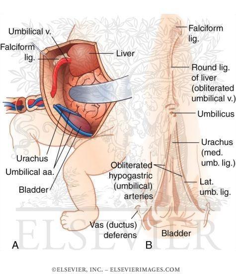
8 Internal surface of anterior abdominal wall SUPRA UMBILICAL PART: The supraumbilical part of the internal surface of the anterior abdominal wall has a sagittally oriented peritoneal reflection. The falciform ligament, that extends between the superior anterior abdominal wall and the liver. It encloses the round ligament of the liver, and the paraumbilical veins in its inferior free edge. 8
9 9
10 Internal surface of anterior abdominal wall The round ligament is a fibrous remnant of the umbilical vein, which passed from umbilicus to the liver prenatally. 10

11 Median umbilical ligament The median umbilical ligament or Xander s ligament. It extends from the apex of the bladder to the umbilicus, on the deep surface of the anterior abdominal wall. It is unpaired. It is covered by the median umbilical fold. It should not be confused with the medial umbilical ligament, as: Median Umbilical ligament.urachus. Medial Umbilical ligament..obliterated Umbilical arteries. It may be used as a landmark for surgeons who are performing laparoscopy, such as laparoscopic inguinal hernia repair. 11

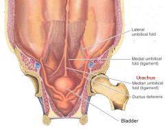
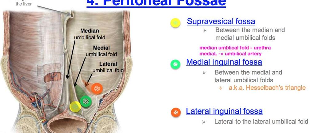
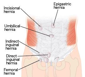
12 Internal surface of the anterolateral abdominal wall The depression lateral to the umbilical folds are the peritoneal fossae, each of which is a potential site for the hernia. 12

 Lateral inguinal fossae (A in")
13 Internal surface of the anterolateral abdominal wall The shallow fossae between the umbilical folds are the : Supravesical fossae (C in Figure) Medial inguinal fossae (B in Figure) Lateral inguinal fossae (A in Figure) 13

14 Supravesical fossae Supravesical fossae lies between the median and the medial umbilical folds. It is formed as the peritoneum reflects from the anterior abdominal wall onto the bladder. The level of the supravesical fossae rises and falls with filling and emptying of the bladder. 14
15 15
.")
16 Medial inguinal fossae Medial inguinal fossae between the medial and the lateral umbilical folds, areas also commonly called inguinal triangles ( Hessel bach s triangles). It is the potential sites for the less common direct inguinal hernias. 16

17 Hesselbach s triangles The inguinal triangle/ Hesselbach s triangle is a triangular fossa of the abdominal wall. Boundaries Medial border: Lateral margin of the rectus sheath Superolateral border: Inferior epigastric vessels Inferior border: Inguinal ligament 17
18 18

19 Lateral inguinal fossae Lateral inguinal fossae, lateral to the lateral umbilical folds, include the deep inguinal rings. It is a potential sites for the most common type of hernia in the lower abdominal wall, the Indirect Inguinal Hernia. 19


20 ARCUATE LINE The arcuate line of the abdomen, linea semicircularis or Douglas' line. It is a horizontal line that demarcates the lower limit of the posterior layer of the rectus sheath. It is also where the inferior epigastric vessels perforate the rectus abdominis. 20
21 21
22 22

23 Post natal patency of the umbilical vein The umbilical vein is a vein present during fetal development that carries oxygenated blood from the placenta into the growing fetus. The umbilical vein provides convenient access to the central circulation of a neonate for restoration of blood volume and for administration of glucose and drugs. Closure of the umbilical vein usually occurs after the umbilical arteries have closed. 23
.")
24 post natal patency of the umbilical vein..con t Within a week of birth, the neonate's umbilical vein is completely obliterated and is replaced by a fibrous cord called the round ligament of the liver (also called ligamentum teres hepatis). It extends from the umbilicus to the transverse fissure, where it joins with the falciform ligament of the liver. It separate segment 4 from segments 2 and 3 of the left hepatic lobe. 24

25 Supravesical hernia A rare cause of intestinal obstruction Supravesical hernia is an unusual type of hernia. It is of two types: Internal and External. It was reported by Sir Astley Cooper in The internal type is more difficult to diagnose and commonly presents as intestinal obstruction or undiagnosed abdominal pain. The external type is medial to the direct inguinal hernia. The iliohypogastric nerve is in danger of injury during the repair of this type of hernia. 25

26 Internal surface of the anterolateral abdominal wall The location of a hernia in one of these fossae determines how the hernia is classified. 26
27 27
28 28
29 29
30 30
31 31


32
Internal abdominal wall and inguinal region. Mathew Wedel, 2015
 Internal abdominal wall and inguinal region Mathew Wedel, 2015 gut tube umbilicus gut tube dorsal mesentery visceral peritoneum gut tube FOREGUT dorsal mesentery parietal peritoneum MIDGUT & HINDGUT gut
This presentation will discuss the anatomy of the anterior abdominal wall as it pertains to gynaecological and obstetric surgery.
 This presentation will discuss the anatomy of the anterior abdominal wall as it pertains to gynaecological and obstetric surgery. 1 The border of the anterior abdominal wall is defined superiorly by the
Lecture 02 Anatomy of the LIVER
 Lecture 02 Anatomy of the LIVER BY Dr Farooq Khan Aurakzai Dated: 02.01.2018 Introduction to Liver Largest gland in the body. 2 nd largest organ of the body. Weight approximately 1500 gm, and is roughly
The peritoneum. Prof. Oluwadiya KS, MBBS, FMCS(Orthop) Website:
 Website:") The peritoneum Prof. Oluwadiya KS, MBBS, FMCS(Orthop) Website: http://oluwadiya.com The peritoneum Serous membrane that lines the abdominopelvic cavity and invests the viscera The largest serous membrane
The peritoneum Prof. Oluwadiya KS, MBBS, FMCS(Orthop) Website: http://oluwadiya.com The peritoneum Serous membrane that lines the abdominopelvic cavity and invests the viscera The largest serous membrane
ABDOMINAL WALL & RECTUS SHEATH
 ABDOMINAL WALL & RECTUS SHEATH Learning Objectives Describe the anatomy, innervation and functions of the muscles of the anterior, lateral and posterior abdominal walls. Discuss their functional relations
Borders of the Abdomen
 Abdominal wall Borders of the Abdomen Abdomen is the region of the trunk that lies between the diaphragm above and the inlet of the pelvis below Borders Superior: Costal cartilages 7-12. Xiphoid process:
GI anatomy Lecture: 2 د. عصام طارق
 GI anatomy Lecture: 2 د. عصام طارق Objectives: To define rectus sheath. To describe anatomy of inguinal canal. To relates types of inguinal hernia to the region. To explore spermatic cord. Rectus Abdominis
BLOCK IV: OFFICIAL BODY PARTS LIST FOR ANTERIOR ABDOMINAL WALL AND ABDOMINAL CONTENTS
 BLOCK IV: OFFICIAL BODY PARTS LIST FOR ANTERIOR ABDOMINAL WALL AND ABDOMINAL CONTENTS External oblique muscle Muscular portion Aponeurotic portion Superficial inguinal ring Lateral (inferior) crus Medial
The front of the thigh. Dr.Amjad shatarat
 The front of the thigh Femoral triangle (Scarpa s triangle) Is a triangular depressed area located in the upper part of the medial aspect of the thigh immediately below the inguinal ligament. Superiorly:
STRUCTURAL BASIS OF MEDICAL PRACTICE EXAMINATION 3. October 16, 2015
 STRUCTURAL BASIS OF MEDICAL PRACTICE EXAMINATION 3 October 16, 2015 PART l. Answer in the space provided. (12 pts) 1. Identify the structures. (2 pts) A. B. A B C. D. C D 2. Identify the structures. (2
Abdomen: Introduction. Prof. Oluwadiya KS
 Abdomen: Introduction Prof. Oluwadiya KS www.oluwadiya.com Abdominopelvic Cavity Abdominal Cavity Pelvic Cavity Extends from the inferior margin of the thorax to the superior margin of the pelvis and the
STRUCTURAL BASIS OF MEDICAL PRACTICE EXAMINATION 3. October 17, 2014
 STRUCTURAL BASIS OF MEDICAL PRACTICE EXAMINATION 3 October 17, 2014 PART l. Answer in the space provided. (12 pts) 1. Identify the structures. (2 pts) A. B. A B C. D. C D 2. Identify the structures. (2
Tor Chiu. Deep Inferior Epigastric Artery Perforator Flap 161
 18 Deep Inferior Epigastric Artery Perforator Flap Tor Chiu Deep Inferior Epigastric Artery Perforator Flap 161 Deep Inferior Epigastric Artery Perforator Flap FLAP TERRITORY The deep inferior epigastric
Accessory Glands of Digestive System
 Accessory Glands of Digestive System The liver The liver is soft and pliable and occupies the upper part of the abdominal cavity just beneath the diaphragm. The greater part of the liver is situated under
HERNIAS .(A) .(B) 5. .(A) 7..( (Lumbar hernia),
 .(B) 5. .(A) 7..( (Lumbar hernia),") HERNIAS ysms91@wonju.yonsei.ac.kr 1..(B) 2..(B) 3..(A) 4. (Hesselbach's striangle).(b) 5.,.(A) 6. (Sliding hernia).(a) 7..( (Lumbar hernia), (Obturator hernia), (Sciatica hernia)).(b) Hernia = rupture
HERNIAS ysms91@wonju.yonsei.ac.kr 1..(B) 2..(B) 3..(A) 4. (Hesselbach's striangle).(b) 5.,.(A) 6. (Sliding hernia).(a) 7..( (Lumbar hernia), (Obturator hernia), (Sciatica hernia)).(b) Hernia = rupture
Abdominal muscles. Subinguinal hiatus and ingiunal canal. Femoral and adductor canals. Neurovascular system of the lower limb. Sándor Katz M.D.,Ph.D.
 Abdominal muscles. Subinguinal hiatus and ingiunal canal. Femoral and adductor canals. Neurovascular system of the lower limb. Sándor Katz M.D.,Ph.D. External oblique muscle Origin: outer surface of the
Mousa Salah. Dr. Mohammad Al. Mohtasib. 1 P a g e
 8 Mousa Salah Dr. Mohammad Al. Mohtasib 1 P a g e In the previous lecture we talked about the peritoneum, and we said that the peritonium is a serous sac, and it consists of two layers, visceral and parietal.
Femoral Triangle and Adductor Canal. Dr. Heba Kalbouneh Associate Professor of Anatomy and Histology
 Femoral Triangle and Adductor Canal Dr. Heba Kalbouneh Associate Professor of Anatomy and Histology Femoral Triangle and Adductor Canal Femoral triangle Is a triangular depressed area located in the upper
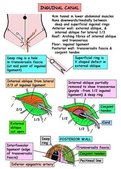
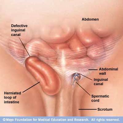
Inguinal Canal. It is an oblique passage through the lower part of the anterior abdominal wall. Present in both sexes
 Inguinal canal Inguinal Canal It is an oblique passage through the lower part of the anterior abdominal wall Present in both sexes It allows structures to pass to and from the testis to the abdomen in
Duodenum retroperitoneal
 Duodenum retroperitoneal C shaped Initial region out of stomach into small intestine RETROperitoneal viscus Superior 1 st part duodenal cap ; moves upwards and backwards to lie on the R crura medial to
The Anterolateral Abdominal Wall By Prof. Dr. Muhammad Imran Qureshi
 1 P age The Anterolateral Abdominal Wall By Prof. Dr. Muhammad Imran Qureshi Introduction The abdomen is the region of the trunk located between the thorax and the pelvis. It includes the anterolateral
Gross Anatomy ABDOMEN/SESSION 1 Dr. Firas M. Ghazi
 Anterior Abdominal Wall Structure, muscles and surface anatomy Curricular Objectives By the end of this session students are expected to: Practical 1. Identify the hip and distinguish the three bones forming
Abdominal Hernia Omar alnoubani MD,MRCS
 Abdominal Hernia Omar alnoubani MD,MRCS Definition of hernia Anatomical landmarks Overview of types of hernia Presentation and Management of common types of hernia What is the definition of a hernia? An
Netter's Anatomy Flash Cards Section 4 List 4 th Edition
 Netter's Anatomy Flash Cards Section 4 List 4 th Edition https://www.memrise.com/course/1577335/ Section 4 Abdomen (31 cards) Plate 4-1 Bony Framework of Abdomen 1.1 Costal cartilages 1.2 Iliac crest 1.3
To describe the liver. To list main structures in porta hepatis.
 GI anatomy Lecture: 6 د. عصام طارق Objectives: To describe the liver. To list main structures in porta hepatis. To define portal system & portosystemic anastomosis. To list parts of biliary system. To
1 Right & left Hepatic ducts Gastric Impression of spleen
 Pancreatic Model 1 Right & left Hepatic ducts 14 Gastric Impression of spleen 2 Common hepatic duct 15 Renal Impression of spleen 3 Cystic Duct 16 Colic Impression of spleen 4 Common Bile Duct 17 Splenic
Lecture 08 THIGH MUSCLES ANTERIOR COMPARTMENT. Dr Farooq Khan Aurakzai. Dated:
 Lecture 08 THIGH MUSCLES ANTERIOR COMPARTMENT BY Dr Farooq Khan Aurakzai Dated: 11.02.2017 INTRODUCTION to the thigh Muscles. The musculature of the thigh can be split into three sections by intermuscular
ABSITE Review: Hernias
 ABSITE Review: Inguinal and Femoral Hernias Sybile Val M.D. SUNY Downstate Medical Center Department of Surgery June 27, 2008 Objectives www.downstatesurgery.org Correctly identify anatomical landmarks
INGUINAL HERNIA REPAIR PROCEDURE GUIDE
 ROOM CONFIGURATION The following figure shows an overhead view of the recommended OR configuration for a da Vinci Inguinal Hernia Repair (Figure 1). NOTE: Configuration of the operating room suite is dependent
In the name ofgod. Abdomen 3. Dr. Zahiri
 In the name ofgod Abdomen 3 Dr. Zahiri Peritoneum Peritoneum It is the serous membrane(a type of loose connective tissue and is covered by mesothelium) that lines the abdominal cavity. Extensions of the
Anatomy: Know Your Abdomen
 Anatomy: Know Your Abdomen Glossary Abdomen - part of the body below the thorax (chest cavity); separated by the diaphragm. Anterior - towards the front of the body. For example, the umbilicus is anterior
大體老師無語良師 大體解剖學實驗 HUMAN DISSECTION ANTERIOR ABDOMINAL WALL & INGUINAL REGION 盧家鋒助理教授 臺北醫學大學醫學系解剖學暨細胞生物學科 臺北醫學大學醫學院轉譯影像研究中心.
 大體老師無語良師 大體解剖學實驗 HUMAN DISSECTION ANTERIOR ABDOMINAL WALL & INGUINAL REGION 盧家鋒助理教授 臺北醫學大學醫學系解剖學暨細胞生物學科 臺北醫學大學醫學院轉譯影像研究中心 http://www.ym.edu.tw/~cflu REFERENCES Dissector s guide [1] Dissection Guide for
Exploring Anatomy: the Human Abdomen
 Exploring Anatomy: the Human Abdomen PERITONEUM AND PERITONEAL CAVITY PERITONEUM The peritoneum is a thin serous membrane that lines the abdominal cavity and covers, in variable amounts, the viscera within
The Human Body: An Orientation
 The Human Body: An Orientation Body standing upright Anatomical Position feet slightly apart palms facing forward thumbs point away from body Directional Terms Superior and inferior toward and away from
Surface Anatomy. Location Shape Weight Role of Five Surfaces Borders Fissures Lobes Peritoneal Lig
 The Liver Functions Bile production and secretion Detoxification Storage of glycogen Protein synthesis Production of heparin and bile pigments Erythropoiesis (in fetus) Surface Anatomy Location Shape Weight
NBME Anatomy Review. Sylvia Nelsen, Ph.D. March 19, 2015
 NBME Anatomy Review Sylvia Nelsen, Ph.D. March 19, 2015 UPPER & LOWER LIMBS 1. What is the most likely diagnosis in this case? A. Rotator cuff tendinitis: pain w/o weakness B. Adhesive capsulitis: absolute
THE INS AND OUTS OF HERNIAS WHERE TO START? WHAT IS A HERNIA? CLINICAL INDICATIONS THE INGUINAL CANAL THE CLINICAL QUESTION 18/09/2018
 THE INS AND OUTS OF HERNIAS Cassandra Harrison BA/BSc, MMRU, AMS WHERE TO START? The Clinical Question Essential anatomy Inguinal hernia Scanning technique Variations WHAT IS A HERNIA? CLINICAL INDICATIONS
Introduction Facts you should know:
 Introduction Facts you should know: - Mid inguinal point = ASIS to pubis symphysis (femoral artery) - Midpoint of inguinal ligament = ASIS to pubic tubercle (deep inguinal ring: 1 to 2cm above femoral
Inguinal and Femoral Hernias. August 10, 2016 Basic Science Lecture Department of Surgery University of Tennessee Health Science Center
 Inguinal and Femoral Hernias August 10, 2016 Basic Science Lecture Department of Surgery University of Tennessee Health Science Center Background Approximately 20 million groin hernias are repaired each
GI module Lecture: 9 د. عصام طارق. Objectives:
 GI module Lecture: 9 د. عصام طارق Objectives: To list structures forming posterior abdominal wall. To follow aorta & its main branches. To describe IVC & its main tributaries. To list nerves of posterior
Anatomy of the Large Intestine
 Large intestine Anatomy of the Large Intestine 2 Large Intestine Extends from ileocecal valve to anus Length = 1.5-2.5m = 5 feet Regions Cecum = 2.5-3 inch Appendix= 3-5 inch Colon Ascending= 5 inch Transverse=
Surgical management of the undescended testis is performed
 Undescended Testes/Orchiopexy James C.Y. Dunn, MD, PhD, 1 Akemi L. Kawaguchi, MD, 2 and Eric W. Fonkalsrud, MD 1 Surgical management of the undescended testis is performed to prevent the potential complications
The sinus venosus represent the venous end of the heart It receives 3 veins: 1- Common cardinal vein body wall 2- Umbilical vein from placenta 3-
 1 2 The sinus venosus represent the venous end of the heart It receives 3 veins: 1- Common cardinal vein body wall 2- Umbilical vein from placenta 3- Vitelline vein from yolk sac 3 However!!!!! The left
Inguinal Hernia. Dr. Budi Irwan, SpB-KBD. Department of Surgery Faculty of Medicine University of North Sumatera Adam Malik National Hospital
 Inguinal Hernia Dr. Budi Irwan, SpB-KBD Division of Digestive Surgery Department of Surgery Faculty of Medicine University of North Sumatera Adam Malik National Hospital Definition Abnormal protrusion
NOTES FROM GUTMAN LECTURE 10/26 Use this outline to study from. As you go through Gutman s lecture, fill in the topics.
 NOTES FROM GUTMAN LECTURE 10/26 Use this outline to study from. As you go through Gutman s lecture, fill in the topics. Anatomy above the arcuate line Skin Camper s fascia Scarpa s fascia External oblique
The Thoracic wall including the diaphragm. Prof Oluwadiya KS
 The Thoracic wall including the diaphragm Prof Oluwadiya KS www.oluwadiya.com Components of the thoracic wall Skin Superficial fascia Chest wall muscles (see upper limb slides) Skeletal framework Intercostal
LECTURE -I. Intercostal Spaces & Its Content. BY Dr Farooq Khan Aurakzai. Date:
 LECTURE -I Intercostal Spaces & Its Content BY Dr Farooq Khan Aurakzai Date: 18.04.18 Layers of IC space: Following are the layers of the thoracic region: Skin Subcutaneous CT External IC muscle and membrane
Biology 218 Human Anatomy. Adapted from Martini Human Anatomy 7th ed. Chapter 1 Foundations: An Introduction to Anatomy
 Adapted from Martini Human Anatomy 7th ed. Chapter 1 Foundations: An Introduction to Anatomy Introduction Anatomy The study of external structures The study of internal structures The study of the relationship
-2 ة يمجع وبأ اه م - - Dr Muhtaseb Al - 1
 -2 م ها أبو عجمي ة - - Dr Al - Muhtaseb 1 Refer to Snell for clinical notes (as the doctor said in his first lecture O_O) and to the slides for illustrations. This sheet is about abdomen, there are anterior
The posterior abdominal wall. Prof. Oluwadiya KS
 The posterior abdominal wall Prof. Oluwadiya KS www.oluwadiya.sitesled.com Posterior Abdominal Wall Lumbar vertebrae and discs. Muscles opsoas, quadratus lumborum, iliacus, transverse, abdominal wall
Adductor canal (Subsartorial) or Hunter s canal
 or Hunter s canal") Adductor canal (Subsartorial) or Hunter s canal John Hunter described the exposure and ligation of the femoral artery in this canal for aneurysm of the popliteal artery; this method has the advantage that
Adductor canal (Subsartorial) or Hunter s canal John Hunter described the exposure and ligation of the femoral artery in this canal for aneurysm of the popliteal artery; this method has the advantage that
THIEME. Anterior and Medial Compartments of the Thigh
 CHAPTER Anterior and Medial Compartments of the Thigh Learning Objectives 2 At the end of the dissection, you should be able to identify the following: Cutaneous nerves innervating the skin of the anterior
Hernia 9 Springer-Verlag 1997
 Hemia (1997) h 101-110 Hernia 9 Springer-Verlag 1997 Applied anatomy Surgical anatomy of the preperitoneal fasciae and posterior transversalis fasciae in the inguinal region M.E. Arregui St. Vincent Hospital
OPEN ACCESS ATLAS OF OTOLARYNGOLOGY, HEAD & NECK OPERATIVE SURGERY
 OPEN ACCESS ATLAS OF OTOLARYNGOLOGY, HEAD & NECK OPERATIVE SURGERY RECTUS ABDOMINIS FLAP FOR HEAD & NECK RECONSTRUCTION Patrik Pipkorn, Brian Nussenbaum The rectus abdominis flap is based on the deep inferior
بسم هللا الرحمن الرحيم
 بسم هللا الرحمن الرحيم **As we remember from the last lecture: The arterial supply which comes from the single branches of the aorta drains in the portal vein (venous drainage of the gut = portal vein).
M. Al-Mohtaseb. Tala Saleh. Faisal Nimri
 4 5 M. Al-Mohtaseb Tala Saleh Faisal Nimri Inguinal Hernia - An abdominal hernia is the protrusion of part of the abdominal content beyond the normal confines of the abdominal wall through weak points
ANTERIOR CERVICAL TRIANGLE (Fig. 2.1 )
") 2 Neck Anatomy ANTERIOR CERVICAL TRIANGLE (Fig. 2.1 ) The boundaries are: Lateral: sternocleidomastoid muscle Superior: inferior border of the mandible Medial: anterior midline of the neck This large triangle
2 Neck Anatomy ANTERIOR CERVICAL TRIANGLE (Fig. 2.1 ) The boundaries are: Lateral: sternocleidomastoid muscle Superior: inferior border of the mandible Medial: anterior midline of the neck This large triangle
PLEURAE and PLEURAL RECESSES
 PLEURAE and PLEURAL RECESSES By Dr Farooq Aman Ullah Khan PMC 26 th April 2018 Introduction When sectioned transversely, it is apparent that the thoracic cavity is kidney shaped: a transversely ovoid space
Peritoneal Dialysis Catheter Placement. Peritoneal Dialysis Catheter Placement. Peritoneal Dialysis Catheter Placement
 ASDIN Advanced Techniques Pre-course Feb. 24, 2012 New Orleans, La Randall L. Rasmussen, MD Special thank you to Drs. Rajeev Narayan, San Antonio, Tx and Hemant Dhingra, Fresno Ca for lending me slides
Surgical Physiopathology of the Inguinal Region
 Surgical Physiopathology of the Inguinal Region The myriad of procedures for the treatment of hernias raises the suspicion that some unknown element conditions the not always perfect outcome of surgery;
Rama Nada. - Ensherah Mokheemer. - Ahmed salman. 1 P a g e
 - 5 - Rama Nada - Ensherah Mokheemer - Ahmed salman 1 P a g e We will continue talking about the urinary bladder The ligaments of the bladder: 1-Median umbilical ligament: Continuous with apex of the bladder
musculoskeletal system anatomy nerves of the lower limb 1 done by: dina sawadha & mohammad abukabeer
 musculoskeletal system anatomy nerves of the lower limb 1 done by: dina sawadha & mohammad abukabeer What is the importance of plexuses? plexuses provides us the advantage of a phenomenon called convergence
Courtesy of Dr. Susan Maskel Western Connecticut State University
 Courtesy of Dr. Susan Maskel Western Connecticut State University 1 2 SECTION vs. PLANE SECTION = a cut PLANE: when the section is made through the body wall or through an organ, it is made along an imaginary
Lab 9 Abdomen MUSCLES
 Lab 9 Abdomen MUSCLES External abdominal oblique continuous with the external intercostal muscle; its fibers point in a caudal direction as it moves anteriorly until it inserts on the linea alba via its
Peritoneum: Def. : It is a thin serous membrane that lines the walls of the abdominal and pelvic cavities and clothes the viscera.
 Peritoneum: Def. : It is a thin serous membrane that lines the walls of the abdominal and pelvic cavities and clothes the viscera. Layers of the peritoneum: 1. Outer Layer ( Parietal Peritoneum) : lines
Medial Groin and Hernia: Sonographic Evaluation. Adam M. Pourcho DO Swedish Sports Medicine
 Medial Groin and Hernia: Sonographic Evaluation Adam M. Pourcho DO Swedish Sports Medicine Disclosures Hernia Eval Takes Practice: Fake it till you make it Objectives Understand anatomy of medial hip and
EFSUMB Course Book, 2 nd Edition
 Ultrasound of the liver. 11.04.2018 10:01 1 EFSUMB Course Book, 2 nd Edition Editor: Christoph F. Dietrich Ultrasound of the liver Christoph F. Dietrich, Carla Serra 2, Maciej Jedrzejczyk 3 1 Caritas-Krankenhaus
The Human Body: An Overview of Anatomy. Anatomy. Physiology. Anatomy - Study of internal and external body structures
 C H A P T E R 1 The Human Body: An Orientation An Overview of Anatomy Anatomy The study of the structure of the human body Physiology The study of body function Anatomy - Study of internal and external
Abdomen. Retroperitoneal space
 Abdomen. Retroperitoneal space Abdominal cavity The space bounded by: Anterolateral abdominal wall Posterior abdominal wall Diaphragm Pelvic walls and pelvic floor. Subdivided into: True abdominal cavity
Abdomen... PART ONE. Anterolateral abdominal muscles. Anterior abdominal wall. External oblique
 Abdomen... PART ONE Anterior abdominal wall The skin and subcutaneous tissues of the anterior abdominal wall have been dealt with as part of the body wall (see p. 185). For clinical purposes, such as the
Urinary Bladder. Prof. Imran Qureshi
 Urinary Bladder Prof. Imran Qureshi Urinary Bladder It develops from the upper end of the urogenital sinus, which is continuous with the allantois. The allantois degenerates and forms a fibrous cord in
Study of the course of inferior epigastric artery with reference to laparoscopic portal
 Original Article Study of the course of inferior epigastric artery with reference to laparoscopic portal Manvikar Purushottam Rao, Vatsala Swamy, Vasanti Arole, Paramatma Mishra Department of Anatomy,
LAB 1: INTRODUCTION TO ANATOMY AND PHYSIOLOGY
 LAB 1: INTRODUCTION TO ANATOMY AND PHYSIOLOGY ANSWERS TO Pre- Lab Assignments Pre-Lab Activity 1: 1. b 2. a. 3 b. 7 c. 5 d. 6 e. 4 f. 1 g. 8 h. 2 i. 10 j. 9 3. a. frontal b. cervical c. antecubital d.
A COMPARATIVE STUDY OF LAPROSCOPIC (TOTAL EXTRA PERITONEAL) AND OPEN LICHENSTEIN REPAIR OF INGUINAL HERNIA
 AND OPEN LICHENSTEIN REPAIR OF INGUINAL HERNIA") A COMPARATIVE STUDY OF LAPROSCOPIC (TOTAL EXTRA PERITONEAL) AND OPEN LICHENSTEIN REPAIR OF INGUINAL HERNIA Nishant Khurana, *Raghav Tantia, Devansh Arora, Sanjay Singhal, Dheeraj Aggarwal and Shireesh
A COMPARATIVE STUDY OF LAPROSCOPIC (TOTAL EXTRA PERITONEAL) AND OPEN LICHENSTEIN REPAIR OF INGUINAL HERNIA Nishant Khurana, *Raghav Tantia, Devansh Arora, Sanjay Singhal, Dheeraj Aggarwal and Shireesh
The thigh. Prof. Oluwadiya KS
 The thigh Prof. Oluwadiya KS www.oluwadiya.com The Thigh: Boundaries The thigh is the region of the lower limb that is approximately between the hip and knee joints Anteriorly, it is separated from the
Development of pancreas and Small Intestine. ANATOMY DEPARTMENT DR.SANAA AL-AlSHAARAWY DR.ESSAM Eldin Salama
 Development of pancreas and Small Intestine ANATOMY DEPARTMENT DR.SANAA AL-AlSHAARAWY DR.ESSAM Eldin Salama OBJECTIVES At the end of the lecture, the students should be able to : Describe the development
Venous drainage of the lower limb
 Venous drainage of the lower limb INTRODUCTION It is of immense clinical and surgical importance. The venous blood against gravity. FACTORS HELPING THE VENOUS DRAINAGE OF THE LOWER LIMB The contraction
Emergency presentation of hernias of the torso: What your surgeon wants to know.
 Emergency presentation of hernias of the torso: What your surgeon wants to know. Ken F Linnau, MD, MS Emergency Radiology UW Medicine Harborview Medical Center klinnau@uw.edu Nordic Forum 2017 Helsinki,
Gross Anatomy of the Urinary System
 Gross Anatomy of the Urinary System Lecture Objectives Overview of the urinary system. Describe the external and internal anatomical structure of the kidney. Describe the anatomical structure of the ureter
Pancreas & Biliary System. Dr. Vohra & Dr. Jamila
 Pancreas & Biliary System Dr. Vohra & Dr. Jamila 1 Objectives At the end of the lecture, the student should be able to describe the: Location, surface anatomy, parts, relations & peritoneal reflection
Laparoscopic total mesorectal excision (TME) with electric hook for rectal cancer
 with electric hook for rectal cancer") Technical Note Page 1 of 8 Laparoscopic total mesorectal excision (TME) with electric hook for rectal cancer Gong Chen, Rong-Xin Zhang, Zhi-Tao Xiao Department of Colorectal Surgery, Sun Yat-sen University
Technical Note Page 1 of 8 Laparoscopic total mesorectal excision (TME) with electric hook for rectal cancer Gong Chen, Rong-Xin Zhang, Zhi-Tao Xiao Department of Colorectal Surgery, Sun Yat-sen University
Essentials of Anatomy and Physiology 6th Edition Scanlon Sanders Test Bank
 Essentials of Anatomy and Physiology 6th Edition Scanlon Sanders Test Bank Link full download: http://testbankcollection.com/download/essentials-of-anatomy-and-physiology- 6th-edition-scanlon-sanders-test-bank
Basic Body Structure
 Basic Body Structure The Cell All life consists of microscopic living structures called cells. They perform various functions throughout the body. All cells are similar in structure, but not identical.
The abdominal Esophagus, Stomach and the Duodenum. Prof. Oluwadiya KS
 The abdominal Esophagus, Stomach and the Duodenum Prof. Oluwadiya KS www.oluwadiya.com Viscera of the abdomen Abdominal esophagus: Terminal part of the esophagus The stomach Intestines: Small and Large
Perineum. done by : zaid al-ghnaneem
 Perineum done by : zaid al-ghnaneem Hello everyone, this sheet will talk about 2 nd Lecture which is Perineum but there are some slides and info from 1 st Lecture. Everything included Slides + Pics Let
CT abdomen and pelvis
 CT abdomen and pelvis General indications: Assessment of vague abdominal symptoms (pain, colics,distenstion,...) Varifecation of a lesion discovered by other diagnostic modalities as US, barium,ivp, Staging
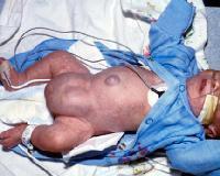
Internal supravesical hernia - a rare cause of intestinal obstruction: report of two cases
 Case report Open Access Internal supravesical hernia - a rare cause of intestinal obstruction: report of two cases Mahdi Bouassida 1,&, Selim Sassi 1, Hassen Touinsi 1, Helmi Kallel 1, Mohamed Mongi Mighri
The Abdomen. Surface Anatomy, Vessels, Muscles, and Peritoneum
 The Abdomen Surface Anatomy, Vessels, Muscles, and Peritoneum Surface Anatomy Anterior abdominal wall extends from costal margin to inferior boundaries: Iliac crest Anterior superior iliac spine Inguinal
2. List the 8 pelvic spaces: list one procedure or dissection which involves entering that space.
 Name: Anatomy Quiz: Pre / Post 1. In making a pfannensteil incision you would traverse through the following layers: a) Skin, Camper s fascia, Scarpa s fascia, external oblique aponeurosis, internal oblique
CHAPTER 2 Terms Pertaining to the Body as a Whole
 CHAPTER 2 Terms Pertaining to the Body as a Whole OBJECTIVES 1. Define terms that apply to the structural organization of the body. 2. Identify the body cavities and the organs contained within the cavities.
thoracic cage inlet and outlet landmarks of the anterior chest wall muscles of the thoracic wall sternum joints ribs intercostal spaces diaphragm
 Thoracic Wall Lecture Objectives Describe the shape and outline of the thoracic cage including inlet and outlet. Describe the anatomical landmarks of the anterior chest wall. List various structures making
Technique Guide. Bard MK Hernia Repair. Featuring Modified Onflex Mesh SOFT TISSUE REPAIR. Anterior Approach to a Preperitoneal Inguinal Hernia Repair
 Bard MK Hernia Repair Featuring Modified Onflex Mesh Technique Guide Anterior Approach to a Preperitoneal Inguinal Hernia Repair SOFT TISSUE REPAIR Right Procedure. Right Product. Right Outcome. The opinions
Hernias of the Abdominal Wall:
 Hernias of the Abdominal Wall: Inguinal Anatomy in the Male Bob Caruthers. CST. PhD The surgical repair of an inguinal hernia, although one of the most common of surgical procedures, presents a special
Liver o The liver is the largest gland in the body and has a wide variety of functions. - It s an accessory organ of GIT
 بسم رلاهللا You don t need to refer to the slides, we included everything here In this lecture we will talk about Liver & Gallbladder Liver o The liver is the largest gland in the body and has a wide variety
ANATYOMY OF The thigh
 ANATYOMY OF The thigh 1- Lateral cutaneous nerve of the thigh Ι) Skin of the thigh Anterior view 2- Femoral branch of the genitofemoral nerve 5- Intermediate cutaneous nerve of the thigh 1, 2 and 3 are
THE ABDOMEN SUPRARENAL GLANDS KIDNEY URETERS URINARY BLADDER
 THE ABDOMEN SUPRARENAL GLANDS KIDNEY URETERS URINARY BLADDER THE SUPRARENAL GLANDS The suprarenal (adrenal) glands lie immediately superior and slightly anterior to the upper pole of either kidney. Golden
Test Bank for Essentials of Anatomy and Physiology 6th edition by Valerie C. Scanlon and Tina Sanders
 Test Bank for Essentials of Anatomy and Physiology 6th edition by Valerie C. Scanlon and Tina Sanders Link download full: https://digitalcontentmarket.org/download/test-bank-foressentials-of-anatomy-and-physiology-6th-edition-by-scanlon-and-sanders/
Dr.Susan Maskel. BIO 105 Western Connecticut State University A&P I BIO 211 Naugatuck Valley Community College A&P I.
 1 Please wait 20 seconds before starting slide show. Mouse click to advance. Arrow keys etc.also work. Dr.Susan Maskel BIO 105 Western Connecticut State University A&P I BIO 211 Naugatuck Valley Community
JlntSocPlastination, Vol4:16-22,
 JlntSocPlastination, Vol4:16-22, 1990 16 SECTIONAL ANATOMY: STANDARDIZED METHODOLOGY Alexander Lane, Coordinator of Anatomy and Physiology, Triton College, Visiting Associate Professor, University of Illinois
Variations in Course of Inferior Epigastric Artery- Importance in Laproscopic Surgery
 Quest Journals Journal of Medical and Dental Science Research Volume 3~ Issue 9 (2016) pp: 47-52 ISSN(Online) : 2394-076X ISSN (Print):2394-0751 www.questjournals.org Research Paper Variations in Course