ANATOMY OF THE SMALL & LARGE INTESTINES. Semester 1, 2011 A. Mwakikunga
|
|
|
- Peter Payne
- 5 years ago
- Views:
Transcription
1 ANATOMY OF THE SMALL & LARGE INTESTINES Semester 1, 2011 A. Mwakikunga
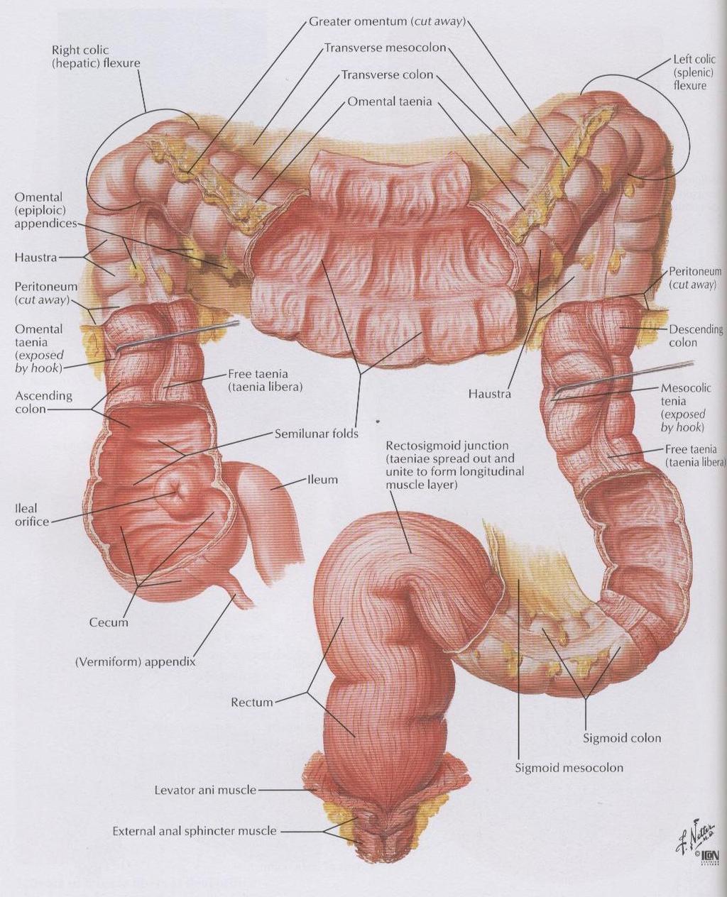
2 LEARNING OBJECTIVES 1. List the parts and anatomical regions of the small and large intestines 2. State anatomical relations of the small and large intestines 3. Mention the arterial supply, venous and lymphatic drainage and innervation of the small and large intestines 4. Discuss the importance of the mesenteries that are related to the small and large intestines 5. Relate the significance of McBurney s point to the anatomical position of the vermiform appendix in clinical practice 6. Describe the clinical relevance of the ligament of Treitz 7. Explain the following clinical terms: ischemia of intestines, appendicitis, hemorrhoids, rectal examination

3 Small intestine Divided into duodenum jejunum ileum
4 DUODENUM C-shaped Extent : pyloric sphincter - duodenojejunal flexure Mostly Retroperitoneal except near stomach Characterized by Brunner s glands in submucosa These tubular glands secrete mucus Primary function - food digestion & absorption
5 DUODENUM Four anatomical parts of the duodenum Superior (first) part Related to the pylorus Intraperitoneal - found in the hepatoduodenal lig - freely mobile Posteriorly - bile duct, gastrodudenal art, portal vein
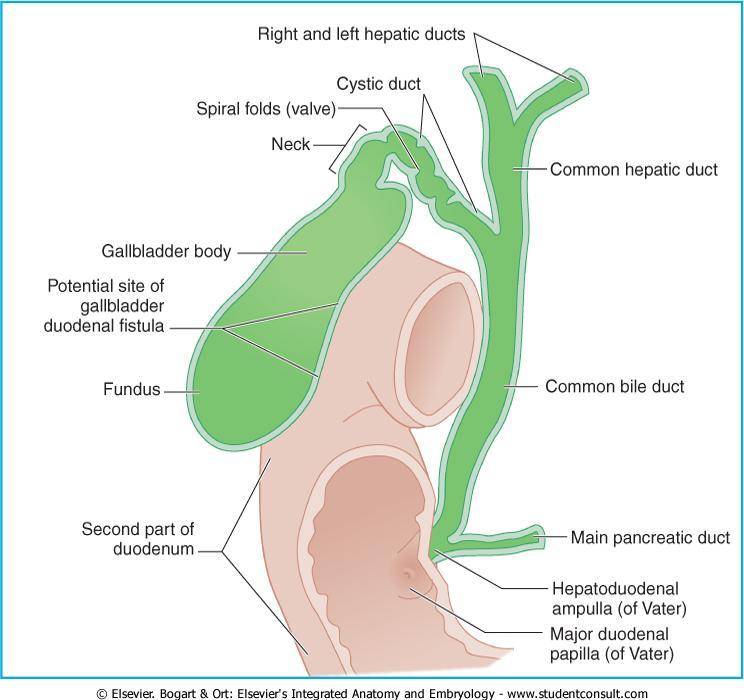
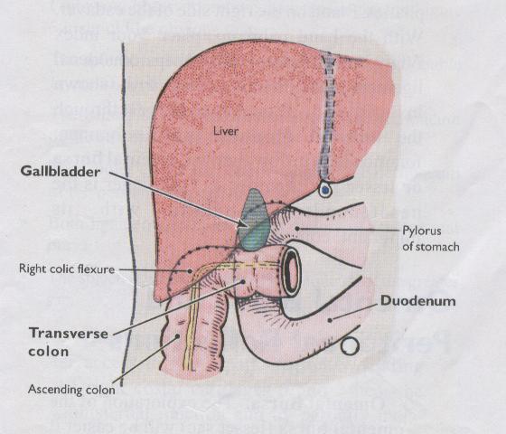
6 DUODENUM CONTD Descending (second) part Retroperitoneal Bile duct and main pancreatic duct join to form hepatopancreatic ampulla Major duodenal papilla (of Vater) located posteromedial Also related to the Fundus & body of gallbladder, right kidney, transverse colon, head of pancreas
7
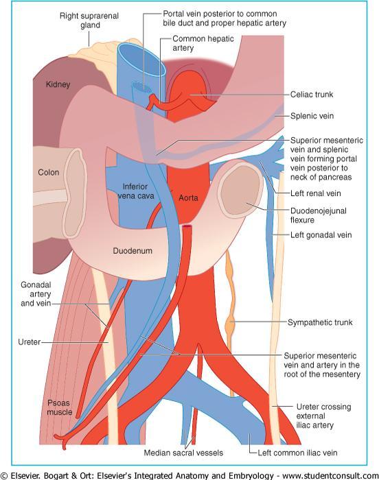
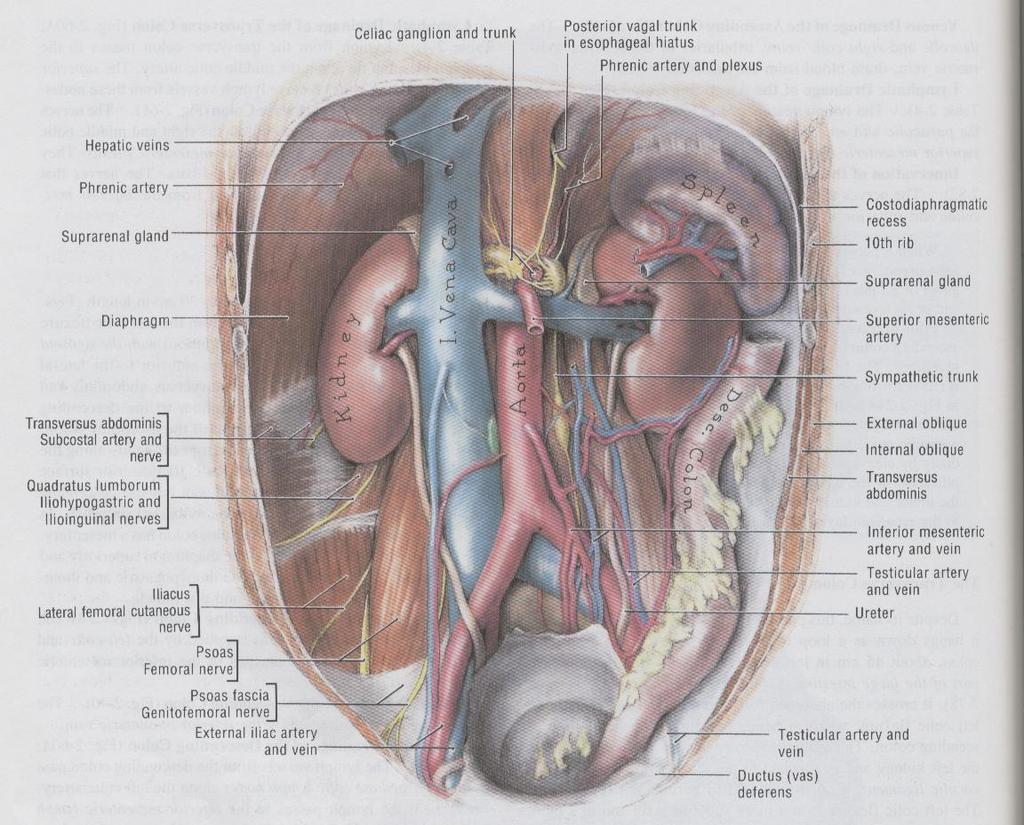
8 DUODENUM CONTD Horizontal (third) part Retroperitoneal Related to the: Psoas major muscle IVC & aorta Right ureter Gonadal vessels Superior mesenteric art & vein
9 DUODENUM CONTD Ascending (fourth) part Retroperitoneal Duodenojejunal flexure supported by suspensory ligament of duodenum (of Treitz) Lig of Treitz used to locate duodenojejunal flexure Clinical dividing line between upper and lowergastrointestinal tracts Most gastrointestinal hemorrhage is above the ligament of Treitz, coming from esophagus, stomach or duodenum Doudenal ulcers may cause peritonitis
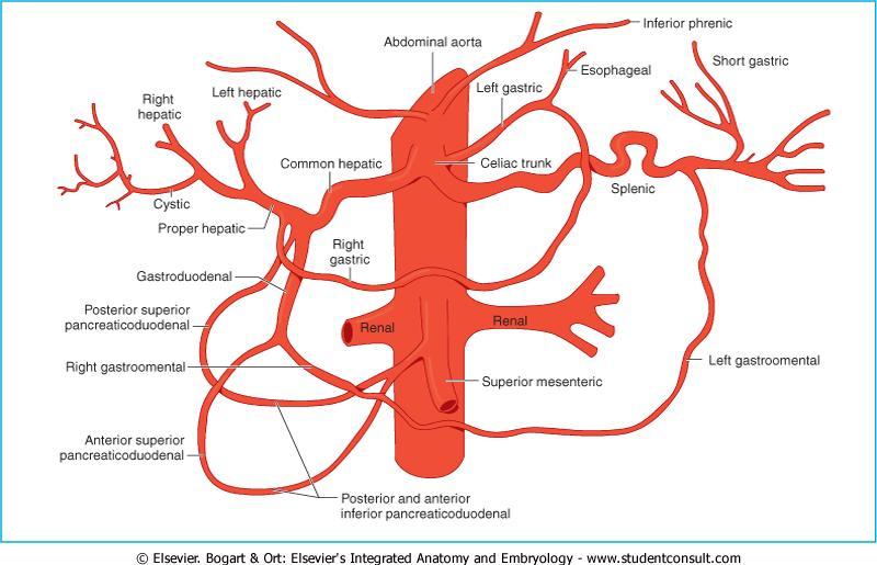
10 BLOOD & NERVE SUPPLY TO THE Arterial Supply DUODENUM Supraduodenal art - can be used to identify the first part of the duodenum sup & inf pancreaticoduodenal arts with their ant & post branches that form arcades Right gastric art Gastroduodenal art Venous Drainage Duodenal veins draining into the portal vein
11
12 BLOOD & NERVE SUPPLY TO THE Lymphatic Drainage DUODENUM Ant lymphatic vessels drain into pancreaticoduodenal nodes, pyloric lymph nodes Post lymphatic vessels drain into sup mesenteric lymph nodes Celiac lymph nodes Innervation Vagus nerve Celiac & sup mesenteric plexuses

13 JEJUNUM Extent: duodenum - ileum Intraperitoneal Most of it lies in Left upper quadrant Lumen slightly larger than ileum More internal folds than ileum - plicae circulare & villi Deeper red with greater vascularity
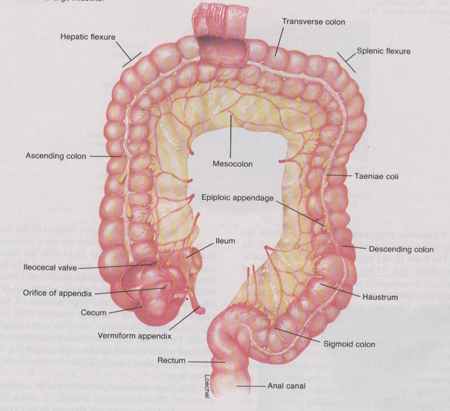
14 ILEUM Terminal portion Most of it lies in Right lower quadrant Ends at ileocecal junction Joins cecum medially through ileocecal valve Have shorter straight arteries than jejunum Has abundant lymph nodules - Peyer s patches or Mesenteric patches - GALT
15 MESENTERY ASSOCIATED WITH JEJUNUM & ILEUM Jejunum & ileum attached to posterior abdominal wall by mesentery Extent of the root (origin) of mesentery duodenojejunal junction - ileocolic junction The root of mesentery crosses Ascending & horizontal duodenum Abdominal aorta & IVC Right ureter, Right psoas major Right testicular or ovarian vessels
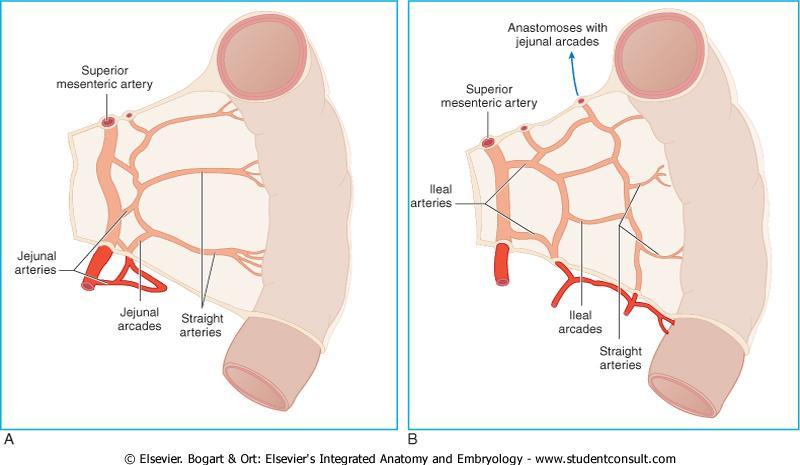
16 BLOOD & NERVE SUPPLY TO THE Arterial supply JEJUNUM & ILEUM Sup mesenteric art - arterial arcades that give rise to vasa recta - ischemia of intestines
17
18 BLOOD & NERVE SUPPLY TO THE Venous Drainage JEJUNUM & ILEUM Sup mesenteric vein Lymphatic Drainage Lacteals, sup mesenteric lymph nodes, ileocolic lymph nodes Innervation Parasympathetic - Vagus nerve Sympathetic - Lesser splanchnic nerve Superior mesenteric plexus
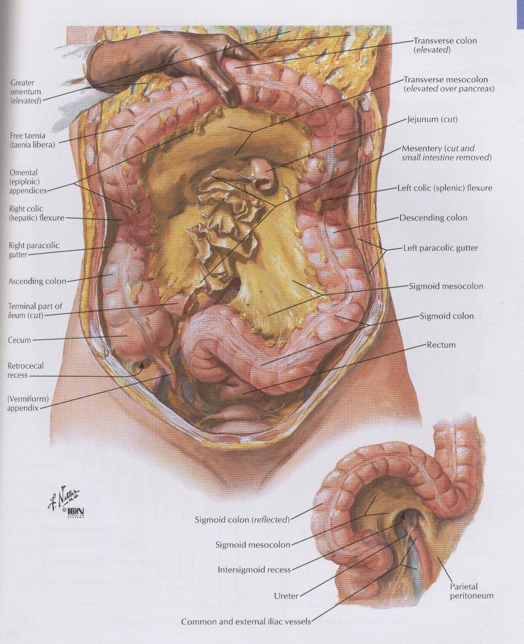
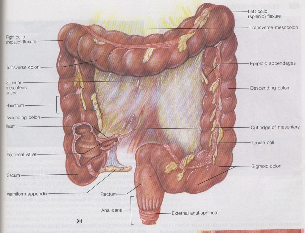
19 LARGE INTESTINE OR COLON Extent: Ileocecal junction - anus Length: About 1.5 m Parts: Cecum with attached vermiform appendix Colon: ascending, transverse, descending, sigmoid Rectum and anal canal
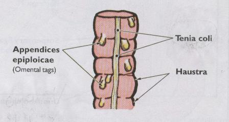
20 SPECIAL FEATURES Teniae coli - three thickened bands of longitudinal muscle Haustra - sacculations of its wall Omental (epiploic) appendages - small pouches of omentum (peritoneum) filled with fat Villi - Mucosa has no villi but numerous mucus cells
21
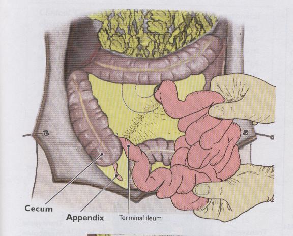
22 CECUM Blind sac invested in peritoneum 8 cm W x 8 cm L, located in RIF Vermiform appendix attached to posteromedial wall & taenia coli converge on appendix
23
24 CECUM CONTD Lat & med - attached by peritoneal cecal folds to iliac fossa This produces a small sac of peritoneal cavity called retrocecal recess Retrocecal recess lies post to cecum It may extend sup, post to inferior end of ascending colon as retrocolic recess In 64% of people - appendix lies in retrocolic recess
25 RELATIONS OF THE CECUM Post - lies on iliacus & psoas Ant - small intestines & ant abdominal wall
26 VERMIFORM APPENDIX About 8 cm long & worm-shaped Joins cecum about 2.5 cm inf to ileocecal junction Longer in children than in adults Very mobile & its position is variable
27 VERMIFORM APPENDIX CONTD Has a mesentery called mesoappendix which joins it to terminal ileum Appendicular artery (an end artery) is within this fold Usually retrocecal - post to cecum The 3 teniae coli of cecum converge at base of appendix

28 VERMIFORM APPENDIX
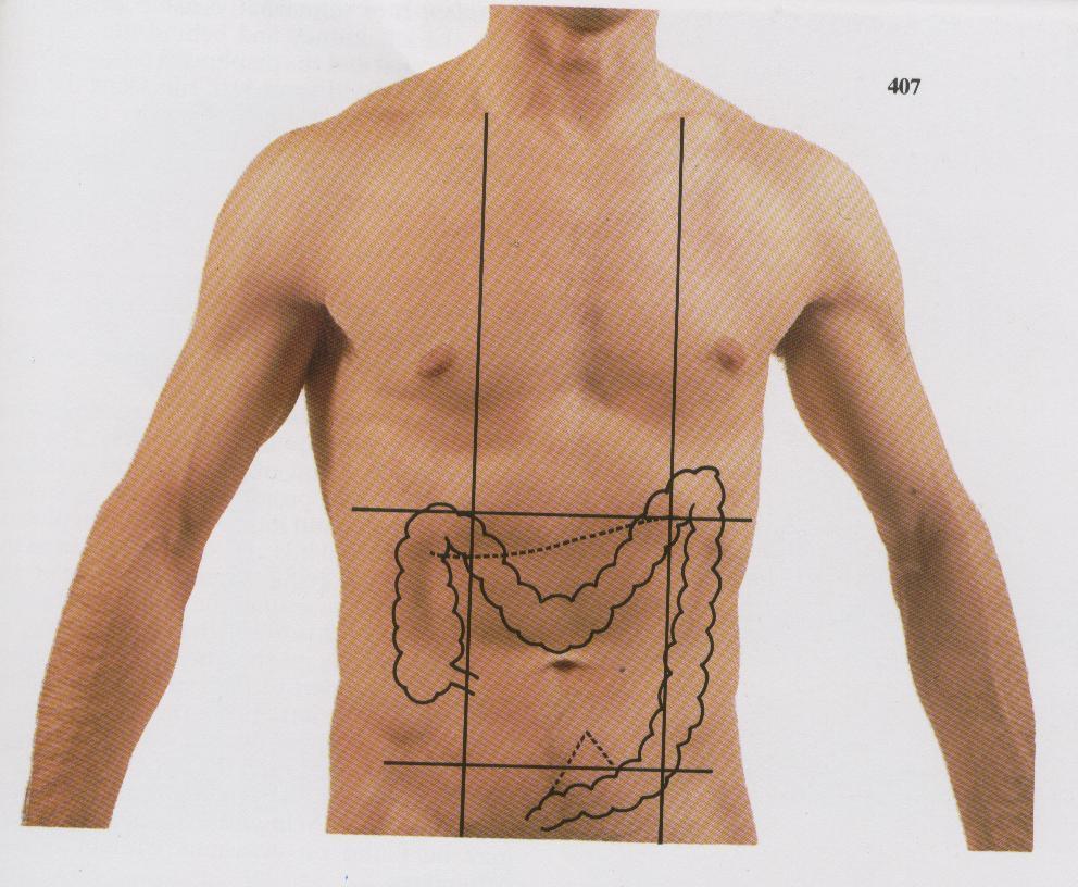
29 VERMIFORM APPENDIX CONTD Its base usually lies deep at Mc Burney s point - junction of lat & mid thirds of line joining ant sup iliac spine & umbilicus In appendectomy, incisions are made about 2.5 cm superomedial to ant. sup. iliac spine Appendicitis - inflammation of appendix Rupture of inflamed appendix causes general peritonitis - increased abdominal pain
30
31 ASCENDING COLON About 15 cm long Ascends on right side of abdominal cavity From cecum to right lobe of liver It turns to the left as right colic (hepatic) flexure It usually has no mesentery - 25% of pple have a short mesentery Lies retroperitoneally along right side of posterior abdominal wall
32
33 ASCENDING COLON - CONTD Posterior relations Back muscles: iliacus & quadratus lumborum; right kidney Nerves of posterior abdominal wall: ilioinguinal & iliohypogastric Anterior relations - small intestines & greater omentum Laterally - covered by peritonium, which attaches it to posterior abdominal wall Its peritonium forms a trench or groove called right paracolic gutter
34
35
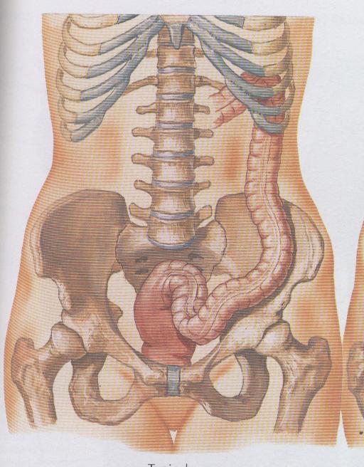
36 ASCENDING COLON - CONTD VOLVULUS Abnormal mobility of cecum & proximal part of ascending colon because inferior part of ascending colon has a mesentery It may cause obstruction of intestines resulting from twisting CECOPEXY - Anchoring procedure where tenia colia of cecum & ascending colon are sutured to abdominal wall to avoid volvulus
37 TRANSVERSE COLON Length: About 50 cm Extent: Right colic to left colic flexures Largest & most mobile part of L intestine Splenic flexure: lies ant. to the inferior part of left kidney & is attached to the diaphragm by phrenicocolic ligament - shelf to support spleen is more sup. & post. to right colic flexure & more acute & less mobile than hepatic flexure It has a mesentery called transverse mesocolon Mesocolon suspends transverse colon from posterior abdominal wall T. colon is variable in position
38
39
40 DESCENDING COLON Length: About 30 cm Extent: Left colic (splenic) flexure into left iliac fossa where it is continuous with sigmoid colon Caliber - smaller than ascending colon Relations: Sup - related to diaphragm & quadratus lumborum muscles Passes ant. to lat. border of left kidney, transversus abdominis & quadratus lumborum muscles
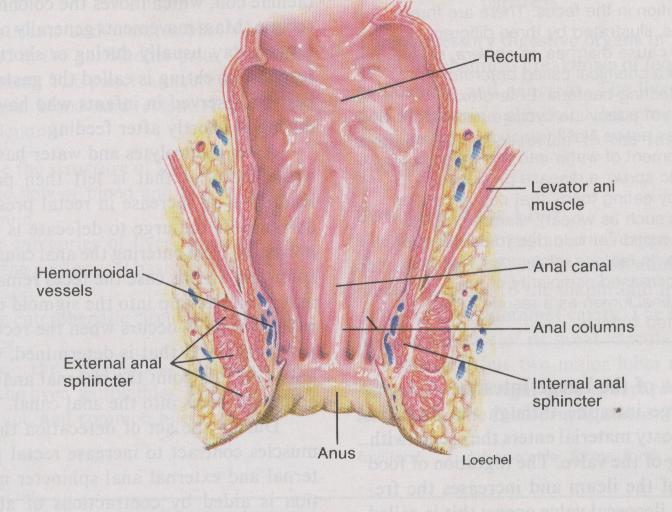
41 DESCENDING COLON CONTD It usually has no mesentery (33% of pple have) Has left paracolic gutter on lat aspect
42
43 SIGMOID COLON Length: About 40 cm Extent: Btwn descending colon & rectum Forms S-shaped loop - Greek letter sigma (S) It is also called pelvic colon Termination of teniae coli indicates beginning of rectum Has a long mesentery - sigmoid mesocolon whose root has an inverted V - shaped attachment sup
44
45 SIGMOID COLON CONTD Left ureter & a division of left common iliac artery are post to the apex of the mesentery It occupies rectovesicle pouch in males & rectouterine pouch in females Has long omental appendages Rectosigmoid junction is about 15 cm from anus Post - Left external iliac vessels & piriformis muscle

46 SIGMOID COLON CONTD
47
48 RECTUM 12 cm long Sup - continuous with sigmoid colon at the level of S3 - rectosigmoid junction Inf - continuous with anal canal
49 PERITONEAL RELATIONS Sup 1/3 ant & lat peritoneal cover Mid 1/3 ant peritoneal cover Inf 1/3 no peritoneal cover Rectal examination - prostate gland
50
51 ANAL CANAL Terminal part of GI tract - 4 cm long Anus - external opening of anal canal Two sphincters guard the anus External anal sphincter - composed of skeletal muscle Internal anal sphincter - composed of smooth muscle fibers Has anal columns & anal valves with anastomoses; anal sinuses Hemorrhoids - veins in anal area
52
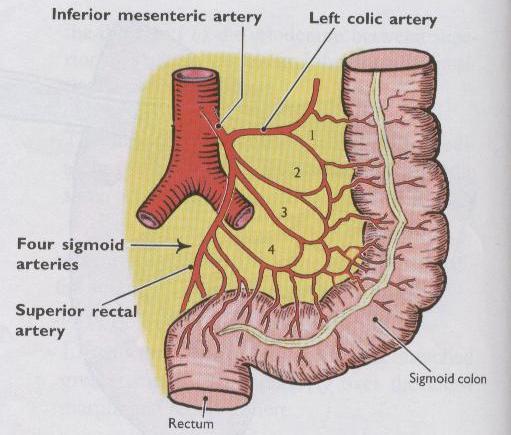
53 BLOOD & NERVE SUPPLY TO THE Arterial supply Branches of SMA LARGE INTESTINE Middle colic art with its branches - R & L Right colic with its branches - ascending & descending Ileocolic with its branches - colic and ileal Branches of IMA Left colic art Sigmoid art
54 BLOOD & NERVE SUPPLY TO THE LARGE INTESTINE Venous Drainage Superior mesenteric vein Inferior mesenteric vein Splenic vein Portal vein Lymphatic Drainage Follow arteries Preaortic lymph nodes
55 BLOOD & NERVE SUPPLY TO THE LARGE INTESTINE Innervation Parasympathetic Vagus nerve Pelvic splanchnic nerves Sympathetic Superior mesenteric & aorticorenal ganglia Lesser splanchnic nerves Inferior mesenteric ganglia Lumbar splanchnic nerves
Anatomy of the Large Intestine
 Large intestine Anatomy of the Large Intestine 2 Large Intestine Extends from ileocecal valve to anus Length = 1.5-2.5m = 5 feet Regions Cecum = 2.5-3 inch Appendix= 3-5 inch Colon Ascending= 5 inch Transverse=
Dr. Zahiri. In the name of God
 Dr. Zahiri In the name of God small intestine = small bowel is the part of the gastrointestinal tract Boundaries: Pylorus Ileosecal junction Function: digestion and absorption of food It receives bile
Bushra Arafa Zayed & Hanan Jamal. - Dana AF
 - 10 - Bushra Arafa Zayed & Hanan Jamal - Dana AF - Mohammad Al Muhtaseb Notes: This sheet was written in the same order as the slides, and everything in the slides is mentioned in this sheet. Pictures
Anatomy of the SMALL INTESTINE. Dr. Noman Ullah Wazir PMC
 Anatomy of the SMALL INTESTINE Dr. Noman Ullah Wazir PMC SMALL INTESTINE The small intestine, consists of the duodenum, jejunum, and illium. It extends from the pylorus to the ileocecal junction were the
Small Plicae Circularis. Short Closely packed together. Sparse, completely absent at distal part Lymphoid Nodule
 Intestines Differences Between Jejunum and Ileum Types Jejunum Ileum Color Deeper red Paler pink Calibre Bigger Smaller Thickness of wall Thick and Heavy Thin and Lighter Vascularity Highly vascularised
THE ORAL CAVITY
 THE ORAL CAVITY WALL OF ABDOMEN (ANTERIOR) The paraumbilical vein drains into the portal vein and then through the liver. This is an important clinical connection. THE ABDOMINAL VISCERA The small
Duodenum retroperitoneal
 Duodenum retroperitoneal C shaped Initial region out of stomach into small intestine RETROperitoneal viscus Superior 1 st part duodenal cap ; moves upwards and backwards to lie on the R crura medial to
BLOCK IV: OFFICIAL BODY PARTS LIST FOR ANTERIOR ABDOMINAL WALL AND ABDOMINAL CONTENTS
 BLOCK IV: OFFICIAL BODY PARTS LIST FOR ANTERIOR ABDOMINAL WALL AND ABDOMINAL CONTENTS External oblique muscle Muscular portion Aponeurotic portion Superficial inguinal ring Lateral (inferior) crus Medial
The abdominal Esophagus, Stomach and the Duodenum. Prof. Oluwadiya KS
 The abdominal Esophagus, Stomach and the Duodenum Prof. Oluwadiya KS www.oluwadiya.com Viscera of the abdomen Abdominal esophagus: Terminal part of the esophagus The stomach Intestines: Small and Large
Block 3: DISSECTION 2 CELIAC TRUNK, JEJUNUM/ILEUM, LARGE INTESTINE, DUODENUM, PANCREAS, PORTAL VEIN; MOBILIZATION OF THE LIVER
 1 Block 3: DISSECTION 2 CELIAC TRUNK, JEJUNUM/ILEUM, LARGE INTESTINE, DUODENUM, PANCREAS, PORTAL VEIN; MOBILIZATION OF THE LIVER Attempt to complete as much as you can of the dissection explained in the
د. عصام طارق. Objectives:
 GI anatomy Lecture: 5 د. عصام طارق Objectives: To describe anatomy of stomach, duodenum & pancreas. To list their main relations. To define their blood & nerve supply. To list their lymph drainage. To
Preview from Notesale.co.uk Page 1 of 34
 Abdominal viscera and digestive tract Digestive tract Abdominal viscera comprise majority of the alimentary system o Terminal oesophagus, stomach, pancreas, spleen, liver, gallbladder, kidneys, suprarenal
Al-Mohtaseb. Saba Alfayoumi. Mo Alfarra
 8 Al-Mohtaseb Saba Alfayoumi Mo Alfarra For the comparison purposes refer to the last page where you can find a table that summarizes them. Enjoy Jejunum and Ileum -They're intraperitoneal and freely mobile
The jejunum and the Ileum. Prof. Oluwadiya KS
 The jejunum and the Ileum Prof. Oluwadiya KS www.oluwadiya.siteled.com Introduction Introduction The small intestine (SI) comprises of the duodenum, jejunum and the ileum The jejunum is the second part
-Ensherah Mokheemer. -Shatha Al-Jaberi محمد المحتسب- 1 P a g e
 9-9 -Ensherah Mokheemer -Shatha Al-Jaberi محمد المحتسب- 1 P a g e Small intestine has three regions: ( االثني عشر( The duodenum The jejunum The ileum Small intestine Duodenum: -c-shaped -The concavity
Pancreas & Biliary System. Dr. Vohra & Dr. Jamila
 Pancreas & Biliary System Dr. Vohra & Dr. Jamila 1 Objectives At the end of the lecture, the student should be able to describe the: Location, surface anatomy, parts, relations & peritoneal reflection
Peritoneal cavity. Infracolic compartment. Assoc. prof. dr. S. Delchev, MD, PhD
 Peritoneal cavity. Infracolic compartment Assoc. prof. dr. S. Delchev, MD, PhD Infracolic compartment The infracolic compartment lies inferior to the transverse mesocolon and posterior to the greater omentum
Netter's Anatomy Flash Cards Section 4 List 4 th Edition
 Netter's Anatomy Flash Cards Section 4 List 4 th Edition https://www.memrise.com/course/1577335/ Section 4 Abdomen (31 cards) Plate 4-1 Bony Framework of Abdomen 1.1 Costal cartilages 1.2 Iliac crest 1.3
ABDOMEN - GI. Duodenum
 TALA SALEH ABDOMEN - GI Duodenum - Notice the shape of the duodenum, it looks like capital G shape tube which extends from the pyloroduodenal junction to the duodenojejunal junction. - It is 10 inches
Mousa Salah. Dr. Mohammad Al. Mohtasib. 1 P a g e
 8 Mousa Salah Dr. Mohammad Al. Mohtasib 1 P a g e In the previous lecture we talked about the peritoneum, and we said that the peritonium is a serous sac, and it consists of two layers, visceral and parietal.
To describe the liver. To list main structures in porta hepatis.
 GI anatomy Lecture: 6 د. عصام طارق Objectives: To describe the liver. To list main structures in porta hepatis. To define portal system & portosystemic anastomosis. To list parts of biliary system. To
The peritoneum. Prof. Oluwadiya KS, MBBS, FMCS(Orthop) Website:
 Website:") The peritoneum Prof. Oluwadiya KS, MBBS, FMCS(Orthop) Website: http://oluwadiya.com The peritoneum Serous membrane that lines the abdominopelvic cavity and invests the viscera The largest serous membrane
The peritoneum Prof. Oluwadiya KS, MBBS, FMCS(Orthop) Website: http://oluwadiya.com The peritoneum Serous membrane that lines the abdominopelvic cavity and invests the viscera The largest serous membrane
Lab Monitor Images Dissection of the Abdominal Vasculature + Lower Digestive System
 Lab Monitor Images Dissection of the Abdominal Vasculature + Lower Digestive System Stomach & Duodenum Frontal (AP) View Nasogastric tube 2 1 3 4 Stomach Pylorus Duodenum 1 Duodenum 2 Duodenum 3 Duodenum
GASTROINTESTINAL SYSTEM
 GASTROINTESTINAL SYSTEM Topographic Anatomy of the Abdomen Surface Landmarks Xiphoid process T9/T10 Inferior costal margin L2/L3 Iliac Crest L4 level ASIS L5/S1 level Pubic symphysis level of greater trochanter
Biology Human Anatomy Abdominal and Pelvic Cavities
 Biology 351 - Human Anatomy Abdominal and Pelvic Cavities Please place your name and I.D. number on the back of the last page of this exam. You must answer all questions on this exam. Because statistics
The Digestive System Laboratory
 The Digestive System Laboratory 1 The Digestive Tract The alimentary canal is a continuous tube stretching from the mouth to the anus. Liver Gallbladder Small intestine Anus Parotid, sublingual, and submaxillary
Peritoneum: Def. : It is a thin serous membrane that lines the walls of the abdominal and pelvic cavities and clothes the viscera.
 Peritoneum: Def. : It is a thin serous membrane that lines the walls of the abdominal and pelvic cavities and clothes the viscera. Layers of the peritoneum: 1. Outer Layer ( Parietal Peritoneum) : lines
Nasogastric tube. Stomach. Pylorus. Duodenum 1. Duodenum 2. Duodenum 3. Duodenum 4
 Esophagus Barium Swallow Stomach and Duodenum 4 year old Upper GI Nasogastric tube Stomach and Duodenum 4 year old Upper GI Nasogastric tube Stomach Pylorus Duodenum 1 Duodenum 2 Duodenum 3 Duodenum 4
-12. -Renad Habahbeh. -Dr Mohammad mohtasib
 -12 -Renad Habahbeh - -Dr Mohammad mohtasib The Gallbladder -The gallbladder has a body, a fundus (a rounded end), a neck, Hartmann s pouch before the neck and a cystic duct that meets the common hepatic
SUBJECTS 2nd year, 1st semester I. 1. Primitive gut - limits, derivatives 2. Foregut -limits, evolution, derivatives 3. Midgut -limits, evolution,
 SUBJECTS 2nd year, 1st semester I. 1. Primitive gut - limits, derivatives 2. Foregut -limits, evolution, derivatives 3. Midgut -limits, evolution, derivatives 4. Hindgut- limits, evolution, derivatives
Pancreas and Biliary System
 Pancreas and Biliary System Please view our Editing File before studying this lecture to check for any changes. Color Code Important Doctors Notes Notes/Extra explanation Objectives At the end of the lecture,
The Digestive System and Body Metabolism
 14 PART B The Digestive System and Body Metabolism PowerPoint Lecture Slide Presentation by Jerry L. Cook, Sam Houston University ESSENTIALS OF HUMAN ANATOMY & PHYSIOLOGY EIGHTH EDITION ELAINE N. MARIEB
ANATOMY OF THE DIGESTIVE SYSTEM PART II
 ANATOMY OF THE DIGESTIVE SYSTEM PART II 9.12.2014 Kaan Yücel M.D., Ph.D. http://fhs121.org Dr.Kaan Yücel http://fhs121.org Digestive system Part II 1. LIVER The liver is the largest gland in the body and,
Exploring Anatomy: the Human Abdomen
 Exploring Anatomy: the Human Abdomen PERITONEUM AND PERITONEAL CAVITY PERITONEUM The peritoneum is a thin serous membrane that lines the abdominal cavity and covers, in variable amounts, the viscera within
GI module Lecture: 9 د. عصام طارق. Objectives:
 GI module Lecture: 9 د. عصام طارق Objectives: To list structures forming posterior abdominal wall. To follow aorta & its main branches. To describe IVC & its main tributaries. To list nerves of posterior
Anatomy: Know Your Abdomen
 Anatomy: Know Your Abdomen Glossary Abdomen - part of the body below the thorax (chest cavity); separated by the diaphragm. Anterior - towards the front of the body. For example, the umbilicus is anterior
Surface Anatomy. Location Shape Weight Role of Five Surfaces Borders Fissures Lobes Peritoneal Lig
 The Liver Functions Bile production and secretion Detoxification Storage of glycogen Protein synthesis Production of heparin and bile pigments Erythropoiesis (in fetus) Surface Anatomy Location Shape Weight
Jhia Anjela D. Rivera 1 1. BS Biology, Department of Biology, College of Science, Polytechnic University of the Philippines
 DIGESTIVE SYSTEM Jhia Anjela D. Rivera 1 1 BS Biology, Department of Biology, College of Science, Polytechnic University of the Philippines DIGESTIVE SYSTEM Consists of the digestive tract (gastrointestinal
- Digestion occurs during periods of low activity - Produces more energy than it uses. - Mucosa
 Introduction Digestive System Chapter 29 Provides processes to break down molecules into a state easily used by cells - A disassembly line: Starts at the mouth and ends at the anus Digestive functions
Midterm 2 is Tuesday 5/28/13
 Business Reminder: No class Monday (Memorial Day) Midterm 2 is Tuesday 5/28/13 Optional review session tomorrow @ 5pm Homework due in Lab 1. PreLab 8 (1pt) 2. Replace a Missing Assignment (4 pts) Homework
The posterior abdominal wall. Prof. Oluwadiya KS
 The posterior abdominal wall Prof. Oluwadiya KS www.oluwadiya.sitesled.com Posterior Abdominal Wall Lumbar vertebrae and discs. Muscles opsoas, quadratus lumborum, iliacus, transverse, abdominal wall
Accessory Glands of Digestive System
 Accessory Glands of Digestive System The liver The liver is soft and pliable and occupies the upper part of the abdominal cavity just beneath the diaphragm. The greater part of the liver is situated under
ACTIVITY 11: RESPIRATORY AND DIGESTIVE SYSTEMS RESPIRATORY SYSTEM
 ACTIVITY 11: RESPIRATORY AND DIGESTIVE SYSTEMS OBJECTIVES: 1) How to get ready: Read Chapters 25 and 26, McKinley et al., Human Anatomy, 4e. All text references are for this textbook. 2) Identify structures
1 Right & left Hepatic ducts Gastric Impression of spleen
 Pancreatic Model 1 Right & left Hepatic ducts 14 Gastric Impression of spleen 2 Common hepatic duct 15 Renal Impression of spleen 3 Cystic Duct 16 Colic Impression of spleen 4 Common Bile Duct 17 Splenic
RESPIRATORY SYSTEM. described: pp. 744,746 fig. 25.1, described: p. 746 fig described: p. 776 fig. 26.3
 ACTIVITY 11: RESPIRATORY AND DIGESTIVE SYSTEMS OBJECTIVES: 1) How to get ready: Read Chapters 25 and 26, McKinley et al., Human Anatomy, 5e. All text references are for this textbook. 2) Identify structures
-the stones will obstruct the common bile duct and it might also be precancerous. -so the best treatment is chlolycyctoctomy.
 At the beginning this sheet includes the rest of last lecture s slides liver +gallbladder and the new lecture posterior abdominal wall and its vessels. We will start talking about Cholelithiasis -it means
Anatomical Considerations for Lab Practical II
 Anatomical Considerations for Lab Practical II For each of the following please be prepared to provide: Identification System Organ(s) or ducts to Function(s) location which it is attached Use your lecture
LECTURE 11 & 12: ABDOMINAL VISCERA ABDOMINAL CONTENTS DIVISION. The location of abdominal viscera is divided into 4 quadrants:
 LECTURE 11 & 12: ABDOMINAL VISCERA ABDOMINAL CONTENTS DIVISION The location of abdominal viscera is divided into 4 quadrants: - horizontal line across the umbilicus divides the upper quadrants from the
- Digestion occurs during periods of low activity - Produces more energy than it uses. 3 Copyright 2016 by Elsevier Inc. All rights reserved.
 Introduction Digestive System Chapter 29 Provides processes to break down molecules into a state easily used by cells - A disassembly line: Starts at the mouth and ends at the anus Digestive functions
This lab activity is aligned with Visible Body s Human Anatomy Atlas app. Learn more at visiblebody.com/professors
 1 This lab activity is aligned with Visible Body s Human Anatomy Atlas app. Learn more at visiblebody.com/professors 2 A. Digestive System Overview To Start: Go to the Views menu and scroll down to the
Lab 5 Digestion and Hormones of Digestion. 7/16/2015 MDufilho 1
 Lab 5 Digestion and Hormones of Digestion 1 Figure 23.1 Alimentary canal and related accessory digestive organs. Mouth (oral cavity) Tongue* Parotid gland Sublingual gland Submandibular gland Salivary
BY DR NOMAN ULLAH WAZIR
 BY DR NOMAN ULLAH WAZIR The stomach (from ancient Greek word stomachos, stoma means mouth) is a muscular, hollow and the most dilated part of the GIT. It starts from the point where esophagus ends. It
Gastrointestinal Tract. Anatomy of GI Tract. Anatomy of GI Tract. (Effective February 2007) (1%-5%)
 (1%-5%)") Gastrointestinal Tract (Effective February 2007) (1%-5%) Anatomy of GI Tract Esophagus bulls-eye or target EG junction seen on sagittal scan posterior to left lobe of liver and anterior to aorta Anatomy
Gastrointestinal Tract (Effective February 2007) (1%-5%) Anatomy of GI Tract Esophagus bulls-eye or target EG junction seen on sagittal scan posterior to left lobe of liver and anterior to aorta Anatomy
Embryology of the Midgut and Hind gut
 Embryology of the Midgut and Hind gut Prof. Abdulameer Al-Nuaimi E-mail: a.al-nuaimi@sheffield.ac.uk E-mail: abdulameerh@yahoo.com Abdominal organs www.google.co.uk/search? Development of Duodenum The
Abdomen. Retroperitoneal space
 Abdomen. Retroperitoneal space Abdominal cavity The space bounded by: Anterolateral abdominal wall Posterior abdominal wall Diaphragm Pelvic walls and pelvic floor. Subdivided into: True abdominal cavity
In the name ofgod. Abdomen 3. Dr. Zahiri
 In the name ofgod Abdomen 3 Dr. Zahiri Peritoneum Peritoneum It is the serous membrane(a type of loose connective tissue and is covered by mesothelium) that lines the abdominal cavity. Extensions of the
ABDOMEN. 2. The highest branch of the abdominal aorta is: (a) R suprarenal a (b) Coeliac trunk (c) L renal a (d) L gonadal a (e) SMA
 R suprarenal a (b) Coeliac trunk (c) L renal a (d) L gonadal a (e) SMA") ABDOMEN 1. The duodenum: (a) is a retroperitoneal structure (b) is 25cm long (c) lies between the levels of L2-L4 (d) in its fourth part lies to the R of the aorta (e) all of the above 2. The highest branch
ABDOMEN 1. The duodenum: (a) is a retroperitoneal structure (b) is 25cm long (c) lies between the levels of L2-L4 (d) in its fourth part lies to the R of the aorta (e) all of the above 2. The highest branch
Done by: Dina Sawadha & Mohammad Abukabeer
 Done by: Dina Sawadha & Mohammad Abukabeer The stomach *the stomach is a dilated part of the gastro intestinal tract, it's "J" shape. *the lower surface of the stomach ( the greater curvature ) reaches
Posterior Abdominal wall-
 Structures of posterior abdominal wall: o Bony boundaries: 5 lumber vertebra and their intervertebral disc, iliac fossa and iliac crest. o Muscles: psoas major, quadrates lumborum, transversus abdominis,
SMALL INTESTINE LARGE INTESTINE RECTUM ABDOMINAL AORTA INFERIOR VENA CAVA THE ABDOMEN
 SMALL INTESTINE LARGE INTESTINE RECTUM ABDOMINAL AORTA INFERIOR VENA CAVA THE ABDOMEN THE SMALL INTESTINE THE SMALL INTESTINE: Is the longest part of the gastrointesanal tract - length of 5 metres (3 7
The Spleen. Dr Fahad Ullah
 The Spleen BY Dr Fahad Ullah Spleen The spleen is an largest lymphoid organ shaped like a shoe that lies relative to the 9th and 11th ribs and is located in the left hypochondrium. Thus, the spleen is
Lab 9 Abdomen MUSCLES
 Lab 9 Abdomen MUSCLES External abdominal oblique continuous with the external intercostal muscle; its fibers point in a caudal direction as it moves anteriorly until it inserts on the linea alba via its
1. A stab wound into the abdomen transected the hepatoduodenal ligament. Each of the following structures would have been cut EXCEPT the:
 YR 1 GROSS ANATOMY UNIT EXAM 3 -- November 07, 1997. CHOOSE THE SINGLE BEST ANSWER FOR QUESTION 1-42. 1. A stab wound into the abdomen transected the hepatoduodenal ligament. Each of the following structures
Lab 8: Digestive System
 BIOL 221 A&P II Lab 8: Digestive System Become familiar with the gross anatomy of the digestive system (Exercise 38) using the models, Fig. 38.1 (Activity 1), and the rat. Recognize and know the functions
Omran Saeed. Mohammad Al-muhtaseb. 1 P a g e
 13 Omran Saeed Mohammad Al-muhtaseb 1 P a g e Posterior abdominal wall - The diaphragm separates between thoracic cavity and abdominal cavity. Structures of posterior abdominal wall: (below diaphragm)
Biology Human Anatomy Abdominal and Pelvic Cavities
 Biology 351 - Human Anatomy Abdominal and Pelvic Cavities You must answer all questions on this exam. Because statistics demonstrate that, on average, between 2-5 questions on every 100-point exam are
The stomach is formed of three parts: -
 The stomach is formed of three parts: - (a) CARDIAC STOMACH: - It receives the oesophagus through Cardiac aperture guarded by a cardiac sphincter which prevents regurgitation of food. (b) FUNDIC PART:
Digestive System. In one end and out the other.
 Digestive System In one end and out the other. Overview Every cell in the body needs nourishment, yet most cells cannot leave their position in the body and travel to a food source, so the food must be
Inferior Pelvic Border
 Pelvis + Perineum Pelvic Cavity Enclosed by bony, ligamentous and muscular wall Contains the urinary bladder, ureters, pelvic genital organs, rectum, blood vessels, lymphatics and nerves Pelvic inlet (superior
It passes through the diaphragm at the level of the 10th thoracic vertebra to join the stomach
 The esophagus is a tubular structure (muscular, collapsible tube ) about 10 in. (25 cm) long that is continuous above with the laryngeal part of the pharynx opposite the sixth cervical vertebra The esophagus
458 Essentials of Human Anatomy and Physiology
 458 Essentials of Human Anatomy and Physiology Visceral peritoneum Intrinsic nerve plexuses: Myenteric nerve plexus Submucosal nerve plexus Submucosal glands Mucosa: Surface epithelium Lamina propria Muscle
Exercise. Digestive System. Digestive system function. 1. Define the following terms: a. Chemical digestionb. Mechanical digestionc.
 Exercise 7 The Digestive System NAME: DATE: INSTRUCTOR: SECTION: Digestive system function 1. Define the following terms: a. Chemical digestionb. Mechanical digestionc. Ingestiond. Digestione. Absorptionf.
Midgut. Over its entire length the midgut is supplied by the superior mesenteric artery
 Gi Embryology 3 Midgut the midgut is suspended from the dorsal abdominal wall by a short mesentery and communicates with the yolk sac by way of the vitelline duct or yolk stalk Over its entire length the
Gastrointestinal System!
 Gastrointestinal System! Assoc. Prof. Prasit Suwannalert, Ph.D. (Email: prasit.suw@mahidol.ac.th)! Objectives: After learning, student should be able to describe and discuss in topics of! 1. Anatomical
DIGESTIVE SYSTEM. Chapter 25
 DIGESTIVE SYSTEM Chapter 25 DIGESTIVE SYSTEM Digestive Tract Mouth Pharynx Esophagus Stomach Small intestines Large intestines Anus Accessory Organs Teeth Tongue Salivary glands Pancreas Liver Gallbladder
STRUCTURAL BASIS OF MEDICAL PRACTICE EXAMINATION 3. October 16, 2015
 STRUCTURAL BASIS OF MEDICAL PRACTICE EXAMINATION 3 October 16, 2015 PART l. Answer in the space provided. (12 pts) 1. Identify the structures. (2 pts) A. B. A B C. D. C D 2. Identify the structures. (2
Bio 104 Digestive System
 13 Lecture Outline: Digestive System Hole s HAP [Chapters 17 & 18] General Characteristics of the Alimentary Canal A. Functions 1. Ingestion 2. Mechanical digestion 3. Chemical digestion 4. Propulsion
بسم االه الرحمن الرحيم
 MAY 3, 2012 [POSTERIOR ABDOMINAL WALL] LECTURE 26 ANATOMY Quick Revision: بسم االه الرحمن الرحيم Last time we started with the anterior abdominal wall and said that: 1. Diaphragm is the root of the abdomen.
The Digestive System
 The Digestive System Identify the Structure and Function. Mesentery of the Large Intestine The mesentery functions to connect the visceral organs to the abdominal wall. Identify the Structure. Nasal Cavity
The Whipple Operation Illustrations
 The Whipple Operation Illustrations Fig. 1. Illustration of the sixstep pancreaticoduodenectomy (Whipple operation) as described in a number of recent text books by Dr. Evans. The operation is divided
Anatomy of the renal system. Professor Nawfal K. Al-Hadithi
 Anatomy of the renal system Professor Nawfal K. Al-Hadithi Objectives To describe the posterior abdominal wall To identify the main anatomical landmarks of the kidneys & ureters To describe the suprarenal
STRUCTURAL BASIS OF MEDICAL PRACTICE EXAMINATION 3. October 17, 2014
 STRUCTURAL BASIS OF MEDICAL PRACTICE EXAMINATION 3 October 17, 2014 PART l. Answer in the space provided. (12 pts) 1. Identify the structures. (2 pts) A. B. A B C. D. C D 2. Identify the structures. (2
Dissection Lab Manuals: Required Content
 Dissection Lab Manuals: Required Content 1. Introduction a. Basic terminology (directions) b. External features of the cat c. Adaptations to predatory niche d. How to skin a cat e. How to make the incisions
GI Tract Lynn Ta Jennifer Zhang July 6, 2006 GI TRACT. 1) Other Names: Gastrointestinal tract Digestive tract Alimentary tract
 Other Names: Gastrointestinal tract Digestive tract Alimentary tract") GI Tract Lynn Ta Jennifer Zhang July 6, 2006 GI TRACT 1) Other Names: Gastrointestinal tract Digestive tract Alimentary tract 2) Definition/Location: Digestion and absorption are the primary functions
GI Tract Lynn Ta Jennifer Zhang July 6, 2006 GI TRACT 1) Other Names: Gastrointestinal tract Digestive tract Alimentary tract 2) Definition/Location: Digestion and absorption are the primary functions
consists of: Muscular, hollow tube (= digestive tract ) + Various accessory organs
 + Various accessory organs") DIGESTIVE SYSTEM consists of: Muscular, hollow tube (= digestive tract ) + Various accessory organs FUNCTION Individual parts function in: ingestion mechanical digestion chemical and enzymatic digestion
DIGESTIVE SYSTEM consists of: Muscular, hollow tube (= digestive tract ) + Various accessory organs FUNCTION Individual parts function in: ingestion mechanical digestion chemical and enzymatic digestion
Overview of the Digestive
 Overview of the Digestive System Bởi: OpenStaxCollege The function of the digestive system is to break down the foods you eat, release their nutrients, and absorb those nutrients into the body. Although
Lab activity manual - Histology of the digestive system. Lab activity 1: esophagus stomach - small intestines
 Lab activity manual - Histology of the digestive system Jeanne Adiwinata Pawitan Prerequisite: Histology of the 4 basic tissues In this module we learn about the histology of the digestive system, from
The Digestive System PowerPoint Lecture Presentations prepared by Steven Bassett Southeast Community College Lincoln, Nebraska
 25 The Digestive System PowerPoint Lecture Presentations prepared by Steven Bassett Southeast Community College Lincoln, Nebraska Introduction The digestive system consists of: The digestive tract Accessory
CEA (CARCINOEMBRYONIC ANTIGEN)
") (CARCINOEMBRYONIC ANTIGEN) 428 C15.3 Malignant neoplasm of upper third of esophagus C15.4 Malignant neoplasm of middle third of esophagus C15.5 Malignant neoplasm of lower third of esophagus C15.8 Malignant
(CARCINOEMBRYONIC ANTIGEN) 428 C15.3 Malignant neoplasm of upper third of esophagus C15.4 Malignant neoplasm of middle third of esophagus C15.5 Malignant neoplasm of lower third of esophagus C15.8 Malignant
Dr. Fawzy Elnady Professor of Anatomy and Embryology Faculty of Veterinary Medicine Cairo University
 Dr. Fawzy Elnady Professor of Anatomy and Embryology Faculty of Veterinary Medicine Cairo University Objectives By the end of this lecture you should be able to: 1. Understand the components and functions
ANATOMY OF PELVICAYCEAL SYSTEM -DR. RAHUL BEVARA
 1 ANATOMY OF PELVICAYCEAL SYSTEM -DR. RAHUL BEVARA 2 KIDNEY:ANATOMY OVERVIEW Kidneys are retroperitoneal, in posterior abdominal region, extending from T12 L3 Bean-shaped Right kidney is lower than left
GASTROINTESTINAL SYSTEM
 GASTROINTESTINAL SYSTEM I. Topographic Anatomy of the Abdomen A. Surface landmarks 1. Xiphoid process 2. Costal margin 3. Iliac crest 4. ASIS 5. Pubic symphysis 6. Inguinal groove B. Anterior abdominal
THE ABDOMEN SUPRARENAL GLANDS KIDNEY URETERS URINARY BLADDER
 THE ABDOMEN SUPRARENAL GLANDS KIDNEY URETERS URINARY BLADDER THE SUPRARENAL GLANDS The suprarenal (adrenal) glands lie immediately superior and slightly anterior to the upper pole of either kidney. Golden
Anatomy of the spleen. Oluwadiya KS
 Anatomy of the spleen Oluwadiya KS www.oluwadiya.com Introduction The spleen is an ovoid, usually purplish, pulpy mass about the size and shape of one's fist. It is the largest lymphoid tissue in the body
Lecture 02 Anatomy of the LIVER
 Lecture 02 Anatomy of the LIVER BY Dr Farooq Khan Aurakzai Dated: 02.01.2018 Introduction to Liver Largest gland in the body. 2 nd largest organ of the body. Weight approximately 1500 gm, and is roughly
Small Intestine, Large Intestine and anal cannel
 Small Intestine, Large Intestine and anal cannel 32409 Small intestine Large intestine Small intestine General Structure of the Digestive Tract rat 32409 Epithelium with goblet cells and absorptive cells
Soft palate elevates, closing off the nasopharynx. Hard palate Tongue Bolus Epiglottis. Glottis Larynx moves up and forward.
 The Cephalic Phase Chemical and mechanical digestion begins in the mouth Saliva is an exocrine secretion Salivary secretion is under autonomic control Softens and lubricates food Chemical digestion: salivary
Digestive System 7/15/2015. Outline Digestive System. Digestive System
 Digestive System Biology 105 Lecture 18 Chapter 15 Outline Digestive System I. Functions II. Layers of the GI tract III. Major parts: mouth, pharynx, esophagus, stomach, small intestine, large intestine,
DIGESTIVE SYSTEM ALIMENTARY CANAL / GI TRACT & ACCESSORY ORGANS. Mar 16 10:34 PM
 DIGESTIVE SYSTEM ALIMENTARY CANAL / GI TRACT & ACCESSORY ORGANS Mar 16 10:34 PM 1 I. Digestive System Functions > Ingestion the taking in of food > Propulsion movement caused by force > Digestion breakdown
Benha University. Faculty of Medicine. Anatomy Department Course code (MED 0701) Model answer of Anatomy examination. (Abdomen,Pelvis and Thorax)
 Model answer of Anatomy examination. (Abdomen,Pelvis and Thorax)") 1 Benha University Faculty of Medicine Anatomy Department Course code (MED 0701) Model answer of Anatomy examination (Abdomen,Pelvis and Thorax) 1 st year 2 nd term Date :18 /5 /2013 2 I-Short account
1 Benha University Faculty of Medicine Anatomy Department Course code (MED 0701) Model answer of Anatomy examination (Abdomen,Pelvis and Thorax) 1 st year 2 nd term Date :18 /5 /2013 2 I-Short account