Ovarian carcinoma classification. Robert A. Soslow, MD
|
|
|
- Laureen Henderson
- 5 years ago
- Views:
Transcription
1 Ovarian carcinoma classification Robert A. Soslow, MD
2 WHO classification Serous Mucinous Endometrioid Clear cell Transitional Squamous Mixed epithelial Undifferentiated Introduction
3 Rationale for histotyping Distinct disease entities Diagnostic criteria for carcinoma Carcinoma grading Personal and family cancer risk Therapeutic relevance
4 Take Home Message Distinct disease entities Diagnostic criteria for carcinoma Carcinoma grading Personal and family cancer risk Therapeutic relevance
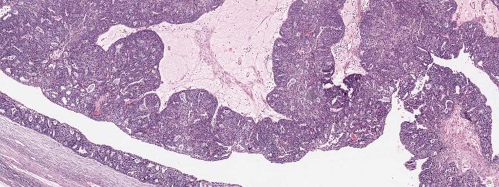
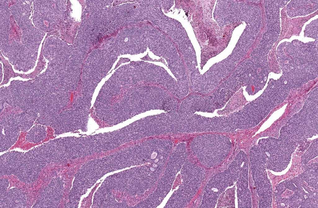
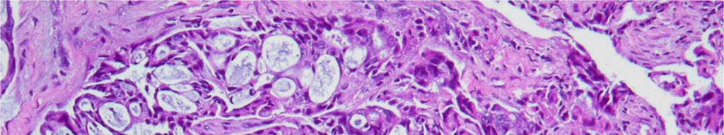
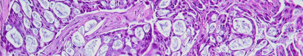
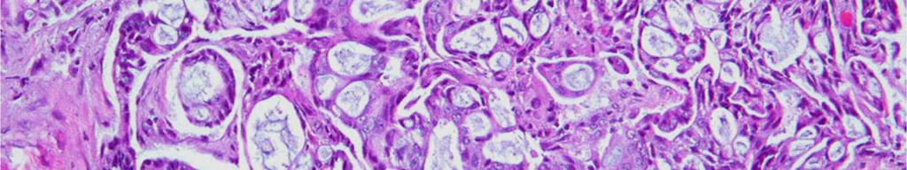
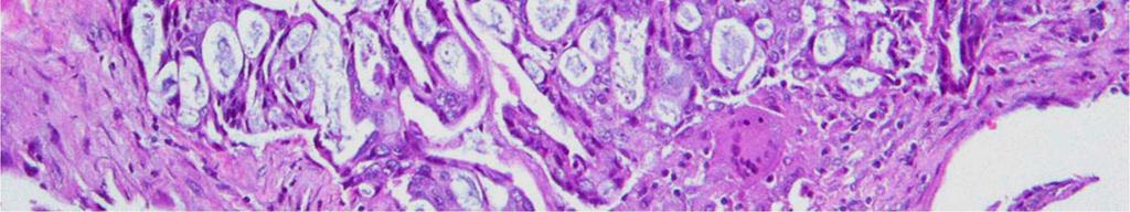
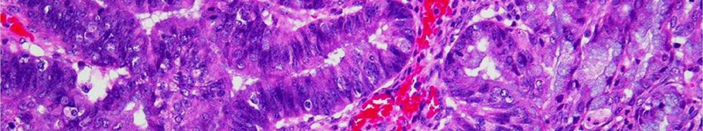
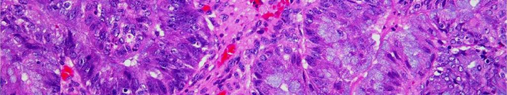
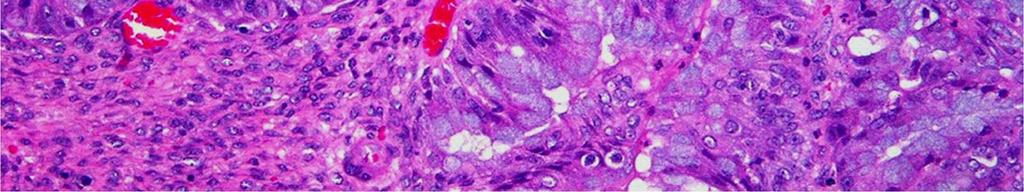
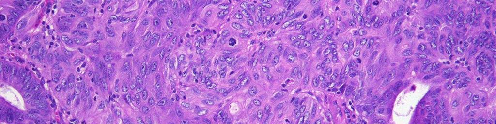
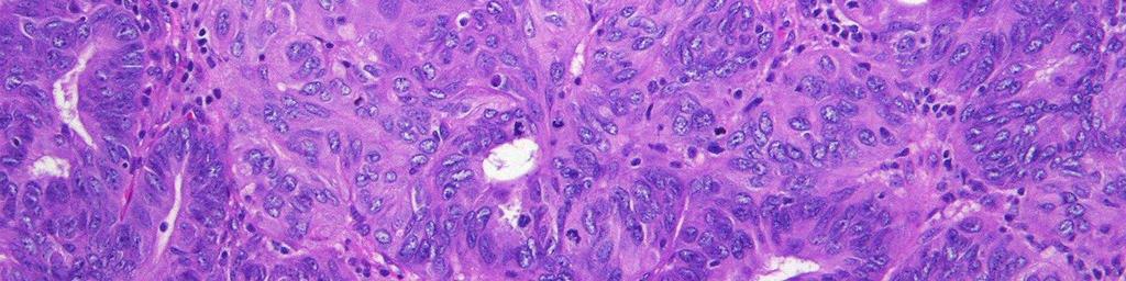
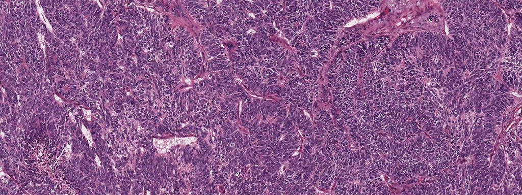
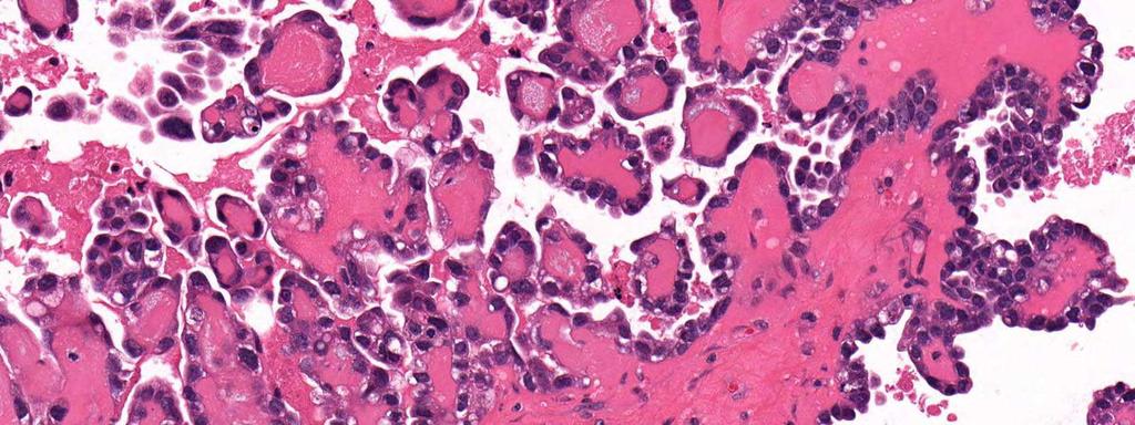
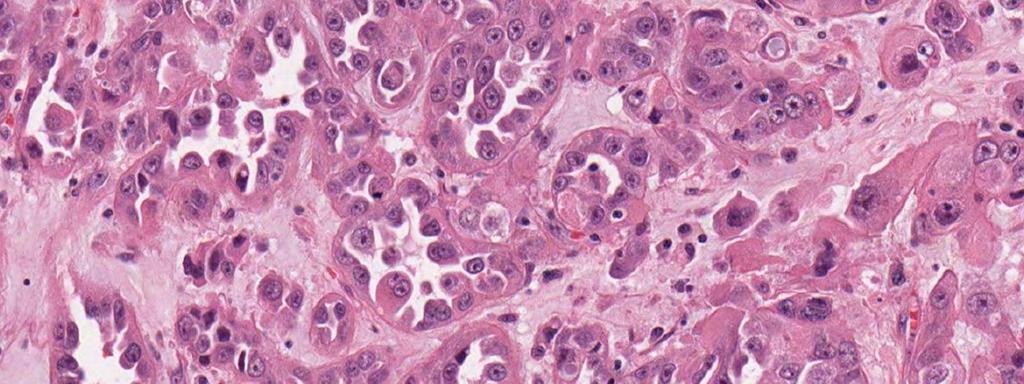
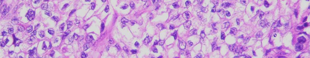
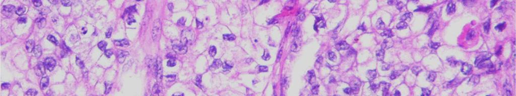
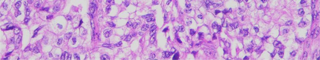
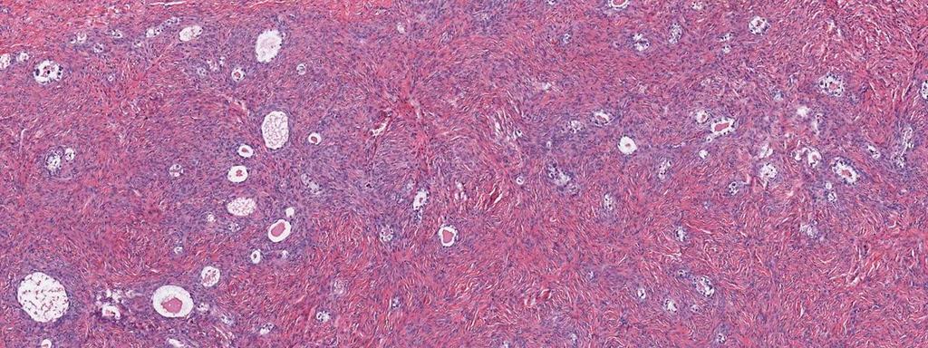
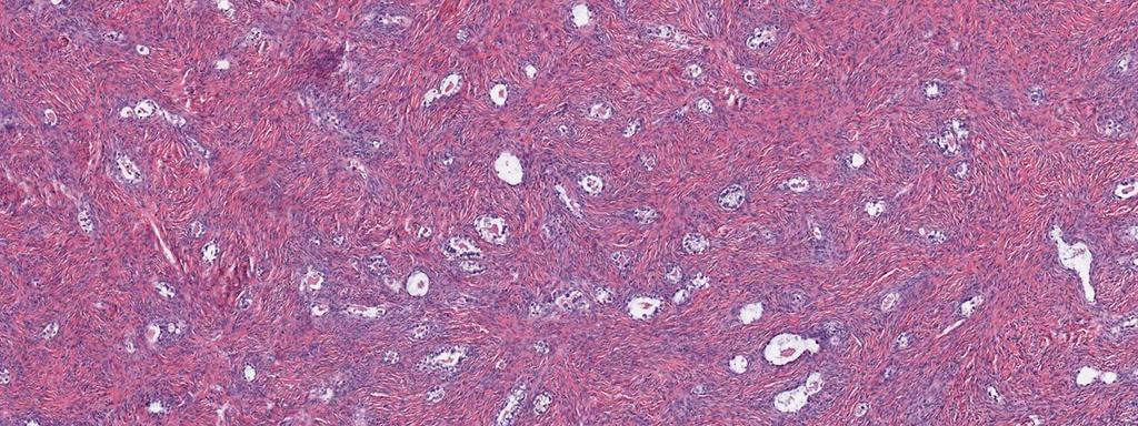
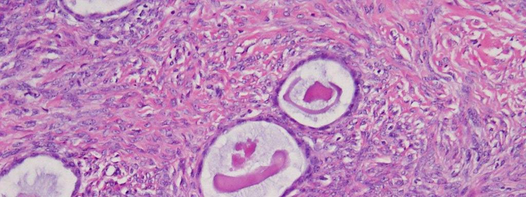
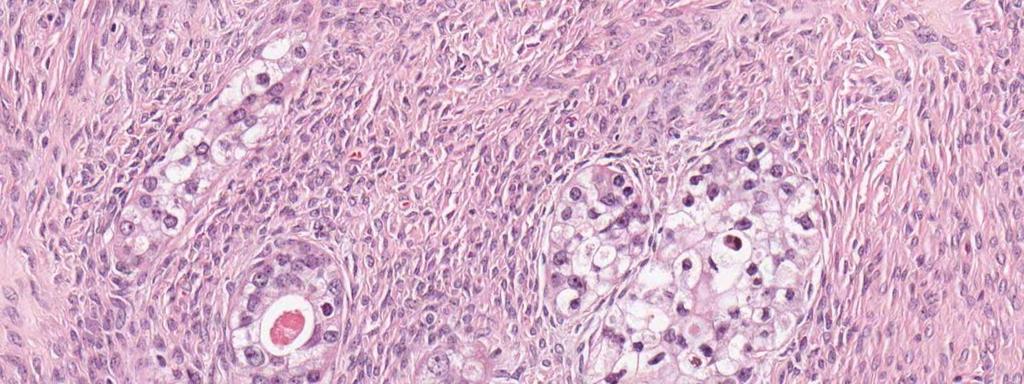
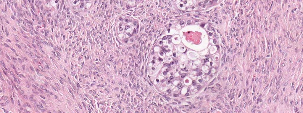
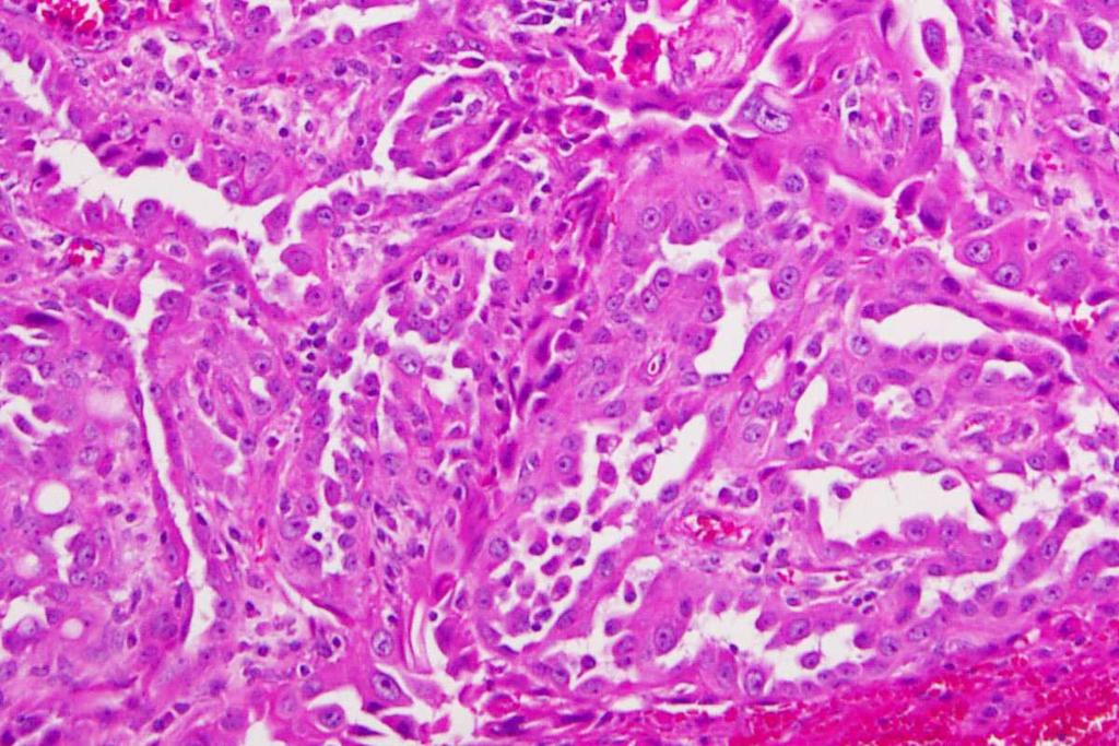
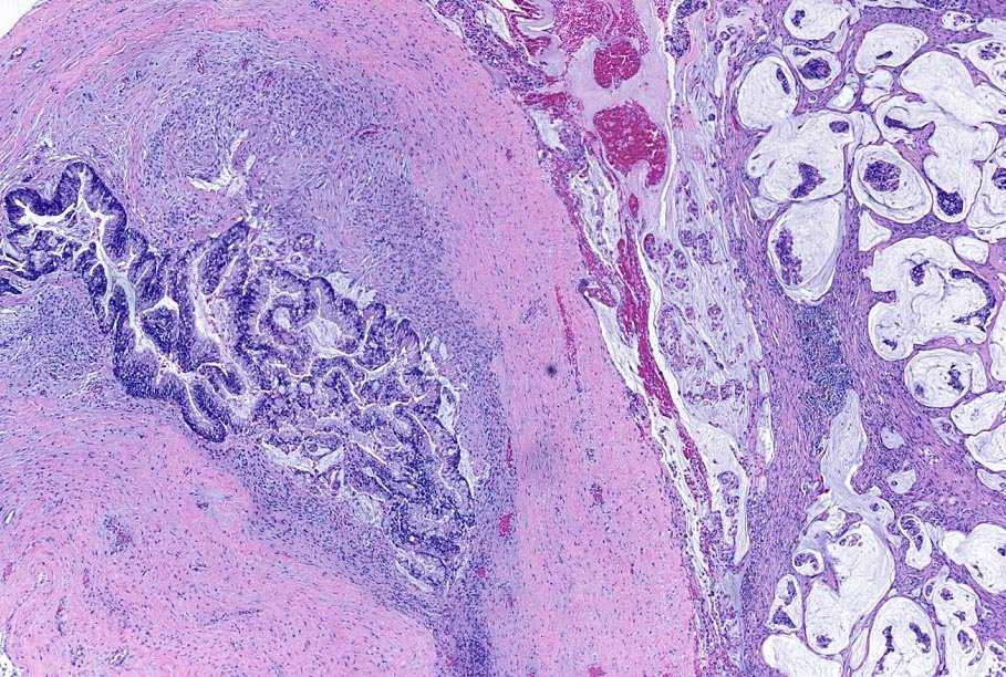
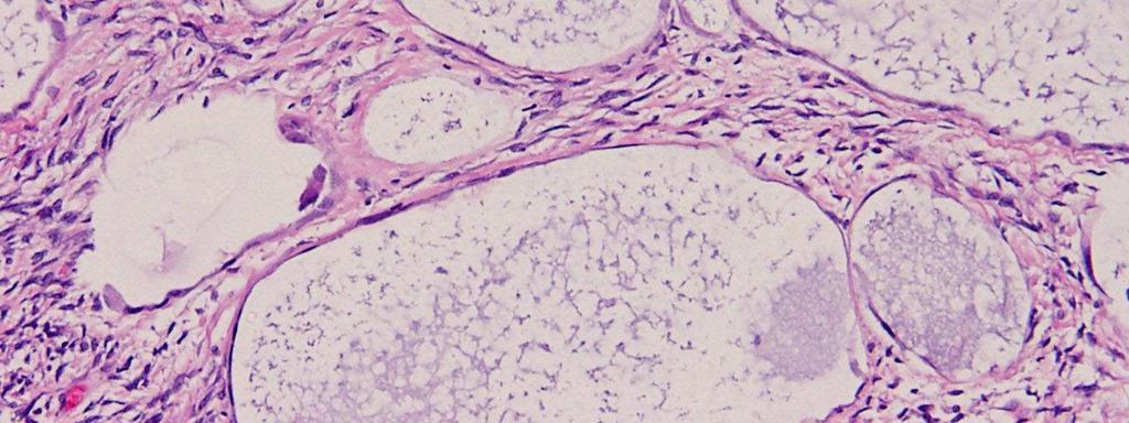
5 Ovarian cancer: distinct disease entities Hi grade serous Lo grade serous Architecture Nuclear Mitotic Endometrioid Clear cell Mucinous Shimizu Silverberg grading
6 Ovarian cancer: distinct disease entities Hi grade serous Lo grade serous p53 PIK3CA RAF/RAS Endometrioid Clear cell Mucinous Genotype
7 Ovarian cancer: distinct disease entities Hi grade serous Lo grade serous Stage Grade 5 yr III, IV Hi 20-40% Yes III Lo yr No Endo I Lo 95% Yes Clear cell I, II Intermed 70% No Mucinous I Lo >90% No Chemosens
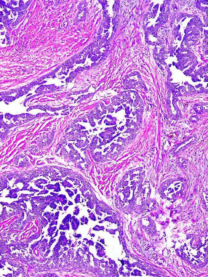
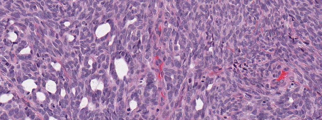
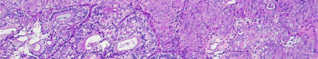
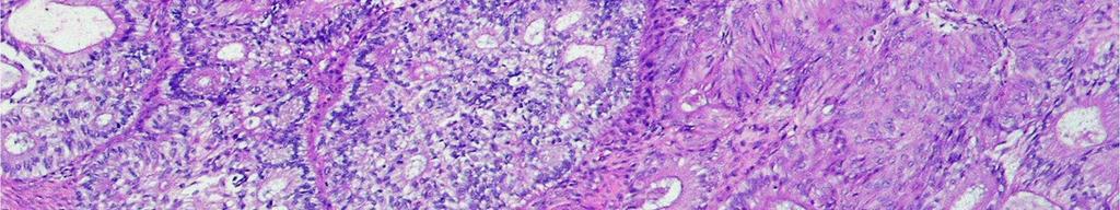
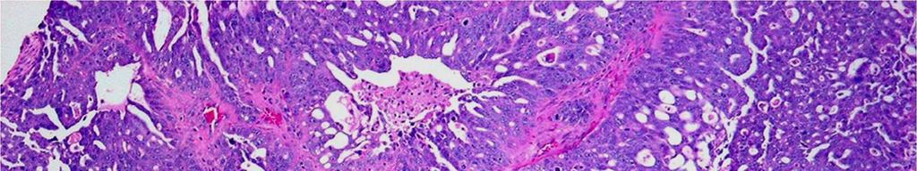
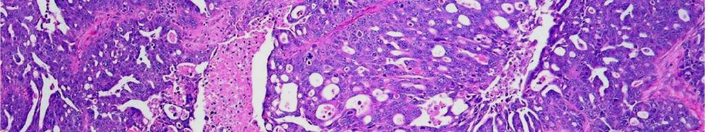
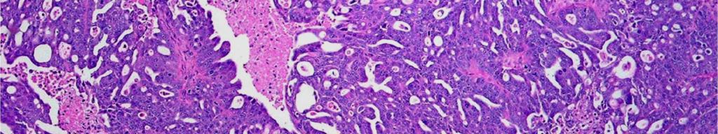
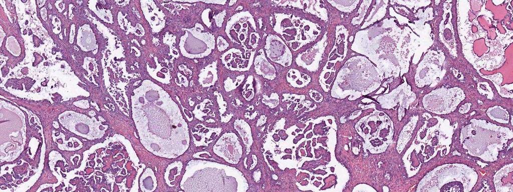
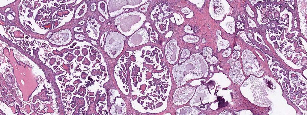
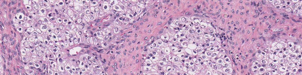
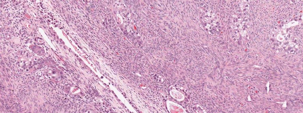
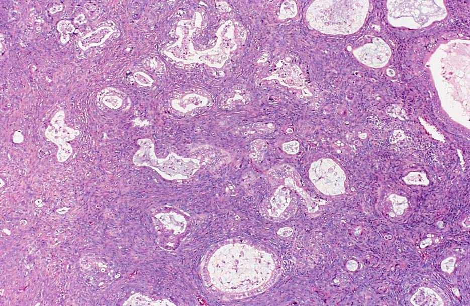
8 Problems with the traditional approach Mucinous carcinoma Clear cell carcinoma Moderately differentiated serous carcinoma Poorly differentiated endometrioid carcinoma Mixed epithelial carcinoma

9 Gilks CB, et al. Hum Pathol 2008;39:
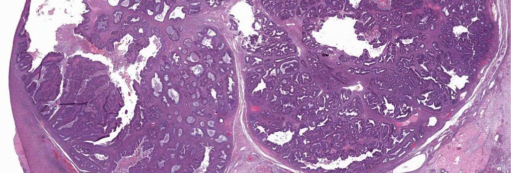
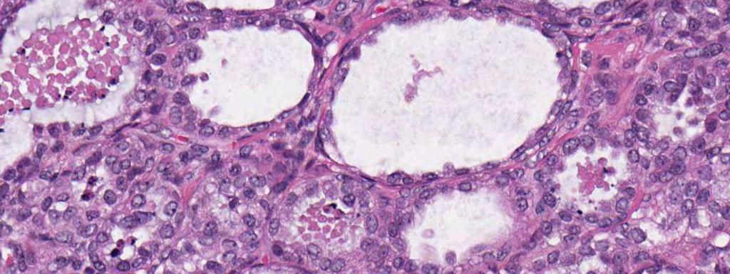
10 Revised scheme High grade serous carcinoma Clear cell carcinoma Endometrioid carcinoma Mucinous carcinoma Low grade serous carcinoma
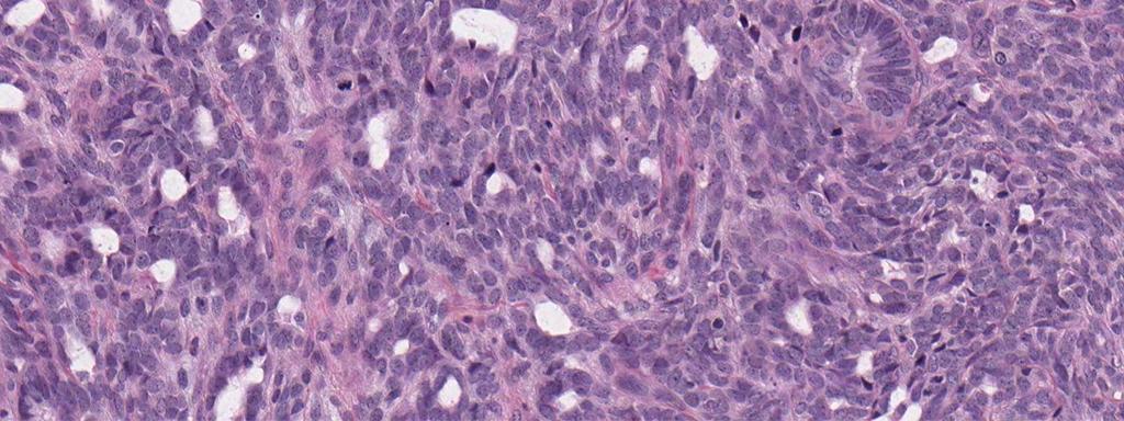
11 Revised scheme Separates low- and high-grade serous carcinoma into different categories Reclassifies as high-grade serous most: Poorly differentiated endometrioid carcinoma Undifferentiated carcinoma Transitional cell carcinoma Mixed epithelial carcinoma
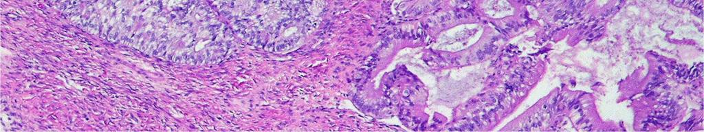
12 Revised scheme Recognizes that architectural attributes and nuclear features of clear cell carcinoma take precedence over cytoplasmic features Excludes metastatic mucinous carcinomas Recognizes contributions of clinical correlation, epidemiology, immunophenotype and genotype

13 FIGO stage and histologic subtype are independently associated with survivals after histologic reclassification Gilks CB, et al. Hum Pathol 2008;39:
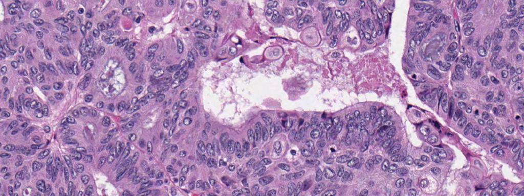
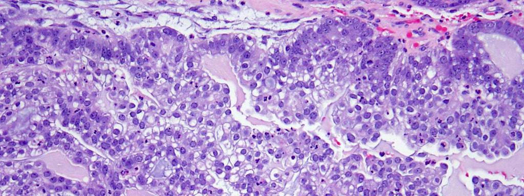
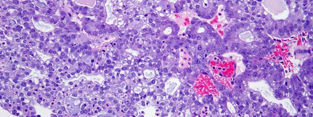
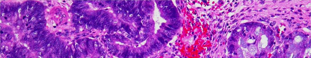
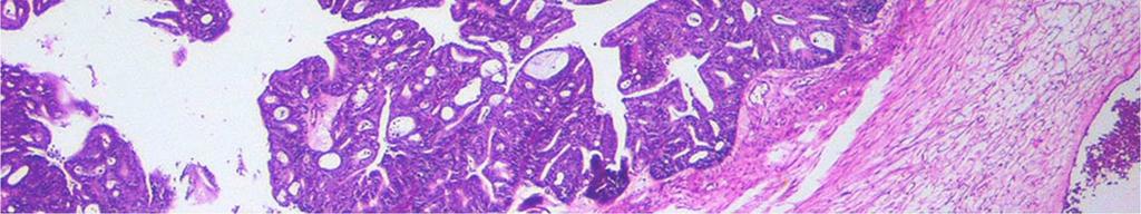
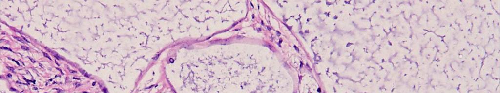
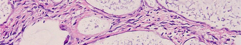
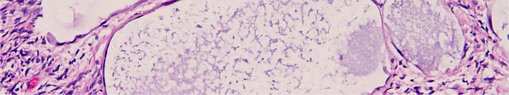
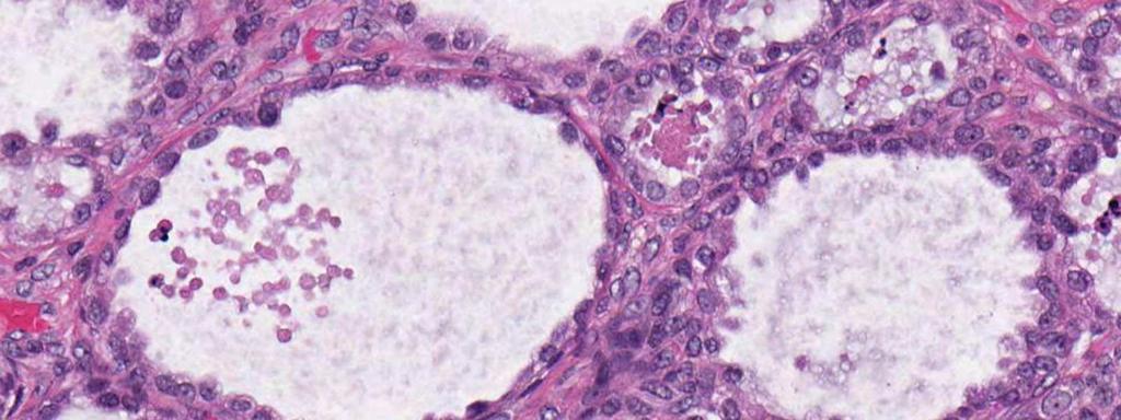
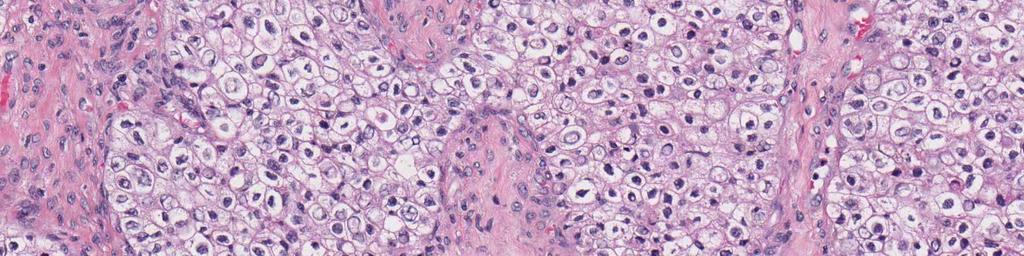
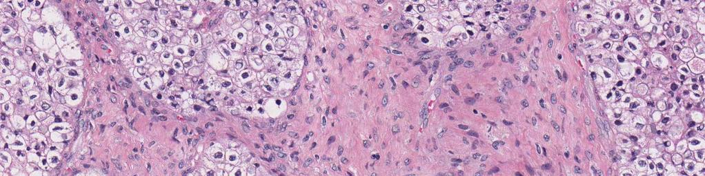
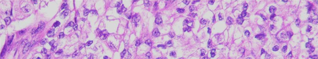
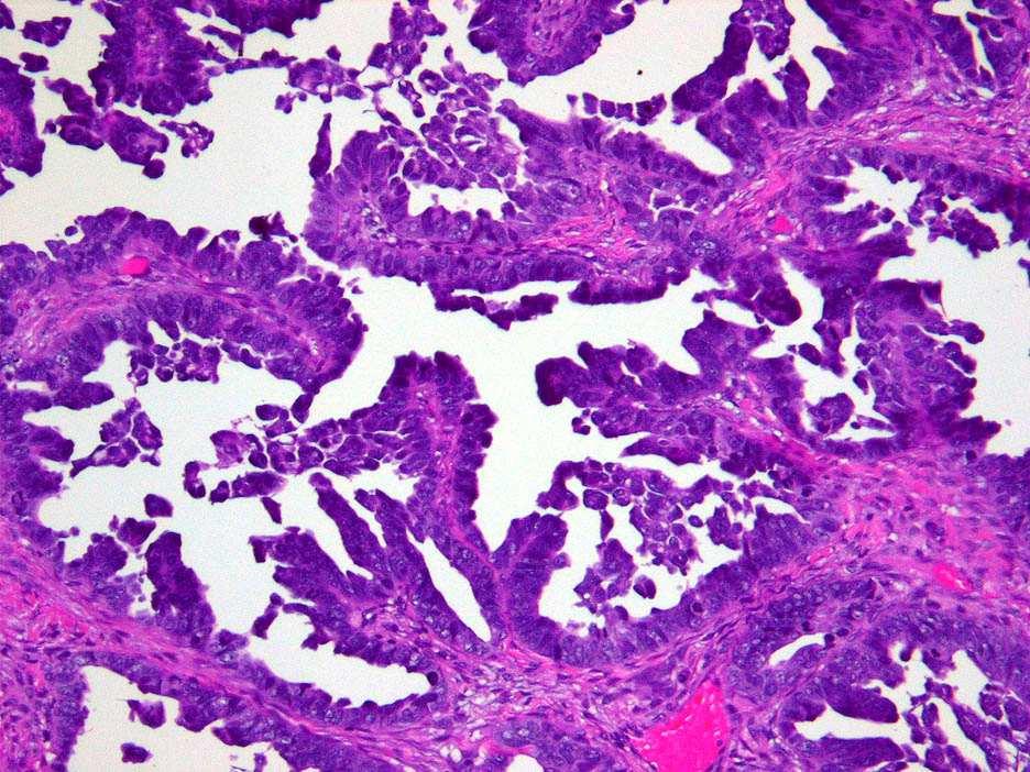
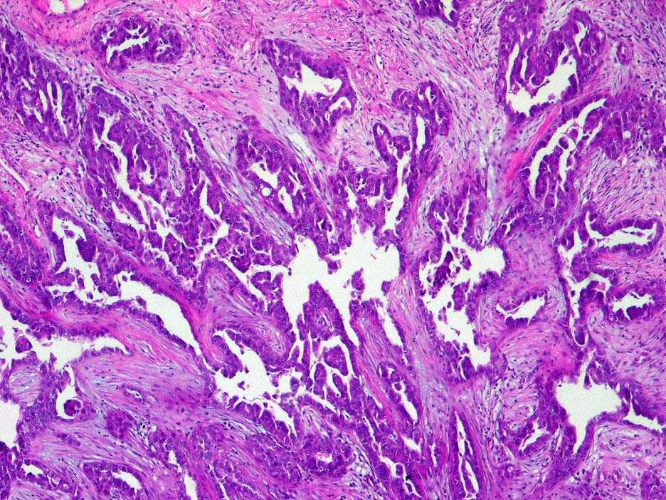
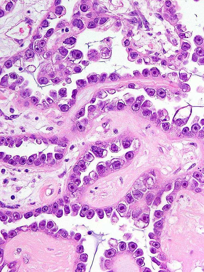
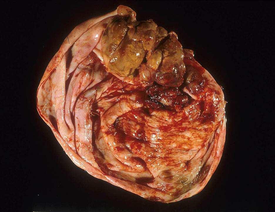
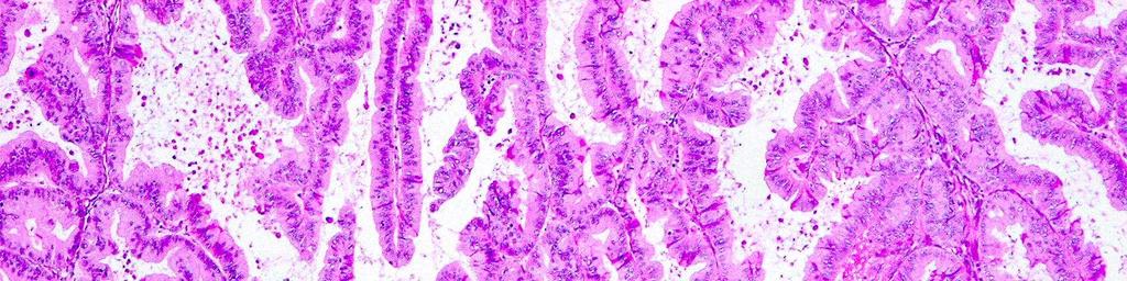
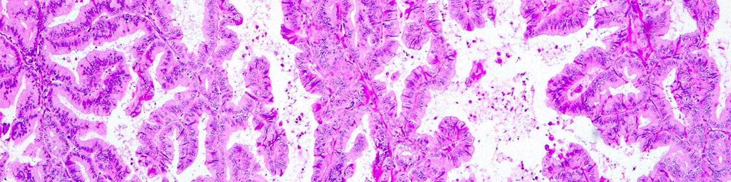
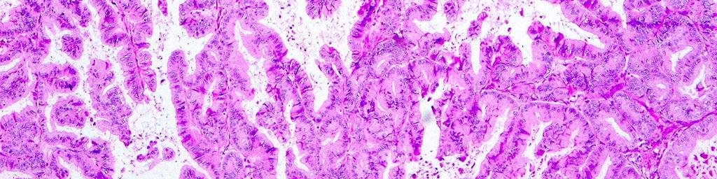
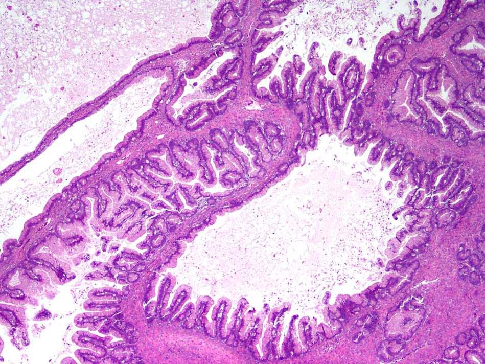
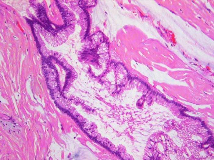
14 Serous
15 Serous tumors: demographics 80-85% of ovarian carcinomas 95% of stage III-IV ovarian carcinoma Low stage serous carcinomas are rare <5% of serous carcinomas are stage I Association with BRCA1 and 2 Leitao MM, et al. Am J Surg Pathol 28:147-59, 2004 Seidman JD, et al. Int J Gynecol Pathol 23:41-4, 2004
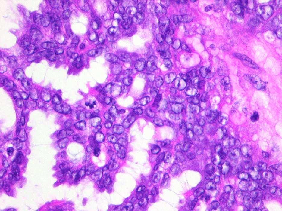
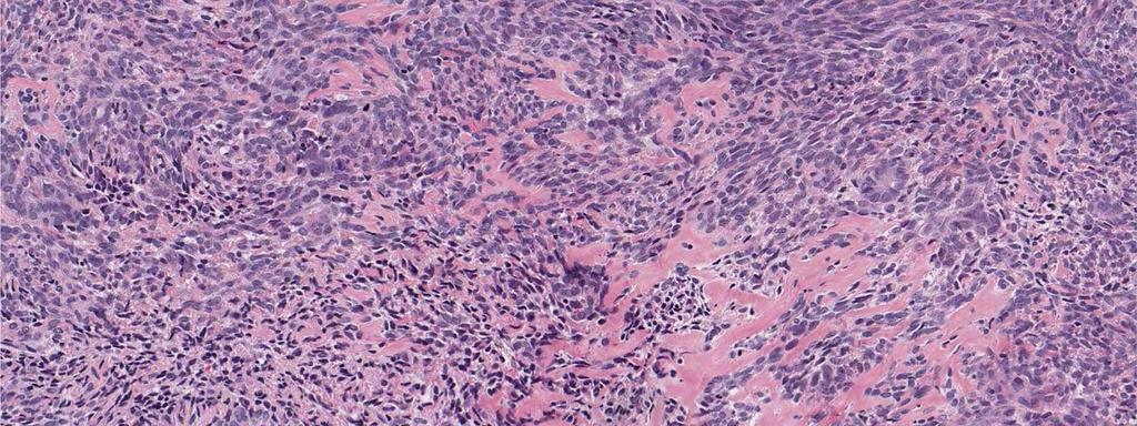
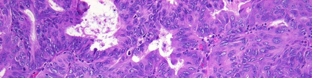
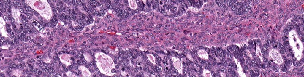
16 Serous carcinoma: problems Differential diagnosis Serous vs endometrioid carcinoma Serous vs clear cell carcinoma Serous vs transitional cell carcinoma Serous vs mucinous carcinoma Serous carcinoma vs borderline tumor Grading
17
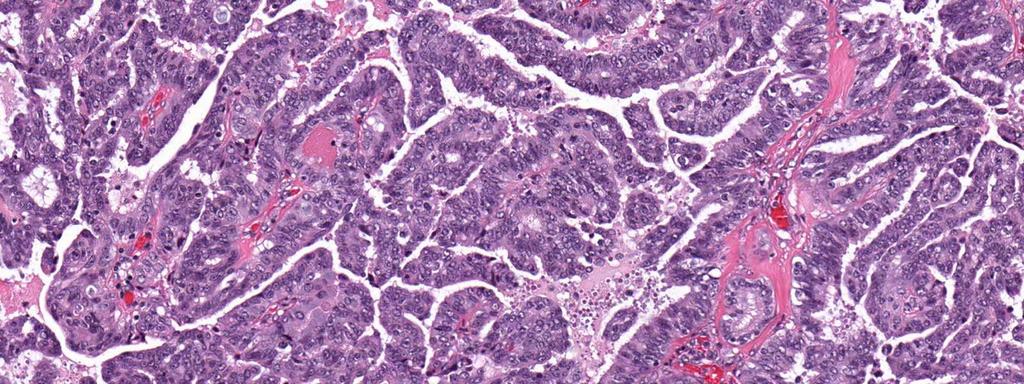
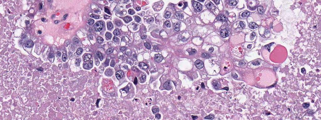
18 Pseudo-endometrioid HGSC immunophenotype WT1 p53 Pseudoendometrioid HGSC ~80% ~90% HGSC ~80% ~90% Endometrioid 10% 10%
19
20
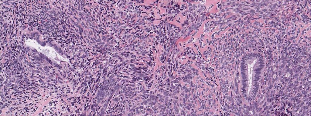
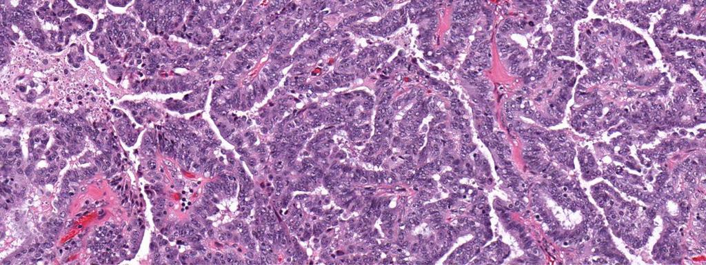
21 Transitional cell-like HGSC immunophenotype WT1 CK20 UROIII THR TCC-like HGSC 82% 0% 6% 18% HGSC ~80% <5% TCC of bladder 0% 50% 40% 61% Logani S, et al. Am J Surg Pathol 27(11): , 2003
22
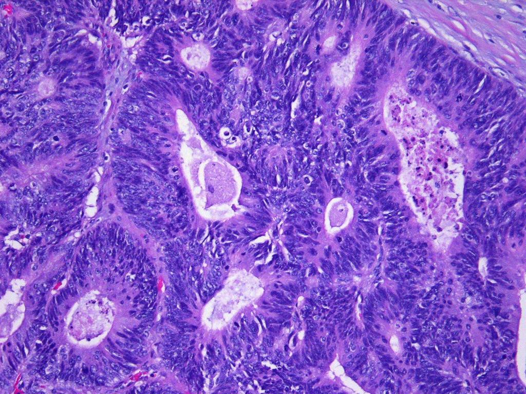
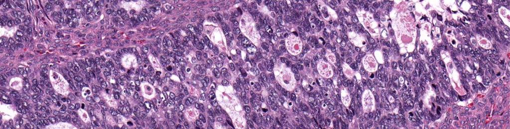
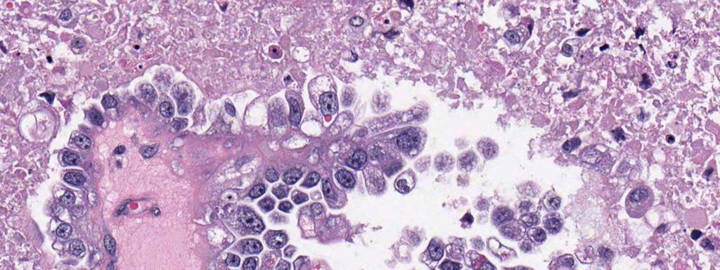
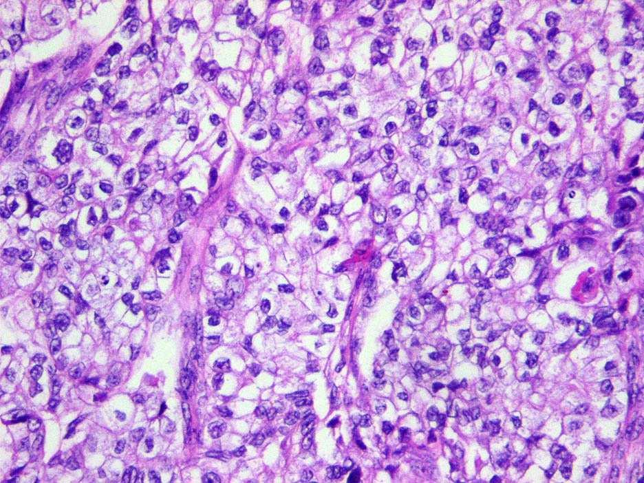
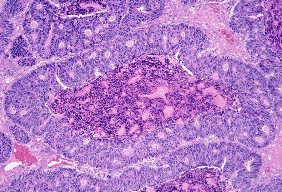
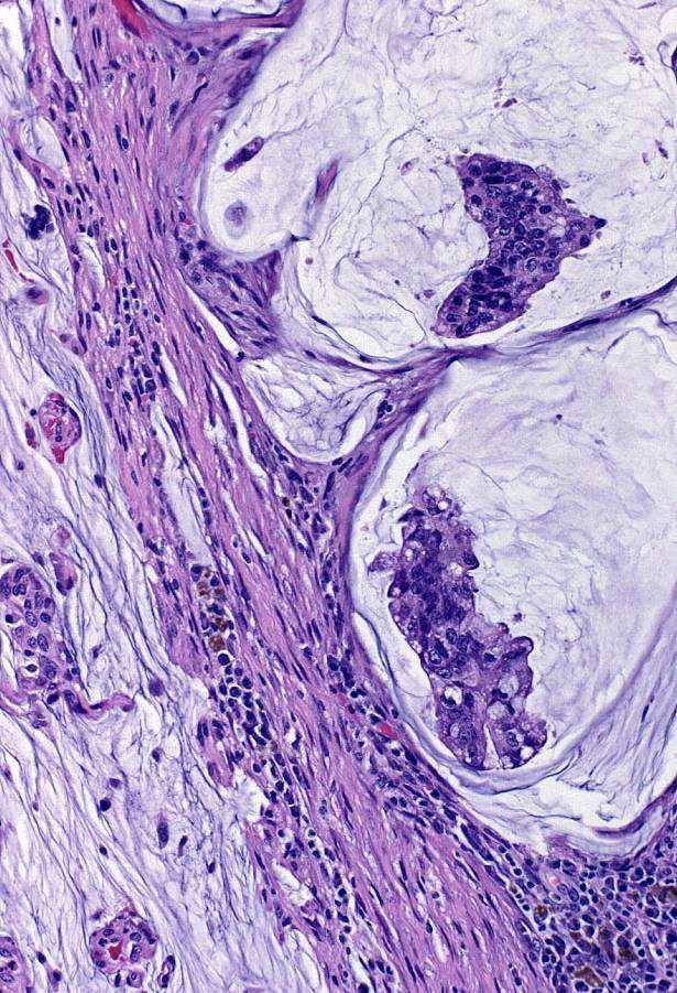
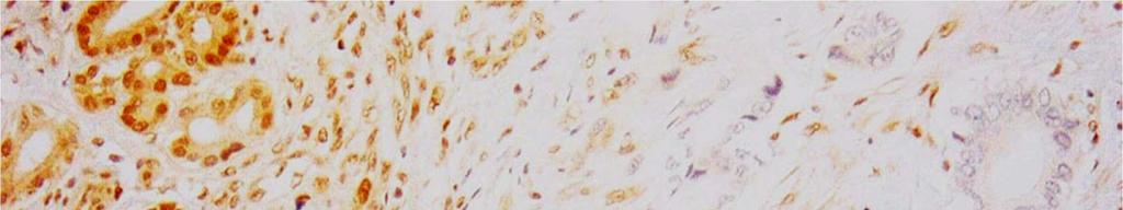
23 Clear cell-like HGSC immunophenotype WT1 ER p53 HNF-1β Clear celllike HGSC 90% 90% 60% <5% HGSC 90% 90% 70% <5% CCC 10% 10% 0% 95% Han G, Gilks CB, et al. Am J Surg Pathol 32(7):955-64, 2008 DeLair D. Am J Surg Pathol Jan;35(1):36-44
24 Serous carcinoma: pathogenesis Familial Tubal fimbria/inclusion BRCA mutation TP53 mutation STIC TP53 mutation Sporadic BRAF K-ras Surface Epithelium Borderline tumor (BT) Micropapillary BT High grade carcinoma Low grade carcinoma Singer G, et al. Am J Surg Pathol 29:218-24, 2005
25
26
27
28
29
30 Serous carcinoma: immunophenotype WT1 (>75%, all grades) Histologic subtype assignment Primary site p53 (>70%, high grade) Grade ER/PR (>90% low grade; variable high grade) Therapeutics Differential diagnosis viz clear cell carcinoma Other P16
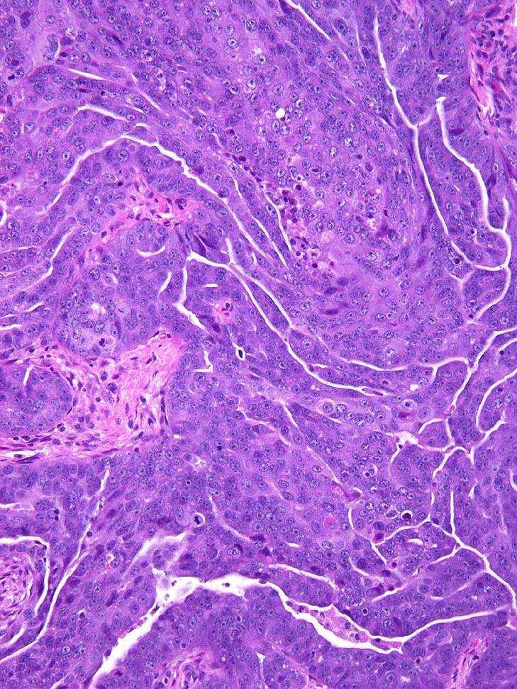
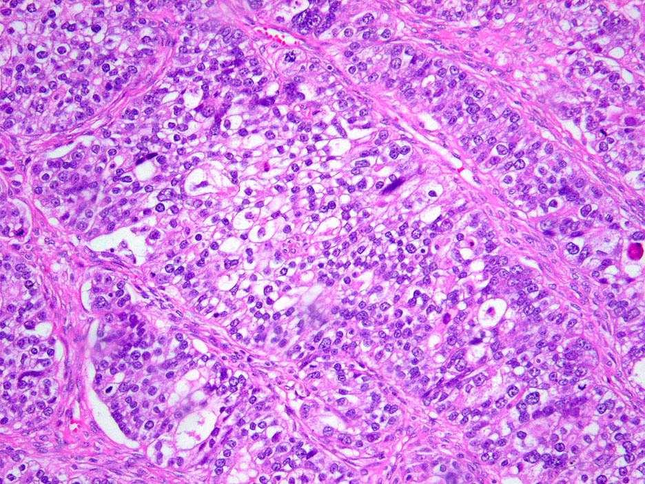
31 Grading MD Anderson 2-tier grading scheme High grade (Shimizu/Silverberg grades 2 and 3) Low grade (Shimizu/Silverberg grade 1) Uniform nuclear size (less than 3x variation) Less than 12 mitotic figures/10 high power fields


32 MD Anderson grading serous carcinoma Low grade High grade

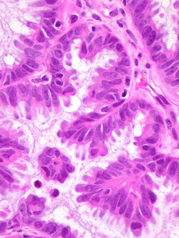
33 Serous WHO Ovarian tumours characterized in their better-differentiated forms by cell types resembling those of the fallopian tube
34 Summary: serous carcinomas Usually high stage (Stage IIC or greater)
35 Summary: serous carcinomas Usually high stage Broad range of histologic features Slit-like spaces, irregular luminal contours
36 Summary: serous carcinomas Usually high stage Broad range of histologic features Slit-like spaces, irregular luminal contours Frequent WT1 Low-grade: serous borderline tumor, BRAF/Kras, ER/PR High-grade: tubal intraepithelial carcinoma, p53, p16, loss of BRCA1, BRCA1 or 2 family
37 Summary: serous carcinomas Usually high stage Broad range of histologic features Slit-like spaces, irregular luminal contours Frequent WT1 Low-grade: serous borderline tumor, BRAF/Kras, ER/PR High-grade: tubal intraepithelial carcinoma, p53, p16, loss of BRCA1, BRCA1 or 2 family Other entities are excluded
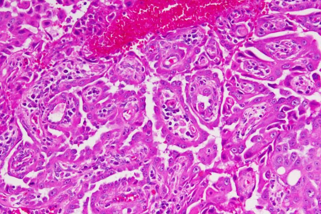
38 Endometrioid
39 Endometrioid tumors: demographics 10% of ovarian carcinomas Most common stage I carcinoma (40-50%) High stage endometrioid carcinomas are rare Leitao MM, et al. Am J Surg Pathol 28:147-59, 2004 Seidman JD, et al. Int J Gynecol Pathol 23:41-4, 2004
40
41
42
43
44
45
46
47 Endometrioid tumors: problems Differential diagnosis: Endometrioid vs mucinous Endometrioid vs sex cord stromal tumor Endometrioid vs carcinosarcoma Endometrioid vs metastasis Endometrioid vs clear cell carcinoma Endometrioid vs serous carcinoma Borderline tumor versus carcinoma Grading Shimizu/Silverberg
48
49
50
51
52
53
54
55
56
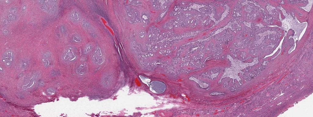
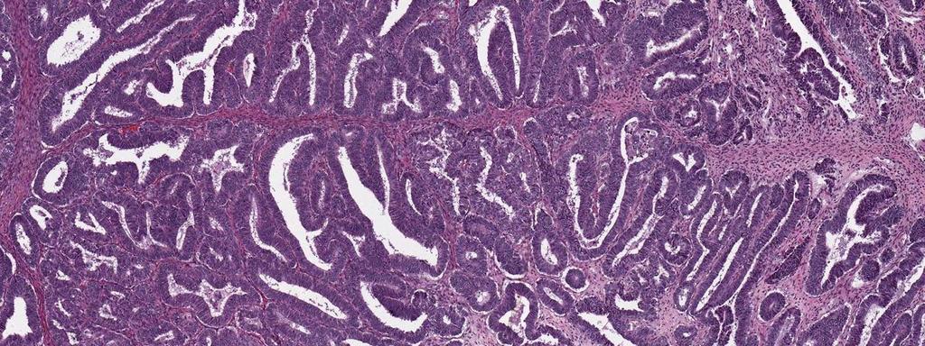
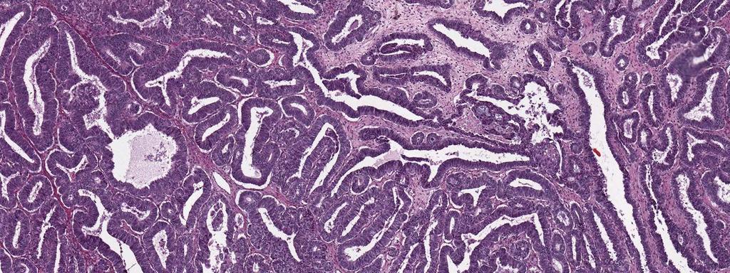
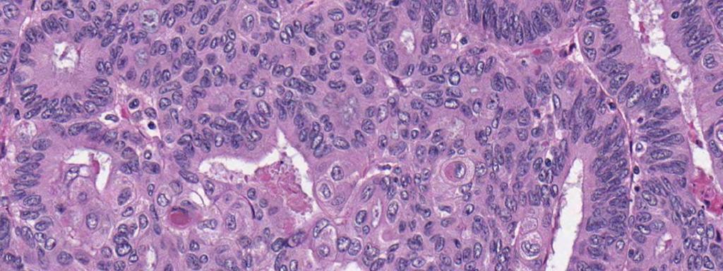
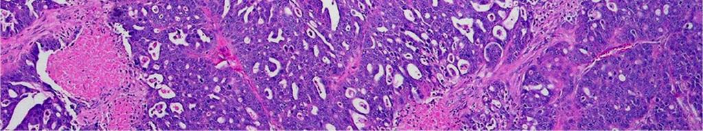
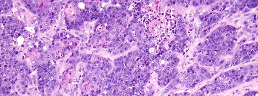
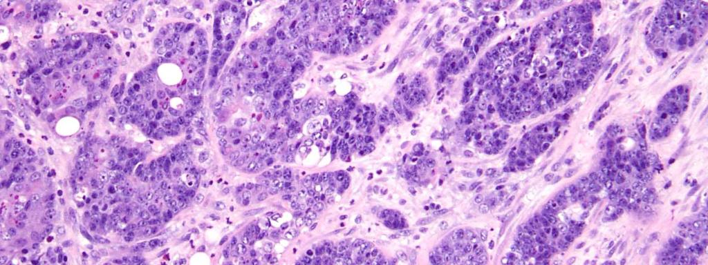
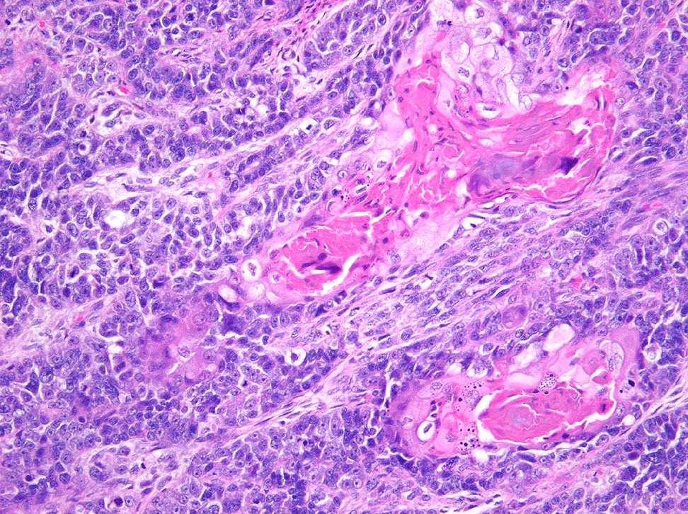
57
58
59
60

61
62
63
64 High grade endometrioid carcinoma Wu R, Cho KR, et al. Cancer Cell Apr;11(4):321-33
65 High grade endometrioid carcinoma Wu R, Cho KR, et al. Cancer Cell Apr;11(4):321-33
66
67
68 Endometrioid tumors: immunophenotype WT1 (<5%) Histologic subtype assignment Nuclear β-catenin (~30%) Histologic subtype assignment ER/PR (>75%) Therapeutic Differential diagnoses viz clear cell carcinoma P53 in high grade examples
69 Endometrioid carcinoma grading Differentiation Broder FIGO endometrial (GOG) Shimizu/Silverberg
70 Endometrioid carcinoma grading Differentiation Broder FIGO endometrial (GOG) Shimizu/Silverberg
71 Shimizu/Silverberg grading Architecture Glandular (1 point) Papillary (2 points) Solid (3 points) Nuclear pleomorphism Slight (1 point) Moderate (2 points) Marked (3 points) Mitotic activity 0-9 mf/10 hpf (1 point) (2 points) >24 (3 points) Shimizu, Silverberg, et al. Cancer 1998 Mar 1;82(5): Shimizu, Silverberg, et al. Gynecol Oncol 1998 Jul;70(1):2-12
72 Shimizu/Silverberg grading Architecture Glandular (1 point) Papillary (2 points) Solid (3 points) Nuclear pleomorphism Slight (1 point) Moderate (2 points) Marked (3 points) Mitotic activity 0-9 mf/10 hpf (1 point) (2 points) >24 (3 points) Grade 1 (3-5 points) Grade 2 (6-7 points) Grade 3 (8-9 points) Correlates with survival Do NOT use with clear cell carcinoma Shimizu, Silverberg, et al. Cancer 1998 Mar 1;82(5): Shimizu, Silverberg, et al. Gynecol Oncol 1998 Jul;70(1):2-12
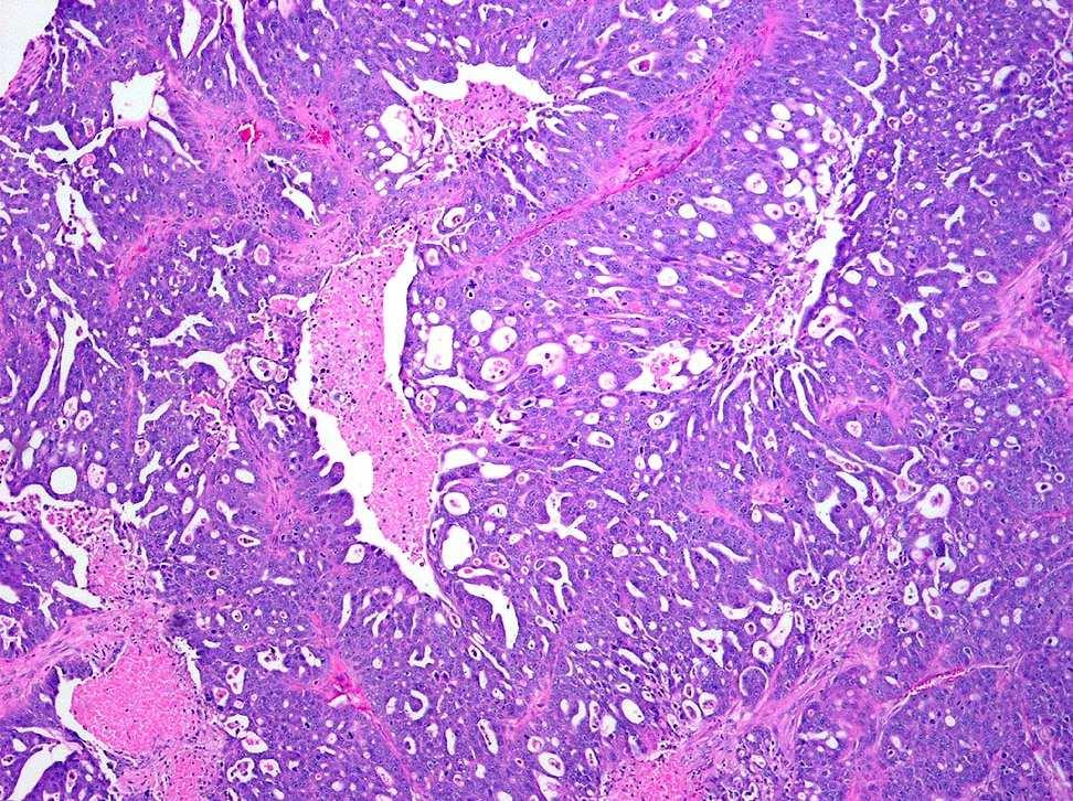
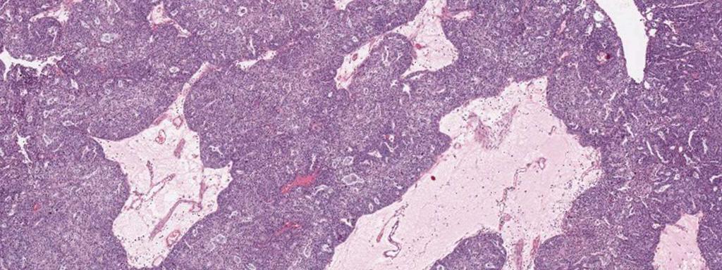
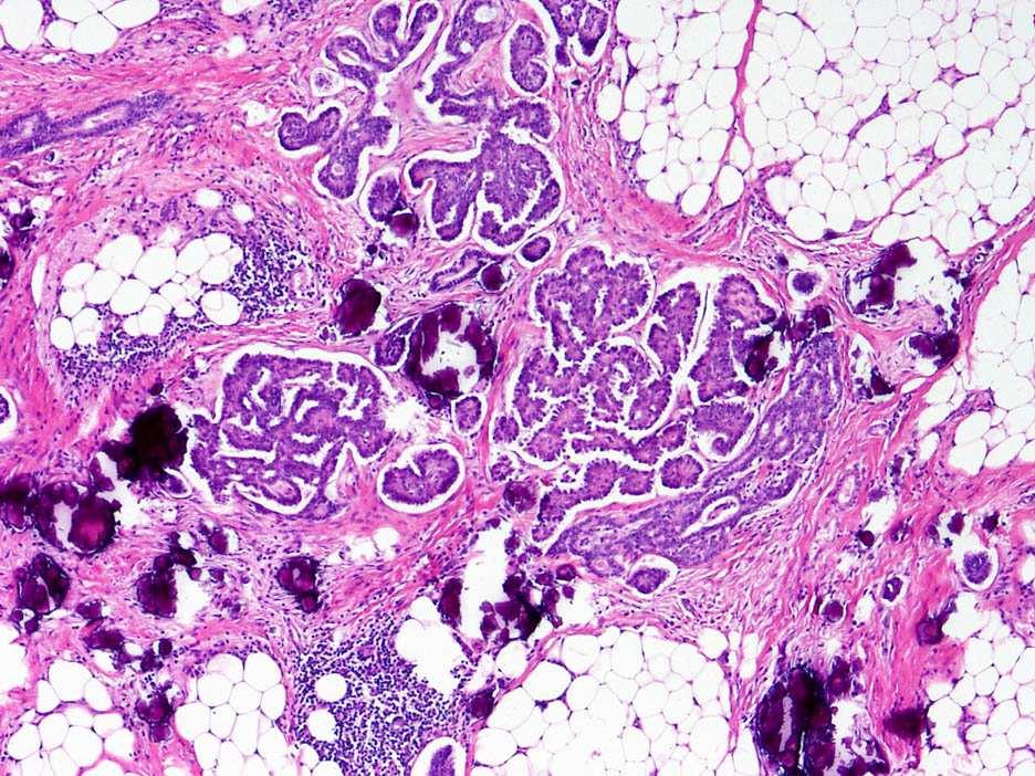
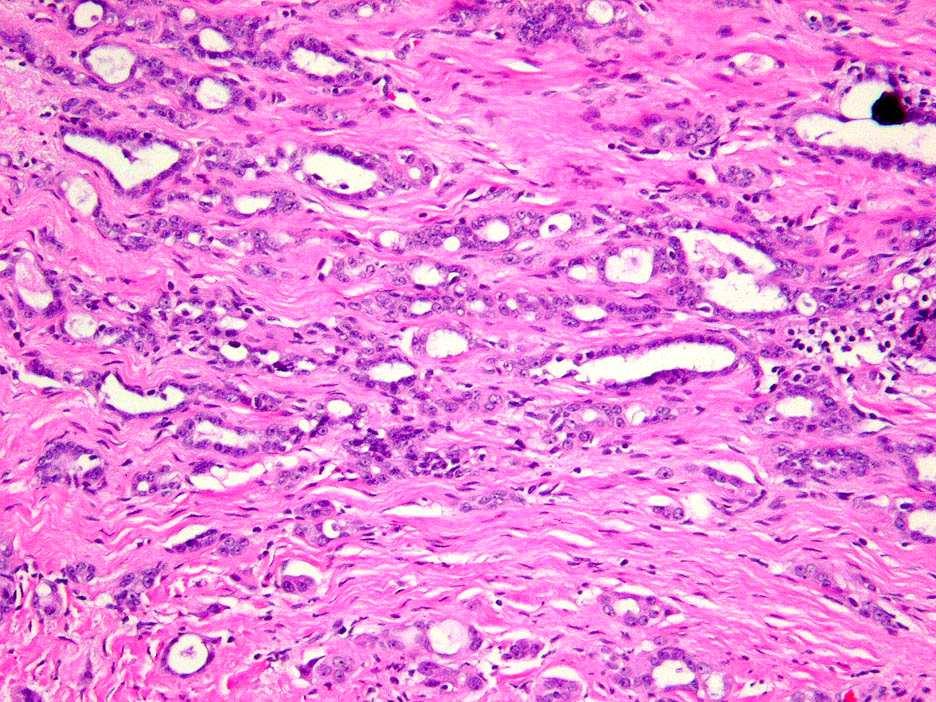
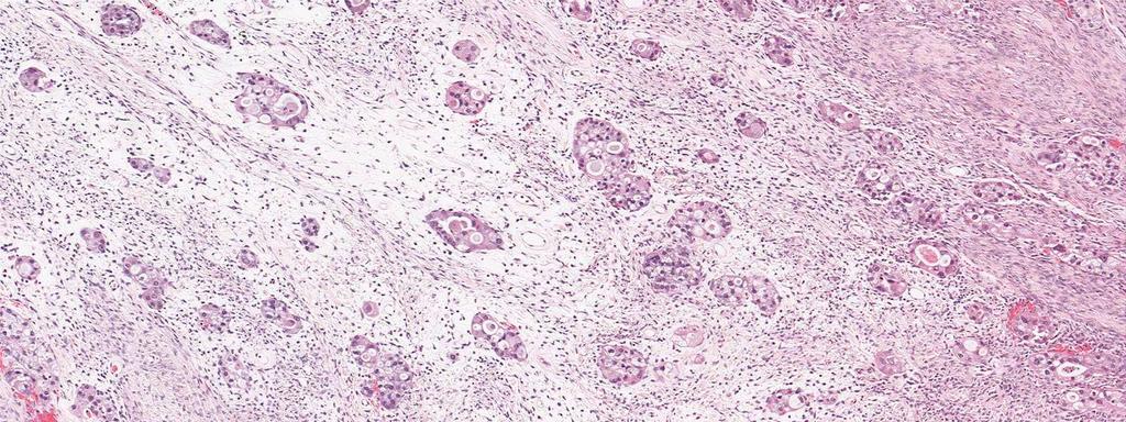
73 SHIMIZU GRADE?
74 Endometrioid WHO Tumours of the ovary that closely resemble the various types of endometrioid tumors of the uterine corpus
75 Summary: Endometrioid tumors Low stage, low grade
76 Summary: Endometrioid tumors Low stage, low grade Endometrial-like, metaplasias, secretory change, expansile invasion Endometriosis, endometrioid borderline tumor, endometrioid uterine carcinoma
77 Summary: Endometrioid tumors Low stage, low grade Endometrial-like, metaplasias, secretory change, expansile invasion Endometriosis, endometrioid borderline tumor, endometrioid uterine carcinoma ER/PR, nuclear β-catenin; not WT1 CTNNB-1 (β-catenin), PTEN, PIK3CA, ARID 1A, MSI-H
78 Summary: Endometrioid tumors Low stage, low grade Endometrial-like, metaplasias, secretory change, expansile invasion Endometriosis, endometrioid borderline tumor, endometrioid uterine carcinoma ER/PR, nuclear β-catenin; not WT1 CTNNB-1 (β-catenin), PTEN, PIK3CA, ARID 1A, MSI-H Other entities are excluded

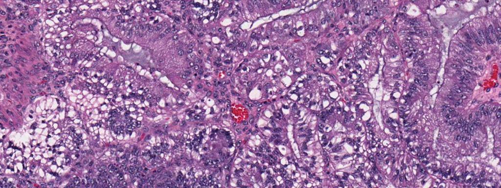
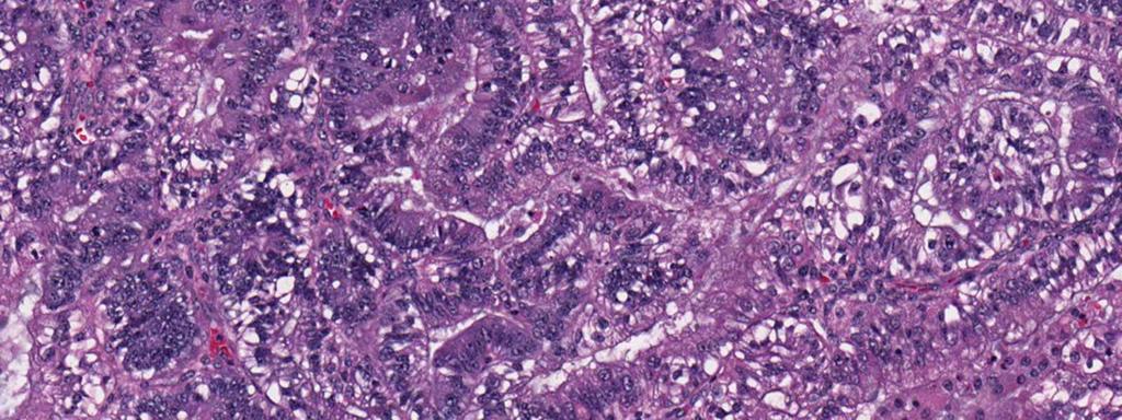
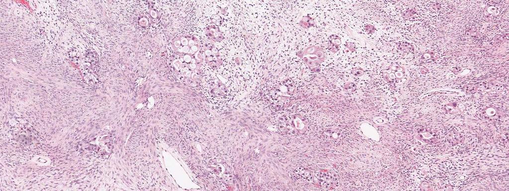
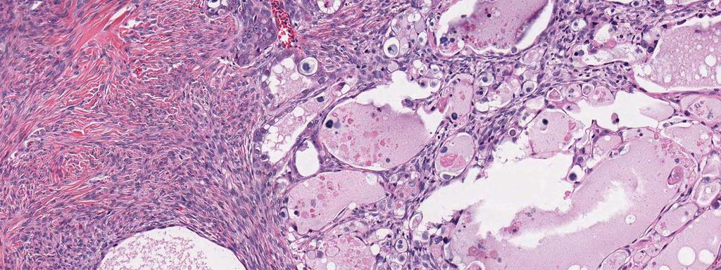
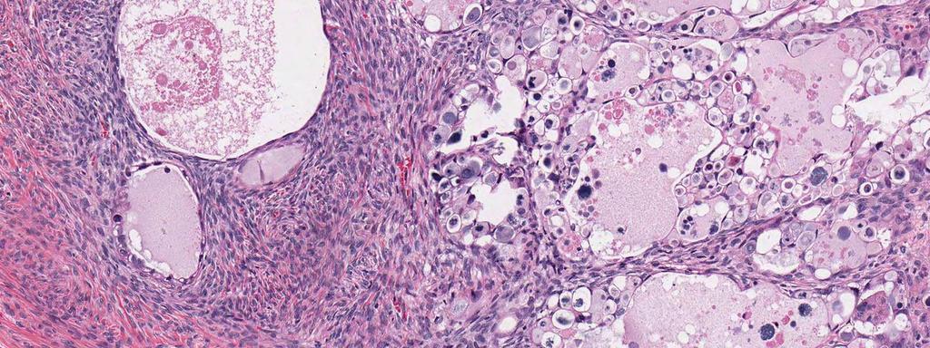
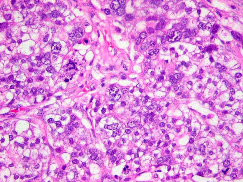
79 Clear cell
80 Clear cell tumors: demographics 5-10% of ovarian carcinomas (in the West) Disproportionately low stage (and unilateral) 25-30% of stage I and II carcinomas are clear cell High stage clear cell carcinomas are uncommon HNPCC/Lynch syndrome in ~20% of pts <50 yrs* Leitao MM, et al. Am J Surg Pathol 28:147-59, 2004 Seidman JD, et al. Int J Gynecol Pathol 23:41-4, 2004 *Jensen KC, et al. Am J Surg Pathol 32: , 2008
81

82 Shoo-ron-pou 82
83
84
85
86
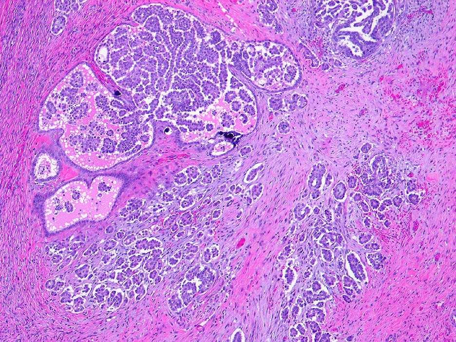
87
88
89
90
91
92
93
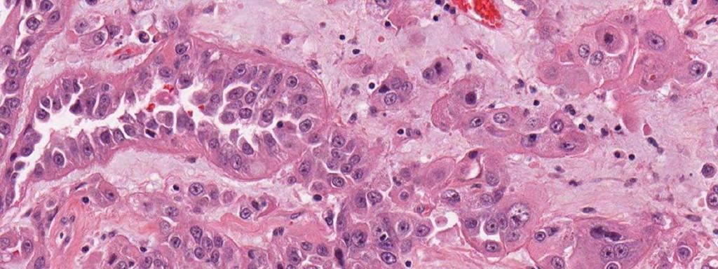
94 Clear cell tumors: problems Differential diagnosis: Clear cell vs serous carcinoma Mixed clear cell and serous carcinoma vs serous carcinoma Clear cell vs endometrioid carcinoma Clear cell borderline tumor Grading No grading scheme
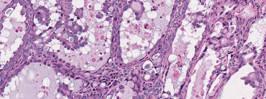
95 Clear cell tumors: diagnostic reproducibility, immunophenotype, lessons learned Clear cell carcinomas have a distinctive architectural repertoire and immunophenotype Reproducibly diagnosed Han G, Gilks CB, et al. Am J Surg Pathol 32(7):955-64, 2008
96 Clear cell tumors: diagnostic reproducibility, immunophenotype, lessons learned Clear cell carcinomas have a distinctive architectural repertoire and immunophenotype Mixed epithelial carcinomas containing clear cells (MEP-C) are not reproducibly diagnosed Han G, Gilks CB, et al. Am J Surg Pathol 32(7):955-64, 2008
97 Clear cell tumors: diagnostic reproducibility, immunophenotype, lessons learned Clear cell carcinomas have a distinctive architectural repertoire and immunophenotype Mixed epithelial carcinomas containing clear cells (MEP-C) are not reproducibly diagnosed MEP-Cs are seldom clear cell carcinomas most are serous carcinomas Han G, Gilks CB, et al. Am J Surg Pathol 32(7):955-64, 2008
98 Clear cell immunophenotype WT1 ER p53 HNF-1β CCC (n=11) 10% 10% 0% 95% SC (n=10) 90% 90% 70% <5% Clear component (n=11) Serous component (n=11) 90% 90% 60% 90% 90% 60%
99
100
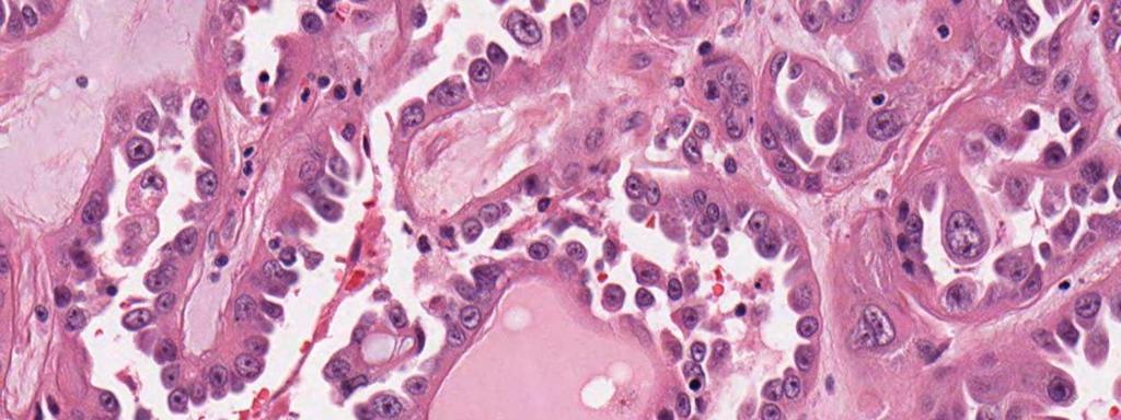
101 ER/PR (<10%) Clear cell carcinoma: immunophenotype Histologic subtype assignment (viz serous and endometrioid) WT1 (<10%) Histologic subtype assignment (viz serous) p53 (<10%) Histologic subtype assignment (viz hi grade serous) HNF-1 β (95%) Histologic subtype assignment (viz serous and endometrioid) DeLair D, et al. Am J Surg Pathol 2011 Jan;35(1):36-44.

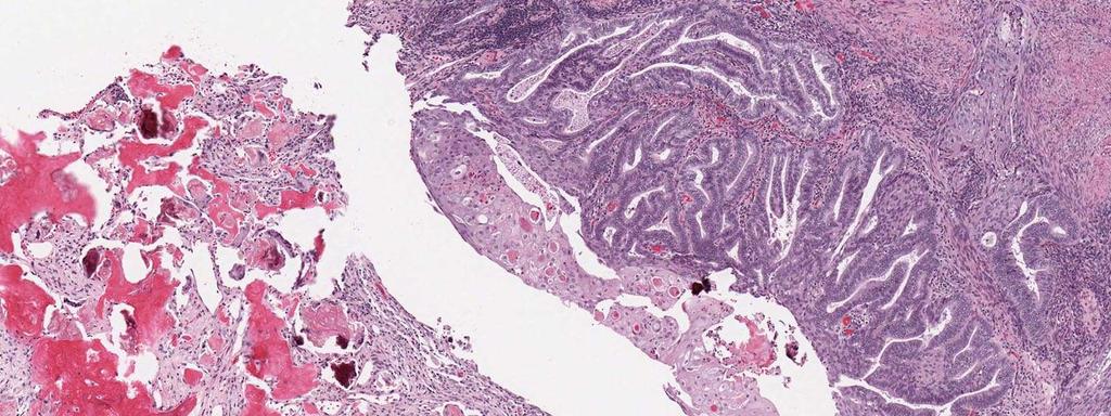
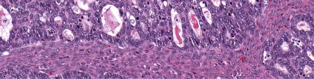
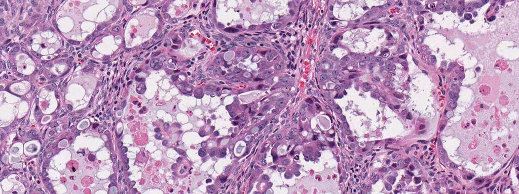
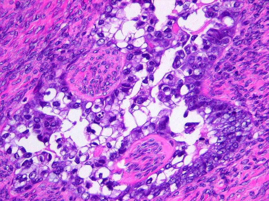
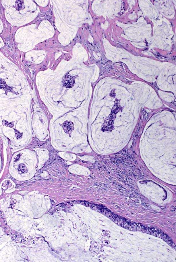
102 Clear cell tumors with papillary architecture Clear cell tumors with papillary architecture are carcinomas, not borderline tumors Clear cell borderline tumor
103
104

105
106
107
108
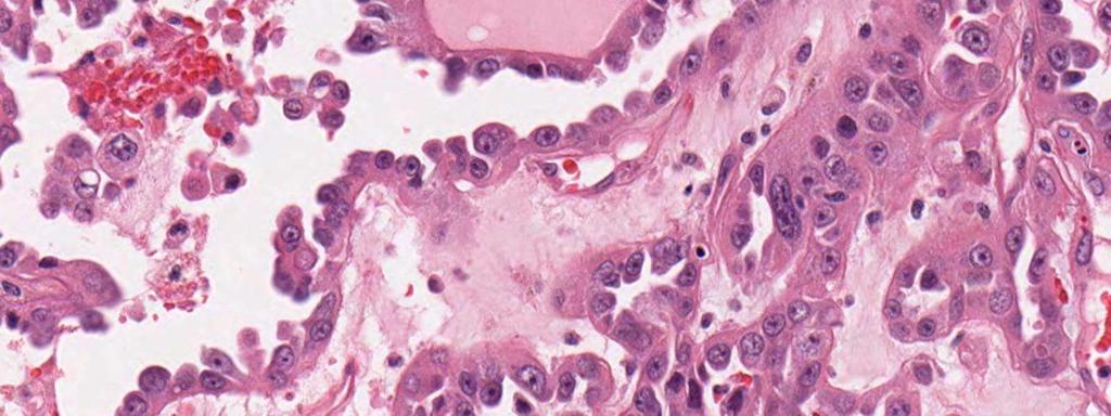
109
110 Clear cell tumors with papillary architecture High nuclear grade Clear cell vs serous carcinoma Not-so-high-nuclear grade Clear cell vs Serous carcinoma Endometrioid carcinoma Mesothelioma Serous borderline tumor Sangoi AR, Soslow RA, Longacre TA. Am J Surg Pathol 2008;32:269-74
111
112
113
114
115


116 Oxyphilic clear cell carcinoma mimicking mesothelioma

117 Well-differentiated papillary mesothelioma
118 Papillary ovarian tumors Clear cell CA Unilateral, low stage Round papillae Hyaline, edematous stroma Hobnail cells, cuboidal Monolayer Uniform nuclei Decreased mitotic activity* Other CCC patterns Endometriosis WT1-/ER-/p53- Serous CA Bilateral, high stage Elongate, hierarchical branching Fibrous stroma Columnar cells Cellular tufting, micropapillae Pleomorphic nuclei High mitotic rate Slit-like spaces No endometriosis WT1+/ER variable/p53+
119 Summary: Clear cell carcinomas Low stage and unilateral
120 Summary: Clear cell carcinomas Low stage and unilateral Papillary, tubulocystic, solid, hobnail, frequently clear cytoplasm Endometriosis, clear cell borderline tumor
121 Summary: Clear cell carcinomas Low stage and unilateral Papillary, tubulocystic, solid, hobnail, frequently clear cytoplasm Endometriosis, clear cell borderline tumor Low ER/PR, WT1, p53, mib-1 Positive HNF-1ß
122 Summary: Clear cell carcinomas Low stage and unilateral Papillary, tubulocystic, solid, hobnail, frequently clear cytoplasm Endometriosis, clear cell borderline tumor Low ER/PR, WT1, p53, mib-1; HNF-1β+ Lack of features that define other entities Metaplasias, secretory changes Multilayering, serrated luminal profiles
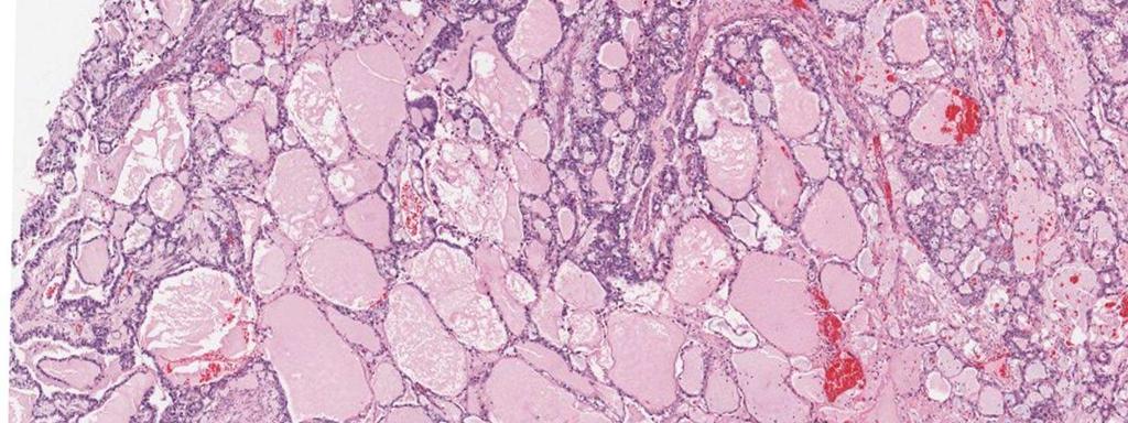
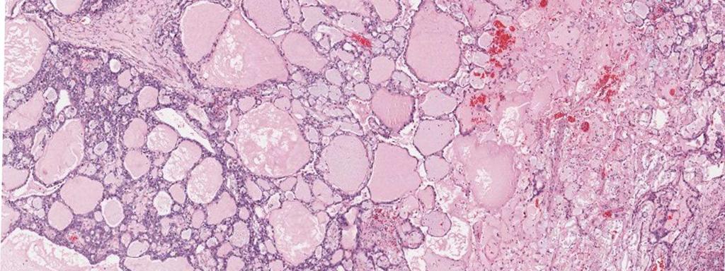
123 Summary: Clear cell carcinomas Low stage and unilateral Papillary, tubulocystic, solid, hobnail, frequently clear cytoplasm Endometriosis, clear cell borderline tumor Low ER/PR, WT1, p53, mib-1; HNF-1β+ Lack of features that define other entities Metaplasias, secretory changes Multilayering, serrated luminal profiles Association with HNPCC in women <50 yrs PIK3CA, ARID 1A, MSI-H
124 Can we apply these criteria to practice? Trans-Canadian study of diagnostic reproducibility Training: 6 gynecological pathologists participated in a training session using 40 ovarian cancers ( Testing: First round: another 40 cancers selected to ensure a representative distribution of cell types Second round: IHC data provided Kobel M, et al. Mod Pathol vol 22, abstract 1007
125 Can we apply these criteria to Results: practice? 92.3% concordance (remained essentially unchanged after 2 nd round) Kobel M, et al. Mod Pathol vol 22, abstract 1007
126 Take Home Message Distinct disease entities Diagnostic criteria for carcinoma Carcinoma grading Personal and family cancer risk Therapeutic relevance
127 Ovarian cancer: distinct disease entities Hi grade serous Lo grade serous Stage Grade 5 yr Chemosens III, IV Hi (2, 3) 40% (20%) Yes III Lo (1) 50%* No Endometrioid I Lo (1) 95% Yes Clear cell I, II Intermediate (2) 75% No Mucinous I Lo (1, 2) >90% No *Survival at 10 years

128

129

130 Mucinous Ronnett B,
131
132
133 Intestinal-type mucinous tumors: demographics Only <3% of all ovarian carcinomas 2/3 are stage I 10-15% of all stage I tumors Leitao MM, et al. Am J Surg Pathol 28:147-59, 2004 Seidman JD, et al. Am J Surg Pathol 27(7):985-93, 2003 Riopel MA, et al. Am J Surg Pathol 23(6):617-35, 1999 Gilks CB, et al. Hum Pathol 2008;39:
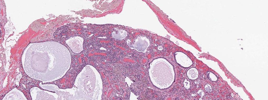
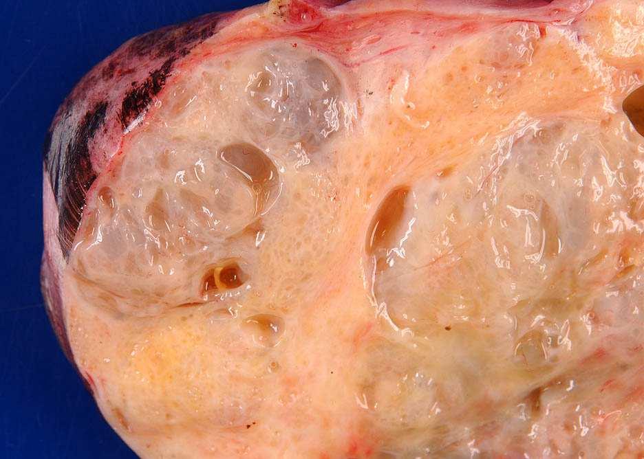
134 Intestinal-type mucinous tumors: problems Primary versus metastasis Borderline tumor versus carcinoma Same as endometrioid tumors Grading Shimizu/Silverberg
135 Intestinal-type mucinous tumors: features favoring metastasis Bilateral disease Surface involvement Destructive stromal invasion Nodular growth pattern Single cells/signet ring cells Vascular invasion Lee KR, Young RH. Am J Surg Pathol Mar; 27(3):
136
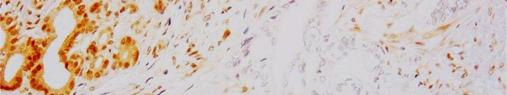
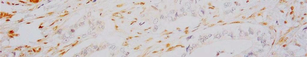
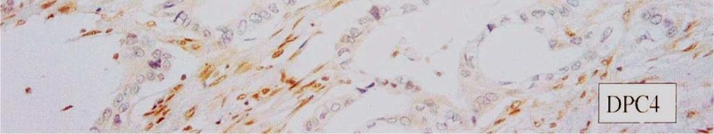
137
138
139
140 Algorithm for distinguishing primary and metastatic mucinous carcinoma Bilateral mucinous carcinomas: metastastic Unilateral mucinous carcinomas <12 cm: metastatic Unilateral mucinous carcinomas >12 cm: primary ovarian Yemelyanova AV, et al. Am J Surg Pathol Jan;32(1):128-38
141 Pseudomyxoma peritonei (PMP) PMP associated with ovarian tumor(s) is almost never derived from the ovaries themselves
142 Pseudomyxoma peritonei low grade
143 Pseudomyxoma high grade
144 Primary ovarian mucinous carcinoma: immunophenotype CK7>20 Retained SMAD4 Negative: Racemase (positive in colorectal) β-catenin (positive in colorectal and endometrioid) ER (positive in endometrioid and breast) P16 (positive in endocervical and serous) Mesothelin (positive in pancreatic, serous and mesothelial) Fascin (positive in pancreatic)
145 Mucinous carcinoma immunohistochemistry Informative IHC pattern: CK7<CK20 NOT ovarian primary (if algorithm suggests metastasis) Rule out colorectal and appendiceal primaries Consider other possibilities CK7 < CK20
 Rule out")
 Consider other")
146 Mucinous carcinoma immunohistochemistry Uninformative IHC patterns (CK7=CK20; CK7>CK20) Rule out pancreatobiliary, upper gastrointestinal primaries (if algorithm suggests metastasis) Consider other possibilities CK7 > CK20
147 SMAD4 Smad4 (DPC4) mediates TGFb pathway suppressing epithelial cell growth Germline mutations FJP Somatic alterations Panc CA (55%) Colon CA (10-35%) Immunohistochemical loss of expression Pancreatic CA (46%) Colon CA (11%) Ovarian mucinous CA (0%) Endometrioid CA (?>0%) Biochem Biophys Res Commun 2003; 306:
148
149 Mucinous carcinoma immunohistochemistry Algorithm suggests metastasis or CK7>20 or CK7=CK20 SMAD4 retained SMAD4 lost Not informative Pancreatic>colorectal

150 Summary: intestinal mucinous carcinomas of ovary Unilateral ovarian tumor; no pertinent medical history Intracytoplasmic mucin, expansile invasion Intestinal mucinous borderline tumor CK7>20, retained SMAD4 Negative racemase, β-catenin, ER, p16, mesothelin, fascin K-ras Other entities are excluded: exclude metastasis
151
International Society of Gynecological Pathologists Symposium 2007
 International Society of Gynecological Pathologists Symposium 2007 Anais Malpica, M.D. Department of Pathology The University of Texas M.D. Anderson Cancer Center Grading of Ovarian Cancer Histologic grade
Current Concept in Ovarian Carcinoma: Pathology Perspectives
 Current Concept in Ovarian Carcinoma: Pathology Perspectives Rouba Ali-Fehmi, MD Professor of Pathology The Karmanos Cancer Institute, Wayne State University School of Medicine Current Concept in Ovarian
of 20 to 80 and subsequently declines [2].
![of 20 to 80 and subsequently declines [2].](/thumbs/80/81450506.jpg "of 20 to 80 and subsequently declines [2].") - - According to the 2014 World Health Organization (WHO) classification and tumor morphology, primary ovarian tumors are subdivided into three categories: epithelial (60%), germ cell (30%), and sex-cord
- - According to the 2014 World Health Organization (WHO) classification and tumor morphology, primary ovarian tumors are subdivided into three categories: epithelial (60%), germ cell (30%), and sex-cord
Ovarian Clear Cell Carcinoma
 Ovarian Clear Cell Carcinoma Rouba Ali-Fehmi, MD Professor of Pathology The Karmanos Cancer Institute, Wayne State University School of Medicine 50 year old woman with chief complaint of shortness of breath
Case presentation 04/13/2017. Genomic/morphological classification of endometrial carcinoma
 Genomic/morphological classification of endometrial carcinoma Robert A. Soslow, MD soslowr@mskcc.org architecture.about.com Case presentation 49 year old woman with vaginal bleeding Underwent endometrial
5/26/2016. Pelvic Serous Carcinoma: 2014 W.H.O. Update. Outline of Talk. Changes to 2014 WHO system for pelvic serous tumors
 Pelvic Serous Carcinoma: 2014 W.H.O. Update Outline of Talk Practical Implications for Pathologists Changes to 2014 WHO system for pelvic serous tumors High grade serous carcinoma versus low grade serous
Bibliography. Serous Tumors of the Ovary. Nomenclature
 Bibliography Serous Tumors of the Ovary Nomenclature 1. Allison KH, Swisher EM, Kerkering KM, et al. Defining an appropriate threshold for the diagnosis of serous borderline tumor of the ovary: when is
Mucinous Tumors of the Ovary Beirut, Lebanon. Anaís Malpica, M.D. Professor Department of Pathology
 Mucinous Tumors of the Ovary Beirut, Lebanon Anaís Malpica, M.D. Professor Department of Pathology Primary Mucinous Tumors of the Ovary Cystadenoma Borderline (Tumor of Low Malignant Potential/Atypical
Section 1. Biology of gynaecological cancers: our current understanding
 Section 1 Biology of gynaecological cancers: our current understanding Chapter 1 Morphological sub-types of ovarian carcinoma: new developments and pathogenesis W Glenn McCluggage 1 Introduction In most
Low-grade serous neoplasia. Robert A. Soslow, MD
 Low-grade serous neoplasia Robert A. Soslow, MD soslowr@mskcc.org Outline Orientation Ovarian tumor overview Non serous borderline tumors Serous borderline tumors Clinical summary Morphologic description
The Diagnostic Challenges of Low Grade and High Grade Tubo-Ovarian Serous Carcinomas. W Glenn McCluggage Belfast, Northern Ireland
 The Diagnostic Challenges of Low Grade and High Grade Tubo-Ovarian Serous Carcinomas W Glenn McCluggage Belfast, Northern Ireland Enterprise Interest None OVARIAN SEROUS CARCINOMA (OSC) RECENT DEVELOPMENTS
Ovarian cancer: 2012 Update Srini Prasad MD Univ Texas MD Anderson Cancer Center
 Ovarian cancer: 2012 Update Srini Prasad MD Univ Texas MD Anderson Cancer Center Ovarian cancer is not a single disease Ovarian Epithelial Tumors: Histological Spectrum* Type Frequency Histology High-Grade
ENODMETRIAL CARCINOMA: SPECIAL & NOT SO SPECIAL VARIANTS
 ENODMETRIAL CARCINOMA: SPECIAL & NOT SO SPECIAL VARIANTS Pacific Northwest Society of Pathologists Vancouver, B.C. September 26, 2015 Teri A. Longacre, M.D. longacre@stanford.edu Stanford University, Stanford,
How to Recognize Gynecologic Cancer Cells from Pelvic Washing and Ascetic Specimens
 How to Recognize Gynecologic Cancer Cells from Pelvic Washing and Ascetic Specimens Wenxin Zheng, M.D. Professor of Pathology and Gynecology University of Arizona zhengw@email.arizona.edu http://www.zheng.gynpath.medicine.arizona.edu/index.html
ACCME/Disclosures. Risk of Gyne Ca in HBOC. Molecular basis of HBOC. Hereditary Ovarian and Breast Cancer Syndrome
 Hereditary Ovarian and Breast Cancer Syndrome C. Blake Gilks, MD Dept of Pathology Vancouver General Hospital University of British Columbia Blake.gilks@vch.ca The USCAP requires that anyone in a position
Case 1. Pathology of gynecological cancer. What do we need to know (Case 1) Luca Mazzucchelli Istituto cantonale di patologia Locarno
 Luca Mazzucchelli Istituto cantonale di patologia Locarno") Case 1 Pathology of gynecological cancer. What do we need to know (Case 1) Luca Mazzucchelli Istituto cantonale di patologia Locarno SAMO Interdisciplinary Workshop on Gynecological Tumors Lucern, October
Case 1 Pathology of gynecological cancer. What do we need to know (Case 1) Luca Mazzucchelli Istituto cantonale di patologia Locarno SAMO Interdisciplinary Workshop on Gynecological Tumors Lucern, October
Patologia Molecular del Carcinoma de Ovario
 Curso de Patologia Molecular XXVI Congreso Nacional de la SEAP Cadiz Patologia Molecular del Carcinoma de Ovario Jaime Prat Barcelona Ovarian Epithelial Tumors WHO 1999 and 2003 Serous Mucinous Endometrioid
What s (new) and Important in Reporting of Uterine Cancers Katherine Vroobel The Royal Marsden
 and Important in Reporting of Uterine Cancers Katherine Vroobel The Royal Marsden") What s (new) and Important in Reporting of Uterine Cancers Katherine Vroobel The Royal Marsden Maastricht Pathology 2018 Wednesday 20 th June Endometrioid adenocarcinoma High grade carcinomas (common)
What s (new) and Important in Reporting of Uterine Cancers Katherine Vroobel The Royal Marsden Maastricht Pathology 2018 Wednesday 20 th June Endometrioid adenocarcinoma High grade carcinomas (common)
Mody. AIS vs. Invasive Adenocarcinoma of the Cervix
 Common Problems in Gynecologic Pathology Michael T. Deavers, M.D. Houston Methodist Hospital, Houston, Texas Common Problems in Gynecologic Pathology Adenocarcinoma in-situ (AIS) of the Cervix vs. Invasive
Bases biológicas del cáncer de ovario en el siglo XXI
 Bases biológicas del cáncer de ovario en el siglo XXI Iñigo Espinosa, M.D. Clínica Universidad de Navarra Epithelial Ovarian Tumors WHO 1973-2014 Serous Mucinous Endometrioid Clear cell Transitional Squamous
Mucinous Adenocarcinoma Involving the Ovary: Comparative Evaluation of the Classification Algorithms using Tumor Size and Laterality
 J Korean Med Sci 2010; 25: 220-5 ISSN 1011-8934 DOI: 10.3346/jkms.2010.25.2.220 Mucinous Adenocarcinoma Involving the Ovary: Comparative Evaluation of the Classification Algorithms using Tumor Size and
Pathobiology of ovarian carcinomas
 Chinese Journal of Cancer Review Mojgan Devouassoux-Shisheboran 1 and Catherine Genestie 2 Abstract Ovarian tumors comprise a heterogeneous group of lesions, displaying distinct tumor pathology and oncogenic
Low-Grade Serous Ovarian Tumors Debra A. Bell, MD Mayo Clinic and Mayo Medical School Rochester, MN
 1 Low-Grade Serous Ovarian Tumors Debra A. Bell, MD Mayo Clinic and Mayo Medical School Rochester, MN It is very appropriate to discuss low-grade ovarian serous neoplasms in a symposium in honor of Dr.
Borderline tumors. Borderline tumors. Serous borderline tumor are NOT benign. Low grade serous carcinoma: pathogenesis. Serous carcinoma: pathogenesis
 Serous borderline tumor are NOT benign Robert A. Soslow, MD Memorial Sloan-Kettering Cancer Center soslowr@mskcc.org Borderline tumors Serous BTs and seromucinous BTs are both histopathologically borderline
Dall istologia alla caratterizzazione biomolecolare
 Il carcinoma ovarico: approccio multidisciplinare e prospettive terapeutiche Dall istologia alla caratterizzazione biomolecolare Anna Pesci Ospedale SC Don Calabria, Negrar anna.pesci@sacrocuore.it Ovarian
Interpretation of p53 Immunostains. P53 Mutations are Ubiquitous in High Grade Serous Carcinoma. Diffuse strong positive nuclear staining
 Stains for Tumor Classification p53 p16 WT1 HMGA2 P53 Mutations are Ubiquitous in High Grade Serous Carcinoma Source Ahmed et al Australian Ovarian Cancer Study Cancer Genome Atlas Research Network Cases
Molecular Subtyping of Endometrial Cancer: A ProMisE ing Change
 Molecular Subtyping of Endometrial Cancer: A ProMisE ing Change Charles Matthew Quick, M.D. Associate Professor of Pathology Director of Gynecologic Pathology University of Arkansas for Medical Sciences
3/24/2017. Disclosure of Relevant Financial Relationships. Mixed Epithelial Endometrial Carcinoma. ISGyP Endometrial Cancer Project
 Disclosure of Relevant Financial Relationships USCAP requires that all planners (Education Committee) in a position to influence or control the content of CME disclose any relevant financial relationship
USCAP 2013: THE ORIGINS OF OVARIAN CANCER: MUCINOUS TUMORS
 USCAP 2013: THE ORIGINS OF OVARIAN CANCER: MUCINOUS TUMORS Russell Vang, M.D. Associate Professor Division of Gynecologic Pathology The Johns Hopkins Hospital Table of contents: Powerpoint handout Text
3 cell types in the normal ovary
 Ovarian tumors 3 cell types in the normal ovary Surface (coelomic epithelium) the origin of the great majority of ovarian tumors (neoplasms) 90% of malignant ovarian tumors Totipotent germ cells Sex cord-stromal
Mousa. Najat kayed &Renad Al-Awamleh. Nizar Alkhlaifat
 6 Mousa Najat kayed &Renad Al-Awamleh Nizar Alkhlaifat P a g e 1 This sheet written based on record 13 on website Cover slide( 95-117 ) No need to go back to slide FALLOPIAN TUBE PATHOLOGY In general fallopian
Introduction. Agenda. Pathogenesis. Pathogenesis and Clinical Implications. Pathogenesis and Clinical Implications
 Surface Epithelial Tumors of the Ovary Part II Invasive Carcinomas Moderator Robert J. Kurman, M.D. Discussants Kathleen R. Cho, M.D. Anais Malpica, M.D. Patricia A. Shaw, M.D. Robert A. Soslow, M.D. C.
Clinical History USCAP Specialty Conference. Gynecologic Pathology Case 3
 2010 USCA Specialty Conference Gynecologic athology Case Kathleen R. Cho, M.D. Department of athology Clinical History 46 yo woman presented with bilateral ovarian masses and elevated CA-125 TAH/BSO, pelvic
05/07/2018. Types of challenges. Challenging cases in uterine pathology. Case 1 ` 65 year old female Post menopausal bleeding Uterine Polyp
 Types of challenges Challenging cases in uterine pathology Nafisa Wilkinson Gynaecological Pathologist UCLH London Lack of complete history often, NO clinical history at all! Cases from other centres often
Adenocarcinoma of Mullerian origin: review of pathogenesis, molecular biology, and emerging treatment paradigms
 Cobb et al. Gynecologic Oncology Research and Practice (2015) 2:1 DOI 10.1186/s40661-015-0008-z REVIEW Adenocarcinoma of Mullerian origin: review of pathogenesis, molecular biology, and emerging treatment
3 cell types in the normal ovary
 Ovarian tumors 3 cell types in the normal ovary Surface (coelomic epithelium) the origin of the great majority of ovarian tumors 90% of malignant ovarian tumors Totipotent germ cells Sex cord-stromal cells
Objectives. Atypical Glandular Cells. Atypical Endocervical Cells. Reactive Endocervical Cells
 2013 California Society of Pathologists 66 th Annual Meeting San Francisco, CA Atypical Glandular Cells to Early Invasive Adenocarcinoma: Cervical Cytology and Histology Christina S. Kong, MD Associate
Important Recent Advances in Gynaecological Pathology
 Important Recent Advances in Gynaecological Pathology Sanjiv Manek Consultant Gynaecological Pathologist Oxford, UK In recent years there have been a significant number of changes in gynaecological pathology
Case Report Ovarian Seromucinous Borderline Tumor and Clear Cell Carcinoma: An Unusual Combination
 Case Reports in Obstetrics and Gynecology Volume 2015, Article ID 690891, 5 pages http://dx.doi.org/10.1155/2015/690891 Case Report Ovarian Seromucinous Borderline Tumor and Clear Cell Carcinoma: An Unusual
The role of immunohistochemistry in surgical pathology of the uterine corpus and cervix
 The role of immunohistochemistry in surgical pathology of the uterine corpus and cervix Prof. Ben Davidson, MD PhD Department of Pathology, Norwegian Radium Hospital, Oslo University Hospital, Oslo, Norway
GOBLET CELL CARCINOID. Hanlin L. Wang, MD, PhD University of California Los Angeles
 GOBLET CELL CARCINOID Hanlin L. Wang, MD, PhD University of California Los Angeles Disclosure of Relevant Financial Relationships USCAP requires that all planners (Education Committee) in a position to
GOBLET CELL CARCINOID
 GOBLET CELL CARCINOID Hanlin L. Wang, MD, PhD University of California Los Angeles Disclosure of Relevant Financial Relationships USCAP requires that all planners (Education Committee) in a position to
Endometrial Metaplasia, Hyperplasia & Other Cancer Mimics: a Consultant s Experience
 Endometrial Metaplasia, Hyperplasia & Other Cancer Mimics: a Consultant s Experience Pacific Northwest Society of Pathologists Vancouver, B.C. September 26, 2015 Teri A. Longacre, M.D. longacre@stanford.edu
When Immunostains Can Get You in Trouble: Gynecologic Pathology p16: Panacea or Pandora s Box?
 When Immunostains Can Get You in Trouble: Gynecologic Pathology p16: Panacea or Pandora s Box? Teri A. Longacre, MD Stanford Medicine Stanford California pi6 in Gynecologic Pathology: Panacea or Pandora
Survival Analysis and Prognosis for Patients with Serous and Mucinous Borderline Ovarian Tumors: 14-Year Experience from a Tertiary Center in Iran
 ORIGINAL ARTICLE Survival Analysis and Prognosis for Patients with Serous and Mucinous Borderline Ovarian Tumors: 14-Year Experience from a Tertiary Center in Iran Katayoun Ziari, Ebrahim Soleymani, and
Dr Sanjiv Manek Oxford. Oxford Pathology Course 2010 for FRCPath Illustration-Cellular Pathology. Oxford Radcliffe NHS Trust
 Dr Sanjiv Manek Oxford Oxford Pathology Course 2010 for FRCPath Illustration-Cellular Pathology. Oxford Radcliffe NHS Trust Ovarian Endometrial Vulvo-vaginal Cervical Illustration-Cellular Pathology. Oxford
New Developments in Immunohistochemistry for Gynecologic Pathology
 New Developments in Immunohistochemistry for Gynecologic Pathology Michael T. Deavers, M.D. Professor, Departments of Pathology and Gynecologic Oncology Immunohistochemistry in Gynecologic Pathology Majority
Int. J. Curr. Res. Med. Sci. (2017). 3(1): International Journal of Current Research in Medical Sciences
. 3(1): International Journal of Current Research in Medical Sciences") International Journal of Current Research in Medical Sciences ISSN: 2454-5716 www.ijcrims.com Volume 3, Issue 1-2017 Case Report DOI: http://dx.doi.org/10.22192/ijcrms.2017.03.01.006 A rare case report
International Journal of Current Research in Medical Sciences ISSN: 2454-5716 www.ijcrims.com Volume 3, Issue 1-2017 Case Report DOI: http://dx.doi.org/10.22192/ijcrms.2017.03.01.006 A rare case report
Case 3 - GYN. History: 66 year old, routine Pap test. Dr. Stelow
 Case 3 - GYN History: 66 year old, routine Pap test Dr. Stelow Case 3 66 year year old woman Routine Pap Test Cytologic Features 3 dimensional clusters of cells with small to moderate amount of
Select problems in cystic pancreatic lesions
 Disclosure Select problems in cystic pancreatic lesions Five Prime Therapeutics shareholder Adicet Bio shareholder Bristol-Meyer Squibb advisory board grace.kim@ucsf.edu Pancreatic cystic lesions Intraductal
ACCME/Disclosures. Cribriform Lesions of the Prostate. Case
 Cribriform Lesions of the Prostate Ming Zhou, MD, PhD Departments of Pathology and Urology New York University Langone Medical Center New York, NY Ming.Zhou@NYUMC.ORG ACCME/Disclosures The USCAP requires
64 YO lady THBSO for prolapse At gross : A 3 cm endometrial polyp in the fundus
 Case 6 64 YO lady THBSO for prolapse At gross : A 3 cm endometrial polyp in the fundus Numerous irregular, large glands with leaf-like pattern Large glands with broad-based papillary infolding into the
Primary Mucinous Ovarian Cancer (PMOC) Michael Frumovitz
 Michael Frumovitz") Primary Mucinous Ovarian Cancer (PMOC) Michael Frumovitz Epithelial Subtypes Serous Endometrioid Mucinous Transitional Clear Cell Mixed Undifferentiated Squamous Ovarian Surface Epithelium Naora et al.,
Primary Mucinous Ovarian Cancer (PMOC) Michael Frumovitz Epithelial Subtypes Serous Endometrioid Mucinous Transitional Clear Cell Mixed Undifferentiated Squamous Ovarian Surface Epithelium Naora et al.,
Interobserver Variability in the Diagnosis of Uterine High-Grade Endometrioid Carcinoma
 Interobserver Variability in the Diagnosis of Uterine High-Grade Endometrioid Carcinoma Sumi Thomas, MD; Yaser Hussein, MD; Sudeshna Bandyopadhyay, MD; Michele Cote, PhD; Oudai Hassan, MD; Eman Abdulfatah,
A Survay on Appendiceal Involvement in Ovarian Mucinous Tumors
 http://www.ijwhr.net Open Access doi 10.15296/ijwhr.2018.33 Original Article International Journal of Women s Health and Reproduction Sciences Vol. 6, No. 2, April 2018, 199 203 ISSN 2330-4456 A Survay
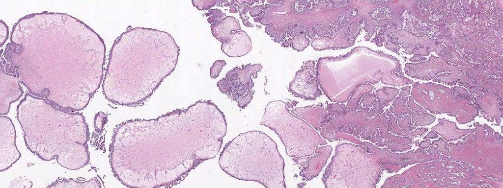
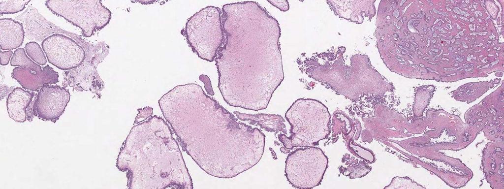
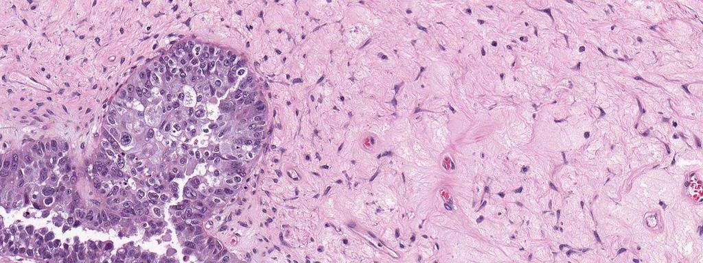
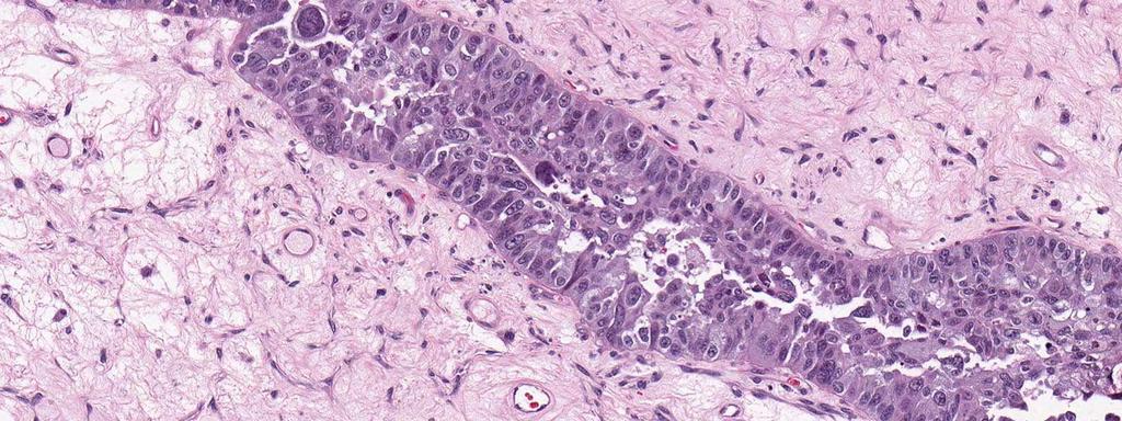
Case # 4 Low-Grade Serous Carcinoma (Macropapillary) of the Ovary Arising in an Atypical Proliferative Serous Tumor
 of the Ovary Arising in an Atypical Proliferative Serous Tumor") Case # 4 Low-Grade Serous Carcinoma (Macropapillary) of the Ovary Arising in an Atypical Proliferative Serous Tumor Robert J Kurman, M.D. Johns Hopkins University School of Medicine Case History A 53 year
Case # 4 Low-Grade Serous Carcinoma (Macropapillary) of the Ovary Arising in an Atypical Proliferative Serous Tumor Robert J Kurman, M.D. Johns Hopkins University School of Medicine Case History A 53 year
Wendy L Frankel. Chair and Distinguished Professor
 1 Wendy L Frankel Chair and Distinguished Professor Case 1 59 y/o woman Abdominal pain No personal or family history of cancer History of colon polyps Colonoscopy Polypoid rectosigmoid mass Biopsy 3 4
Unknown Slides Conference
 Unknown Slides Conference Jae Y. Ro, MD, PhD Weill Medical College of Cornell Univ. The Methodist Hospital, and UT MD Anderson Cancer Center Houston, TX November 9, 2013 Amman, Jordan 25 th Congress of
Pathology of Ovarian Tumours. Dr. Jyothi Ranganathan MD ( Path) AFMC Pune PDCC (Cytopathology) PGI Chandigarh
 AFMC Pune PDCC (Cytopathology) PGI Chandigarh") Pathology of Ovarian Tumours Dr. Jyothi Ranganathan MD ( Path) AFMC Pune PDCC (Cytopathology) PGI Chandigarh Outline Incidence Risk factors Classification Pathology of tumours Tumour markers Prevention
Pathology of Ovarian Tumours Dr. Jyothi Ranganathan MD ( Path) AFMC Pune PDCC (Cytopathology) PGI Chandigarh Outline Incidence Risk factors Classification Pathology of tumours Tumour markers Prevention
6/5/2010. Outline of Talk. Endometrial Alterations That Mimic Cancer & Vice Versa: Metaplastic / reactive changes. Problems in Biopsies/Curettages
 Outline of Talk Endometrial Alterations That Mimic Cancer & Vice Versa: Problems in Biopsies/Curettages Metaplastic / reactive changes Mucinous change Microglandular hyperplasia-like change Squamous metaplasia
RESEARCH COMMUNICATION
 RESEARCH COMMUNICATION Clinicopathologic Analysis of Women with Synchronous Primary Carcinomas of the Endometrium and Ovary: 10- Year Experience from Chiang Mai University Hospital Jiraprapa Natee 1 *,
Pacific Northwest Society of Pathologists Fall Meeting September 2015 Intraoperative Consultation in Gynecological Pathology: The Adnexal Mass
 Pacific Northwest Society of Pathologists Fall Meeting September 2015 Intraoperative Consultation in Gynecological Pathology: The Adnexal Mass Julie Irving, MD Department of Pathology, University of British
Papillary Lesions of the breast
 Papillary Lesions of the breast Emad Rakha Professor of Breast Pathology The University of Nottingham Papillary lesions of the breast are a heterogeneous group of disease, which are characterised by neoplastic
Page # 1. Endometrium. Cellular Components. Anatomical Regions. Management of SIL Thomas C. Wright, Jr. Most common diseases:
 Endometrium Pathology of the Endometrium Thomas C. Wright Columbia University, New York, NY Most common diseases: Abnormal uterine bleeding Inflammatory conditions Benign neoplasms Endometrial cancer Anatomical
Presenter: Yeh-Han Wang M.D.
 Korea-Taiwan-Japan Joint Meeting for Gynecological Pathology Mini-lecture Female Adnexal Tumor of Probable Wolffian Origin (FATWO) in Taiwan: A Small Case Series and Literature Review Presenter: Yeh-Han
Intrahepatic cholangiocarcinoma Histologic spectrum, novel markers and molecular assays
 2018 Current Issues in Surgical Pathology Summary (not actual lecture) Intrahepatic cholangiocarcinoma Histologic spectrum, novel markers and molecular assays Sanjay Kakar, MD University of California,
ACCME/Disclosures. Case History 4/13/2016. USCAP GU Specialty Conference Case 3. Ann Arbor, MI
 USCAP GU Specialty Conference Case 3 March 2016 L. Priya Kunju, M.D. University of Michigan Health System Ann Arbor, MI University of Michigan Health System ACCME/Disclosures The USCAP requires that anyone
Institute of Pathology First Faculty of Medicine Charles University. Ovary
 Ovary Barrett esophagus ph in vagina between 3.8 and 4.5 ph of stomach varies from 1-2 (hydrochloric acid) up to 4-5 BE probably results from upward migration of columnar cells from gastroesophageal junction
Endometrial pathology. Dr Tom Dodd and Dr Georgina England
 Endometrial pathology Dr Tom Dodd and Dr Georgina England Case 1 Female age 35 Case 1 Proliferative endometrium Case 2 Female age 38 Case 2 Secretory endometrium Dating endometrium Assessed on the
Normal endometrium: A, proliferative. B, secretory.
 Normal endometrium: A, proliferative. B, secretory. Nội mạc tử cung Nội mạc tử cung Cyclic changes in endometrium.. Approximate relationship of useful microscopic changes. Arias-Stella reaction in endometrial
Diagnostically Challenging Cases in Gynecologic Pathology
 Diagnostically Challenging Cases in Gynecologic Pathology Eric C. Huang, M.D., Ph.D. Department of Pathology and Laboratory Medicine University of California, Davis Medical Center Case 1 Presentation 38
In situ and Invasive Endocervical Carcinoma: Problems and Pitfalls in Diagnosis
 In situ and Invasive Endocervical Carcinoma: Problems and Pitfalls in Diagnosis Rouba Ali-Fehmi,MD The Karmanos Cancer Institute, Wayne State University School of Medicine Global incidence of cervical
The Origin of Pelvic Low-Grade Serous Proliferative Lesions
 The Origin of Pelvic Low-Grade Serous Proliferative Lesions Ovarian Atypical Proliferative (Borderline) Serous Tumors, Noninvasive Implants and Endosalpingiosis Robert J. Kurman, M.D. Kurman RJ, Vang R,
Original contribution
 Human Pathology (2012) 43, 747 752 www.elsevier.com/locate/humpath Original contribution The presence and location of epithelial implants and implants with epithelial proliferation may predict a higher
FREQUENCY OF NAPSIN A POSITIVITY IN OVARIAN CLEAR CELL CARCINOMA AND SEROUS CARCINOMA
 Open Access Original Article Napsin A Positivity in Ovarian Clear Cell Carcinoma Pak Armed Forces Med J 2018; 68 (4): 723-28 FREQUENCY OF NAPSIN A POSITIVITY IN OVARIAN CLEAR CELL CARCINOMA AND SEROUS
Gynecologic Cancers are many diseases. Gynecologic Cancers in the Age of Precision Medicine Advances in Internal Medicine. Speaker Disclosure:
 Gynecologic Cancer Care in the Age of Precision Medicine Gynecologic Cancers in the Age of Precision Medicine Advances in Internal Medicine Lee-may Chen, MD Department of Obstetrics, Gynecology & Reproductive
ARTHUR PURDY STOUT SOCIETY COMPANION MEETING: DIFFICULT NEW DIFFERENTIAL DIAGNOSES IN PROSTATE PATHOLOGY. Jonathan I. Epstein.
 1 ARTHUR PURDY STOUT SOCIETY COMPANION MEETING: DIFFICULT NEW DIFFERENTIAL DIAGNOSES IN PROSTATE PATHOLOGY Jonathan I. Epstein Professor Pathology, Urology, Oncology The Reinhard Professor of Urological
Gynecologic Cancers are many diseases. Speaker Disclosure: Gynecologic Cancer Care in the Age of Precision Medicine. Controversies in Women s Health
 Gynecologic Cancer Care in the Age of Precision Medicine Gynecologic Cancers in the Age of Precision Medicine Controversies in Women s Health Lee-may Chen, MD Department of Obstetrics, Gynecology & Reproductive
Intravascular Endometrium Mimicking Vascular Invasion
 ISPUB.COM The Internet Journal of Pathology Volume 12 Number 1 A Papanicolau, G Lin Citation A Papanicolau, G Lin.. The Internet Journal of Pathology. 2010 Volume 12 Number 1. Abstract Intravascular endometrium
Atypical Hyperplasia/EIN
 EIN Atypical Hyperplasia/EIN Based on scientific and diagnostic advances, in 2014 the WHO moved that the precursor lesion for endometrioid carcinoma be atypical hyperplasia/ein, rather than what was previously
4/12/2018. MUSC Pathology Symposium Kiawah Island April 18, Jesse K. McKenney, MD
 MUSC Pathology Symposium Kiawah Island April 18, 2018 Jesse K. McKenney, MD 1 Urothelial Carcinoma with Alternative Differentiation 2 Urothelial Carcinoma with Alternative Differentiation Recognition as
Department of Pathology, Royal Group of Hospitals Trust, Belfast, Northern Ireland.
 UTERINE ADENOSARCOMA W Glenn McCluggage Department of Pathology, Royal Group of Hospitals Trust, Belfast, Northern Ireland. Definition of Adenosarcoma: A mixed tumor composed of benign neoplastic glandular
5/21/2018. Prostate Adenocarcinoma vs. Urothelial Carcinoma. Common Differential Diagnoses in Urological Pathology. Jonathan I.
 Common Differential Diagnoses in Urological Pathology Jonathan I. Epstein Prostate Adenocarcinoma vs. Urothelial Carcinoma 1 2 NKX3.1 NKX3.1 3 4 5 6 Proposed ISUP Recommendations Option to use PSA as a
Epithelial Ovarian Cancer 8/2/2013. Tu-be or Not Tu-be: Is the Fallopian Tube the Source of Ovarian Cancer?
 Tu-be or Not Tu-be: Is the Fallopian Tube the Source of Ovarian Cancer? Ann E. Smith Sehdev, MD Director, Center for Gynecologic Pathology Cascade Pathology, Portland, Oregon Ann E. Smith Sehdev has no
Advanced Stage Mucinous Adenocarcinoma of the Ovary Is Both Rare and Highly Lethal
 Advanced Stage Mucinous Adenocarcinoma of the Ovary Is Both Rare and Highly Lethal A Gynecologic Oncology Group Study Richard J. Zaino, MD 1 ; Mark F. Brady, PhD 2 ; Subodh M. Lele, MD 3 ; Helen Michael,
Borderline Ovarian Mucinous Tumors: Consensus Points and Persistent Controversies Regarding Nomenclature, Diagnostic Criteria, and Behavior
 Borderline Ovarian Mucinous Tumors: Consensus Points and Persistent Controversies Regarding Nomenclature, Diagnostic Criteria, and Behavior Brigitte M. Ronnett, M.D.; C. Blake Gilks, M.D., Maria J. Merino,
Gynecologic Cytopathology: Glandular lesions
 Gynecologic Cytopathology: Glandular lesions Lin Wai Fung (MSc, MPH, CMIAC) 17/4/2014 Glandular lesions of the uterus Endocervix Endometrium Normal endocervical cells Sheets, strips well-preserved architecture:
Identification of Potential Therapeutic Targets by Molecular and Genomic Profiling of 628 Cases of Uterine Serous Carcinoma
 Identification of Potential Therapeutic Targets by Molecular and Genomic Profiling of 628 Cases of Uterine Serous Carcinoma Nathaniel L Jones 1, Joanne Xiu 2, Sandeep K. Reddy 2, Ana I. Tergas 1, William
Histologic and Immunohistochemical Analyses of Endometrial Carcinomas. Experiences From Endometrial Biopsies in 358 Consultation Cases
 Histologic and Immunohistochemical Analyses of Endometrial Carcinomas Experiences From Endometrial Biopsies in 358 Consultation Cases Jian-Jun Wei, MD; Ajit Paintal, MD; Pacita Keh, MD Context. Uterine
David Nunns on behalf of the Gynae Guidelines Group Date:
 Title of Guideline (must include the word Guideline (not protocol, policy, procedure etc) Borderline tumours of the ovary management and follow-up Author: Contact Name and Job Title Directorate & Speciality
Enterprise Interest None
 Enterprise Interest None What are triple negative breast cancers? A synopsis of their histological patterns Ian Ellis Molecular Medical Sciences, University of Nottingham Department of Histopathology,
Immunohistochemical Expression of Cytokeratin 5/6 in Gynaecological Tumors.
 ISPUB.COM The Internet Journal of Pathology Volume 13 Number 2 Immunohistochemical Expression of Cytokeratin 5/6 in Gynaecological Tumors. A Baghla, S Choudhry, A Kataria Citation A Baghla, S Choudhry,
Update in Salivary Gland Pathology. Benjamin L. Witt University of Utah/ARUP Laboratories February 9, 2016
 Update in Salivary Gland Pathology Benjamin L. Witt University of Utah/ARUP Laboratories February 9, 2016 Objectives Review the different appearances of a selection of salivary gland tumor types Establish
Endometrial Stromal Tumors
 Endometrial Stromal Tumors WHO Categories: Endometrial Stromal Nodule (ESN) Endometrial Stromal Sarcoma, low grade (LGESS) Endometrial Stromal Sarcoma, high grade (HGESS) Undifferentiated Uterine Sarcoma
Borderline Brenner tumor of the ovary: a case report with immunohistochemical and molecular study
 De Cecio et al. Journal of Ovarian Research 2014, 7:101 CASE REPORT Open Access Borderline Brenner tumor of the ovary: a case report with immunohistochemical and molecular study Rossella De Cecio 1, Monica
BLADDER CANCER EPIDEMIOLOGY
 BLADDER CANCER WHAT IS NEW AND CLINICALLY RELEVANT Canadian Geese - Geist Reservoir (my backyard), Indianapolis, USA BLADDER CANCER EPIDEMIOLOGY Urinary bladder 17,960 2% Urinary bladder 4,390 1.6% Siegel
Hepatocyte Nuclear Factor-1b Is Not a Specific Marker of Clear Cell Carcinoma in Serous Effusions
 Hepatocyte Nuclear Factor-1b Is Not a Specific Marker of Clear Cell Carcinoma in Serous Effusions Ben Davidson, MD, PhD 1,2 BACKGROUND: The transcription factor hepatocyte nuclear factor-1b (HNF1b) has
Adenocarcinoma of the Cervix
 Question 1. Each of the following statements about cervical adenocarcinoma is true except: Adenocarcinoma of the Cervix SAMS a) A majority of women with cervical adenocarcinoma have stage I tumors at diagnosis.
Shina Oranratanaphan, Tarinee Manchana*, Nakarin Sirisabya
 Comparison of Synchronous Endometrial and Ovarian Cancers versus Primary with Metastasis RESEARCH COMMUNICATION Clinicopathologic Variables and Survival Comparison of Patients with Synchronous Endometrial
Loss of Mismatch Repair Protein Expression in Epithelial Ovarian Carcinoma: A Histomorphologic Guide to Targeted Screening
 Loss of Mismatch Repair Protein Expression in Epithelial Ovarian Carcinoma: A Histomorphologic Guide to Targeted Screening Eman Abdulfatah a, Sharif Sakr b, Sumi Thomas a, Sudeshna Bandyopadhyay a, Kavita