Macro- and microacinar proliferations of the prostate
|
|
|
- Marvin Harris
- 5 years ago
- Views:
Transcription
")
 Polytechnic University of Marche")

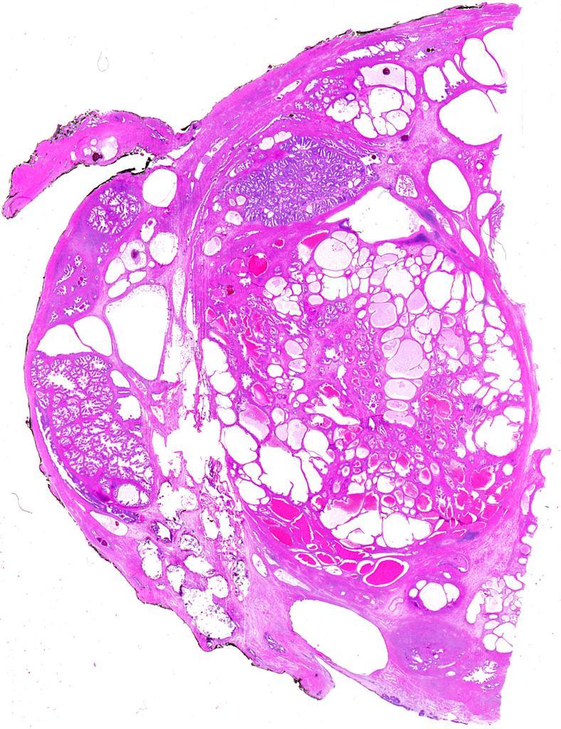
1 Macro- and microacinar proliferations of the prostate (with emphasis on cancer mimics) Rodolfo Montironi, MD (IT), FRCPath (UK), IFCAP (USA) Polytechnic University of Marche Region (Ancona) School of Medicine, Ancona, Italy and Arizona Cancer Center, Tucson, AZ, USA

2 Lesions with 1. small 2. large and cribriform 3. solid and nonglandular patterns Further subdivided into 1. those of prostatic epithelial origin and 2. those of nonprostatic epithelial origin
3 - Architectural pattern: Small gland - PCa mimicked: Gleason pattern 3 Lesions of prostatic epithelial origin 1.Atrophy 2.Adenosis (Atypical adenomatous hyperplasia, AAH)) 3.Sclerosing adenosis 4.Radiation atypia in benign glands 5.Verumontanum mucosal gland hyperplasia
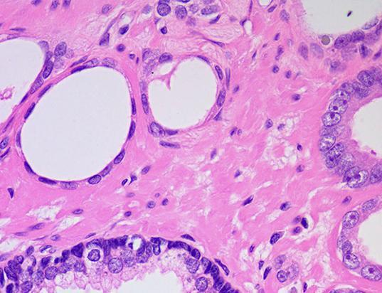
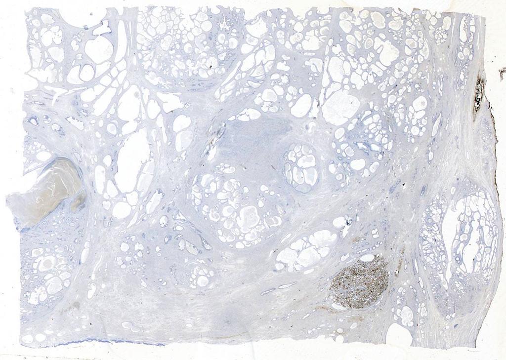
4 Small gland pattern Lesions of prostatic epithelial origin Atrophy 1.Simple atrophy (cyst formation) 2.Post-atrophic hyperplasia 3.Partial atrophy Proliferative atrophy and proliferative inflammatory atrophy (PIA) are optional designations
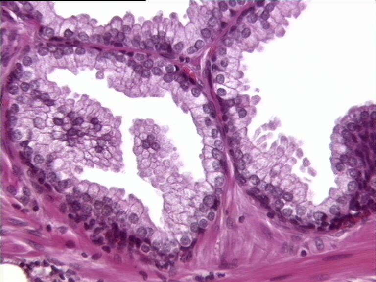
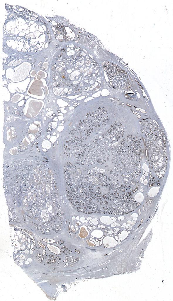
5 Small gland pattern Simple atrophy and postatrophic hyperplasia 1. Lobular configuration 2. Basophilia from the lack of cytoplasm, both apically and laterally compared to normal epithelium, such that the nuclei appear crowded and a nuclear outline of the glands is seen at lowpower 3. Stroma altered by a pale fibrosis, with periacinar collagen deposition, which can impart a sclerotic appearance
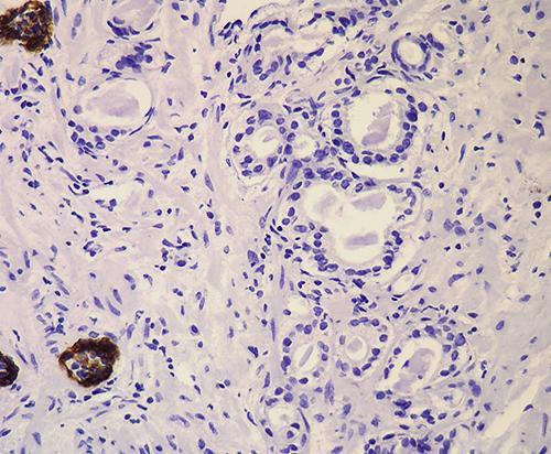
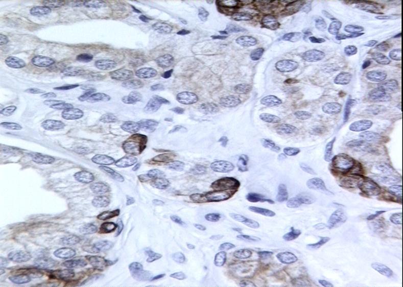
6 Small gland pattern: Atrophy Simple atrophy and postatrophic hyperplasia do not generally pose a diagnostic problem IHC for basal cell markers shows uniform staining of the basal cells in simple atrophy and postatrophic hyperplasia, ruling out PCa AMACR is generally negative in simple atrophy and very uncommonly expressed in postatrophic hyperplasia
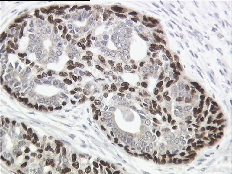
7
8
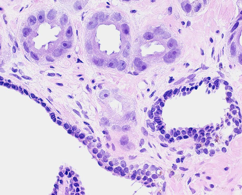
9 Small gland pattern: Partial atrophy Differs from simple atrophy and postatrophic hyperplasia in several aspects: Although it generally retains a lobular architectural pattern of growth, it can show a more disorganized diffuse growth pattern
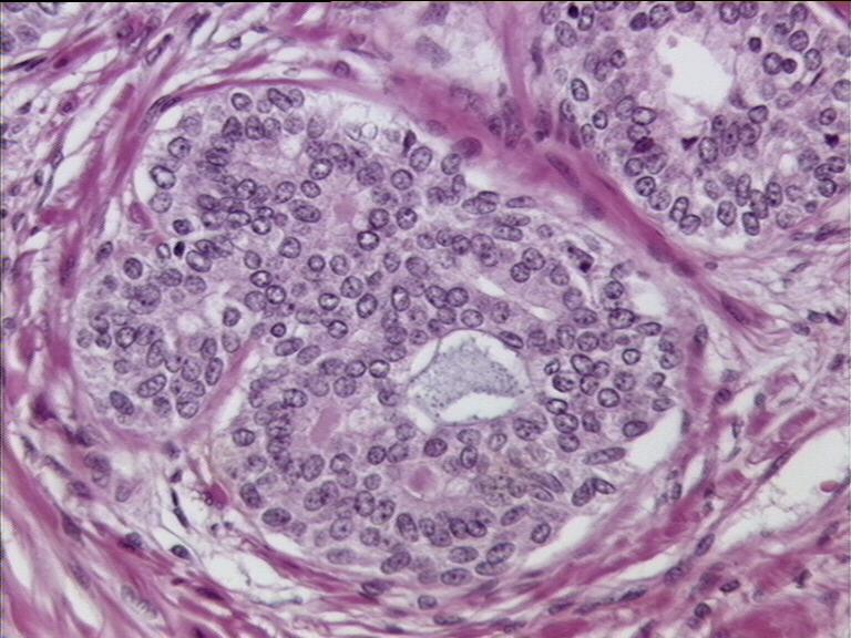
10 Small gland pattern: Partial atrophy Focus of crowded glands with pale scant cytoplasm The attenuated cytoplasm is mostly apical with most of the nuclei in the glands reaching the full height of the cells The lateral aspect of the cytoplasm is preserved which results in nuclei that are more spaced and less crowded than simple atrophy or postatrophic hyperplasia


11 Small gland pattern Atrophy 1 Simple atrophy Post-atrophic 2 hyperplasia 3 Partial atrophy Montironi R et al, Histopathology 2011
12 Small gland pattern: Partial atrophy Features seen within partial atrophy that create difficulty in its distinction from acinar (conventional) Pca: 1. Crowded and sometimes disorganized pattern of growth 2. Relative high nuclear to cytoplasmic ratio with slightly enlarged nuclei 3. Presence of visible yet small nucleoli 4. Straight luminal borders in some glands 5. Negativity of some of the glands for basal cell markers 6. Positivity of some glands for AMACR
13 Small gland pattern: Partial atrophy The disorganized pattern can give the focus a pseudoinfiltrative appearance in which the smaller glands of partial atrophy seem to be present between larger clearly benign glands 1. It is usually focal and, most importantly, close inspection reveals that the smaller suspicious glands are cytologically similar to the larger benign glands which they appear to infiltrate amongst 2. The cytoplasm in partial atrophy is clear or pale in contrast to the typical amphophilic cytoplasm of PCa
14 Small gland pattern: Partial atrophy 1. The high nuclear to cytoplasmic ratio seen in partial atrophy is the result of the lack of apical cytoplasm rather than marked nuclear enlargement which is seen in Pca 2. Nucleoli are usually absent to visible in partial atrophy and not as prominent as seen in PCa
15 Small gland pattern: Partial atrophy Differs from atrophic acinar PCa in that atrophic cancers have one or more of the following features: 1. A more infiltrative appearance where the cancer glands infiltrate as isolated glands in between benign glands 2. Associated nonatrophic cancer 3. Prominent cytological atypia beyond what can be seen with benign atrophy 4. A focus of atrophic cancer should be supported by negative IHC stains for basal cell markers
16
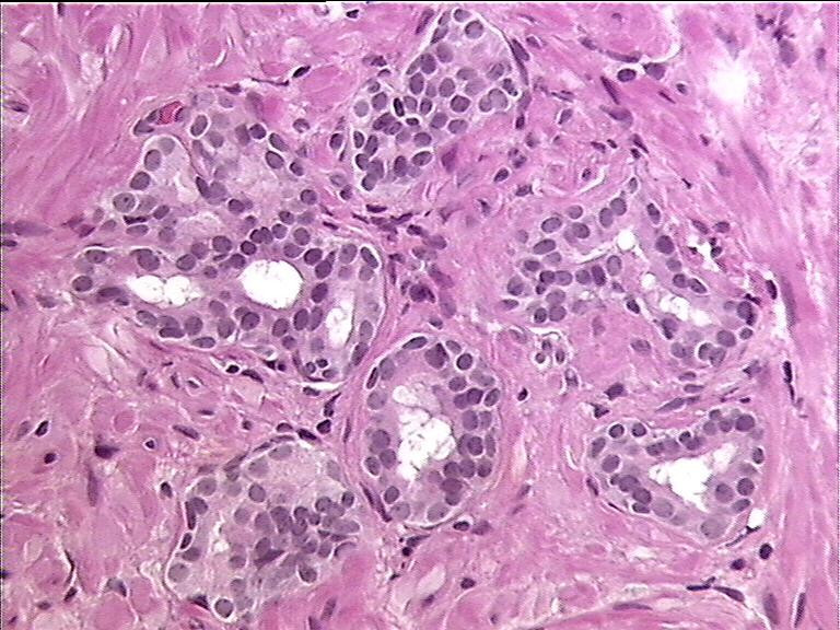
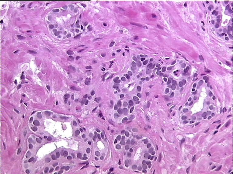
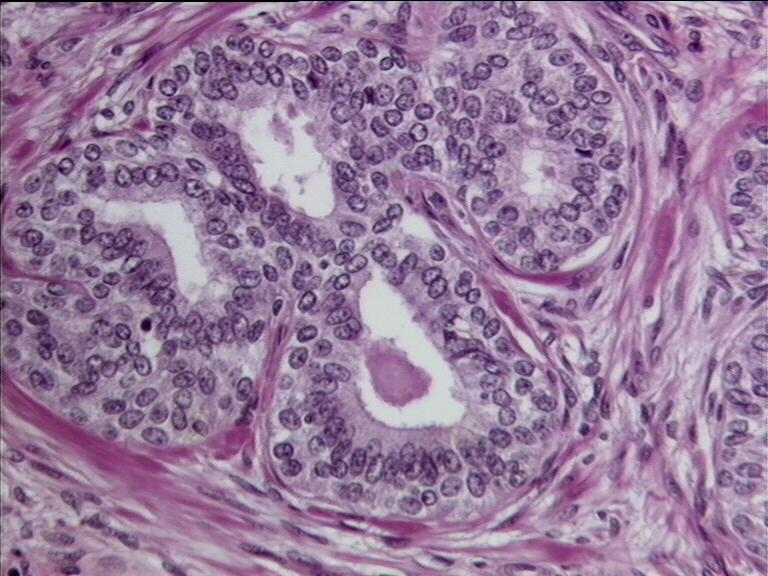
17 Small gland pattern: Adenosis (AAH) 1. Proliferation of small acini 2. Prominent perinodular distribution of the abnormal glands (within or adjacent to typical hyperplastic nodules) 3. Some variation in size and shape 4. Individual glands are closely packed but separate and show no evidence of fusion
18
19
20 Small gland pattern: Adenosis 1. Pushing rather than infiltrating border but may show a limited degree of infiltration 2. Uncommonly, the small acini exhibit a more extensive, crowded, and nonlobular distribution, in a pattern termed diffuse adenosis
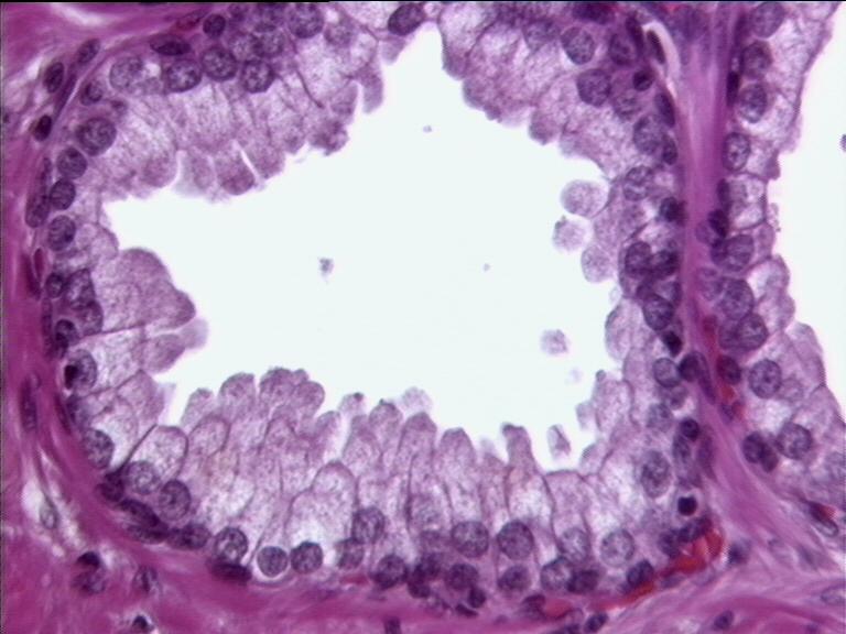
21 Small gland pattern: Adenosis 1. Lined by cuboidal to low columnar cells with moderate to abundant clear or lightly eosinophilic cytoplasm 2. The nuclei are round to oval and there is uniform fine chromatin 3. Nucleoli may be present but they are generally small. Uncommonly, enlarged nucleoli are identified in a subset of cells
22
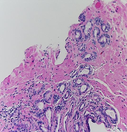
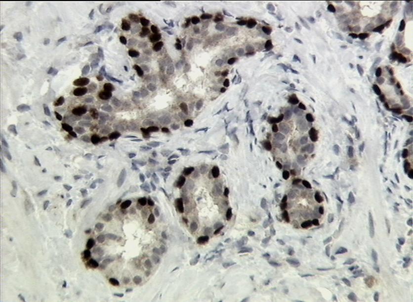
23 Small gland pattern: Adenosis 1. May contain corpora amylacea and, in some instances, luminal eosinophilic crystalloids Occasionally, basophilic luminal mucus may be seen 2. Up to 18% of cases express AMACR 3. Immunohistochemical staining demonstrates absence of basal cells in about one-half of all glands
24

25 Small gland pattern: Adenosis Common mimicker of PCa The features shared in adenosis and prostate cancer are: 1. Crowded glands 2. Crystalloids 3. Medium-sized nucleoli 4. Scattered poorly formed glands and single cells 5. Minimal infiltration at periphery 6. AMACR immunoreactivity
26 Adenosis Prostate cancer Lobular Small glands share features with admixed larger glands Pale-clear cytoplasm Haphazard growth pattern Small glands differ from adjacent benign glands Amphophilic cytoplasm Medium-sized nucleoli Occasionally large nucleoli Blue mucinous secretions rare Blue mucinous secretions common Basal cells present Basal cells absent Corpora amylacea common Corpora amylacea rare
27 Small gland pattern: Adenosis 1. There is lack of proof of a relationship between adenosis and PCa 2. Adenosis should be considered as a benign lesion and patients followed conservatively 3. The term should not be used as a wastebasket for small glandular lesions that are difficult to classify or for suspicious atypical small gland proliferations
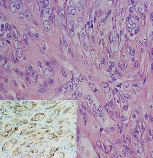
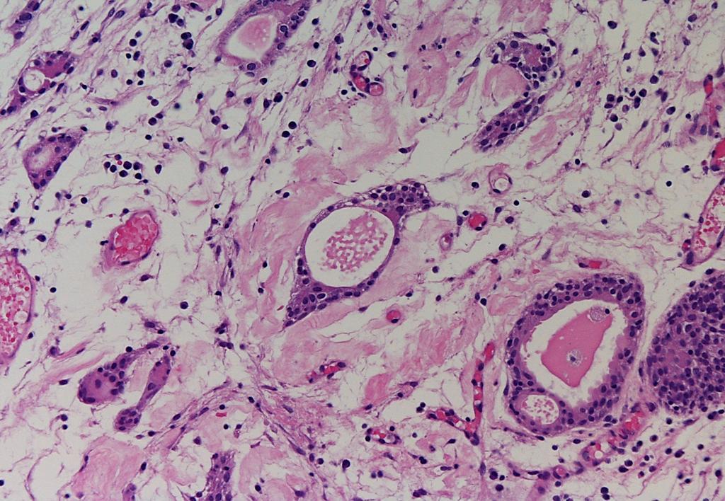
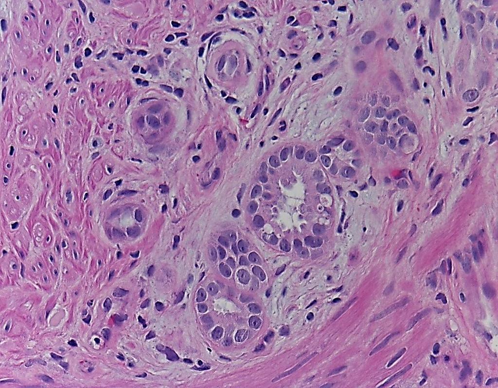
28 Small gland pattern: Sclerosing Adenosis (SA) 1. Benign, small, acinar proliferation in dense spindle cell stroma, with a distinct immunohistochemical profiles 2. Tiny microacini, cords, solid clusters, and single cells 3. Rarely seen in needle biopsy specimens, may simulate PCa and accounts for up to 10% of cases overdiagnosed as PCa
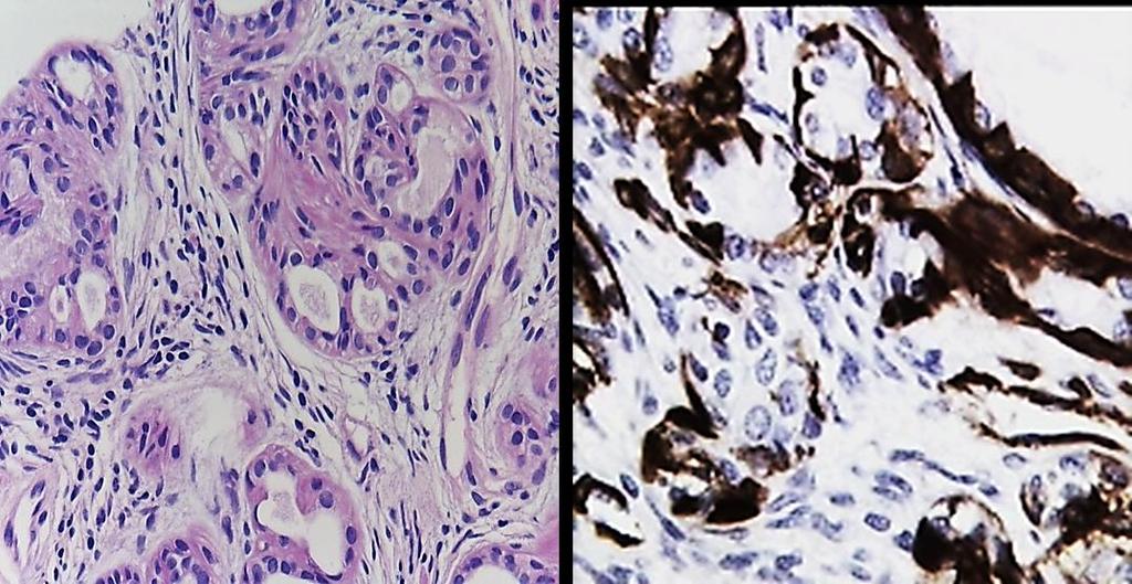
29
30 Small gland pattern: Sclerosing Adenosis Multiple light microscopical and immunohistochemical features separate SA from PCa: 1. Intact basal cell layer 2. Unique immunophenotype of the basal cells, including abundant S-100 protein and smooth muscle actin (SMA) reactivity (i.e., myoepithelial metaplasia) 3. Cellular spindle cell stroma 4. Variably thickened basement membrane 5. Absence of significant cytological atypia

31 p63 SMA S100
32 Atypical sclerosing adenosis (ASA) A series of five cases of the so-called atypical sclerosing adenosis was by Cheng L. and Bostwick DG The overall architecture is identical to that of SA Differs from it by the presence of enlarged nuclei, prominent nucleoli, and aneuploid DNA content in the majority of cases The term ASA does not imply that the lesion is premalignant Benign lesion that does not require treatment ASA may be mistaken for PCa, and should be distinguished from other mimics
33 SMA
34 - Architectural pattern: Small gland - Pca mimicked: Gleason pattern 3 Lesions of nonprostatic epithelial origin 1. Nephrogenic adenoma 2. Mesonephric remnants 3. Ejaculatory duct epithelium and seminal vesicle 4. Mucinous metaplasia 5. Cowper s (bulbourethral) glands 6. Colonic glands Montironi R et al, Histopathology 2011
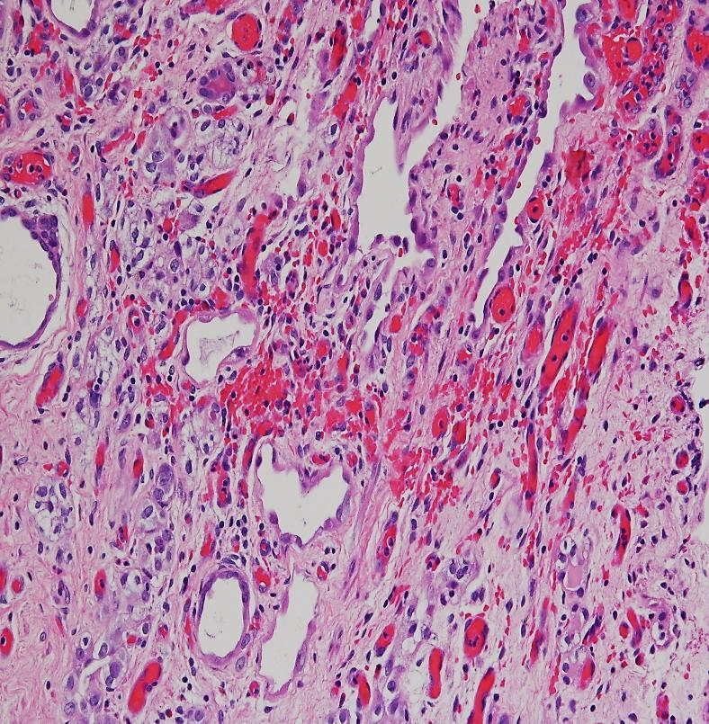
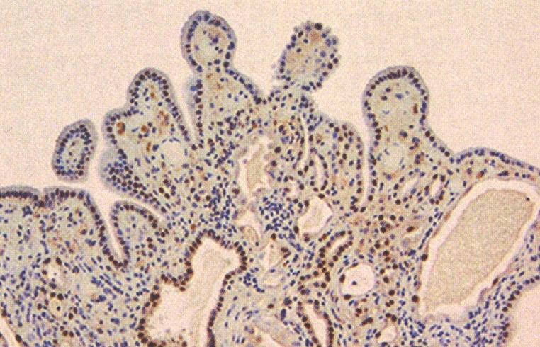
35 Small gland pattern: Nephrogenic adenoma Histologically, papillary structures, small tubules, or cystically dilated tubules lined by cuboidal, low columnar, or hobnail-shaped eosinophilic cells are seen A lesion of presumed renal tubular origin; previous diagnostic and therapeutic procedures Different locations within the urinary system, including the prostatic urethra where it might cause confusion with PCa and UC with small tubules
36
37 NA vs. PCa Lesions predominantly composed of small tubules are those most likely to be confused with PCa Frequently negative for basal cell markers and not infrequently positive for AMACR, PSA, and/or PSAP PAX2 and/or PAX8 immunostains can be used to arrive at the correct diagnosis

38 AMACR PAX8
39 NA vs. UC with small tubules The tubules of urothelial carcinoma are lined by attenuated urothelial cells in contrast to the varying admixture of cuboidal, columnar and occasionally flattened cells that line the tubules of NA CK 20, high-molecular-weight cytokeratin and p63: positive in more than half of urothelial carcinomas
40
41 Features Admixed nested, tubular, cystic, polypoid, papillary Oedematous and inflammatory background UC with small tubules No No NA Yes Yes Cell lining Attenuated Mixture Prominent BM no Yes Invasion of the muscularis propria Yes Possible IHC Distinctive for UC Distinctive for NA
42 Atypical nephrogenic metaplasia 18 cases described by Cheng et al Nuclear enlargement, nuclear hyperchromasia, and enlarged nucleoli Two patients had recurrent nephrogenic metaplasia, and the remainder were alive without recurrence or urothelial carcinoma No direct evidence that links atypical nephrogenic metaplasia to cancer Awareness of the spectrum of cytological changes is critical to prevent overdiagnosis of cancer
43


44 - Architectural pattern: Small gland - Pca mimicked: Gleason pattern 3 Lesions of nonprostatic epithelial origin Mucinous metaplasia Cowper s (bulbourethral) glands Colonic glands
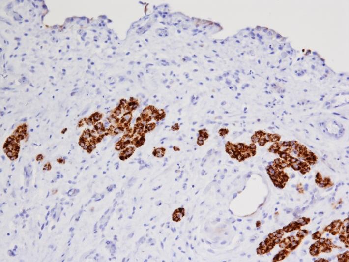
45 Expected immunoreactivity in benign mimickers of prostate carcinoma of nonprostatic epithelial origin Differential Diagnosis PSA/PAP* Basal Cell Associated Markers AMACR* Ejaculatory duct/seminal vesicle -/+ + (basal cells) - Cowper s gland - -/+ - Mesonephric remnants - -/+ - Nephrogenic adenoma -/+ -/+ +/-
46 Architectural pattern: Large and cribriform glands 1. Basal cell hyperplasia 2. Clear cell cribriform hyperplasia 3. Medium- to large-sized hyperplastic glands 4. Reactive epithelial atypia Types of prostate carcinoma mimicked 1. Cribriform Gleason patterns 3, 4, and 5 2. Ductal adenocarcinoma 3. Pseudohyperplastic Montironi R et al, Histopathology 2011
47 Basal cell proliferations of the prostate 1. Ordinary (or usual) BCH 2. BCH with prominent nucleoli (or atypical BCH) 3. Basal cell carcinoma (adenoid cystic carcinoma)
48 p63
49 NP
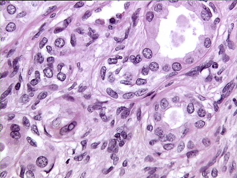
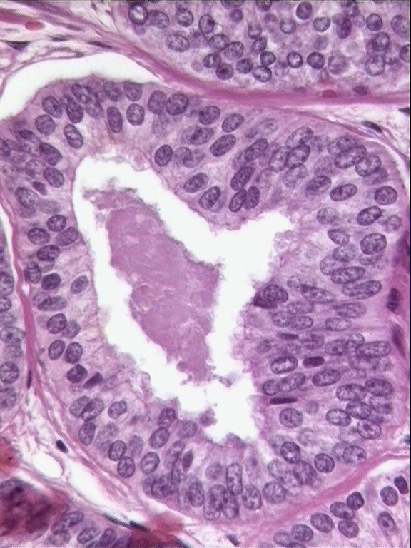
50 BCH (with prominent nucleoli ) vs. HGPIN 1.The nuclei in BCH tend to be round whereas, at times, the cells form small solid basal cell nests. In contrast, the cells in HGPIN tend to be more pseudostratified and columnar and do not occlude the glandular lumina 2.Within areas of BCH, atypical looking basal cells can be seen underling the overlying benign appearing secretory cells. HGPIN has full-thickness cytological atypia with the nuclei oriented perpendicular to the basement membrane
 BCH")

51 Ordinary (or usual) BCH BCH with prominent nucleoli
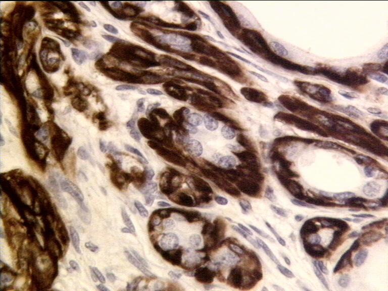
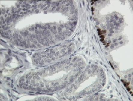
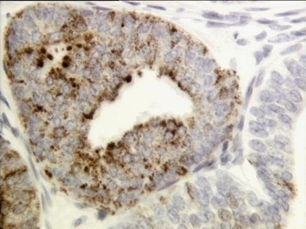
52 BCH vs. HGPIN In BCH, IHC shows multilayered staining of the basal cells, whereas an interrupted immuno-reactive basal cell layer is seen in HGPIN AMACR is negative in florid BCH and positive in HGPIN and Pca Glutathione-S-transferase pi is positive in florid BCH and negative in PCa


")
53 BCH with prominent nucleoli (or atypical BCH)

54 Central zone epithelium BCH HGPIN
55 BCH (with a glandular architecture or when florid ) vs. PCa BCH can be distinguished from PCa by its very basal cell appearance 1. The glands in BCH appear basophilic at low power due to multilayering of basal cells which have scant cytoplasm 2. Gland-forming PCa of the prostate almost always has more abundant cytoplasm resulting in a more eosinophilic appearance to the glands Utilization of IHC with basal cell specific antibodies (34betaE12 or p63) can differentiate between these two lesions
56 Expected immunoreactivity in glandular lesions of prostatic epithelial origin Differential Diagnosis Basal Cell Associated Markers AMACR Benign glands including atrophy, postatrophic hyperplasia, etc + - Adenosis +/- (patchy) -/+ Basal cell hyperplasia + - Prostate carcinoma - +
57 Prostatic adenocarcinoma, PIN-like

58 BCH vs. basal cell carcinoma Characteristics of basal cell carcinoma that can help in the differential diagnostic distinction from basal cell hyperplasia 1. Extensive infiltration between normal prostate glands 2. Extension out of the prostate 3. Perineural invasion 4. Necrosis 5. IHC studies for Ki67 and BCL2 may also be of value: the proliferative index in basal cell hyperplasia is low (<5%) BCL2 is not overexpressed.
59 Clear cell cribriform hyperplasia At low-power a nodular appearance and intervening cellular stroma Cribriform glands have clear cytoplasm and uniform round lumina The cells comprising the central cribriform areas are cuboidal to low columnar secretory-type cells with uniform round nuclei and clear cytoplasm They lack nuclear atypia and nucleolar enlargement Basal cells are prominently displayed around the periphery.
60
61 CCCH mimics CCCH enters the differential diagnosis of Gleason grade 4 cribriform Pca The distinction of clear cell cribriform hyperplasia from cribriform carcinoma is based on 1. the low power nodularity 2. cellular stroma 3. presence of basal cells, and 4. lack of significant cytological atypia

62 Gleason pattern 3 Gleason pattern 4
63 Take home message The diagnosis of PCa and its mimickers should not be based on one feature only (i.e., nucleoli or lack of basal cell markers) but rather on a case's constellation of architectural, cytological, and ancillary features
PROSTATIC ADENOCARCINOMA: DIAGNOSTIC CRITERIA AND IMPORTANT MIMICKERS PROSTATIC ADENOCARCINOMA: DIAGNOSTIC CRITERIA
 PROSTATIC ADENOCARCINOMA: DIAGNOSTIC CRITERIA AND IMPORTANT MIMICKERS PROSTATIC ADENOCARCINOMA: DIAGNOSTIC CRITERIA 1 A good H & E helps! ADENOCARCINOMA DIAGNOSTIC CRITERIA Relatively uniform proliferation
They Do Look Alike : Mimics of Prostate Cancer in Biopsy Samples
 They Do Look Alike : in Biopsy Samples Gladell P. Paner, MD Departments of Pathology and Surgery (Urology) University of Chicago, IL USA Gladell.paner@uchospitals.edu Benign in Needle Biopsy 1. Benign
3/28/2017. Disclosure of Relevant Financial Relationships. GU Evening Subspecialty Case Conference. Differential Diagnosis:
 GU Evening Subspecialty Case Conference Rajal B. Shah, M.D. VP, Medical Director, Urologic Pathology Miraca Life Sciences, Irving, Texas Clinical Associate Professor of Pathology Baylor College of Medicine,
ARTHUR PURDY STOUT SOCIETY COMPANION MEETING: DIFFICULT NEW DIFFERENTIAL DIAGNOSES IN PROSTATE PATHOLOGY. Jonathan I. Epstein.
 1 ARTHUR PURDY STOUT SOCIETY COMPANION MEETING: DIFFICULT NEW DIFFERENTIAL DIAGNOSES IN PROSTATE PATHOLOGY Jonathan I. Epstein Professor Pathology, Urology, Oncology The Reinhard Professor of Urological
Synonyms. Nephrogenic metaplasia Mesonephric adenoma
 Nephrogenic Adenoma Synonyms Nephrogenic metaplasia Mesonephric adenoma Definition Benign epithelial lesion of urinary tract with tubular, glandular, papillary growth pattern Most frequently in the urinary
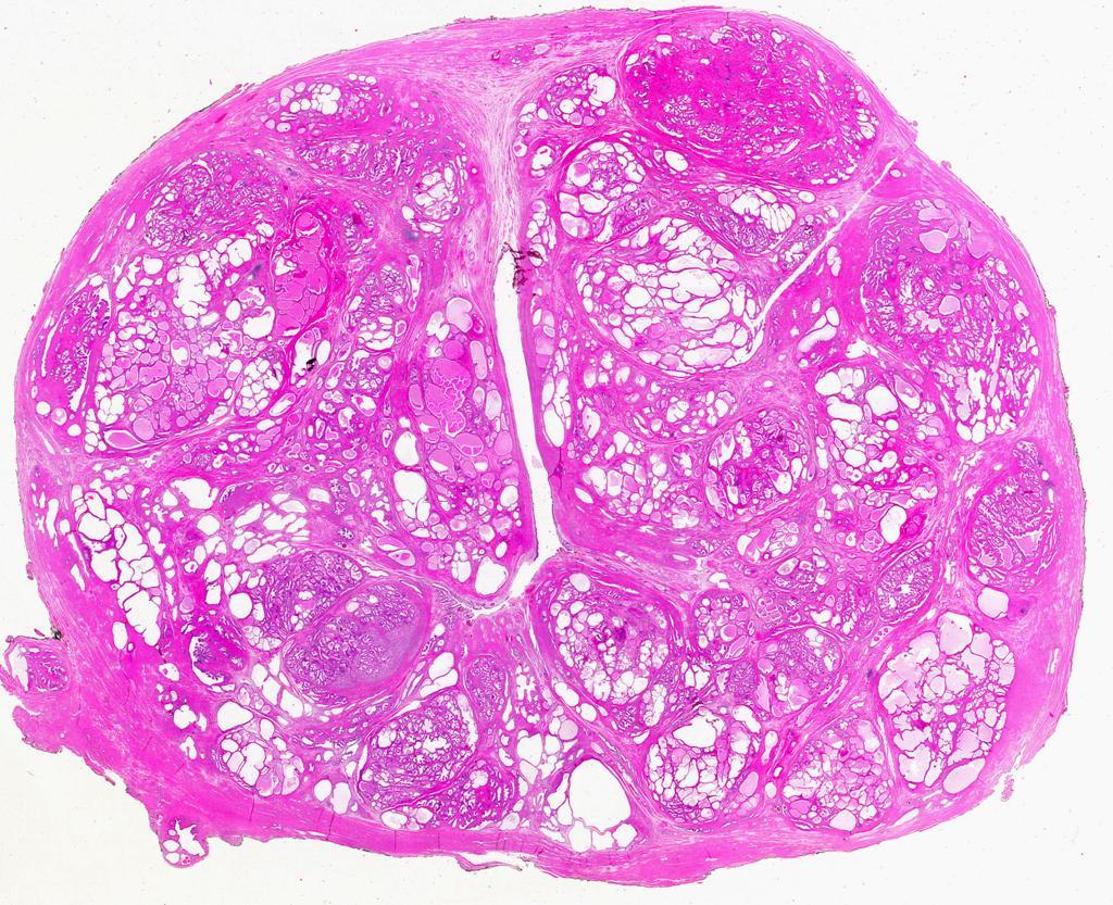
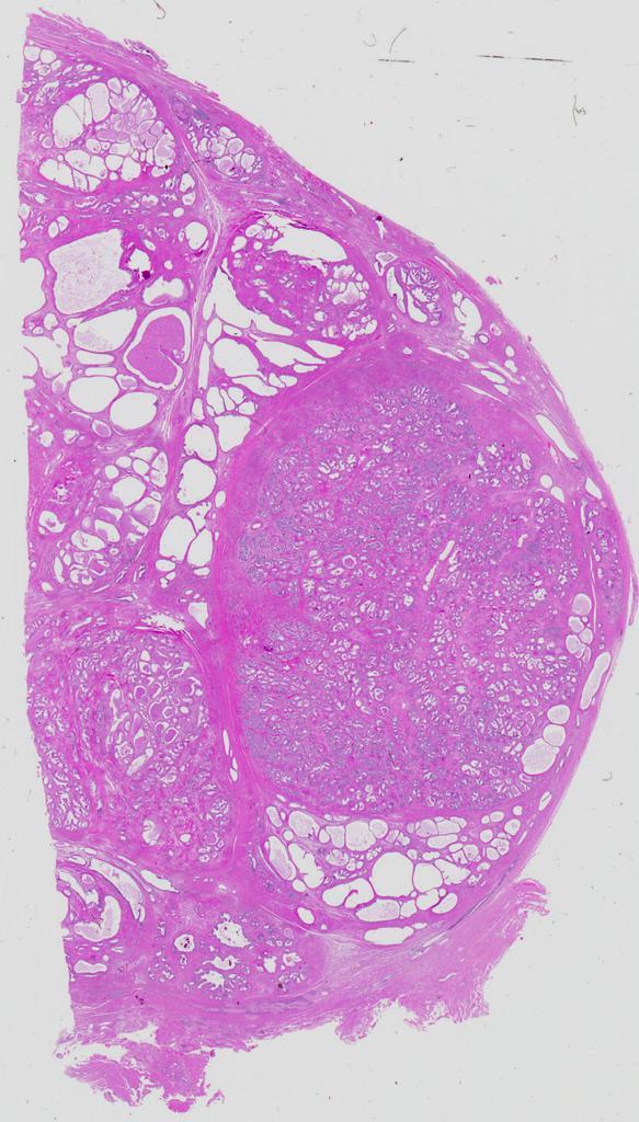
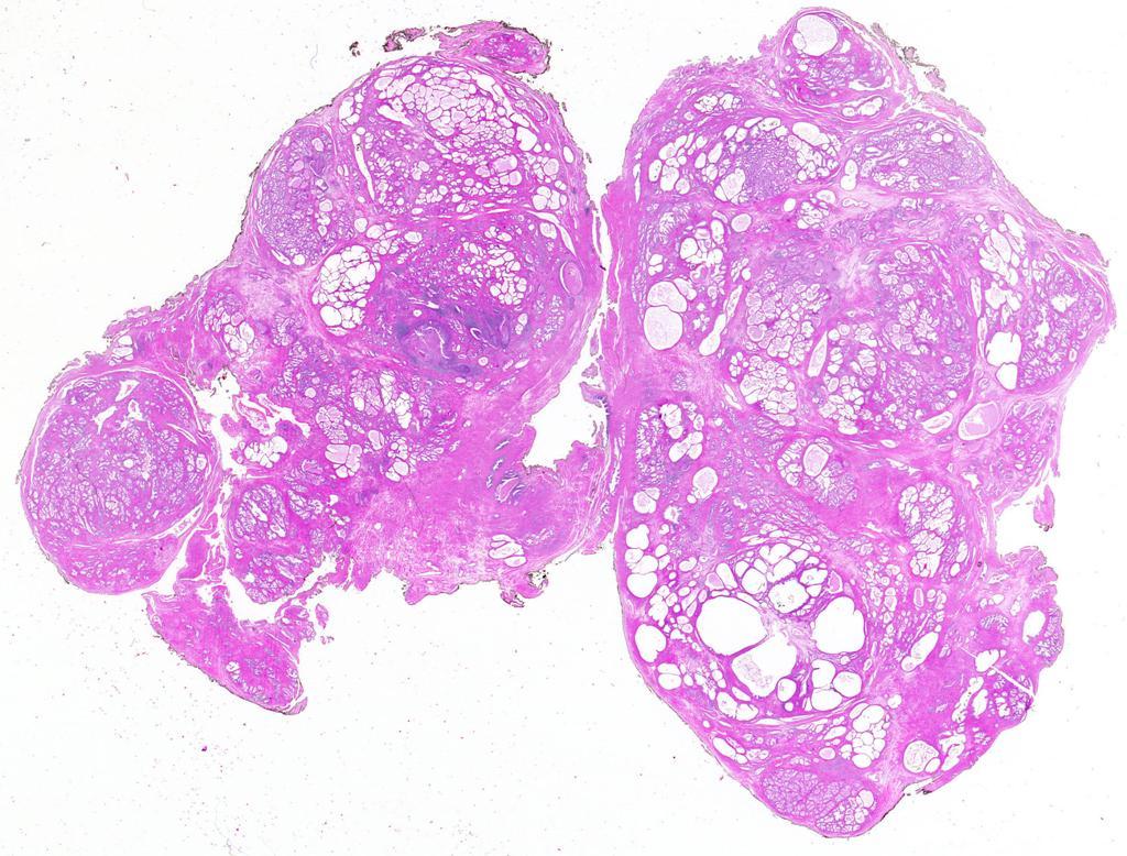
Gross appearance of nodular hyperplasia in material obtained from suprapubic prostatectomy. Note the multinodular appearance and the admixture of
 Tiền liệt tuyến Tiền liệt tuyến Gross appearance of nodular hyperplasia in material obtained from suprapubic prostatectomy. Note the multinodular appearance and the admixture of solid and microcystic areas.
PSA. HMCK, p63, Racemase. HMCK, p63, Racemase
 Case 1 67 year old male presented with gross hematuria H/o acute prostatitis & BPH Urethroscopy: small, polypoid growth with a broad base emanating from the left side of the verumontanum Serum PSA :7 ng/ml
Prostate Pathology: Prostate Carcinoma, variants and Gleason Grading (Part 1)
") Prostate Pathology: Prostate Carcinoma, variants and Gleason Grading (Part 1) Jae Y. Ro, MD, PhD June 7, 2012 Ten Leading Cancer Types for the Estimated New Cancer Cases and Deaths By Sex, United States,
Prostate Pathology: Prostate Carcinoma, variants and Gleason Grading (Part 1) Jae Y. Ro, MD, PhD June 7, 2012 Ten Leading Cancer Types for the Estimated New Cancer Cases and Deaths By Sex, United States,
5/21/2018. Difficulty in Underdiagnosing Prostate Cancer. Diagnosis of Prostate Cancer. Evaluation of Prostate Cancer and Atypical on Needle Biopsy
 Evaluation of Prostate Cancer and Atypical on Needle Biopsy Jonathan I. Epstein Difficulty in Underdiagnosing Prostate Cancer Limited tissue on needle biopsy (1 cm. x
ACCME/Disclosures. Cribriform Lesions of the Prostate. Case
 Cribriform Lesions of the Prostate Ming Zhou, MD, PhD Departments of Pathology and Urology New York University Langone Medical Center New York, NY Ming.Zhou@NYUMC.ORG ACCME/Disclosures The USCAP requires
Although partial atrophy is one of the most common
 ORIGINAL ARTICLE Partial Atrophy on Prostate Needle Biopsy Cores: A Morphologic and Immunohistochemical Study Wenle Wang, MD, PhD,* Xinlai Sun, MD,w and Jonathan I. Epstein, MD*zy Abstract: Partial atrophy
Papillary Lesions of the Breast A Practical Approach to Diagnosis. (Arch Pathol Lab Med. 2016;140: ; doi: /arpa.
 Papillary Lesions of the Breast A Practical Approach to Diagnosis (Arch Pathol Lab Med. 2016;140:1052 1059; doi: 10.5858/arpa.2016-0219-RA) Papillary lesions of the breast Span the spectrum of benign,
1 NORMAL HISTOLOGY AND METAPLASIAS
 1 NORMAL HISTOLOGY AND METAPLASIAS, MD Anatomy and Histology 1 Metaplasias 2 ANATOMY AND HISTOLOGY The female breast is composed of a branching duct system, which begins at the nipple with the major lactiferous
Proliferative Epithelial lesions of the Breast. Sami Shousha, MD, FRCPath Charing Cross Hospital & Imperial College, London
 Proliferative Epithelial lesions of the Breast Sami Shousha, MD, FRCPath Charing Cross Hospital & Imperial College, London Amman, November2013 Proliferative Epithelial Lesions of the Breast Usual type
INTRADUCTAL LESIONS OF THE PROSTATE. Jonathan I. Epstein
 INTRADUCTAL LESIONS OF THE PROSTATE Jonathan I. Epstein Topics Prostatic intraepithelial neoplasia (PIN) Intraductal adenocarcinoma (IDC-P) Intraductal urothelial carcinoma Ductal adenocarcinoma High Prostatic
Intraductal carcinoma of the prostate on needle biopsy: histologic features and clinical significance
 & 2006 USCAP, Inc All rights reserved 0893-3952/06 $30.00 www.modernpathology.org Intraductal carcinoma of the prostate on needle biopsy: histologic features and clinical significance Charles C Guo 1 and
Papillary Lesions of the breast
 Papillary Lesions of the breast Emad Rakha Professor of Breast Pathology The University of Nottingham Papillary lesions of the breast are a heterogeneous group of disease, which are characterised by neoplastic
5/21/2018. Prostate Adenocarcinoma vs. Urothelial Carcinoma. Common Differential Diagnoses in Urological Pathology. Jonathan I.
 Common Differential Diagnoses in Urological Pathology Jonathan I. Epstein Prostate Adenocarcinoma vs. Urothelial Carcinoma 1 2 NKX3.1 NKX3.1 3 4 5 6 Proposed ISUP Recommendations Option to use PSA as a
Pathology of the Prostate. PathoBasic Tatjana Vlajnic
 Pathology of the Prostate PathoBasic 24.01.17 Tatjana Vlajnic Overview Adenocarcinoma of the prostate Grading Special variants Mimickers of prostate adenocarcinoma Atrophy Inflammatory conditions Granulomatous
B asal cell proliferations in the prostate gland exhibit a
 290 ORIGINAL ARTICLE Basal cell hyperplasia and basal cell carcinoma of the prostate: a comprehensive review and discussion of a case with c-erbb-2 expression R Montironi, R Mazzucchelli, D Stramazzotti,
Prostate Immunohistochemistry. Literature Interpretation: Caveats. Must be aware of staining pattern of antibody in the relevant tissue
 IHC Interpretation: General Principles (1) Prostate Immunohistochemistry Murali Varma Cardiff, UK wptmv@cf.ac.uk Sarajevo Nov 2013 Must be aware of staining pattern of antibody in the relevant tissue Nuclear/cytoplasmic/membranous
Diseases of the breast (1 of 2)
") Diseases of the breast (1 of 2) Introduction A histology introduction Normal ducts and lobules of the breast are lined by two layers of cells a layer of luminal cells overlying a second layer of myoepithelial
Diseases of the breast (1 of 2) Introduction A histology introduction Normal ducts and lobules of the breast are lined by two layers of cells a layer of luminal cells overlying a second layer of myoepithelial
Salivary Glands 3/7/2017
 Salivary Glands 3/7/2017 Goals and objectives Focus on the entities unique to H&N Common board type facts Information for your future practice Salivary Glands Salivary Glands Major gland. Paratid. Submandibular.
Immunohistochemistry in prostate pathology: Recent Advances
 Immunohistochemistry in prostate pathology: Recent Advances Jae Y. Ro, M.D., Ph.D. Weill Medical College of Cornell University, Methodist Hospital, and UT MD Anderson Cancer Center, Houston, TX Yonsei
Diagnostically Challenging Cases in Gynecologic Pathology
 Diagnostically Challenging Cases in Gynecologic Pathology Eric C. Huang, M.D., Ph.D. Department of Pathology and Laboratory Medicine University of California, Davis Medical Center Case 1 Presentation 38
Although current American Cancer Society guidelines
 ORIGINAL ARTICLE Diffuse Adenosis of the Peripheral Zone in Prostate Needle Biopsy and Prostatectomy Specimens Tamara L. Lotan, MD* and Jonathan I. Epstein, MD*w z Abstract: We have observed a group of
Dissertation submitted in. Partial fulfilment of the regulations required for the award of M.D. DEGREE PATHOLOGY BRANCH III THE TAMILNADU
 THE EXPRESSION AND DIAGNOSTIC UTILITY OF AMACR AND 34βE12 IN PROSTATIC LESIONS Dissertation submitted in Partial fulfilment of the regulations required for the award of M.D. DEGREE In PATHOLOGY BRANCH
Columnar Cell Lesions. Columnar Cell Lesions and Flat Epithelial Atypia
 Columnar Cell Lesions and Stuart J. Schnitt, M.D. Beth Israel Deaconess Medical Center and Harvard Medical School Boston, MA, USA Columnar Cell Lesions Lesions characterized by columnar epithelial cells
Normal Morphology. Anatomic Considerations. Normal Urothelial Histology and Cytology
 1 Normal Morphology Anatomic Considerations The urinary tract can be divided into three regions: the kidney; the calyces, pelves and ureters (upper collecting system or upper tract); and the bladder and
CLINICAL SIGNIFICANCE OF BENIGN EPITHELIAL CHANGES
 Papillomas. Papillomas are composed of multiple branching fibrovascular cores, each having a connective tissue axis lined by luminal and myoepithelial cells ( Fig. 23-11 ). Growth occurs within a dilated
Pancreatitis: A Potential Pitfall in Endoscopic Ultrasound Guided Pancreatic FNA
 Pancreatitis: A Potential Pitfall in Endoscopic Ultrasound Guided Pancreatic FNA Jack Yang, MD Department of Pathology, Medical University of South Carolina Objectives Understand the indication of EUS
Uropathology January Jon Oxley
 Uropathology January 2012 Jon Oxley Background to seminar These slides were available to view via the web from scanned slides The junior pathologists answered questions on them via the web The answers
2016 WHO CLASSIFICATION OF TUMOURS OF THE PROSTATE. Peter A. Humphrey, MD, PhD Yale University School of Medicine New Haven, CT
 2016 WHO CLASSIFICATION OF TUMOURS OF THE PROSTATE Peter A. Humphrey, MD, PhD Yale University School of Medicine New Haven, CT 2016 WHO CLASSIFICATION OF TUMOURS OF THE PROSTATE AUTHORS : PROSTATE CHAPTER
Prostatic ductal adenocarcinoma is a subtype of
 ORIGINAL ARTICLE High-grade Prostatic Intraepithelial Neoplasialike Ductal Adenocarcinoma of the Prostate: A Clinicopathologic Study of 28 Cases Fabio Tavora, MD* and Jonathan I. Epstein, MD*w z Abstract:
5/2/2018. Low Grade Dysplasia of GI Tract. High Grade Dysplasia of GI Tract. Dysplasia in Gastrointestinal Tract: Practical Pearls and Issues
 Dysplasia in Gastrointestinal Tract: Practical Pearls and Issues Arief Suriawinata, M.D. Professor of Pathology and Laboratory Medicine Geisel School of Medicine at Dartmouth Department of Pathology and
Lesions Mimicking Adenoid Cystic Carcinoma. Diagnostic Problems in Salivary Gland Pathology An Update 5/29/2009
 Diagnostic Problems in Salivary Gland Pathology An Update Lesions Mimicking Adenoid Cystic Carcinoma Stacey E. Mills, M.D. W.S. Royster Professor of Pathology Director of Surgical and Cytopathology University
Some prostatic diseases
 Some prostatic diseases Benign Prostatic Hyperplasia (Nodular Hyperplasia) Extremely common Present in a significant number of men by the age of 40 & its frequency rises progressively with age, reaching
Breast pathology. 2nd Department of Pathology Semmelweis University
 Breast pathology 2nd Department of Pathology Semmelweis University Breast pathology - Summary - Benign lesions - Acute mastitis - Plasma cell mastitis / duct ectasia - Fat necrosis - Fibrocystic change/
Spectrum of Preneoplastic and Neoplastic Cystic Lesions of the Kidney in Adult. by dr. Banan Burhan Mohammed Lecturer in Pathology Department
 Spectrum of Preneoplastic and Neoplastic Cystic Lesions of the Kidney in Adult by dr. Banan Burhan Mohammed Lecturer in Pathology Department Various hereditary, acquired, and neoplastic conditions can
Dr Sanjiv Manek Oxford. Oxford Pathology Course 2010 for FRCPath Illustration-Cellular Pathology. Oxford Radcliffe NHS Trust
 Dr Sanjiv Manek Oxford Oxford Pathology Course 2010 for FRCPath Illustration-Cellular Pathology. Oxford Radcliffe NHS Trust Ovarian Endometrial Vulvo-vaginal Cervical Illustration-Cellular Pathology. Oxford
Mody. AIS vs. Invasive Adenocarcinoma of the Cervix
 Common Problems in Gynecologic Pathology Michael T. Deavers, M.D. Houston Methodist Hospital, Houston, Texas Common Problems in Gynecologic Pathology Adenocarcinoma in-situ (AIS) of the Cervix vs. Invasive
Objective. Atypical/Atypia. Atypical Glandular Lesions of the Prostate 12/27/2011
 Atypical Glandular Lesions of the Prostate V.O.Speights,Jr.,D.O. Scott and White Memorial Hospital Texas A & M Health Science Center January 13,2012 Objective To identify histological abnormalities in
04/10/2018. Intraductal Papillary Neoplasms Of Breast INTRADUCTAL PAPILLOMA
 Intraductal Papillary Neoplasms Of Breast Savitri Krishnamurthy MD Professor of Pathology Deputy Division Head The University of Texas MD Anderson Cancer Center 25 th Annual Seminar in Pathology Pittsburgh,
Minimal Adenocarcinoma in Prostate Needle Biopsy Tissue
 Anatomic Pathology / MINIMAL PROSTATE CANCER Minimal Adenocarcinoma in Prostate Needle Biopsy Tissue Phataraporn Thorson, MD, and Peter A. Humphrey, MD, PhD Key Words: Prostate; Cancer; Biopsy; Minimal
Title malignancy. Issue Date Right 209, 12, (2013)
") NAOSITE: Nagasaki University's Ac Title Author(s) A case of intracystic apocrine papi malignancy Hayashi, Hiroko; Ohtani, Hiroshi; Y Citation Pathology - Research and Practice, Issue Date 2013-12 URL Right
NAOSITE: Nagasaki University's Ac Title Author(s) A case of intracystic apocrine papi malignancy Hayashi, Hiroko; Ohtani, Hiroshi; Y Citation Pathology - Research and Practice, Issue Date 2013-12 URL Right
ISPUB.COM. Interpretation Of Prostatic Biopsies: A Review. A Chitale, S Khubchandani INTRODUCTION NON-NEOPLASTIC LESIONS GRADING: GLEASON'S SCORE
 ISPUB.COM The Internet Journal of Urology Volume 3 Number 1 A Chitale, S Khubchandani Citation A Chitale, S Khubchandani.. The Internet Journal of Urology. 2004 Volume 3 Number 1. Abstract The incidence
Science & Technologies ATYPICAL NEPHROGENIC METAPLASIA OF THE URINARY BLADDER: A CASE REPORT AND REVIEW OF THE LITERATURE
 ATYPICAL NEPHROGENIC METAPLASIA OF THE URINARY BLADDER: A CASE REPORT AND REVIEW OF THE LITERATURE Vesela Ivanova *, Milen Karaivanov ** * Department of General and Clinical Pathology, Medical Faculty,
Pancreas. Atrophy, acinar cell. Pathogenesis: Diagnostic key features:
 Pancreas Atrophy, acinar cell Pathogenesis: Decrease in number and/or size of acinar cells may be due to spontaneous or experimentally induced degenerative changes, apoptosis, or a sequel of chronic inflammation.
CHRONIC PANCREATITIS OR DUCTAL ADENOCARCINOMA? N. Volkan Adsay, \ MD
 CHRONIC PANCREATITIS OR DUCTAL ADENOCARCINOMA? N. Volkan Adsay, \ MD Case for discussion 67 y/o male Back pain and weight loss CT: 4.5 cm ill-defined, solid lesion in the head FNA/Core bx: Inconclusive
Pitfalls in thyroid tumor pathology. Prof.Valdi Pešutić-Pisac MD, PhD
 Pitfalls in thyroid tumor pathology Prof.Valdi Pešutić-Pisac MD, PhD Too many or... Tumour herniation through a torn capsule simulating capsular invasion fibrous capsule with a sharp discontinuity, suggestive
Outline 11/2/2017. Pancreatic EUS-FNA general aspects. Cytomorphologic features of solid neoplasms/lesions of the pancreas
 ENDOSCOPIC ULTRASOUND GUIDED-FINE NEEDLE ASPIRATION CYTOLOGY OF PANCREAS Khalid Amin M.D. Assistant Professor Department of Laboratory Medicine and Pathology University of Minnesota Outline Pancreatic
Normal endometrium: A, proliferative. B, secretory.
 Normal endometrium: A, proliferative. B, secretory. Nội mạc tử cung Nội mạc tử cung Cyclic changes in endometrium.. Approximate relationship of useful microscopic changes. Arias-Stella reaction in endometrial
Prostate Biopsy Interpretation: An Illustrated Guide
 Prostate Biopsy Interpretation: An Illustrated Guide Authors Prostate Biopsy Interpretation: An Illustrated Guide Rajal B. Shah, M.D. Director, Urologic Pathology Caris Life Sciences 6655 North MacArthur
Endometrial Metaplasia, Hyperplasia & Other Cancer Mimics: a Consultant s Experience
 Endometrial Metaplasia, Hyperplasia & Other Cancer Mimics: a Consultant s Experience Pacific Northwest Society of Pathologists Vancouver, B.C. September 26, 2015 Teri A. Longacre, M.D. longacre@stanford.edu
Objectives. Atypical Glandular Cells. Atypical Endocervical Cells. Reactive Endocervical Cells
 2013 California Society of Pathologists 66 th Annual Meeting San Francisco, CA Atypical Glandular Cells to Early Invasive Adenocarcinoma: Cervical Cytology and Histology Christina S. Kong, MD Associate
Condyloma Acuminatum. Mimics of Bladder Cancer. Squamous Papilloma. Squamous epithelium in bladder
 Mimics of Bladder Cancer Murali Varma Cardiff, UK wptmv@cf.ac.uk Squamous epithelium in bladder Non-keratinising vaginal type mucosa common in trigone region in women Normal variant Sarajevo Nov 2013 Squamous
Oncocytic-Appearing Salivary Gland Tumors. Oncocytic, Cystic, Mucinous, and High Grade Salivary Gland Tumors SALIVARY GLAND FNA: PART II
 William C. Faquin, MD, PhD Professor of Pathology Harvard Medical School Director of Head and Neck Pathology Massachusetts Eye and Ear Massachusetts General Hospital SALIVARY GLAND FNA: PART II Oncocytic,
Urinary Bladder: WHO Classification and AJCC Staging Update 2017
 Urinary Bladder: WHO Classification and AJCC Staging Update 2017 Houston Society of Clinical Pathologists 58 th Annual Spring Symposium Houston, TX April 8, 2017 Jesse K. McKenney, MD Classification
Supplemental Figure 1
 A1 A A5 A7 A9 A A4 A6 A8 A10 A1/A Gleason Score +=6 A/A4 Gleason Score +=6 Small glands with well defined Prominent feature of size variation lumen of various size infiltrating in to and distinct lumina
Notice of Faculty Disclosure
 California Society of Pathology Diagnostic Problems in Surgical Pathology December 2015 Case 2 Laura C. Collins, M.D. Associate Professor of Pathology Associate Director of Anatomic Pathology Beth Israel
Salivary Gland Cytology
 Salivary Gland Cytology Diagnostic challenges and potential pitfalls Tarik M. Elsheikh, MD Professor and Medical Director Anatomic Pathology Cleveland Clinic FNA Salivary Gland Lesions Indications Distinguish
Columnar Cell Lesions
 Columnar Cell Lesions Laura C. Collins, M.D. Department of Pathology Beth Israel Deaconess Medical Center and Harvard Medical School Boston, MA Question? Columnar cell lesions are: a) Annoying lesions
CINtec p16 INK4a Staining Atlas
 CINtec p16 INK4a Staining Atlas Rating Rating Positive The rating positive will be assigned if the p16 INK4a -stained slide shows a continuous staining of cells of the basal and parabasal cell layers of
Pathological Classification of Hepatocellular Carcinoma
 3 rd APASL Single Topic Conference: HCC in 3D Pathological Classification of Hepatocellular Carcinoma Glenda Lyn Y. Pua, M.D. HCC Primary liver cancer is the 2 nd most common cancer in Asia HCC is the
Atypical Hyperplasia/EIN
 EIN Atypical Hyperplasia/EIN Based on scientific and diagnostic advances, in 2014 the WHO moved that the precursor lesion for endometrioid carcinoma be atypical hyperplasia/ein, rather than what was previously
Gynecologic Cytopathology: Glandular lesions
 Gynecologic Cytopathology: Glandular lesions Lin Wai Fung (MSc, MPH, CMIAC) 17/4/2014 Glandular lesions of the uterus Endocervix Endometrium Normal endocervical cells Sheets, strips well-preserved architecture:
number Done by Corrected by Doctor Maha Shomaf
 number 16 Done by Waseem Abo-Obeida Corrected by Zeina Assaf Doctor Maha Shomaf MALIGNANT NEOPLASMS The four fundamental features by which benign and malignant tumors can be distinguished are: 1- differentiation
Various hereditary, acquired and neoplastic conditions can lead to cyst formation in the kidney.
 Dr. Fatima AlAl-Hashimi Hashimi,, MD, FRCPath Salmaniya Medical Complex, Bahrain Various hereditary, acquired and neoplastic conditions can lead to cyst formation in the kidney. The most frequently encountered
Ductal Proliferations of the Breast: The Good, the Bad, and the Ugly
 Ductal Proliferations of the Breast: The Good, the Bad, and the Ugly Melinda F. Lerwill, MD CRITERIA FOR DISTINGUISHING LOW-GRADE DUCTAL CARCINOMA IN SITU FROM USUAL DUCTAL HYPERPLASIA CYTOLOGY Low-grade
DIAGNOSTIC SLIDE SEMINAR: PART 1 RENAL TUMOUR BIOPSY CASES
 DIAGNOSTIC SLIDE SEMINAR: PART 1 RENAL TUMOUR BIOPSY CASES Dr. Andrew J. Evans MD, PhD, FACP, FRCPC Consultant in Genitourinary Pathology University Health Network, Toronto, ON Case 1 43 year-old female,
MALE REPRODUCTIVE SYSTEM
 MALE REPRODUCTIVE SYSTEM The male reproductive system consists of primary sex organs (testes) and secondary or accessory sex organs. The secondary organs consist of a series of genital ducts (ductules
LYMPHATIC DRAINAGE AXILLARY (MOSTLY) INTERNAL MAMMARY SUPRACLAVICULAR
 INTERNAL MAMMARY SUPRACLAVICULAR") BREAST LYMPHATIC DRAINAGE AXILLARY (MOSTLY) INTERNAL MAMMARY SUPRACLAVICULAR HISTOLOGY LOBE: (10 in whole breast) LOBULE: (many per lobe) ACINUS/I, aka ALVEOLUS/I: (many per lobule) DUCT(S): INTRA- or
BREAST LYMPHATIC DRAINAGE AXILLARY (MOSTLY) INTERNAL MAMMARY SUPRACLAVICULAR HISTOLOGY LOBE: (10 in whole breast) LOBULE: (many per lobe) ACINUS/I, aka ALVEOLUS/I: (many per lobule) DUCT(S): INTRA- or
Disclosures. Parathyroid Pathology. Objectives. The normal parathyroid 11/10/2012
 Disclosures Parathyroid Pathology I have nothing to disclose Annemieke van Zante MD/PhD Assistant Professor of Clinical Pathology Associate Chief of Cytopathology Objectives 1. Review the pathologic features
6/3/2010. Outline of Talk. Lobular Breast Cancer: Definition of lobular differentiation. Common Problems in Diagnosing LCIS in Core Biopsies
 Outline of Talk Lobular Breast Cancer: Common Problems in Diagnosing LCIS in Core Biopsies Definition of lobular differentiation Variants of LCIS that: carry risk for unsampled invasive cancer mimic DCIS
Disorders of Cell Growth & Neoplasia. Histopathology Lab
 Disorders of Cell Growth & Neoplasia Histopathology Lab Paul Hanna April 2010 Case #84 Clinical History: 5 yr-old, West Highland White terrier. skin mass from axillary region. has been present for the
New Diagnoses Need New Approaches: A Glimpse into the Near Future of Gynecologic Pathology
 New Diagnoses Need New Approaches: A Glimpse into the Near Future of Gynecologic Pathology United States and Canadian Academy of Pathology 102 nd Annual Meeting Baltimore, Maryland Christina S. Kong, M.D.
Proliferative Breast Disease: implications of core biopsy diagnosis. Proliferative Breast Disease
 Proliferative Breast Disease: implications of core biopsy diagnosis Jean F. Simpson, M.D. Breast Pathology Consultants, Inc. Nashville, TN Proliferative Breast Disease Must be interpreted in clinical and
Pathology Slides. [Pathology]
![Pathology Slides. [Pathology]](/thumbs/94/120604575.jpg "Pathology Slides. [Pathology]") Pathology Slides MedicoNotes provides real laboratory pathological slides to aid you to differentiate between different pathological structures under microscope. www.mediconotes.com Histology slides example
Pathology Slides MedicoNotes provides real laboratory pathological slides to aid you to differentiate between different pathological structures under microscope. www.mediconotes.com Histology slides example
Benign Mimics of Malignancy in Breast Pathology
 Arthur Purdy Stout Society of Surgical Pathologists Companion Meeting Benign Mimics of Malignancy in Breast Pathology Stuart J. Schnitt, M.D. Beth Israel Deaconess Medical Center and Harvard Medical School,
Clinicopathological Study of Associated Lesions in Benign Prostatic Hyperplasia and Prostatic Adenocarcinoma in Surgical Biopsy Specimens
 Original Article DOI: 10.21276/APALM.1608 Clinicopathological Study of Associated Lesions in Benign Prostatic Hyperplasia and Prostatic Adenocarcinoma in Surgical Biopsy Specimens Esakki Muthuvel, Vimal
DISCUSSION: PLGA accounts for about 2% of all salivary gland tumours and occurs almost exclusively in the minor salivary glands.
 SWELLING ON THE HARD PALATE PRESENTING AS POLYMORPHOUS LOW GRADE ADENOCARCINOMA: A AND REVIEW OF LITERATURE Swapnil D. Chandekar 1, Sunita S. Dantkale 2, Rahul R. Narkhede 3, Snehal V. Chavhan 4, Khushboo
Papillary Lesions of the Breast
 Papillary Lesions of the Breast Laura C. Collins, M.D. Associate Professor of Pathology Associate Director, Division of Anatomic Pathology Beth Israel Deaconess Medical Center and Harvard Medical School
Coordinate Expression of Cytokeratins 7 and 20 in Prostate Adenocarcinoma and Bladder Urothelial Carcinoma
 Anatomic Pathology / CYTOKERATINS 7 AND 20 IN PROSTATE AND BLADDER CARCINOMAS Coordinate Expression of Cytokeratins 7 and 20 in Prostate Adenocarcinoma and Bladder Urothelial Carcinoma Nader H. Bassily,
Mammary Nodular Hyperplasia in Intact R hesus Monkeys
 Vet. Path. 10: 130-134 (1973) Mammary Nodular Hyperplasia in Intact R hesus Monkeys L. W NELSON and L. D. SHOTT Department of Pathology and Toxicology, Mead Johnson Research Center, Evansville, Ind., and
Thyroid follicular neoplasms in cytology. Ulrika Klopčič Institute of Oncology, Department of Cytopathology, Ljubljana, Slovenia
 Thyroid follicular neoplasms in cytology Ulrika Klopčič Institute of Oncology, Department of Cytopathology, Ljubljana, Slovenia Lecture overview importance of FNAB in assessing thyroid lesions follicular
Basement membrane in lobule.
 Bahram Memar, MD Basement membrane in lobule. Normal lobule-luteal phase Normal lobule-follicular phase Lactating breast Greater than 95% are adenocarcinomas in situ carcinomas and invasive carcinomas.
S1.04 Principal clinician. G1.01 Comments. G2.01 *Specimen dimensions (prostate) S2.02 *Seminal vesicles
 S2.02 *Seminal vesicles") Prostate Cancer Histopathology Reporting Proforma (Radical Prostatectomy) Includes the International Collaboration on Cancer reporting dataset denoted by * Family name Given name(s) Date of birth Sex Male
Prostate Cancer Histopathology Reporting Proforma (Radical Prostatectomy) Includes the International Collaboration on Cancer reporting dataset denoted by * Family name Given name(s) Date of birth Sex Male
Case study 1. Rie Horii, M.D., Ph.D. Division of Pathology Cancer Institute Hospital, Japanese Foundation for Cancer Research
 NCCN/JCCNB Seminar in Japan April 15, 2012 Case study 1 Rie Horii, M.D., Ph.D. Division of Pathology Cancer Institute Hospital, Japanese Foundation for Cancer Research Present illness: A 50y.o.premenopausal
Demystifying Endometrial Hyperplasia
 Demystifying Endometrial Hyperplasia A review from Diagnostic Histopathology 19:7 Dr R Hadden ST5 Histopathology Derriford Hospital Plymouth Endometrium Target for sex-steroid hormones Glands Stroma Proliferate
Ducts of Luschka as a Mimicker of Well-Differentiated Adenocarcinoma of Gallbladder: A Case Report and Review of Literature
 North American Journal of Medicine and Science Oct 2016 Vol 9 No.4 187 Case Report Ducts of Luschka as a Mimicker of Well-Differentiated Adenocarcinoma of Gallbladder: A Case Report and Review of Literature
Index 179. Genital tract contaminants, 17, 20, 22, 150 papilloma virus-infected cells, 47 squamous cells, sources of, 7
 Index Accuracy of urinary cytology, 166 Acute inflammatory cells, 38 catheter sample, 39 herpes simplex infections, 44 carcinomas, 104, 105 non-viral inclusions, 52, 53 voided urine, 17 Adenocarcinoma
Diagnosis, pathology and prognosis including variant pathology
 PROSTATE CANCER Diagnosis, pathology and prognosis including variant pathology No Conflict of Interest Universitat Autónoma de Barcelona F.Algaba Section of Pathology PROSTATE CANCER Diagnosis, pathology
Case: The patient is a 62 year old woman with a history of renal cell carcinoma that was removed years ago. A 2.4 cm liver mass was found on CT
 Case: The patient is a 62 year old woman with a history of renal cell carcinoma that was removed years ago. A 2.4 cm liver mass was found on CT during follow- up. ALT, AST, Alk Phos and bilirubin were
Microcystic transitional cell carcinoma: a rare tumor of the urinary bladder
 PATHOLOGICA 2017;109:151-155 Case report Microcystic transitional cell carcinoma: a rare tumor of the urinary bladder M. TRIKI 1, L. AYADI 1, R. KALLEL 1, S. CHARFI 1, I. SAGUEM 1, N. MHIRI 2, T.S. BOUDAWARA
USE OF IMMUNOHISTOCHEMISTRY AS AN ADJUNCT IN THE DIAGNOSIS OF LIMITED ADENOCARCINOMA OF THE PROSTATE CANCER. Jonathan Epstein
 USE OF IMMUNOHISTOCHEMISTRY AS AN ADJUNCT IN THE DIAGNOSIS OF LIMITED ADENOCARCINOMA OF THE PROSTATE CANCER Jonathan Epstein The use of immunohistochemistry for basal cell markers and AMACR for the diagnosis
6/5/2010. Outline of Talk. Endometrial Alterations That Mimic Cancer & Vice Versa: Metaplastic / reactive changes. Problems in Biopsies/Curettages
 Outline of Talk Endometrial Alterations That Mimic Cancer & Vice Versa: Problems in Biopsies/Curettages Metaplastic / reactive changes Mucinous change Microglandular hyperplasia-like change Squamous metaplasia
Epithelial Columnar Breast Lesions: Histopathology and Molecular Markers
 29th Annual International Conference Advances in the Application of Monoclonal Antibodies in Clinical Oncology and Symposium on Cancer Stem Cells 25 th -27t h June, 2012, Mykonos, Greece Epithelial Columnar
Muhammad Kashif Baig, Usman Hassan and Samina Mansoor
 ORIGINAL ARTICLE Role of p63 in Differentiating Morphologically Ambiguous Lesions of Prostate Muhammad Kashif Baig, Usman Hassan and Samina Mansoor ABSTRACT Objective: To observe the role of p63 staining
Chronic inflammation of long-standing duration has
 Inflammatory Atrophy of the Prostate Prevalence and Significance Athanase Billis, MD; Luis A. Magna, MD Context. Recently, prostatic atrophy associated with chronic inflammation has been linked to carcinoma
The incidence of cervical adenocarcinoma (ADC) has
 has") Cervical Adenocarcinoma of Human Papillomavirus Positive and Human Papillomavirus Negative Tumors Edyta C. Pirog, MD, PhD Context. Cervical adenocarcinomas span a diverse group of tumors with several distinct
Cervical Adenocarcinoma of Human Papillomavirus Positive and Human Papillomavirus Negative Tumors Edyta C. Pirog, MD, PhD Context. Cervical adenocarcinomas span a diverse group of tumors with several distinct