Mucinous Tumors of the Ovary Beirut, Lebanon. Anaís Malpica, M.D. Professor Department of Pathology
|
|
|
- Jewel Merritt
- 5 years ago
- Views:
Transcription


1 Mucinous Tumors of the Ovary Beirut, Lebanon Anaís Malpica, M.D. Professor Department of Pathology
2 Primary Mucinous Tumors of the Ovary Cystadenoma Borderline (Tumor of Low Malignant Potential/Atypical Proliferative Tumor) Mucinous, WHO 2014 (Intestinal Type) Seromucinous, WHO 2014 (Endocervical Type) Microinvasion
3 Primary Mucinous Tumors of the Ovary Mucinous Carcinoma - Microinvasive Carcinoma - Intraepithelial Carcinoma - Invasive Carcinoma - Expansile Type - Infiltrative Type Anaplastic Carcinoma
4 Primary Mucinous Tumors of the Ovary Molecular alterations Treatment Frozen section handling
5 Primary Mucinous Tumors of the Ovary <5% 15% Mucinous Cystadenomas 80% Mucinous Borderline Tumors (Neoplasms of Low Malignant Potential) Mucinous Carcinomas Hart WR, 2004; Seidman JD et al, 2003

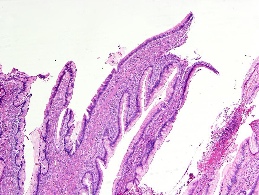
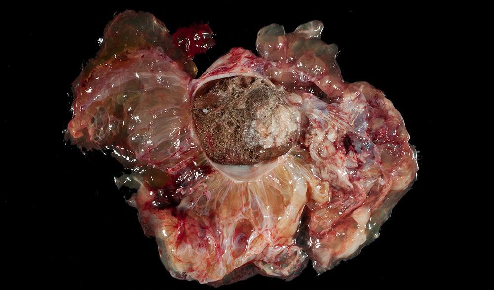
6 Mucinous Cystadenoma Usually unilateral Only 5% of the cases are bilateral Size: Usually large Multilocular cyst in most cases

7 Mucinous Cystadenoma Occasionally, unilocular cyst Smooth outer surface


8 Mucinous Cystadenoma 5% Associated with Dermoid Cyst 5% Associated with Brenner Tumor


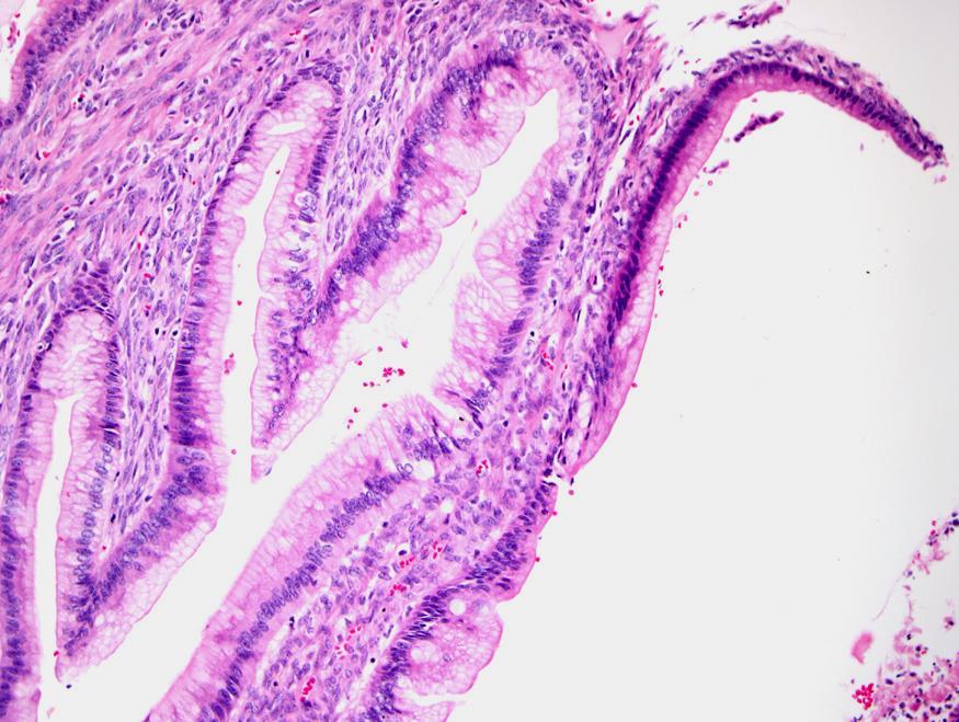
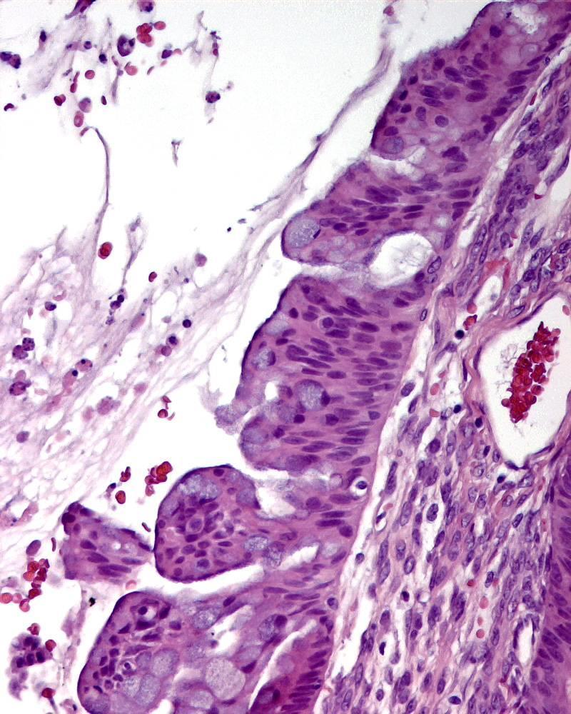
9 Mucinous Cystadenoma Single layer of columnar cells with abundant intracellular mucin and small basilar nuclei


10 Mucinous Cystadenoma with Mucin Granulomas Disruption of the Cyst Wall


11 Mucinous Cystadenoma in a Mature Cystic Teratoma Mucin dissecting the ovarian stroma


12 Be Attentive Mucinous cystadenomas should not have: Nuclear pleomorphism Numerous mitotic figures or apoptosis Abundant mucin dissecting the stroma unless there is an association with teratoma
13 Mucinous Cystadenoma Behavior Benign neoplasm Recurrences have been reported after cystectomies
14
15 Borderline Tumors with Mucinous Differentiation (Mucinous Tumors of Low Malignant Potential/Atypical Proliferative Tumors) Nomenclature Issues WHO 2014 nomenclature changes Intestinal Type Mucinous Borderline/Atypical Proliferative Tumor Tumor of Low Malignant Potential, not recommended Endocervical type Seromucinous Borderline/Atypical Proliferative Tumor Tumor of Low Malignant Potential, not recommended
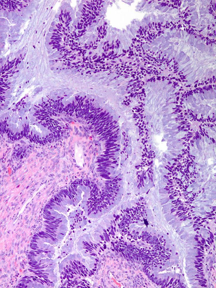
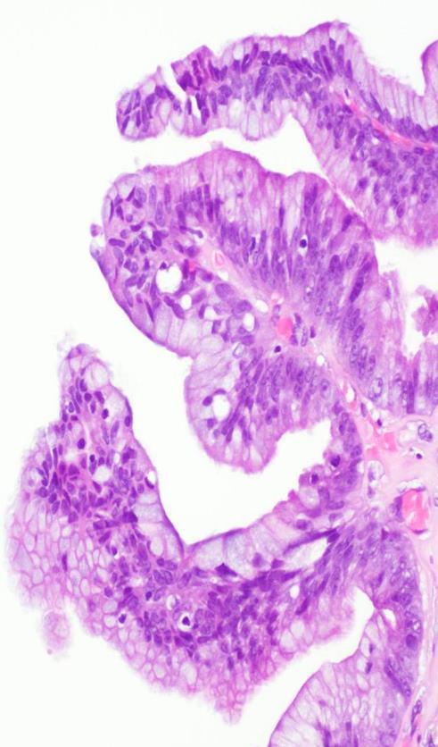
16 Should We Abandon the Term Borderline/Low Malignant Potential? In our opinion, this nomenclature change is not safe The terms borderline/low malignant potential allow us to deal with the potential sampling artifact that we can encounter while examining these tumors Malignant component will determine the disease outcome


17 Why Should We Worry About Potential Sampling Artifact? Cystadenoma Borderline Mucinous Tumor Intraepithelial carcinoma Invasive carcinoma, expansile pattern Invasive carcinoma, infiltrative pattern Large tumors Heterogeneous There is no evidence-based protocol to ensure a perfect sampling
18 33 FIGO stage 1 cases with at least 5 years of follow-up Two patients with recurrences at 12 and 14 months, respectively first patient with tumor incompletely excised due to adhesions to the ileum and sigmoid second patient treated with cystectomy only After the completion of this study, we have encountered rare patients with tumors initially diagnosed as a mucinous tumors of low malignant potential, intestinal type, who developed pelvic carcinomatosis or lung metastasis within a few years after dx, 1-3 years (SAMPLING ARTIFACT ISSUE) In our opinion, the terms borderline/tumor of low malignant potential should be retained to ensure the follow-up of patients affected by this disease until an evidence based sampling protocol becomes available
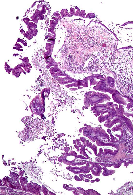
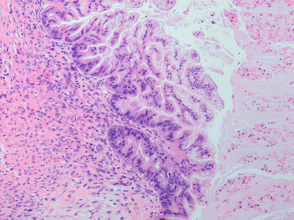
19 2006
20 2009
21 Clinicopathologic Features of 6 Recurrent Cases of Mucinous LMP Tumors Khunamornpong S et al, 2011
22 Borderline Tumors of the Ovary with Mucinous Differentiation (Old Classification and 2014 WHO Nomenclature) 15% 85% Mucinous Borderline Tumor (WHO 2014) Intestinal Type Seromucinous Borderline (WHO 2014) Endocervical Type
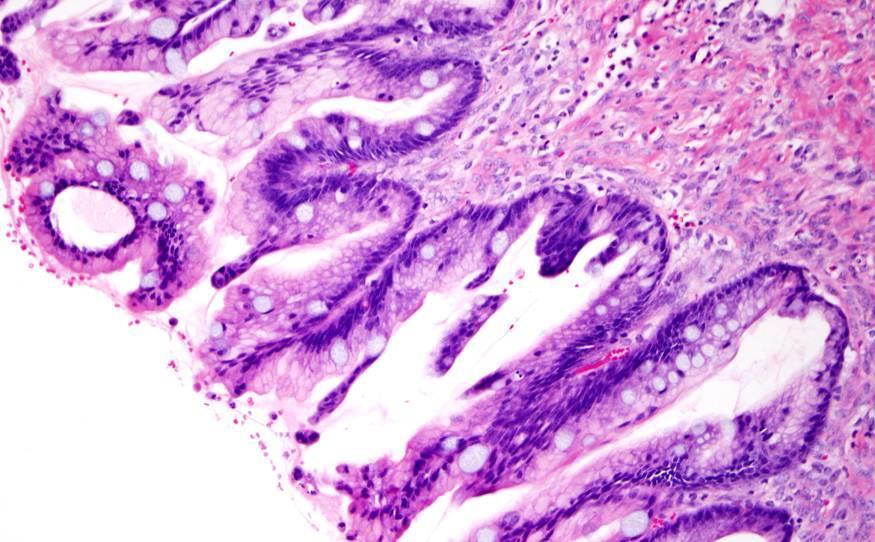
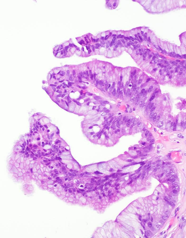
 Gross Features Most")

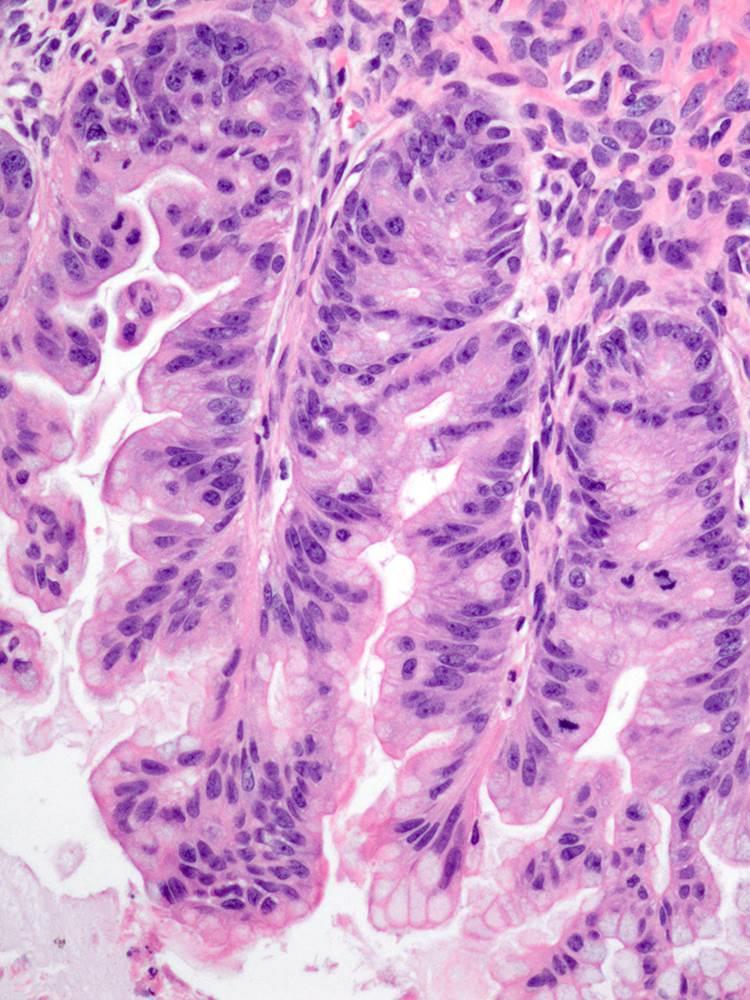
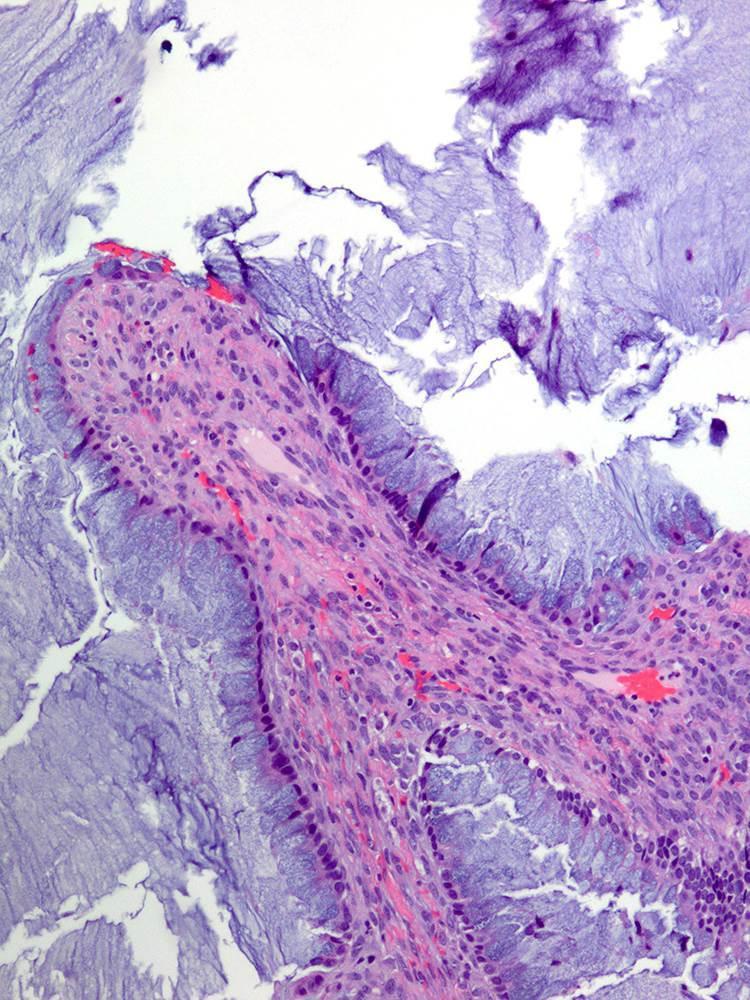
23 Mucinous Borderline Tumor, WHO 2014 (Mucinous Tumor of Low Malignant Potential, Intestinal Type) Gross Features Most cases are unilateral Only 6% of the cases are bilateral Large, average diameter 17 cm Most cases are multilocular Smooth outer surface
24
25
26 Cystadenoma Area Borderline Area


27 Abundant Acellular Mucin in the Ovary and Peritoneum Associated with an Ovarian Mucinous Borderline Tumor Arising in a Dermoid Cyst
28 Mucinous Borderline Tumor, WHO 2014 (Mucinous Tumor of Low Malignant Potential, Intestinal Type) IHC CK 7 + (93%) CK 20 + (87%) CDX-2 + (42%) PAX 8 + (50-60%) Vimentin ( ) ER (-) PR (-) WT-1 (-) SATB2 (-) Vang R, et al Vang R, et al Yasunaga M, et al WHO 2014 Perez Montiel D, et al, 2015 Moh M, et al. 2016
29 Cystectomy or Salpingo-oophorectomy A total of 8 recurrences All cases had either residual ovarian tissue or had extensive adhesions

30 Seromucinous Borderline Tumor, WHO 2014 (Mucinous Tumor of Low Malignant Potential, Endocervical Type) Gross Features Variable size, 2-36 cm Mean, 7 to 8 cm Most cases are multilocular 40% of cases are bilateral Papillary excrescences can be seen on the ovarian surface Endometriosis,30% of the cases

31 Broad Base Papillae with Stromal Edema

32 Columnar Cells Mimicking Endocervical Cells, Squamoid Areas and Inflammatory Cells

33 Columnar Cells Mimicking Endocervical Cells and Inflammatory Cells
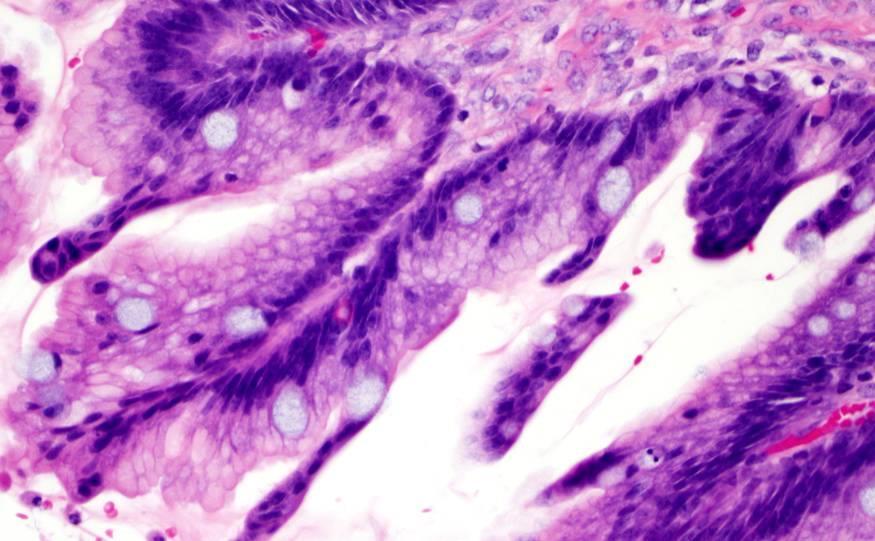
 or lymph node")
34 Seromucinous Borderline Tumor, WHO 2014 (Mucinous Tumors of Low Malignant Potential, Endocervical Type) Up to 20% of the cases are associated with peritoneal implants (usually desmoplastic non-invasive) or lymph node involvement
35 Seromucinous Borderline Tumor, WHO 2014 (Mucinous Tumors of Low Malignant Potential, Endocervical Type) IHC CK 7 + (100%) CK 20 - CDX-2 - ER + (100%) PR + (67-75%) Vimentin + (55%) WT-1 + (8%-11%) PAX-8 + Vang R, et al Yasunaga M, et al Zannoni GF, et al. 2014
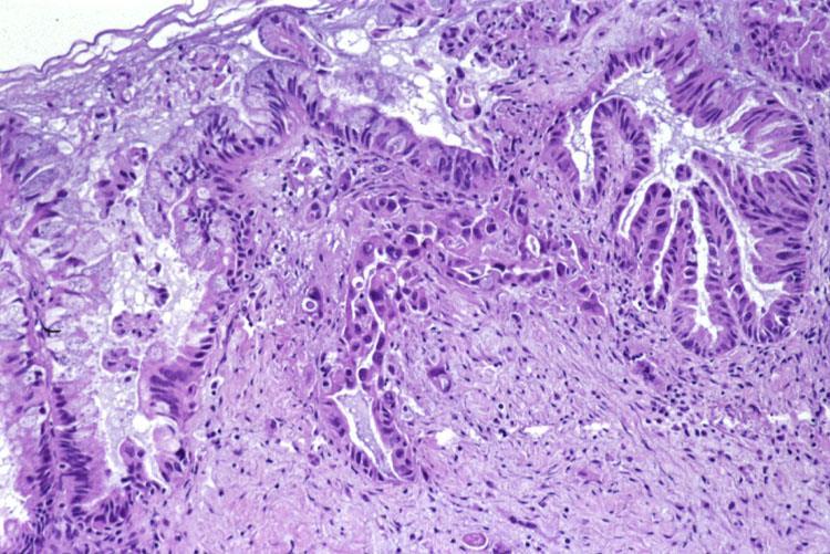
36 Mucinous Borderline Tumor, WHO 2014, with Microinvasion Focus of invasion into the stroma < 5mm in greatest linear extent Mild to moderate cytologic atypia within the invasive focus WHO 2014
37
38 Mucinous or Seromucinous Borderline Tumor with Microinvasion Behavior? Limited experience So far, excellent outcome
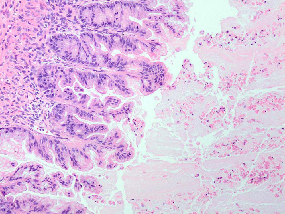
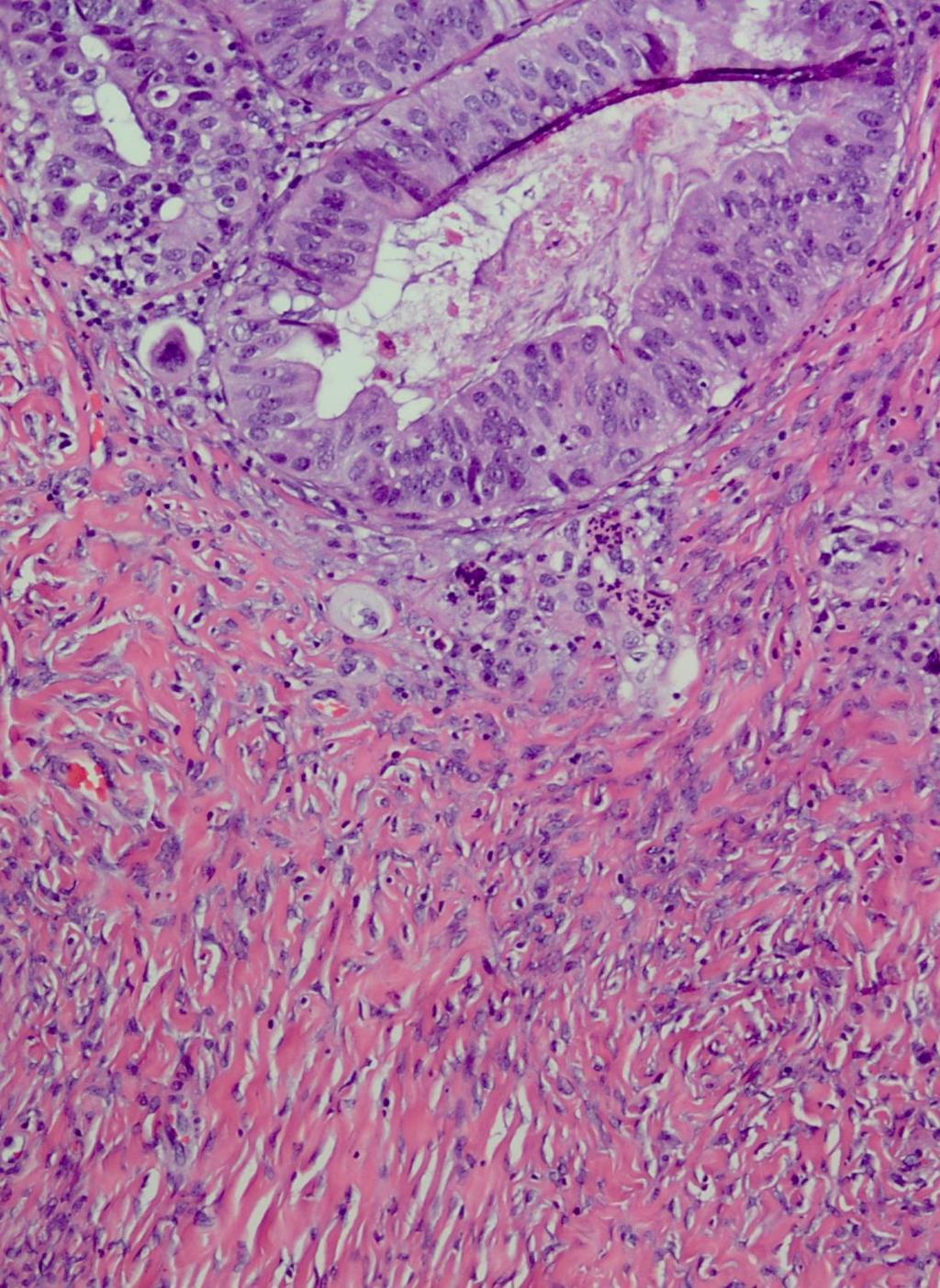
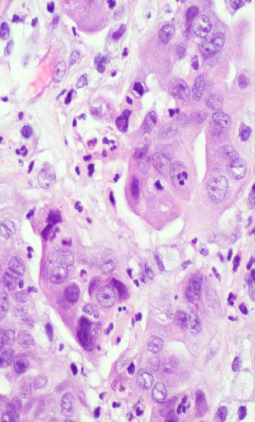
39 Microinvasive Mucinous Carcinoma Focus of invasion into the stroma < 5mm in greatest linear extent Marked cytologic atypia within the invasive focus WHO 2014
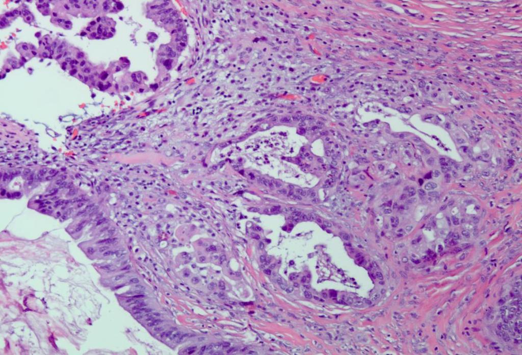
40 Microinvasive Carcinoma
41 Microinvasive Mucinous Carcinoma
42 Microinvasive Mucinous Carcinoma Experience is limited Rare cases have been reported with recurrences cause of death Nomura K, Aizawa S, Cancer 2000 Khunamornpong S. et al, Int J Gynecol Path 2011 WHO 2014
43 Microinvasive Carcinoma Should Not Be Mistaken for Mucinous Borderline Tumor with Microinvasion


44 Microinvasive Mucinous Carcinoma Mucinous Borderline Tumor with Microinvasion
")

45 Non-invasive (Intraepithelial) Carcinoma Marked atypia of the epithelium
46 Non-invasive (Intraepithelial) Carcinoma Behavior Risk of recurrence for FIGO stage I cases: 5.8%

47 Mucinous Carcinoma Infrequent Usually unilateral Gross Features Usually large and multicystic They can have solid or nodular areas Seldom they can be totally solid
48 Invasive Mucinous Carcinoma Expansile or Confluent Type Confluent glandular pattern uninterrupted by normal ovarian stroma This growth occupies an area measuring more than 5 mm in diameter (WHO 2014)
")
49 Invasive Mucinous Carcinoma Infiltrative Type Small glands, nests of cells or individual cells infiltrating the stroma in an area measuring more than 5 mm in diameter (WHO 2014)
50 Invasive Mucinous Carcinoma 5-year survival of 91% for stage I cases; advanced stage cases all died of disease Riopel MA et al Infiltrative stromal invasion appears to be more aggressive than expansile invasion Lee KR and Scully RE Rodriguez IM and Prat J Cases with expansile stromal invasion can have a fatal outcome Ludwick C, et al. 2005
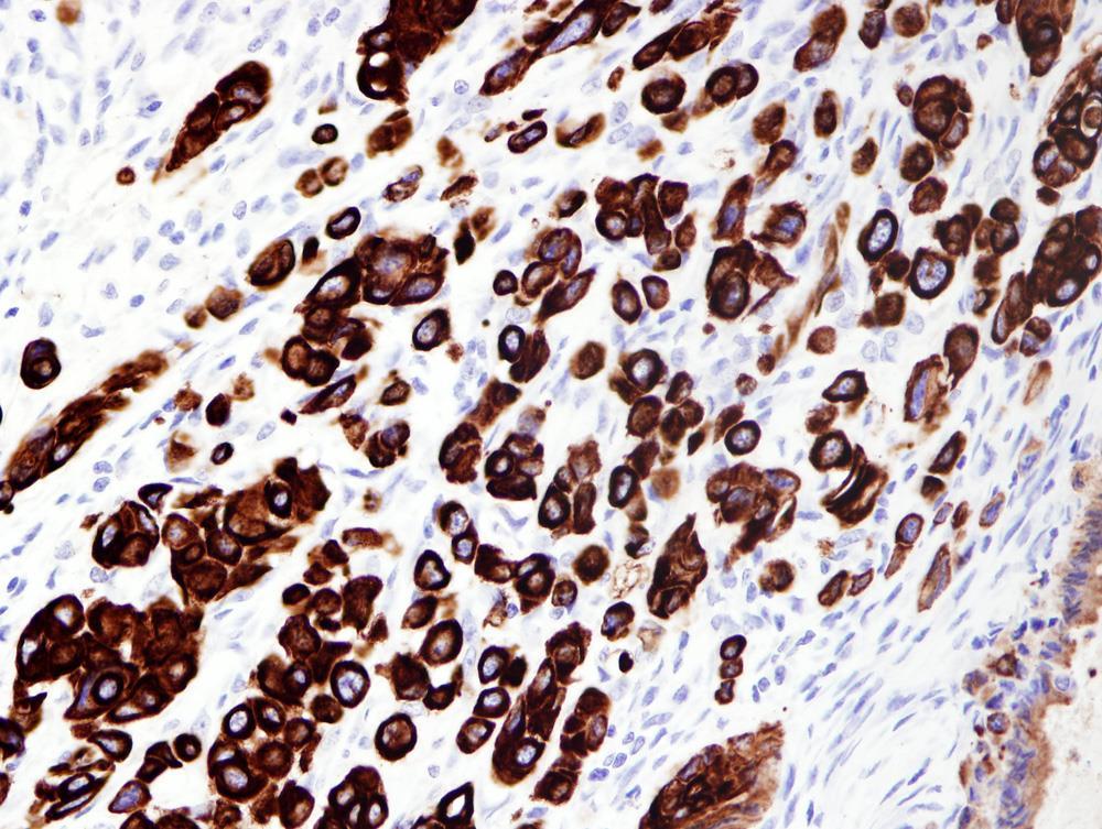
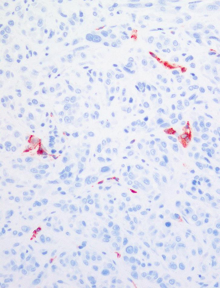
51 Invasive Carcinoma Associated with Seromucinous Borderline Tumor (WHO 2014) Posligua L, et al (in preparation) 16% (14/88) of these tumors had an invasive carcinoma, endometrioid type

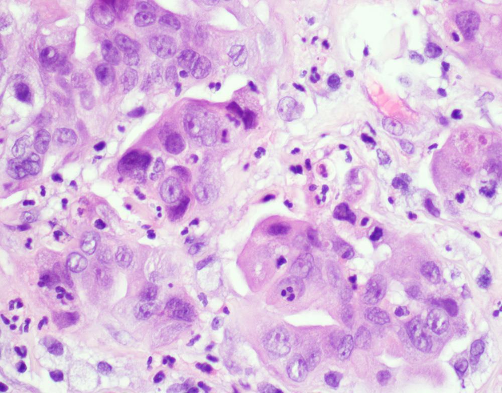
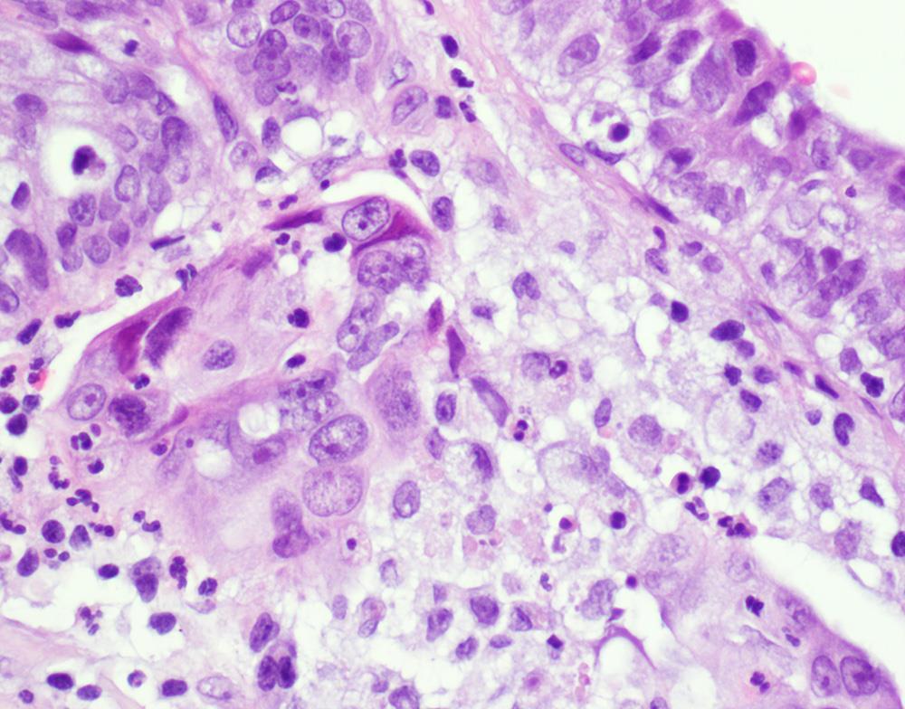
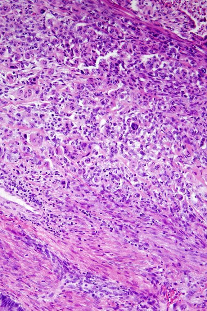
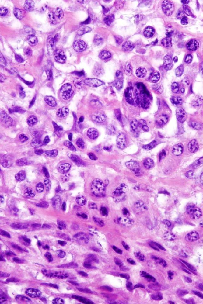
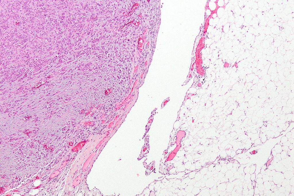
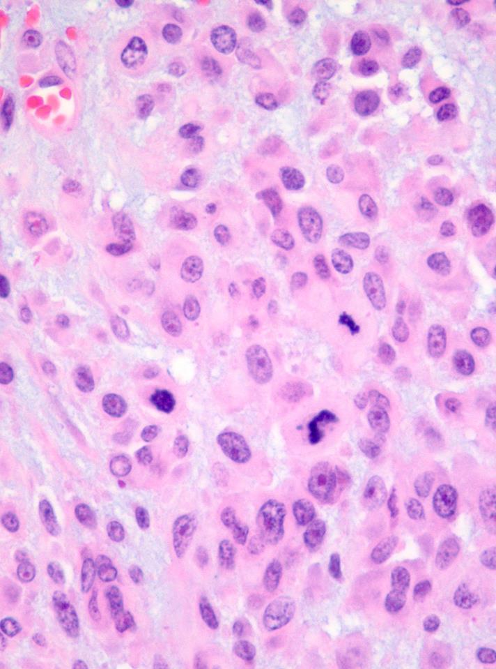
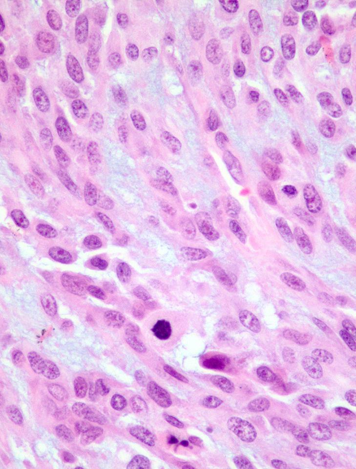
52 Papillae Lined by Endocervical-Type Epithelium with Squamoid Areas

53 Papillae Lined by Endocervical-Type Epithelium with Squamoid Areas

54 Endometrioid Carcinoma with Mucinous and Squamous Metaplasia


55 Seromucinous Carcinoma Arising in a Seromucinous Borderline is a Rare Tumor
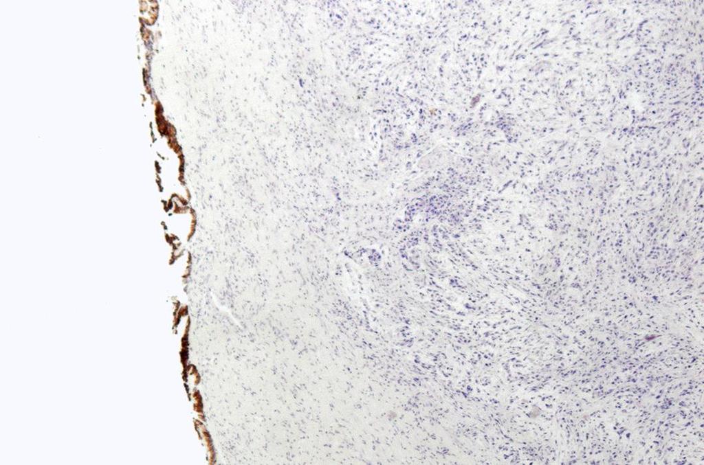
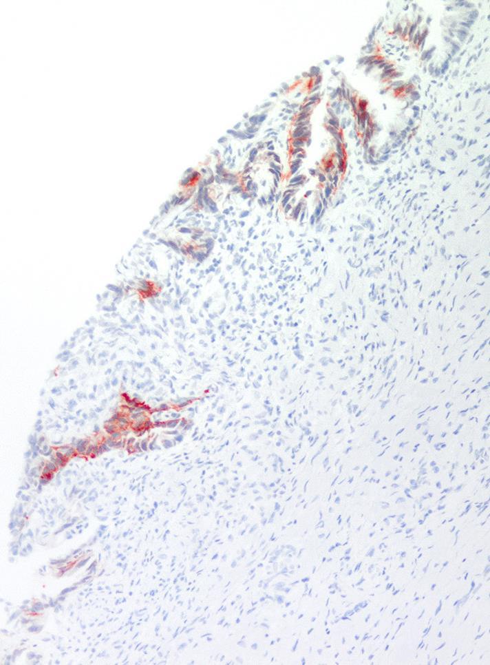
56 19 cases 10 cases associated with endometriosis 10 cases associated with a mucinous tumors of low malignant potential, endocervical type (seromucinous borderline tumors) IHC: CK 7(+), ER(+), PR(+), PAX-8(+) WT-1 + (2/13 cases), p53 wild-type They are similar to endometrioid carcinomas
57 37 cases Association with: borderline tumor, 89% mixed or seromucinous type endometriosis, 48% endometrial endometrioid carcinoma, 43% Modern Pathology, Feb 2015
58 71% expansile pattern of invasion 59% initially diagnosed as mucinous carcinoma IHC CK7 + (100%) CK20 + (35%) ER + (100%) PR + (65%) Modern Pathology, Feb 2015


59 POEM with expansile pattern of invasion POEM arising in endometriosis

60
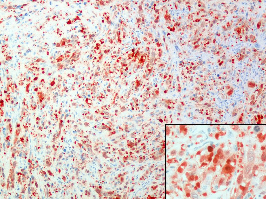
61
62 Keratin +
63 Anaplastic Carcinoma in Ovarian Mucinous Tumors Tumor size: 7 to 62 cm Gross: One or multiple nodules No discrete nodule (microscopic finding)
64 Anaplastic Carcinoma in Ovarian Mucinous Tumors The mucinous epithelium can have the features of: A borderline mucinous tumor Intraepithelial mucinous carcinoma Invasive mucinous carcinoma The anaplastic component can show: Epithelioid Rhabdoid Spindle cells
65 Anaplastic Carcinoma in Ovarian Mucinous Tumors FIGO stage (known in 31 cases): Stage Ia:15 Stage Ic: 6 Stage II: 2 Stage III: 7 Stage IV: 1 Prognosis 7 patients died of disease (either with FIGO stage IC disease or advanced stage disease) 10 patients alive with no evidence of disease after a mean follow up of 5 years All of them with FIGO stage Ia disease


66 Epulis-like area not to be confused with anaplastic carcinoma Epulis-like area with a mixture of giant cells, histiocytes and inflammatory cells


67 Cystic spaces lined by gastrointestinal-type mucinous epithelium with nuclear stratification Mesothelioma associated with a mucinous borderline tumor


68 Adjacent solid component with pleomorphic epithelioid and spindle cells


69 Left ovarian surface with a proliferation of mesothelial cells


70 Right ovary with serous adenofibroma and a marked proliferation of mesothelial cells
71 Omentum

72 Ker 7 Ker 20
73 Calretinin

74 Ker5/6 Podoplanin BerEP4
75 Molecular Alterations in Ovarian Mucinous Carcinoma Ovarian Mucinous Carcinoma MSI-H 22% KRAS mutation 43% BRAF mutation 0% HER2 amplification 18% APC or CTNNB1 mutations 9% TP53 mutations 26% Kelemen LE and Köbel M. 2011
76 Ovarian Mucinous Carcinoma Primary surgery is the standard of care Most cases are FIGO stage I Routine lymphadenectomy is not recommended due to the low risk of lymph node involvement Schmeler K, et al For FIGO stage I disease, there is little evidence to support additional adjuvant therapy after primary surgery Groen RS, et al. 2015
77 Ovarian Mucinous Carcinoma Chemotherapy with platinum and paclitaxel is currently used to treat advanced disease The response to these drugs is poor Schiavone MB, et al Ledermann JA, et al. 2014
78 Ovarian Mucinous Carcinoma Given the platinum resistance exhibited by ovarian mucinous carcinoma Current treatment option 6 cycles of intravenous oxaliplatin and 5-FU or capecitabine with or without bevacizumab after primary surgery for FIGO stage IC-IV disease Groen RS, et al. 2015
79 Proposed Treatment Algorithm for Primary Ovarian Mucinous Carcinoma Anglesio MS, et al. 2013
80 Mucinous Tumors: Frozen Section Handling If the tumor does not have features suggestive of metastasis or borderline tumor or carcinoma FSDX: Mucinous neoplasm, definitive diagnosis deferred for permanent
81 Mucinous Tumors: Frozen Section Handling Appendix should be removed Routine appendectomy in these cases seldom reveals an unsuspected appendiceal primary Elias KM, et al Surgeon should inspect the pelvis and omentum To sample these areas if necessary


82 Primary Ovarian Mucinous Tumors - Sampling Tumor < 10 cm 1 section per cm Tumor 10 cm Tumor of any size 2 sections per cm Microinvasion Intraepithelial Carcinoma


83 Take Home Messages Mucinous cystadenoma Usually unilateral No nuclear pleomorphism, apoptosis or conspicuous mitotic activity should be seen Can recur after cystectomy

84 Ovarian mucinous tumors associated with an ovarian teratoma can have pseudomyxoma ovarii or peritonei Take Home Messages

85 Take Home Messages Borderline mucinous tumor (WHO 2014), mucinous tumor of low malignant potential, intestinal type Usually unilateral and confined to the ovary Undersampling can be an issue

86 Take Home Messages Borderline seromucinous tumor (WHO 2014), mucinous tumor of low malignant potential, endocervical type It can be bilateral It can be associated with endometriosis It can have implants

87 Take Home Messages Mucinous Borderline Tumor with Microinvasion Experience limited So far, excellent outcome


88 Take Home Messages Microinvasive mucinous carcinoma Experience limited Poor outcome
89 Take Home Messages Intraepithelial mucinous carcinoma Recurrence rate: 5.8%
90 Take Home Messages Invasive mucinous carcinoma, expansile pattern More common than the infiltrative type of invasion Usually stage I disease
91 Take Home Messages Invasive mucinous carcinoma, infiltrative pattern Less common than the expansile pattern
92 Take Home Messages Seromucinous borderline tumor If carcinoma is detected, it is usually an endometrioid carcinoma with mucinous metaplasia


93 Take Home Messages Anaplastic Carcinoma It can be associated with different types of mucinous tumors of the ovary Cases of stage IA disease can have a good prognosis


94


95 Dan Flavin
International Society of Gynecological Pathologists Symposium 2007
 International Society of Gynecological Pathologists Symposium 2007 Anais Malpica, M.D. Department of Pathology The University of Texas M.D. Anderson Cancer Center Grading of Ovarian Cancer Histologic grade
Bibliography. Serous Tumors of the Ovary. Nomenclature
 Bibliography Serous Tumors of the Ovary Nomenclature 1. Allison KH, Swisher EM, Kerkering KM, et al. Defining an appropriate threshold for the diagnosis of serous borderline tumor of the ovary: when is
of 20 to 80 and subsequently declines [2].
![of 20 to 80 and subsequently declines [2].](/thumbs/80/81450506.jpg "of 20 to 80 and subsequently declines [2].") - - According to the 2014 World Health Organization (WHO) classification and tumor morphology, primary ovarian tumors are subdivided into three categories: epithelial (60%), germ cell (30%), and sex-cord
- - According to the 2014 World Health Organization (WHO) classification and tumor morphology, primary ovarian tumors are subdivided into three categories: epithelial (60%), germ cell (30%), and sex-cord
Low-grade serous neoplasia. Robert A. Soslow, MD
 Low-grade serous neoplasia Robert A. Soslow, MD soslowr@mskcc.org Outline Orientation Ovarian tumor overview Non serous borderline tumors Serous borderline tumors Clinical summary Morphologic description
Mody. AIS vs. Invasive Adenocarcinoma of the Cervix
 Common Problems in Gynecologic Pathology Michael T. Deavers, M.D. Houston Methodist Hospital, Houston, Texas Common Problems in Gynecologic Pathology Adenocarcinoma in-situ (AIS) of the Cervix vs. Invasive
Case Report Ovarian Seromucinous Borderline Tumor and Clear Cell Carcinoma: An Unusual Combination
 Case Reports in Obstetrics and Gynecology Volume 2015, Article ID 690891, 5 pages http://dx.doi.org/10.1155/2015/690891 Case Report Ovarian Seromucinous Borderline Tumor and Clear Cell Carcinoma: An Unusual
Ovarian carcinoma classification. Robert A. Soslow, MD
 Ovarian carcinoma classification Robert A. Soslow, MD soslowr@mskcc.org WHO classification Serous Mucinous Endometrioid Clear cell Transitional Squamous Mixed epithelial Undifferentiated Introduction Rationale
3 cell types in the normal ovary
 Ovarian tumors 3 cell types in the normal ovary Surface (coelomic epithelium) the origin of the great majority of ovarian tumors 90% of malignant ovarian tumors Totipotent germ cells Sex cord-stromal cells
3 cell types in the normal ovary
 Ovarian tumors 3 cell types in the normal ovary Surface (coelomic epithelium) the origin of the great majority of ovarian tumors (neoplasms) 90% of malignant ovarian tumors Totipotent germ cells Sex cord-stromal
Mousa. Najat kayed &Renad Al-Awamleh. Nizar Alkhlaifat
 6 Mousa Najat kayed &Renad Al-Awamleh Nizar Alkhlaifat P a g e 1 This sheet written based on record 13 on website Cover slide( 95-117 ) No need to go back to slide FALLOPIAN TUBE PATHOLOGY In general fallopian
Section 1. Biology of gynaecological cancers: our current understanding
 Section 1 Biology of gynaecological cancers: our current understanding Chapter 1 Morphological sub-types of ovarian carcinoma: new developments and pathogenesis W Glenn McCluggage 1 Introduction In most
Current Concept in Ovarian Carcinoma: Pathology Perspectives
 Current Concept in Ovarian Carcinoma: Pathology Perspectives Rouba Ali-Fehmi, MD Professor of Pathology The Karmanos Cancer Institute, Wayne State University School of Medicine Current Concept in Ovarian
USCAP 2013: THE ORIGINS OF OVARIAN CANCER: MUCINOUS TUMORS
 USCAP 2013: THE ORIGINS OF OVARIAN CANCER: MUCINOUS TUMORS Russell Vang, M.D. Associate Professor Division of Gynecologic Pathology The Johns Hopkins Hospital Table of contents: Powerpoint handout Text
Survival Analysis and Prognosis for Patients with Serous and Mucinous Borderline Ovarian Tumors: 14-Year Experience from a Tertiary Center in Iran
 ORIGINAL ARTICLE Survival Analysis and Prognosis for Patients with Serous and Mucinous Borderline Ovarian Tumors: 14-Year Experience from a Tertiary Center in Iran Katayoun Ziari, Ebrahim Soleymani, and
Case 1. Pathology of gynecological cancer. What do we need to know (Case 1) Luca Mazzucchelli Istituto cantonale di patologia Locarno
 Luca Mazzucchelli Istituto cantonale di patologia Locarno") Case 1 Pathology of gynecological cancer. What do we need to know (Case 1) Luca Mazzucchelli Istituto cantonale di patologia Locarno SAMO Interdisciplinary Workshop on Gynecological Tumors Lucern, October
Case 1 Pathology of gynecological cancer. What do we need to know (Case 1) Luca Mazzucchelli Istituto cantonale di patologia Locarno SAMO Interdisciplinary Workshop on Gynecological Tumors Lucern, October
MPH Quiz. 1. How many primaries are present based on this pathology report? 2. What rule is this based on?
 MPH Quiz Case 1 Surgical Pathology from hysterectomy performed July 11, 2007 Final Diagnosis: Uterus, resection: Endometrioid adenocarcinoma, Grade 1 involving most of endometrium, myometrial invasion
Pathology of Ovarian Tumours. Dr. Jyothi Ranganathan MD ( Path) AFMC Pune PDCC (Cytopathology) PGI Chandigarh
 AFMC Pune PDCC (Cytopathology) PGI Chandigarh") Pathology of Ovarian Tumours Dr. Jyothi Ranganathan MD ( Path) AFMC Pune PDCC (Cytopathology) PGI Chandigarh Outline Incidence Risk factors Classification Pathology of tumours Tumour markers Prevention
Pathology of Ovarian Tumours Dr. Jyothi Ranganathan MD ( Path) AFMC Pune PDCC (Cytopathology) PGI Chandigarh Outline Incidence Risk factors Classification Pathology of tumours Tumour markers Prevention
Pathology of the female genital tract
 Pathology of the female genital tract Common illnesses of the female genital tract Before menarche Developmental anomalies Tumors (ovarial teratoma) Amenorrhea Fertile years PCOS, ovarian cysts Endometriosis
64 YO lady THBSO for prolapse At gross : A 3 cm endometrial polyp in the fundus
 Case 6 64 YO lady THBSO for prolapse At gross : A 3 cm endometrial polyp in the fundus Numerous irregular, large glands with leaf-like pattern Large glands with broad-based papillary infolding into the
Presenter: Yeh-Han Wang M.D.
 Korea-Taiwan-Japan Joint Meeting for Gynecological Pathology Mini-lecture Female Adnexal Tumor of Probable Wolffian Origin (FATWO) in Taiwan: A Small Case Series and Literature Review Presenter: Yeh-Han
How to Recognize Gynecologic Cancer Cells from Pelvic Washing and Ascetic Specimens
 How to Recognize Gynecologic Cancer Cells from Pelvic Washing and Ascetic Specimens Wenxin Zheng, M.D. Professor of Pathology and Gynecology University of Arizona zhengw@email.arizona.edu http://www.zheng.gynpath.medicine.arizona.edu/index.html
Borderline Ovarian Mucinous Tumors: Consensus Points and Persistent Controversies Regarding Nomenclature, Diagnostic Criteria, and Behavior
 Borderline Ovarian Mucinous Tumors: Consensus Points and Persistent Controversies Regarding Nomenclature, Diagnostic Criteria, and Behavior Brigitte M. Ronnett, M.D.; C. Blake Gilks, M.D., Maria J. Merino,
Ovarian Clear Cell Carcinoma
 Ovarian Clear Cell Carcinoma Rouba Ali-Fehmi, MD Professor of Pathology The Karmanos Cancer Institute, Wayne State University School of Medicine 50 year old woman with chief complaint of shortness of breath
Borderline tumors. Borderline tumors. Serous borderline tumor are NOT benign. Low grade serous carcinoma: pathogenesis. Serous carcinoma: pathogenesis
 Serous borderline tumor are NOT benign Robert A. Soslow, MD Memorial Sloan-Kettering Cancer Center soslowr@mskcc.org Borderline tumors Serous BTs and seromucinous BTs are both histopathologically borderline
Joseph Misdraji, M.D. GI pathology Unit Massachusetts General Hospital
 Joseph Misdraji, M.D. GI pathology Unit Massachusetts General Hospital jmisdraji@partners.org Low-grade appendiceal mucinous neoplasm (LAMN) High-grade appendiceal mucinous neoplasm (HAMN) Adenocarcinoma
Invited Re vie W. Molecular genetics of ovarian carcinomas. Histology and Histo pathology
 Histol Histopathol (1 999) 14: 269-277 http://www.ehu.es/histol-histopathol Histology and Histo pathology Invited Re vie W Molecular genetics of ovarian carcinomas J. Diebold Pathological Institute, Ludwig-Maximilians-University
Mucinous Adenocarcinoma Involving the Ovary: Comparative Evaluation of the Classification Algorithms using Tumor Size and Laterality
 J Korean Med Sci 2010; 25: 220-5 ISSN 1011-8934 DOI: 10.3346/jkms.2010.25.2.220 Mucinous Adenocarcinoma Involving the Ovary: Comparative Evaluation of the Classification Algorithms using Tumor Size and
Dr Sanjiv Manek Oxford. Oxford Pathology Course 2010 for FRCPath Illustration-Cellular Pathology. Oxford Radcliffe NHS Trust
 Dr Sanjiv Manek Oxford Oxford Pathology Course 2010 for FRCPath Illustration-Cellular Pathology. Oxford Radcliffe NHS Trust Ovarian Endometrial Vulvo-vaginal Cervical Illustration-Cellular Pathology. Oxford
The Origin of Pelvic Low-Grade Serous Proliferative Lesions
 The Origin of Pelvic Low-Grade Serous Proliferative Lesions Ovarian Atypical Proliferative (Borderline) Serous Tumors, Noninvasive Implants and Endosalpingiosis Robert J. Kurman, M.D. Kurman RJ, Vang R,
Normal endometrium: A, proliferative. B, secretory.
 Normal endometrium: A, proliferative. B, secretory. Nội mạc tử cung Nội mạc tử cung Cyclic changes in endometrium.. Approximate relationship of useful microscopic changes. Arias-Stella reaction in endometrial
Low-Grade Serous Ovarian Tumors Debra A. Bell, MD Mayo Clinic and Mayo Medical School Rochester, MN
 1 Low-Grade Serous Ovarian Tumors Debra A. Bell, MD Mayo Clinic and Mayo Medical School Rochester, MN It is very appropriate to discuss low-grade ovarian serous neoplasms in a symposium in honor of Dr.
Pathobiology of ovarian carcinomas
 Chinese Journal of Cancer Review Mojgan Devouassoux-Shisheboran 1 and Catherine Genestie 2 Abstract Ovarian tumors comprise a heterogeneous group of lesions, displaying distinct tumor pathology and oncogenic
OVARIES. MLS Basic histological diagnosis MLS HIST 422 Semester 8- batch 7 L13 Dr: Ali Eltayb.
 OVARIES MLS Basic histological diagnosis MLS HIST 422 Semester 8- batch 7 L13 Dr: Ali Eltayb. OBJECTIVES Recognize different disease of ovaries Classify ovarian cyst Describe the pathogenesis, morphology
Gross appearance of peritoneal cysts. They have a thin, translucent wall and contain a clear fluid.
 Gross appearance of peritoneal cysts. They have a thin, translucent wall and contain a clear fluid. So-called multicystic benign mesothelioma. A, Gross appearance. So-called multicystic benign mesothelioma.
A Survay on Appendiceal Involvement in Ovarian Mucinous Tumors
 http://www.ijwhr.net Open Access doi 10.15296/ijwhr.2018.33 Original Article International Journal of Women s Health and Reproduction Sciences Vol. 6, No. 2, April 2018, 199 203 ISSN 2330-4456 A Survay
Borderline Ovarian Tumours. Andreas Obermair Brisbane
 Borderline Ovarian Tumours Andreas Obermair Brisbane Definition First described in 1929 Cellular features of malignancy Cellular atypia Mitotic activity No stromal invasion An entity per se??? (or precursor
Female Genital Tract Lab. Dr. Nisreen Abu Shahin Assistant Professor of Pathology University of Jordan
 Female Genital Tract Lab Dr. Nisreen Abu Shahin Assistant Professor of Pathology University of Jordan Ovarian Pathology A 20-year-old female presented with vague left pelvic pain. Pelvic exam revealed
GOBLET CELL CARCINOID. Hanlin L. Wang, MD, PhD University of California Los Angeles
 GOBLET CELL CARCINOID Hanlin L. Wang, MD, PhD University of California Los Angeles Disclosure of Relevant Financial Relationships USCAP requires that all planners (Education Committee) in a position to
GOBLET CELL CARCINOID
 GOBLET CELL CARCINOID Hanlin L. Wang, MD, PhD University of California Los Angeles Disclosure of Relevant Financial Relationships USCAP requires that all planners (Education Committee) in a position to
Institute of Pathology First Faculty of Medicine Charles University. Ovary
 Ovary Barrett esophagus ph in vagina between 3.8 and 4.5 ph of stomach varies from 1-2 (hydrochloric acid) up to 4-5 BE probably results from upward migration of columnar cells from gastroesophageal junction
Ovarian cancer: 2012 Update Srini Prasad MD Univ Texas MD Anderson Cancer Center
 Ovarian cancer: 2012 Update Srini Prasad MD Univ Texas MD Anderson Cancer Center Ovarian cancer is not a single disease Ovarian Epithelial Tumors: Histological Spectrum* Type Frequency Histology High-Grade
Case Report Angiosarcoma Arising in Ovarian Mucinous Tumor: A Challenge in Intraoperative Frozen Section Diagnosis
 Case Reports in Pathology Volume 2016, Article ID 8508624, 5 pages http://dx.doi.org/10.1155/2016/8508624 Case Report Angiosarcoma Arising in Ovarian Mucinous Tumor: A Challenge in Intraoperative Frozen
Interesting Cases in Gynecologic Pathology. Michael Ward, MD Surgical Pathology Fellow University of Utah Health Sciences Center Salt Lake City, UT
 Interesting Cases in Gynecologic Pathology Michael Ward, MD Surgical Pathology Fellow University of Utah Health Sciences Center Salt Lake City, UT Case 1 History: 50 year old woman with a uterine mass
Published Ahead of Print on September 28, 2012 as /theoncologist
 The Oncologist Gynecologic Oncology Diagnosis, Treatment, and Follow-Up of Borderline Ovarian Tumors DANIELA FISCHEROVA, a MICHAL ZIKAN, a PAVEL DUNDR, b DAVID CIBULA a a Gynecological Oncology Center,
5/26/2016. Pelvic Serous Carcinoma: 2014 W.H.O. Update. Outline of Talk. Changes to 2014 WHO system for pelvic serous tumors
 Pelvic Serous Carcinoma: 2014 W.H.O. Update Outline of Talk Practical Implications for Pathologists Changes to 2014 WHO system for pelvic serous tumors High grade serous carcinoma versus low grade serous
The relative frequency and histopathological patterns of ovarian lesions: study of 116 cases
 Original article: The relative frequency and histopathological patterns of ovarian lesions: study of 116 cases Dr Dimple Mehta*,Dr Alpesh Chavda**, Dr Hetal Patel*** *Assistant Professor, **Tutor, ***3
Papillary Lesions of the Breast A Practical Approach to Diagnosis. (Arch Pathol Lab Med. 2016;140: ; doi: /arpa.
 Papillary Lesions of the Breast A Practical Approach to Diagnosis (Arch Pathol Lab Med. 2016;140:1052 1059; doi: 10.5858/arpa.2016-0219-RA) Papillary lesions of the breast Span the spectrum of benign,
6/5/2010. Outline of Talk. Endometrial Alterations That Mimic Cancer & Vice Versa: Metaplastic / reactive changes. Problems in Biopsies/Curettages
 Outline of Talk Endometrial Alterations That Mimic Cancer & Vice Versa: Problems in Biopsies/Curettages Metaplastic / reactive changes Mucinous change Microglandular hyperplasia-like change Squamous metaplasia
The Diagnostic Challenges of Low Grade and High Grade Tubo-Ovarian Serous Carcinomas. W Glenn McCluggage Belfast, Northern Ireland
 The Diagnostic Challenges of Low Grade and High Grade Tubo-Ovarian Serous Carcinomas W Glenn McCluggage Belfast, Northern Ireland Enterprise Interest None OVARIAN SEROUS CARCINOMA (OSC) RECENT DEVELOPMENTS
Case Report Ovarian mucinous cystic tumor with sarcoma-like mural nodules and multifocal anaplastic carcinoma: a case report
 Int J Clin Exp Pathol 2013;6(8):1688-1692 www.ijcep.com /ISSN:1936-2625/IJCEP1306008 Case Report Ovarian mucinous cystic tumor with sarcoma-like mural nodules and multifocal anaplastic carcinoma: a case
David Nunns on behalf of the Gynae Guidelines Group Date:
 Title of Guideline (must include the word Guideline (not protocol, policy, procedure etc) Borderline tumours of the ovary management and follow-up Author: Contact Name and Job Title Directorate & Speciality
Original contribution
 Human Pathology (2012) 43, 747 752 www.elsevier.com/locate/humpath Original contribution The presence and location of epithelial implants and implants with epithelial proliferation may predict a higher
Pacific Northwest Society of Pathologists Fall Meeting September 2015 Intraoperative Consultation in Gynecological Pathology: The Adnexal Mass
 Pacific Northwest Society of Pathologists Fall Meeting September 2015 Intraoperative Consultation in Gynecological Pathology: The Adnexal Mass Julie Irving, MD Department of Pathology, University of British
ENODMETRIAL CARCINOMA: SPECIAL & NOT SO SPECIAL VARIANTS
 ENODMETRIAL CARCINOMA: SPECIAL & NOT SO SPECIAL VARIANTS Pacific Northwest Society of Pathologists Vancouver, B.C. September 26, 2015 Teri A. Longacre, M.D. longacre@stanford.edu Stanford University, Stanford,
Adenocarcinoma of the Cervix
 Question 1. Each of the following statements about cervical adenocarcinoma is true except: Adenocarcinoma of the Cervix SAMS a) A majority of women with cervical adenocarcinoma have stage I tumors at diagnosis.
Papillary Lesions of the breast
 Papillary Lesions of the breast Emad Rakha Professor of Breast Pathology The University of Nottingham Papillary lesions of the breast are a heterogeneous group of disease, which are characterised by neoplastic
Key Words. Borderline ovarian tumor Prognostic parameter Ultrasound Fertility Conservative surgery Recurrence
 The Oncologist Gynecologic Oncology Diagnosis, Treatment, and Follow-Up of Borderline Ovarian Tumors DANIELA FISCHEROVA, a MICHAL ZIKAN, a PAVEL DUNDR, b DAVID CIBULA a a Gynecological Oncology Center,
Salivary Glands 3/7/2017
 Salivary Glands 3/7/2017 Goals and objectives Focus on the entities unique to H&N Common board type facts Information for your future practice Salivary Glands Salivary Glands Major gland. Paratid. Submandibular.
Atypical Hyperplasia/EIN
 EIN Atypical Hyperplasia/EIN Based on scientific and diagnostic advances, in 2014 the WHO moved that the precursor lesion for endometrioid carcinoma be atypical hyperplasia/ein, rather than what was previously
Pancreatic Cystic Lesions 원자력병원
 Pancreatic Cystic Lesions 원자력병원 박선 후 Lines of cellular differentiation Ductal Acinar Undetermined Ductal adenocarcinoma Serous/ mucinous tumor Intraductal papillary mucinous neoplasm Acinar cell carcinoma
Primary Mucinous Ovarian Cancer (PMOC) Michael Frumovitz
 Michael Frumovitz") Primary Mucinous Ovarian Cancer (PMOC) Michael Frumovitz Epithelial Subtypes Serous Endometrioid Mucinous Transitional Clear Cell Mixed Undifferentiated Squamous Ovarian Surface Epithelium Naora et al.,
Primary Mucinous Ovarian Cancer (PMOC) Michael Frumovitz Epithelial Subtypes Serous Endometrioid Mucinous Transitional Clear Cell Mixed Undifferentiated Squamous Ovarian Surface Epithelium Naora et al.,
Exploring the Histogenesis of Ovarian Mucinous and Transitional Cell (Brenner) Neoplasms and Their Relationship With Walthard Cell Nests
 Neoplasms and Their Relationship With Walthard Cell Nests") Exploring the Histogenesis of Ovarian Mucinous and Transitional Cell (Brenner) Neoplasms and Their Relationship With Walthard Cell Nests A Study of 120 Tumors Jeffrey D. Seidman, MD; Fatemeh Khedmati,
Exploring the Histogenesis of Ovarian Mucinous and Transitional Cell (Brenner) Neoplasms and Their Relationship With Walthard Cell Nests A Study of 120 Tumors Jeffrey D. Seidman, MD; Fatemeh Khedmati,
Ovarian Tumors. Andrea Hayes-Jordan MD FACS, FAAP Section Chief, Pediatric Surgery/Surgical Onc. UT MD Anderson Cancer Center
 Ovarian Tumors Andrea Hayes-Jordan MD FACS, FAAP Section Chief, Pediatric Surgery/Surgical Onc. UT MD Anderson Cancer Center Case 13yo female with abdominal pain Ultrasound shows huge ovarian mass Surgeon
CASE 4 21/07/2017. Ectopic Prostatic Tissue in Cervix. Female 31. LLETZ for borderline nuclear abnormalities
 Female 31 CASE 4 LLETZ for borderline nuclear abnormalities PSA Ectopic Prostatic Tissue in Cervix AJSP 2006;30;209-215 usually incidental microscopic finding usually in ectocervical stroma? developmental
04/10/2018. Intraductal Papillary Neoplasms Of Breast INTRADUCTAL PAPILLOMA
 Intraductal Papillary Neoplasms Of Breast Savitri Krishnamurthy MD Professor of Pathology Deputy Division Head The University of Texas MD Anderson Cancer Center 25 th Annual Seminar in Pathology Pittsburgh,
Select problems in cystic pancreatic lesions
 Disclosure Select problems in cystic pancreatic lesions Five Prime Therapeutics shareholder Adicet Bio shareholder Bristol-Meyer Squibb advisory board grace.kim@ucsf.edu Pancreatic cystic lesions Intraductal
A Serous Borderline Tumor of the Fallopian Tube Detected Incidentally
 A Serous Borderline Tumor of the Fallopian Tube Detected Incidentally Imrana Tanvir, Ghania Ali, Haseeb Ahmed Khan and Ahmed Nasir Hanifi* Dept. of Histopathology, FMH College of Medicine & Dentistry,
Case # 4 Low-Grade Serous Carcinoma (Macropapillary) of the Ovary Arising in an Atypical Proliferative Serous Tumor
 of the Ovary Arising in an Atypical Proliferative Serous Tumor") Case # 4 Low-Grade Serous Carcinoma (Macropapillary) of the Ovary Arising in an Atypical Proliferative Serous Tumor Robert J Kurman, M.D. Johns Hopkins University School of Medicine Case History A 53 year
Case # 4 Low-Grade Serous Carcinoma (Macropapillary) of the Ovary Arising in an Atypical Proliferative Serous Tumor Robert J Kurman, M.D. Johns Hopkins University School of Medicine Case History A 53 year
CASE 1A DIAGNOSIS. Problems in the Evaluation of Surface Epithelial-Stromal Tumors of the Ovary: an Update
 Problems in the Evaluation of Surface Epithelial-Stromal Tumors of the Ovary: an Update John H. Eichhorn C. Blake Gilks CASE 1A A 27-year-old woman with primary infertility had a negative work-up, except
Serous Borderline Tumors of the Ovary: Implants, Manifestations, Biology & New Insights in Progression
 Serous Borderline Tumors of the Ovary: Implants, Manifestations, Biology & New Insights in Progression Stanley J. Robboy, MD Professor of Pathology Professor of Obstetrics & Gynecology Vice Chairman for
Endosalpingiosis. Case report
 Case report Endosalpingiosis Michael D. Holmes, M.D. Howard S. Levin M.D. Department of Pathology Lester A. Ballard, Jr., M.D. Department of Gynecology Endosalpingiosis, a term referring to tuballike epithelium
L/O/G/O. Ovarian Tumor. Xiaoyu Niu Obstetrics and Gynecology Department Sichuan University West China Second Hospital
 L/O/G/O Ovarian Tumor Xiaoyu Niu Obstetrics and Gynecology Department Sichuan University West China Second Hospital Essentials classification of ovarian tumor clinical manifestation of ovarian tumor metastatic
Diseases of the breast (1 of 2)
") Diseases of the breast (1 of 2) Introduction A histology introduction Normal ducts and lobules of the breast are lined by two layers of cells a layer of luminal cells overlying a second layer of myoepithelial
Diseases of the breast (1 of 2) Introduction A histology introduction Normal ducts and lobules of the breast are lined by two layers of cells a layer of luminal cells overlying a second layer of myoepithelial
Department of Pathology, Breast, and Gynecologic Pathology, Magee-Womens Hospital of UPMC, PA 15213, USA
 Case Reports in Obstetrics and Gynecology Volume 2012, Article ID 269489, 5 pages doi:10.1155/2012/269489 Case Report Malignant Transformation of a Mature Cystic Ovarian Teratoma into Thyroid Carcinoma,
Icd 10 ovarian stroma
 Icd 10 ovarian stroma Struma ovarii; Micrograph of a struma ovarii. Characteristic thyroid follicles are seen on the right, and ovarian stroma on the left. H&E stain. Classification and. Free, official
Pathology of the Thyroid
 Pathology of the Thyroid Thyroid Carcinoma Arising from Follicular Cells 2015-01-19 Prof. Dr. med. Katharina Glatz Pathologie Carcinomas Arising from Follicular Cells Differentiated Carcinoma Papillary
C ORPUS UTERI C ARCINOMA STAGING FORM (Carcinosarcomas should be staged as carcinomas)
") C ORPUS UTERI C ARCINOMA STAGING FORM CLINICAL Extent of disease before any treatment y clinical staging completed after neoadjuvant therapy but before subsequent surgery Tis * T1 I T1a IA NX N0 N1 N2
C ORPUS UTERI C ARCINOMA STAGING FORM CLINICAL Extent of disease before any treatment y clinical staging completed after neoadjuvant therapy but before subsequent surgery Tis * T1 I T1a IA NX N0 N1 N2
What s (new) and Important in Reporting of Uterine Cancers Katherine Vroobel The Royal Marsden
 and Important in Reporting of Uterine Cancers Katherine Vroobel The Royal Marsden") What s (new) and Important in Reporting of Uterine Cancers Katherine Vroobel The Royal Marsden Maastricht Pathology 2018 Wednesday 20 th June Endometrioid adenocarcinoma High grade carcinomas (common)
What s (new) and Important in Reporting of Uterine Cancers Katherine Vroobel The Royal Marsden Maastricht Pathology 2018 Wednesday 20 th June Endometrioid adenocarcinoma High grade carcinomas (common)
Neoplasias Quisticas del Páncreas
 SEAP -Aproximación Práctica a la Patología Gastrointestinal- Madrid, 26 de mayo, 2006 Neoplasias Quisticas del Páncreas Gregory Y. Lauwers, M.D. Director, Service Massachusetts General Hospital Harvard
In situ and Invasive Endocervical Carcinoma: Problems and Pitfalls in Diagnosis
 In situ and Invasive Endocervical Carcinoma: Problems and Pitfalls in Diagnosis Rouba Ali-Fehmi,MD The Karmanos Cancer Institute, Wayne State University School of Medicine Global incidence of cervical
CYSTIC TUMORS OF THE KIDNEY JOHN N. EBLE, M.D. CYSTIC NEPHROMA
 Page 1 CYSTIC TUMORS OF THE KIDNEY JOHN N. EBLE, M.D. Department of Pathology & Laboratory Medicine Phone (317) 274-4806 Medical Science A-128 FAX: (317) 278-2018 635 Barnhill Drive jeble @iupui.edu Indianapolis,
Borderline tumors of the ovary: a separate entity
 Borderline tumors of the ovary: a separate entity Authors Key words A.Ph. Makar Excellent prognosis, conservative surgery, adjuvant therapy Summary Borderline ovarian tumors (BOT) account for 10% to 20%
A Case of Mucinous Cystadenocarcinoma Ovary Mimicking Psuedomyxoma Peritonei
 A Case of Mucinous Cystadenocarcinoma Ovary Mimicking Psuedomyxoma Peritonei Dr. Abdul haleem & Dr. Vishwanath sherigar Department of General Surgery, K S Hegde Medical Academy, Mangalore, Karnataka, India
TUMOR AND TUMOR-LIKE CONDITIONS OF THE PERITONEUM AND OMENTUM/MESENTERY 40 th. Annual Meeting SCBTMR September 9-13, 2017, Nashville, Tennessee
 TUMOR AND TUMOR-LIKE CONDITIONS OF THE PERITONEUM AND OMENTUM/MESENTERY 40 th. Annual Meeting SCBTMR September 9-13, 2017, Nashville, Tennessee Isaac R Francis University of Michigan Department of Radiology
Dr Agata T Kochman Wishaw General Hospital
 Dr Agata T Kochman Wishaw General Hospital Case E1 84 year old male Symptoms: R shoulder pain CT = thymic mass and (R) LL nodules + (L) lung nodule Clinically metastatic lesions in lung with primary thymic
Page # 1. Endometrium. Cellular Components. Anatomical Regions. Management of SIL Thomas C. Wright, Jr. Most common diseases:
 Endometrium Pathology of the Endometrium Thomas C. Wright Columbia University, New York, NY Most common diseases: Abnormal uterine bleeding Inflammatory conditions Benign neoplasms Endometrial cancer Anatomical
Adenocarcinoma of Mullerian origin: review of pathogenesis, molecular biology, and emerging treatment paradigms
 Cobb et al. Gynecologic Oncology Research and Practice (2015) 2:1 DOI 10.1186/s40661-015-0008-z REVIEW Adenocarcinoma of Mullerian origin: review of pathogenesis, molecular biology, and emerging treatment
Diagnostically Challenging Cases in Gynecologic Pathology
 Diagnostically Challenging Cases in Gynecologic Pathology Eric C. Huang, M.D., Ph.D. Department of Pathology and Laboratory Medicine University of California, Davis Medical Center Case 1 Presentation 38
Intrarenal Extension. sinus
 Intrarenal Extension into sinus Document Capsular Penetration sinus 16 Pediatric Renal Tumor Staging Stage I Limited to Kidney & Completely Resected Intact Renal Capsule No Previous Rupture or Biopsy Renal
C ORPUS UTERI C ARCINOMA STAGING FORM (Carcinosarcomas should be staged as carcinomas)
") CLINICAL C ORPUS UTERI C ARCINOMA STAGING FORM PATHOLOGIC Extent of disease before S TAGE C ATEGORY D EFINITIONS Extent of disease through any treatment completion of definitive surgery y clinical staging
CLINICAL C ORPUS UTERI C ARCINOMA STAGING FORM PATHOLOGIC Extent of disease before S TAGE C ATEGORY D EFINITIONS Extent of disease through any treatment completion of definitive surgery y clinical staging
Ovarian mucinous borderline tumor accompanied by LGESS with myxoid change: a case report and literature review
 https://doi.org/10.1186/s40001-017-0295-4 European Journal of Medical Research CASE REPORT Open Access Ovarian mucinous borderline tumor accompanied by LGESS with myxoid change: a case report and literature
Disclosure. Relevant Financial Relationship(s) None. Off Label Usage None MFMER slide-1
 None. Off Label Usage None MFMER slide-1") Disclosure Relevant Financial Relationship(s) None Off Label Usage None 2013 MFMER slide-1 Case Presentation A 43 year old male, with partial nephrectomy for a right kidney mass 2013 MFMER slide-2 2013
Disclosure Relevant Financial Relationship(s) None Off Label Usage None 2013 MFMER slide-1 Case Presentation A 43 year old male, with partial nephrectomy for a right kidney mass 2013 MFMER slide-2 2013
Chapter 8 Adenocarcinoma
 Page 80 Chapter 8 Adenocarcinoma Overview In Japan, the proportion of squamous cell carcinoma among all cervical cancers has been declining every year. In a recent survey, non-squamous cell carcinoma accounted
INTERNATIONAL JOURNAL OF WOMEN'S HEALTH AND REPRODUCTION SCIENCES
 Case Report INTERNATIONAL JOURNAL OF WOMEN'S HEALTH AND REPRODUCTION SCIENCES http://www.ijwhr.net doi: 10.15296/ijwhr.2014.15 Primary Retroperitoneal Mucinous Tumor; Report of Two Cases and Review of
Case Report Serous Ovarian Carcinoma Recurring as Malignant Mixed Mullerian Tumor
 Case Reports in Obstetrics and Gynecology Volume 2015, Article ID 612824, 5 pages http://dx.doi.org/10.1155/2015/612824 Case Report Serous Ovarian Carcinoma Recurring as Malignant Mixed Mullerian Tumor
Endometrial Metaplasia, Hyperplasia & Other Cancer Mimics: a Consultant s Experience
 Endometrial Metaplasia, Hyperplasia & Other Cancer Mimics: a Consultant s Experience Pacific Northwest Society of Pathologists Vancouver, B.C. September 26, 2015 Teri A. Longacre, M.D. longacre@stanford.edu
Clinical History USCAP Specialty Conference. Gynecologic Pathology Case 3
 2010 USCA Specialty Conference Gynecologic athology Case Kathleen R. Cho, M.D. Department of athology Clinical History 46 yo woman presented with bilateral ovarian masses and elevated CA-125 TAH/BSO, pelvic
H&E, IHC anti- Cytokeratin
 Cat No: OVC2281 - Ovary cancer tissue array Lot# Cores Size Cut Format QA/QC OVC228101 228 1.1mm 4um 12X19 H&E, IHC anti- Cytokeratin Recommended applications: For Research use only. RNA or protein ovary
Study of ascitic fluid cytology in ovarian tumors
 International Journal of Research in Medical Sciences Janagam C et al. Int J Res Med Sci. 2017 Dec;5(12):5227-5231 www.msjonline.org pissn 2320-6071 eissn 2320-6012 Original Research Article DOI: http://dx.doi.org/10.18203/2320-6012.ijrms20175382
Chapter 2: Initial treatment for endometrial cancer (including histologic variant type)
") Chapter 2: Initial treatment for endometrial cancer (including histologic variant type) CQ01 Which surgical techniques for hysterectomy are recommended for patients considered to be stage I preoperatively?
Chapter 2: Initial treatment for endometrial cancer (including histologic variant type) CQ01 Which surgical techniques for hysterectomy are recommended for patients considered to be stage I preoperatively?