The embryonic endoderm initially is widely connected with the yolk sac. As a consequence of cephalocaudal and lateral folding, a portion of the
|
|
|
- Shana Douglas
- 5 years ago
- Views:
Transcription
1 DIGESTIVE SYSTEM
2 The embryonic endoderm initially is widely connected with the yolk sac. As a consequence of cephalocaudal and lateral folding, a portion of the endoderm-lined yolk sac cavity is incorporated into the embryo to form the primivive gut.
3 The gut system extends from the buccopharyngeal membrane to the cloacal membrane. It consists of: - pharyngeal gut (give rise to the pharynx and related glands) - foregut - midgut - hindgut

4 The epithelium of the digestive system and the parenchyma of its derivatives originate in the endoderm The connective tissue, muscular and peritoneal components originate in the splanchnic mesoderm.
5
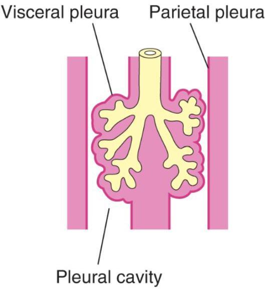
6 Developing gut and its derivatives are suspended from the dorsal to the ventral body wall by mesenteries. The mesenteries consist of two layers of peritoneum, provide pathways for blood and lympatic vessels and nerves, enclose organs and connect it to the body wall.
7 The organs enclosed between peritonal layers ale called intraperitoneal, the organs that lie against th eposterior body wall and are covered by peritoneum on their anterior wall only are called retroperitoneal.
8 Initially, primitive gut is in broad contact with the mesenchyme of the posterior abdominal wall. By the fifth week, connective tissue bridge becomes thinner, and the caudal part of the foregut, midgut and a major part of the hindgut are suspended from the posterior abdominal wall by the dorsal mesentery (which extends from the lower end of the esophagus to the cloacal region of the hindgut).
9 The dorsal mesentery is finally divided into: - dorsal mesogastrium (greater omentum) in the region of the stomach - dorsal mesoduodenum in the region of duodenum - mesentery proper in the region of the jejunal and ileal loops - dorsal mesocolon in the region of the colon
10 The ventral mesentery is formed only in the region of the terminal part of the esophagus, the stomach and the upper part of duodenum and is derived from the septum transversum.
11 The ventral mesentery is divided by growing liver bud into: - the lesser omentum (the dorsal portion - extending from the lower portion of the esophagus, the stomach and the upper portion of the duodenum to the liver) - the falciform ligament extending from the liver to the ventral body wall
12 The foregut gives rise to: - the esophagus - the trachea and lungs buds - the stomach - the duodenum (proximal to the entrance to the bile duct) - the liver - the biliary apparatus - the pancreas
13 The liver primodium appears in the middle of third week as an outgrowth of the endodermal epithelium at the distal end of the foregut. The liver bud consists of rapidly proliferating cells that penetrate septum transversum. While hepatic cells continue to penetrate the septum, the connection between the hepatic diverticulum and the duodenum narrows, forming the bile duct.
14 The outgrowth of the bile duct gives rise to the gallbladder and the cystic duct. Hematopoietic cells, Kupffer cells and connective tissue cells are derived from mesoderm of the septum transversum.
15
16 The pancreas is formed by two buds originating from the endodermal lining of the duodenum: - the dorsal pancreatic bud (in the dorsal mesentery) - the ventral pancreatic bud growing close to the bile duct The pancreatic buds fuse to form the definitive pancreas
 - the jejunum - the ileum - the ascending colon - the transverse colon (proximal two-thirds)")
17 The midgut forms the primary intestinal loop which gives rise to: - the duodenum (distal to the entrance of the bile duct) - the jejunum - the ileum - the ascending colon - the transverse colon (proximal two-thirds)


18 The apex of the primary intestinal loop remains temporarilly in open connection with the yolk sac by through the vitelline duct (Meckel s diverticulum).
 During the tenth week it returns into the abdominal cavity.")
19 During the sixth week, the intestinal loop grows rapidely and protrudes into the umbilical cord (physiological herniation) During the tenth week it returns into the abdominal cavity.

20 ROTATION OF THE MIDGUT The primary intestinal loop rotates around an axis formed by superior mesenteric artery. When viewed from the front this rotation is counterclockwise, and amounts approximately 270o when is complete.
21 Elongation of the small intestinal loop continues during rotation the jejunum and ileum form form a number of coiled loops The large intestine also elongates but not participate in the coiling phenomenon. Rotation of the intestinal loop occurs during herniation (90) as well as during return of the intestinal loop into the abdominal cavity (180).
22 The hindgut gives rise to the: - distal third of transverse colon - descending colon - sigmoid - rectum - upper part of the anal canal
23 The hindgut enters the posterior region of the cloaca (future anorectal canal) and the allantois enters the anterior region of the cloaca (future urogenital sinus). Breakdown of the cloacal membrane covering this area provides communication to the exterior for the anus and urogenital sinus.
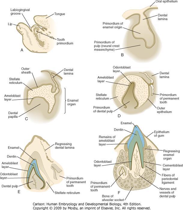
24 tooth development

25 enamel formation
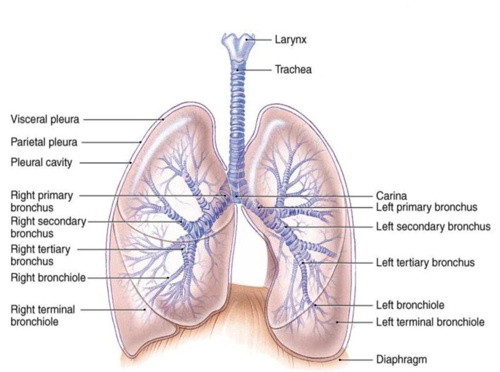
26 RESPIRATOTY SYSTEM
27
28
29
30 The lung bud appears at 4th week after fertilisation and arise from an invagination of the median groove of the primitive pharynx (on the border of the pharynx and foregut). Formation of the primodium is regulated by signals from the surrounding mesenchyme.
31 The epithelium of the larynx, trachea, bronchi, broncholi and alveoli originates in the endoderm. The cartilaginous, muscular and connective tissue components of the lungs arise in the splanchnic mesoderm.
32 Initially, the lung bud is in open communication with the foregut. Then the diverticulum expands caudally and tracheoesophageal ridges separate it from the foregut.
33 Subsequently, tracheoesophageal ridges fuse to form tracheoesophageal septum and the foregut is divided into a dorsal portion: the esophagus and a ventral portion: the trachea and lung buds. The respiratory primodium maintains its communication with the pharynx through the laryngeal orifice.
34 Larynx The internal lining of the larynx originates from endoderm, whereas the cartilages and muscles originate from mesenchyme of the fourth and sixth pharyngeal arches all laryngeal muscles are innervated by branches of the vagus nerve.

35 The derivatives of the fourth arch are innervated by the superior pharyngeal nerve. The derivatives of the sixth pharyngeal arch are innervated by the recurrent laryngeal nerve.
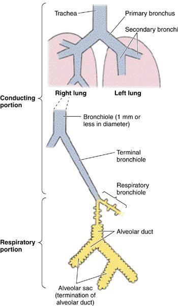
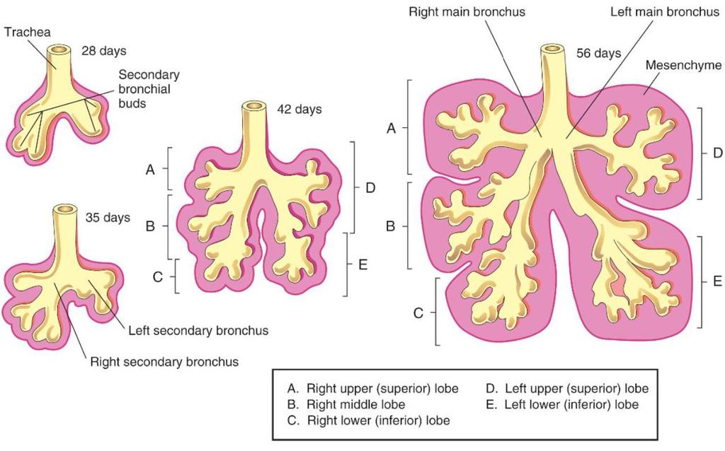
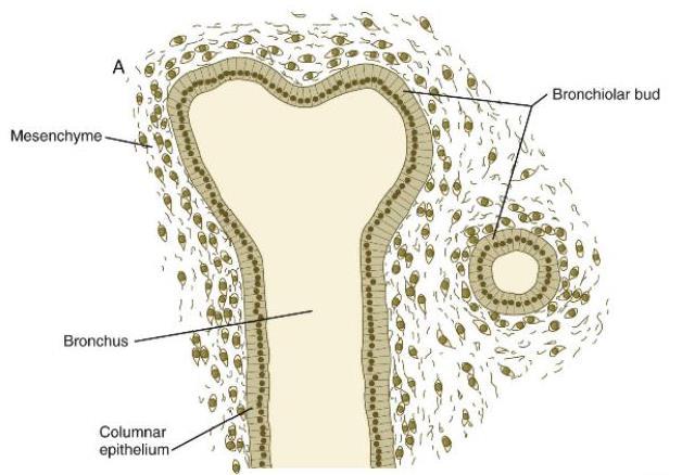
36 Trachea, bronchi, lungs At the begining of the fifth week, the bronchial buds enlarge to form right and left main bronchi. The right main bronchus forms three secondary bronchi, and the left, two.
37
38
39 With time, the lung primodia growth in caudal and lateral directions and expand into the body cavity. At the begining, the intraembryonic cavity is a common large space with the thoracic and the pelvic regions. At the fourth week between upper (prospective thoracic cavity) and lower (prospective abdomen) portion of the intraembryonic cavity, septum transversum develops.

40 The septum transversum is thick plate of mesodermal tissue occupying the space between the thoracic cavity and the stalk of the yolk sac. The septum transversum is the primodium of the diaphragm.

41 The septum transversum does not separate the thoracic and abdominal cavities completely but leaves large openings the pericardioperitoneal canals.

42 The pericardioperitoneal canals the spaces for the lungs growth lie on each side of the foregut. The pericardioperitoneal canals are narrow and are gradually filled by the expanding lung buds.

43 Ultimately the pleuroperitoneal folds separate the pericardioperitoneal canals from the peritoneal cavity and the pleuropericardial folds separate the pericardioperitoneal canals from the pericardial cavity and as a consequence the primitive pleural cavities are formed.

44 The mesoderm, which covers the outside of the lung, develops into the visceral pleura. The somatic mesoderm layer, covering the body wall from the inside develops into the parietal pleura. The space between the parietal and visceral pleura becomes the pleural cavity.
45 During further development, secondary bronchi divides repeatedly in a dichotomous fasion, forming 10 tertiary (segmental) bronchi in the right lung and 8 in the left. By the end of the sixth month, approximately 17 generations of subdivisions are formed. An additional 6 divisions form during postnatal life.
46

47 Pseudoglandular period (5-16 weeks) - branching has continued to form terminal bronchioles - no respiratory bronchioles or alveoli are present

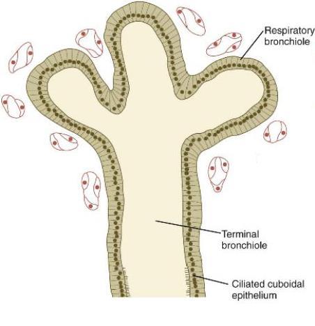
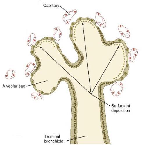
48 Canalicular period (16-26 weeks) - each terminal bronchiole divides into 2 or more respiratory bronchioles - each respiratory bronchiole divides into 3-6 alveolar sacs - vascular supply increases steadily
 form - capilaries establish close")
49 Terminal sac period (26 weeks to birth) - terminal sacs (primitive alveoli) form - capilaries establish close contact

50 Alveolar period (8 months to childhood) - mature alveoli have weel-developed epithelial-endothelial contacts
51 Fetal breathing movements begin before birth and cause aspiration of amniotic fluid. These movement are necessery for stimulating lung development and conditioning respiratory muscles.


52 When respiration begins at birth, most of amniotic fluid is rapidly absorbed by the blood and lymph capilaries. Small amount of the lung fluid is expelled via the trachea and bronchi during delivery.

53 The diaphragm develops from four components: - septum transversum (central tendon) - pleuroperitoneal membranes - dorsal mesentery of the esophagus - muscular components of the body wall
54
55 A congenital diaphragmatic hernia is one of the more common malformations in the newborn (1/2000). In case of cdh the peritoneal and pleural cavities are continuous with one another mostly along the posterior body wall.
56 The diaphragmatic hernias allows abdominal viscera (intestinal loops, stomach, spleen, liver) to enter the pleural cavity push the heart, compress the lungs and cause larg edefects of developing lungs (pulmonary hypoplasia and dysfunction).
57 Meckel s diverticulum is commonly located in the: A ileum B ascending colon C jejunum D transverse colon E duodenum
58 The primodium of which structure is located in the septum transversum? A dorsal pancreas B lung C liver D thymus E heart
59 The yolk stalk is most closely associated with which artery? A celiac B umbilical C superior mesenteric D inferior mesenteric E aorta
The Foregut. At first the esophagus is short. but with descent of the heart and lungs it lengthens rapidly
 GI embryology 2 The Foregut At first the esophagus is short but with descent of the heart and lungs it lengthens rapidly The muscular coat, which is formed by surrounding splanchnic mesenchyme, is striated
Development of the Digestive System. W.S. O The University of Hong Kong
 Development of the Digestive System W.S. O The University of Hong Kong Plan for the GI system Then GI system in the abdomen first develops as a tube suspended by dorsal and ventral mesenteries. Blood
Development of the Digestive System. W.S. O School of Biomedical Sciences, University of Hong Kong.
 Development of the Digestive System W.S. O School of Biomedical Sciences, University of Hong Kong. Organization of the GI tract: Foregut (abdominal part) supplied by coeliac trunk; derivatives include
Midgut. Over its entire length the midgut is supplied by the superior mesenteric artery
 Gi Embryology 3 Midgut the midgut is suspended from the dorsal abdominal wall by a short mesentery and communicates with the yolk sac by way of the vitelline duct or yolk stalk Over its entire length the
Embryology of the Midgut and Hind gut
 Embryology of the Midgut and Hind gut Prof. Abdulameer Al-Nuaimi E-mail: a.al-nuaimi@sheffield.ac.uk E-mail: abdulameerh@yahoo.com Abdominal organs www.google.co.uk/search? Development of Duodenum The
2/2/2011. Primitive Gut Tube Proctodeum and Stomodeum Stomach Duodenum Pancreas Liver and Biliary Apparatus Spleen Midgut
 DEVELOPMENT OF THE DIGESTIVE SYSTEM Development of Endodermal Organs Primitive Gut Tube Proctodeum and Stomodeum Stomach Duodenum Pancreas Liver and Biliary Apparatus Spleen Midgut Wednesday, February
Fareed Khdair, MD Assistant Professor Chief, Section of Pediatric Gastroenterology, Hepatology, and Nutrition University of Jordan School of Medicine
 Fareed Khdair, MD Assistant Professor Chief, Section of Pediatric Gastroenterology, Hepatology, and Nutrition University of Jordan School of Medicine Outline Lecture one : Gut formation Foregut: esophagus,
Development of Respiratory System. Dr. Sanaa Alshaarawy& Dr. Saeed Vohra
 Development of Respiratory System Dr. Sanaa Alshaarawy& Dr. Saeed Vohra OBJECTIVES At the end of the lecture the students should be able to: Identify the development of the laryngeotracheal (respiratory)
- Tamara Wahbeh. - Fareed Khdair. 0 P a g e
 -1 - Tamara Wahbeh - - Fareed Khdair 0 P a g e GI Embryology Note: I included everything in the records and slides; anything in the slide not included in this sheet was not mentioned by the doctor during
Development of pancreas and Small Intestine. ANATOMY DEPARTMENT DR.SANAA AL-AlSHAARAWY DR.ESSAM Eldin Salama
 Development of pancreas and Small Intestine ANATOMY DEPARTMENT DR.SANAA AL-AlSHAARAWY DR.ESSAM Eldin Salama OBJECTIVES At the end of the lecture, the students should be able to : Describe the development
8. Development of digestive system II. Rotation if intestine. Liver, pancreas, spleen. Development of respiratory passages and lung.
 8. Development of digestive system II. Rotation if intestine. Liver, pancreas, spleen. Development of respiratory passages and lung. Duodenum originates partially from the terminal part of the foregut,
Lecture 21Development of respiratory system Dr. Rehan Asad At the end of session students should able to Describe formation of lung buds Describe
 Lecture 21Development of respiratory system Dr. Rehan Asad At the end of session students should able to Describe formation of lung buds Describe development of larynx, trachea and bronchi. Describe the
Lectures of Human Embryology
 Lectures of Human Embryology "Body Cavities & GIT" By DR. ABDEL-MONEM AWAD HEGAZY M.B. with honor 1983, Dipl."Gynecology and Obstetrics "1989, Master "Anatomy and Embryology" 1994, M.D. "Anatomy and Embryology"
Embryology - GIT - Lecture 2
 Embryology - GIT - Lecture 2 Last time we talked about embryology of the GIT. We said that the development of the stomach is accompanied with the development of the duodenum and the pancreas. Also we talked
Bronchioles. Alveoli. Type I alveolar cells are very thin simple squamous epithelial cells and form most of the lining of an alveolus.
 276 Bronchioles Bronchioles continue on to form bronchi. The primary identifying feature is the loss of hyaline cartilage. The epithelium has become simple ciliated columnar, and there is a complete ring
Development of the nasal cavity :
 Development of the nasal cavity : several processes contribute to the development of the nose, the nose consists of 2 cavities separated by a septum, and the nasal cavity is separated from the oral cavity
Exploring Anatomy: the Human Abdomen
 Exploring Anatomy: the Human Abdomen PERITONEUM AND PERITONEAL CAVITY PERITONEUM The peritoneum is a thin serous membrane that lines the abdominal cavity and covers, in variable amounts, the viscera within
Development of the Liver and Pancreas
 Development of the Liver and Pancreas Professor Alfred Cuschieri Department of Anatomy University of Malta Three glandular buds arise from the distal end of the foregut during the fourth week Day 22 -The
The peritoneum. Prof. Oluwadiya KS, MBBS, FMCS(Orthop) Website:
 Website:") The peritoneum Prof. Oluwadiya KS, MBBS, FMCS(Orthop) Website: http://oluwadiya.com The peritoneum Serous membrane that lines the abdominopelvic cavity and invests the viscera The largest serous membrane
The peritoneum Prof. Oluwadiya KS, MBBS, FMCS(Orthop) Website: http://oluwadiya.com The peritoneum Serous membrane that lines the abdominopelvic cavity and invests the viscera The largest serous membrane
Respiratory System Embryology
 Respiratory System Embryology Development of the nose and Palate Development of the nose At the end of the fourth week, facial prominences consisting primarily of neural crest-derived mesenchyme and formed
Anatomy: Know Your Abdomen
 Anatomy: Know Your Abdomen Glossary Abdomen - part of the body below the thorax (chest cavity); separated by the diaphragm. Anterior - towards the front of the body. For example, the umbilicus is anterior
Accessory Glands of Digestive System
 Accessory Glands of Digestive System The liver The liver is soft and pliable and occupies the upper part of the abdominal cavity just beneath the diaphragm. The greater part of the liver is situated under
Embryology: Development of digestive system
 Embryology: Development of digestive system Embryo folding incorporation of endoderm to form primitive gut. Outside of embryo yolk sac and allantois. Vitelline duct Stomodeum (primitive mouth) the oral
Chapter 7 The digestive system
 41 Chapter 7 The digestive system Primitive gut tube 41 Foregut 42 Other foregut derivatives 44 Midgut 45 Hindgut 47 4 Fig. 7.1 The gut tube in a 4-week embryo. Pharynx Foregut Respiratory diverticulum
Anatomical Considerations for Lab Practical II
 Anatomical Considerations for Lab Practical II For each of the following please be prepared to provide: Identification System Organ(s) or ducts to Function(s) location which it is attached Use your lecture
Development of Gastrointestinal Tract
 11 Development of Gastrointestinal Tract Learning Objectives At the end of this chapter, students would be able to define and understand the following: Development of the esophagus and stomach Rotation
Histology and development of the respiratory system
 Histology and development of the respiratory system Árpád Dobolyi Semmelweis University, Department of Anatomy, Histology and Embryology Outline of the lecture 1. Structure of the trachea 2. Histology
Preview from Notesale.co.uk Page 1 of 34
 Abdominal viscera and digestive tract Digestive tract Abdominal viscera comprise majority of the alimentary system o Terminal oesophagus, stomach, pancreas, spleen, liver, gallbladder, kidneys, suprarenal
Pharyngeal apparatus. - At the third week, it is a 3 layered structure: ectoderm, mesoderm and endoderm. This is called trilaminar disc
 Pharyngeal apparatus Remember from the first year embryology - The embryo was disc shaped in the second week of development (this is called embryonic disc) and it is a 2 layered disc (composed of two layers)---bilaminar
SUBJECTS 2nd year, 1st semester I. 1. Primitive gut - limits, derivatives 2. Foregut -limits, evolution, derivatives 3. Midgut -limits, evolution,
 SUBJECTS 2nd year, 1st semester I. 1. Primitive gut - limits, derivatives 2. Foregut -limits, evolution, derivatives 3. Midgut -limits, evolution, derivatives 4. Hindgut- limits, evolution, derivatives
When you see this diagram, remember that you are looking at the embryo from above, through the amniotic cavity, where the epiblast appears as an oval
 When you see this diagram, remember that you are looking at the embryo from above, through the amniotic cavity, where the epiblast appears as an oval disc 2 Why the embryo needs the vascular system? When
RESPIRATORY SYSTEM. described: pp. 744,746 fig. 25.1, described: p. 746 fig described: p. 776 fig. 26.3
 ACTIVITY 11: RESPIRATORY AND DIGESTIVE SYSTEMS OBJECTIVES: 1) How to get ready: Read Chapters 25 and 26, McKinley et al., Human Anatomy, 5e. All text references are for this textbook. 2) Identify structures
Embryology of the Heart
 *Page 1A: Embryology of the Heart Human embryonic disc is divided into three layers: ectoderm, intraembryonic mesoderm, and endoderm. The embryonic disc lies between the amniotic cavity and the primary
ACTIVITY 11: RESPIRATORY AND DIGESTIVE SYSTEMS RESPIRATORY SYSTEM
 ACTIVITY 11: RESPIRATORY AND DIGESTIVE SYSTEMS OBJECTIVES: 1) How to get ready: Read Chapters 25 and 26, McKinley et al., Human Anatomy, 4e. All text references are for this textbook. 2) Identify structures
In the name ofgod. Abdomen 3. Dr. Zahiri
 In the name ofgod Abdomen 3 Dr. Zahiri Peritoneum Peritoneum It is the serous membrane(a type of loose connective tissue and is covered by mesothelium) that lines the abdominal cavity. Extensions of the
Jhia Anjela D. Rivera 1 1. BS Biology, Department of Biology, College of Science, Polytechnic University of the Philippines
 DIGESTIVE SYSTEM Jhia Anjela D. Rivera 1 1 BS Biology, Department of Biology, College of Science, Polytechnic University of the Philippines DIGESTIVE SYSTEM Consists of the digestive tract (gastrointestinal
Peritoneum: Def. : It is a thin serous membrane that lines the walls of the abdominal and pelvic cavities and clothes the viscera.
 Peritoneum: Def. : It is a thin serous membrane that lines the walls of the abdominal and pelvic cavities and clothes the viscera. Layers of the peritoneum: 1. Outer Layer ( Parietal Peritoneum) : lines
Development of the Urinary System. 3 Distinct Embryonic Kidney Structures
 Development of the Urinary System Excretory portion of urinary system derived from intermediate mesoderm Week 4: 1 st nephrons/renal corpuscles form Nephrotomes form and develop hollow lumens to form nephric
MICROSCOPIC STRUCTURE OF LIVER, GALLBLADDER, GALL DUCTS, AND PANCREAS OVERVIEW OF DEVELOPMENT OF THE ALIMENTARY CANAL
 Lecture 2 ESS_3rd semester MICROSCOPIC STRUCTURE OF LIVER, GALLBLADDER, GALL DUCTS, AND PANCREAS OVERVIEW OF DEVELOPMENT OF THE ALIMENTARY CANAL MICROSCOPIC STRUCTURE OF LIVER - is the largest gland of
Anatomy of the Large Intestine
 Large intestine Anatomy of the Large Intestine 2 Large Intestine Extends from ileocecal valve to anus Length = 1.5-2.5m = 5 feet Regions Cecum = 2.5-3 inch Appendix= 3-5 inch Colon Ascending= 5 inch Transverse=
The abdominal Esophagus, Stomach and the Duodenum. Prof. Oluwadiya KS
 The abdominal Esophagus, Stomach and the Duodenum Prof. Oluwadiya KS www.oluwadiya.com Viscera of the abdomen Abdominal esophagus: Terminal part of the esophagus The stomach Intestines: Small and Large
Embryology of the Gut and Mesenteries, slide 7 This is very similar to a cross-section we looked at when were talking about formation of the diaphragm
 Embryology of the Gut and Mesenteries, slide 2 This is a median sagittal section of a four week embryo, most of you can probably draw it in your sleep. I made a few modifications from some of the earlier
Respiratory & Digestive Organs of the Head and Neck, Human;
 Name Date Lab Exercise 5: Lab Exercise 6: Lab Exercise 7: Lab Exercise 8: Respiratory & Digestive Organs of the Head and Neck, Human; Histology of the Respiratory System Digestive System Models, Human
The jejunum and the Ileum. Prof. Oluwadiya KS
 The jejunum and the Ileum Prof. Oluwadiya KS www.oluwadiya.siteled.com Introduction Introduction The small intestine (SI) comprises of the duodenum, jejunum and the ileum The jejunum is the second part
OVARIES URETER FALLOPIAN TUBES BLADDER UROGENITAL OPENINGS (BOTH SEXES) PENIS VAGINA UTERUS
 PENIS VAGINA UTERUS") URETER OVARIES FALLOPIAN TUBES BLADDER UROGENITAL OPENINGS (BOTH SEXES) PENIS VAGINA UTERUS REPRODUCTIVE PRODUCE FEMALE HORMONES EXCRETORY FROM KIDNEY TO BLADDER EXCRETORY STORES URINE REPRODUCTIVE TRANSPORTS
URETER OVARIES FALLOPIAN TUBES BLADDER UROGENITAL OPENINGS (BOTH SEXES) PENIS VAGINA UTERUS REPRODUCTIVE PRODUCE FEMALE HORMONES EXCRETORY FROM KIDNEY TO BLADDER EXCRETORY STORES URINE REPRODUCTIVE TRANSPORTS
-Tamara Wahbeh. -Razan Abu Rumman. Dr. Mohammed Al-Muhtaseb
 -2 -Tamara Wahbeh -Razan Abu Rumman Dr. Mohammed Al-Muhtaseb I tried to include everything the doctor mentioned in both the lecture and his slides in the simplest way possible, so hopefully there would
Small Plicae Circularis. Short Closely packed together. Sparse, completely absent at distal part Lymphoid Nodule
 Intestines Differences Between Jejunum and Ileum Types Jejunum Ileum Color Deeper red Paler pink Calibre Bigger Smaller Thickness of wall Thick and Heavy Thin and Lighter Vascularity Highly vascularised
Pancreas & Biliary System. Dr. Vohra & Dr. Jamila
 Pancreas & Biliary System Dr. Vohra & Dr. Jamila 1 Objectives At the end of the lecture, the student should be able to describe the: Location, surface anatomy, parts, relations & peritoneal reflection
Anatomy of the SMALL INTESTINE. Dr. Noman Ullah Wazir PMC
 Anatomy of the SMALL INTESTINE Dr. Noman Ullah Wazir PMC SMALL INTESTINE The small intestine, consists of the duodenum, jejunum, and illium. It extends from the pylorus to the ileocecal junction were the
BY DR NOMAN ULLAH WAZIR
 BY DR NOMAN ULLAH WAZIR The stomach (from ancient Greek word stomachos, stoma means mouth) is a muscular, hollow and the most dilated part of the GIT. It starts from the point where esophagus ends. It
Block 3: DISSECTION 2 CELIAC TRUNK, JEJUNUM/ILEUM, LARGE INTESTINE, DUODENUM, PANCREAS, PORTAL VEIN; MOBILIZATION OF THE LIVER
 1 Block 3: DISSECTION 2 CELIAC TRUNK, JEJUNUM/ILEUM, LARGE INTESTINE, DUODENUM, PANCREAS, PORTAL VEIN; MOBILIZATION OF THE LIVER Attempt to complete as much as you can of the dissection explained in the
Dr. Weyrich G07: Superior and Posterior Mediastina. Reading: 1. Gray s Anatomy for Students, chapter 3
 Dr. Weyrich G07: Superior and Posterior Mediastina Reading: 1. Gray s Anatomy for Students, chapter 3 Objectives: 1. Subdivisions of mediastinum 2. Structures in Superior mediastinum 3. Structures in Posterior
Duodenum retroperitoneal
 Duodenum retroperitoneal C shaped Initial region out of stomach into small intestine RETROperitoneal viscus Superior 1 st part duodenal cap ; moves upwards and backwards to lie on the R crura medial to
Group B: Organ systems (digestive, respiratory, urinary, genital system, heart, glands and skin) green
 green") Group B: Organ systems (digestive, respiratory, urinary, genital system, heart, glands and skin) green Digestive system 1. Teeth Main points: external and internal structure of a tooth, fixation of a tooth
Group B: Organ systems (digestive, respiratory, urinary, genital system, heart, glands and skin) green Digestive system 1. Teeth Main points: external and internal structure of a tooth, fixation of a tooth
THE RESPIRATORY SYSTEM
 THE RESPIRATORY SYSTEM Functions of the Respiratory System Provides extensive gas exchange surface area between air and circulating blood Moves air to and from exchange surfaces of lungs Protects respiratory
BLOCK IV: OFFICIAL BODY PARTS LIST FOR ANTERIOR ABDOMINAL WALL AND ABDOMINAL CONTENTS
 BLOCK IV: OFFICIAL BODY PARTS LIST FOR ANTERIOR ABDOMINAL WALL AND ABDOMINAL CONTENTS External oblique muscle Muscular portion Aponeurotic portion Superficial inguinal ring Lateral (inferior) crus Medial
Dissection Lab Manuals: Required Content
 Dissection Lab Manuals: Required Content 1. Introduction a. Basic terminology (directions) b. External features of the cat c. Adaptations to predatory niche d. How to skin a cat e. How to make the incisions
It passes through the diaphragm at the level of the 10th thoracic vertebra to join the stomach
 The esophagus is a tubular structure (muscular, collapsible tube ) about 10 in. (25 cm) long that is continuous above with the laryngeal part of the pharynx opposite the sixth cervical vertebra The esophagus
Organizational Overview of Thorax, Abdomen, Pelvis. Introduction to Blood
 Biology 224 Human Anatomy and Physiology - II Week 1; Lecture 2; Wednesday Dr. Stuart S. Sumida Organizational Overview of Thorax, Abdomen, Pelvis Introduction to Blood Organizational Overview of Thorax,
Lab Monitor Images Dissection of the Abdominal Vasculature + Lower Digestive System
 Lab Monitor Images Dissection of the Abdominal Vasculature + Lower Digestive System Stomach & Duodenum Frontal (AP) View Nasogastric tube 2 1 3 4 Stomach Pylorus Duodenum 1 Duodenum 2 Duodenum 3 Duodenum
Chest and cardiovascular
 Module 1 Chest and cardiovascular A. Doss and M. J. Bull 1. Regarding the imaging modalities of the chest: High resolution computed tomography (HRCT) uses a slice thickness of 4 6 mm to identify mass lesions
Biology Human Anatomy Abdominal and Pelvic Cavities
 Biology 351 - Human Anatomy Abdominal and Pelvic Cavities Please place your name and I.D. number on the back of the last page of this exam. You must answer all questions on this exam. Because statistics
Dr. Zahiri. In the name of God
 Dr. Zahiri In the name of God small intestine = small bowel is the part of the gastrointestinal tract Boundaries: Pylorus Ileosecal junction Function: digestion and absorption of food It receives bile
Biology 340 Comparative Embryology Lecture 10 Dr. Stuart Sumida. Further Development of the Mesoderm (and Endoderm)
") Biology 340 Comparative Embryology Lecture 10 Dr. Stuart Sumida Further Development of the Mesoderm (and Endoderm) Further Development: Digestive System Foregut, Midgut, Hindgut Heart and Aortic Arches
Biology 340 Comparative Embryology Lecture 10 Dr. Stuart Sumida Further Development of the Mesoderm (and Endoderm) Further Development: Digestive System Foregut, Midgut, Hindgut Heart and Aortic Arches
#1 - Chapter 1 - Anatomy. General Anatomical Terms The Anatomical Position
 #1 - Chapter 1 - Anatomy General Anatomical Terms The Anatomical Position The anatomical position is a stance in which a person stands erect with the feet flat on the floor and close together, arms at
THE ORAL CAVITY
 THE ORAL CAVITY WALL OF ABDOMEN (ANTERIOR) The paraumbilical vein drains into the portal vein and then through the liver. This is an important clinical connection. THE ABDOMINAL VISCERA The small
Lab 9 Abdomen MUSCLES
 Lab 9 Abdomen MUSCLES External abdominal oblique continuous with the external intercostal muscle; its fibers point in a caudal direction as it moves anteriorly until it inserts on the linea alba via its
ANATOMY OF THE DIGESTIVE SYSTEM PART II
 ANATOMY OF THE DIGESTIVE SYSTEM PART II 9.12.2014 Kaan Yücel M.D., Ph.D. http://fhs121.org Dr.Kaan Yücel http://fhs121.org Digestive system Part II 1. LIVER The liver is the largest gland in the body and,
Embryo#1. Mohammad Hisham Al-Mohtaseb باشق جهاد. 0 P a g e
 Embryo#1 Mohammad Hisham Al-Mohtaseb باشق جهاد 0 P a g e Before you start, it is important to link what you learn in gross anatomy with developmental stages discussed in embryology. Cells that form organs
Name: Fetal Pig Dissection Internal Anatomy
 In this lab exercise you will open the thoracic cavities and abdominal pelvic of a fetal pig and identify its major organs. Remember you are dissecting not butchering. The goal is for you to identify all
Chapter 16. Respiratory System
 Chapter 16 Respiratory System Introduction Respiration = the entire process of exchanging gases between the atmosphere and body cells 1. Ventilation 2. Gas exchange 3. Gas transport : 4. Cellular respiration
د. عصام طارق. Objectives:
 GI anatomy Lecture: 5 د. عصام طارق Objectives: To describe anatomy of stomach, duodenum & pancreas. To list their main relations. To define their blood & nerve supply. To list their lymph drainage. To
To describe the liver. To list main structures in porta hepatis.
 GI anatomy Lecture: 6 د. عصام طارق Objectives: To describe the liver. To list main structures in porta hepatis. To define portal system & portosystemic anastomosis. To list parts of biliary system. To
The Human Body: An Overview of Anatomy. Anatomy. Physiology. Anatomy - Study of internal and external body structures
 C H A P T E R 1 The Human Body: An Orientation An Overview of Anatomy Anatomy The study of the structure of the human body Physiology The study of body function Anatomy - Study of internal and external
CEA (CARCINOEMBRYONIC ANTIGEN)
") (CARCINOEMBRYONIC ANTIGEN) 428 C15.3 Malignant neoplasm of upper third of esophagus C15.4 Malignant neoplasm of middle third of esophagus C15.5 Malignant neoplasm of lower third of esophagus C15.8 Malignant
(CARCINOEMBRYONIC ANTIGEN) 428 C15.3 Malignant neoplasm of upper third of esophagus C15.4 Malignant neoplasm of middle third of esophagus C15.5 Malignant neoplasm of lower third of esophagus C15.8 Malignant
ABDOMEN - GI. Duodenum
 TALA SALEH ABDOMEN - GI Duodenum - Notice the shape of the duodenum, it looks like capital G shape tube which extends from the pyloroduodenal junction to the duodenojejunal junction. - It is 10 inches
The Digestive System
 The Digestive System Identify the Structure and Function. Mesentery of the Large Intestine The mesentery functions to connect the visceral organs to the abdominal wall. Identify the Structure. Nasal Cavity
Anatomy (embryo 2) Aseel Al- khader 29/11/2015. Mohammad al Haidari. 1 P a g e
 Aseel Al- khader 29/11/2015. Mohammad al Haidari. 1 P a g e") 14 Anatomy (embryo 2) Aseel Al- khader 29/11/2015 Mohammad al Haidari 1 P a g e بسم هللا الرحمن الرحيم *I advice you to watch this video ( from omar outom s post ) before studying this lecture for better
14 Anatomy (embryo 2) Aseel Al- khader 29/11/2015 Mohammad al Haidari 1 P a g e بسم هللا الرحمن الرحيم *I advice you to watch this video ( from omar outom s post ) before studying this lecture for better
Welcome to ANAT 10A! What is Anatomy? Different levels of Anatomy The Language of Anatomy Pearson Education, Inc.
 Welcome to ANAT 10A! What is Anatomy? Different levels of Anatomy The Language of Anatomy Introduction Anatomy means to dissect: (ANAT 10A) The study of internal & external body structures The study of
Digestive system (Systema digestorium/ alimentarium) Lecture #1
 Lecture #1") Digestive system (Systema digestorium/ alimentarium) Lecture #1 Internal organs are grouped into 1. System - Have the same functions and development Digestive system Respiratory system 2. Apparatus - Have
Digestive system (Systema digestorium/ alimentarium) Lecture #1 Internal organs are grouped into 1. System - Have the same functions and development Digestive system Respiratory system 2. Apparatus - Have
FORMS OF EMBRYONIC PRIMORDIA
 FORMS OF EMBRYONIC PRIMORDIA BY PROF. ANTHONY OBIOMA NWAOPARA UNIVERSITY OF MEDICAL SCIENCES ONDO CITY, ONDO STATE LEARNING OBJECTIVES To recognise the different forms of embryonic primordia. To recognise
Lungs a. d. b. c. e.
 Lungs d. e. Lungs Right superior lobe Right middle lobe Right inferior lobe d. Left superior lobe e. Left inferior lobe Sinuses d. Nasal Cavity & Sinuses g. g. i. Nasal Cavity & Sinuses g. h. d. f. e.
- Digestion occurs during periods of low activity - Produces more energy than it uses. - Mucosa
 Introduction Digestive System Chapter 29 Provides processes to break down molecules into a state easily used by cells - A disassembly line: Starts at the mouth and ends at the anus Digestive functions
Netter's Anatomy Flash Cards Section 4 List 4 th Edition
 Netter's Anatomy Flash Cards Section 4 List 4 th Edition https://www.memrise.com/course/1577335/ Section 4 Abdomen (31 cards) Plate 4-1 Bony Framework of Abdomen 1.1 Costal cartilages 1.2 Iliac crest 1.3
Cardiovascular System Anatomy & Embryology
 بسم رلاهللا Cardiovascular System Anatomy & Embryology Portal circulation: Portal vessel: a vessel or a vein running between two sets of capillaries, the portal vein of the liver starts from a set of capillaries
Nasogastric tube. Stomach. Pylorus. Duodenum 1. Duodenum 2. Duodenum 3. Duodenum 4
 Esophagus Barium Swallow Stomach and Duodenum 4 year old Upper GI Nasogastric tube Stomach and Duodenum 4 year old Upper GI Nasogastric tube Stomach Pylorus Duodenum 1 Duodenum 2 Duodenum 3 Duodenum 4
THE DESCENDING THORACIC AORTA
 Intercostal Arteries and Veins Each intercostal space contains a large single posterior intercostal artery and two small anterior intercostal arteries. The anterior intercostal arteries of the lower spaces
LECTURE 11 & 12: ABDOMINAL VISCERA ABDOMINAL CONTENTS DIVISION. The location of abdominal viscera is divided into 4 quadrants:
 LECTURE 11 & 12: ABDOMINAL VISCERA ABDOMINAL CONTENTS DIVISION The location of abdominal viscera is divided into 4 quadrants: - horizontal line across the umbilicus divides the upper quadrants from the
Exercise. Digestive System. Digestive system function. 1. Define the following terms: a. Chemical digestionb. Mechanical digestionc.
 Exercise 7 The Digestive System NAME: DATE: INSTRUCTOR: SECTION: Digestive system function 1. Define the following terms: a. Chemical digestionb. Mechanical digestionc. Ingestiond. Digestione. Absorptionf.
STRUCTURAL BASIS OF MEDICAL PRACTICE EXAMINATION 3. October 16, 2015
 STRUCTURAL BASIS OF MEDICAL PRACTICE EXAMINATION 3 October 16, 2015 PART l. Answer in the space provided. (12 pts) 1. Identify the structures. (2 pts) A. B. A B C. D. C D 2. Identify the structures. (2
B. Correct! As air travels through the nasal cavities, it is warmed and humidified.
 Human Anatomy - Problem Drill 20: The Respiratory System Question No. 1 of 10 1. Which of the following statements about the portion of the respiratory system labeled in the image below is correct? Question
Introduction to The Human Body
 1 Introduction to The Human Body FOCUS: The human organism is often examined at seven structural levels: chemical, organelle, cell, tissue, organ, organ system, and the organism. Anatomy examines the structure
Thorax Lecture 2 Thoracic cavity.
 Thorax Lecture 2 Thoracic cavity. Spring 2016 Dr. Maher Hadidi, University of Jordan 1 Enclosed by the thoracic wall. Extends between (thoracic inlet) & (thoracic outlet). Thoracic inlet At root of the
Mediastinum and pericardium
 Mediastinum and pericardium Prof. Abdulameer Al-Nuaimi E-mail: a.al-nuaimi@sheffield.ac.uk E. mail: abdulameerh@yahoo.com The mediastinum: is the central compartment of the thoracic cavity surrounded by
Al s 202 study guide answers Answers Respiratory System 1 External nares (nostrils) 33 Carina 2 Vestibule 34 Left primary bronchus 3 Nasal cavity 35
 33 Carina 2 Vestibule 34 Left primary bronchus 3 Nasal cavity 35") Trachea & Respiratory Histology 1 Epiglottis 26 Capillary 2 Larynx 27 Alveolar sac 3 Thyroid cartilage 28 Alveoli/Alveolus 4 Cricoid cartilage 29 Basement membrane 5 Vocal folds (True vocal cords) 30 Cilia
Trachea & Respiratory Histology 1 Epiglottis 26 Capillary 2 Larynx 27 Alveolar sac 3 Thyroid cartilage 28 Alveoli/Alveolus 4 Cricoid cartilage 29 Basement membrane 5 Vocal folds (True vocal cords) 30 Cilia
Date Lab Pd. Lecture Notes (57)
") Name SECTION OBJECTIVES Describe the locations of the major body cavities List the organs located in each major body cavity Name the membranes associated with the thoracic and abdominopelvic cavities Name
Name SECTION OBJECTIVES Describe the locations of the major body cavities List the organs located in each major body cavity Name the membranes associated with the thoracic and abdominopelvic cavities Name
Ex. 1 :Language of Anatomy
 Collin College BIOL 2401 : Human Anatomy & Physiology Ex. 1 :Language of Anatomy The Anatomical Position Used as a reference point when referring to specific areas of the human body Body erect Head and
Anatomy of the spleen. Oluwadiya KS
 Anatomy of the spleen Oluwadiya KS www.oluwadiya.com Introduction The spleen is an ovoid, usually purplish, pulpy mass about the size and shape of one's fist. It is the largest lymphoid tissue in the body
LESSON ASSIGNMENT. After completing this lesson, you should be able to:
 LESSON ASSIGNMENT LESSON 6 The Human Digestive System. TEXT ASSIGNMENT Paragraphs 6-1 through 6-16. LESSON OBJECTIVES After completing this lesson, you should be able to: 6-1. Define the human digestive