Case Report Intra-Abdominal Testicular Seminoma in a Woman with Testicular Feminization Syndrome
|
|
|
- Dortha Cameron
- 5 years ago
- Views:
Transcription
1 Case Reports in Radiology Volume 2011, Article ID , 5 pages doi: /2011/ Case Report Intra-Abdominal Testicular Seminoma in a Woman with Testicular Feminization Syndrome Darshana D. Rasalkar, 1 Bhawan K. Paunipagar, 1 Alex Ng, 1 Fernand. Lai, 2 and Shalini Jain Bagaria 3 1 Department of Imaging and Interventional Radiology, Prince of Wales Hospital, The Chinese University of Hong Kong, Ngan Shing Street, Shatin, New Territories, Hong Kong 2 Department of Anatomical and Cellular Pathology, Prince of Wales Hospital, The Chinese University of Hong Kong, Ngan Shing Street, Shatin, New Territories, Hong Kong 3 Department of Obstetrics and Gynecology, UCS and GTB Hospital, Dilshad Garden, Delhi , India Correspondence should be addressed to Darshana D. Rasalkar, drdarshanar@yahoo.com Received 12 ay 2011; Accepted 5 July 2011 Academic Editors: B. J. Barron and C. Chaskis Copyright 2011 Darshana D. Rasalkar et al. This is an open access article distributed under the Creative Commons Attribution License, which permits unrestricted use, distribution, and reproduction in any medium, provided the original work is properly cited. We report a case of intra-abdominal testicular tumor in a 36-year-old married lady presenting with chief complaints of primary amenorrhea. The patient was later diagnosed with testicular feminization syndrome, a form of male pseudohermaphroditism. This testicular tumor was histologically proven as seminoma. Due to rarity, imaging findings in patients with testicular feminization syndrome and intraabdominal testicular tumor have been poorly documented. So far, only one case report had described the combined role of CT and R imaging in intraabdominal testicular sex-cord stromal tumor. To our knowledge, this case is first to document USG and R imaging in addition to R spectroscopy features in intraabdominal testicular seminoma. 1. Introduction As many as 1 in 3000 babies are born intersexed [1]. ale pseudohermaphroditism is one of the intersexed type, inherited as a sex-linked recessive disorder ranging from the mild cases with hypospadias, cleft scrotum, persistent urogenital sinus, and undescended testes, to the severe alteration in phenotype resulting in testicular agenesis (Turner s syndrome), or dysgenesis (testicular feminization syndrome). Testicular dysgenesis leads to inhibition of spermatogenesis and dominance of sertoli cells, so-called feminizing testis also known as an androgen insensitivity syndrome (AIS) [2]. Thus, these patients despite having a male genotype present as asymptomatic, functionally normal but reproductively sterile females. AIS is caused by mutations in the androgen receptor gene and is associated with abnormal testicular development with an increased risk of germ cell malignancy [3]. The risk of neoplasia is also known in a maldescended testis and often increases as the age advances [4]. 2. Case History A thirty-nine-year old married lady presented with chief complaints of primary amenorrhea. She also had abdominal distension and palpable swelling over the abdominopelvic regions since last two years. There were no constitutional symptoms or weight loss. There was no history of prior biochemical or radiological investigations during her teens. The physical examination revealed normally developed breasts but sparse pubic and axillary hair. On local examination, there was a large firm nontender mass extending over the abdominopelvic regions. Gynecological examinations revealed normal labia; however, vagina was blind (5cm)withabsentcervix.Asolidmasswasfeltatanterior aspect of pelvis. Her hormonal assay revealed raised testosterone levels up to 3.94 (normal levels in women nmol/liter). Tumor markers showed raised beta- HCG levels up to 159 IU/L (normal levels in nonpregnant premenopausal women is less than or equal to 1 IU/L).
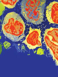
 Figure 1: (a) Transabdominal ultrasound showing a")
 and central necrosis (arrowhead).")


 T2W axial image showing the tubular gonadal structure in the")
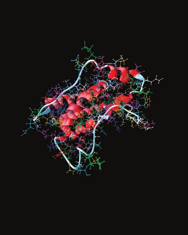
2 2 Case Reports in Radiology (a) (b) RK (c) (d) R H L F R A P L Cho (e) Figure 1: (a) Transabdominal ultrasound showing a heterogeneously hypoechoic mass () with internal septations (white arrows) and central necrosis (arrowhead). (b) Color and spectral Doppler images showing few internal vascular channels with arterial flow. (c) T1W axial image of the abdomen. The tumor is hypointense causing mass effect on to the adjacent structures. The right kidney is pushed more posteriorly. (d) T2W axial image showing the tubular gonadal structure in the region of right inguinal canal. (e) R spectroscopy showing a high choline peak compatible with a tumor spectrum.
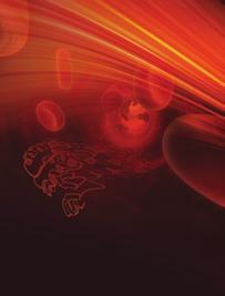
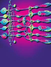
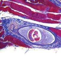
 classical seminoma, with tumor cells with nests, anastomosing cords, and sheets, ill-formed tubular growth pattern, accompanied by a heavy lymphocytic infiltrate, H&E 100.")
 and alpha-fetoprotein (AFP) levels were within normal limits. Blood karyotyping was performed which surprisingly revealed a chromosomal abnormality with 46, XY.")
). Few vascular channels were detected on colour Doppler (Figure 1(b)).")

3 Case Reports in Radiology 3 Lt-A Lt-B (a) (b) Lt-C Lt-D (c) (d) Figure 2: Histology of the left gonad (Lt). (a) classical seminoma, with tumor cells with nests, anastomosing cords, and sheets, ill-formed tubular growth pattern, accompanied by a heavy lymphocytic infiltrate, H&E 100. (b) Large polygonal tumor cells, with glycogen-rich clear cytoplasm, PAS 200. (c) Tumor cells are immunoreactive to both placental alkaline phosphatase and to C-Kit (d), both 100. Carcinoembryonic antigen (CEA) and alpha-fetoprotein (AFP) levels were within normal limits. Blood karyotyping was performed which surprisingly revealed a chromosomal abnormality with 46, XY. Further assessment of mass by an ultrasound showed a large predominantly hypoechoic solid mass measuring 22 cm 17 cm cm with minimal echogenic internal septations and central necrosis (Figure 1(a)). Few vascular channels were detected on colour Doppler (Figure 1(b)). The uterus was absent and no distinct ovarian tissue identified. On RI, the mass was homogenously T1 hypointense and T2 hyperintense (Figures 1(c)and 1(d)). oderate contrast enhancement with central necrosis was identified on postcontrast administration. The right kidney was mildly compressed posteriorly and the aorta, IVC were slightly displaced towards left. A 17 mm long tubular structure with a 14 mm higher signal intensity nodule were detected extending from the right groin to the right inguinal region which were thought to represent a rudimentary gubernaculum of testis +/ undescended testis (Figure 1(d)). The uterus and both ovaries were absent. Overall imaging features were compatible with testicular feminization with possibility of neoplasm arising from cryptorchid intraabdominal type testis. Further R spectroscopy showed markedly raised choline peak suggestive of tumor spectrum (Figure 1(e)). The other visceral organs were unremarkable. The patient underwent laparotomy removal of pelvic mass and bilateral gonadectomies were performed. Left gonadal tumor was adhered to sigmoid colon and omentum. Right gonad was ectopically situated inside the right inguinal canal. Histopathology of the resected specimen revealed classical seminoma from the left gonad (Figure 2) while right gonad showed presence of inactive sertoli cell tubules with no evidence of malignancy (Figure 3). Omental biopsy and peritoneal fluid were negative for malignant cells. 3. Discussion Intersex disorders result from a genetic defect in chromosomal presentation. They often present with ambiguous external genitalia and on the basis of their gonadal presentation, are categorized into true hermaphrodites and mixed gonadal dysgenesis (pseudohermaphrodites) [5]. Remarkable propensity of the gonads to develop malignant tumors has been documented in 46XY with mixed gonadal dysgenesis and male pseudohermaphroditism [2]. Feminizing testis (testicular feminization syndrome), is an inherited sex-linked recessive disorder, a rare form of a male pseudohermaphroditism, characterized by androgen insensitivity resulting from an absence or abnormal cytosol receptor for androgens [6]. Thus, despite possessing a male karyotype (46, XY); phenotypically patient presents as an asymptomatic female usually discovered during perimenarchal stage when the individual fails to menstruate, often presenting in their early 2nd to 3rd decades. Testicular
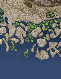
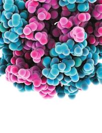
 Atrophic gonadal composed mainly of tubules of sertoli cells, with absence of germ cells, and the presence of interstitial Leydig cells, H&E 100.")
 Both Sertoli cells and Leydig cells are immunoreactive to inhibin 100, while only Leydig cells react to Neuron-Specific Enolase 100 (d).")
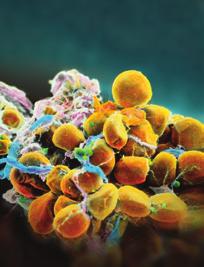
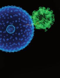
![feminization syndrome is rarely diagnosed in the 4th or 5th decades [7].](/docs-images/84/89101594/images/4-2.jpg "The testis is often undescended and the ovaries, uterus, fallopian tubes, and upper third of the vagina are typically absent [2, 6]. Externally, the labia majora and minora are usually well formed.")
![The estimated risk of malignancy in testicular feminization is 5% [8]. In comparison to other intersex disorders, the premalignant risk is relatively low before puberty [2, 7].](/docs-images/84/89101594/images/4-3.jpg "However, the overall risk increases in patients older than 30 years and reaches up to 33% in patients above 50 years of age [9].")
4 4 Case Reports in Radiology Rt-A Rt-B (a) (b) Rt-C Rt-D (c) (d) Figure 3: Histology of the right gonad (Rt). (a) Atrophic gonadal composed mainly of tubules of sertoli cells, with absence of germ cells, and the presence of interstitial Leydig cells, H&E 100. (b) The Sertoli tubules show inactive epithelial cells, absence of germ cells or atypia, H&E 200. (c) Both Sertoli cells and Leydig cells are immunoreactive to inhibin 100, while only Leydig cells react to Neuron-Specific Enolase 100 (d). feminization syndrome is rarely diagnosed in the 4th or 5th decades [7]. The testis is often undescended and the ovaries, uterus, fallopian tubes, and upper third of the vagina are typically absent [2, 6]. Externally, the labia majora and minora are usually well formed. The estimated risk of malignancy in testicular feminization is 5% [8]. In comparison to other intersex disorders, the premalignant risk is relatively low before puberty [2, 7]. However, the overall risk increases in patients older than 30 years and reaches up to 33% in patients above 50 years of age [9]. Gonadectomy/orchidectomy is therefore can be delayed to allow for a natural puberty [3]. Gonadoblastoma is the most commonly seen tumor in dysgenetic gonad [9]. The seminoma-dysgerminoma can also occur and is most commonly seen in streak gonad in mixed gonadal dysgenesis [8]. Rutgers et al. [2] had reported malignant change in their series of 43 patients with testicular feminization in which four (9%) malignant tumors (two seminomas, a germ cell neoplasm, and a malignant sexcord stromal tumor). Germ cell tumor remains the most common tumor in cryptorchidism as well as in testicular feminization [2]. Ramaswamy et al. [10] reported a large multicystic sex-cord tumor in a testicular feminization syndrome. Imaging features of testicular feminization syndrome are rarely described in the literature. Single imaging report by Karabulut et al. [4] has described CT and RI imaging features of stromal tumor of the sex cord in a woman with testicular feminization syndrome which showed internal necrotic and haemorrhagic components. We observed mild necrotic components whereas haemorrhages were not seen. Seminomas complicating undescended intraabdominal testes in a man with cryptorchidism are usually well delineated without evidence of hemorrhage or necrosis [11] and sex-cord stromal tumor in intraabdominal testis with testicular feminization syndrome exhibited heterogeneity in both texture and contrast enhancement [4]. Imaging feature in our case were in between (intraabdominal testicular seminomas in cryptorchidism and intraabdominal testicular sex-cord stromal tumor in testicular feminization syndrome) with no and mild heterogeneity in texture and contrast enhancement, respectively. In conclusion, though definitive diagnosis of testicular feminization syndrome relies on biochemical tests, imaging findings are useful in documenting the absence of mullerian duct derivatives, identifying the location of the undescended testes, and evaluating the accompanying intraabdominal masses. Imaging may also be helpful in providing tissue characteristics of a potential neoplasm arising from the gonads, their accurate extent and staging which are essential for further management.
5 Case Reports in Radiology 5 References [1] L. elton, New perspectives on the management of intersex, The Lancet, vol. 357, no. 9274, p. 2110, [2] J. L. Rutgers and R. E. Scully, The androgen insensitivity syndrome (testicular feminization) a clinicopathologic study of 43 cases, International Gynecological Pathology, vol. 10, no. 2, pp , [3] S.E.Hannema,I.S.Scott,E.Rajpert-Deeyts,N.E.Skakkebæk, N. Coleman, and I. A. Hughes, Testicular development in the complete androgen insensitivity syndrome, Pathology, vol. 208, no. 4, pp , [4] N. Karabulut, A. Karabulut, E. Pakdemirli, N. Sabir, S. K. Soysal,and.E.Soysal, Stromaltumorofthesexcord in a woman with testicular feminization syndrome: imaging features, American Roentgenology, vol. 178, no. 6, pp , [5] Z. Hrabovszky and J.. Hutson, Surgical treatment of intersex abnormalities: a review, Surgery, vol. 131, no. 1, pp , [6] C. B. Coulam,. L. Graham II, and T. C. Spelsberg, Androgen insensitivity syndrome: gonadal androgen receptor activity, American Obstetrics and Gynecology, vol. 150, no. 5, part 1, pp , [7] S. S. Lentz and J. O. Cappellari, Postmenopausal diagnosis of testicular feminization, American Obstetrics and Gynecology, vol. 179, no. 1, pp , [8] B. Salle and C. Hedinger, Gonadal histology in children with male pseudohermaphroditism and mixed gonadal dysgenesis, Acta Endocrinologica, vol. 64, no. 2, pp , [9]P.K.Donahoe,J.D.Crawford,andW.H.Hendren, ixed gonadal dysgenesis, pathogenesis, and management, Journal of Pediatric Surgery, vol. 14, no. 3, pp , [10] G. Ramaswamy, V. Jagadha, and V. Tchertkoff, Atesticular tumor resembling the sex cord with annular tubules in a case of the androgen insensitivity syndrome, Cancer, vol. 55, no. 7, pp , [11] F. H. iller, W. S. Whitney, S. W. Fitzgerald, and E. I. iller, Seminomas complicating undescended intraabdominal testes in patients with prior negative findings from surgical exploration, American Roentgenology, vol. 172, no. 2, pp , 1999.


6 EDIATORS of INFLAATION The Scientific World Journal Gastroenterology Research and Practice Diabetes Research International Endocrinology Immunology Research Disease arkers Submit your manuscripts at Bioed Research International PPAR Research Obesity Ophthalmology Evidence-Based Complementary and Alternative edicine Stem Cells International Oncology Parkinson s Disease Computational and athematical ethods in edicine AIDS Behavioural Neurology Research and Treatment Oxidative edicine and Cellular Longevity
Case Report Müllerian Remnant Cyst as a Cause of Acute Abdomen in a Female Patient with Müllerian Agenesis: Radiologic and Pathologic Findings
 Volume 2016, Article ID 6581387, 4 pages http://dx.doi.org/10.1155/2016/6581387 Case Report üllerian Remnant Cyst as a Cause of Acute Abdomen in a Female Patient with üllerian Agenesis: Radiologic and
Approach to Disorders of Sex Development (DSD)
") Approach to Disorders of Sex Development (DSD) Old name: The Approach to Intersex Disorders Dr. Abdulmoein Al-Agha, FRCP Ass. Professor & Consultant Pediatric Endocrinologist, KAUH, Erfan Hospital & Ibn
Approach to Disorders of Sex Development (DSD) Old name: The Approach to Intersex Disorders Dr. Abdulmoein Al-Agha, FRCP Ass. Professor & Consultant Pediatric Endocrinologist, KAUH, Erfan Hospital & Ibn
IN SUMMARY HST 071 NORMAL & ABNORMAL SEXUAL DIFFERENTIATION Fetal Sex Differentiation Postnatal Diagnosis and Management of Intersex Abnormalities
 Harvard-MIT Division of Health Sciences and Technology HST.071: Human Reproductive Biology Course Director: Professor Henry Klapholz IN SUMMARY HST 071 Title: Fetal Sex Differentiation Postnatal Diagnosis
-The cause of testicular neoplasms remains unknown
 - In the 15- to 34-year-old age group, they are the most common tumors of men. - include: I. Germ cell tumors : (95%); all are malignant. II. Sex cord-stromal tumors: from Sertoli or Leydig cells; usually
Let s Talk About Hormones!
 Let s Talk About Hormones! This lesson was created by Serena Reves and Nichelle Penney, with materials from the BCTF and The Pride Education Network. Hormones are responsible for the regulation of many
When testes make no testosterone: Identifying a rare cause of 46, XY female phenotype in adulthood
 When testes make no testosterone: Identifying a rare cause of 46, XY female phenotype in adulthood Gardner DG, Shoback D. Greenspan's Basic & Clinical Endocrinology, 10e; 2017 Sira Korpaisarn, MD Endocrinology
Ethiopian patients and a brief review
 Postgraduate Medical Journal (November 1979) 55, 844-848 Testicular feminization syndrome: a report of three Ethiopian patients and a brief review EDEMARIAM TSEGA M.D., D.C.M.T.(L), F.R.C.P.(C) Summary
Case Report Primary Malignancy in a Supernumerary Testicle Presenting as a Large Pelvic Mass
 Hindawi Volume 2017, Article ID 4529853, 4 pages https://doi.org/10.1155/2017/4529853 Case Report Primary Malignancy in a Supernumerary Testicle Presenting as a Large Pelvic Mass Justin Noroozian, 1 Daniel
W.S. O University of Hong Kong
 W.S. O University of Hong Kong Development of the Genital System 1. Sexual differentiation 2. Differentiation of the gonads a. Germ cells extragonadal in origin b. Genital ridge intermediate mesoderm consisting
Note: The cause of testicular neoplasms remains unknown
 - In the 15- to 34-year-old age group, they are the most common tumors of men. - Tumors of the testis are a heterogeneous group of neoplasms that include: I. Germ cell tumors : 95%; all are malignant.
Normal and Abnormal Development of the Genital Tract. Dr.Raghad Abdul-Halim
 Normal and Abnormal Development of the Genital Tract Dr.Raghad Abdul-Halim objectives: Revision of embryology. Clinical presentation, investigations and clinical significance of most common developmental
PHYSIOLOGY AND PATHOLOGY OF SEXUAL DIFFERENTIATION
 PHYSIOLOGY AND PATHOLOGY OF SEXUAL DIFFERENTIATION Prof. Dr med. Jolanta Słowikowska-Hilczer Department of Andrology and Reproductive Endocrinology Medical University of Łódź, Poland Sexual determination
DEFINITION: Masculinization of external genitalia in patients with normal 46XX karyotype.
 INTERSEX DISORDERS DEFINITION: Masculinization of external genitalia in patients with normal 46XX karyotype. - Degree of masculinization variable: - mild clitoromegaly - complete fusion of labia folds
AMBIGUOUS GENITALIA. Dr. HAKIMI, SpAK. Dr. MELDA DELIANA, SpAK
 AMBIGUOUS GENITALIA (DISORDERS OF SEXUAL DEVELOPMENT) Dr. HAKIMI, SpAK Dr. MELDA DELIANA, SpAK Dr. SISKA MAYASARI LUBIS, SpA Pediatric Endocrinology division USU/H. ADAM MALIK HOSPITAL 1 INTRODUCTION Normal
Maldescended testis in Adults. Dr. BG GAUDJI Urologist STEVE BIKO ACADEMIC HOSPITAL
 Maldescended testis in Adults Dr. BG GAUDJI Urologist STEVE BIKO ACADEMIC HOSPITAL Definitions Cryptorchid: testis neither resides nor can be manipulated into the scrotum Ectopic: aberrant course Retractile:
Management of gonads in DSD
 Management of gonads in DSD Martine Cools, paediatric endocrinologist, Katja Wolffenbuttel and Piet Hoebeke, paediatric urologists, all at University Hospital Ghent, Belgium Sten Drop, paediatric endocrinologist
COMPLETE GONADAL DYSGENESIS WITH XY CHROMOSOMAL CONSTITUTION
 Tipar Cap coada final.qxd 1/22/2007 11:57 PM Page 465 Case report COMPLETE GONADAL DYSGENESIS WITH XY CHROMOSOMAL CONSTITUTION Dorina Stoicanescu *,1, Valerica Belengeanu 1, Dana Amzar 2, Cristina Popa
Development of the urogenital system
 Development of the urogenital system Location of the pronephros, mesonephros and metanephros Differentiation of the intermedierm mesoderm into nephrotome and mesonephric tubules Connection between aorta
Disordered Sex Differentiation Mixed gonadal dysgenesis Congenital adrenal hyperplasia Mixed gonadal dysgenesis
 Disordered Sex Differentiation DSD has superceded intersex in describing genital anomalies in childhood DSD results from hormonal imbalances due to (i) abnormal genetic status, (ii) enzyme defects, or
Intersex Genital Mutilations in ICD-10 Zwischengeschlecht.org / StopIGM.org 2014 (v2.1)
") Intersex Genital Mutilations in ICD-10 Zwischengeschlecht.org / StopIGM.org 2014 (v2.1) ICD-10 Codes and Descriptions: http://apps.who.int/classifications/icd10/browse/2010/en 1. Reference: 17 Most Common
Intersex Genital Mutilations in ICD-10 Zwischengeschlecht.org / StopIGM.org 2014 (v2.1) ICD-10 Codes and Descriptions: http://apps.who.int/classifications/icd10/browse/2010/en 1. Reference: 17 Most Common
Persistent Mullerian Duct Syndrome (PMDS) With Large Intraabdominal Seminoma
 With Large Intraabdominal Seminoma") BMH Med. J. 2017;4(3):103-108 Case Report Persistent Mullerian Duct Syndrome (PMDS) With Large Intraabdominal Seminoma Della Harigovind, Harish Babu, Sana Sherfin, Shilpa Asokan, Shylesh Aikot, SP Zebunnisa
BMH Med. J. 2017;4(3):103-108 Case Report Persistent Mullerian Duct Syndrome (PMDS) With Large Intraabdominal Seminoma Della Harigovind, Harish Babu, Sana Sherfin, Shilpa Asokan, Shylesh Aikot, SP Zebunnisa
Case Report Bilateral Sclerosing Stromal Ovarian Tumor in an Adolescent
 Case Reports in Radiology Volume 2015, Article ID 271394, 4 pages http://dx.doi.org/10.1155/2015/271394 Case Report Bilateral Sclerosing Stromal Ovarian Tumor in an Adolescent Anjani Naidu, 1 Betty Chung,
Cardiff MRCS OSCE Courses Testicular Cancer
 Testicular Cancer Scenario: A 40-year-old male presents to the surgical out-patient clinic with a 6-8 week history of a painless lump in his left scrotum. He however complains of a dull ache in the scrotum
DISORDERS OF MALE GENITALS
 Wit JM, Ranke MB, Kelnar CJH (eds): ESPE classification of paediatric endocrine diagnosis. 9. Testicular disorders/disorders of male genitals. Horm Res 2007;68(suppl 2):63 66 ESPE Code Diagnosis OMIM ICD10
Primary Amenorrhea, age 16: Recent Reflections. David A Grainger MD, MPH February 1, 2017
 Primary Amenorrhea, age 16: Recent Reflections David A Grainger MD, MPH February 1, 2017 Primary Amenorrhea No menses by age 13-14 WITHOUT BREAST DEVELOPMENT No menses by age 15-16 WITH BREAST DEVELOPMENT
Disclosure. Session Objectives. I have no actual or potential conflict of interest in relation to this program/presentation.
 46, XY Female: A Case of Complete Androgen Insensitivity Syndrome (CAIS) MICHELLE MCLOUGHLINMSN, CRNP, CPNP-AC THE CHILDREN S HOSPITAL OF PHILADELPHIA DIVISION OF ENDOCRINOLOGY AND DIABETES Disclosure
OVOTESTIS Background Pathophysiology
 OVOTESTIS Background Ovotestis refers to the histology of a gonad that contains both ovarian follicles and testicular tubular elements. Such gonads are found exclusively in people with ovotesticular disorder
Female with 46, XY karyotype
 Case Report Obstet Gynecol Sci 2017;60(4):378-382 https://doi.org/10.5468/ogs.2017.60.4.378 pissn 2287-8572 eissn 2287-8580 Female with 46, XY karyotype Eun Jung Jung 1, Do Hwa Im 1, Yong Hee Park 1, Jung
Case Report Intracranial Capillary Hemangioma in the Posterior Fossa of an Adult Male
 Case Reports in Radiology Volume 2016, Article ID 6434623, 4 pages http://dx.doi.org/10.1155/2016/6434623 Case Report Intracranial Capillary Hemangioma in the Posterior Fossa of an Adult Male Jordan Nepute,
Development of the Genital System
 Development of the Genital System Professor Alfred Cuschieri Department of Anatomy University of Malta The mesonephros develops primitive nephrotomes draining into a mesonephric duct nephrotome mesonephric
Endometrial Stromal Sarcoma
 May 26, 2011 By Sushila Ladumor, MD [1] Endometrial stromal sarcoma (ESS) is a rare malignant tumor of the endometrium, occurring in the age group of 40-50 years. History The 50-year-old, female patient
Testicular feminization. The features of this syndrome in its typical form are these (Fig. 1): (1) Female bodily configuration.
: (1) Female bodily configuration.") Personal Practice Archives of Disease in Childhood, 1970, 45, 595. The XY Female Child C. J. DEWHURST From the Institute of Obstetrics and Gynaecology, Queen Charlotte's Maternity Hospital, and Chelsea
Personal Practice Archives of Disease in Childhood, 1970, 45, 595. The XY Female Child C. J. DEWHURST From the Institute of Obstetrics and Gynaecology, Queen Charlotte's Maternity Hospital, and Chelsea
Case Report Five-Year Survival after Surgery for Invasive Micropapillary Carcinoma of the Stomach
 Case Reports in Surgery Volume 2013, Article ID 560712, 4 pages http://dx.doi.org/10.1155/2013/560712 Case Report Five-Year Survival after Surgery for Invasive Micropapillary Carcinoma of the Stomach Shigeo
International Journal of Case Reports in Medicine
 International Journal of Case Reports in Medicine Vol. 2013 (2013), Article ID 326535, 19 minipages. DOI:10.5171/2013.326535 www.ibimapublishing.com Copyright 2013 Ahmet Ay, Aybala Agac Ay, Ozan Turgut,
Incidental gonadal tumours at the time of gonadectomy in women with Swyer syndrome: a case series. Journal of Pediatric and Adolescent Gynecology
 Accepted Manuscript Incidental gonadal tumours at the time of gonadectomy in women with Swyer syndrome: a case series Amie JM. Hanlon, MBBS Rebecca M. Kimble, MBBS, FRANZCOG PII: S1083-3188(14)00272-1
Please Take Seats by Gender as Shown Leave Three Seats Empty in the Middle
 Please Take Seats by Gender as Shown Leave Three Seats Empty in the Middle Women Men Sexual Differentiation & Development Neal G. Simon, Ph.D. Professor Dept. of Biological Sciences Signaling Cascade &
Case Report Role of Imaging in the Diagnosis and Management of Complete Androgen Insensitivity Syndrome in Adults
 Hindawi Publishing Corporation Case Reports in Radiology Volume 2013, Article ID 158484, 6 pages http://dx.doi.org/10.1155/2013/158484 Case Report Role of Imaging in the Diagnosis and Management of Complete
State of the art review in gonadal dysgenesis: challenges in diagnosis and management
 McCann-Crosby et al. International Journal of Pediatric Endocrinology 2014, 2014:4 PES REVIEW State of the art review in gonadal dysgenesis: challenges in diagnosis and management Open Access Bonnie McCann-Crosby
Goals. Disorders of Sex Development (Intersex): An Overview. Joshua May, MD Pediatric Endocrinology
: An Overview. Joshua May, MD Pediatric Endocrinology") Disorders of Sex Development (Intersex): An Overview Joshua May, MD Pediatric Endocrinology Murphy, et al., J Ped Adol Gynecol, 2011 Goals Objectives: Participants will be able to: 1. Apply the medical
Disorders of Sex Development (Intersex): An Overview Joshua May, MD Pediatric Endocrinology Murphy, et al., J Ped Adol Gynecol, 2011 Goals Objectives: Participants will be able to: 1. Apply the medical
46 XY gonadal dysgenesis in adulthood pitfalls of late diagnosis
 Reminder of important clinical lesson 46 XY gonadal dysgenesis in adulthood pitfalls of late diagnosis Jarna Naing Hamin,1 Francis Raymond P Arkoncel,2 Frances Lina Lantion-Ang,1 Mark Anthony S Sandoval1
Urogenital System. PUMC Dept. of Anat. Hist. & Embry. 钱晓菁 XIAO-JING QIAN Dept. of Anatomy, Histology & Embryology Peking Union Medical College
 Urogenital System 钱晓菁 XIAO-JING QIAN Dept. of Anatomy, Histology & Embryology Peking Union Medical College intermediate mesoderm urogenital ridge mesonephric ridge genital ridge I. Urinary System 1. kidney
DEVELOPMENT (DSD) 1 4 DISORDERS OF SEX
 1 4 DISORDERS OF SEX") Wit JM, Ranke MB, Kelnar CJH (eds): ESPE classification of paediatric endocrine diagnosis. 4. Disorders of sex development (DSD). Horm Res 2007;68(suppl 2):21 24 ESPE Code Diagnosis OMIM ICD 10 4 DISORDERS
Wit JM, Ranke MB, Kelnar CJH (eds): ESPE classification of paediatric endocrine diagnosis. 4. Disorders of sex development (DSD). Horm Res 2007;68(suppl 2):21 24 ESPE Code Diagnosis OMIM ICD 10 4 DISORDERS
Testicular leydig cell tumor with metachronous lesions: Outcomes after metastasis resection and cryoablation
 Washington University School of Medicine Digital Commons@Becker Open Access Publications 2015 Testicular leydig cell tumor with metachronous lesions: Outcomes after metastasis resection and cryoablation
American Journal of Oral Medicine and Radiology
 American Journal of Oral Medicine and Radiology e - ISSN - XXXX-XXXX ISSN - 2394-7721 Journal homepage: www.mcmed.us/journal/ajomr ULTRASONOGRAPHIC EVALUATION OF ADNEXAL MASSES Nageswar Rao* Professor,
1) Intersexuality - Dr. Huda
 Intersexuality - Dr. Huda") 1) Intersexuality - Dr. Huda DSD (Disorders of sex development) occur when there is disruption of either: Gonadal differentiation Fetal sex steroid production or action Mullerian abnormalities and Wolffian
1) Intersexuality - Dr. Huda DSD (Disorders of sex development) occur when there is disruption of either: Gonadal differentiation Fetal sex steroid production or action Mullerian abnormalities and Wolffian
Chapter 18 Development. Sexual Differentiation
 Chapter 18 Development Sexual Differentiation There Are Many Levels of Sex Determination Chromosomal Sex Gonadal Sex Internal Sex Organs External Sex Organs Brain Sex Gender Identity Gender Preference
Case Report Uncommon Mixed Type I and II Choledochal Cyst: An Indonesian Experience
 Case Reports in Surgery Volume 2013, Article ID 821032, 4 pages http://dx.doi.org/10.1155/2013/821032 Case Report Uncommon Mixed Type I and II Choledochal Cyst: An Indonesian Experience Fransisca J. Siahaya,
Pathology of Ovarian Tumours. Dr. Jyothi Ranganathan MD ( Path) AFMC Pune PDCC (Cytopathology) PGI Chandigarh
 AFMC Pune PDCC (Cytopathology) PGI Chandigarh") Pathology of Ovarian Tumours Dr. Jyothi Ranganathan MD ( Path) AFMC Pune PDCC (Cytopathology) PGI Chandigarh Outline Incidence Risk factors Classification Pathology of tumours Tumour markers Prevention
Pathology of Ovarian Tumours Dr. Jyothi Ranganathan MD ( Path) AFMC Pune PDCC (Cytopathology) PGI Chandigarh Outline Incidence Risk factors Classification Pathology of tumours Tumour markers Prevention
Intersex is a group of conditions where there is a discrepancy between the external genitals and the internal genitals (the testes and ovaries).
.") Intersex to use the sharing features on this page, please enable JavaScript. Share on facebookshare on IwitterBookmark & SharePrintcr-lnendly version Intersex is a group of conditions where
Intersex to use the sharing features on this page, please enable JavaScript. Share on facebookshare on IwitterBookmark & SharePrintcr-lnendly version Intersex is a group of conditions where
A Young Asian Girl with MRKH Type B Syndrome: A Case Report
 A Young Asian Girl with MRKH Type B Syndrome: A Case Report Nidhi Jain 1, Pardaman Singh 2, Deepak Goel 3, Jyotsna kamra 4 1,4 Department of Obstetrics and Gynecology, Maharaja Agarsein Medical College,
10. Development of genital system. Gonads. Genital ducts. External genitalia.
 10. Development of genital system. Gonads. Genital ducts. External genitalia. Gonads, genital ducts and the external genital organs initially pass through an indifferent period of development, which is
46,XY Female: SRY and AR Basis: Genotype & Phenotype Correlation
 Original Article Indian Journal of Genetics and Molecular Research 53 Volume 1 Number 2, July - December 2012 46,XY Female: SRY and AR Basis: Genotype & Phenotype Correlation Amudha S.*, MSc; Sayee Rajangam**,
PERSISTANT MULLERIAN DUCT SYNDROME ASSOCIATED WITH TRANSVERSE TESTICULAR ECTOPIA
 PERSISTANT MULLERIAN DUCT SYNDROME ASSOCIATED WITH TRANSVERSE TESTICULAR ECTOPIA Dr. Abdulrahman A. Al-Bassam, FRCS(Ed) Assistant Professor & Consultant Paediatric Surgeon King Khalid University Hospital
Bilateral Segmental Testicular Infarction
 Case Study TheScientificWorldJOURNAL (2007) 7, 779 783 TSW Urology ISSN 1537-744X; DOI 10.1100/tsw.2007.146 Bilateral Segmental Testicular Infarction Aaron Bayne 1, Brad Koslin 2, and Siamak Daneshmand
C. Patrick Shahan, MD University of Tennessee Health Science Center Department of Surgery
 C. Patrick Shahan, MD University of Tennessee Health Science Center Department of Surgery Drop use of hermaphrodite and derivatives 1 in 15,000 live births Congenital Adrenal Hyperplasia Mixed Gonadal
Key words: Dysgerminoma, Ovarian Neoplasms, Diagnostic Imaging, Germinoma, Seminoma.
 JOURNAL OF CASE REPORTS 2014;4(1):225-229 Massive Ovarian Dysgerminoma Presenting as an Unusual Large Intra-abdominal Mass Arvinder Singh 1, Surinderpal 2, Sohan Singh 1, Aditi Mahajan 1, Mohit Khandelwal
Hamartomatous Nodule, Sertoli Cell Adenoma in Complete Androgen Insensitivity Syndrome with Wolffian/Müllerian Duct Remnants: An Unusual Case Report
 Case Report DOI: 10.21276/APALM.1132 Hamartomatous Nodule, Sertoli Cell Adenoma in Complete Androgen Insensitivity Syndrome with Wolffian/Müllerian Duct Remnants: An Unusual Case Report Prashant Vijay
FLASH CARDS. Kalat s Book Chapter 11 Alphabetical
 FLASH CARDS www.biologicalpsych.com Kalat s Book Chapter 11 Alphabetical alpha-fetoprotein alpha-fetoprotein Alpha-Fetal Protein (AFP) or alpha-1- fetoprotein. During a prenatal sensitive period, estradiol
Clinical Study Changing Trends in Use of Laparoscopy: A Clinical Audit
 Minimally Invasive Surgery, Article ID 562785, 4 pages http://dx.doi.org/10.1155/2014/562785 Clinical Study Changing Trends in Use of Laparoscopy: A Clinical Audit Ritu Khatuja, 1 Geetika Jain, 1 Sumita
Case Report Traumatic Haemorrhagic Cervical Lymphadenopathy with Underlying Infectious Mononucleosis
 Hindawi Case Reports in Radiology Volume 2017, Article ID 3097414, 4 pages https://doi.org/10.1155/2017/3097414 Case Report Traumatic Haemorrhagic Cervical Lymphadenopathy with Underlying Infectious Mononucleosis
Analysis of the Sex-determining Region of the Y Chromosome (SRY) in a Case of 46, XX True Hermaphrodite
 in a Case of 46, XX True Hermaphrodite") Clin Pediatr Endocrinol 1994; 3(2): 91-95 Copyright (C) 1994 by The Japanese Society for Pediatric Endocrinology Analysis of the Sex-determining Region of the Y Chromosome (SRY) in a Case of 46, XX True
Clin Pediatr Endocrinol 1994; 3(2): 91-95 Copyright (C) 1994 by The Japanese Society for Pediatric Endocrinology Analysis of the Sex-determining Region of the Y Chromosome (SRY) in a Case of 46, XX True
Case Report Denosumab Chemotherapy for Recurrent Giant-Cell Tumor of Bone: A Case Report of Neoadjuvant Use Enabling Complete Surgical Resection
 Case Reports in Oncological Medicine Volume 2013, Article ID 496351, 4 pages http://dx.doi.org/10.1155/2013/496351 Case Report Denosumab Chemotherapy for Recurrent Giant-Cell Tumor of Bone: A Case Report
Chapter 7 DEVELOPMENT AND SEX DETERMINATION
 Chapter 7 DEVELOPMENT AND SEX DETERMINATION Chapter Summary The male and female reproductive systems produce the sperm and eggs, and promote their meeting and fusion, which results in a fertilized egg.
Case Report Internal Jugular Vein Thrombosis in Isolated Tuberculous Cervical Lymphadenopathy
 Volume 2016, Article ID 5184196, 4 pages http://dx.doi.org/10.1155/2016/5184196 Case Report Internal Jugular Vein Thrombosis in Isolated Tuberculous Cervical Lymphadenopathy Sanjay Khaladkar, Avadhesh
Long-term outcome of ovotesticular disorder of sex development: A single center experience
 International Journal of Urology (2011) 18, 231 236 doi: 10.1111/j.1442-2042.2010.02700.x Original Article: Clinical Investigationiju_2700 231..236 Long-term outcome of ovotesticular disorder of sex development:
Sexual Development. 6 Stages of Development
 6 Sexual Development 6 Stages of Development Development passes through distinct stages, the first of which is fertilization, when one sperm enters one ovum. To enter an ovum, a sperm must undergo the
M Tayfur, R Kocabas, A Kaygisiz, S Tiryaki, M Polat, K Cefle
 ISPUB.COM The Internet Journal of Pathology Volume 7 Number 2 Dysgerminoma arising in Swyer Syndrome M Tayfur, R Kocabas, A Kaygisiz, S Tiryaki, M Polat, K Cefle Citation M Tayfur, R Kocabas, A Kaygisiz,
R. F. Falkenstern-Ge, 1 S. Bode-Erdmann, 2 G. Ott, 2 M. Wohlleber, 1 and M. Kohlhäufl Introduction. 2. Histology
 Case Reports in Oncological Medicine Volume 2013, Article ID 167585, 4 pages http://dx.doi.org/10.1155/2013/167585 Case Report Late Lung Metastasis of a Primary Eccrine Sweat Gland Carcinoma 10 Years after
Ovarian Tumors. Andrea Hayes-Jordan MD FACS, FAAP Section Chief, Pediatric Surgery/Surgical Onc. UT MD Anderson Cancer Center
 Ovarian Tumors Andrea Hayes-Jordan MD FACS, FAAP Section Chief, Pediatric Surgery/Surgical Onc. UT MD Anderson Cancer Center Case 13yo female with abdominal pain Ultrasound shows huge ovarian mass Surgeon
Clinical Study Laparoscopic Management of Intra-Abdominal Testis: 5-Year Single-Centre Experience A Retrospective Descriptive Study
 Minimally Invasive Surgery Volume 2012, Article ID 878509, 4 pages doi:10.1155/2012/878509 Clinical Study Laparoscopic Management of Intra-Abdominal Testis: 5-Year Single-Centre Experience A Retrospective
Testicular Tumors: What s New, True, Important Cristina Magi-Galluzzi, MD, PhD
 Testicular Tumors: What s New, True, Important Cristina Magi-Galluzzi, MD, PhD Director, Genitourinary Pathology R.J. Tomsich Pathology & Laboratory Medicine Institute Professor of Pathology, Lerner College
Downloaded from amuj.arakmu.ac.ir at 22: on Thursday June 21st 2018 FSH.
 * Downloaded from amuj.arakmu.ac.ir at 22:26 +0430 on Thursday June 21st 2018 CT Email: mjamilian@yahoo.com * X 1 - T F: Testicular feminization. TF T F xy TSH immature testicular tissue CT TF patients
Case Based Urology Learning Program
 Case Based Urology Learning Program Resident s Corner: UROLOGY Case Number 3 CBULP 2011 019 Case Based Urology Learning Program Editor: Associate Editors: Manager: Case Contributors: Steven C. Campbell,
Sexual differentiation:
 Abnormal Development of Female Genitalia Dr. Maryam Fetal development of gonads, external genitalia, Mullerian ducts and Wolffian ducts can be disrupted at a variety of points, leading to a wide range
Sex Determination and Development of Reproductive Organs
 Sex Determination and Development of Reproductive Organs Sex determination The SRY + gene is necessary and probably sufficient for testis development The earliest sexual difference appears in the gonad
11. SEXUAL DIFFERENTIATION. Germinal cells, gonocytes. Indifferent stage INDIFFERENT STAGE
 11. SEXUAL DIFFERENTIATION INDIFFERENT STAGE Early in pregnancy, (within 10-15 % of the pregnancy s expected length) a genital ridge is formed in the sides of the embryonic tissue, ventral to the mesonephros
Bilateral Primary Fallopian Tube Carcinoma: Findings on Sequential MRI
 Hosokawa et al. MRI in Fallopian Tube Carcinoma Women s Imaging Case Report WOMEN S IMAGING Chisa Hosokawa 1 Mitsuo Tsubakimoto 2 Yuichi Inoue 3 Tetsuo Nakamura 2 Hosokawa C, Tsubakimoto M, Inoue Y, Nakamura
Case Fibrothecoma of the ovary
 Case 10646 Fibrothecoma of the ovary Elisa Melo Abreu, Teresa Margarida Cunha Section: Genital (Female) Imaging Published: 2015, Jan. 2 Patient: 70 year(s), female Authors' Institution Department of Radiology,
Stump the Professors. Professor Panelists 4/13/2015. Challenger. Kylie Fowler, MD
 4/13/2015 Stump the Professors Professor Panelists Cynthia Holland-Hall MD, MPH Mary Anne Jamieson, MD Julie Strickland, MD, MPH Challenger Kylie Fowler, MD Medical degree: University of Washington School
7 Mousa. Obada Zalat. Mohammad Badi
 7 Mousa Obada Zalat Mohammad Badi Tumors of the ovaries Last lecture we talked about surface epithelial tumors of the ovaries (the most common type). But there are many other types of tumors of germ cell
Gynecologic Cancer Surveillance and Survivorship: Informing Practice and Policy
 Gynecologic Cancer Surveillance and Survivorship: Informing Practice and Policy Stephanie Yap, M.D. University Gynecologic Oncology Northside Cancer Institute Our Learning Objectives Review survival rates,
ISSN International Journal of Innovative and Applied Research (2017) Journal home page: RESEARCH ARTICLE
 Journal home page: RESEARCH ARTICLE") Journal home page: http://www.journalijiar.com RESEARCH ARTICLE MAYER-ROKITANSKY-KUSTER-HAUSER SYNDROME II (Rare Case). * Dr. Verinder Dhar 1 and Dr. Renu Hashia 2. 1. MD(Medicine), DM(Endo),Medicare Nursing
Journal home page: http://www.journalijiar.com RESEARCH ARTICLE MAYER-ROKITANSKY-KUSTER-HAUSER SYNDROME II (Rare Case). * Dr. Verinder Dhar 1 and Dr. Renu Hashia 2. 1. MD(Medicine), DM(Endo),Medicare Nursing
Bios 90/95. Jennifer Swann, PhD
 Sexual Differentiation Fall 2007 Bios 90/95 Jennifer Swann, PhD Dept Biol Sci, Lehigh University Why have sexes? What determines sex? Environment Genetics Hormones What causes these differences? The true
Biology of Reproduction- Zool 346 Exam 2
 Biology of Reproduction- Zool 346 Exam 2 ANSWER ALL THE QUESTIONS ON THE ANSWER SHEET. THE ANSWER ON THE ANSWER SHEET IS YOUR OFFICIAL ANSWER. Some critical words are boldfaced. This exam is 7 pages long.
Action of reproductive hormones through the life span 9/22/99
 Action of reproductive hormones through the life span Do reproductive hormones affect the life span? One hypothesis about the rate of aging asserts that there is selective pressure for either high rate
Case Report Crossed Renal Ectopia without Fusion An Unusual Cause of Acute Abdominal Pain: A Case Report
 Case Reports in Urology Volume 2012, Article ID 728531, 4 pages doi:10.1155/2012/728531 Case Report Crossed Renal Ectopia without Fusion An Unusual Cause of Acute Abdominal Pain: A Case Report D. P. Ramaema,
Case Report A Case of Cystic Basal Cell Carcinoma Which Shows a Homogenous Blue/Black Area under Dermatoscopy
 Volume 20, Article ID 450472, 4 pages doi:0.55/20/450472 Case Report A Case of Cystic Basal Cell Carcinoma Which Shows a Homogenous Blue/Black Area under Dermatoscopy Akihiro Yoneta, Kohei Horimoto, Keiko
- production of two types of gametes -- fused at fertilization to form zygote
 Male reproductive system I. Sexual reproduction -- overview - production of two types of gametes -- fused at fertilization to form zygote - promotes genetic variety among members of a species -- each offspring
Solitary Contralateral Adrenal Metastases after Nephrectomy for Renal Cell Carcinoma
 Original Report ISSN 1537-744X; DOI 10.1100/tsw.2004.39 Solitary Contralateral Adrenal after Nephrectomy for Renal Cell Carcinoma Nikolaos Antoniou, M.D. and Demetrios Karanastasis, M.D. General Hospital
Intratubular Germ Cell Neoplasia of the Testis
 Intratubular Germ Cell Neoplasia of the Testis KS Ngoo Department of Urology Hospital Selayang Advanced Urology Course 15 Aug 2014 MUA Office Clinical scenario A 33 years old man has bilateral testicular
Bilateral Renal Angiomyolipomas with Invasion of the Renal Vein: A Case Report
 Case Study TheScientificWorldJOURNAL (2008) 8, 145 148 TSW Urology ISSN 1537-744X; DOI 10.1100/tsw.2008.29 Bilateral Renal Angiomyolipomas with Invasion of the Renal Vein: A Case Report C. Blick, N. Ravindranath,
CLINICAL PRESENTATION AND RADIOLOGY QUIZ QUESTION
 Donald L. Renfrew, MD Radiology Associates of the Fox Valley, 333 N. Commercial Street, Suite 100, Neenah, WI 54956 2/12/2011 Radiology Quiz of the Week # 7 Page 1 CLINICAL PRESENTATION AND RADIOLOGY QUIZ
TitleA case of metachronous bilateral te. Citation 泌尿器科紀要 (1993), 39(6):
, 39(6):") TitleA case of metachronous bilateral te Takashi, Munehisa; Hirata, Yoshifum Author(s) Hideo; Shimoji, Toshio; Miyake, Koj Nagasaka, Tetsuro Citation 泌尿器科紀要 (1993), 39(6): 577-580 Issue Date 1993-06 URL
TitleA case of metachronous bilateral te Takashi, Munehisa; Hirata, Yoshifum Author(s) Hideo; Shimoji, Toshio; Miyake, Koj Nagasaka, Tetsuro Citation 泌尿器科紀要 (1993), 39(6): 577-580 Issue Date 1993-06 URL
DAX1, testes development role 7, 8 DFFRY, spermatogenesis role 49 DMRT genes, male sex differentiation role 15
 Subject Index N-Acetylcysteine, sperm quality effects 71 Ambiguous genitalia, origins 1, 2 Anti-Müllerian hormone function 13 receptors 13 Sertoli cell secretion 10, 38 Apoptosis assays in testes 73, 74
Female Genital Tract Lab. Dr. Nisreen Abu Shahin Assistant Professor of Pathology University of Jordan
 Female Genital Tract Lab Dr. Nisreen Abu Shahin Assistant Professor of Pathology University of Jordan Ovarian Pathology A 20-year-old female presented with vague left pelvic pain. Pelvic exam revealed
UNIVERSITY OF MEDICINE AND PHARMACY OF CRAIOVA DOCTORAL SCHOOL. PHD THESIS UNDESCENDED TESTICLE IN CHILD CLINICAL AND THERAPEUTIC ASPECTS - Abstract -
 UNIVERSITY OF MEDICINE AND PHARMACY OF CRAIOVA DOCTORAL SCHOOL PHD THESIS UNDESCENDED TESTICLE IN CHILD CLINICAL AND THERAPEUTIC ASPECTS - Abstract - SCIENTIFIC COORDINATOR: Prof. Dr. Ion GEORGESCU PHD
Case Report Renal Cell Carcinoma Metastatic to Thyroid Gland, Presenting Like Anaplastic Carcinoma of Thyroid
 Case Reports in Urology Volume 2013, Article ID 651081, 4 pages http://dx.doi.org/10.1155/2013/651081 Case Report Renal Cell Carcinoma Metastatic to Thyroid Gland, Presenting Like Anaplastic Carcinoma
Imaging Ejaculatory Disorders and Hematospermia
 ATHENS 4-6 October 2018 European Society of Urogenital Radiology Imaging Ejaculatory Disorders and Hematospermia Parvati Ramchandani, MD Professor, Radiology and Surgery University of Pennsylvania Medical