Endogenous TNFα orchestrates the trafficking of neutrophils into and within lymphatic vessels during acute inflammation
|
|
|
- Emil Parrish
- 5 years ago
- Views:
Transcription
1 SUPPLEMENTARY INFORMATION Endogenous TNFα orchestrates the trafficking of neutrophils into and within lymphatic vessels during acute inflammation Samantha Arokiasamy 1,2, Christian Zakian 1, Jessica Dilliway 1, Wen Wang 2#, Sussan Nourshargh 1# & Mathieu-Benoit Voisin 1 *. 1
2 VIDEO LEGENDS Video 1 Neutrophil breaching of the lymphatic endothelium in a TNF-stimulated tissue (Figure 1e). The video shows a cremaster lymphatic vessel of a LysM-GFP mouse (exhibiting GFP-labelled leukocytes (green), immunostained in vivo for ECs with Alexa647-labelled anti-pecam-1 mab (blue) and stimulated with TNF (300ng/mouse i.s.). The video shows the migration of a neutrophil (recorded from 4hrs after injection of the cytokine) into the lumen of the lymphatic vessel. Images were captured every minute for 90min. Still images are shown in Fig. 1e. Video 2 Neutrophil breaching of the lymphatic endothelium in a TNF-stimulated (300ng/mouse i.s.) cremaster lymphatic vessel of a LysM-GFP mouse (exhibiting GFP-labelled leukocytes (green), and immunostained in vivo for LECs with non-blocking dose of an Alexa555-labelled anti-lyve-1 mab (red). The video shows the migration of a neutrophil (in green and isolated from the rest of the inflammatory response by creating an isosurface on it using IMARIS software for clarity of the image) into the lumen of the lymphatic vessel (recorded from 2hrs after injection of the cytokine). Images were captured every minute for 90min. Video 3 Intraluminal neutrophil crawling along the lumen of the lymphatic endothelium (Figure 1f). The video captures the intraluminal crawling of neutrophils (in green) along the lymphatic endothelial cell wall in a TNF-stimulated cremaster of a LysM-GFP mouse (exhibiting GFP-labelled leukocytes (green). Lymphatic vessels were immunostained in vivo with Alexa555-labelled anti LYVE-1 mab (red) and Alexa647-labelled anti-pecam-1 mab (blue). Images were captured from 4hrs post-inflammation at one stack per minute for a duration of 90 min. Still images of this video are shown in Fig. 1f. Video 4 Intraluminal neutrophil crawling with isotype control (IgG1, κ) mab (Figure 5a). The video captures the intraluminal crawling of several neutrophils (in green and isolated from the rest of the inflammatory response by creating an isosurface on them using IMARIS software for clarity of the image) along the lymphatic endothelial cell wall in a CFA+Ag-stimulated cremaster of a LysM-GFP mouse (exhibiting 2
3 GFP-labelled leukocytes (green) immunostained in vivo with Alexa555 labelled anti LYVE-1 mab (red) and Alexa647-labelled anti-pecam-1 mab (blue), in the presence of an IgG1, κ isotype control (50μg/mouse). The IgG1, κ isotype control mab was injected i.s. 4hrs post inflammation before exteriorisation of the cremaster muscles to perform intravital confocal microscopy 2hrs later. At the end of the sequence, the track followed by the neutrophils during their crawling on the luminal side of the lymphatic endothelium is shown, alongside the displacement (arrow). Images were captured at one stack per minute for a duration of 90min. A still image of this is shown in Fig. 5a. Video 5 Intraluminal neutrophil crawling with anti-tnf blocking mab (Figure 5a). The video captures the intraluminal crawling of several neutrophils (in green and isolated from the rest of the inflammatory response by creating an isosurface on them using IMARIS software for clarity of the image) along the lymphatic endothelial cell wall in a CFA+Ag-stimulated cremaster of a LysM-GFP mouse (exhibiting GFP-labelled leukocytes (green) and immunostained in vivo with Alexa555 labelled anti LYVE-1 mab (red) and Alexa647-labelled anti-pecam-1 mab (blue), in the presence of an anti-tnf blocking Ab (50μg/mouse). The anti-tnf blocking Ab was injected i.s. 4hrs post-inflammation before exteriorisation of the cremaster muscles to perform intravital confocal microscopy 2hrs later. At the end of the sequence, the track followed by the neutrophils during their crawling on the luminal side of the lymphatic endothelium is shown, alongside the displacement (arrow). Images were captured at one stack per minute for a duration of 90min. A still image of this is shown in Fig. 5a. Video 6 Intraluminal neutrophil crawling with isotype control (IgG2b, κ) mab (Figure 7a). The video captures the intraluminal crawling of several neutrophils (in green and isolated from the rest of the inflammatory response by creating an isosurface on them using IMARIS software for clarity of the image) along the lymphatic endothelial cell wall in a CFA+Ag-stimulated cremaster of a LysM-GFP mouse (exhibiting GFP-labelled leukocytes (green) immunostained in vivo with Alexa555 labelled anti LYVE-1 mab (red) and Alexa647-labelled anti-pecam-1 mab (blue), in the presence of an IgG2b, κ isotype control (10μg/mouse). Antibodies were injected i.s. 90min before exteriorisation of the cremaster muscles and visualisation by intravital confocal microscopy from 6hrs post inflammation. At the end of the sequence, the track followed by the neutrophils during their crawling on the luminal side of the lymphatic endothelium is shown, alongside the displacement (arrow). Images were captured at one stack per minute for a duration of 90min. A still image of this is shown in Fig. 7a. 3
4 Video 7 Intraluminal neutrophil crawling with anti-icam-1 blocking mab (Figure 7a). The video captures the intraluminal crawling of several neutrophils (in green and isolated from the rest of the inflammatory response by creating an isosurface on them using IMARIS software for clarity of the image) along the lymphatic endothelial cell wall in a CFA+Ag-stimulated cremaster of a LysM-GFP mouse (exhibiting GFP-labelled leukocytes (green) immunostained in vivo with Alexa555 labelled anti LYVE-1 mab (red) and Alexa647-labelled anti-pecam-1 mab (blue), in the presence of an anti-icam-1 blocking mab (10μg/mouse). Antibodies were injected i.s. 90min before exteriorisation of the cremaster muscles and visualisation by intravital confocal microscopy. At the end of the sequence, the track followed by the neutrophils during their crawling on the luminal side of the lymphatic endothelium is shown, alongside the displacement (arrow). Images were captured at one stack per minute for a duration of 90min. A still image of this is shown in Fig. 7a. Video 8 Intraluminal neutrophil crawling with anti-mac-1 blocking mab (Figure 7a). The video captures the intraluminal crawling of several neutrophils (in green and isolated from the rest of the inflammatory response by creating an isosurface on them using IMARIS software for clarity of the image) along the lymphatic endothelial cell wall in a CFA+Ag-stimulated cremaster of a LysM-GFP mouse (exhibiting GFP-labelled leukocytes (green) immunostained in vivo with Alexa555 labelled anti LYVE-1 mab (red) and Alexa647-labelled anti-pecam-1 mab (blue), in the presence of an anti-mac-1 blocking mab (10μg/mouse). Antibodies were injected i.s. 90min before exteriorisation of the cremaster muscles and visualisation by intravital confocal microscopy. At the end of the sequence, the track followed by the neutrophils during their crawling on the luminal side of the lymphatic endothelium is shown, alongside the displacement (arrow). Images were captured at one stack per minute for a duration of 90min. A still image of this is shown in Fig. 7a. 4

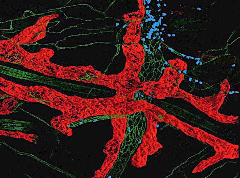
5 SUPPLEMENTARY FIGURES Figure S1 a PECAM-1 LYVE-1 MRP14 b PECAM-1 VE-Cadherin 5
6 Figure S1: Lymphatic architecture of the cremasteric tissue (a) Representative 3D-reconstructed confocal image of a whole-mount fixed cremaster muscle of a C57BL/6 WT animal and immunostained with antibodies against LYVE-1 (Alexa555-conjugated, red), PECAM-1 (Alexa488-conjugated, green) and MRP14 (Alexa647-conjugated, blue) to visualise the lymphatic vessels, the endothelial cell junctions and neutrophils, respectively, showing the presence of a fully developed lymphatic vasculature in this tissue with blind-ended lymphatic capillary vessels (arrow). (b) Representative confocal image of a fixed cremasteric lymphatic vessel immunostained for VE-Cadherin (Alexa555-conjugated, red) and PECAM-1 (Alexa647-conjugated, blue). The inserts on the right area show a magnified view of the heterogeneous distribution of the adhesion molecules (with VE-cadherin-rich buttons and PECAM-1-rich flaps) expressed by the endothelial cells of the cremasteric lymphatic vessels and exhibiting an oak-leaf shape morphology. Bar=20 µm. 6
7 Figure S2 Figure S2: Blood neutrophilia following stimulation of the cremaster muscles with CFA+Ag. Percentage of neutrophils with the blood circulation of WT mice subjected to cremaster muscle inflammation with CFA+Ag as analysed by flow cytometry. Data are expressed as mean±sem from 4 independent experiments. Statistically significant differences between the stimulated and control groups are indicated by asterisks: ***, P < 0.001; ****, P <
8 Figure S3 a b Figure S3: neutrophil migration response in the cremaster muscle following stimulation with CFA+Ag for 8hrs in WT, TNRdbKO and CCR7KO mice WT, TNFRdbKO and CCR7KO mice were subjected to CFA+Ag-induced inflammation of the cremaster muscles. Controlled mice were injected with PBS. Eight hours later, the cremaster muscles were dissected away, fixed and immunostained for LYVE-1, PECAM-1 and MRP14 to visualise the lymphatic vasculatures the endothelial cells junctions and neutrophils, respectively before analysis of the neutrophil migration responses by confocal microscopy. (a) Number of extravasated neutrophils in the cremaster muscles. (b) Number of neutrophils within the cremaster lymphatic vessels. Data are expressed as mean±sem from n = at least 4 animals per group. Statistically significant difference between stimulated and unstimulated animals are indicated by asterisks: **, P< 0.01; ***, P < 0.001; ****, P < Statistically significant difference between WT and KO animals are indicated by dashes: ##, P < 0.01; ###, P <

 and their surface expression of TNFR p55 and p75 were analysed by flow")
")
 or WT (WT WT) bone marrow hematopoietic cells (blue) and")
9 Figure S4 a (p75) TNFRdbKO WT WT WT b (p55) TNFRdbKO WT WT WT Figure S4: Phenotypic analysis of blood neutrophils from TNFRs chimeric animals Blood leukocytes from TNFRdbKO or chimeric animals were immunostained for neutrophils specific markers (CD45+ Ly6G+) and their surface expression of TNFR p55 and p75 were analysed by flow cytometry. The figure shows representative histograms of the fluorescence intensity for P75 (a) and P55 (b) of blood neutrophils from lethally irradiated WT mice and reconstituted with TNFRdbKO (TNFRdbKO WT) or WT (WT WT) bone marrow hematopoietic cells (blue) and compared to the intensity of staining from neutrophils of TNFRdbKO animals (as control, red). 9
10 Figure S5 a b c d e f g h Figure S5: Neither CXCL1:CXCR1/2 nor CXCL12:CXCR4 axes play a significant role in neutrophil migration across the lymphatic endothelium during antigen sensitisation. WT mice were subjected to CFA+Ag-induced inflammation of the cremaster muscles. Four hours later, mice received an i.s. injection of an anti-cxcl1 blocking mab (or an isotype control mab) or the CXCR4 specific inhibitor AMD3100 (or vehicle as control). At the end of the inflammation period, 10
11 cremaster muscles were dissected away, fixed and immunostained for LYVE-1, PECAM-1 and MRP14 to visualise the lymphatic vasculatures the endothelial cells junctions and neutrophils, respectively before analysis of the neutrophil migration responses by confocal microscopy. (a) Number of extravasated neutrophils in the cremaster muscles of unstimulated and CFA+Ag-stimulated (8hrs) mice injected with anti-cxcl1 blocking antibody or isotype control. (b) Number of neutrophils within the cremaster lymphatic vessels of unstimulated and CFA+Ag-stimulated (8hrs) mice injected with anti- CXCL1 blocking antibody or isotype control. (c) Number of neutrophils found in the dlns of the cremastermuscle of unstimulated and CFA+Ag-stimulated (8hrs) mice injected with anti-cxcl1 blocking antibody or isotype control. (d) Number of extravasated neutrophils in inflamed cremaster muscles of unstimulated and CFA+Ag-stimulated (16hrs) mice treated with AMD3100 or vehicle control. (e) Number of neutrophils within the cremaster lymphatic vessels of unstimulated and CFA+Ag-stimulated (16hrs) mice treated with AMD3100 or vehicle control. (f) Number of neutrophils found within the dlns of unstimulated and CFA+Ag-stimulated (16hrs) mice treated with AMD3100 or vehicle control. (g) Percentage of neutrophils into the blood circulation as assessed by flow cytometry. (h) Surface expression of CXCR4 (RFI) on blood circulating neutrophils as assessed by flow cytometry. Data are expressed as mean±sem from at least 4 independent experiments with n = 5-12 animals per group. Statistically significant difference between stimulated and unstimulated animals are indicated by asterisks: *, P< 0.05; **, P< 0.01; ***, P < 0.001; ****, P <
.")
12 Figure S6 Figure S6: low dose of TNFα induces CCR7 expression on blood neutrophils in vitro. Whole blood leukocytes from WT and CCR7KO animals were stimulated in vitro with different doses of TNFα for 4hrs in the presence of the endocytic inhibitor, nystatin (50 µm). Expression of CCR7 on the surface of neutrophils was then assessed by flow cytometry. Dotted line represent the RFI of the isotype control antibody. Data are expressed as mean±sem from 7 independent experiments (at least 7 mice per condition). Statistically significant difference specific CCR7 staining and isotope control antibody are indicated by asterisks: **, P< Data were analysed using a two-way analysis of variance (ANOVA), followed by Holm-Sidak's multiple comparisons test. Statistically significant difference TNFα-stimulated cells and PBS control group are indicated by dash symbols: #, P< 0.05, ### P < Statistically significant difference TNFα-stimulated cells and PBS control group are indicated by dagger symbol; P < ND: not done. 12
 was induced in WT and TNFRdbKO animals as well as in")
13 Figure S7: a b c f e d Figure S7: CXCL12:CXCR4 axis is responsible for the high number of neutrophils found into the lymph nodes of CCR7KO mice under steady state condition. Neutrophil migration into the lymphatic system of the cremaster muscle following antigen sensitisation with complete Freund s adjuvant (CFA+Ag) was induced in WT and TNFRdbKO animals as well as in chimeric animals exhibiting neutrophils deficient for CCR7. (a) Percentage of circulating neutrophils in naïve WT and CCR7KO animals, as quantified by flow cytometry. (b) Surface expression of CXCR4 on circulating neutrophils from WT and CCR7KO animals, as quantified by flow cytometry. (c) CXCL12 expression in the LNs of naïve WT and CCR7KO mice, as quantified by ELISA. (d) Number of extravasated neutrophils in inflamed cremaster muscles of unstimulated and CFA+Ag-stimulated CCR7KO mice treated with AMD3100 or the vehicle. (e) Number of neutrophils within the cremaster lymphatic vessels of unstimulated and CFA+Ag-stimulated CCR7KO mice treated with AMD3100 or the vehicle. (f) Number of neutrophils found into the dlns of unstimulated and CFA+Ag-stimulated CCR7KO mice treated with AMD3100 or the vehicle. Data are expressed as mean±sem with 5-12 animals per group. Statistically significant differences between stimulated and unstimulated treatment groups are indicated by asterisks: *, P < 0.05; **, P < Significant differences between responses PBS and AMD3100 treated groups are indicated by hash symbols: #, P <0.05; ##, P <
14 Figure S8 a b c d e Figure S8: CCL21 expression in the tissue. WT mice were subjected to CFA+Ag-induced inflammation of the cremaster muscles for 8hrs before harvesting the tissues for subsequent analysis for the expression of CCL21. (a) Quantification of CCL21 generation in the tissue of naïve and stimulated mice by ELISA. (b) WT mice subjected to CFA+Aginduced inflammation also received a local (i.s.) injection of the anti-tnfα blocking antibody (or an isotype control antibody, 50µg/mouse) before quantifying the expression of CCL21 by ELISA. (c) In some experiments, at the end of the inflammatory period (8hrs), tissues were fixed and whole-mount immunostained for Lyve-1 and CCL21 (or isotype control antibody) before being analysed by confocal 14
15 microscopy. The intensity of staining for CCL21 was then quantified within both the lymphatic vessels and the interstitial tissue using IMARIS software. (d) A line intensity profile was generated perpendicular to the direction of the vessel (10 lines/vessel) from the abluminal surface into the tissue (10 µm length) to look at a potential gradient of CCL21 guiding the leukocytes toward lymphatic vessels. Data are presented as percentage change from the first pixel of the line. (e) Similarly, a surface intensity profile within the lymphatic vessels was generated in the direction of the flow (100 µm length). Data are presented as percentage change from the first measured pixels of the surface. Data are expressed as mean±sem from at least with 4 animals per group (with at least 5 vessels per mouse fore confocal images). Statistically significant differences for CCL21 staining between the isotype control antibody vs. anti-ccl21 Ab (c) and for the intensity profiles from the first pixel to the last pixel measured (d & e) are indicated by asterisks: *, P < 0.05; **, P < 0.01; ****, P < Significant differences between the interstitial tissue vs. the lymphatic vessels and unstimulated vs. stimulated, are indicated by hash symbols: ####, P <
16 Figure S9 Figure S9: IL-17 and GM-CSF release upon antigen stimulation of the cremaster muscle. Time course of IL17 (left panel) and GM-CSF (right panel) generated in the cremaster muscles of WT mice following intra-scrotal injection of CFA+Ag and as quantified by ELISA. Data are expressed as mean±sem from 6-10 mice (from 4 independent experiments). 16
17 17
Suppl Video: Tumor cells (green) and monocytes (white) are seeded on a confluent endothelial
 and monocytes (white) are seeded on a confluent endothelial") Supplementary Information Häuselmann et al. Monocyte induction of E-selectin-mediated endothelial activation releases VE-cadherin junctions to promote tumor cell extravasation in the metastasis cascade
Supplementary Information Häuselmann et al. Monocyte induction of E-selectin-mediated endothelial activation releases VE-cadherin junctions to promote tumor cell extravasation in the metastasis cascade
Supplementary Information
 Nature Immunology doi:1.138/ni.2477 Supplementary Information Capillary and arteriolar pericytes attract innate leukocytes exiting through venules and instruct them with pattern recognition and motility
Supplementary Information. Tissue-wide immunity against Leishmania. through collective production of nitric oxide
 Supplementary Information Tissue-wide immunity against Leishmania through collective production of nitric oxide Romain Olekhnovitch, Bernhard Ryffel, Andreas J. Müller and Philippe Bousso Supplementary
Distinct Compartmentalization of the Chemokines CXCL1 and CXCL2 and the Atypical Receptor ACKR1 Determine Discrete Stages of Neutrophil Diapedesis
 Article Distinct Compartmentalization of the Chemokines CXCL1 and CXCL2 and the Atypical Receptor ACKR1 Determine Discrete Stages of Neutrophil Diapedesis Graphical Abstract Authors Tamara Girbl, Tchern
Supplementary Figure 1. Characterization of basophils after reconstitution of SCID mice
 Supplementary figure legends Supplementary Figure 1. Characterization of after reconstitution of SCID mice with CD4 + CD62L + T cells. (A-C) SCID mice (n = 6 / group) were reconstituted with 2 x 1 6 CD4
Evaluation of directed and random motility in microslides Assessment of leukocyte adhesion in flow chambers
 Evaluation of directed and random motility in microslides Motility experiments in IBIDI microslides, image acquisition and processing were performed as described. PMN, which ended up in an angle < 180
Supplementary material page 1/10
 Supplementary Figure 1. Metoprolol administration during ongoing AMI reduces MVO in STEMI patients (a, b) Complete representative CMR exams (short-axis covering the entire left ventricle (LV) from base
and follicular helper T cells is Egr2-dependent. (a) Diagrammatic representation of the
 Diagrammatic representation of the") Supplementary Figure 1. LAG3 + Treg-mediated regulation of germinal center B cells and follicular helper T cells is Egr2-dependent. (a) Diagrammatic representation of the experimental protocol for the
Supplementary Figure 1. LAG3 + Treg-mediated regulation of germinal center B cells and follicular helper T cells is Egr2-dependent. (a) Diagrammatic representation of the experimental protocol for the
Fisher et al. Supplemental Figure 1
 Supplemental Figure 1 A TNF IL-1 IL-6 CCL2 CCL5 CXCL10 pg/mg total protein 50 30 10 4,000 3,000 2,000 1,000 n.d. 1 1 14,000 12,000 10,000 8,000 6,000 4,000 2,000 6,000,000 CT26 5,000 16,000 B16 4,000 12,000
Supplementary Materials. for Garmy-Susini, et al, Integrin 4 1 signaling is required for lymphangiogenesis and tumor metastasis
 Supplementary Materials for Garmy-Susini, et al, Integrin 4 1 signaling is required for lymphangiogenesis and tumor metastasis 1 Supplementary Figure Legends Supplementary Figure 1: Integrin expression
Chronic variable stress activates hematopoietic stem cells
 SUPPLEMENTARY INFORMATION Chronic variable stress activates hematopoietic stem cells Timo Heidt *, Hendrik B. Sager *, Gabriel Courties, Partha Dutta, Yoshiko Iwamoto, Alex Zaltsman, Constantin von zur
Santulli G. et al. A microrna-based strategy to suppress restenosis while preserving endothelial function
 ONLINE DATA SUPPLEMENTS Santulli G. et al. A microrna-based strategy to suppress restenosis while preserving endothelial function Supplementary Figures Figure S1 Effect of Ad-p27-126TS on the expression
Supplementary Materials for
 www.sciencetranslationalmedicine.org/cgi/content/full/4/117/117ra8/dc1 Supplementary Materials for Notch4 Normalization Reduces Blood Vessel Size in Arteriovenous Malformations Patrick A. Murphy, Tyson
Supplementary Figure 1 Chemokine and chemokine receptor expression during muscle regeneration (a) Analysis of CR3CR1 mrna expression by real time-pcr
 Analysis of CR3CR1 mrna expression by real time-pcr") Supplementary Figure 1 Chemokine and chemokine receptor expression during muscle regeneration (a) Analysis of CR3CR1 mrna expression by real time-pcr at day 0, 1, 4, 10 and 21 post- muscle injury. (b)
Supplementary Figure 1 Chemokine and chemokine receptor expression during muscle regeneration (a) Analysis of CR3CR1 mrna expression by real time-pcr at day 0, 1, 4, 10 and 21 post- muscle injury. (b)
activation with anti-cd3/cd28 beads and 3d following transduction. Supplemental Figure 2 shows
 Supplemental Data Supplemental Figure 1 compares CXCR4 expression in untreated CD8 + T cells, following activation with anti-cd3/cd28 beads and 3d following transduction. Supplemental Figure 2 shows the
Supplementary information
 Supplementary information Intrahepatic myeloid cell-aggregates enable local CD8 + T cell expansion and successful immunotherapy against chronic viral liver infection Li- Rung Huang, Dirk Wohlleber, Florian
Nature Immunology: doi: /ni eee Supplementary Figure 1
 eee Supplementary Figure 1 Hyphae induce NET release, but yeast do not. (a) NET release by human peripheral neutrophils stimulated with a hgc1 yeast-locked C. albicans mutant (yeast) or pre-formed WT C.
Supplementary Figure 1. Generation of knockin mice expressing L-selectinN138G. (a) Schematics of the Sellg allele (top), the targeting vector, the
 Schematics of the Sellg allele (top), the targeting vector, the") Supplementary Figure 1. Generation of knockin mice expressing L-selectinN138G. (a) Schematics of the Sellg allele (top), the targeting vector, the targeted allele in ES cells, and the mutant allele in
Supplementary Figure 1. Generation of knockin mice expressing L-selectinN138G. (a) Schematics of the Sellg allele (top), the targeting vector, the targeted allele in ES cells, and the mutant allele in
% of live splenocytes. STAT5 deletion. (open shapes) % ROSA + % floxed
 % ROSA + % floxed") Supp. Figure 1. a 14 1 1 8 6 spleen cells (x1 6 ) 16 % of live splenocytes 5 4 3 1 % of live splenocytes 8 6 4 b 1 1 c % of CD11c + splenocytes (closed shapes) 8 6 4 8 6 4 % ROSA + (open shapes) % floxed
Supp. Figure 1. a 14 1 1 8 6 spleen cells (x1 6 ) 16 % of live splenocytes 5 4 3 1 % of live splenocytes 8 6 4 b 1 1 c % of CD11c + splenocytes (closed shapes) 8 6 4 8 6 4 % ROSA + (open shapes) % floxed
Chapter 3, Part A (Pages 37-45): Leukocyte Migration into Tissues
: Leukocyte Migration into Tissues") Allergy and Immunology Review Corner: Chapter 3, Part A (pages 37-45) of Cellular and Molecular Immunology (Seventh Edition), by Abul K. Abbas, Andrew H. Lichtman and Shiv Pillai. Chapter 3, Part A (Pages
Allergy and Immunology Review Corner: Chapter 3, Part A (pages 37-45) of Cellular and Molecular Immunology (Seventh Edition), by Abul K. Abbas, Andrew H. Lichtman and Shiv Pillai. Chapter 3, Part A (Pages
Brain barriers control immune cell trafficking to the CNS. Britta Engelhardt Theodor Kocher Institute University of Bern - Switzerland
 Brain barriers control immune cell trafficking to the CNS Britta Engelhardt Theodor Kocher Institute University of Bern - Switzerland The brain barriers 2 The BBB establishes a physical and metabolic barrier
B220 CD4 CD8. Figure 1. Confocal Image of Sensitized HLN. Representative image of a sensitized HLN
 B220 CD4 CD8 Natarajan et al., unpublished data Figure 1. Confocal Image of Sensitized HLN. Representative image of a sensitized HLN showing B cell follicles and T cell areas. 20 µm thick. Image of magnification
Supplemental Materials
 Supplemental Materials Programmed death one homolog maintains the pool size of regulatory T cells by promoting their differentiation and stability Qi Wang 1, Jianwei He 1, Dallas B. Flies 2, Liqun Luo
ECM1 controls T H 2 cell egress from lymph nodes through re-expression of S1P 1
 ZH, Li et al, page 1 ECM1 controls T H 2 cell egress from lymph nodes through re-expression of S1P 1 Zhenhu Li 1,4,Yuan Zhang 1,4, Zhiduo Liu 1, Xiaodong Wu 1, Yuhan Zheng 1, Zhiyun Tao 1, Kairui Mao 1,
CD4 and CD8 T cells show a similar accumulation in the tumor stroma.
 Fig S1 CD4 Fibronectin EpCM CD8 CD4 and CD8 T cells show a similar accumulation in the tumor stroma. Fluorescently-labeled CD4 (CMFD, green) and CD8 (Hoechst, yellow) T cells were added to a human lung
Macrophages form functional vascular mimicry channels in vivo. SI Figures and Legend
 Macrophages form functional vascular mimicry channels in vivo Authors: *Faith H. Barnett, *Mauricio Rosenfeld, Malcolm Wood, William Kiosses, Yoshihiko Usui, Valentina Marchetti, Edith Aguilar, and Martin
SUPPLEMENTARY INFORMATION
 doi:10.1038/nature10134 Supplementary Figure 1. Anti-inflammatory activity of sfc. a, Autoantibody immune complexes crosslink activating Fc receptors, promoting activation of macrophages, and WWW.NATURE.COM/NATURE
Thomas HAIDER Journal Club
 Thomas HAIDER Journal Club 20.10.2014 Background Immunology of the CNS - History Ehrlich, 1885 & 1904 dye did not stain brain -> BBB Shirai, Y. (1921) On the transplantation of the rat sarcoma in adult
Supplementary fig. 1. Crystals induce necroptosis does not involve caspases, TNF receptor or NLRP3. A. Mouse tubular epithelial cells were pretreated
 Supplementary fig. 1. Crystals induce necroptosis does not involve caspases, TNF receptor or NLRP3. A. Mouse tubular epithelial cells were pretreated with zvad-fmk (10µM) and exposed to calcium oxalate
Afferent lymph-derived T cells and dendritic cells use different CCR7-dependent routes for lymph node entry and intranodal migration
 Braun et al. Supplementary Information 1 Supplementary Information Afferent lymph-derived T cells and dendritic cells use different CCR7-dependent routes for lymph node entry and intranodal migration Asolina
Type of file: PDF Title of file for HTML: Supplementary Information Description: Supplementary Figures
 Type of file: PDF Title of file for HTML: Supplementary Information Description: Supplementary Figures Type of file: MOV Title of file for HTML: Supplementary Movie 1 Description: NLRP3 is moving along
Lymphoid architecture & Leukocyte recirculation. Thursday Jan 26th, 2017
 Lymphoid architecture & Leukocyte recirculation Thursday Jan 26th, 2017 Topics The life of immune cells Where are they born? Where are they educated? Where do they function? How do they get there? The
SUPPLEMENTARY METHODS
 SUPPLEMENTARY METHODS Histological analysis. Colonic tissues were collected from 5 parts of the middle colon on day 7 after the start of DSS treatment, and then were cut into segments, fixed with 4% paraformaldehyde,
Supplementary Figure 1. NAFL enhanced immunity of other vaccines (a) An over-the-counter, hand-held non-ablative fractional laser (NAFL).
 An over-the-counter, hand-held non-ablative fractional laser (NAFL).") Supplementary Figure 1. NAFL enhanced immunity of other vaccines (a) An over-the-counter, hand-held non-ablative fractional laser (NAFL). (b) Depiction of a MTZ array generated by NAFL. (c-e) IgG production
Supplementary Figure 1. NAFL enhanced immunity of other vaccines (a) An over-the-counter, hand-held non-ablative fractional laser (NAFL). (b) Depiction of a MTZ array generated by NAFL. (c-e) IgG production
Figure S1. Western blot analysis of clathrin RNA interference in human DCs Human immature DCs were transfected with 100 nm Clathrin SMARTpool or
 Figure S1. Western blot analysis of clathrin RNA interference in human DCs Human immature DCs were transfected with 100 nm Clathrin SMARTpool or control nontargeting sirnas. At 90 hr after transfection,
DC were seeded into tissue culture dishes in IMDM 2% FCS, and added with PMN. (1:1; PMN: DC) for 16h also in the presence of DNAse (100 U/ml); DC were
 for 16h also in the presence of DNAse (100 U/ml); DC were") Supplementary methods Flow cytometric analysis of DCs. DC were seeded into tissue culture dishes in IMDM 2% FCS, and added with PMN (1:1; PMN: DC) for 16h also in the presence of DNAse (100 U/ml); DC were
Supplementary methods Flow cytometric analysis of DCs. DC were seeded into tissue culture dishes in IMDM 2% FCS, and added with PMN (1:1; PMN: DC) for 16h also in the presence of DNAse (100 U/ml); DC were
Newly Recognized Components of the Innate Immune System
 Newly Recognized Components of the Innate Immune System NOD Proteins: Intracellular Peptidoglycan Sensors NOD-1 NOD-2 Nod Protein LRR; Ligand Recognition CARD RICK I-κB p50 p65 NF-κB Polymorphisms in Nod-2
SUPPLEMENTARY FIGURES
 SUPPLEMENTARY FIGURES Supplementary Figure 1: Chemokine receptor expression profiles of CCR6 + and CCR6 - CD4 + IL-17A +/ex and Treg cells. Quantitative PCR analysis of chemokine receptor transcript abundance
Supplementary Figures
 Supplementary Figures Supplementary Fig. 1. Galectin-3 is present within tumors. (A) mrna expression levels of Lgals3 (galectin-3) and Lgals8 (galectin-8) in the four classes of cell lines as determined
F-actin VWF Vinculin. F-actin. Vinculin VWF
 a F-actin VWF Vinculin b F-actin VWF Vinculin Supplementary Fig. 1. WPBs in HUVECs are located along stress fibers and at focal adhesions. (a) Immunofluorescence images of f-actin (cyan), VWF (yellow),
ILC1 and ILC3 isolation and culture Following cell sorting, we confirmed that the recovered cells belonged to the ILC1, ILC2 and
 Supplementary Methods and isolation and culture Following cell sorting, we confirmed that the recovered cells belonged to the, ILC2 and subsets. For this purpose we performed intracellular flow cytometry
The Immune Response in Time and Space
 The Immune Response in Time and Space Chapters 14 & 4 Sharon S. Evans, Ph.D. Department of Immunology 845-3421 sharon.evans@roswellpark.org September 18 & 23, 2014 Inflammation Inflammation Complex response
Supplemental Figure 1. Quantification of proliferation in thyroid of WT, Ctns -/- and grafted
 Supplemental Figure 1. Quantification of proliferation in thyroid of WT, Ctns -/- and grafted Ctns -/- mice. Cells immunolabeled for the proliferation marker (Ki-67) were counted in sections (n=3 WT, n=4
well for 2 h at rt. Each dot represents an individual mouse and bar is the mean ±
 Supplementary data: Control DC Blimp-1 ko DC 8 6 4 2-2 IL-1β p=.5 medium 8 6 4 2 IL-2 Medium p=.16 8 6 4 2 IL-6 medium p=.3 5 4 3 2 1-1 medium IL-1 n.s. 25 2 15 1 5 IL-12(p7) p=.15 5 IFNγ p=.65 4 3 2 1
Supplementary Figure 1. ETBF activate Stat3 in B6 and Min mice colons
 Supplementary Figure 1 ETBF activate Stat3 in B6 and Min mice colons a pstat3 controls Pos Neg ETBF 1 2 3 4 b pstat1 pstat2 pstat3 pstat4 pstat5 pstat6 Actin Figure Legend: (a) ETBF induce predominantly
CD4 T cell sphingosine 1-phosphate receptor (S1PR)1 and S1PR4 and endothelial S1PR2 regulate afferent lymphatic migration
1 and S1PR4 and endothelial S1PR2 regulate afferent lymphatic migration") LYMPHOCYTE MIGRATION CD4 T cell sphingosine 1-phosphate receptor (S1PR)1 and S1PR4 and endothelial S1PR2 regulate afferent lymphatic migration Yanbao Xiong 1,2 *, Wenji Piao 1,2 *, C. Colin Brinkman 2,
LYMPHOCYTE MIGRATION CD4 T cell sphingosine 1-phosphate receptor (S1PR)1 and S1PR4 and endothelial S1PR2 regulate afferent lymphatic migration Yanbao Xiong 1,2 *, Wenji Piao 1,2 *, C. Colin Brinkman 2,
ONLINE SUPPLEMENT MATERIAL. CD70 limits atherosclerosis and promotes macrophage function.
 ONLINE SUPPLEMENT MATERIAL CD7 limits atherosclerosis and promotes macrophage function. Holger Winkels* 1,2, Svenja Meiler* 1,2, Esther Smeets* 2, Dirk Lievens 1, David Engel 3, Charlotte Spitz 1, Christina
Supplementary Figure 1. Properties of various IZUMO1 monoclonal antibodies and behavior of SPACA6. (a) (b) (c) (d) (e) (f) (g) .
 (b) (c) (d) (e) (f) (g) .") Supplementary Figure 1. Properties of various IZUMO1 monoclonal antibodies and behavior of SPACA6. (a) The inhibitory effects of new antibodies (Mab17 and Mab18). They were investigated in in vitro fertilization
Supplementary Figure 1. Properties of various IZUMO1 monoclonal antibodies and behavior of SPACA6. (a) The inhibitory effects of new antibodies (Mab17 and Mab18). They were investigated in in vitro fertilization
Supplementary Materials for
 www.sciencemag.org/content/348/6241/aaa825/suppl/dc1 Supplementary Materials for A mucosal vaccine against Chlamydia trachomatis generates two waves of protective memory T cells Georg Stary,* Andrew Olive,
Figure S2
 Figure S1 Figure S2 Figure S3 Figure S4 Figure S5 Figure S6 SUPPLEMENTARY FIGURE LEGENDS Figure S1 Biosynthetic pathway for gangliosides showing activity of GalNAcT and GD3s and the structures of the major
Supplementary Figure 1. Nature Neuroscience: doi: /nn.4547
 Supplementary Figure 1 Characterization of the Microfetti mouse model. (a) Gating strategy for 8-color flow analysis of peripheral Ly-6C + monocytes from Microfetti mice 5-7 days after TAM treatment. Living
The encephalitogenicity of TH17 cells is dependent on IL-1- and IL-23- induced production of the cytokine GM-CSF
 CORRECTION NOTICE Nat.Immunol. 12, 568 575 (2011) The encephalitogenicity of TH17 cells is dependent on IL-1- and IL-23- induced production of the cytokine GM-CSF Mohamed El-Behi, Bogoljub Ciric, Hong
PHENOTYPIC DYNAMICS OF MICROGLIAL AND MONOCYTE-DERIVED CELLS IN GLIOBLASTOMA-BEARING MICE.
 SUPPLEMENTARY FIGURES, TABLES AND VIDEOS PHENOTYPIC DYNAMICS OF MICROGLIAL AND MONOCYTE-DERIVED CELLS IN GLIOBLASTOMA-BEARING MICE. Clément Ricard 1,2,3,4, Aurélie Tchoghandjian 2,4, Hervé Luche 5, Pierre
Nature Immunology: doi: /ni.3866
 Nature Immunology: doi:10.1038/ni.3866 Supplementary Figure 1 The effect of TIPE2 on chemotaxis. a, The expression of TIPE2 in dhl-60c, dhl-60t, TIPE2-expressing and 15/16Q-expressing dhl-60t neutrophils
TGF-β Signaling Regulates Neuronal C1q Expression and Developmental Synaptic Refinement
 Supplementary Information Title: TGF-β Signaling Regulates Neuronal C1q Expression and Developmental Synaptic Refinement Authors: Allison R. Bialas and Beth Stevens Supplemental Figure 1. In vitro characterization
Supporting Information Table of Contents
 Supporting Information Table of Contents Supporting Information Figure 1 Page 2 Supporting Information Figure 2 Page 4 Supporting Information Figure 3 Page 5 Supporting Information Figure 4 Page 6 Supporting
Eosinophils are required. for the maintenance of plasma cells in the bone marrow
 Eosinophils are required for the maintenance of plasma cells in the bone marrow Van Trung Chu, Anja Fröhlich, Gudrun Steinhauser, Tobias Scheel, Toralf Roch, Simon Fillatreau, James J. Lee, Max Löhning
McWilliams et al., http :// /cgi /content /full /jcb /DC1
 Supplemental material JCB McWilliams et al., http ://www.jcb.org /cgi /content /full /jcb.201603039 /DC1 THE JOURNAL OF CELL BIOLOGY Figure S1. In vitro characterization of mito-qc. (A and B) Analysis
Leukocyte rolling and adhesion both contribute to regulation of microvascular permeability to albumin via ligation of ICAM-1
 Am J Physiol Cell Physiol 301: C804 C813, 2011. First published June 8, 2011; doi:10.1152/ajpcell.00135.2011. Leukocyte rolling and adhesion both contribute to regulation of microvascular permeability
Supplemental Material
 Supplemental Material Supplementary Fig. 1. EETs stimulate primary tumor growth. a) Schematic presentation of genetic and pharmacological tools used to manipulate endogenous EET levels. b) Endothelial
Hua Tang, Weiping Cao, Sudhir Pai Kasturi, Rajesh Ravindran, Helder I Nakaya, Kousik
 SUPPLEMENTARY FIGURES 1-19 T H 2 response to cysteine-proteases requires dendritic cell-basophil cooperation via ROS mediated signaling Hua Tang, Weiping Cao, Sudhir Pai Kasturi, Rajesh Ravindran, Helder
Supplementary Figure 1. Deletion of Smad3 prevents B16F10 melanoma invasion and metastasis in a mouse s.c. tumor model.
 A B16F1 s.c. Lung LN Distant lymph nodes Colon B B16F1 s.c. Supplementary Figure 1. Deletion of Smad3 prevents B16F1 melanoma invasion and metastasis in a mouse s.c. tumor model. Highly invasive growth
Loss of HS1 inhibits neutrophil extravasation during. inflammation via disturbed PKA signaling. IV. Immunology Summit, Houston,
 Loss of HS1 inhibits neutrophil extravasation during inflammation via disturbed PKA signaling IV. Immunology Summit, Houston, 29.09.2015 Michael Schnoor, PhD Center for Research and Advanced Studies, (Cinvestav-IPN),
SUPPLEMENTARY INFORMATION
 doi:1.138/nature1554 a TNF-α + in CD4 + cells [%] 1 GF SPF 6 b IL-1 + in CD4 + cells [%] 5 4 3 2 1 Supplementary Figure 1. Effect of microbiota on cytokine profiles of T cells in GALT. Frequencies of TNF-α
W/T Itgam -/- F4/80 CD115. F4/80 hi CD115 + F4/80 + CD115 +
 F4/8 % in the peritoneal lavage 6 4 2 p=.15 n.s p=.76 CD115 F4/8 hi CD115 + F4/8 + CD115 + F4/8 hi CD115 + F4/8 + CD115 + MHCII MHCII Supplementary Figure S1. CD11b deficiency affects the cellular responses
Supplementary Figure 1
 Supplementary Figure 1 Identification of IFN-γ-producing CD8 + and CD4 + T cells with naive phenotype by alternative gating and sample-processing strategies. a. Contour 5% probability plots show definition
Supplementary Figure 1. Characterization of NMuMG-ErbB2 and NIC breast cancer cells expressing shrnas targeting LPP. NMuMG-ErbB2 cells (a) and NIC
 and NIC") Supplementary Figure 1. Characterization of NMuMG-ErbB2 and NIC breast cancer cells expressing shrnas targeting LPP. NMuMG-ErbB2 cells (a) and NIC cells (b) were engineered to stably express either a LucA-shRNA
Supplementary Figure 1. Characterization of NMuMG-ErbB2 and NIC breast cancer cells expressing shrnas targeting LPP. NMuMG-ErbB2 cells (a) and NIC cells (b) were engineered to stably express either a LucA-shRNA
Aquaporin-9-expressing neutrophils are required for the establishment of contact hypersensitivity
 Supplemental Material quaporin-9-expressing neutrophils are required for the establishment of contact hypersensitivity atharina Sagita Moniaga 1, Sachiko Watanabe 1, Tetsuya Honda 1, 2, Søren Nielsen 3,
(A) RT-PCR for components of the Shh/Gli pathway in normal fetus cell (MRC-5) and a
 RT-PCR for components of the Shh/Gli pathway in normal fetus cell (MRC-5) and a") Supplementary figure legends Supplementary Figure 1. Expression of Shh signaling components in a panel of gastric cancer. (A) RT-PCR for components of the Shh/Gli pathway in normal fetus cell (MRC-5) and
Supplementary figure legends Supplementary Figure 1. Expression of Shh signaling components in a panel of gastric cancer. (A) RT-PCR for components of the Shh/Gli pathway in normal fetus cell (MRC-5) and
Pearson r = P (one-tailed) = n = 9
 = n = 9") 8F4-Specific Lysis, % 1 UPN1 UPN3 8 UPN7 6 Pearson r =.69 UPN2 UPN5 P (one-tailed) =.192 4 UPN8 n = 9 2 UPN9 UPN4 UPN6 5 1 15 2 25 8 8F4, % Max MFI Supplementary Figure S1. AML samples UPN1-UPN9 show variable
8F4-Specific Lysis, % 1 UPN1 UPN3 8 UPN7 6 Pearson r =.69 UPN2 UPN5 P (one-tailed) =.192 4 UPN8 n = 9 2 UPN9 UPN4 UPN6 5 1 15 2 25 8 8F4, % Max MFI Supplementary Figure S1. AML samples UPN1-UPN9 show variable
Nature Immunology: doi: /ni Supplementary Figure 1. Examples of staining for each antibody used for the mass cytometry analysis.
 Supplementary Figure 1 Examples of staining for each antibody used for the mass cytometry analysis. To illustrate the functionality of each antibody probe, representative plots illustrating the expected
SUPPLEMENTARY INFORMATION. Supp. Fig. 1. Autoimmunity. Tolerance APC APC. T cell. T cell. doi: /nature06253 ICOS ICOS TCR CD28 TCR CD28
 Supp. Fig. 1 a APC b APC ICOS ICOS TCR CD28 mir P TCR CD28 P T cell Tolerance Roquin WT SG Icos mrna T cell Autoimmunity Roquin M199R SG Icos mrna www.nature.com/nature 1 Supp. Fig. 2 CD4 + CD44 low CD4
VEGFR2-Mediated Vascular Dilation as a Mechanism of VEGF-Induced Anemia and Bone Marrow Cell Mobilization
 Cell Reports, Volume 9 Supplemental Information VEGFR2-Mediated Vascular Dilation as a Mechanism of VEGF-Induced Anemia and Bone Marrow Cell Mobilization Sharon Lim, Yin Zhang, Danfang Zhang, Fang Chen,
Supplementary Figure 1
 Supplementary Figure 1 AAV-GFP injection in the MEC of the mouse brain C57Bl/6 mice at 4 months of age were injected with AAV-GFP into the MEC and sacrificed at 7 days post injection (dpi). (a) Brains
Figure S1: Effects on haptotaxis are independent of effects on cell velocity A)
") Supplemental Figures Figure S1: Effects on haptotaxis are independent of effects on cell velocity A) Velocity of MV D7 fibroblasts expressing different GFP-tagged Ena/VASP family proteins in the haptotaxis
Supplemental Figures Figure S1: Effects on haptotaxis are independent of effects on cell velocity A) Velocity of MV D7 fibroblasts expressing different GFP-tagged Ena/VASP family proteins in the haptotaxis
Supplementary Figure 1. Expression of EPO and EPOR during self-limited versus delayed
 Supplementary Figure 1. Expression of EPO and EPOR during self-limited versus delayed inflammation resolution. a: Flow cytometry analysis showing the electronic gating strategy used to identify peritoneal
Immune response to infection
 Immune response to infection Dr. Sandra Nitsche (Sandra.Nitsche@rub.de ) 20.06.2018 1 Course of acute infection Typical acute infection that is cleared by an adaptive immune reaction 1. invasion of pathogen
Supplemental Table 1. Primer sequences for transcript analysis
 Supplemental Table 1. Primer sequences for transcript analysis Primer Sequence (5 3 ) Primer Sequence (5 3 ) Mmp2 Forward CCCGTGTGGCCCTC Mmp15 Forward CGGGGCTGGCT Reverse GCTCTCCCGGTTTC Reverse CCTGGTGTGCCTGCTC
Supplementary Figure 1. SA-β-Gal positive senescent cells in various cancer tissues. Representative frozen sections of breast, thyroid, colon and
 Supplementary Figure 1. SA-β-Gal positive senescent cells in various cancer tissues. Representative frozen sections of breast, thyroid, colon and stomach cancer were stained with SA-β-Gal and nuclear fast
In Vivo Imaging of Virological Synapses
 In Vivo Imaging of Virological Synapses Xaver Sewald 1, David G. Gonzalez 2, Ann M. Haberman 2, and Walther Mothes 1 * 1 Department of Microbial Pathogenesis, Yale University School of Medicine, New Haven,
Sharon S. Evans, Ph.D. Dept. Immunology, RPCI Jan. 29 & Feb 3, 2015
 Adhesion Molecules And Lymphocyte Homing Sharon S. Evans, Ph.D. Dept. Immunology, RPCI Jan. 29 & Feb 3, 2015 Outline General introduction to lymphocyte trafficking homeostatic recirculation vs active recruitment.
Nature Immunology: doi: /ni Supplementary Figure 1. Production of cytokines and chemokines after vaginal HSV-2 infection.
 Supplementary Figure 1 Production of cytokines and chemokines after vaginal HSV-2 infection. C57BL/6 mice were (a) treated intravaginally with 20 µl of PBS or infected with 6.7x10 4 pfu of HSV-2 in the
Programmed necrosis, not apoptosis, is a key mediator of cell loss and DAMP-mediated inflammation in dsrna-induced retinal degeneration
 Programmed necrosis, not apoptosis, is a key mediator of cell loss and DAMP-mediated inflammation in dsrna-induced retinal degeneration The Harvard community has made this article openly available. Please
Dual Targeting Nanoparticle Stimulates the Immune
 Dual Targeting Nanoparticle Stimulates the Immune System to Inhibit Tumor Growth Alyssa K. Kosmides, John-William Sidhom, Andrew Fraser, Catherine A. Bessell, Jonathan P. Schneck * Supplemental Figure
Pathologic Stage. Lymph node Stage
 ASC ASC a c Patient ID BMI Age Gleason score Non-obese PBMC 1 22.1 81 6 (3+3) PBMC 2 21.9 6 6 (3+3) PBMC 3 22 84 8 (4+4) PBMC 4 24.6 68 7 (3+4) PBMC 24. 6 (3+3) PBMC 6 24.7 73 7 (3+4) PBMC 7 23. 67 7 (3+4)
Supplementary information. The proton-sensing G protein-coupled receptor T-cell death-associated gene 8
 1 Supplementary information 2 3 The proton-sensing G protein-coupled receptor T-cell death-associated gene 8 4 (TDAG8) shows cardioprotective effects against myocardial infarction 5 Akiomi Nagasaka 1+,
Diphtheria toxin-mediated ablation of lymphatic endothelial cells results in progressive lymphedema
 Diphtheria toxin-mediated ablation of lymphatic endothelial cells results in progressive lymphedema Jason C. Gardenier 1, Geoffrey E. Hespe 1, Raghu P. Kataru 1, Ira L. Savetsky 1, Jeremy S. Torrisi 1,
Supplementary Figures
 Supplementary Figures Supplementary Figure 1. NKT ligand-loaded tumour antigen-presenting B cell- and monocyte-based vaccine induces NKT, NK and CD8 T cell responses. (A) The cytokine profiles of liver
Supplementary Figures
 Supplementary Figures Supplementary Fig. 1. Surface thiol groups and reduction of activated T cells. (a) Activated CD8 + T-cells have high expression levels of free thiol groups on cell surface proteins.
Question 1. Kupffer cells, microglial cells and osteoclasts are all examples of what type of immune system cell?
 Abbas Chapter 2: Sarah Spriet February 8, 2015 Question 1. Kupffer cells, microglial cells and osteoclasts are all examples of what type of immune system cell? a. Dendritic cells b. Macrophages c. Monocytes
Fang et al. NMuMG. PyVmT unstained Anti-CCR2-PE MDA-MB MCF MCF10A
 A NMuMG PyVmT 16.5+.5 47.+7.2 Fang et al. unstained Anti-CCR2-PE 4T1 Control 37.6+6.3 56.1+.65 MCF1A 16.1+3. MCF-7 3.1+5.4 MDA-M-231 42.1+5.5 unstained Secondary antibody only Anti-CCR2 SUPPLEMENTAL FIGURE
Supplementary Figure S1. PTPN2 levels are not altered in proliferating CD8+ T cells. Lymph node (LN) CD8+ T cells from C57BL/6 mice were stained with
 CD8+ T cells from C57BL/6 mice were stained with") Supplementary Figure S1. PTPN2 levels are not altered in proliferating CD8+ T cells. Lymph node (LN) CD8+ T cells from C57BL/6 mice were stained with CFSE and stimulated with plate-bound α-cd3ε (10µg/ml)
Supplementary Figure S1. PTPN2 levels are not altered in proliferating CD8+ T cells. Lymph node (LN) CD8+ T cells from C57BL/6 mice were stained with CFSE and stimulated with plate-bound α-cd3ε (10µg/ml)
Supplementary Information:
 Supplementary Information: Follicular regulatory T cells with Bcl6 expression suppress germinal center reactions by Yeonseok Chung, Shinya Tanaka, Fuliang Chu, Roza Nurieva, Gustavo J. Martinez, Seema
a surface permeabilized
 a surface permeabilized RAW 64.7 P388D1 J774 b CD11b + Ly-6G - Blood Monocytes WT Supplementary Figure 1. Cell surface expression on macrophages and DCs. (a) RAW64.7, P388D1, and J774 cells were subjected
Supplementary Materials for
 www.sciencesignaling.org/cgi/content/full/8/381/ra59/dc1 Supplementary Materials for Analysis of single-cell cytokine secretion reveals a role for paracrine signaling in coordinating macrophage responses
Supporting Information
 Supporting Information Aldridge et al. 10.1073/pnas.0900655106 Fig. S1. Flow diagram of sublethal (a) and lethal (b) influenza virus infections. (a) Infection of lung epithelial cells by influenza virus
Supplemental Figure 1. Cell-bound Cetuximab reduces EGFR staining intensity. Blood
 Antibody-mediated depletion of CD19-CAR T cells Supplemental 1 Supplemental Materials Supplemental Figure 1. Supplemental Figure 1. Cell-bound Cetuximab reduces EGFR staining intensity. Blood cells were
MATERIALS AND METHODS. Neutralizing antibodies specific to mouse Dll1, Dll4, J1 and J2 were prepared as described. 1,2 All
 MATERIALS AND METHODS Antibodies (Abs), flow cytometry analysis and cell lines Neutralizing antibodies specific to mouse Dll1, Dll4, J1 and J2 were prepared as described. 1,2 All other antibodies used
Supplementary Materials
 Supplementary Materials 43 Figure S1. CD123 in acute lymphoblastic leukemia and leukemia-initiating cells. A. CD123 (histograms) is highly and homogenously expressed in B-ALL blasts (as defined by live,