THE ULTRASTRUCTURE OF LEISHMANIA DONOVANI
|
|
|
- Arthur Campbell
- 6 years ago
- Views:
Transcription

1 University of Nebraska - Lincoln DigitalCommons@University of Nebraska - Lincoln Journal of Parasitology Archives Parasitology, Harold W. Manter Laboratory of April 1956 THE ULTRASTRUCTURE OF LEISHMANIA DONOVANI Patricia C. H. Chang The Johns Hopkins University, Baltimore, Maryland Follow this and additional works at: Part of the Parasitology Commons Chang, Patricia C. H., "THE ULTRASTRUCTURE OF LEISHMANIA DONOVANI" (1956). Journal of Parasitology Archives This Article is brought to you for free and open access by the Parasitology, Harold W. Manter Laboratory of at DigitalCommons@University of Nebraska - Lincoln. It has been accepted for inclusion in Journal of Parasitology Archives by an authorized administrator of DigitalCommons@University of Nebraska - Lincoln.
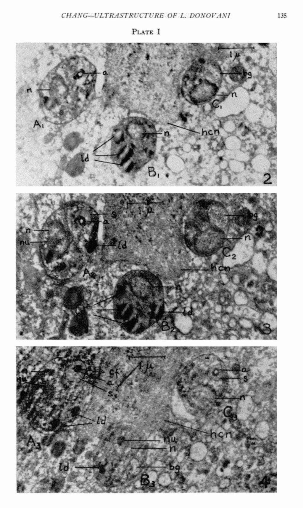
2 THE ULTRASTRUCTURE OF LEISHMANIA DONOVAN1 The structure of the intracellular forin of Leishmania donovani as seen by the light microscope consists of a nucleus and usually a rod-like kinetoplast (Wenyon, 1926) within a homogeneous mass of cytoplasm, while the extracellular or culture form has, in addition to these structures, an anterior flagellum. The intracellular form is usually round or ovoid measuring 24 microns; the extracellular form measures about microns in length and from microns in breadth. Because of the small dimensions of the protozoan parasite and the limited resolution of the light microscope, relatively little information is available on its fundamental organization. With the great resolving power of the electron microscope, several workers have attempted to study its ultrastructure with this instrument. The studies of Emmel, Jakob and Golz (1942), Sen Gupta et d. (1951), Das Gupta et al. (1954) of this species, and Lofgren (1950) of Leishmania tropica using electron microscopy of the culture forms have failed to yield new information on the fine internal morphology because the parasites were examined as whole specimens. The thickness of the specimens does not allow for adequate electron beam penetration, and the parasites appear as dense masses. In this study however, this problem is eliminated by the use of thin sectioning. Fine structural detail is observed in these preparations in both the extra- and intracellular forms. The accumulation of structural knowledge may enable us to understand the relationship of host cells and leishmania ; i.e., the biology of this intracellular parasitism. This paper presents only a few observations of this host-parasite relationship and further work is necessary to make a thorough study of this problem. MATERIALS AND METHODS Intracellular parasites were obtained from infected hamster spleen and the extracellular forms from NIH diphasic medium cultures. The culture forms were washed in Geys' salt solution (Gey and Gey, 1936) before fixation. The culture forn~s and infected spleen material were fixed in 1% veronal-buffered osmium tetraoxide, ph (Palade, 1952), dehydrated by a graded series of ethyl alcohol and embedded in a partially polymerized mixture of one part of methyl to four parts of butyl methacrylate. The embeddings were incubated in the oven overnight at 45 degrees centigrade, sectioned with glass knives on a Servall microtome, mounted on screen and examined in a RCA, Model EMU electron microscope. OBSERVATIONS Intracellular Form The three leishman bodies shown in Figs. 2, 3 and 4 consist of three serial sections of the same three parasites within the cytoplasm of the host cell in hamster spleen. These three parasites are lying in close proximity to the nucleus of the host cell and will hereafter be referred to as Leishmania A, B and C. Received for publication, October 26, *Present address : School of Hygiene and Public Health, The Johns Hopkins University, Baltimore, Maryland. 126
3 CHANG-ULTRASTRUCTURE OF L. DONOVANI 127 Leishmania A. Fig. 2 shows a leishman body with a cell membrane which is distinct from the cytoplasm of the host cell. At this level of sectioning, the leishman body reveals the following structures : 1) A nucleus with fine, granular nucleoplasm surrounded by a nuclear membrane. The nucleoplasm is a homogeneous mass with a clear vacuole on one side. No evidence of a nucleolus or karyosome is recognized in this body. 2) A dense ring with a hollow center whose structure and relationship will be clearly revealed in Figs. 3 and 4 as the axoneme of the reduced flagellum. 3) Three dense, structureless droplets, unequal in size, are tentatively regarded as fat bodies. Droplets similar to these have been described in Try~anosoma cruzi Fig. 1 Diagrammatic interpretation of the proximal flagellum and basal granule structures of the leishman body. The structures observed in serial sections of Figs. 2, 3 and 4 are interpreted with reference to Fig. 1 in the following manner : Leishmania A. Fig. 2 represents a cross section at the level which corresponds to the junction of two adjacent coils of the sheath. The amount of protoplasm enclosed by the sheath membrane is therefore reduced to a minimum making it barely visible. Enclosed within the sheath is the dense axoneme, which appears as a dense ring in cross section. Figs. 3 and 4 are serial sections distal to the basal granule. Fig. 3 shows a cross section of the axoneme and one coil of the sheath. Fig. 4 shows the cross section of the axoneme and two opposite coils of the sheath, one on each side of the axoneme which appears as one continuous circular tube. Leishmania B. Figs. 2 and 3 are sections below the level of the basal granule. In Fig. 4, a small portion of the basal granule is sectioned giving the semi-circular appearance. Leishmania C. Figs. 2 and 3 represent cross sections of the basal granule in the transitional region from which the extended flagellum arises. Fig. 4 represents the first cross section of the flagellum immediate to the basal granule. The three leishman bodies represent three different levels of sectioning through the cell. The presence and absence of certain structures in certain cells would be very difficult to interpret without the aid of serial sections.
 Vacuoles of various sizes are visible in the fine granular cytoplasm. These vacuoles are distributed at random throughout the cell. In Fig.")
4 128 THE JOURNAL OF PARASITOLOGY by Meyer and Porter (1954), and they found that the content of t1.1esc 1,oclies could be removed by xylene. 4) Vacuoles of various sizes are visible in the fine granular cytoplasm. These vacuoles are distributed at random throughout the cell. In Fig. 3, a follow-up on the continuity of the structures described in Fig. 2 reveals the following changes : 1) The nucleus shows up more clearly with a double membrane enclosing the nucleoplasm. The nucleoplasm is finely granular and "patchy," and lying in an eccentric position is an aggregate of densely packed, rather homogeneous granules which is considered to be the nucleolus or karyosome. The structure of nucleoli has been studied by Borysko and Bang (1951). They found a wide variety of forms; included was a homogeneous nucleolus of an eildothelial cell which resembled that observed in Leishmania donovani. 2) The dense ring with the hollow center (axoneme) is still present; however, in addition to this structure, there is another less dense granular ring partially surrounding the axoneme. This is analogous to the sheath of several protozoan flagella described by Brown (1945) and of Trypanoso~wa cruai by Meyer and Porter (1954). 3) The three dense droplets are still present and they are lying in the same relative positions within the leishman body. 4) Vacuoles of various size are still seen and are distributed at random throughout the cytoplasl~~. Fig. 4 shows a very interesting developn~ent of two iiuportant structures of the organism. 1) The nucleus has a nucleolus within it. The nucleoplasm is finely granular and aggregates of nucleoplasm originating from the central nucleolus form a linear, ray-like pattern within the nucleus. 2) The dense ring structure (axoneme) now has a central body within it. This central body is very similar to the internal filaments of several cilia types studied by Fawcett and Porter (1954). The sheath with its well defined membrane encircling the entire axonetne is a coiled structure. The transition of the varying size and thickness of the sheath may be clarified in the diagrammatic interpretation shown in Fig. 1. 3) Only two of the fat droplets are present. 4) The cytoplasmic vacuoles appear as described above. Leishmania B. In Fig. 2 this cell consists of a nucleus without a nucleolus and four fat droplets. The cytoplasm is a homogeneous mass of fine granules enclosed in a double membrane. The outer membrane seems to be a smooth layer, but the inner layer seems to be composed of evenly-spaced fine ridges. Fig. 5 shows this structure very clearly. Fig. 3 shows a similar picture of all structures as described in Fig. 2. Fig. 4 shows changes and addition of structures described in Figs. 2 and 3. The nucleus now has an eccentrically located nucleolus, which appears as a dense homogeneous structure composed of fine granules. There is a semicircular ring with less dense cytoplasm distinct from the cell cytoplasm. There is the appearance of a structure not observed in the cells described above. A reconstruction of the flagellum structure, Fig. 1, and an electron micrograph of a longitudinal section of the extended flagellum of a leptomonad form indicates the relationship of such a structure. This structure is a section of the base of the basal granule. Fat bodies and vacuoles are present in the cell cytoplasm. The double membrane with a ridged inner surface is still evident here. Leishmania C. In Fig. 2, two prominent ovoid structures almost equal in size are clearly seen in this cell. Although both these structures are composed of fine granular substance, they are distinctly different from each other. One of them is

5 CHANG-ULTRASTRUCTURE OF L. DONOVANI 129 easily recognized as the nucleus and the other is identified as the basal granule from which the flagellum arises. Each has its own distinct membrane. The nucleus shows a denser nucleoplasm at the periphery of the nuclear membrane than in the central area and there is no evidence of a nucleolus at this level. One dense fat body and several small vacuoles are scattered throughout the cytoplasm. The cell membrane is a double one. In Fig. 3, the dimensions of the cell nucleus and basal granule are increased. The nucleoplasm is denser and the fine granules are evenly distributed throughout, but there is still no evidence of a nucleolus. The granules in the basal granule are very sharp and distinct; they are centrally located and spread out as a band on the long axis of the basal granule. In Fig. 4, the structures in the cell show a marked change froin the previous series. The'nucleus has a fairly hon~ogeneous nucleoplasm and a faint indication that a very small portion of the nucleolus is present. The basal granule is no longer seen; in its place the dense ring of the axoneme is completely surrounded by a sheath. The flagellar structure has arisen from the basal granule. Again, reference to Fig. 1 will clarify the reconstruction of the basal granule-flagellum relationship. Vacuoles of various sizes are seen throughout the cytoplasm. The single fat droplet in Fig. 2 and 3 is no longer visible; instead, one new droplet may be located near the nucleus. The double meinbrane which surrounds the cell seems to be separated at one end of the cell: the inner layer still surrounds the usual dense cytoplasm of the cell; the outer layer seems to extend at one end of the cell and encloses a relatively clear cytoplasm. This phenomenon has been observed among other leishman bodies; Fig. 6 illustrates another such observation. The significance of this clear area is not apparent. Further study is necessary to clarify these random observations. Extracellular Form Fig. 7 shows two leptoinonad forms cut at different angles. The cell above shows a rounded basal granule which becomes constricted at one end to give rise to the flagellum. The flagellum consists of a cylindrical tube-like axoneme which extends beyond the cell membrane as a free flagellum. A central filament within the axoneme begins at a short distance froin the basal granule and continues along with the axoneme. Surrounding the axoneme is a loosely coiled sheath which begins at the base of the axoneme and continues throughout its entire length. The cytoplasm is granular and there is no evidence of fat bodies or vacuoles. The nucleus is not visible because of the level of the section. The cell below is a longitudinal section. The cytoplasm is granular and homogeneous throughout the cell. Within this cell are the nucleus, the basal granule, axoneme and sheath. The nucleus has a nuclear membrane and within it is a dense, homogeneous nucleolus. The basal granule is a rounded body, and within it is a band of uniform short filaments at right angles to the axoneme. The significance of this band is obscure. The axoneme and sheath are present but only to the edge of the cell membrane; the free end of the flagellum has been deleted at this level of sectioning. The evidence of "cell wall" with fine, longitudinal striations in Leishmania tropica described by Lofgren (1954), the sub-pellicle fibers or striations over the body surface of Trypanosoma cruzi described by Meyer and Porter (1954) and
 are not clearly indicated in this electron micrograph, probably because the techniques employed by these workers were different from those eillployed here.")
6 130 THE JOURNAL OF PARASITOLOGY the myoneme fibrils of Leishnzania donovani noted by Das Gupta et al. (1954) are not clearly indicated in this electron micrograph, probably because the techniques employed by these workers were different from those eillployed here. DISCUSSION The technique of thin sectioning of Leishn$ania donovani for electron microscopy has revealed some details of morphology not obtainable by the whole-mount techniques employed by previous workers. No drastic treatiment of specimens by acid hydrolysis (Das Gupta et al., 1954) or by osinotic rupture by distilled water (Lofgren, 1950) were used in this study. Thus, artefacts have been minimized. Since the structures of the leishillan body and the leptoinonad forin are siiuilar, the morphology of these structures inay be compared. In the Protozoa, the limiting layer which gives the organisin its shape has been variously called the pellicle or periplast. It may be smooth or ridged. In the studies of several flagellates by electron microscopy, Saxe (1947) described the "spiral striations of the pellicle" in Euglena gracilis. Lofgren (1950) described "the cell wall of the Leishmania cells possessed fine, longitudinal striations." Kleinschmidt and Kinder (1950) found fibrillar structure of the periplast in Trypanosoma lewsi and Trypanosoma brucei. Kraneveld, Houwink and Keidal (1951) found that the periplast contains longitudinal and nearly parallel fibrils in Trypanosoma evansi. Wolken and Palade (1953) reported a ridged pellicle of Euglena gracilis. Meyer and Porter (1954) observed a body nlembrane with fibrils in the cytoplasm just beneath the meinbrane in Trypanosmza cruzi. Das Gupta et al. (1954) described "longitudinal myoneme fibrils arranged in interconnecting bundles in the periplast" of Leishmania donovarzi. These observations on the fibrillar nature of the cell meinbrane in a variety of flagellates indicate a close similarity of the cell membrane structure among them, whether they be free-living or parasitic. Most of thse observations were made on whole specimens, and in the observation of sectioned material by Wolken and Palade (1953), the ridged pellicle of Euglena was like a "scalloped" edge on the outside of the cell membrane. The observations of the intracellular ph,ase of Leishmania donovani in the present study reveal a double cell meinbrane, with a smooth layer on the outside and a finely ridged surface in the inner layer. This conforills with the description given by Meyer and Porter (1954) of Trypanoso+$za cruzi. The nuclei of Protozoa, like other types of cell nuclei, are bounded by distinct nuclear membranes. The workers using whole mount technique have provided little or no information on the structure of the nucleus because the organisms are usually too dense for adequate electron bean1 penetration. By using the HCl hydrolysis technique on Leishmania donovani; Das Gupta et al. (1954) obtained electron micrographs showing an oval body that is less dense than the rest of the cytoplasm. A small dark body within the nucleus is presumed to be the karyosome. Sectioned material provides a much clearer picture of this structure. The study of Toxoplasma by Gustafson et al. (1954) shows the nuclei in a variety of shapes, but all show a consistent dense inass within the nucleus. The present study of Leishmania donovani confirms the presence of the nuclear menlbrane; and in optimum sections, a double nuclear membrane may be seen. Within the resting nucleus, a karyosome may be seen. The absence of the karyosome in some cells is n10st probably due to the level of sectioning.
, in study of various flagella, found that the axoneine was composed of fibrils.")
7 CHANG-ULTRASTRUCTURE OF L. DONOVANI 131 Many studies have been made on the flagellar structure in parasitic and freeliving flagellates. All reveal the same basic structure, consisting of an internal axoneme and a sheath surrounding it. Brown (1945), in study of various flagella, found that the axoneine was composed of fibrils. He was uncertain as to the number which constitute an axoneme. Saxe (1947) also reported a number of longitudinal components in the axoneme of Euglena gracilis. Lofgren (1950) in a study of Leishmania tropica, Kleinschmidt and Kinder (1950) in studies of Trypanoso~?za Zewsi and Trypanoso~~za brucei, and Kleinschmidt and Schleich (1951) in studies of Trypanosowza brucei, reported that the axoneme was composed of fibrillae. No mention was made of the number which make up the axoneme. There are other workers who give numerical counts on the fibrils constituting the axoneme. The numbers vary because each study was of a different flagellate. Whether this is a true difference between species is difficult to evaluate at this time. Until improvements in techniques allow good, clearcut electron micrographs, we cannot establish whether the fibrils which constitute an axoneme have a basic number or vary with the organism. Examples of this controversy will be presented as follows: Kraneveld et al. (1951) reported nine parallel isodiainetric fibrils in the axoneme of Trypanoso$$za evansi, Wolken and Palade (1953) found eleven elementary filaments in Euglena and Poteriochromonas in their axonemata. Meyer and Porter (1954) found five or nine fibrils in the axoneme of Trypanoso~iza cruzi. Das Gupta et al. (1954) found a maxiinuin of nine fibrils in Leishvrzania donovani. In the present study of Leish~nania donovani, the fibrillar structure is confirmed; however, the exact number is not readily counted. The fact remains that the axoneme is made up of fibrils. Besides the fibrillar axoneine of flagellates, studies of Schmitt, Hall and Jakus (1943) and Schmitt (1944) show that sperm tails 'and cilia have similar ultrastructure. Their observations showed the presence of subfibrils regarded as the axoneme, which were in turn surrounded by a sheath. The sheath bounded the subfibrils in a tightly coiled helix. Similar regular banded structure was found in the shaft portion of Paramecium caudatum. More recently, Fawcett and Porter (1954) confirmed the same basic structures in the cilia of epithelial cells froin mollusc, amphibia, mouse and man. The sheath always surrounded the axoneine in all the studies mentioned above of flagellates. A helix was the common description of its structure. Some workers just mention a membranous sheath surrounding the axoneme. This study of Leishmania donovani shows a loosely coiled sheath, (Fig. 7). This would appear to be similar to the coiled fiber sheath described by Brown (1945) ; however, a reconstruction of the sheath structure from the serial cross sections proves this is not so of Leishmania donovani. Instead, it is more like a coiled band of protoplasm bounded by a thin membrane. The diagrammatic reconstruction (Fig. 1) from the serial sections shows this interpretation. Not to be dissociated from the flagellum proper, is the basal granule. This structure has been referred to by other names such as the basal body, basal corpuscle, blepharoplast, kinetonucleus, kinetoplast and others. Early workers observed a dense body which was always more or less closely associated with the flagellum. Some observers have reported that this structure has often been associated with another granule and may be regarded as a parabasal body. Chemical studies

8 132 THE JOURNAL OF PARASITOLOGY conducted on flagellates have shown Feulgen-positive material in these granules; this has led workers to regard this as a second nuclear structure. At the present time, very little information is available on the structure of the ' basal granule as revealed by electron microscopy. The difficulty lies in the limited techniques which have been used to reveal the internal structure of flagellates. The thin sectioning technique for electron inicroscopy has been very useful in revealing structures not encountered before, but, at the same time, new problenls of interpretation and identification are created. Spherical bodies in cells showing no other structural detail than a nucleus have been labelled as the blepharoplast (Meyer and Porter, 1954) or the blepharoplast and parabasal bodies (Das Gupta et al., 1954). These descriptions show no relationship to other structures in the cell. Kleinschmidt and Schleich (1951) state that the flagellum does not take its origin from the blepharoplast or the basal granule. Wolken and Palade (1953), studying sectioned material of Poteriochromonas, show the basal body at the base of the flagellum. A cross banding was observed in the basal granule of the flagellate form in this study. What its relationship is to the basal granule or flagellum is not clear. Descriptions of structures resembling this cross banding have been observed in other flagellates. Kraneveld et al. (1951) found a "collar like" structure at the base of the flagellum of Trypanosoma evansi and they think that this structure is in all probability identical to the "ring-like" structure at the base of the axoneme of Leishmania tropica described by Lofgren (1950). Besides the basal granule of the flagellates, there is a similar structure at the base of the cilium. Fawcett and Porter (1954) in studies of various ciliated epithelia, found two different basic types among them: (1) A separate basal corpuscle and cilium structure lield together by cell or ciliary membranes (found in the mollusc and amphibia). (2) A continuous basal corpuscle and ciliuiil structure (found in mammals). The basal granule-flagellum component in Leishnzania donovani observed in this study is similar to the type found in mammals. The cross banding observed in the flagellates may be similar to the periodic cross banding in the basal corpuscle at the cilium in Anodonta described by Worley, Fischbein and Shapiro (1953). The validity of this structure is denied by Fawcett and Porter (1954). This does not seem to be an artefact since Lofgren (1950) saw this by phase microscopy. As yet, there is no proof that these are the same structures and further work would be required to settle this controversy. The cytoplasmic inclusions found in Leishmania donovani are those commonly found in other types of cells; namely, fine granules, vacuoles of various sizes, osmophilic droplets and mitochondria ( 7). The components of the osmophilic droplets have not been definitely established. Meyer and Porter (1954) found that the contents of the osmophilic droplets of Trypanoso~aa cruzi could be dissolved by xylene. This would suggest the lipoidal nature of these droplets. In this study of Leishmania donovani, the number and size of these droplets varies from cell to cell. They are not found in some sections. This may be due to the level of sectioning, or, the cell does not have any. As yet, experiments have not been carried out to determine whether these droplets are really fat. However, Read and Chang (1955) made observations by light microscopy on tissue cultures on chicken macrophages infected with Leishmafzia donovani from culture. Using Sudan I11
9 CHANG-ULTRASTRUCTURE OF L. DONOVANI 133 to demonstate fats in infected and uninfected macrophages, they found that the infected macrophages possess smaller fat droplets and the staining intensity was reduced when compared with the uninfected macrophages. What this means in terms of host-parasite relationship would invite further study. Small bodies in the cytoplasin may be mitochondria, but their internal structure is not clear enough to label them as such. SUMMARY 1. The ultrastructure of the intra- and extracellular phases of Leishf3aania donovani have been studied by the use of thin sectioning and electron microscopy. Fine structural details revealed here have not been observed by previous workers using the whole-mount technique. 2. The intracellular phase or the leishman body found within the hainster spleen cell possesses the following structures : a) A limiting, double cell inembrane which is distinct from the cytoplasin of the host cell: A smooth outer layer and a ridged inner layer. b) A sillooth double nuclear inembrane enclosing the finely granular nucleoplasm. In some sections, a hoinogeneous nucleolus or karyosome may be seen within the nucleus. c) A reduced flagellull? consisting of a fibrillar axoneme surrounded by a coiled band (not coiled rope as described by others) of protoplasm of the flagellar sheath. d) A rounded basal granule located at the proximal end of the flagellum. e) Cytoplasmic inclusions similar to those found in other types of cells: Fine granules, vacuoles of various sizes, osmophilic droplets and mitochondria (?). The con~ponents of the osinophilic droplets have not been established. 3. The extracellular phase or the leptomonad form shom~s slight modification of some of the structures described for the intracellular phase, as follows: a) The cell membrane is a smooth layer without an inner ridged layer. The longitudinal striations on the body surface observed by other workers in whole-mount specimens (not observed in this phase) have been discussed. b) The flagellum extends beyond the cell membrane as a free flagellum. The fibrillar axoneme is evident; a loosely coiled band (the protoplasmic sheath) surrounding it begins at the base of the axoneme and continues throughout its entire length. c) The basal granule has within it a cross banding whose significance is. obscure. d) Osmophilic droplets were not seen in the specimens studied. Whether this is a true absence of them in this phase of their life cycle is difficult to evaluate at this time. 4. It is concluded froin these observations that the intra- and extracellular phases of the parasite possess the same basic structures. LITERATURE CITED BORYSKO, E. AND BANG, I?. B Structure of the nucleolus as revealed by the electron microscope. Bull. Johns Hopkins Hospital 89: BROWN, H. P On the structure and mechanics of the protozoan flagellum. Ohio J. Sci. 45:
10 134 THE JOURNAL OF PARASITOLOGY DAS GUPTA, N. N., GUHA, A., AND DE, M. L Observations on the structure of Leishmania donovani; the kala azar parasite. Exp. Cell Research 6: EMMEL, L., JAKOB, A., AND GOLZ, H Elektronenoptische Untersuchungen an Malaria- Sporozoite und Beobachtungen an Kulturformen von Leishmania donovani. Dtsch. Tropenmed. Z. A. 46: FAWCETT, D. W. AND PORTER, K. R A study of the fine structure of ciliated epithelia. J. Morph. 94: GEY, G. 0. AND GEY, M. K The maintenance of human normal cells and human cells in continuous culture. I. Preliminary report: Cultivation of mesoblastic tumors and normal tissue and notes on cultivation. Amer. J. Cancer 27: GUSTAFSON, P. V., AGAR, H. D., AND CRAMER, D. I An electron microscope study of Toxoplasma. Amer. J. Trop. Med. & Hyg. 3: KLEINSCHMIDT, A Uber den Feinbau von Trypanosomen. Z. Tropenmed. Parasit. 2: AND KINDER, E Electronenoptische Befunde an Rattentrypanosomen. Zentralbl. Bakt. Abt. I. Orig. 156: KRANEVELD, F. C., HOUWINK, A. L., AND KEIDEL, H. J. W Electron microscopical investigations on trypanosomes. I. Some preliminary data regarding the structure of Trypanosoma evansi. Koninkl. Nederland Akad. Wetenschapp. Amsterdam, Proc. 54: LOFGREN, R The structure of Leishmania tropica as revealed by phase and electron microscopy. J. Bact. 60: MEYER, H. AND PORTER, K. R A study of Trypanosoma cruzi with the electron microscope. Parasit., London 44: PALADE, G. E A study of fixation for electron microscopy. J. Exp. Med. 95: READ, C. P. AND CHANG, P Cytochemical observations on cultured macrophages infected with Leishmania donovani. J. Parasit. 41 (Suppl.) : 21. SAXE, L. H., JR Electron microscope observations of flagellated protozoa. Anat. Rec. 99: 687. SCHMITT, F The structural proteins of cells and tissues. Adv. Protein Chem. 1: , HALL, C. E. AND JAKUS, M. A The ultrastructure of protoplasmic fibrils. Biol. Symposia 10: SEN GUPTA, P. C., DAS GUPTA, N. N., AND BHATTACHARYA, D. L Electron and photomicrographic studies of the flagellate form of Leishmania donovani. Nature, London 167 : WENYON, C. M Protozoology. A manual for medical men, veterinarians and zoologists. New York: Wm. Wood and Co. WOLKEN, J. J. AND PALADE, G. E An electron microscopy study of two flagellate?. Chloroplast structure and variation. Ann. N. Y. Acad. Sci. 56: WORI~EY, L. G., FISCHBEIN, E., AND SHAPIRO, J. E The structure of ciliated epithelial cells as revealed by the electron microscope and in phase contrast. J. Morph. 92: PLATE I Figures 2, 3 and 4 show three serial sections of three leishman bodies labelled A, B and C. They are lying within the cytoplasm of a hamster spleen cell. Axoneme (a), basal granule. (bg), central filament (cf), host cell nucleus (hcn), lipid droplet (Id), nucleus (n), karyosome or nucleolus (nu), and sheath (s). Magnification : 19,700 X. Fig. 5. A leishman body showing a distinct double cell membrane: a smooth outer cell membrane (osm) and an inner ridged cell membrane (irm). A nucleus (n) showing distinct nuclear membrane (nm). Within the nucleus is a dense homogeneous karyosome or nucleolus (nu). Magnification : 35,400 X. Fig. 6. Shows a leishman body with an extended outer cell membrane enclosing a relatively clear cytoplasm. The nucleus (n) is present and two lipid droplets (Id). Magnification: 19,700 x. Fig. 7. Shows two leptomonad forms cut longitudinally. The one above shows an extended flagellum. Axoneme (a), basal granule (bg), cross band (cb), nucleus (n), karyosome or nucleolus (nu) and sheath (s). Magnification : 19,700 x.
11

12 136 THE JOCvR.\',4L OF PARASITOLOGY irm -
African Trypanosomes
 African Trypanosomes Giemsa-stained blood smear of African trypanosomes viewed under the 100X objective lens. The block arrows denote trypomastigote forms of the African trypanosomes found within the blood
New aspect of hepatic nuclear glycogenosis
 J. clin. Path. (1968), 21, 19 New aspect of hepatic nuclear glycogenosis in diabetes1 F. CARAMIA, F. G. GHERGO, C. BRANCIARI, AND G. MENGHINI From the Institute of General Pathology, University of Rome,
The Fine Structure of the Epithelial Cells of the Mouse Prostate* II. Ventral Lobe Epithelium
 Published Online: 1 June, 1960 Supp Info: http://doi.org/10.1083/jcb.7.3.511 Downloaded from jcb.rupress.org on September 28, 2018 The Fine Structure of the Epithelial Cells of the Mouse Prostate* II.
Muscle Tissue. General concepts. Classification of muscle. I. Functional classification is based on the type of neural control.
 Muscle Tissue LEARNING OBJECTIVES 1. Identify the three types of muscle tissue at the light microscopic level. 2. List and compare the structural and functional features of each of the three muscle fiber
Morphological forms of hemoflagellates
 Parasitology Lecture: 1 Hemoflagellates (blood and tissue flagellates) *Classification: - Sub-kingdom: Protozoa -Phylum: Sarcomastigophora -Sub-phylum: Mastigiphora -Class: Zoomastigophora د. رائد *Flagellates
The fine structure of the cortical layers in Paramecium aurelia
 129 The fine structure of the cortical layers in Paramecium aurelia By JOHN M. STEWART and ALAN R. MUIR (From the Department of Natural History, University of Aberdeen, and the Department of Anatomy, University
ELECTRON MICROSCOPIC STUDY OF EPIDERMAL PRICKLE CELLS*
 ELECTRON MCROSCOPC STUDY OF EPDERMAL PRCKLE CELLS* EDWARD L. LADEN, M.D., JOHN 0. ERCKSON, Pn.D., AND DOROTHY ARMEN The introduction of the electron microscope by Knoll and Rnska (1) in 1931 has led to
Published Online: 25 November, 1956 Supp Info: on November 16, 2018 jcb.rupress.org Downloaded from
 Published Online: 25 November, 1956 Supp Info: http://doi.org/10.1083/jcb.2.6.799 Downloaded from jcb.rupress.org on November 16, 2018 B~IEF NOrmS 799 Permanganate--A New Fixative for Electron Microscopy.*
IT has been shown (Chou, 1957 a, b) that there are three kinds of lipid
 that there are three kinds of lipid") 279 The Ultra-fine Structure of Lipid Globules in the Neurones of Helix aspersa By J. T. Y. CHOU and G. A. MEEK (From the Cytological Laboratory, Department of Zoology; and Department of Human Anatomy,
279 The Ultra-fine Structure of Lipid Globules in the Neurones of Helix aspersa By J. T. Y. CHOU and G. A. MEEK (From the Cytological Laboratory, Department of Zoology; and Department of Human Anatomy,
Cell Overview. Hanan Jafar BDS.MSc.PhD
 Cell Overview Hanan Jafar BDS.MSc.PhD THE CELL is made of: 1- Nucleus 2- Cell Membrane 3- Cytoplasm THE CELL Formed of: 1. Nuclear envelope 2. Chromatin 3. Nucleolus 4. Nucleoplasm (nuclear matrix) NUCLEUS
(From The Rockefeller Institute) Materials and Methods. Observations with the Electron Microscope
 Materials and Methods. Observations with the Electron Microscope") ELECTRON MICROSCOPE STUDY OF THE DEVELOPMENT OF THE PAPILLOMA VIRUS IN THE SKIN OF THE RABBIT* BY ROBERT S. STONE,~ M.D., RICHARD E. SHOPE, M.D., DAN H. MOORE, P,~.D. (From The Rockefeller Institute) PLATES
ELECTRON MICROSCOPE STUDY OF THE DEVELOPMENT OF THE PAPILLOMA VIRUS IN THE SKIN OF THE RABBIT* BY ROBERT S. STONE,~ M.D., RICHARD E. SHOPE, M.D., DAN H. MOORE, P,~.D. (From The Rockefeller Institute) PLATES
Protozoa from tissues. Leishmania spp. Naegleria fowleri Toxoplasma gondii Trichomonas vaginalis Trypanosoma spp.
 Protozoa from tissues Leishmania spp. Naegleria fowleri Toxoplasma gondii Trichomonas vaginalis Trypanosoma spp. Leishmaniasis Leishmania infantum, Leishmania donovani, in macrophages of man. Female sandflies:
Cell Structure. Present in animal cell. Present in plant cell. Organelle. Function. strength, resist pressure created when water enters
 Cell Structure Though eukaryotic cells contain many organelles, it is important to know which are in plant cells, which are in animal cells and what their functions are. Organelle Present in plant cell
Skeletal muscle. General features :
 Muscular tissues In the first embryonic life the muscular tissues arise from mesoderm, The function of movement in multicellular organisms is usually assumed by specialized cells called muscle fibers which
Ultrastructure of Connective Tissue Cells of Giant African Snails Achatina fulica (Bowdich)
") Kasetsart J. (Nat. Sci.) 36 : 285-290 (2002) Ultrastructure of Connective Tissue Cells of Giant African Snails Achatina fulica (Bowdich) Viyada Seehabutr ABSTRACT The connective tissue sheath of cerebral
Kasetsart J. (Nat. Sci.) 36 : 285-290 (2002) Ultrastructure of Connective Tissue Cells of Giant African Snails Achatina fulica (Bowdich) Viyada Seehabutr ABSTRACT The connective tissue sheath of cerebral
An Electron Microscope Study of the Trichomonas criceti
 320 Cytologia 26 An Electron Microscope Study of the Trichomonas criceti J. Chakraborty, N. N. Das Gupta and N. N. Ray Received January 17, 1961 Biophysics Division, Saha Institute of Nuclear Physics,
STUDIES OF THE HUMAN UNFERTILIZED TUBAL OVUM*t
 FERTILITY AND STERILITY Copyright @ 1973 by The Williams & Wilkins Co. Vol. 24, No.8, August 1973 Printed in U.S.A. STUDIES OF THE HUMAN UNFERTILIZED TUBAL OVUM*t C. NORIEGA, M.D., AND C. OBERTI, M.D.
Chapter 7. (7-1 and 7-2) A Tour of the Cell
 A Tour of the Cell") Chapter 7 (7-1 and 7-2) A Tour of the Cell Microscopes as Windows to the World of Cells Cells were first described in 1665 by Robert Hooke. By the mid-1800s, the accumulation of scientific evidence led
Chapter 7 (7-1 and 7-2) A Tour of the Cell Microscopes as Windows to the World of Cells Cells were first described in 1665 by Robert Hooke. By the mid-1800s, the accumulation of scientific evidence led
Exercise 6. Procedure
 Exercise 6 Procedure Growing of root tips Select a few medium-sized onion bulbs. Carefully remove the dry roots present. Grow root tips by placing the bulbs on glass tubes (of about 3 4 cm. diameter) filled
A Tour of the Cell. reference: Chapter 6. Reference: Chapter 2
 A Tour of the Cell reference: Chapter 6 Reference: Chapter 2 Monkey Fibroblast Cells stained with fluorescent dyes to show the nucleus (blue) and cytoskeleton (yellow and red fibers), image courtesy of
Laboratory diagnosis of Blood and tissue flagellates
 Laboratory diagnosis of Blood and tissue flagellates (Leishmania and trypanosma) Sarah Alharbi Clinical Laboratory department Collage of Applied Medical Sciences King Saud University Leishmania and trypanosma:
BIOLOGY 12 - CELL STRUCTURE & FUNCTION: Chapter Notes THE CELL THEORY
 BIOLOGY 12 - CELL STRUCTURE & FUNCTION: Chapter Notes THE CELL THEORY 1. All living organisms are made up of one or more cells 2. The cell is the basic unit of life 3. All cells come from the division
AN ELECTRON-MICROSCOPIC STUDY OF THE STARCH-CONTAINING PLASTIDS IN THE FERN TODEA BARBARA
 J. Cell Sci. 4, 211-221 (1969) 211 Printed in Great Britain AN ELECTRON-MICROSCOPIC STUDY OF THE STARCH-CONTAINING PLASTIDS IN THE FERN TODEA BARBARA H. M. SMITH* AND D. S. SMITHf Department of Biology,
Eukaryotic Cell Structures
 Comparing the Cell to a Factory Eukaryotic Cell Structures Structures within a eukaryotic cell that perform important cellular functions are known as organelles. Cell biologists divide the eukaryotic cell
ORGANELLES OF THE ENDOMEMBRANE SYSTEM
 Membranes compartmentalize the interior of the cell and facilitate a variety of metabolic activities. Chloroplasts and a rigid cell wall are what distinguish a plant cell from an animal cell. A typical
Skeletal Muscle : Structure
 1 Skeletal Muscle : Structure Dr.Viral I. Champaneri, MD Assistant Professor Department of Physiology 2 Learning objectives 1. Gross anatomy of the skeletal muscle 2. Myofilaments & their molecular structure
Blood Smears Only 6 October Sample Preparation and Quality Control 15B-K
 NEW YORK STATE Parasitology Proficiency Testing Program Blood Smears Only 6 October 5 The purpose of the New York State Proficiency Testing Program in the category of Parasitology - Blood Smears Only is
ELECTRON MICROSCOPY OF A SMALL PIGMENTED CUTANEOUS LESION*
 ELECTRON MICROSCOPY OF A SMALL PIGMENTED CUTANEOUS LESION* The description of the lesion in the title of this rcport is intentionally non-committal. Diagnosed clinically as a lentigo, it was removed as
BIOSC 041. v Today s lecture. v Today s lab. v Note- Monday is a holiday good time to do some reading!
 BIOSC 041 v Today s lecture Review questions Chapter 6, Cells More review questions v Today s lab Quick review of lab safety The Scientific Method start thinking about which environments you might want
CELL PART OF THE DAY. Chapter 7: Cell Structure and Function
 CELL PART OF THE DAY Chapter 7: Cell Structure and Function Cell Membrane Cell membranes are composed of two phospholipid layers. Cell membrane is flexible, not rigid The cell membrane has two major functions.
Muscle Tissue. Xie Fenfen. Department of Histology and Embryology School of Basic Medicine Anhui Medical University
 Muscle Tissue Xie Fenfen Email:xff2005024@126.com Department of Histology and Embryology School of Basic Medicine Key points The structural differences (LM) of 3 types of muscle fibers Molecular structure
Epithelia will be discussed according to the following scheme: Type Number of layers Shape Line drawing. Squamous Cuboidal Columnar
 Epithelia Epithelia will be discussed according to the following scheme: Type Number of layers Shape Line drawing Simple Squamous Cuboidal Columnar Covering and Lining epithelium Pseudostratified Stratified
Starch grains - excess sugars
 (a) Membrane system - site of light reactions (photosynthesis) - chlorpophyll pigments - enzymes - electron carriers - flattened, fluid-filled sacs (called thylakoids which are stacked to form grana) -
Microbiology: A Systems Approach
 Microbiology: A Systems Approach First Edition Cowan & Talaro Chapter 4 Prokaryotic Profiles: the Bacteria and the Archaea Chapter 4 Fig. 4.1 3 3 parts flagella filament long, thin, helical structure composed
basic unit structure and function
 Chapter 3 Cells Introduction The cell is the basic unit of structure and function in living things. Cells vary in their shape, size, and arrangements, but all cells have similar components with a particular
(Plates LXVIII-LXXI)
") [GANN, 54, 481-486; December, 1963] UDC 616.155.392-076.4:578.69 VIRUS-LIKE PARTICLES IN HUMAN CHLOROLEUKEMIA CELLS (Plates LXVIII-LXXI) Zensuke OTA, Shin-ya SUZUKI, and Satoru HIGASHI (Department of Internal
[GANN, 54, 481-486; December, 1963] UDC 616.155.392-076.4:578.69 VIRUS-LIKE PARTICLES IN HUMAN CHLOROLEUKEMIA CELLS (Plates LXVIII-LXXI) Zensuke OTA, Shin-ya SUZUKI, and Satoru HIGASHI (Department of Internal
10/13/11. Cell Theory. Cell Structure
 Cell Structure Grade 12 Biology Cell Theory All organisms are composed of one or more cells. Cells are the smallest living units of all living organisms. Cells arise only by division of a previously existing
ON THE PRESENCE OF A CILIATED COLUMNAR EPITHELIAL CELL TYPE WITHIN THE BOVINE CERVICAL MUCOSA 1
 ON THE PRESENCE OF A CILIATED COLUMNAR EPITHELIAL CELL TYPE WITHIN THE BOVINE CERVICAL MUCOSA 1 R. I. Wordinger, 2 J. B. Ramsey, I. F. Dickey and I. R. Hill, Jr. Clemson University, Clemson, South Carolina
Electron Microscope Studies of HeLa Cells Infected with Herpes Virus
 244 STOKER, M. G. P., SMITH, K. M. & Ross, R. W. (1958). J. gen. Microbiol. 19,244-249 Electron Microscope Studies of HeLa Cells Infected with Herpes Virus BY M: G. P. STOKER, K. M. SMITH AND R. W. ROSS
Plants, Animals, Fungi and Protists have Eukaryotic Cell(s)
") Cell Structure Plants, Animals, Fungi and Protists have Eukaryotic Cell(s) Plant Cell Animal Cell straight edges curved edges Cell Organization cytoplasm cell membrane Eukaryotic cells have 3 major parts:
Cell Structure Plants, Animals, Fungi and Protists have Eukaryotic Cell(s) Plant Cell Animal Cell straight edges curved edges Cell Organization cytoplasm cell membrane Eukaryotic cells have 3 major parts:
:1c.c :& Preliminary and Short Report GRANULE FORMATION IN THE LANGERHANS CELL* structure with rounded ends and a striated lamella
 THE JOURNAL OF INVESTIGATIVE DERMATOLOGY Copyright 1566 by The Williams & Wilkins Co. Vol. 7, No. 5 Printed in U.S.A. Preliminary and Short Report GRANULE FORMATION IN THE LANGERHANS CELL* ALVIN S. ZELICKSON,
D1120 Connective Tissue and Muscle Laboratory Module. 1) Connective tissue
 Connective tissue") D1120 Connective Tissue and Muscle Laboratory Module 1) Connective tissue Objectives: 1) identify the components (cells, fibres) present in "ordinary" connective tissue 2) differentiate the three types
D1120 Connective Tissue and Muscle Laboratory Module 1) Connective tissue Objectives: 1) identify the components (cells, fibres) present in "ordinary" connective tissue 2) differentiate the three types
Biology. Dr. Khalida Ibrahim
 Biology Dr. Khalida Ibrahim BONE TISSUE Bone tissue is a specialized form of connective tissue and is the main element of the skeletal tissues. It is composed of cells and an extracellular matrix in which
PLANT CELL. Department of Biology, College of Science, Polytechnic University of the Philippines 2
 PLANT CELL Jhia Anjela D. Rivera 1,2 1 Department of Biology, College of Science, Polytechnic University of the Philippines 2 Department of Biological Sciences, School of Science and Technology, Centro
Graefe's Archive. Ophthalmology Springer-Verlag Artificial anterior chamber for the growing of membranes on lens implants*
 Graefe's Arch Clin Exp Ophthalmol (1983) 221:55-60 Graefe's Archive for Clinical and Experimental Ophthalmology Springer-Verlag 1983 Artificial anterior chamber for the growing of membranes on lens implants*
1. General characteristics of muscle tissues: 2. A. Skeletal muscle tissue ("striated muscle tissue")
") 1. General characteristics of muscle tissues: Muscle fibers, AKA, muscle cells Vascularized. Other tissues dense and loose C.T. nerves and nerve fibers Muscle fibers (muscle cells) close together. From
1. General characteristics of muscle tissues: Muscle fibers, AKA, muscle cells Vascularized. Other tissues dense and loose C.T. nerves and nerve fibers Muscle fibers (muscle cells) close together. From
On the Filamentous Elements in the Nucleoli of Chick Embryo Fibroblasts
 On the Filamentous Elements in the Nucleoli of Chick Embryo Fibroblasts By EDMUND GUTTES and SOPHIE GUTTES (From the McArdle Memorial Laboratory, Medical School, University of Wisconsin, Madison, Wisconsin.
The Structure of Viruses of the Newcastle Disease- Mumps-Influenza (Myxovirus) Group
 Group") 680 * VALENTINE, R. C. & ISAACS, A. (1957). J. gen. Microbiol. 16, 680-685 The Structure of Viruses of the Newcastle Disease- Mumps-Influenza (Myxovirus) Group BY R. C. VALENTINE AND A. IsAAcS National
680 * VALENTINE, R. C. & ISAACS, A. (1957). J. gen. Microbiol. 16, 680-685 The Structure of Viruses of the Newcastle Disease- Mumps-Influenza (Myxovirus) Group BY R. C. VALENTINE AND A. IsAAcS National
Name: Per/row: Cell Structure and Function Practice: Use Ch 4 in Mader Biology
 Cell Structure and Function Practice: Use Ch 4 in Mader Biology Name: Per/row: 1. Write the name of the cell part in the box next to its description/function. Cell membrane Centrioles Chloroplast Chromatin
CELL PARTS TYPICAL ANIMAL CELL
 AP BIOLOGY CText Reference, Campbell v.8, Chapter 6 ACTIVITY1.12 NAME DATE HOUR CELL PARTS TYPICAL ANIMAL CELL ENDOMEMBRANE SYSTEM TYPICAL PLANT CELL QUESTIONS: 1. Write the name of the cell part in the
Tissues. Tissues - Overview. Bio211 Laboratory 2. Epithelial and Connective Tissues
 Bio211 Laboratory 2 Epithelial and Connective Tissues 1 Tissues Tissues to be examined under the microscope Epithelial Tissue (p. 79 Lab Manual) [TODAY] Connective Tissue (p. 93 Lab Manual) [TODAY] Muscle/Nervous
7-2 : Plasma Membrane and Cell Structures
 7-2 : Plasma Membrane and Cell Structures Plasma Membrane of aveolar sac But first... Let s Review What is cell theory? Light microscopes vs. electron microscopes Prokaryotic vs. eukaryotic Basic Cell
ULTRASTRUCTURAL CHANGES IN THE INFECTIVE LARVAE OF NIPPOSTRONGYLUS BRASILIENSIS IN THE SKIN OF IMMUNE MICE
 ULTRASTRUCTURAL CHANGES IN THE INFECTIVE LARVAE OF NIPPOSTRONGYLUS BRASILIENSIS IN THE SKIN OF IMMUNE MICE by D. L. Lee ABSTRACT Infective stage larvae of Nippostrongylus brasiliensis are immobilized within
the structure of their ducts has been
 Tza JOURNAL 0? INVEa'riGATrVN DEBMATOLOOT Copyright t 1966 by The Williams & Wilkins Co. Vol. 46, No. I Printed in U.S.A. AN ELECTRON MICROSCOPIC STUDY OF THE ADULT HUMAN APOCRINE DUCT* KEN HASHIMOTO,
Tissue Outline (chapter 4) Tissues group of cells that perform structural and roles. List the 4 types:
 Tissues group of cells that perform structural and roles. List the 4 types:") Tissue Outline (chapter 4) Tissues group of cells that perform structural and roles. List the 4 types: 1. 2. 3. 4. I. Epithelial Tissue covers all the surfaces, inside & out. Are the major tissues of,
Tissue Outline (chapter 4) Tissues group of cells that perform structural and roles. List the 4 types: 1. 2. 3. 4. I. Epithelial Tissue covers all the surfaces, inside & out. Are the major tissues of,
Basophilic. Basophilic structures are stained by basic dyes: Mnemonic: Basophilic = Blue
 Cell Overview Basophilic Basophilic structures are stained by basic dyes: Basic dyes are positive Basophilic structures are negative (ex. DNA, RNA, ribosomes, RER) Mnemonic: Basophilic = Blue Acidophilic
Chapter 4. A Tour of the Cell. Lectures by Edward J. Zalisko
 Chapter 4 A Tour of the Cell PowerPoint Lectures for Campbell Essential Biology, Fifth Edition, and Campbell Essential Biology with Physiology, Fourth Edition Eric J. Simon, Jean L. Dickey, and Jane B.
TRANSFER OF PREMELANOSOMES INTO THE KERATINIZING CELLS OF ALBINO HAIR FOLLICLE
 TRANSFER OF PREMELANOSOMES INTO THE KERATINIZING CELLS OF ALBINO HAIR FOLLICLE PAUL F. PARAKKAL. From the Department of Dermatology, Boston University School of Medicine, Boston, Massachusetts 02118 INTRODUCTION
7-2 : Plasma Membrane and Cell Structures
 7-2 : Plasma Membrane and Cell Structures Plasma Membrane of aveolar sac But first... Let s Review What is cell theory? Light microscopes vs. electron microscopes Prokaryotic vs. eukaryotic Basic Cell
A Tour of the Cell. reference: Chapter 6. Reference: Chapter 2
 A Tour of the Cell reference: Chapter 6 Reference: Chapter 2 Monkey Fibroblast Cells stained with fluorescent dyes to show the nucleus (blue) and cytoskeleton (yellow and red fibers), image courtesy of
Blood Smears Only 07 February Sample Preparation and Quality Control 12B A
 NEW YORK STATE Parasitology Proficiency Testing Program Blood Smears Only 07 February 2012 The purpose of the New York State Proficiency Testing Program in the category of Parasitology Blood Smears Only
Eukaryotic cells are essentially two envelope systems. Nuclear materials are separated from cytoplasm by nuclear membrane. Complex structure Also
 Dr. Gugale Pritesh Ramanlal M.Sc., Ph.D., B.Ed., D.M.L.T. Email id - pritesh.gugale09@gmail.com Contact numbernumber- 8446475310 Eukaryotic cells are essentially two envelope systems. Nuclear materials
Cell and Tissue Types. Epithelial, Connective, Muscle, Nerve
 Cell and Tissue Types Epithelial, Connective, Muscle, Nerve Objectives Explain the major stages of the cell cycle and cellular division (mitosis). Describe specific events occurring in each of the phases
Eukaryotic cells contain organelles that allow the specializations and the separation of functions within the cell.
 Section 3: Eukaryotic cells contain organelles that allow the specializations and the separation of functions within the cell. K What I Know W What I Want to Find Out L What I Learned Essential Questions
IN VITRO OF MALIGNANT TUMORS.* BY ALEXIS CARREL AnD MONTROSE T. BURROWS.
 CULTIVATION IN VITRO OF MALIGNANT TUMORS.* BY ALEXIS CARREL AnD MONTROSE T. BURROWS. (From the Laboratories of the Rockefeller Institute for Medical Research, New York.) Pr~AT~ LXXVII. The growth of malignant
Microbiology. Morphology & Ultra-Structure of Microorganism. Prof. Dr. Batool Hassan Al-Ghurabi
 Microbiology Morphology & Ultra-Structure of Microorganism Prof. Dr. Batool Hassan Al-Ghurabi Microbiology: the study of organisms too small to be seen without magnification. Micro - too small to be seen
ELECTRON MICROSCOPY OF THE TISSUE MAST CELL* (From the Dioision of Biological and Medical Research, Argonne National Laboratory, Lemont, Illinois)
") Published Online: 25 January, 1957 Supp Info: http://doi.org/10.1083/jcb.3.1.9 Downloaded from jcb.rupress.org on December 18, 2018 ELECTRON MICROSCOPY OF THE TISSUE MAST CELL* BY DOUGLAS E. SMITH, Pa.D.,
Published Online: 25 January, 1957 Supp Info: http://doi.org/10.1083/jcb.3.1.9 Downloaded from jcb.rupress.org on December 18, 2018 ELECTRON MICROSCOPY OF THE TISSUE MAST CELL* BY DOUGLAS E. SMITH, Pa.D.,
The Cell. BIOLOGY OF HUMANS Concepts, Applications, and Issues. Judith Goodenough Betty McGuire
 BIOLOGY OF HUMANS Concepts, Applications, and Issues Fifth Edition Judith Goodenough Betty McGuire 3 The Cell Lecture Presentation Anne Gasc Hawaii Pacific University and University of Hawaii Honolulu
川北医学院讲稿. Under low power note the testis is enclosed by a strong fibrous. layer of serous epithelium. These fibrous tissue
 川北医学院讲稿 Experiment 5: Male and Female Reproductive System Hello, everybody, class is begin,keep quiet, please. And this is the last experimental class. Today we will learn 5 slices and review all structures
CELLS.
 CELLS http://www.aimediaserver.com/studiodaily/harvard/harvard.swf INTERESTING FACTS The longest cells in the human body are the motor neurons. They can be up to 1.37 meters long and go from the spinal
THE EFFECT OF ROENTGEN RADIATION ON SPINAL GANGLIA OF ALBINO RATS
 THE EFFECT OF ROENTGEN RADIATION ON SPINAL GANGLIA OF ALBINO RATS W. C. MA AND CHIEN-LIANG HSU (From the Departmt-nts of Anatomy and Radiology, Peiping Union M~dical College, Peiping, China) From the extensive
Fine Structure of the Normal Trigeminal Ganglion in the Cat and Monkey*
 Fine Structure of the Normal Trigeminal Ganglion in the Cat and Monkey* DAVID S. MAXWELL, PH.D. Principal Contributor and Leader of Discussion HE inclusion of animal material m a y be justified as a means
Objectives. To determine the differences between plant and animal cells To discover the structure and function of cellular organelles.
 Cell Organelles 3.2 Objectives To determine the differences between plant and animal cells To discover the structure and function of cellular organelles. Basic Cellular Structures Cell membrane (cytoplasmic
1) All organisms are made up of one or more cells and the products of those cells.
 All organisms are made up of one or more cells and the products of those cells.") CELL ORGANELLES - NOTES CELL THEORY Cells are the basic unit of life. The Cell Theory states that: 1) All organisms are made up of one or more cells and the products of those cells. 2) All cells carry
CELL ORGANELLES - NOTES CELL THEORY Cells are the basic unit of life. The Cell Theory states that: 1) All organisms are made up of one or more cells and the products of those cells. 2) All cells carry
Morphogenesis of the residual body of the mouse testis
 93 Morphogenesis of the residual body of the mouse testis By CASIMIR F. FIRLIT and JOSEPH R. DAVIS (From the Department of Pharmacology and Therapeutics, Stritch School of Medicine, and Graduate School,
THE FORM OF HAEMOGLOBIN IN THE ERYTHROCYTES OF THE COD, GADUS CALLARIAS
 J. Cell Set. 8, 407-412 (1971) 407 Printed in Great Britain THE FORM OF HAEMOGLOBIN IN THE ERYTHROCYTES OF THE COD, GADUS CALLARIAS N.W.THOMAS Department of Anatomy, Marischal College, Aberdeen, Scotland
The Extracellular Nature of Enamel in the Rat*
 Published Online: 1 June, 1960 Supp Info: http://doi.org/10.1083/jcb.7.3.489 Downloaded from jcb.rupress.org on September 1, 2018 The Extracellular Nature of Enamel in the Rat* By MICHAEL L. WATSON, Ph.D.
Medical Biology. Dr. Khalida Ibrahim
 Dr. Khalida Ibrahim Medical Biology MUSCLE TISSUE 1. Muscle tissue is characterized by its well-developed properties of contraction. 2. Muscle is responsible for the movements of the body and the various
Human height. Length of some nerve and muscle cells. Chicken egg. Frog egg. Most plant and animal cells Nucleus Most bacteria Mitochondrion
 10 m 1 m 0.1 m 1 cm Human height Length of some nerve and muscle cells Chicken egg Unaided eye 1 mm Frog egg 100 µm 10 µm 1 µm 100 nm 10 nm Most plant and animal cells Nucleus Most bacteria Mitochondrion
Cell Structure & Function. Source:
 Cell Structure & Function Source: http://koning.ecsu.ctstateu.edu/cell/cell.html Definition of Cell A cell is the smallest unit that is capable of performing life functions. http://web.jjay.cuny.edu/~acarpi/nsc/images/cell.gif
History of the Cell. History of the Cell 10/24/2013. Unit 3: Cellular Structure and Function. Robert Hooke (1665) Robert Hooke (1665)
 Robert Hooke (1665)") Unit 3: Cellular Structure and Function Mr. Hulse BVHS 2013-2014 Unit 3: Learning Targets 1-9 History of the Cell Robert Hooke (1665) 1 st person to see a cell Observed a piece of cork using a microscope
Unit 3: Cellular Structure and Function Mr. Hulse BVHS 2013-2014 Unit 3: Learning Targets 1-9 History of the Cell Robert Hooke (1665) 1 st person to see a cell Observed a piece of cork using a microscope
Genus : Chilomastix mesnili Considered to be a non-pathogen, it ' s resides in cecum and colon. The life cycle of this parasite have two stages :
 Lecturer : Nerran K.F.AL- Rubaey Practical parasites Lab - 4 - Genus : Chilomastix mesnili Considered to be a non-pathogen, it ' s resides in cecum and colon. The life cycle of this parasite have two stages
Cell Structure and Function Chapter 3, Day 1 Notes
 Review: Characteristics of Living Things Cell Structure and Function Chapter 3, Day 1 Notes 1. Made of 2. Require 3. (species) 4. Maintain 5. 6. to environment 7. 8. materials with surroundings (water,
Muscular System. Human A & P
 Muscular System Human A & P There are 3 types of muscle tissue: A. Skeletal B. Smooth C. Cardiac The essential function of a muscle is contraction, or shortening, and are responsible for essentially all
Occurrence of Ciliated Vesicle-Containing Reticular Cells in the Mouse Thymus
 Occurrence of Ciliated Vesicle-Containing Reticular Cells in the Mouse Thymus By Takeshi Hoshino Department of Anatomy, Hokkaido University School of Medicine, Sapporo, Japan (Director : Prof. Takashi
Basic Structure of a Cell
 Basic Structure of a Cell 1 Introduction to Cells Cells are the basic units of organisms Cells can only be observed under microscope Basic types of cells: Animal Cell Plant Cell Bacterial Cell 2 Number
An electron microscope study of the tegument and associated structures of Dipylidium Caninum. By L. T. THREADGOLD
 An electron microscope study of the tegument and associated structures of Dipylidium Caninum By L. T. THREADGOLD (From the Department of Anatomy, Dalhousie University, Halifax, Nova Scotia, Canada. Present
VETERINARY HEMATOLOGY ATLAS OF COMMON DOMESTIC AND NON-DOMESTIC SPECIES COPYRIGHTED MATERIAL SECOND EDITION
 VETERINARY HEMATOLOGY ATLAS OF COMMON DOMESTIC AND NON-DOMESTIC SPECIES SECOND EDITION COPYRIGHTED MATERIAL CHAPTER ONE HEMATOPOIESIS GENERAL FEATURES All blood cells have a finite life span, but in normal
Title. Author(s)SONODA, Mitsuo; KOBAYASHI, Kosaku. CitationJapanese Journal of Veterinary Research, 18(3): 125- Issue Date DOI. Doc URL.
SONODA, Mitsuo; KOBAYASHI, Kosaku. CitationJapanese Journal of Veterinary Research, 18(3): 125- Issue Date DOI. Doc URL.") Title PLASMACYTOID CELLS OF CANINE PERIPHERAL BLOOD IN ELE Author(s)SONODA, Mitsuo; KOBAYASHI, Kosaku CitationJapanese Journal of Veterinary Research, 18(3): 125- Issue Date 1970-09 DOI 10.14943/jjvr.18.3.125
Title PLASMACYTOID CELLS OF CANINE PERIPHERAL BLOOD IN ELE Author(s)SONODA, Mitsuo; KOBAYASHI, Kosaku CitationJapanese Journal of Veterinary Research, 18(3): 125- Issue Date 1970-09 DOI 10.14943/jjvr.18.3.125
A Tour of the Cell Chapter 4. Outline. Early contributors to Understanding Cells. Cell Theory. Cell Size s Matt Schleiden & Ted Schann
 A Tour of the Cell Chapter 4 Outline History of the science behind cells Cell theory & its importance Why are cells small? Microscopes Cell structure and function Prokaryotic cells Eukaryotic cells Early
t'za~zs 85 ~o 90 (Received for publication, July 5, 1955)
") THE ROLE OF THE GOLGI COMPLEX IN FAT ABSORPTION AS STUDIED WITH THE ELECTRON MICROSCOPE WITH OBSERVATIONS ON THE CYTOLOGY OF DUODENAL ABSORPTIVE CELLS BY JULES M. WEISS,* M.D. WrrH THE TECHmC.~ ASSISTANCE
THE ROLE OF THE GOLGI COMPLEX IN FAT ABSORPTION AS STUDIED WITH THE ELECTRON MICROSCOPE WITH OBSERVATIONS ON THE CYTOLOGY OF DUODENAL ABSORPTIVE CELLS BY JULES M. WEISS,* M.D. WrrH THE TECHmC.~ ASSISTANCE
Silver-Impregnation of the Golgi Complex in Epididymal Epithelial Cells of Mice
 CELL STRUCTURE AND FUNCTION 8, 339-346 (1984) C by Japan Society for Cell Biology Silver-Impregnation of the Golgi Complex in Epididymal Epithelial Cells of Mice Ikuo Yamaoka, Sumie Katsuta and Yoshimi
[1920], in studies on the human pleural membrane, pointed out the
![[1920], in studies on the human pleural membrane, pointed out the](/thumbs/84/89127006.jpg "[1920], in studies on the human pleural membrane, pointed out the") 'ca -.101 6II.25:6II.OI8.86 NERVES AND NERVE ENDINGS IN THE VISCERAL PLEURA OF THE CAT. BY A. I. G. McLAUGHLIN. (From the Unit Laboratories, University College Hospital Medical School.) (Received September
'ca -.101 6II.25:6II.OI8.86 NERVES AND NERVE ENDINGS IN THE VISCERAL PLEURA OF THE CAT. BY A. I. G. McLAUGHLIN. (From the Unit Laboratories, University College Hospital Medical School.) (Received September
R,;habdomyosarcoma, the most common
 Fine-structural classification of orbital rhabdomyosarcoma Arnold J. Kroll Six cases of orbital rhabdomyosarcoma were studied with the electron microscope. Tumor cells (rhabdomyoblasts) could be classified
EDUCATIONAL COMMENTARY DISTINGUISHING MORPHOLOGIC LOOK-ALIKES
 EDUCATIONAL COMMENTARY DISTINGUISHING MORPHOLOGIC LOOK-ALIKES Educational commentary is provided through our affiliation with the American Society for Clinical Pathology (ASCP). To obtain FREE CME/CMLE
Chapter 3 Cell Structures & Functions
 Biology 12 Name: Cell Biology Per: Date: Chapter 3 Cell Structures & Functions Complete using BC Biology 12, pages 62-107 Diagnostic Questions (mark using the answer key on page 527) 1. 2. 3. 4. 9. What
I. Biology and Society: Antibiotics: Drugs that Target Bacterial Cells
 科目 : 生命科學概論教材單元 : A Tour of the Cell 共 8 頁第 1 頁 I. Biology and Society: Antibiotics: Drugs that Target Bacterial Cells 1. A were first isolated from mold in 1928 2. The widespread use of a drastically
Chapter 4: Cell Structure and Function
 Chapter 4: Cell Structure and Function Robert Hooke Fig. 4-2, p.51 The Cell Smallest unit of life Can survive on its own or has potential to do so Is highly organized for metabolism Senses and responds
 Mrs. B. SAILAJA ASHOK Zoology Senior Faculty UNIT 4 LOCOMOTION AND REPRODUCTION IN PROTOZOA (Continued from Sakshi - Vidya Page 17-12-2009) AMOEBOID LOCOMOTION; Energy required for the flagellar movement
Early scientists who observed cells made detailed sketches of what they saw.
 Early scientists who observed cells made detailed sketches of what they saw. Early scientists who observed cells made detailed sketches of what they saw. CORK Early scientists who observed cells made detailed