DISRUPTION OF BRANCHED-CHAIN AMINO ACID CATABOLISM IMPAIRS RAT MYOBLAST SURVIVAL AND DIFFERENTIATION ZAMEER N. DHANANI
|
|
|
- Coral Barnett
- 6 years ago
- Views:
Transcription
1 DISRUPTION OF BRANCHED-CHAIN AMINO ACID CATABOLISM IMPAIRS RAT MYOBLAST SURVIVAL AND DIFFERENTIATION ZAMEER N. DHANANI A thesis submitted to the Faculty of Graduate Studies in partial fulfillment of the requirements for the degree of MASTER OF SCIENCE GRADUATE PROGRAM IN KINESIOLOGY & HEALTH SCIENCE YORK UNIVERSITY TORONTO, ONTARIO, CANADA APRIL 2016 Zameer Dhanani, April 2016
2 ABSTRACT This study investigates the role of branched-chain amino acid catabolism in rat myoblast differentiation. The branched-chain amino acids (BCAAs), particularly leucine, have been consistently shown to possess anabolic and anti-catabolic effects in regards to skeletal muscle hypertrophy and skeletal muscle differentiation. Metabolites of branched-chain amino acid catabolism have also been shown to induce similar effects, suggesting that production of these metabolites may mediate the effect of BCAA presence. However, the role of BCAA catabolism in skeletal muscle differentiation is not known. In skeletal muscle, the first step of BCAA catabolism is mediated by the branched-chain amino transferase-2 enzyme (BCAT2) to produce corresponding branched-chain keto acids (BCKAs). BCKAs can then be further catabolized by the mitochondrial branched-chain α-keto dehydrogenase complex (BCKD) to produce various acyl-coa derivatives. Our research confirms that the leucine derived BCKA, α-ketoisocaproate (KIC), can positively regulate rat myoblast differentiation and ameliorate conditions of leucine deprivation. Furthermore, we demonstrate that disrupting the enzymes BCAT2 and BCKD that produce KIC and other BCAA metabolites results in impaired myoblast differentiation [MHC and troponin protein reduced by ~70-100%] and proliferation [cell viability reduced by ~15-25%]. Lastly, we show that myoblasts that have impaired BCAA catabolism have elevated levels of apoptosis [cleaved caspase-3 protein increased by ~100%]. Conclusively, our findings demonstrate that BCAA catabolism is an essential process that facilitates myoblast survival and differentiation. This research elucidates mechanisms which regulate skeletal muscle generation and recovery, and thus provides insight into novel targets that may promote muscle development in patients with myopathic diseases. ii
3 Acknowledgements First and foremost, I would like to thank Dr. Adegoke for providing me with this amazing opportunity. Without knowing much about me, he gave me a chance to work in his laboratory and enter the field of academia. His kindness, patience, and knowledge, were some of the many positive factors that facilitated my development into an adequate scientist. I consider myself privileged to have worked under his supervision, and will carry the skillsets he taught me throughout my future career. To my fellow lab mates past and present, thank you for making those long days in the lab fun and light hearted. My experience would have not been the same without you guys and I consider you all friends for life. Lastly, thank you to my two wonderful parents. You taught me the importance of education and motivated me to continually strive for higher knowledge. Your love, support, and guidance are what keep me going and I am eternally indebted to you. iii
4 TABLE OF CONTENTS 1. ABSTRACT... II 2. ACKNOWLEDGEMENTS... III 3. TABLE OF CONTENTS... IV 4. LIST OF FIGURES... V 5. LIST OF ABBREVIATIONS... VI 6. INTRODUCTION LITERATURE REVIEW RESEARCH OBJECTIVES HYPOTHESES EXPERIMENTAL DESIGN & MATERIALS RESULTS FIGURES DISCUSSION CONCLUSION FUTURE WORK REFERENCES DETAILED LABORATORY METHODS AND PROTOCOLS iv
5 LIST OF FIGURES LITERATURE REVIEW Figure 1: Branched-chain amino acid structure Figure 2: BCAA catabolism supports glutamine/alanine synthesis Figure 3: BCAA catabolism via BCAT and BCKD RESULTS Figure 4: The leucine metabolite KIC can regulate L6 myoblast differentiation Figure 5: Expression of BCAA catabolic enzymes BCAT2 and BCKD increase during L6 differentiation Figure 6: Intracellular BCAA concentrations do not change during L6 myoblast differentiation Figure 7: BCAT2 disruption prevents L6 myoblast differentiation Figure 8: BCKD disruption prevents L6 myoblast differentiation Figure 9: BCAT2 and BCKDE1α sirna transfection reduces cell viability Figure 10: Increasing the cell confluency does not rescue differentiation of BCAT2 disrupted myoblasts Figure 11: Supplementing BCKAs to BCAT2 disrupted myoblasts does not rescue myoblast differentiation Figure 12: BCAT2 disruption induces programmed cell death in myoblasts v
6 LIST OF ABBREVIATIONS 4E-BP1 AKT AMPK AT BCAA BCAT2/BCATm BCAT1/BCATc BCKA BCKD/BCKDC/BCKAD BCKDE1α BCKDK bhlh BRG-1 CDK DEPTOR eif4e eif4f eif4g ERK FGF1 FGF2 FKBP12 FoxO GATOR GD GS GSK-3β HMB HPRT IGF-1 IGF-2 IRS JNK KIC KIV KMV Lbx1 MAPK MEF2a eif4e-binding protein-1 also known as protein kinase B (PKB) AMP-activated protein kinase alanine transaminase branched-chain amino acids branched-chain amino acid transferase-2 (mitochondrial isoform) branched-chain amino acid transferase-1 (cytosolic form) branched-chain α-keto acid branched-chain α-keto-dehydrogenase complex branched-chain α-keto-dehydrogenase E1 α-polypeptide branched-chain α-keto-dehydrogenase kinase basic helix-loop-helix brahma-related gene-1 cyclin-dependent kinase DEP domain-containing mtor-interacting protein eurkaryotic translation initiation factor-4e eurkaryotic translation initiation factor-4f eurkaryotic translation initiation factor-4g extracellular signal-regulated kinase fibroblast growth factor-1 fibroblast growth factor-2 12-kDa FK506-binding protein forkhead box-o GAP activity towards Rags glutamate dehydrogenase glutamine synthase glycogen synthase kinase-3β β-hydroxy-β-methylbutyrate Hypoxanthine-guanine phosphoribosyltransferase insulin-like growth factor-1 insulin-like growth factor-2 insulin receptor substrate jun N-terminal kinase α-ketoisocaproate α-ketoisovalerate α-keto-β-methylvalerate ladybird-like homeobox-1 mitogen activated protein kinase myocyte enhancement factor-2a vi
7 MHC-1 myosin heavy chain-1 MLST8 mammalian lethal with SEC13 protein 8 MPS muscle protein synthesis MRFs myogenic regulatory factors MSUD maple syrup urine disease mtdna mitochondrial DNA mtorc1 mammalian/mechanistic target of rapamycin complex-1 mtorc2 mammalian/mechanistic target of rapamycin complex-2 MuRF-1 muscle ring finger protein-1 Myf5 myogenic factor-5 Myf4 myogenic factor-4 (myogenin) NFATC3 nuclear factor of activated T-Cells, cytoplasmic, calcineurin-dependent 3 NRF-1 nuclear respiratory factor-1 PA phosphatidic acid Pax3 paired box-3 Pax7 paired box-7 PDCD4 programmed cell death protein-4 PDK phosphoinositide-dependent kinase PGC-1α PPAR γ coactivator of 1α PHB2 prohibitin-2 PI3K phosphoinositide 3-kinase PLD1 phospholipase D1 PPM1K/PP2CM protein phosphatase 1K PRAS40 proline-rich AKT substrate of 40 kda prb retinoblastoma protein PROTOR protein observed with Rictor Rag RAS-related GTP-binding protein RAPTOR regulatory associated protein of mtor Rheb ras homolog enriched in brain RICTOR rapamycin-insensitive companion of mtor ROS reactive oxygen species S6/rpS6 ribosomal protein S6 S6K1 p70 ribosomal protein S6 kinase 1 SHH sonic hedgehog sirna small interfering RNA shrna small hairpin RNA TGF-β transforming growth factor-β TOR target of rapamycin UPS ubiquitin proteasome system Vps34 vacuolar protein sorting-34 vii
8 INTRODUCTION Skeletal muscle differentiation is a process by which precursor muscle cells called myoblasts mature and fuse with one another to form myotubes, which can also further develop to form skeletal muscle fibers. Skeletal muscle differentiation is an important process that enables the formation of new muscle tissue during embryo development or after muscle injury/damage [1]. This process has been found to be regulated by amino acid presence, especially the branched-chain amino acids (BCAAs) [2], which are a group of three amino acids that include leucine, isoleucine, and valine. Branched-chain amino acids are regarded as important anabolic stimulators of skeletal muscle hypertrophy and growth. In developed muscle, the BCAAs, and in particular leucine, can induce signaling that promotes protein synthesis via the mammalian/mechanistic target of rapamycin complex-1 (mtorc1) [3], [4], and inhibit mechanisms that mediate protein degradation via the ubiquitin-proteasome system [5]. In regards to muscle differentiation, the BCAA leucine has also been shown to be an essential regulator of the myogenic regulatory factors (MRFs) [6], and essential to adequate muscle satellite cell differentiation [7]. However, the mechanisms by which BCAAs exert their anabolic effects in regards to skeletal muscle differentiation have not been completely elucidated. There is evidence that suggests that BCAAs may exert their regulatory effects via metabolites produced from their catabolism. Previous literature has shown that the leucine metabolites α-ketoisocaproate (KIC) and β-hydroxy-β-methylbutyrate (HMB) can positively regulate skeletal muscle hypertrophy in animals and humans [8] [13], while HMB has also been shown to positively regulate the differentiation of skeletal muscle myoblasts [14]. It has also been demonstrated that some of the anabolic effects observed with leucine supplementation are due to KIC production, rather than leucine itself [13]. 1
9 The production of KIC, HMB, and other BCAA metabolites is mediated by the branchedchain aminotransferase enzyme (BCAT) and branched-chain α-keto dehydrogenase complex (BCKD). BCAT reversibly transaminates BCAAs to produce corresponding branched-chain α- keto acids (BCKAs). The BCKD complex mediates the rate-limiting irreversible step that decarboxylates BCKAs to produce corresponding acyl-coa derivatives. Models of disrupted BCAT and BCKD activity in various animal and cell models result in compromises in skeletal muscle metabolism [15], [16], structure [17], function [18], [19], and endurance capacity [15]. Thus, there is evidence to believe that BCAA catabolism may play a critical role in the regulation of muscle differentiation by BCAAs, although this has not been investigated in previous studies. Accordingly, this study seeks to identify the importance of BCAA catabolism to the differentiation of skeletal muscle myoblasts. First, we seek to determine if BCAA metabolites other than HMB can also regulate skeletal muscle myoblast differentiation. More importantly, we attempt to determine what the effect of disrupting the BCAA catabolic enzymes, BCAT and BCKD, has on myoblast differentiation. This significance of this research is that it provides insight in to the mechanisms by which nutrition can affect muscle development. Although it is known branched-chain amino acids are important components of an adequate diet that facilitate the growth and maintenance of skeletal muscle, the mechanisms by which this is achieved are not completely known. Additionally, identifying the reasons by which BCAA catabolism facilitates adequate muscle development may lead to the identification of therapies that could treat patients with abnormal muscle metabolism and development resulting from impaired BCAA catabolism. 2
10 LITERATURE REVIEW 3
11 LITERATURE REVIEW 1. SKELETAL MUSCLE Skeletal muscle structure and function Myogenesis Skeletal muscle differentiation Myogenesis via satellite cells SIGNALING PATHWAYS THAT REGULATE SKELETAL MUSCLE DIFFERENTIATION TGFb/SMAD Notch MAPK Akt mtor AMINO ACID INDUCED REGULATION OF SKELETAL MUSCLE ANABOLISM Amino acid signaling in skeletal muscle hypertrophy Amino acid signaling in skeletal muscle differentiation BRANCHED-CHAIN AMINO ACIDS BCAAs and skeletal muscle hypertrophy Mechanisms mediating BCAA effects in existing muscle BCAAs and skeletal muscle differentiation Mechanisms mediating BCAA effects in skeletal muscle differentiation BRANCHED-CHAIN AMINO ACID METABOLISM BCAA catabolic pathways BCAT structure and distribution Regulation of BCAT BCKD structure and distribution Regulation of BCKD Auxiliary regulation of BCAA metabolism BCAA METABOLITES AND SKELETAL MUSCLE ANABOLISM BCAAs metabolites and skeletal muscle hypertrophy BCAA metabolites and skeletal muscle differentiation DISRUPTION OF BRANCHED-CHAIN AMINO ACID CATABOLISM Disruption of BCAT Disruption of BCKD SUMMARY
12 1.0 Skeletal Muscle 1.1 Skeletal muscle structure and function Skeletal muscle is the most abundant muscle tissue in the body, making up about 35% of the body s total mass [20]. This organ is not only responsible for providing physical movement and support, but it is also involved in a plethora of metabolic reactions and maintenance of energy homeostasis. Skeletal muscle is a form of muscle tissue that is multi-nucleated, voluntarily activated, and striated. Its striated appearance is due to the presence of repeating functional units called sarcomeres. These sarcomeres contain an alternating pattern of dark and light bands that run along the length of muscle fibers, and it is the presence of the dark band which forms the distinct striations [21]. Within the dark bands are complexes of overlapping thick and thin filaments. The interaction between thick and thin filaments are what give skeletal muscle its contractile properties. The thick filament is made from a protein known as myosin, and resembles two hockey-sticks or two golf-clubs twisted together. The two bulbous regions of the thick filament are referred to as the myosin heads. During a muscle contraction, the myosin heads bind to and pull themselves along the thin filament, which serves to shorten and contract sarcomeres, ultimately contracting the muscle fiber. This is referred to as the cross-bridge cycle. The thin filament is primarily made of actin, which is what the myosin heads form cross-bridges with. However, the thin filament also contains two proteins called troponin and tropomyosin. Under non-contractile conditions, tropomyosin blocks the binding site for myosin on actin and allows relaxation, or lengthening, of the muscle fiber. Conversely, when a muscle contraction is desired, calcium released in the muscle binds to troponin, which causes a conformational change in the 5
13 thin filament and moves tropomyosin out of the way of myosin. Thus, myosin can bind to actin and the cross-bridge cycle can occur. (Reviewed in [21]). It is also important to note that the term muscle fiber is synonymous with muscle cell. A muscle cell is a bundle of individual smaller fibers that run the entire length of the muscle. These smaller fibers are called myofibrils, and the sarcomere functional units are contained within these smaller fibers. A muscle cell or fiber is surrounded by a plasma membrane, known as the sarcolemma, while the space between myofibrils is known as the sarcoplasm [21]. The reason muscle fibers are multi-nucleated is due to the way are formed, specifically, because they are formed via the fusion of several individual precursor muscle cells. This occurs as part of a process known as myogenesis [21]. 1.2 Myogenesis Myogenesis refers to the formation of new muscle tissue, usually during embryonic development. In a nutshell, myogenesis involves myogenic progenitor cells committing to become myoblasts, which then proliferate and fuse to form muscle. These progenitor cells originate from the dorsal portion of somites, which are divisions of the embryo consisting of mesodermal tissue [22]. Progenitor cells delaminate from somites and migrate to the area where muscle will be developed. For example, limb muscle will originate from progenitor cells delaminated from limb-level somites. A number of genes control the migration of progenitor cells. Progenitor cells in Pax3 (-/-) mutant embryos (a gene that belongs to the paired-box (PAX) family of transcription factors) fail to migrate to the limb [23], and result in embryos that are missing appendicular, tongue, and diaphragm muscles, and greatly weakened body wall muscles [24]. The c-met tyrosine receptor kinase is also expressed in migrating progenitor cells. Similar to Pax3 mutants, mice lacking c-met do not form muscles of the limbs and diaphragm [25], and 6
14 muscle precursors fail to delaminate [26]. Lbx1 (ladybird-like homeobox-1) disruption has also been found to result in failure of progenitor migration to the limb [27]. Thus, these genes all serve critical functions in progenitor cell migration and the formation of muscle tissue. Once in the limb, the progenitor cells proliferate and become determined to form myoblasts [28]. This specialization of these precursor muscle cells is marked by an induction of the myogenic regulatory factors (MRFs), which are a family of basic helix-loop-helix (bhlh) transcription factors whose expression in a cell reflects their commitment to a myogenic fate [29]. The expression of the MRFs only occurs after the progenitor cells have migrated. The first MRFs to be expressed in embryonic myoblasts are MyoD and Myf5 [30], [31]. Before becoming programmed to differentiate, myoblasts proliferate, and both MyoD and Myf5 have been shown to be important to this process [32] [34]. Sonic Hedgehog (SHH) is another protein that has been found to be implicated in myoblast proliferation, and responsible for activating MRF expression after migration [29], [35]. More recently, Brg1 (Brahma-related gene 1), a chromatin remodeling enzyme, was found to be required for mouse primary myoblast survival and proliferation. Brg1 regulates cell proliferation and survival by inducing the transcription of Pax7, which is required for maintaining the viability of progenitor cells [36]. Lastly, the fibroblast growth factors FGF1 and FGF2 can also regulate myoblast proliferation via interacting with cell surface receptors. These growth factors possess mitogenic activity, stimulating myoblast proliferation and preventing differentiation [37], [38] Skeletal muscle differentiation Following the determination and proliferation of myoblasts, depending on environmental cues and internal signaling they can then differentiate and fuse into multi-nucleated myotubes. Myotubes further fuse with one another and mature to form muscle fibers. To enter 7
15 differentiation, myoblasts must first withdraw from the cell cycle during the G1 phase [39]. After committing to differentiation, myoblasts are unable to reenter the cell cycle. Cyclin dependent kinases (CDKs) are known to propagate the cell cycle, and p21, a CDK inhibitor has been found to be upregulated during muscle differentiation [40]. The retinoblastoma protein (prb) is also a target of CDKs, and its inactivation is required for cell cycle progression. Inactivation occurs via phosphorylation by CDKs, and occurs during the G1 phase [41]. In its hypo-phosphorylated form, prb disrupts G1/S transition by sequestering E2F transcription factors which are required for S phase entry [42]. Thus, it is no surprise that prb is found to be hypo-phosphorylated in myotubes, which is believed to be in part responsible for permanent withdrawal from the cell cycle [43], [44]. Based on these findings, regulation of the cell cycle is pivotal in determining whether or not myoblasts will differentiate. The myogenic regulatory factor MyoD has been shown to also regulate the cell cycle and thus is also important in the onset of differentiation. MyoD can directly upregulate p21 transcription in murine myoblasts as well as in non-myogenic cell lines [45]. Furthermore, MyoD can directly bind to prb in its hypo-phosphorylated, active form [43]. This prevents prb from becoming phosphorylated and thus inactivated, which would promote cell-cycle re-entry. On the other hand, MyoD function can actually be suppressed by the presence of growth factors, ultimately down-regulating myogenic activity [46]. MyoD expression is thus likely involved in cross-talk between cell-cycle signaling pathways, and is a key modulator in determining whether or not myoblasts enter differentiation. Another myogenic regulatory factor involved in myoblast differentiation is myogenin (Myf4). Myogenin has been found to be upregulated in mono-nucleated myoblasts in the postmitotic state (p21 positive) [47], [48]. Upregulation of myogenin then initiates the expression of 8
16 muscle-specific structural proteins [48] and cell-to-cell fusion [48] [50]. Mice that were generated with a homozygous mutation in the myogenin gene survived fetal development, but were postnatally immobile and died soon after birth [51]. These mice also show a severe reduction in skeletal muscle. The essentiality of myogenin in fetal muscle development has also been reported elsewhere [52], and is most important during later stages of myogenesis [53]. Interestingly, in cultured mouse myoblasts it was found that down-regulating myogenin actually caused a reversal of terminal differentiation in differentiated myotubes [54]. sirna mediated knockdown of myogenin caused cellular cleavage of myotubes back into mono-nucleated cells, and induced cell-cycle re-entry. Furthermore, expressing MyoD simultaneously with myogenin knockdown was not able to prevent this phenomenon, indicating that myogenin itself is crucial in maintaining terminal differentiation [54]. Conclusively, myogenin is a transcription factor that is required for the terminal differentiation and fusion of myoblasts into mature and functional muscle tissue Myogenesis via satellite cells Existing muscle tissue also contains a capacity to regenerate and form new muscle fibers, usually in response to muscle damage. In late fetal development, progenitor cells populate muscle in satellite positions around myofibers and are marked by the expression of Pax7. These satellite cells possess an ability to exit quiescence and become myogenic under conditions of stress or trauma. Upon their activation, satellite cells proliferate and differentiate into myoblasts, which can also further proliferate and differentiate to form new muscle fibers. The process has also been found to be regulated by several factors including the MRFs (MyoD, Myf5, myogenin, MRF4), Pax3, and Pax7. (Reviewed in [1]) 9
17 2.0 Signaling pathways that regulate skeletal muscle differentiation The myogenic regulatory factors that induce skeletal muscle differentiation are regulated by many upstream signaling pathways. Based on cues such as energy availability, nutritional status, and ligand binding, these signals can be either inhibitor or stimulatory on the transcriptional and post-transcriptional regulation of the MRFs. The onset and progression of skeletal muscle differentiation has been shown to be extensively regulated by peptide growth factors such as insulin-like growth factor (IGF), fibroblast growth factor (FGF), and transforming growth factor β (TGF-β). IGF and FGF signaling has been shown to have a stimulatory effect on myoblast differentiation [55] [57], while conversely TGF-β has a repressive effect [58], [59]. Myostatin, a member of the TGF-β family of growth factors, has been well characterized as a negative regulator of skeletal muscle anabolism [60], [61], and has been observed to also interfere with myoblast proliferation and differentiation [62], [63]. The presence of myostatin in culture medium causes a down regulation of MyoD, Myf5, myogenin, and p21, while subsequent removal of myostatin allows differentiation to progress and a recovery of these markers [63]. 2.1 TGF-β/SMAD signaling in skeletal muscle differentiation Myostatin is believed to signal through the canonical TGF-β signaling pathway, which begins with myostatin binding to the activin type II receptors [64]. This is followed by the translocation of the type II receptor to the corresponding type I receptor forming a receptor complex [64]. This activated complex then phosphorylates the SMAD2 and SMAD3 proteins [65]. The SMAD family of proteins are intracellular mediators of TGF-β signaling which can modulate gene transcription. After being phosphorylated, SMAD2 and SMAD3 form a heteromultimeric complex with SMAD4, which is then able to translocate to the nucleus. Upon entry into the nucleus, this complex binds directly to or in complex with components of DNA that 10
18 either stimulate or inhibit the transcription of specific target genes. In regards to skeletal muscle differentiation, there is evidence to believe that select SMAD proteins mediate the inhibitory effect of TGF-β signaling on myoblast differentiation. sirna knockdown of SMAD2/3 in mouse myoblasts caused a 100% increase in myogenin expression, as well as an increase in myosin expression during differentiation [66]. Another study also found that myostatin-induced phosphorylation of SMAD3 causes an inhibitory binding with MyoD protein [63]. Additionally, the expression of a dominant-negative construct of SMAD3 was able to rescue luciferase activity of the MyoD promoter-reporter, which was otherwise inhibited by myostatin [63]. Thus, the SMAD proteins can negatively regulate skeletal muscle differentiation in response to TGF-β signaling. 2.2 Notch signaling Another pathway which plays an important role in the regulation of embryonic and postnatal skeletal muscle differentiation is the Notch signaling pathway. Notch signaling is believed to be inhibitory on skeletal muscle differentiation. It has been shown that Notch can localize to the nucleus in mouse myoblasts, and bind to the bhlh domain of MyoD, hindering its effects on regulating transcription of myogenic genes [67], [68]. This mechanism can serve to prevent premature myoblast commitment to differentiation. The Notch inhibitor, Numb, is also expressed in myogenic cell lines [69]. It has been found that Numb expression leads to the commitment of progenitor cells to the myoblast cell fate, as well as expression of myogenic regulatory factors such as Myf5 and myogenin [68], [70]. On the contrary, Numb negative cells depict an opposing expression profile with higher levels of the pre-myoblast marker Pax3 [70]. Thus, down-regulation of Notch via Numb is required for myoblast differentiation. 11
19 Notch signaling can also act as a mediator in sensing environmental status. Reduced oxygen availability, or hypoxia, has been shown to influence the proliferation and differentiation of progenitor cells in various cell lines. Nervous tissue and adipocyte progenitor cells cultured under hypoxic conditions both show increased proliferation and a preference to maintain an undifferentiated state [71] [73]. In regards to skeletal muscle differentiation, one study found that culturing mouse myoblasts in 1% O 2 for four days resulted in a four-fold decrease in the number of differentiated cells [74]. This study also concluded the effects of hypoxia on myoblast differentiation occur through Notch signaling. As they observed, adding an inhibitor of Notch cleavage in combination with exposure to hypoxia resulted in a significant attenuation of hypoxia-induced inhibition of differentiation. Notch signaling has also been shown to be implicated in hypoxia-induced effects on proliferation and differentiation in other cell lines [75] [77]. 2.3 MAPK signaling Extracellular signaling via insulin and insulin-like growth factors have also been found to regulate differentiation through MAPK signaling. The mitogen-activated protein kinases (MAPKs) are a family of proteins that include ERK (extracellular signal-regulated kinase), JNK (jun N-terminal kinase), and p38. These protein kinases are activated in response to growth factor presence, with evidence suggesting that the activation of p38 and ERK are more important in differentiating myoblasts. Cultured rat myoblasts that have dysfunctional p38 regulation express an inability to differentiate [78], [79]. Furthermore, abolishing p38 activation also disrupts the phosphorylation and activation of myocyte enhancement factor-2a (MEF2a) [80]. MEF2a is a transcription factor that is able to translocate to the nucleus and positively regulate the expression of myogenic genes [81], as well as form protein-protein interactions with bhlh 12
20 transcription factors MyoD and myogenin that increases their functional activity [82]. A binding site for MEF2 within the myogenin promoter region has also been found in cultured mouse myoblasts and embryos, and has been shown to be essential for myogenin transcription [83], [84]. Similarly, transcription of the MyoD gene in Xenopus (XMyoDa) is regulated by a MEF2 site that overlaps with the TATA box [85]. MyoD also cannot bind to DNA and regulate transcription without first hetero-dimerizing with E proteins that bind to E-boxes on promoters. E proteins allow binding of transcription factors to DNA elements. Activation of p38 is believed to increase the affinity of MyoD with E47 protein which results in an increase in muscle-specific gene transcription [86]. In addition to p38, ERK1/2 is another MAPK that has regulatory functions in not only in the differentiation of myoblasts, but in their proliferation as well. Growth factors such as IGF-1 and FGF can signal through ERK to maintain myoblast proliferation by preventing cell cycle exit and promoting entry into the S phase [87] [89]. However, once the cells reach confluency IGF-1 stimulates differentiation, although this is believed to occur through the PI3K/Akt pathway [90]. On the other hand, FGF signaling through ERK continues to promote proliferation and prevent differentiation. It is believed this is achieved by preventing nuclear localization and function of MEF2a [91], as well as inhibiting the transcription of MyoD [92], [93]. Thus, it is no surprise that during skeletal muscle differentiation FGF receptors are lost and ERK activity is diminished [94], [95]. 2.4 Akt Insulin and insulin-like growth factors also can induce Akt signaling within skeletal muscle myoblasts. Akt plays a significant role in growth factor and insulin signaling, and has been found to regulate the phosphorylation of downstream targets that regulate skeletal muscle 13
21 differentiation. The Akt pathway is activated by the binding of either insulin to the insulin receptor, or IGF to the IGF receptor. These receptors, being tyrosine kinases, are then able to phosphorylate the insulin-receptor substrates (IRS). Activated IRS then recruits and phosphorylates the lipid kinase phosphatidylinositol 3-kinase (PI3K). Phosphorylated PI3K then produces the membrane-bound PI(3,4)P 2 and PI(3,4,5)P 3 from PI(4)P and PI(4,5)P 2, respectively. The significance of this phosphorylation is that it causes a co-localization of phosphoinositidedependent kinase-1/2 (PDK1/2) with Akt at the plasma membrane. PDK1 and mtorc2 then phosphorylate Akt at two separate sites, Thr 308 and Ser 473, respectively, which leads to its activation. (reviewed in [90], [96]). The phosphorylation and activation of Akt allows it to phosphorylate further downstream targets. One such target is glycogen synthase kinase-3β (GSK-3β), which becomes inactivated upon phosphorylation. GSK-3β is believed to have an inhibitory role in skeletal muscle differentiation, in part by antagonizing the activation of MEF2 via p38-mapk [97]. Suppression of GSK-3β allows the nuclear localization and accumulation of β-catenin and NFATC3, which are genes that have been shown to also upregulate the transcription of myogenic genes [98] [100]. In myoblasts, the activities of MEF2 and MyoD are also suppressed in part by being bound to the transcriptional repressor prohibitin-2 protein (PHB2). An isoform of Akt, Akt2, is able to mitigate this suppression by binding to and downregulating PHB2 [101]. Akt-mediated inhibitory phosphorylation of the FoxO family of transcription factors also facilitates the onset of differentiation [102]. It has been shown that activation of the forkhead box O (FoxO) transcription factors can attenuate expression of MyoD, and that this preclusion is mediated by an increase in Notch signaling [103]. Lastly, Akt2 can bind to p21, and prevent it from becoming 14
22 inhibited via phosphorylation [104]. Recall that p21, a cyclin-dependent kinase inhibitor, promotes cell cycle exit and differentiation. 2.5 mtor The mammalian/mechanistic target of rapamycin (mtor) is a serine/threonine kinase that integrates both intracellular and extracellular signals which regulate skeletal muscle differentiation. In the 1990s, it was discovered that the molecule rapamycin caused toxic and anti-proliferative effects in budding yeast [105]. The proteins TOR1 and TOR2 (target of rapamycin 1 & 2) were found to be mediators of this effect [106], [107]. Further research in mammalian cell lines lead to the discovery and isolation of mtor, which was homologous to the yeast TORs [108]. mtor belongs to the phosphoinositide 3-kinase (PI3K)-related kinase family, and forms two distinct complexes, mtorc1 and mtorc2. These complexes differ in their subunits, upstream/downstream signaling, and sensitivity to rapamycin. In addition to mtor, mtorc1 is comprised of the following proteins: regulatory associated protein of mtor (RAPTOR), mammalian lethal with SEC13 protein 8 (MLST8), proline-rich AKT substrate of 40 kda (PRAS40), DEP domain-containing mtor-interacting protein (DEPTOR), and the Tti1/Tel2 complex of scaffold proteins. On the other hand, mtorc2 is comprised of mtor, MLST8, DEPTOR, Tti1/Tel2 along with the rapamycin-insensitive companion of mtor (RICTOR), mammalian stress-activated map kinase-interacting protein 1 (msin1), and protein observed with Rictor 1/2 (Protor1/2) (reviewed in [109]). The RAPTOR protein is what mediates mtorc1 vulnerability to rapamycin, as rapamycin can form a complex with 12-kDa FK506-binding protein (FKBP12) that directly binds to RAPTOR and inhibits mtorc1 activity [108]. Conversely, mtorc2 does not have the RAPTOR protein, but instead a rapamycin-insensitive 15
23 RICTOR protein. Thus, mtorc2 is much less sensitive to rapamycin treatment, although prolonged exposure to rapamycin in certain cell types can induce inhibition of mtorc2 [110]. An abundance of research has focused on the role of mtorc1, as it has been found to be an indispensable mediator of numerous processes in the cell. mtorc1 is important in regulating autophagy, lipid synthesis, cell cycle progression, and protein synthesis, in response to nutrient/energy availability and growth factor presence (reviewed in [89], [94]). In regards to skeletal muscle growth and hypertrophy, mtorc1 is believed to integrate the effect of extracellular signals such as growth factors and amino acids, to anabolic mechanisms within the cell. In existing muscle, one of the well characterized effects of mtorc1 activity in response to extracellular anabolic stimuli is increased protein synthesis. This is achieved via the kinase activity of mtorc1, which phosphorylates its downstream targets p70 S6 kinase-1 (S6K1) and eif4e-binding protein-1 (4E-BP1). The phosphorylation of 4E-BP1 prevents it from inhibitory binding to eif4e, which enables eif4e to promote cap-dependent translation. The phosphorylation of S6K1 stimulates mrna synthesis, cap-dependent translation and elongation, and the synthesis of ribosomal proteins, via the phosphorylation of further downstream targets such as ribosomal protein S6 (rps6), among others (reviewed in [111]). The role of mtorc2 in skeletal muscle hypertrophy and growth is less clear, although it has been found that mtorc2 is a regulator of actin cytoskeleton reorganization along with cell survival and proliferation [112]. In regards to skeletal muscle differentiation, it has been concretely shown that rapamycin treatment can affect differentiation. Experiments have shown that rapamycin treatment can inhibit differentiation in rat and mouse myoblast lines [113], [114]. Rapamycin has also been shown to abrogate skeletal muscle regeneration in mice [113]. Furthermore, when rapamycin was administered to myoblasts with mutant rapamycin-resistant (RR) mtor, differentiation was 16
24 restored [114]. This supports the idea that rapamycin-sensitive mtor has important functions in skeletal muscle differentiation. It is not entirely clear how mtor positively regulates differentiation; however, so far it has been found that both kinase-dependent and kinaseindependent mechanisms work to regulate this process. The kinase-independent activity of mtor is believed to promote IGF-II transcription during differentiation [115]. IGF-II is then secreted by muscle cells and acts as an extracellular signal that activates the IRS/PI3K/AKT pathways, which promotes differentiation [116]. The kinase-dependent function of mtor has been observed to be critically important in later stages of differentiation involving myoblast fusion and maturation [117]. Further research has shown that this importance is in part due to the fact that mtor can positively regulate the expression of follistatin [118], a myocyte-secreted factor that antagonizes the effects of myostatin and other TGFβ related cytokines that suppress differentiation [119], [120]. The fact that myoblast differentiation is rapamycin sensitive, suggests that the mtorcontaining complex mtorc1 is likely a key positive regulator of muscle differentiation. However, studies that examine mtorc1 function in muscle differentiation do not paint a clear picture that supports this notion. Knocking out RAPTOR, a component of mtorc1, results in enhanced differentiation of C2C12 mouse myoblasts, while overexpression of RAPTOR results in the inhibition of their differentiation [121]. It was shown that this phenomenon is a result of RAPTOR affecting mtorc1 phosphorylation of IRS-1 on its Serine-307 residue. IRS-1 activity is a key step in insulin and growth factor induced IRS/PI3K/AKT signaling, and phosphorylation on this residue results in IRS-1 destabilization and disruption of this pathway [122]. It was observed that knocking out RAPTOR and disrupting mtorc1 function prevented this inhibitory phosphorylation of IRS-1, facilitating myoblast differentiation through adequate AKT signaling. 17
25 Conversely, overexpressing RAPTOR, thus increasing mtorc1 function, promotes destabilizing phosphorylation of IRS-1 and disrupts AKT-mediated myoblast differentiation. In another study, the well characterized target of mtorc1, S6K1, which has been concretely shown to stimulate skeletal muscle hypertrophy, was also found to be dispensable for the adequate differentiation of mouse myoblasts [123]. Thus, it is apparent that mechanisms involving mtorc1 are not consistent across models of skeletal muscle growth and hypertrophy compared to those involving muscle differentiation, as mtorc1 has been found to have a positive role in the former but a dispensable/negative role in the latter. Lastly, the fact that skeletal muscle differentiation was rapamycin sensitive, but not mtorc1 dependent, suggests that other rapamycin-sensitive mtor complexes (such as mtorc2) regulate muscle differentiation. Accordingly, mtorc2 has been found to positively regulate the terminal differentiation of mouse myoblasts [124]. 3.0 Amino acid induced regulation of skeletal muscle anabolism The mechanisms by which signaling pathways in muscle development are regulated are in large part due to a response to upstream stimuli. These stimuli are mostly nutrient/environmental cues, such as amino acid availability. It has been well established that amino acids serve as the building blocks of peptide-chains which form muscle proteins. However, in addition to this canonical role, it has also been observed that amino acids can act as signaling molecules that drive anabolic signaling mechanisms in skeletal muscle. 3.1 Amino acid signaling in skeletal muscle hypertrophy In existing muscle, amino acids have most notably been shown to stimulate the activity of mtorc1, which promotes the transcription and translation of myogenic proteins, while also 18
26 suppressing proteolytic mechanisms such as autophagy (reviewed in [125]). The upstream signals which connect mtorc1 responsiveness to amino acid presence have also been well studied in existing muscle. These include the small GTPase Rag heterodimers, and the class III PI3K protein Vps34. Upon being activated by the guanine nucleotide exchange factor activity of the Ragulator protein complex, the Rag heterodimers bind to RAPTOR and recruit mtorc1 to the lysosomal surface in response to amino acid presence [126] [128]. The translocation of mtorc1 to the lysosomal membrane is required for its activation [129]. Vps34 mediates amino acid induced activation of mtorc1 by facilitating the translocation of phospholipase D1 (PLD1) also to the lysosomal region, where its product phosphatidic acid (PA) can activate mtorc1 by displacing the inhibitory subunit DEPTOR [130], [131]. The activation of mtorc1 can then drive skeletal muscle growth and hypertrophy in response to amino acids. 3.2 Amino acid signaling in skeletal muscle differentiation Although pathways connecting amino acid presence to anabolic mechanisms have been elucidated in existing muscle, such mechanisms have not been as well studied during myogenesis. There is little that is known about how amino acid presence signals to promote anabolic mechanisms during myoblast differentiation. As previously mentioned, the Rag and Vps34 pathways have been shown to act as the amino acid-sensing molecules in existing muscle, which promote growth via mtorc1. However, because mtorc1 is believed to have differential roles when comparing skeletal muscle hypertrophy to skeletal muscle differentiation, the aforementioned model of amino acid sensing might not apply to differentiation. Yoon & Chen (2013) [2] were the first to elucidate the role of mtor and mtorc1 and its upstream signaling in response to amino acids, in the context of differentiating mouse myoblasts. Interestingly, although they found the levels of the Rags to increase during differentiation, they 19
27 concluded the Rags to be negative regulators of myoblast differentiation, as introducing shrna for RagA and RagB resulted in significantly better myoblast fusion and myogenic protein expression. They found that this was because the Rags were necessary for amino acid induced activation of mtorc1. In congruence with previous literature, they found that activation of mtorc1 during differentiation and in response to amino acids caused destabilizing phosphorylation of IRS-1, ultimately disrupting AKT signaling. This result demonstrated that amino-acid-induced activation of the Rags can promote negative signaling in muscle differentiation. Yoon & Chen (2013) [2] then turned to the Vps34 signaling pathway in an attempt to identify its role in regulating differentiation in response to amino acids. In two separate experiments where Vps34 was disrupted either by using a PI3K inhibitor or by shrna, it was found that disrupting Vps34 significantly attenuated myoblast differentiation, suggesting Vps34 activity has a positive role on differentiation. In response to amino acids, they found the activity of PLD (target of Vps34) to increase, which ultimately promoted IGF-II transcription. Introducing Torin1 (an inhibitor of mtorc1/mtorc2) did not affect IGF-11 expression; whereas, using rapamycin abrogated IGF-11 expression, suggesting that a rapamycin-sensitive mtor complex other than mtorc1/2 mediates the anabolic signaling effects of amino acids. The results of this study demonstrated that 1) amino acid presence induces both positive and negative signaling in regards to muscle differentiation, although the positive signaling likely outweighs negative signaling as amino acid presence overall stimulates differentiation 2) the Rag proteins have opposite roles in amino acid sensing when looking at models of skeletal muscle hypertrophy and muscle differentiation 3) Vps34 has a similar positive role in amino acid sensing when comparing muscle hypertrophy and differentiation. This study also provided further evidence to the argument that mtorc1 can act as a negative regulator of myoblast 20

28 differentiation, whereas mtor or another mtor containing complex is a key positive regulator. However, it was odd that disrupting mtorc2 with Torin1 did not affect IGF-11, as mtorc2 has been shown to positively regulate differentiation [124]. Nonetheless, the mechanisms by which amino acids induce and regulate skeletal muscle differentiation are still far from being clearly understood, and there likely exists other unknown mechanisms that coordinate this phenomenon. 4.0 Branched-chain amino acids As discussed in previous sections, amino acids can induce signaling pathways that promote skeletal muscle hypertrophy and muscle differentiation. However, the role and potency of individual amino acids in this model is another question that remains to be completely understood. Evidence to date suggests that the presence of certain amino acids can promote muscle anabolism to a greater extent than others. The most notable amino acids which exhibit such effects are the branched-chain amino acids: leucine, isoleucine, and valine. The branchedchain amino acids (BCAAs) are essential amino acids characterized by having a branched aliphatic side chain from the central carbon atom (Figure 1). In humans, the BCAAs constitute ~35% of the essential amino acids in muscle proteins and 14-18% of the total amino acids in muscle proteins [132]. Figure 1: Chemical structure of the three branched-chain amino acids (BCAAs): valine, leucine, and leucine. Image source: University of Illinois [218]. 21
29 4.1 BCAAs and skeletal muscle hypertrophy Numerous studies have focused on the effects of the branched-chain amino acids on existing skeletal muscle. BCAA supplementation has been shown to promote skeletal muscle protein synthesis [133] [136], reduce proteolysis [133], [137] [139], enhance muscle recovery [137], [140], [141], and increase muscle endurance [142], [143]. Because of their ability to promote muscle growth and maintenance and prevent muscle loss, even under conditions of nutrient deprivation, BCAAs are also considered to be anti-sarcopenic, anti-cachectic, and antianorectic agents [144] [146]. Of the three BCAAs, leucine is believed to be a more potent amino acid in mediating the stimulation of protein synthesis in skeletal muscle. In vitro studies show that incubating diaphragm or gastrocnemius muscle with leucine alone is nearly as effective in stimulating protein synthesis as all three BCAAs [133], [147]. Another study in vivo showed that oral administration of isoleucine and valine did not significantly stimulate protein synthesis in fasted rats, whereas leucine did [148]. However, another study demonstrated that intravenous leucine administration does not stimulate protein synthesis in gastrocnemius or heart muscle of fasted rats [149]. Thus, although the method of administration can affect leucine stimulation of protein synthesis in vivo, leucine has been shown to promote protein synthesis without the presence of the other two BCAAs. Nonetheless, the presence of isoleucine and valine are still important in mediating some of the other beneficial effects observed with BCAA administration [150] Mechanisms mediating BCAA effects in existing muscle The signaling mechanisms that sense BCAA presence and coordinate responses in muscle have been well studied. In existing skeletal muscle, it is believed the BCAAs can regulate skeletal muscle anabolism in part through mtorc1. In regards to protein synthesis, leucine 22
30 supplementation has been shown to increase the phosphorylation of p70-s6k1, 4E-BP1, and S6 in muscle [151] [153]. As previously described, these proteins are targets of the mtorc1 kinase and result in increased levels of protein synthesis. Thus, leucine can affect mtorc1 signaling to increase levels of protein synthesis [4], [154], [155]. The presence of insulin is also believed to regulate the stimulatory action of leucine on mtorc1. Administration of the insulin inhibitor somatostatin to rats supplemented with leucine resulted in an attenuation of leucineinduced increases in 4E-BP1 and S6K1 phosphorylation, depicting an insulin dependent mechanism via mtorc1. However, leucine-induced assembly of eif4e and eif4g was not affected by somatostatin, thus indicating an insulin-independent mechanism for this process. The mechanism by which leucine causes mtorc1 activation is also becoming increasingly more clear. It has been found that leucine can directly bind to Sestrin-2, a GATOR2 interacting protein that inhibits mtorc1 signaling. The binding of leucine to Sestrin-2 prevents inhibitory binding of Sestrin-2 to GATOR2. This allows the GATOR2 complex to inhibit the action of the GTPase activating protein GATOR1 [156], which would normally inhibit the RagA/B proteins from becoming GTP loaded. Via the action of the v-atpase and Ragulator guanine exchange factor, RagA/B become GTP loaded under amino acid presence and recruit mtorc1 to the lysosomal surface where it can be activated [3], [129]. BCAA supplementation has also been shown to attenuate atrophy in skeletal muscle. When rats were subjected to a hind-limb suspension protocol to model disuse atrophy, supplementing the rats with an oral BCAA mixture (600mg/(kg day)) significantly reduced the loss in muscle weight and cross-sectional area of the muscle fibers [157]. Furthermore, the rats supplemented with BCAAs had attenuated levels of atrogin-1 and MuRF1 proteins, which are classified as E3 ubiquitin ligases that are important in regulating protein degradation via the 23
31 ubiquitin-proteasome system. Another study showed that supplementing individuals with BCAAs reduced levels of atrogin-1 in resting vastus lateralis (VL) muscle, and MuRF1 in both rested and exercised VL muscle [158]. Thus, these results indicate that BCAA supplementation can mitigate losses in muscle protein by down-regulating components of the ubiquitinproteasome system. The mechanism by which BCAAs regulate the expression of atrogin-1 and MuRF1 is not completely clear. It has been shown that the attenuation of atrogin-1 via leucine occurs in an mtor dependent manner in C2C12 mouse myoblasts [5]. On the other hand, the mechanism that causes the attenuation of MuRF1 in response to BCAA is not known. Nonetheless, the fact that BCAAs can downregulate atrogin-1 and MuRF1 partially explain why BCAAs can prevent muscle damage and promote faster muscle recovery [159], [160]. The beneficial effect of BCAA supplementation on muscle endurance has also been established. Supplementing individuals with a 0.4% BCAA drink in combination with 4% carbohydrates was shown to increase maximal VO 2 capacity and lactate threshold levels following endurance exercise [161]. Other studies have also shown that BCAA supplementation can increase time to fatigue, lower perceived exertion, lower the respiratory exchange ratio (RER), and increase plasma glucose during exercise [142], [162] [164]. These effects resulting from BCAA supplementation may be in part explained by the fact that BCAAs can increase mitochondrial biogenesis in skeletal muscle. D Antona et al [165] showed that administering a BCAA mixture to adult cardiac myocytes in-vitro resulted in a dose-dependent increase in mitochondrial biogenesis, as indicated by increases in mtdna and transcription of mitochondrial biogenic genes PGC-1α, NRF-1, and the β subunit of the mitochondrial H+-ATP synthase. In-vivo experiments by the same group yielded similar results as they observed increases in mitochondrial biogenesis and expression of mitochondrial regulatory genes in heart, 24
32 diaphragm, soleus, and tibialis muscles following a 3-month supplementation protocol. Additionally, they found that the increases in mitochondrial content were nearly twice as great in mice that were treated with BCAA supplementation and simultaneous endurance exercise. Increased oxidative damage due to reactive oxygen species (ROS) accumulation is a key factor in muscle damage and aging. Compared to untreated mice, they also found that BCAA supplemented mice showed reduced mitochondrial ROS production, as well as an increased capacity to eliminate superoxides in the mitochondria. Conclusively, increased endurance capacity seen with BCAA supplementation likely results from an increase in mitochondrial content in muscle tissues. The increase in ROS elimination may also contribute to enhanced mitochondrial function and the attenuation of muscle damage. 4.2 BCAAs and skeletal muscle differentiation Although the beneficial and anabolic effects of BCAAs have been well established in existing muscles tissue, the effects of BCAAs on muscle myogenesis are less known. More specifically, the question of whether BCAAs play a significant role in regulating the skeletal muscle differentiation program is not clear. Averous et al [6] attempted to elucidate the effect that leucine withdrawal has on mouse myoblasts that were subjected to a differentiation protocol. They observed that removing leucine from the differentiation media completely prevented the myoblasts from differentiating and caused cell cycle arrest. Additionally, when they examined the regulation of the myogenic regulatory factors (MRFs), they observed that the regulation of Myf5 and MyoD was modified as a result of leucine deprivation. Under control conditions, they found Myf5 protein to increase in expression up to 4h of differentiation, then decrease in expression at 8h and all subsequent time points to day 5. In the absence of leucine, they found that Myf5 expression was similarly increased at 4h, however at 8h and onward they 25
33 found its levels to be significantly increased compared to control. Regarding MyoD, in control conditions its protein level remained constant to day 3 of differentiation then decreased upon reaching terminal differentiation. In leucine-deprived cells they observed MyoD protein to be significantly lower at 8h and on to day 5 of differentiation. mrna analysis showed that Myf5 followed the same pattern as its protein expression in treatment and control conditions; however, MyoD mrna expression was not modified by either control or leucine-deprived conditions. Their subsequent experiments in primary mouse satellite cells yielded similar observations in both MyoD and Myf5 protein expression under conditions of leucine presence and leucine deprivation. These results indicate that leucine presence is critical in regulating the normal expression pattern of the myogenic regulatory factors during muscle differentiation. Dai et al [7] examined the role of leucine in differentiating primary rat satellite cells. Their work demonstrated that leucine promoted satellite cell differentiation in a dosedependent manner. Cells cultured in medium containing 0.5 mm leucine resulted in myotubes that were thick, long, and showed rhythmic contraction. However, cells differentiated in medium that only contained 0.1 mm leucine resulted in myotubes that were shorter, thinner, and did not display rhythmic contractions. Thus, these findings also support the notion that the branchedchain amino acid leucine can positively regulate skeletal muscle differentiation. Unfortunately these two studies are the only ones that have looked at the effects of BCAA alone on skeletal muscle differentiation. The effects of other BCAAs, valine and isoleucine, have not been examined in this manner to date. Clearly, further research is required to examine the full contribution of branched-chain amino acids to skeletal muscle differentiation. 26
34 4.2.1 Mechanisms mediating BCAA effects in skeletal muscle differentiation The pathways mediating the effect of leucine on the skeletal muscle differentiation program were also examined in the aforementioned studies. Averous et al [6] demonstrated that although leucine presence was able to regulate mtorc1 activity in mouse myoblasts, neither mtorc1 nor any other rapamycin-sensitive mtor complex was required for the increase in Myf5 and decrease in MyoD expression they observed as a result of leucine withdrawal. The finding that mtorc1 was not involved in nutrient-induced regulation of differentiation falls in line with previous research that mtorc1 is insignificant in the regulation of myoblast differentiation. However, their finding that another rapamycin-sensitive mtor complex was also not involved in the regulation of MyoD and Myf5 contradicts previous literature. Previous work by Sun et al [118] also in mouse myoblasts showed that MyoD transcriptional levels and protein expression was reduced in the presence of rapamycin, indicating a role for a rapamycin-sensitive mtor complex in the regulation of MyoD expression. Thus, the role of mtor in regulating skeletal muscle differentiation is ambiguous, although the literature currently presented suggests mtor may not be involved in mediating the effect of leucine presence on mouse myoblast differentiation. The study by Dai et al [7] also examined the role of rapamycin-sensitive mtor in mediating the effect of leucine on rat muscle satellite cell differentiation. Interestingly, their results showed that rapamycin treatment inhibited MyoD expression in response to leucine treatment indicating a role of rapamycin-sensitive mtor in regulating MyoD. Conversely, rapamycin-sensitive mtor was not involved in regulating the increase in myogenin protein expression in response to leucine treatment. Thus, they concluded that rapamycin-sensitive 27
35 mtor was critical in mediating at least some part of muscle differentiation in response to leucine. Clearly, there appears to be some controversy about the role of rapamycin-sensitive mtor in mediating the effect of leucine on the skeletal muscle differentiation program. Averous et al [6] themselves mentioned they could not find an explanation for their contradictory results compared to the similar study by Dai et al [7]. There is also a paucity of research that examines the mechanism by which mtor activation causes transcriptional regulation of Myf5, MyoD, and myogenin during myoblast differentiation, and if any other pathways mediate this interaction. Further investigation is warranted to draw a clearer picture of the mechanisms that regulate muscle differentiation in response to leucine. 5.0 Branched-chain amino acid metabolism It has been clearly shown that branched-chain amino acids can positively regulate aspects of existing skeletal muscle metabolism and skeletal muscle differentiation. What is not clear is how these effects are achieved, including the intermediates that facilitate this process. In various tissues, including skeletal muscle, all three BCAAs undergo similar multi-step catabolic pathways that yield various metabolites. There is evidence to believe that these metabolites of BCAAs are also responsible for mediating at least some aspects of the anabolic effects observed in response to BCAAs. In this section, evidence will be presented that implicates products of BCAA catabolism in regulating skeletal muscle anabolism, as well as information regarding the characteristics, function, and regulation of the catabolic pathways and their intermediates. 28
36 5.1 BCAA catabolic pathways The first step of leucine, isoleucine, and valine catabolism involves the reversible transamination of these amino acids to their α-keto acid derivatives via the branched-chain amino transferase (BCAT) enzyme. Transamination of leucine produces α-ketoisocaproate (KIC), of isoleucine produces α-keto-methylvalerate (KMV), and of valine produces α- ketoisovalerate (KIV). At the same time that the BCAT enzyme removes the amino group from a BCAA, it donates the amino group removed to α-ketoglutarate to produce glutamate. Glutamate can then be converted to glutamine via glutamine synthetase (GS). Thus, not only is BCAT important in catabolizing the BCAAs, but it is also important in supporting glutamine synthesis [166]. The reverse reaction of glutamate to α-ketoglutarate by alanine transaminase (AT) simultaneously catalyzes the conversion of pyruvate to alanine. The liver can uptake alanine from the blood, and use the alanine to produce pyruvate to be used in gluconeogenesis. Glutamate can also be converted back to α-ketoglutarate in the liver, which removes the amino group and produces urea to be excreted. Thus, BCAA catabolism allows nitrogen cycling that allows various substrates to be produced and used in other reactions in skeletal muscle and the liver. This paradigm is illustrated in Figure 2. 29
. Glutamate can then be used to produce glutamine in skeletal muscle via glutamate synthetase (GS).")
37 Figure 2: The three BCAAs share a similar initial step in their catabolism. The branched-chain amino transferase (BCAT) enzyme reversibly transaminates BCAAs to yield corresponding α-keto acids. This reaction simultaneously donates an amino group to α-ketoglutarate to yield glutamate (Glu). Glutamate can then be used to produce glutamine in skeletal muscle via glutamate synthetase (GS). Glutamate can also be converted back to α-ketoglutarate using pyruvate, to produce alanine. Alanine can then travel in the blood to the liver where alanine transaminase (AT) can deaminate alanine back to pyruvate to be used in gluconeogenesis. In the liver, glutamate dehydrogenase (GD) disposes of the amino group transferred from alanine to glutamate by producing urea. 30
38 The branched-chain α-keto acids (KMV, KIV, and KIC) produced by BCAT in the first transamination reaction can themselves be further catabolized in a separate pathway. The three branched-chain α-keto acids (BCKAs) are irreversibly decarboxylated by the branched-chain α- keto acid dehydrogenase complex (BCKDC or BCKD or BCKAD) to produce acyl-coa derivatives with one less carbon. The subsequent pathways resemble those for fatty acid oxidation, and produce metabolites that can be used in the tricarboxylic acid cycle. KMV (from isoleucine) catabolism ultimately produces propionyl-coa and acetyl-coa, which are respectively glucogenic and ketogenic products. KIC (from leucine) catabolism yields acetyl- CoA and acetoacetate, which are ketogenic. KIV (from valine) finally produces succinyl-coa and is therefore glucogenic. In the liver, KIC can also be catabolized by the KIC-dioxygenase enzyme through a separate pathway to produce the metabolite β-hydroxy-β-methylbutyrate (HMB) [167]. (Figure 3). 31
39 Figure 3. The first step in branched-chain amino acid catabolism is transamination by the BCAT enzyme to produce a corresponding α-keto acid. The BCKD complex then irreversibly decarboxylates keto acids to produce corresponding CoA derivatives. In the liver, KIC can also be metabolized by the KIC-dixoygenase cytosolic enzyme to produce β-hydroxy-β-methylbutyrate (HMB). Modified from She P, Olson KC, Kadota Y, Inukai A, Shimomura Y, Hoppel CL, Adams SH, Kawamata Y, Matsumoto H, Sakai R, Lang CH, Lynch CJ. Leucine and protein metabolism in obese Zucker rats. PLoS One. 2013;8(3) [219] 5.2 BCAT structure and distribution The branched-chain aminotransferase enzyme occupies both the cytosol and mitochondria. The mitochondrial isozyme, BCAT2 or BCATm, exists in most rat tissues but is not found in the liver or brain. The cytosolic isozyme, BCAT1 or BCATc, exists only in the brain, liver, and has been detected at lower levels in rat testes, ovary, and placenta [168]. The Michaelis constants (K m ) for the mitochondrial and cytosolic enzymes differ little in rat brain, kidney, skeletal muscle, and mammary glands [169]. In various tissues, both enzymes also show the highest affinity towards leucine and isoleucine with a K m ranging from 0.4 to 0.8mM, followed by 1.2 to 4mM for valine [169], [170]. In rats, total BCAT activity per gram of tissue is believed to be highest in the heart and kidney, intermediary in skeletal muscle, and lowest in the liver [171]. In humans, BCAT activity is highest in the kidney, intermediary in the brain and stomach, and lowest in the heart, muscle, liver, and intestines [172]. However, since skeletal muscle represents a much larger portion of tissue relative to the body, skeletal muscle is responsible for the bulk of BCAA transamination. The BCAT enzymes contain a redox-sensitive CXXC center that plays an important role in enabling its catalytic activity. The C s represent cysteine residues while the X s represent other amino acids. The significance of these residues is that they allow the BCAT enzymes to catalyze both forward and reverse transamination between BCAAs and BCKAs [173]. 32
40 5.2.1 Regulation of BCAT There are currently no unique mechanisms that have been identified to regulate BCAT2 or BCAT1 activity. Thus, its enzymatic activity primarily depends on the concentration of the enzyme and its substrates [174]. BCAT enzymes operate close to equilibrium in most tissues, with the concentrations of product and substrate being at or below the K m value [175]. Because the K m of BCAT for α-ketoglutarate is lower than the K m for the BCAAs, there is a higher potential for α-ketoglutarate to become the limiting substrate for transamination [174]. In this situation, re-amination of BCKAs via the reverse reaction to reproduce α-ketoglutarate would be expected [174]. Both BCAT2 and BCAT1 also use Vitamin B-6 cofactors to catalyze transamination [176]. Since the rate of reaction for BCAT depends on reactant and product concentrations, BCKAs must be eliminated from the tissue for the forward transamination reaction to proceed. This can occur via transport of the BCKAs out of the cell, or by catabolism of the BCKAs via the second catabolic step. The second catabolic step in BCAA metabolism is mediated by the BCKD enzyme and involves irreversible decarboxylation of BCKAs as previously described. 5.3 BCKD structure and distribution Once BCAAs are transaminated by the BCAT enzyme, the BCKD enzyme complex can further catabolize the α-keto acid produced. The structure and function of BCKD is said to resemble that of the pyruvate dehydrogenase complex, and is comprised of 3 enzyme subunits [174]. The branched-chain α-keto acid decarboxylase is identified as the E1 (α 2 β 2 ) subunit, the dihydrolipoyl transacylase as the E2 subunit, and the dihydrolipoyl dehydrogenase as the E3 subunit. It is the E1 subunit that catalyzes the decarboxylation of BCKAs, and uses thiamine diphosphate (ThDP) as a cofactor. BCKD also requires coenzyme A (CoA) and NAD + as 33
41 cofactors, and is located within the inner mitochondrial wall. Similar to BCAT, BCKD can catabolize the products of all three BCAAs. However, unlike BCAT2 which allows reversible transamination, the decarboxylation by BCKD is an irreversible and rate-limiting step. In rat liver, BCKD has been shown to have a higher affinity towards KIC and KMV (BCKAs of leucine and isoleucine) with a K m of 15 and 14 µm, respectively. BCKD has a lower affinity for KIV (BCKA of valine), with a K m of 28 µm [177]. BCKD is present in all cells throughout the body and has been found to have varying activity levels in different tissues and different animals. In rats, BCKD oxidative activity has been found to be highest in the liver, followed by the kidney, and lowest in skeletal muscle, brain, and intestines. In humans, BCKD activity is highest in skeletal muscle, followed by the brain and liver, and lowest in the kidney, and stomach [172]. Unlike BCAT which has no identified regulators, the BCKD complex has been found to be regulated by multiple mechanisms Regulation of BCKD The BCKD complex can be regulated by inhibitory or activating phosphorylation/dephosphorylation by distinct protein kinases. The branched-chain α-keto acid dehydrogenase kinase (BCKDK) enzyme phosphorylates the E1 subunit of BCKD and causes inhibition of BCKD activity. There are at least three phosphorylation sites on the E1α subunit that can be phosphorylated by BCKD kinase. Phosphorylation on Ser 293, Ser 332, and Ser 337 blocks the binding site for BCKAs and inhibits the function of the BCKD complex [178], [179]. BCKD kinase expression has been found to be regulated by exercise, diet, and hormonal state. BCKD kinase activity decreases in response to exercise, high-protein diets, and insulin withdrawal (reviewed in [180]. The net effect of reduced BCDK kinase activity is increased BCKA oxidation under these circumstances. BCKD kinase can also be directly inhibited by KIC 34
42 accumulation [181]. Conversely, BCKD kinase activity can be increased by starvation, lowprotein diets, and insulin presence, and results in decreased BCKA oxidation [180], [182]. While BCKD can be inactivated by BCKD kinase, it can be activated by the mitochondrial isoform of protein phosphatase 1K (PPM1K, also known as PP2CM). Under conditions of excess BCAA, the phosphorylation of Ser 293 on the E1α subunit of BCKD becomes dephosphorylated by PPM1K [179]. PPM1K is regulated at the transcriptional level in response to nutrient status. In mice, nutrient deprivation and BCAA depletion causes down-regulation of PPM1K mrna [183]. Lastly, BCKD activity is also regulated by its end products. NADH and the branchedchain acyl-coa derivatives produced can competitively inhibit the BCKD complex and prevent further BCKA oxidation [184]. 5.4 Auxiliary regulation of BCAA catabolism Although factors such as nutrient presence, exercise, and insulin can regulate the BCAA catabolic enzymes, pathways that mediate this phenomenon have not been completely elucidated. Recently, Zhen et al [185] attempted to identify targets that mediate BCKD activity in response to the BCAA leucine. They found that oral administration of a 2% leucine solution in 7-week-old mice resulted in a significant increase in heart BCKD activity. However, when they provided the leucine solution to mice that were simultaneously treated with rapamycin, they observed that there was no increase in BCKD activity. Thus, this study suggests that a rapamycin-sensitive mtor complex regulates BCKD responsiveness to BCAAs. It is known that exercise promotes BCAA oxidation via upregulation of BCAA catabolic enzymes [186]. It is also known that exercise can promote mitochondrial function and biogenesis via the PPAR γ coactivator of 1α (PGC-1α) [187]. Hatazawa et al [188] sought to determine if PGC-1α can regulate BCAA metabolism by affecting the expression of its key 35
43 catabolic enzymes. To do this, they created a model of transgenic mice overexpressing PGC-1α and examined the regulation of BCAT2, BCKD, and BCKDK, in skeletal muscle. Compared to WT mice, they found a significant increase in the mrna expression of BCAT2, BCKD, but no change in BCKDK. When examining the protein expression, they correspondingly observed an increase in BCKD, but no change in BCKDK. Oddly, the study did not present any data on BCAT2 protein expression. When examining BCAA levels, they found decreased levels of valine and leucine along with increased levels of glutamic acid/glutamate (a metabolite of BCAT2). These findings support the notion that transgenic mice overexpressing PGC-1α have increased BCAA catabolism. Their experiments in C2C12 mouse myoblasts that were overexpressing PGC-1α cells yielded similar results. Thus, this study provides evidence that PGC-1α can control BCAA catabolism by positively regulating the expression of BCAA catabolic enzymes. Adiponectin is another molecule which has been shown to regulate BCAA catabolism. Adiponectin is an adipocytokine that helps regulate proper glucose and lipid metabolism [189], [190]. In cases of insulin attenuation, such as diabetes, adiponectin levels are decreased [191]. BCAA metabolism can also be regulated by insulin levels [180], [182]. Based on these findings, Lian et al [192] tried to determine if there was any connection between adiponectin and BCAA metabolism. They demonstrated that mice that were adiponectin deficient and diabetic exhibited significantly decreased BCKD activity along with decreased PPM1K (PP2CM) protein levels. This was complemented with an increase in BCKD kinase expression, and a concurrent increase in plasma BCAAs and BCKAs. Interestingly, subsequent treatment with adiponectin in these mice restored the expression of PPM1K, BCKD, along with BCKD activity, and BCAA and BCKA levels. Lastly, the adiponectin-mediated amelioration in PPM1K expression and 36
44 BCKD activity were eliminated when AMPK (a kinase that plays a role in cell energy homeostasis) was inhibited. Conclusively, the work by Lian et al [192] showed that adiponectin-ampk signaling is required for adequate function of BCAA catabolism. Furthermore, the dysregulation of BCAA metabolism in cases of diabetes may be in part due to the fact that there is reduced adiponectin. Despite being separate enzymes with distinct functions, Islam et al. have shown that BCATm and BCKD can directly interact with one another to form a structural relationship referred to as a metabolon. Islam et al [193] found that human BCATm can directly associate with the E1 decarboxylase component of either rat or human BCKD. NADH, a metabolite of BCKD, dissociates the complex. Furthermore, they found the rate of E1-catalyzed decarboxylation was enhanced 12-fold when BCATm was also present. In a separate study by the same group, they demonstrated that the binding of BCATm to the E1 decarboxylase allows direct channeling of BCKAs from BCATm to BCKD [194]. This evidence suggests that there is direct cooperativity between BCAT and BCKD to facilitate BCAA/BCKA oxidation, and the presence of one can enhance the function of the other. 6.0 BCAA metabolites and skeletal muscle anabolism As covered in section 4, branched-chain amino acids can positively regulate many aspects of skeletal muscle anabolism. However, it is not entirely clear whether it is BCAAs themselves or products made from BCAAs that regulate muscle anabolism. Interestingly, there is evidence that suggests the metabolites of BCAAs, particularly those of leucine, play important roles in mediating the anabolic effects of BCAAs in skeletal muscle. In this section, studies 37
45 examining the roles of the metabolites of BCAAs on skeletal muscle hypertrophy and skeletal muscle differentiation will be reviewed. 6.1 BCAA metabolites and skeletal muscle hypertrophy One of the most well characterized BCAA metabolites that has anabolic effects on existing skeletal muscle tissue is the leucine metabolite β-hydroxy-β-methylbutyrate (HMB). HMB is a metabolite produced from KIC decarboxylation via the liver KIC-dixoygenase enzyme (Figure 3). In 1996, Nissen et al. [8] were the first to show that supplementing HMB to individuals undertaking resistance exercise enhanced gains in lean body mass and strength, while lowering rates of post-workout muscle proteolysis. Since then, numerous studies have examined the effects of HMB in different cohorts. HMB supplementation has been shown to promote gains in lean body mass, strength, aerobic performance, in trained, untrained, elderly, and sedentary populations (reviewed in [9]). The anabolic effects of HMB in existing muscle have been shown to be mediated in part by the mtorc1 pathway. Eley et al [10] demonstrated that HMB supplementation was able to induce protein synthesis in mouse myoblasts. This increase in protein synthesis correlated with an increase in phosphorylation of mtor along with p70-s6k1 and 4E-BP1 (targets that regulate mrna translation; see section 2.5). Furthermore, the increase in protein synthesis was abolished in the presence of rapamycin (an mtorc1 inhibitor). Aversa et al [11] also showed that HMB supplementation promotes protein synthesis in rats in an mtor-dependent manner. Similar to leucine, HMB can also suppress dexamethasone and cancer-induced muscle proteolysis via down-regulation of the ubiquitin-proteasome system and its atrogenes MuRF-1 and atrogin-1 [195] [197]. Clearly, many of the positive effects seen with leucine treatment in muscle (described in section 4) can be emulated by its metabolite HMB. 38
46 The leucine metabolite KIC (α-ketoisocaproate) has also been shown to stimulate anabolic mechanisms in muscle. Escobar et al [12] sought to compare the effect of leucine vs. KIC administration in neonatal pigs. Infusion of leucine or KIC (400µm/kg) both resulted in a ~60% increase in longissimus dorsi muscle protein synthesis compared to mock infused piglets. Both leucine and KIC also caused a 3-fold increase in 4E-BP1 phosphorylation and eif4e/eif4g complex formation in muscle tissues, compared to control. This study suggests KIC can mimic leucine-induced stimulation of protein synthesis and regulation of mtor signaling. Another study by Tischler et al [13] examined the role of leucine and its catabolism in regulating protein synthesis and protein degradation in rat diaphragm and cardiac muscle. They observed that administration of 0.2 to 0.5 mm of leucine was able to both increase protein synthesis and prevent breakdown (measured by 14 C labeled amino acid incorporation and release). To determine if leucine catabolism was required for these effects, they inhibited the BCAT2 enzyme with the drug L-cycloserine. Recall that BCAT2 transaminates leucine (along with valine and isoleucine) to produce corresponding BCKAs. L-cycloserine treatment completely blocked the attenuation of protein degradation observed with leucine treatment in both diaphragm and atrial muscle. This result suggests that KIC and/or other metabolites of BCAA catabolism mediate BCAA-induced attenuation of skeletal muscle atrophy. Interestingly, L-cycloserine treatment did not affect leucine-induced stimulation of protein synthesis, suggesting leucine itself is responsible for the stimulation of protein synthesis in rat diaphragm and atrial muscle. Nonetheless, the overall findings of these studies implicate a role for BCAA metabolites, particularly those of leucine, in mediating the anabolic effects seen with BCAA supplementation in skeletal muscle. 39
47 6.2 BCAA metabolites and skeletal muscle differentiation Metabolites of BCAAs have been shown to have similar anabolic properties as their branched-chain amino acid precursors in existing skeletal muscle. In regards to skeletal muscle differentiation, there is evidence to suggest that BCAA metabolites also have anabolic properties. Kornasio et al [14] demonstrated that supplementing HMB to serum-starved human and chick myoblasts stimulated their proliferation and differentiation in a dose-dependent manner. In serum-starved chicken and human cells, HMB administration for 17 hours caused increased cell number and DNA synthesis (markers of cell proliferation), along with increased MyoD mrna and protein accumulation. 24 hours of HMB treatment to serum-starved cells caused an increase in the levels of muscle differentiation factors myogenin and MEF2 in chicken myoblasts. Myosin Heavy Chain (MHC) levels were also increased in both chicken and human myoblasts in a dosedependent manner. Cell fusion was also observed in the C2 mouse myogenic line in response to HMB treatment. At higher levels of HMB, a significant increase in the number of myotubes containing five or more nuclei was observed. Lastly, HMB treatment was found to increase levels of IGF-1, along with MAPK/ERK signaling and AKT signaling. Thus, this study provides evidence the leucine metabolite HMB can alone promote skeletal muscle myoblast proliferation and differentiation, and upregulate pathways that induce these effects. Unfortunately, this was the only study that was found that examined the specific effect of a BCAA metabolite on skeletal muscle differentiation. 7.0 Disruption of branched-chain amino acid catabolism Because metabolites of BCAAs have been shown to possess important anabolic properties in skeletal muscle hypertrophy and skeletal muscle differentiation, the enzymes that produce these metabolites likely have great importance in facilitating muscle anabolism. This 40
48 section will review studies that have examined the effects of disrupting the BCAA catabolic enzymes (BCAT and BCKD) in muscle. 7.1 Disruption of BCAT She et al [198] conducted a study in which they sought to determine the effect of a whole body BCAT2 knockout in mice. Knockout mice grew at the same rate as wildtype (WT) mice up until ~6 weeks, after which the knockout mice exhibited a 10-15% reduction in body weight compared to WT. The loss in body mass was found to be from reduced epididymal fat and kidney weight. There was no reduction in gastrocnemius muscle, liver, or heart mass. Knockout mice also consumed less of the normal chow diet than WT mice, but consumed more of the BCAA-free chow than WT mice. Although knockout mice consumed less BCAAs, their plasma leucine, isoleucine, and valine levels were increased 14, 21, and 31 fold, respectively. Interestingly, knockout mice were also protected from diet-induced obesity, as feeding mice a high fat diet for 15 weeks resulted in knockout mice that were still significantly leaner than WT mice. Plasma levels of the BCAA transamination product KIC was not changed, whereas KMV and KIV levels were reduced. The leanness of BCAT knockout mice was found to be a result of increased energy expenditure. In regards to the effect of BCAT knockout on skeletal muscle, it was found that both protein synthesis and protein degradation was significantly higher in gastrocnemius muscle, indicating higher rates of protein turnover, although muscle weight was not changed. The increased rate of protein turnover was found to correlate with increased mtor signaling as indicated by higher phosphorylation of the mtor downstream targets 4E-BP1 and rps6. Surprisingly, the results from this study do not convey any negative consequences for skeletal muscle as a result of BCAT knockout. Although there was an increased protein turnover rate, there was no difference in gastrocnemius muscle mass or general 41
49 locomotor activity. In fact, BCAT2 knockout provides mice with an advantage of being protected from diet-induced obesity via increased energy expenditure. It should be noted that this study only looked at the change in mass of one muscle (gastrocnemius), and did not measure other functional measurements of muscle such as strength and endurance. In a separate study, however, She et al [15] examined the effect of BCAT2 knockout (KO) on exercise metabolism and endurance in mice. These mice were subjected to inclined treadmill running to exhaustion. They observed that BCAT2 knockout mice had significantly lower running times and running distances. Additionally, it was observed that this impairment in BCAT2 KO mice coincided with a decrease in glutamine levels, and increase in lactate and ammonia production. Thus, BCAT2 disruption likely can impair metabolic homeostasis in skeletal muscle that can negatively affect skeletal muscle performance. They also observed no change to structural components of muscle fibers in BCAT2 KO mice, further suggesting it is impaired muscle metabolism that contributes to exercise impairment. 7.2 Disruption of BCKD There are multiple studies that have investigated impaired BCKD function, since this enzyme serves as the rate limiting irreversible step in BCAA oxidation and because it is regulated by various mechanisms. Mutations in genes encoding subunits of the BCKD complex or PPM1K can lead to a condition known as maple syrup urine disease (MSUD). The name of the disease is in reference to the sweet smelling urine that results from increased concentrations of BCAAs/BCKAs. Human and animal models of MSUD both show elevated levels of BCAA and BCKAs [199], [200]. Although the disease can be managed by eating a low protein diet, this disease can produce deleterious effects if not treated promptly. Neural cell models of MSUD exhibit increased oxidative stress and mitochondrial dysfunction in response to BCKAs [201], 42
50 [202], which can serve as a contributor to brain damage seen in MSUD patients [203]. Neural cells and fibroblasts isolated from patients with MSUD also undergo apoptosis in response to BCKAs [204], [205]. In regards to possible effects on muscle, zebra fish with MSUD show dysfunctional locomotor activity that results in aberrant swimming behavior [18]. Clinical reports also show that adult human patients can develop muscle dystonia [19], and newborns with the disease can display hypotonia and develop muscle spasms [17]. Quadriceps femoris muscle biopsies from these newborns show that some of their muscle fibers have irregular shapes along with nuclei that are abnormally large and also irregularly shaped. There were muscle fibers that also showed indications of myofibrillar destruction indicated by damage to sarcomeres, and in some instances damaged fibers were converted to empty sarcolemmic sacs. Thus, MSUD, which involves dysregulation of the BCKD enzyme, can produce negative consequences for skeletal muscle, among other tissues. On the other side of the spectrum, hyperactivity of the BCKD complex can also produce negative effects. Joshi et al [206] developed a transgenic mouse line that was deficient for the BCKD kinase (BCKDK). Recall that the BCKD kinase mediates inhibitory phosphorylation of the BCKD complex enzyme, thus these mice had hyperactive BCKD activity. These mice had lower plasma and tissue levels of BCAAs, and by 12 weeks of age were 15% smaller than wildtype mice. Muscle, brain, and adipose tissue weights were attenuated in knockout mice, whereas liver and kidney weights were elevated. These mice had abnormal hindlimb flexion throughout life, and developed epileptic seizures after 6-7 months of age, indicating defects in neurological and/or neuromuscular systems. Conclusively, the negative consequences of BCKD disruption and BCKD hyper activation indicate that adequate BCKD regulation is an important process in many tissues 43
51 including skeletal muscle. The role of BCKD disruption in skeletal muscle differentiation is not known, although, the fact that newborn infants with MSUD have defective muscle tissue indicates that defective embryonic myogenesis due to disrupted BCKD function is likely to blame. SUMMARY Skeletal muscle differentiation is a process by which precursor muscle cells called myoblasts mature and fuse with one another to form myotubes, which themselves can further develop to form skeletal muscle fibers. Skeletal muscle differentiation is an important process that enables the formation of new muscle tissue during embryo development or after muscle injury/damage. Skeletal muscle differentiation is regulated by many pathways, some of which are responsive to nutrient presence. Specifically, amino acid presence can regulate and stimulate skeletal muscle differentiation. Out of all amino acids, the branched-chain amino acids, particularly leucine, have been shown to potently stimulate anabolic pathways and suppress atrophic pathways in skeletal muscle hypertrophy and differentiation. One of the major questions is how BCAAs, particularly leucine, exert their anabolic effects in skeletal muscle differentiation. There is evidence that suggests that it may be in part due to the metabolites that are produced from BCAA catabolism, as these metabolites themselves have been shown to exert similar anabolic and anti-catabolic effects. Furthermore, disruption of BCAA catabolism in models of existing muscle tissue exhibit impaired skeletal muscle function, structure, and metabolism. Thus, BCAA catabolism likely represents a critical process in the maintenance and growth of existing muscle tissue. However, the importance of BCAA 44
52 catabolism during skeletal muscle differentiation, where new muscle tissue is being formed, is not known. RESEARCH OBJECTIVES The aim of our study was to examine the role of branched-chain amino acid catabolism in skeletal muscle differentiation. Previous research has shown that branched-chain amino acids, particularly leucine, can positively regulate skeletal muscle hypertrophy and skeletal muscle differentiation. Studies that examine metabolites of leucine catabolism also show that these metabolites can induce anabolic effects similar to leucine in both skeletal muscle hypertrophy and skeletal muscle differentiation. Furthermore, models of disrupted BCAA catabolism, such as in maple syrup urine disease, exhibit compromised skeletal muscle structure and function. Thus, we wondered if BCAA catabolism is critical in mediating the anabolic effect of BCAA presence in skeletal muscle differentiation. In this study, we aim to complete the following objectives: 1) Although the leucine metabolite HMB has been shown to regulate skeletal muscle differentiation, the leucine metabolite KIC has not been studied in this context. Thus, we aim to determine if KIC can also regulate skeletal muscle differentiation. 2) Expression levels of enzymes that mediate BCAA catabolism during skeletal muscle differentiation are not known. Hence, we seek to elucidate the expression of both muscle specific isoform of BCAT (BCAT2), and BCKD during skeletal muscle differentiation. 3) The role of BCAA catabolism in skeletal muscle differentiation is not known. We attempt to examine how skeletal muscle differentiation is affected by disrupting the BCAA catabolic enzymes, BCAT2 and BCKD. To attain these objectives, we utilized an in-vitro model of differentiating rat skeletal muscle myoblasts for a period of five days. Under normal conditions, these myoblasts can 45
53 differentiate and fuse with one another to form elongated premature muscle fibers called myotubes. We conducted various experiments throughout this model of differentiation and ultimately attempted to determine if BCAA catabolism regulates this process. HYPOTHESES 1) Based on literature that suggests KIC can regulate skeletal muscle hypertrophy to a similar extent as leucine, I hypothesize that KIC will be able to also regulate skeletal muscle differentiation. 2) Based on previous literature that suggests that BCAA metabolites can positively regulate skeletal muscle differentiation, I hypothesize that expression of BCAT2 and BCKD will increase during myoblast differentiation so as to produce higher levels of their metabolites. 3) It has been shown that whole-body animal knockouts of BCAT2 demonstrate disrupted muscle energy metabolism and exercise capacity. Thus, I hypothesize that disrupting BCAT2 during skeletal muscle differentiation will disrupt myoblast differentiation. In regards to BCKD, it has been shown that disruption of BCKD negatively affects skeletal muscle structure and function, thus I hypothesize that disrupting BCKD in myoblasts will also negatively affect skeletal muscle differentiation. 46
54 EXPERIMENTAL DESIGN & MATERIALS Cell culture & differentiation protocol: To produce a model of skeletal muscle cell differentiation, we cultured L6 rat myoblasts and differentiated them for a period of 5 days. L6 rat myoblasts were obtained from the American Type Culture Collection. Approximately 5.0 x 10 5 cells were seeded in a 10-cm plate and incubated at 37ºC and 5% CO 2. The growth medium (GM) was α modified MEM (AMEM) obtained from Wisent Bioproducts (# CL), supplemented with 10% fetal bovine serum (Gibco # ) and 1% antibiotic-antimycotic (Ab) (Wisent # EL). Cells were passed every 48 hours or at approximately 70% confluency. To initiate differentiation, cells were first grown to ~90% confluency. Cells were then shifted to a differentiation medium (DM) consisting of AMEM, 1% antibiotic-antimycotic, and 2% horse serum (Gibco # ). The differentiation medium was replaced every 24 hours. Leucine deprivation + KIC rescue experiment: To observe how differentiation was affected by depriving myoblasts of leucine, the following was conducted: 1.5 x 10 5 cells were seeded in 6- well plates using growth media (AMEM + 10% FBS+ 1%Ab). Once cells reached ~90% confluency, the media was shifted to differentiation media consisting of RPMI 1640 (US Biologicals R ), + 2% HS + 1% Ab. RPMI media was used because it lacks L-leucine (along with L-glutamine, L-alanine, and sodium bicarbonate). L-glutamine and sodium bicarbonate were re-added to all RPMI media at concentrations to mimic AMEM. L-leucine and L-alanine were added to respective treatments to replicate AMEM concentrations. RPMI differentiation media was changed every 24 hours. To examine the effect of adding the leucine metabolite KIC to leucine-deprived cells, the following was conducted: cells were differentiated in media that was leucine-free but supplemented with 200 µm of α-ketoisocaproate (KIC) 47
55 (Sigma #K0629). The supplemented differentiation media was changed every 24 hours. Following all treatments, cells were washed with cold PBS and harvested with 100µl lysis buffer and stored at -20ºC. The lysis buffer was formulation was: 1mM EDTA, 2% sodium dodecyl sulfate (SDS), 25mM Tris, protease cocktail inhibitor (10 µl/ml) (Sigma #P8340), phosphatase inhibitor cocktail (10 µl/ml) (Sigma #P5726), and DTT (1 µl/ml). Western blotting: To obtain protein expression levels of various proteins during the differentiation protocol, samples harvested from each treatment day were subjected to western blots. Sample protein concentrations were first determined using the Pierce BCA Protein Assay Kit (Thermo Scientific #OJI94919). Samples were then mixed with 4x laemmli loading buffer at a ratio of 3 parts sample to 1 part buffer. Samples were run on either 10% or 15% SDS-page gels and transferred to PVDF membranes. MHC-1, BCKDE1α, and troponin were blotted using the 10% gel membranes, whereas, BCAT2, ph-s6, caspase-3, and myogenin were blotted using the 15% gel membranes. Membranes were incubated for 1 hour in 5% non-fat milk at room temperature. Membranes were then washed twice briefly and then once for 5 minutes with TBS- T, then incubated with primary antibody diluted in 2.5% BSA overnight at 4ºC. Following this, membranes were again washed twice briefly and then another three times for 5 minutes each with TBS-T, then incubated in either rabbit or mouse secondary antibody diluted in 5% non-fat milk for 3 hours at room temperature. Membranes were then washed again twice briefly followed by three 5-minute rinses, then incubated with HRP chemiluminescent substrate (Millipore #WBKLS0500). Immediately after, luminescence was detected using the Kodak Image Station 4000mm Pro & Carestream molecular imaging software. Primary antibodies: Myosin Heavy Chain-1 1:500 dilution (Developmental Studies Hybridoma Bank #MF20), Troponin 1:500 dilution (Developmental Studies Hybridoma Bank #CT3), Myogenin 1:500 48
56 dilution (Developmental Studies Hybridoma Bank #F5D), BCKDE1α 1:2500 dilution (Sigma #SAB ), BCAT2 1:3000 (Sigma #B7312), ph-s6 1:3000 dilution (CST #4858), Gamma Tubulin 1:10,000 (Sigma #T6557), Caspase-3 1:2000 dilution (CST #9662). Secondary Antibodies: Anti-rabbit IgG-HRP-conjugated (CST #7074), Anti-mouse IgG-HRP-conjugated (CST #7076); 1:10,000 dilutions. mrna analysis: To observe mrna levels of BCAT2 and BCKDE1α during myoblast differentiation, the following was conducted: 1.5 x 10 5 cells were seeded in 6-well plates using growth media (AMEM + 10% FBS+ 1%Ab). Once cells reached ~90% confluency, the media was shifted to differentiation media consisting of (AMEM + 2%HS + 1%Ab). RNA was isolated from sample wells using an RNA isolation kit (Thermo-Fisher # A). The RNA isolation procedure was carried out according to the manufacturer s instructions except for the fact that the reaction mixture was incubated at 42 C for 60 mins instead of 30 mins. After RNA was isolated it was stored at -80 C. cdna was synthesized from RNA using a cdna synthesis kit (Bio-rad # ), and the procedure was also followed according to the provided protocol. Quantitative PCR was conducted in 20μl reactions using a kit according to manufacturer s instructions (Bio- Rad # ), and analyzed using a real-time detection system (Bio-Rad CFX96 ). The following primers were used: BCAT2 [Forward: 5 -TCCAGAACCTCACAGTGC-3 Reverse: 5 -CCTGCTTGTCAAAGTCTG-3 ], BCKDE1α [Forward: 5 -GGGCTTGGCTAGATTCA-3 Reverse: 5 -GGGGATCTTCACTGGGGT-3 ], HPRT [Forward: 5 - GCTTTCCTTGGTCAAGCAC-3 Reverse: 5 -TCCAACAAAGTCTGGCCTGA-3 ]. Measuring intracellular BCAA concentrations: To measure intracellular levels of BCAAs during differentiation the following protocol was followed: Briefly, leucine dehydrogenase can catalyze the oxidative deamination of BCAAs to their corresponding α-keto acid. The reaction 49
57 requires NAD + and produces NADH in an equal molar ratio to the amount of reactant (BCAA). The production of NADH can be measured via a fluorometer and a standard curve can be generated using different concentrations of BCAAs. This standard curve can then be used to estimate sample concentrations of BCAAs. First, 275 µl of assay buffer containing 0.1 M potassium phosphate, 2% EDTA, and 0.1% β-mercaptoethanol is added to wells of a 96-well plate. Then 10 µl of 120 mm of NAD + diluted in 0.1 M sodium carbonate buffer is added to the wells. 14 µl of sample or standard is then added to each well and a blank fluorescence reading is taken. Then, 10 µl of an enzyme solution [ units of leucine dehydrogenase diluted in (25 mm sodium phosphate buffer + 1 mg/ml BSA)] is added to each well. Another blank reading is taken, followed by a reading after incubation at 37ºC for 15 minutes. sirna transfection experiments: To examine the effect of disrupting BCAA catabolic enzymes on myoblast differentiation, we utilized sirna reverse transfection to disrupt BCAT2 and BCKDE1α. The protocol is as follows: 2.5 x 10 5 cells were seeded in 6 well plates along with either a control or treatment transfection media. The control medium consisted of scrambled sirna (Sigma #SIC001) + lipofectamine RNAiMAX (Life Technologies # ) + Opti- MEM (Life Technologies # ) + 1 ml growth medium without antibiotic at ratios according to manufacturer protocol. The treatment media consisted of either BCAT2 sirna (Sigma # NM_022400) or BCKDEIα sirna (Sigma # NM_012782) + lipofectamine RNAiMAX + Opti-MEM at a similar ratio. Twenty-four hours after transfection, 1 ml of growth medium with antibiotic was added to all wells. Cells were incubated in this media for another 24 hours and grown to ~90% confluency. Cells were then washed twice with warm PBS and shifted to differentiation media. Following treatment, samples were harvested with 100μl lysis buffer, and stored at -20ºC for later analysis. 50
58 Cell viability analysis: We measured cell viability to quantify the relative amount of viable cells remaining after BCAT2 and BCKDE1α sirna transfections. Cell viability was measured in sirna transfected myoblasts using the CCK-8 cell viability assay kit (Sigma #96992). Briefly, 8.0 x 10 3 cells were seeded along with transfection media in 96-well tissue culture plates. Twenty-four hours after transfection, the transfection solution was removed and replaced with 100μl of growth medium, and 10μl of CCK-8 solution. The absorbance was read at 450nm using a spectrophotometer. 24 hours later, the media of the remaining wells was changed to 100μl of DM, and cell viability was measured at each day of differentiation. Cell viability is expressed relative to control untreated cells that did not receive any sirna treatment. Increasing number of cells after sirna transfection experiment: Because BCAT2 sirna transfection reduced the number of viable cells, we wondered if adding more cells could rescue BCAT2-disrupted myoblast differentiation. To answer this we conducted the following: 24 hours following reverse transfection of control or BCAT2 sirna in 6-well plates, we trypsinized 3 wells of the BCAT2 sirna-treated cells and combined them into one new well. Similarly, for the control sirna treated cells, we trypsinized 1 well and simply moved the cells into 1 new well. We only used one well for control cells because cell death was minimal in control cells compared to BCAT2 disrupted cells. Once the cells from both treatments were placed in to new wells, they were allowed to grow in GM for another 24 hours. Following this, cells were shifted to DM and their ability to differentiate was observed. The DM was changed every 24 hours. BCKA rescue experiment in BCAT2 sirna cells: To examine the effect of adding branchedchain α-keto acids (KIC, KMV, KIV) to BCAT2 disrupted cells, the following was conducted: 2.5 x 10 5 cells were seeded in 6-well plates along with either a control or BCAT2 sirna transfection mix (described earlier). Twenty-four hours after transfection, 1 ml of regular GM 51
59 was added to all wells. In BCAT2 sirna wells 200µM of KIC, 200 µm of KIV, and 200 µm of KMV was also added. After another 24 hours of incubation, the cells were shifted to DM, and again KIC, KIV, and KMV was re-added to BCAT2 sirna wells. The DM was changed every subsequent 24 hours, and KIC, KMV, KIV was also re-added to BCAT2 sirna cells when fresh DM was added. Statistical analysis: Two-tailed paired t-test was used to assess the difference between two groups. One-way ANOVA with Tukey Kramer post-hoc test was conducted on experiments with more than two groups. Values are means ± SEM. p< 0.05 was considered significantly different. 52
60 RESULTS The leucine metabolite KIC can regulate L6 myoblast differentiation. We first sought to demonstrate the essentiality of the branched-chain amino acid leucine to L6 rat myoblast differentiation. Myoblasts differentiated in a differentiation media (DM) that contained all amino acids (CTR) demonstrated a robust ability to differentiate as visible myotubes formed after 5 days (D5) of differentiation (Fig. 4A). Myoblasts differentiated in media that lacked the non-essential, non- BCAA amino acid alanine also were able to differentiate well (Fig. 4A). However, when we tried to differentiate myoblasts in media that lacked the BCAA leucine, no differentiation was observed as indicated by the absence of myotube formation (Fig. 4A). The cells also appeared to enter a nonproliferative and quiescent state. Interestingly, when we added the leucine metabolite α-ketoisocaproate (KIC) to leucine-deprived myoblasts, the myoblasts were able to differentiate and form visible myotubes (Fig. 4A). We also examined the protein expression of various markers of differentiation in these cells. Levels of the myofibrillar proteins myosin heavy chain (MHC) and troponin were both abundantly present in CTR and alanine-deprived conditions by D5 of differentiation (Fig. 4B). On the other hand, myoblasts differentiated in leucine-free DM showed no expression of MHC or troponin. However, myoblasts differentiated in the leucine-free DM supplemented with KIC demonstrated robust MHC and troponin expression at D5 (Fig. 4B). Protein level of the myogenic regulatory factor myogenin followed a similar expression profile as expression of the myofibrillar proteins. The mtorc1 target S6 (or rps6) that is phosphorylated in response to nutrient stimulation was also found to be hypo-phosphorylated at D5 in leucine-deprived myoblasts, but phosphorylated in all other conditions (Fig. 4C). Conclusively, these results demonstrate the BCAA leucine is essential for L6 myoblast differentiation, and furthermore, in the absence of leucine, KIC can rescue myoblast differentiation. KIC supplementation can also rescue myofibrillar protein expression, expression of the myogenic regulatory factor myogenin, and nutrient-induced mtorc1 signaling. 53
61 Expression of BCAA catabolic enzymes BCAT2 and BCKD increase during L6 differentiation Because we observed that metabolites of leucine catabolism could regulate L6 myoblast differentiation, we wondered how the enzymes that produce BCAA metabolites are regulated during myoblast differentiation. Expression of BCAT2 (the BCAT isoform found in skeletal muscle) showed a non-significant trend to increase to day 3 (D3) of differentiation (Fig. 5A). Expression of the BCKD subunit BCKDE1α showed a significant trend to increase on all days of differentiation, where D4 and D5 were significantly higher compared to D1 of differentiation (p<0.05) (Fig. 5A). We also investigated mrna levels of BCAT2 and BCKDE1α during differentiation. We found a non-significant increase in BCAT2 mrna at D1 and D4 of differentiation (Fig. 5B). BCKDE1α mrna analysis showed a trend to increase between D0 and D2 of differentiation, followed by a significant decrease to D5 (p<0.05) (Fig. 5B). In summary, our evidence suggests there is an upregulation of BCAT2 and BCKDE1α transcription and protein expression during portions of rat myoblast differentiation. Intracellular BCAA concentrations do not change during L6 myoblast differentiation Since we observed that levels of the enzymes responsible for BCAA catabolism increase during differentiation, we wondered if there were concurrent changes in intracellular concentrations of BCAA. We observed no significant difference in levels of BCAAs within myoblasts over the five-day differentiation period (Fig. 6). BCAT2 disruption prevents L6 myoblast differentiation The increase in BCAT2 and BCKD expression during myoblast differentiation led us to wonder if these enzymes have critical functions that facilitate differentiation. To establish the significance of the BCAT2 enzyme during rat myoblast differentiation, we knocked down BCAT2 protein in myoblasts using reverse sirna transfection. Forty-eight hours after growing transfected myoblasts, the myoblasts were then placed in differentiation media (D0) and allowed to differentiate for five days. Cell transfected 54
62 with control sirna were able to differentiate well, whereas BCAT2 knockdown cells showed no visible cell-to-cell fusion or myotube formation (Fig. 7A). Furthermore, BCAT2 sirna treated wells showed a visible loss in cell number at D1 and D2 compared to D0 (Fig. 7A). Protein levels of MHC were significantly reduced by over 80% at D3 in BCAT2-siRNA myoblasts (p<0.05), and there was no expression of troponin at any days in these cells (Fig. 7B and C). Expression of myogenin was also completed abolished by BCAT2 knockdown (Fig. 7B and C). Lastly, phosphorylation of S6 was significantly reduced at D2 (p<0.05) and showed a trend to be reduced at D3 and D4 as well (Fig. 7B and C). Clearly, these results demonstrate that BCAT2 serves an essential role in the differentiation of rat myoblasts. BCKD disruption prevents L6 myoblast differentiation We next observed the effect of disrupting the BCKD subunit BCKDE1α in rat myoblasts. Similarly to the previous experiment, we reverse transfected myoblasts with BCKDE1α sirna, then placed these cells in differentiation media 48 hours later. The results of this experiment are similar to those seen in BCAT2 knockdown. Control transfected cells robustly fused to form healthy myotubes, whereas BCKD knockdown cells showed no visible fusion or myotube formation (Fig. 8A). Also similar to BCAT2 knockdown cells, BCKDE1α disrupted cells showed a visible reduction in cell number at D1 and D2 compared to D0 (Fig. 8A). Levels of troponin were also completely abolished in BCKDE1α knocked-down cells, while levels of MHC were significantly reduced by over 90% by D4 (p<0.05) (Fig. 8B and C). Myogenin levels were also almost completely abolished by BCKDE1α knockdown, and ph-s6 levels were significantly reduced by ~20% at D2 of differentiation (p<0.05) (Fig. 8B and C). Thus, similar to BCAT2, adequate BCKD function is essential to myoblast differentiation. 55
63 BCAT2 and BCKDE1α sirna transfection reduces cell viability To quantify the portion of living and/or viable cells in BCAT2 and BCKDE1α knockdowns, we performed a cell viability assay. Myoblasts transfected with two different BCAT2 targeting sirna oligonucleotides both resulted in a reductions in cell viability at D1 and D2, where myoblasts treated with BCAT2 sirna-1 at D2 showed significant reductions (p<0.05) (Fig. 9A). Similarly, myoblasts transfected with two different BCKDE1α targeting sirna oligonucleotides also both showed a significant reduction in cell viability at D1 and D2 (p<0.05) (Fig. 9B). These results demonstrate that knockdown of BCAT2 or BCKDE1α result in a reduction in the number of healthy viable cells, which confirms what we visually observed in Figure 7A and 8A. Conclusively, disruption of these enzymes impairs myoblast proliferation and survival. From this figure onward, we attempt to determine the mechanism by which BCAT2 disrupted cells do not differentiate. We focused on BCAT2 as it is the first step of BCAA catabolism, and has additional regulatory functions in conjunction with BCAA catabolism such as glutamine/α-ketoglutarate production. Increasing the cell confluency does not rescue differentiation of BCAT2 disrupted myoblasts We next sought to determine if we could rescue the differentiation of BCAT2 disrupted myoblasts by adding a greater number of viable cells after transfection. Since BCAT2 knockdown resulted in a loss of viable cells at D1 and D2 (Fig. 7A & 9A), we wondered if the lack of myoblast fusion and differentiation was due to a lack of cell confluency. To address this question, 24 hours posttransfection we combined three wells worth of BCAT2 deficient cells into one new well (Fig. 10A). To control transfected cells, we did not combine wells but we similarly trypsinized them and also transferred them to new wells (Fig. 10A). After another 24 hours, we placed these cells in differentiation media (D0). As expected, increasing the number of cells increased the cell confluency at D1 of differentiation, as there were minimal empty spaces between cells in the BCAT2 sirna condition (Fig. 10B). However, despite there being more cells, BCAT2 disrupted cells still showed an absence of differentiation and 56
64 exhibited a marked reduction in cell number at D3 of differentiation (Fig. 10B). In conclusion, these findings suggest that the reason BCAT2 deficient myoblasts do not fuse and differentiate is not due to there being a lack of adherent cells. Supplementing BCKAs to BCAT2 disrupted myoblasts does not rescue myoblast differentiation To determine an alternate reason as to why BCAT2 disrupted myoblasts do not differentiate, we wondered if it was due to BCAT2-mediated production of branched-chain α-keto acids (BCKAs) being disrupted (Fig. 3). Thus, we tried to determine if supplementing BCKAs to BCAT2 deficient myoblasts could rescue their differentiation. Supplementing the BCKAs KIC, KMV, and KIV (BCKAs of leucine, isoleucine, and valine, respectively) to BCAT2 sirna treated cells resulted in no visible amelioration of myoblast fusion or cell death (Fig. 11A). Supplementation of BCKAs to BCAT2 disrupted cells also did not rescue the attenuation of myogenic proteins (MHC-1, troponin, myogenin) or mtorc1 signaling (ph- S6) (Fig. 11B). Hence, the reason BCAT2 disruption negatively affects myoblast differentiation is likely due to another BCAT2-mediated function other than BCKA production. BCAT2 disruption induces programmed cell death in myoblasts To determine a mechanism by which BCAT2 disruption affects cell survival and differentiation, we wondered if these cells became apoptotic in response to BCAT2 knockdown. Thus, we probed for caspase-3 protein, which when cleaved, induces programmed cell death via apoptosis. We observed that BCAT2 knockdown significantly increased the amount of cleaved caspase-3 at D1 of differentiation (p<0.05) (Fig. 12). Additionally, supplementing BCKAs to BCAT2 disrupted myoblasts did not rescue levels of cleaved caspase-3 (Fig. 12). Thus, these findings demonstrate that BCAT2 disrupted myoblasts become apoptotic when they attempt to differentiate. 57

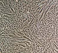
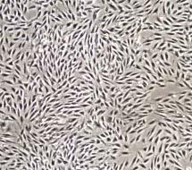
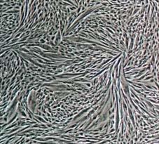
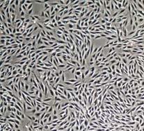
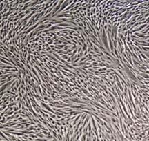
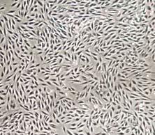
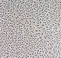
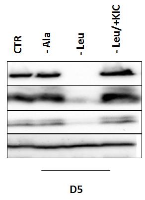
65 FIGURE 4. The leucine metabolite KIC can regulate L6 myoblast differentiation A) CTR - Alanine - Leucine - Leu / + KIC D0 D5 B) D0 58
66 D0 59
67 C) CTR - Ala - Leu - Leu/ + KIC CTR - Ala - Leu - Leu/ + KIC ph-s6 ƴ-tubulin Figure 4. A) L6 rat myoblasts were differentiated in either differentiation media (DM) with all amino acids (CTR), DM without L- alanine, DM without L-leucine, or DM without L-leucine supplemented with 200µM KIC for a period of 5 days. Day 0 (D0) represents the moment immediately before being shifting the cells to differentiation media, and day 5 (D5) represents the moment after allowing the cells to differentiate for five days. Leucine deprived cells showed an inability to differentiate, as indicated by the absence of myotube formation. Supplementing KIC to leucine deprived cells was able to rescue myotube formation. B) Cells were harvested on day 0 (D0) and day 5 (D5) and probed for myosin heavy chain-1 (MHC-1), troponin, & myogenin, which are markers of differentiation. The absence of leucine impaired the expression of these proteins after 5 days of differentiation. However, supplementing the leucine-deprived differentiation medium with KIC was able to rescue levels of MHC-1, myogenin, and troponin. * denotes significantly different compared to all other conditions in the D5 group (p<0.05). C) mtorc1 signaling was attenuated by leucine deprivation, but rescued by KIC supplementation. * denotes significantly different compared to all other conditions in the D5 group (p<0.05). n = 3, with 2 replicates per treatment. Data are mean ± SEM. 60


")

68 FIGURE 5. Expression of BCAA catabolic enzymes BCAT2 and BCKD increase during L6 differentiation A) Day of differentiation Day of differentiation B) 61
.")
69 Figure 5. A) BCAT2 (branched-chain aminotransferase-2) and BCKDE1α (branched-chain alphaketo dehydrogenase E1α) protein expression during a 5-day differentiation of L6 myoblasts. BCAT2 levels showed a non-significant trend to increase to day 3 of differentiation (D3). BCKDE1α showed a consistent increase on each day of differentiation, where day 4 (D4) and day 5 (D5) were significantly greater than day 1 (D1) (p<0.05). B) BCAT2 mrna and BCKDE1α mrna expressed relative to HPRT control mrna over a 5 day differentiation of L6. BCAT2 showed non-significant increases at D1 and D4, whereas BCKDE1α showed significant elevation on D2 compared to D5 (p<0.05). n = 3, with 2-3 replicates per treatment. Data are mean ± SEM. FIGURE 6. Intracellular BCAA concentrations do not change during L6 myoblast differentiation Day of differentiation Figure 6. Intracellular BCAA concentration at each day of differentiation was measured using a fluorometric assay. Values were corrected using sample protein concentration. No significant change in intracellular BCAA concentrations was observed during differentiation. n = 3, with 3 replicates per treatment. Data are mean ± SEM. 62


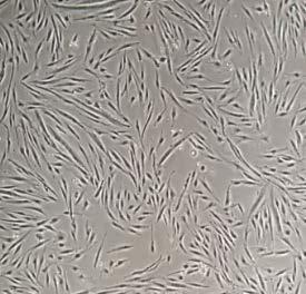
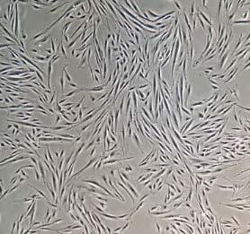

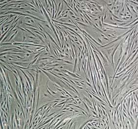

70 FIGURE 7. BCAT2 disruption prevents L6 myoblast differentiation A) D0 D1 D2 D3 D4 D5 CTR sirna BCAT2 sirna B) 63
 L6 rat myoblasts were transfected with BCAT2 sirna, or control (CTR) sirna.")
 Cells were harvested and probed for myogenic proteins MHC-1, troponin, and myogenin. The expression of myogenic proteins was severely attenuated by BCAT2 knockdown.")
71 BCAT2 sirna: BCAT2 sirna: D0 D1 D2 D3 D4 D5 D0 D1 D2 D3 D4 D5 BCAT2 sirna: D0 D1 D2 D3 D4 D5 BCAT2 sirna: D0 D1 D2 D3 D4 D5 BCAT2 sirna: D0 D1 D2 D3 D4 D5 Figure 7. A) L6 rat myoblasts were transfected with BCAT2 sirna, or control (CTR) sirna. Myoblasts transfected with BCAT2 sirna showed no ability to differentiate over 5 days, as indicated by the absence of myotube formation. A visible loss in cell number was also observed at D1 and D2. B) Cells were harvested and probed for myogenic proteins MHC-1, troponin, and myogenin. The expression of myogenic proteins was severely attenuated by BCAT2 knockdown. mtorc1 signaling was also impaired by BCAT2 knockdown. * with bar denotes significantly different (p<0.05). n = 3, with 2 replicates per treatment. Data are mean ± SEM. 64

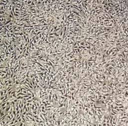
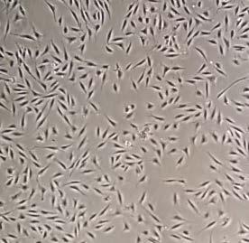
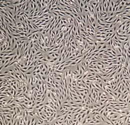
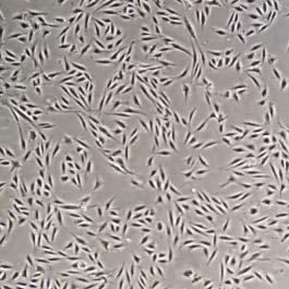
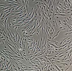

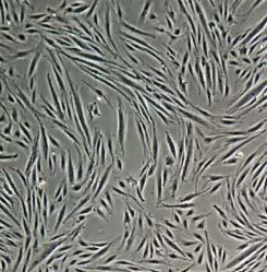
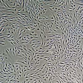
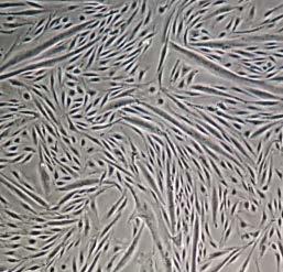

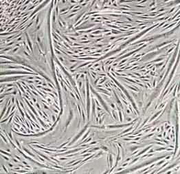
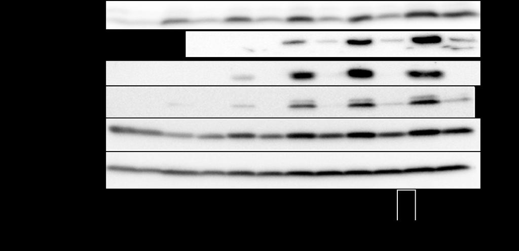
72 FIGURE 8. BCKD disruption prevents L6 myoblast differentiation A) D0 D1 D2 D3 D4 D5 CTR sirna BCKDE1α sirna B) 65
 L6 rat myoblasts were transfected with BCKDE1α sirna, or control (CTR) sirna.")
73 BCKDE1α sirna: D0 D1 D2 D3 D4 D5 BCKDE1α sirna: D0 D1 D2 D3 D4 D5 BCKDE1α sirna: D0 D1 D2 D3 D4 D5 BCKDE1α sirna: D0 D1 D2 D3 D4 D5 BCKDE1α sirna: D0 D1 D2 D3 D4 D5 Figure 8. A) L6 rat myoblasts were transfected with BCKDE1α sirna, or control (CTR) sirna. Myoblasts transfected with BCKDE1α sirna showed no ability to differentiate over 5 days, as indicated by absent myotube formation. A visible loss in cell number was also observed at D1 and D2. B) Cells were harvested and probed for myogenic proteins MHC-1, troponin, and myogenin. The expression of myogenic proteins was severely attenuated by BCKDE1α knockdown. mtorc1 signaling was also impaired by BCKDE1α knockdown. * with bar denotes significantly different (p<0.05). n = 3, with 2 replicates per treatment. Data are mean ± SEM. 66
74 FIGURE 9. BCAT2 and BCKDE1α sirna transfection reduces cell viability A) B) Figure 9. A) Cell viability was measured in wells transfected with two different BCAT2 sirna oligonucleotides. Both sirna oligos induced reduced cell viability in treatment wells at day 1 (D1) and day 2 (D2) of differentiation compared to control transfected wells. B) Cell viability was measured in wells transfected with two different BCKDE1α sirna oligonucleotides. Both sirna oligos induced reduced cell viability in treatment wells also at D1 and D2 compared to control transfected myoblasts. * with bar denotes significantly different (p<0.05). n = 3, with 2 replicates per treatment. Data are mean ± SEM. 67


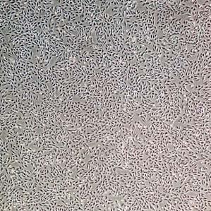
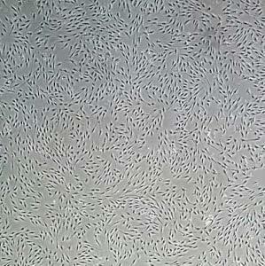

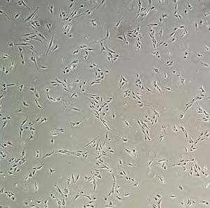
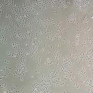
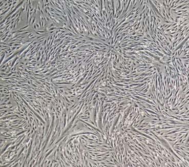
75 FIGURE 10. Increasing the cell confluency does not rescue differentiation of BCAT2 disrupted myoblasts A) CTR sirna BCAT2 sirna 3x as many cells 1x as many cells 24 hours after transfection 48 hours after transfection (D0) B) CTR sirna BCAT2 sirna D1 D3 D5 68


 Increasing the cell number")
,")


76 Figure 10. Increasing the cell confluency does not rescue differentiation of BCAT2 disrupted myoblasts A) 24 hours after being transfected, cells from 3 BCAT2 transfected wells were combined into one new well. Cells from one control well were also moved into one new well. Twenty-four hours after wells were combined, the cells were shifted to differentiation media. B) Increasing the cell number did not rescue myoblast differentiation by day 5 (D5), as absent myotube formation and a loss in cell number was still observed by D3 of differentiation. Number of experiments = 2. FIGURE 11. Supplementing BCKAs to BCAT2 disrupted myoblasts does not rescue myoblast differentiation A) CTR sirna BCAT2 sirna BCAT2 sirna + KMV KIV KIC D0 D1 D3 D5 69

77 B) CTR sirna BCAT2i BCAT2i + KIC/KMV/KIV CTR sirna BCAT2i BCAT2i + KIC/KMV/KIV CTR sirna BCAT2i BCAT2i + KIC/KMV/KIV CTR sirna BCAT2i BCAT2i + KIC/KMV/KIV CTR sirna BCAT2i BCAT2i + KIC/KMV/KIV CTR sirna BCAT2i BCAT2i + KIC/KMV/KIV D0 D1 D2 D3 D4 D5 BCAT2 MHC-1 troponin myogenin ph-s6 γ-tubulin 70
78 Figure 11. To determine if supplementing BCAT2 sirna transfected myoblasts with BCKAs could rescue their differentiation, transfected myoblasts were supplemented with 200µM α-ketoisocaproate (KIC), 200µM α-ketomethylvalerate (KMV), and 200µM α-ketoisovalerate (KIV). A) Adding BCKAs did not rescue myoblast differentiation, as the absence of myotube formation and a loss in cell number was still observed. B) Expression of myogenic proteins MHC-1, troponin, and myogenin, and mtorc1 signaling, were not rescued by BCKA supplementation. BCATi = BCAT2 sirna. * with bar denotes significantly different (p<0.05). n = 3, with 2 replicates per treatment. Data are mean ± SEM. 71

 of differentiation.")
79 FIGURE 12. BCAT2 disruption induces programmed cell death in myoblasts CTR sirna BCAT2i BCAT2i + KIC/KMV/KIV CTR sirna BCAT2i BCAT2i + KIC/KMV/KIV CTR sirna BCAT2i BCAT2i + KIC/KMV/KIV CTR sirna BCAT2i BCAT2i + KIC/KMV/KIV CTR sirna BCAT2i BCAT2i + KIC/KMV/KIV CTR sirna BCAT2i BCAT2i + KIC/KMV/KIV Caspase-3 ~30 kda D0 D1 D2 D3 D4 D5 Cleaved caspase-3 ~15 kda Figure 12. BCAT2 transfected myoblasts show increased apoptotic signaling as indicated by increases in caspase-3 cleavage at day 1 (D1) of differentiation. Addition of 200µM α-ketoisocaproate (KIC), 200µM α-ketomethylvalerate (KMV), and 200µM α-ketoisovalerate (KIV) to BCAT2 transfected cells did not rescue increased apoptotic signaling in myoblasts. BCATi = BCAT2 sirna. * with bar denotes significantly different (p<0.05). n = 2, with 2 replicates per treatment. Data are mean ± SEM. 72
A particular set of insults induces apoptosis (part 1), which, if inhibited, can switch to autophagy. At least in some cellular settings, autophagy se
, which, if inhibited, can switch to autophagy. At least in some cellular settings, autophagy se") A particular set of insults induces apoptosis (part 1), which, if inhibited, can switch to autophagy. At least in some cellular settings, autophagy serves as a defence mechanism that prevents or retards
A particular set of insults induces apoptosis (part 1), which, if inhibited, can switch to autophagy. At least in some cellular settings, autophagy serves as a defence mechanism that prevents or retards
Enzyme-coupled Receptors. Cell-surface receptors 1. Ion-channel-coupled receptors 2. G-protein-coupled receptors 3. Enzyme-coupled receptors
 Enzyme-coupled Receptors Cell-surface receptors 1. Ion-channel-coupled receptors 2. G-protein-coupled receptors 3. Enzyme-coupled receptors Cell-surface receptors allow a flow of ions across the plasma
Cell Signaling part 2
 15 Cell Signaling part 2 Functions of Cell Surface Receptors Other cell surface receptors are directly linked to intracellular enzymes. The largest family of these is the receptor protein tyrosine kinases,
KEY CONCEPT QUESTIONS IN SIGNAL TRANSDUCTION
 Signal Transduction - Part 2 Key Concepts - Receptor tyrosine kinases control cell metabolism and proliferation Growth factor signaling through Ras Mutated cell signaling genes in cancer cells are called
Optimizing Nutritional Strategies to Promote Growth in Newborns
 Optimizing Nutritional Strategies to Promote Growth in Newborns Teresa A. Davis, Ph.D. Professor of Pediatrics USDA/ARS Children s Nutrition Research Center, Baylor College of Medicine, Houston, TX Disclosure
ANSC/FSTC 607 Biochemistry and Physiology of Muscle as a Food PRIMARY, SECONDARY, AND TERTIARY MYOTUBES
 ANSC/FSTC 607 Biochemistry and Physiology of Muscle as a Food PRIMARY, SECONDARY, AND TERTIARY MYOTUBES I. Satellite Cells A. Proliferative, myoblastic cells that lie in invaginations in the sarcolemma
Branched Chain Amino Acid, Leucine: The Effects of Leucine on Skeletal Tissue in Relation to Aerobic Exercise. Shea Teresi. For
 Branched Chain Amino Acid, Leucine: The Effects of Leucine on Skeletal Tissue in Relation to Aerobic Exercise By Shea Teresi For Dr. William R. Proulx, RD Associate Professor of Nutrition & Dietetics In
RAS Genes. The ras superfamily of genes encodes small GTP binding proteins that are responsible for the regulation of many cellular processes.
 ۱ RAS Genes The ras superfamily of genes encodes small GTP binding proteins that are responsible for the regulation of many cellular processes. Oncogenic ras genes in human cells include H ras, N ras,
Receptor mediated Signal Transduction
 Receptor mediated Signal Transduction G-protein-linked receptors adenylyl cyclase camp PKA Organization of receptor protein-tyrosine kinases From G.M. Cooper, The Cell. A molecular approach, 2004, third
Medical Biology. Dr. Khalida Ibrahim
 Dr. Khalida Ibrahim Medical Biology MUSCLE TISSUE 1. Muscle tissue is characterized by its well-developed properties of contraction. 2. Muscle is responsible for the movements of the body and the various
Signal Transduction Cascades
 Signal Transduction Cascades Contents of this page: Kinases & phosphatases Protein Kinase A (camp-dependent protein kinase) G-protein signal cascade Structure of G-proteins Small GTP-binding proteins,
Chapter Skeletal Muscle Structure and Function
 Chapter 10.2 Skeletal Muscle Structure and Function Introduction to Muscle Physiology Movement is a fundamental characteristic of all living things All muscle cells (skeletal, cardiac, and smooth) are
The Cell Cycle M G2 G1 G0 S 1
 The Cell Cycle M G2 G1 G0 S 1 Cell Cycle G1 (Gap 1) 3 to 12 hours in length Respond to cues from the environment External cues Growth factors that signal the cell to stay in G1 or continue to through the
Principles of cell signaling Lecture 4
 Principles of cell signaling Lecture 4 Johan Lennartsson Molecular Cell Biology (1BG320), 2014 Johan.Lennartsson@licr.uu.se 1 Receptor tyrosine kinase-induced signal transduction Erk MAP kinase pathway
Muscle Tissue- 3 Types
 AN INTRODUCTION TO MUSCLE TISSUE Muscle Tissue- 3 Types Skeletal muscle (focus on these) Cardiac muscle Smooth muscle FUNCTIONS OF SKELETAL MUSCLES Produce movement of the skeleton Maintain posture and
G-Protein Signaling. Introduction to intracellular signaling. Dr. SARRAY Sameh, Ph.D
 G-Protein Signaling Introduction to intracellular signaling Dr. SARRAY Sameh, Ph.D Cell signaling Cells communicate via extracellular signaling molecules (Hormones, growth factors and neurotransmitters
FACTORS AFFECTING SKELETAL MUSCLE PROTEIN SYNTHESIS IN THE HORSE
 University of Kentucky UKnowledge Theses and Dissertations--Animal and Food Sciences Animal and Food Sciences 2011 FACTORS AFFECTING SKELETAL MUSCLE PROTEIN SYNTHESIS IN THE HORSE Ashley Leigh Wagner University
9/16/2009. Fast and slow twitch fibres. Properties of Muscle Fiber Types Fast fibers Slow fibers
 Muscles, muscle fibres and myofibrils Fast and slow twitch fibres Rat hindlimb muscle ATPase staining at different ph and NADH Muscle fibre shortening velocity lengths/second Properties of Muscle Fiber
Propagation of the Signal
 OpenStax-CNX module: m44452 1 Propagation of the Signal OpenStax College This work is produced by OpenStax-CNX and licensed under the Creative Commons Attribution License 3.0 By the end of this section,
Principles of Genetics and Molecular Biology
 Cell signaling Dr. Diala Abu-Hassan, DDS, PhD School of Medicine Dr.abuhassand@gmail.com Principles of Genetics and Molecular Biology www.cs.montana.edu Modes of cell signaling Direct interaction of a
Muscles, muscle fibres and myofibrils
 Muscles, muscle fibres and myofibrils Properties of Muscle Fiber Types Fast fibers Slow fibers Characteristic IIb IIx IIa Type I V max (speed of shortening) Highest Intermediate Low Resistance to fatigue
Role of Map4k4 in Skeletal Muscle Differentiation: A Dissertation
 University of Massachusetts Medical School escholarship@umms GSBS Dissertations and Theses Graduate School of Biomedical Sciences 5-1-2013 Role of Map4k4 in Skeletal Muscle Differentiation: A Dissertation
1. Activated receptor tyrosine kinases (RTKs) phosphorylates themselves
 phosphorylates themselves") Enzyme-coupled receptors Transmembrane proteins Ligand-binding domain on the outer surface Cytoplasmic domain acts as an enzyme itself or forms a complex with enzyme 1. Activated receptor tyrosine kinases
Enzyme-coupled receptors Transmembrane proteins Ligand-binding domain on the outer surface Cytoplasmic domain acts as an enzyme itself or forms a complex with enzyme 1. Activated receptor tyrosine kinases
E3 ligase TRIM72 negatively regulates myogenesis by IRS-1 ubiquitination
 E3 ligase negatively regulates myogenesis by IRS-1 ubiquitination Young-Gyu Ko College of Life Science and Biotechnology Korea University MyHC immunofluorescence Lipid rafts are plasma membrane compartments
About This Chapter. Skeletal muscle Mechanics of body movement Smooth muscle Cardiac muscle Pearson Education, Inc.
 About This Chapter Skeletal muscle Mechanics of body movement Smooth muscle Cardiac muscle Skeletal Muscle Usually attached to bones by tendons Origin: closest to the trunk or to more stationary bone Insertion:
Muscle Cells & Muscle Fiber Contractions. Packet #8
 Muscle Cells & Muscle Fiber Contractions Packet #8 Skeletal muscle is attached to bones and is responsible for movement. Introduction Introduction II Skeletal muscle is composed of bundles of muscle fibers
Regulation of Gene Expression in Eukaryotes
 Ch. 19 Regulation of Gene Expression in Eukaryotes BIOL 222 Differential Gene Expression in Eukaryotes Signal Cells in a multicellular eukaryotic organism genetically identical differential gene expression
Microanatomy of Muscles. Anatomy & Physiology Class
 Microanatomy of Muscles Anatomy & Physiology Class Three Main Muscle Types Objectives: By the end of this presentation you will have the information to: 1. 2. 3. 4. 5. 6. Describe the 3 main types of muscles.
Post-translational modifications of proteins in gene regulation under hypoxic conditions
 203 Review Article Post-translational modifications of proteins in gene regulation under hypoxic conditions 1, 2) Olga S. Safronova 1) Department of Cellular Physiological Chemistry, Tokyo Medical and
Chapter 8 Notes. Muscles
 Chapter 8 Notes Muscles 8.1 Intro Three muscle types Skeletal Smooth cardiac 8.2 Structure of Skeletal Muscle Composition Skeletal muscle tissue Nervous tissue Blood Connective tissue Connective tissue
Biol403 MAP kinase signalling
 Biol403 MAP kinase signalling The mitogen activated protein kinase (MAPK) pathway is a signalling cascade activated by a diverse range of effectors. The cascade regulates many cellular activities including
Signaling Through Immune System Receptors (Ch. 7)
") Signaling Through Immune System Receptors (Ch. 7) 1. General principles of signal transduction and propagation. 2. Antigen receptor signaling and lymphocyte activation. 3. Other receptors and signaling
Signaling Through Immune System Receptors (Ch. 7) 1. General principles of signal transduction and propagation. 2. Antigen receptor signaling and lymphocyte activation. 3. Other receptors and signaling
Revision. camp pathway
 االله الرحمن الرحيم بسم Revision camp pathway camp pathway Revision camp pathway Adenylate cyclase Adenylate Cyclase enzyme Adenylate cyclase catalyses the formation of camp from ATP. Stimulation or inhibition
Cellular Signaling Pathways. Signaling Overview
 Cellular Signaling Pathways Signaling Overview Signaling steps Synthesis and release of signaling molecules (ligands) by the signaling cell. Transport of the signal to the target cell Detection of the
Chapter 10 Muscle Tissue and Physiology Chapter Outline
 Chapter 10 Muscle Tissue and Physiology Chapter Outline Module 10.1 Overview of muscle tissue (Figures 10.1 10.2) A. Types of Muscle Tissue (Figure 10.1) 1. The three types of cells in muscle tissue are,,
Muscle Cell Anatomy & Function (mainly striated muscle tissue)
") Muscle Cell Anatomy & Function (mainly striated muscle tissue) General Structure of Muscle Cells (skeletal) several nuclei (skeletal muscle) skeletal muscles are formed when embryonic cells fuse together
Muscle Cell Anatomy & Function (mainly striated muscle tissue) General Structure of Muscle Cells (skeletal) several nuclei (skeletal muscle) skeletal muscles are formed when embryonic cells fuse together
Muscle and Muscle Tissue
 Muscle and Muscle Tissue Make up about half of total body mass Exerts force by converting chemical energy, ATP, to mechanical energy Muscle tissue is classified based on Shape Number and position of nuclei
MUSCLE TISSUE (MUSCLE PHYSIOLOGY) PART I: MUSCLE STRUCTURE
 PART I: MUSCLE STRUCTURE") PART I: MUSCLE STRUCTURE Muscle Tissue A primary tissue type, divided into: skeletal muscle cardiac muscle smooth muscle Functions of Skeletal Muscles Produce skeletal movement Maintain body position Support
PART I: MUSCLE STRUCTURE Muscle Tissue A primary tissue type, divided into: skeletal muscle cardiac muscle smooth muscle Functions of Skeletal Muscles Produce skeletal movement Maintain body position Support
Connective tissue MUSCLE TISSUE
 Connective tissue MUSCLE TISSUE Part 1 General features of MT Develop from mesoderm Many cells, less intercellular matrix Function contraction (shortening) Skeletal (striated, voluntary) Types of MT Cardiac
The PI3K/AKT axis. Dr. Lucio Crinò Medical Oncology Division Azienda Ospedaliera-Perugia. Introduction
 The PI3K/AKT axis Dr. Lucio Crinò Medical Oncology Division Azienda Ospedaliera-Perugia Introduction Phosphoinositide 3-kinase (PI3K) pathway are a family of lipid kinases discovered in 1980s. They have
SUPPLEMENTARY INFORMATION
 doi:10.1038/nature22314 Supplementary Discussion In mammals, BCAAs are essential amino acids that must be supplied from food. These dietary BCAAs are subsequently used for protein synthesis or are catabolized
Signal Transduction Pathway Smorgasbord
 Molecular Cell Biology Lecture. Oct 28, 2014 Signal Transduction Pathway Smorgasbord Ron Bose, MD PhD Biochemistry and Molecular Cell Biology Programs Washington University School of Medicine Outline 1.
Lipids and Membranes
 Lipids and Membranes Presented by Dr. Mohammad Saadeh The requirements for the Pharmaceutical Biochemistry I Philadelphia University Faculty of pharmacy Membrane transport D. Endocytosis and Exocytosis
Optimizing Protein in a Carbohydrate World
 Optimizing Protein in a Carbohydrate World Donald K. Layman, Ph.D. Professor Emeritus Department of Food Science & Human Nutrition University of Illinois at Urbana Champaign The confused consumer 1 Myth:
Molecular Cell Biology - Problem Drill 19: Cell Signaling Pathways and Gene Expression
 Molecular Cell Biology - Problem Drill 19: Cell Signaling Pathways and Gene Expression Question No. 1 of 10 1. Which statement about cell signaling is correct? Question #1 (A) Cell signaling involves receiving
BCHM3972 Human Molecular Cell Biology (Advanced) 2013 Course University of Sydney
 2013 Course University of Sydney") BCHM3972 Human Molecular Cell Biology (Advanced) 2013 Course University of Sydney Page 2: Immune Mechanisms & Molecular Biology of Host Defence (Prof Campbell) Page 45: Infection and Implications for Cell
BCHM3972 Human Molecular Cell Biology (Advanced) 2013 Course University of Sydney Page 2: Immune Mechanisms & Molecular Biology of Host Defence (Prof Campbell) Page 45: Infection and Implications for Cell
Chapter 15: Signal transduction
 Chapter 15: Signal transduction Know the terminology: Enzyme-linked receptor, G-protein linked receptor, nuclear hormone receptor, G-protein, adaptor protein, scaffolding protein, SH2 domain, MAPK, Ras,
Lecture 9A. Muscle structure. Outline
 Lecture 9A Muscle structure Outline Smooth, skeletal, and cardiac muscle tissues Structure and function of skeletal muscle cells. Sarcomeres structure and contraction Actin-myosin interaction and sliding
The elements of G protein-coupled receptor systems
 The elements of G protein-coupled receptor systems Prostaglandines Sphingosine 1-phosphate a receptor that contains 7 membrane-spanning domains a coupled trimeric G protein which functions as a switch
Companion to Biosynthesis of Ketones & Cholesterols, Regulation of Lipid Metabolism Lecture Notes
 Companion to Biosynthesis of Ketones & Cholesterols, Regulation of Lipid Metabolism Lecture Notes The major site of acetoacetate and 3-hydorxybutyrate production is in the liver. 3-hydorxybutyrate is the
Lecture 10. G1/S Regulation and Cell Cycle Checkpoints. G1/S regulation and growth control G2 repair checkpoint Spindle assembly or mitotic checkpoint
 Lecture 10 G1/S Regulation and Cell Cycle Checkpoints Outline: G1/S regulation and growth control G2 repair checkpoint Spindle assembly or mitotic checkpoint Paper: The roles of Fzy/Cdc20 and Fzr/Cdh1
Lecture 7: Signaling Through Lymphocyte Receptors
 Lecture 7: Signaling Through Lymphocyte Receptors Questions to Consider After recognition of its cognate MHC:peptide, how does the T cell receptor activate immune response genes? What are the structural
BIOL 4374/BCHS 4313 Cell Biology Exam #1 February 13, 2001
 BIOL 4374/BCHS 4313 Cell Biology Exam #1 February 13, 2001 SS# Name This exam is worth a total of 100 points. The number of points each question is worth is shown in parentheses. Good luck! 1. (2) The
Cell Physiology Final Exam Fall 2008
 Cell Physiology Final Exam Fall 2008 Guys, The average on the test was 69.9. Before you start reading the right answers please do me a favor and remember till the end of your life that GLUCOSE TRANSPORT
Cell cycle and Apoptosis. Chalermchai Mitrpant
 Cell cycle and Apoptosis 2556 Chalermchai Mitrpant Overview of the cell cycle Outline Regulatory mechanisms controlling cell cycle Progression of the cell cycle Checkpoint of the cell cycle Phases of the
Summary of Endomembrane-system
 Summary of Endomembrane-system 1. Endomembrane System: The structural and functional relationship organelles including ER,Golgi complex, lysosome, endosomes, secretory vesicles. 2. Membrane-bound structures
Growth and Differentiation Phosphorylation Sampler Kit
 Growth and Differentiation Phosphorylation Sampler Kit E 0 5 1 0 1 4 Kits Includes Cat. Quantity Application Reactivity Source Akt (Phospho-Ser473) E011054-1 50μg/50μl IHC, WB Human, Mouse, Rat Rabbit
Molecular Biology (BIOL 4320) Exam #2 May 3, 2004
 Exam #2 May 3, 2004") Molecular Biology (BIOL 4320) Exam #2 May 3, 2004 Name SS# This exam is worth a total of 100 points. The number of points each question is worth is shown in parentheses after the question number. Good
Molecular Biology (BIOL 4320) Exam #2 May 3, 2004 Name SS# This exam is worth a total of 100 points. The number of points each question is worth is shown in parentheses after the question number. Good
What would you observe if you fused a G1 cell with a S cell? A. Mitotic and pulverized chromosomes. B. Mitotic and compact G1 chromosomes.
 What would you observe if you fused a G1 cell with a S cell? A. Mitotic and pulverized chromosomes. B. Mitotic and compact G1 chromosomes. C. Mostly non-compact G1 chromosomes. D. Compact G1 and G2 chromosomes.
Muscle Physiology. Introduction. Four Characteristics of Muscle tissue. Skeletal Muscle
 Muscle Physiology Introduction Muscle = tissue capable of forceful shortening or contraction Converts chemical energy (ATP) into mechanical energy Important in: Respiration Urine collection & flow Gastrointestinal
Chapter 10! Chapter 10, Part 2 Muscle. Muscle Tissue - Part 2! Pages !
 ! Chapter 10, Part 2 Muscle Chapter 10! Muscle Tissue - Part 2! Pages 308-324! SECTION 10-5! Sarcomere shortening and muscle fiber stimulation produce tension! 2! Tension Production - Muscle FIBER! All-or-none
Sphingosine-1-phosphate signaling and cardiac fibrosis. Department of Physiology, Kanazawa University School of Medicine, Kanazawa, Japan
 96 Special Issue: Cellular and Molecular Bases for Fibrotic Diseases Review Article Sphingosine-1-phosphate signaling and cardiac fibrosis Noriko Takuwa 1, 2, ), Yasuo Okamoto 1), Kazuaki Yoshioka 1) and
DOWNLOAD PDF STRUCTURE AND REGULATION OF CARDIAC AND SKELETAL MUSCLE THIN FILAMENTS
 Chapter 1 : ACTC1 - Wikipedia LARRY S. TOBACMAN STRUCTURE AND REGULATION OF CARDIAC AND The biological production of force and movement can be understood only when it is. Structure[ edit ] There are three
Muscle Tissue. Dr. Heba Kalbouneh Associate Professor of Anatomy and Histology
 Muscle Tissue Dr. Heba Kalbouneh Associate Professor of Anatomy and Histology Functions of muscle tissue Movement Maintenance of posture Joint stabilization Heat generation Tendon Belly Tendon Types of
Cell Quality Control. Peter Takizawa Department of Cell Biology
 Cell Quality Control Peter Takizawa Department of Cell Biology Cellular quality control reduces production of defective proteins. Cells have many quality control systems to ensure that cell does not build
Outline. Bio 105: Muscular System. Muscular System. Types of Muscles. Smooth Muscle. Cardiac Muscle 4/6/2016
 Outline Bio 105: Muscular System Lecture 11 Chapter 6 Characteristics of muscles 3 types of muscles Functions of muscles Structure of skeletal muscles Mechanics of muscle contraction Energy sources for
Lecture: CHAPTER 13 Signal Transduction Pathways
 Lecture: 10 17 2016 CHAPTER 13 Signal Transduction Pathways Chapter 13 Outline Signal transduction cascades have many components in common: 1. Release of a primary message as a response to a physiological
Control of Cell Cycle Progression by mtor
 City University of New York (CUNY) CUNY Academic Works Dissertations, Theses, and Capstone Projects Graduate Center 5-2015 Control of Cell Cycle Progression by mtor Amrita Chatterjee Graduate Center, City
Molecular Cell Biology Problem Drill 16: Intracellular Compartment and Protein Sorting
 Molecular Cell Biology Problem Drill 16: Intracellular Compartment and Protein Sorting Question No. 1 of 10 Question 1. Which of the following statements about the nucleus is correct? Question #01 A. The
Skeletal Muscle and the Molecular Basis of Contraction. Lanny Shulman, O.D., Ph.D. University of Houston College of Optometry
 Skeletal Muscle and the Molecular Basis of Contraction Lanny Shulman, O.D., Ph.D. University of Houston College of Optometry Like neurons, all muscle cells can be excited chemically, electrically, and
PCB 3023 Exam 4 - Form A First and Last Name
 PCB 3023 Exam 4 - Form A First and Last Name Student ID # (U Number) A Before beginning this exam, please complete the following instructions: 1) Write your name and U number on the first page of this
We are IntechOpen, the first native scientific publisher of Open Access books. International authors and editors. Our authors are among the TOP 1%
 We are IntechOpen, the first native scientific publisher of Open Access books 3,350 108,000 1.7 M Open access books available International authors and editors Downloads Our authors are among the 151 Countries
Muscle Histology. Dr. Heba Kalbouneh Assistant Professor of Anatomy and Histology
 Muscle Histology Dr. Heba Kalbouneh Assistant Professor of Anatomy and Histology Functions of muscle tissue Movement Maintenance of posture Joint stabilization Heat generation Types of Muscle Tissue Skeletal
1. Locomotion. 2. Repositioning. 3. Internal movement
 MUSCLE and MOVEMENT Chapters 20, 8, 21 1. Locomotion A. Movement B. 2. Repositioning A. 3. Internal movement A. 1 Muscle Cells 1. Contractile 2. Myocytes 3. Striated A. Skeletal B. Cardiac 4. Smooth 5.
MOLECULAR CELL BIOLOGY
 1 Lodish Berk Kaiser Krieger scott Bretscher Ploegh Matsudaira MOLECULAR CELL BIOLOGY SEVENTH EDITION CHAPTER 13 Moving Proteins into Membranes and Organelles Copyright 2013 by W. H. Freeman and Company
1. Locomotion. 2. Repositioning. 3. Internal movement
 MUSCLE and MOVEMENT Chapters 20, 8, 21 1. Locomotion A. Movement B. 2. Repositioning A. 3. Internal movement A. Muscle Cells 1. Contractile 2. Myocytes 3. Striated A. Skeletal B. Cardiac 4. Smooth 5. Striated
PREPARED FOR: U.S. Army Medical Research and Materiel Command Fort Detrick, Maryland
 AD Award Number: W81XWH-09-1-0279 TITLE: Regulation of mtor by Nutrients PRINCIPAL INVESTIGATOR: Kun-Liang Guan CONTRACTING ORGANIZATION: University of San Diego La Jolla, CA 92093 REPORT DATE: July 2010
1. to understand how proteins find their destination in prokaryotic and eukaryotic cells 2. to know how proteins are bio-recycled
 Protein Targeting Objectives 1. to understand how proteins find their destination in prokaryotic and eukaryotic cells 2. to know how proteins are bio-recycled As a protein is being synthesized, decisions
Cell Cycle. Trends in Cell Biology
 Cell Cycle Trends in Cell Biology Cell Cycle The orderly sequence of events by which a cell duplicates its contents and divides into two Daughter Cells Activities of a cell from one cell division to the
The Musculoskeletal System. Chapter 46
 The Musculoskeletal System Chapter 46 Types of Skeletal Systems Changes in movement occur because muscles pull against a support structure Zoologists recognize three types: 1. Hydrostatic skeletons a fluid
Tala Saleh. Ahmad Attari. Mamoun Ahram
 23 Tala Saleh Ahmad Attari Minna Mushtaha Mamoun Ahram In the previous lecture, we discussed the mechanisms of regulating enzymes through inhibitors. Now, we will start this lecture by discussing regulation
Chapter 9 Muscle. Types of muscle Skeletal muscle Cardiac muscle Smooth muscle. Striated muscle
 Chapter 9 Muscle Types of muscle Skeletal muscle Cardiac muscle Smooth muscle Striated muscle Chapter 9 Muscle (cont.) The sliding filament mechanism, in which myosin filaments bind to and move actin
Signaling. Dr. Sujata Persad Katz Group Centre for Pharmacy & Health research
 Signaling Dr. Sujata Persad 3-020 Katz Group Centre for Pharmacy & Health research E-mail:sujata.persad@ualberta.ca 1 Growth Factor Receptors and Other Signaling Pathways What we will cover today: How
Integration Of Metabolism
 Integration Of Metabolism Metabolism Consist of Highly Interconnected Pathways The basic strategy of catabolic metabolism is to form ATP, NADPH, and building blocks for biosyntheses. 1. ATP is the universal
HOMEWORK RUBRICS MECHANOTRANSDUCTION UNIT: HOMEWORK #1 (20 pts towards your grade)
") HOMEWORK RUBRICS MECHANOTRANSDUCTION UNIT: HOMEWORK #1 (20 pts towards your grade) 1. Mesenchymal stem cells (MSC) cultured on extracellular matrices with different stiffness exhibit diverse lineage commitment
HOMEWORK RUBRICS MECHANOTRANSDUCTION UNIT: HOMEWORK #1 (20 pts towards your grade) 1. Mesenchymal stem cells (MSC) cultured on extracellular matrices with different stiffness exhibit diverse lineage commitment
AperTO - Archivio Istituzionale Open Access dell'università di Torino
 AperTO - Archivio Istituzionale Open Access dell'università di Torino From the nucleus to the mitochondria and backthe odyssey of a multitask STAT3 This is the author's manuscript Original Citation: From
Lecture 15. Signal Transduction Pathways - Introduction
 Lecture 15 Signal Transduction Pathways - Introduction So far.. Regulation of mrna synthesis Regulation of rrna synthesis Regulation of trna & 5S rrna synthesis Regulation of gene expression by signals
Vets 111/Biov 111 Cell Signalling-2. Secondary messengers the cyclic AMP intracellular signalling system
 Vets 111/Biov 111 Cell Signalling-2 Secondary messengers the cyclic AMP intracellular signalling system The classical secondary messenger model of intracellular signalling A cell surface receptor binds
Cell Cell Communication
 IBS 8102 Cell, Molecular, and Developmental Biology Cell Cell Communication January 29, 2008 Communicate What? Why do cells communicate? To govern or modify each other for the benefit of the organism differentiate
Muscle Tissue. PowerPoint Lecture Presentations prepared by Jason LaPres. Lone Star College North Harris Pearson Education, Inc.
 10 Muscle Tissue PowerPoint Lecture Presentations prepared by Jason LaPres Lone Star College North Harris An Introduction to Muscle Tissue Muscle Tissue A primary tissue type, divided into: Skeletal muscle
BIOL212 Biochemistry of Disease. Metabolic Disorders - Obesity
 BIOL212 Biochemistry of Disease Metabolic Disorders - Obesity Obesity Approx. 23% of adults are obese in the U.K. The number of obese children has tripled in 20 years. 10% of six year olds are obese, rising
PSK4U THE NEUROMUSCULAR SYSTEM
 PSK4U THE NEUROMUSCULAR SYSTEM REVIEW Review of muscle so we can see how the neuromuscular system works This is not on today's note Skeletal Muscle Cell: Cellular System A) Excitation System Electrical
Chapter 9. Cellular Signaling
 Chapter 9 Cellular Signaling Cellular Messaging Page 215 Cells can signal to each other and interpret the signals they receive from other cells and the environment Signals are most often chemicals The
PRMT BIOLOGY DURING SKELETAL MUSCLE DISUSE
 PRMT BIOLOGY DURING SKELETAL MUSCLE DISUSE PROTEIN ARGININE METHYLTRANSFERASE EXPRESSION, LOCALIZATION, AND ACTIVITY DURING DISUSE-INDUCED SKELETAL MUSCLE PLASTICITY By DEREK W. STOUTH, B.Sc. Kin Honours
Amino acids. Side chain. -Carbon atom. Carboxyl group. Amino group
 PROTEINS Amino acids Side chain -Carbon atom Amino group Carboxyl group Amino acids Primary structure Amino acid monomers Peptide bond Peptide bond Amino group Carboxyl group Peptide bond N-terminal (
Cell cycle, signaling to cell cycle, and molecular basis of oncogenesis
 Cell cycle, signaling to cell cycle, and molecular basis of oncogenesis MUDr. Jiří Vachtenheim, CSc. CELL CYCLE - SUMMARY Basic terminology: Cyclins conserved proteins with homologous regions; their cellular
REGULATION OF ENZYME ACTIVITY. Medical Biochemistry, Lecture 25
 REGULATION OF ENZYME ACTIVITY Medical Biochemistry, Lecture 25 Lecture 25, Outline General properties of enzyme regulation Regulation of enzyme concentrations Allosteric enzymes and feedback inhibition
CHAPTER 6 2/9/2016. Learning Objectives List the four traits that all muscle types have in common.
 Learning Objectives List the four traits that all muscle types have in common. CHAPTER 6 The Muscular System Demonstrate and explain the use of antagonistic muscle pairs. Describe the attachment of muscle
Phospho-AKT Sampler Kit
 Phospho-AKT Sampler Kit E 0 5 1 0 0 3 Kits Includes Cat. Quantity Application Reactivity Source Akt (Ab-473) Antibody E021054-1 50μg/50μl IHC, WB Human, Mouse, Rat Rabbit Akt (Phospho-Ser473) Antibody
Muscle Regeneration: Cellular and Molecular Events
 Review Muscle Regeneration: Cellular and Molecular Events MARIA KARALAKI, SOFIA FILI, ANASTASSIOS PHILIPPOU and MICHAEL KOUTSILIERIS Department of Experimental Physiology, Medical School of the National