THE ANTIMICROBIAL EFFICACY OF FIJI HONEYS AND PROPOLIS AGAINST CLINICAL ISOLATES FROM DIABETIC FOOT ULCERS
|
|
|
- Lester Allen Joseph
- 5 years ago
- Views:
Transcription
1
2 THE ANTIMICROBIAL EFFICACY OF FIJI HONEYS AND PROPOLIS AGAINST CLINICAL ISOLATES FROM DIABETIC FOOT ULCERS by Rajneeta Saraf A thesis submitted in partial fulfillment of the requirements for the degree of Master of Science Copyright 2011 by Rajneeta Saraf Division of Biology School of Biological, Chemical and Environmental Sciences Faculty of Science and Technology University of the South Pacific March, 2011
3 DECLARATION OF ORIGINALITY Statement by Author: I, Rajneeta Saraf, hereby declare that this thesis is the result of my own work and that, to the best of my knowledge; it contains no material previously published (except my own research papers published/presented in conferences) or substantially overlapping with material submitted for the award of any other degree at any institution, except where due acknowledgement is made in the text. Signature: Date: 15 th, March, 2011 Name: Ms. Rajneeta Saraf Student ID Number: S Statement by Supervisor(s): The research work reported in this thesis was performed under our supervision and to our knowledge is the sole work of Mrs. Rajneeta Saraf. Any help sought to complete this research work is duly acknowledged. Signature: Date: 15 th March 2011 Name: Professor Anand P. Tyagi Designation: Professor of Biology and Head of SBCS II
4 ACKNOWLEDGEMENTS I would like to thank my supervisors Dr Vincent Bowry and Dr Dhana Rao for their contribution and support at the initial stages of my research. I would like to express my sincere gratitude to my current supervisor Dr Anand Tyagi and Co-Supervisor Dr Permal Deo who were so kind to take over from Dr Bowry and Dr Rao and assisted me greatly especially when combining everything together towards the end of my thesis. Without their support and guidance, this journey of mine wouldn t have been complete. I would also like to thank the biology technicians especially Babita Narayan, Shiva Padyachi and Pranita Dutt for all the many questions thrown their way and their valued technical know-how and advice. My external supervisor, Prof. Peter Molan, Director, Honey Research Unit, Waikato University, Hamilton, New Zealand whose expertise on honey research and advice was my source of inspiration. Thank you so much sir for your guidance and patience even when I called you at your busiest times to seek advice and for the translated versions of the published papers that were foreign to me. My sincere gratitude also goes to Mr. Parmod Chand and Shalini Singh of Microbiology Lab, CWM Hospital for the supply of clinical isolates from Diabetic Foot Ulcers. I would like to thank Mr. Kamal Prasad, Apiculture Coordinator, Ministry of Agriculture, Nausori for the supply of honey and propolis samples from different geographical regions in Fiji. Financial assistance is gratefully acknowledged from London Insulin Dependent Diabetes Trust (IDDT). Finally, I would like to dedicate this thesis to my two boys; my husband, Dr. Prashant Saraf and my baby, Prasun Saraf who came as a gift to us on Christmas day, I love you both so much. Thank you so much for your patience and understanding while I was writing up this thesis. III
5 ABSTRACT A diverse range of illnesses have been treated with honey and propolis since ancient civilizations. There has been growing interest by health care professionals in wound care products based on New Zealand Manuka honey and Australian honey of similar Leptospermum spp. Similar interest has been evoked by propolis for its antimicrobial properties. Even though honey and propolis are both gaining popularity amongst researchers worldwide, the knowledge of the healing or antimicrobial properties of propolis is limited compared to honey. In Fiji, local honeys have been used in homes to treat diabetic foot ulcers which have failed to heal by conventional therapeutic methods. This suggests that Fiji honeys might confer antimicrobial activity against isolates from diabetic foot ulcers and the aim of the study was to investigate the efficacy of Fiji honeys as well as Fiji propolis against clinical isolates from diabetic foot ulcers. The antimicrobial activity of 30 natural honeys and two processed honeys together were determined using clinical isolates from diabetic foot ulcers namely: Staphylococcus aureus, Escherichia coli, Proteus mirabilis, Pseudomonas aeruginosa, Klebsiella pneumoniae and Candida albicans. The antimicrobial activity of the natural honeys, determined by agar well diffusion assay and expressed as the concentration of phenol with equivalent activity, was found to be between 4.1 and 14.5% w/v phenol. The Minimum Inhibitory Concentrations (MIC) of the honeys determined by agar incorporation technique was found to range from 4.8% to more than 9.1% (v/v) honey (9.1% being the highest concentration tested). In comparison, the activities of two processed honeys were between % phenol equivalence and did not inhibit the clinical isolates from diabetic foot ulcers at the highest concentration of honey tested (9.1%). The antimicrobial activities of 70% ethanol extract of propolis (EEP) and 40% EEP were determined by disc diffusion. The mean zones of inhibition of 70% EEP were in the range from 20.4 mm ± 0.20 to 41.3mm ± 0.60 and for 40% EEP ranged from 17.8mm ± 0.30 to 22.0mm ± This shows that the activity of 70% EEP was greater than the activity for the 40% EEP. The results also demonstrate that Fiji propolis is effective as an antimicrobial agent against clinical isolates from diabetic foot ulcers. In comparison the mean zones of inhibition for Fiji honeys was found to be between IV
6 28.2mm ± 0.50 to 38.5mm ± There was significant difference between the antimicrobial activities of Fiji honeys and propolis. The effects of storage and heating were also examined for six selected honeys. Honeys were heated at 80 0 C for 1 hour and the activity was determined by agar well diffusion assay. For the effects of storage on the antimicrobial activities, six honeys were stored for 10, 8, 5, 3 and 2 years and for 7 months respectively. The activities of the six honeys prior to storage and after the storage period were determined using agar well diffusion assay. The activities of the heated honeys were in the range from not being detectable to 7.2% phenol with equivalent activity (Not detectable implies having activity of less than 4.1% phenol activity) and for the stored honeys to be in the range from not being detectable to 12.3% phenol activity. The MICs determined by agar incorporation technique for the six heated honeys was found to range from 9.1% to more than 9.1% (v/v) honey and for the stored honeys to range from 7.0% to more than 9.1% (v/v) honey. This implies that heating and storage of honeys lead to reduced efficacy against isolates from diabetic foot ulcers. However, the results also indicate that the loss of antimicrobial activity is significantly greater through heating then through storage. The region providing the most potent honey was assessed in this study. It was found that honey from Kabara, Lau region had the highest activity against three isolates (S. aureus, P. aeruginosa and K. pneumoniae) from diabetic foot ulcers and thus was the most potent among the other 31 honeys analyzed. The results of this study demonstrate that Fiji honeys and propolis have potential as herbal remedies for the treatment of diabetic foot ulcers. However, to assess the true potential of Fiji honeys and Fiji propolis on diabetic foot ulcers, there is a need for actual clinical trials on these wounds. V
7 ABBREVIATIONS DFU Diabetic foot ulcers MIC Minimum inhibitory concentration E. coli Escherichia coli K. pneumoniae Klebsiella pneumoniae P. aeruginosa Pseudomonas aeruginosa P. mirabilis Proteus mirabilis S. aureus Staphylococcus aureus C. albicans Candida albicans EEP Ethanol extracted propolis VI
8 TABLE OF CONTENTS Acknowledgement Abstract Abbreviations Table of contents List of tables List of figures III IV VI VII XII XIII CHAPTER 1: General Introduction 1.1 Introduction Rationale of the study Aims of the study 4 CHAPTER 2: Literature Review 2.1 Introduction History Nature of antimicrobial activity of honey High osmolality Acidity Hydrogen peroxide Additional antibacterial factors Manuka honey Spectrum and potency of antimicrobial activity of honey Other actions Clinical uses of honey as an antimicrobial agent Clinical evidence for effectiveness of honey on infected wounds Resistant bacteria Benefits apart from control of infection in topical treatment with honey Propolis as a therapeutic agent 21 VII
9 2.10 Antimicrobial properties of propolis Effects of propolis on microorganisms Diabetes Diabetes foot ulceration Microbes associated with diabetic foot ulcers Microbes studied in this thesis Staphylococcus aureus Escherichia coli Klebsiella pneumonia Proteus mirabilis Pseudomonas aeruginosa Candida albicans 32 CHAPTER 3: Experimental 3.1 Materials Honey samples Clinical isolates for honey analysis Culturing media Propolis samples Clinical isolates for propolis analysis Methods Agar well diffusion technique Standardization of inoculums Plate preparation Preparation of honey samples Preparation of phenol standards Sample and standard application Plate incubation Calculation of antimicrobial activity Agar incorporation technique: MIC determination 38 VIII
10 Effect of heating on the antimicrobial activities of honeys Storage and antimicrobial activities of honeys Physical properties of Fiji honeys Antimicrobial analysis of Fiji propolis Extraction of propolis samples Preparation of inoculum Plate preparation Preparation of ethanol solution Control for the propolis assay Antimicrobial assay of propolis samples Statistical analysis 42 CHAPTER 4: Results 4.1 Physical properties of honeys Antimicrobial analysis of Fiji honeys Activity expressed as percentage phenol activity Minimum Inhibitory Concentration (MIC) Effects of heat on honey activity Effects of storage on honey activity Geographical region sourcing the most potent honey Microbiological analysis of Fiji propolis Zone of inhibition Comparison of activity between Fiji honey and propolis 66 CHAPTER 5: Discussion 5.1 Physical properties of Fiji honeys Variation in antimicrobial activity of honey Degrees of variances observed Probable reasons for the variation in antimicrobial activities among Fiji honeys 69 IX
11 5.3 Effects of heat on the antimicrobial properties of Fiji honeys Probable reasons for the reduction in antimicrobial activity due to heating Effects of storage on the antimicrobial properties of Fiji honeys Probable reasons for the reduction in antimicrobial activity due to storage Geographical region providing the most potent honey Antimicrobial analysis of Fiji propolis Probable reasons for the different patterns of activity among Fiji propolis Comparison of antimicrobial activities between Fiji honeys and Fiji propolis Limitations of the study 75 CHAPTER 6: Conclusions 76 Appendices Appendix A: Floral sources of Fiji honeys 77 Appendix B B1: Zone of inhibition of phenol standard 78 B2: Zone of inhibition against S. aureus 79 B3: Zone of inhibition against E. coli 80 B4: Zone of inhibition against P. aeruginosa 81 B5: Zone of inhibition against K. pneumonia 82 B6: Zone of inhibition against P. mirabilis 83 B7: Zone of inhibition against C. albicans 84 Appendix C: Calculation of antimicrobial activity 85 C1: Graph of S. aureus 85 C2: Graph of E. coli 86 C3: Graph of P. aeruginosa 86 C4: Graph of K. pneumonia 87 C5: Graph of C. albicans 87 X
12 Appendix D D1: MIC against S. aureus 88 D2: MIC against E. coli 89 D3: MIC against P. aeruginosa 90 D4: MIC against K. pneumonia 91 D5: MIC against P. mirabilis 92 D6: MIC against C. albicans 93 Appendix E E1: Honey collection from unsealed comb 94 Special Enclosures 95 Publication 1 The antimicrobial efficacy of Fijian honeys against clinical isolates from DFU 96 Publication 2 The antimicrobial activity of Fijian propolis against clinical isolates from DFU 104 References 107 XI
13 LIST OF TABLES Table 2.1 MICs of honeys against different bacteria for different honey studies Table 2.2 MICs of honeys against various species of bacteria with standardized honey Table 2.3 Clearance of infection in wounds when treated with honey Table 2.4 Microflora of diabetic foot ulcers Table 3.1 Guidelines for honey sampling for bee-farmers Table 3.2 Conditions for sub-culturing the isolates from DFU Table 3.3 Storage time for honey samples Table 4.1 Physical properties of Fiji honeys Table 4.2 Antimicrobial activities of Fiji honeys as percentage phenol activity Table 4.3 The MIC of Fiji honeys as determined by agar incorporation technique Table 4.4 The activities of Fiji honeys after heating expressed as percentage phenol Table 4.5 The MIC of Fiji honeys after heating Table 4.6 The activities of Fiji honeys after storage expressed as percentage phenol Table 4.7 The MIC of Fiji honeys after storage Table 4.8 Zone of inhibition for 70% EEP Table 4.9 Zone of inhibition for 40% EEP Table 4.10 Comparison of activities of Fiji honeys with 70% EEP and 40% EEP XII
14 LIST OF FIGURES Fig 2.1 Reaction showing production of hydrogen peroxide Fig 3.1 Marked geographical locations of Fiji where honey were sampled Fig 4.1 Graph of inhibition versus ph Fig 4.2 Graph of inhibition versus moisture content Fig 4.3 The mean antimicrobial activities of Fiji honeys against each isolates of DFU Fig 4.4 Results: plate showing zone of inhibition- honey 3 against E. coli Fig 4.5 Results: plate showing zone of inhibition- honey 15 against E. coli Fig 4.6 Results: plate showing zone of inhibition- honey 16 against S. aureus Fig 4.7 Results: plate showing zone of inhibition- honey 19 against S. aureus Fig 4.8 Results: plate showing zone of inhibition- honey 23 against S. aureus Fig 4.9 Results: plate showing zone of inhibition- before heating Fig 4.10 Results: plate showing zone of inhibition- after heating Fig 4.11 Results: plate showing zone of inhibition- before storage Fig 4.12 Results: plate showing zone of inhibition- after storage Fig 4.13 Graph of % loss in efficacy versus storage time Fig 4.14 Graph: effects of storage and heating on the activities of honeys Fig 4.15 The geographical region providing the most potent honey Fig 4.16 Plate showing zone of inhibition of 70% EEP Fig 4.17 Plate showing zone of inhibition of 40% EEP Fig 4.18 Illustration of the sensitivity of Fiji propolis against isolates from DFU XIII
15 CHAPTER Introduction According to a recent survey done by Fred Hollows Foundation New Zealand, shocking new figures show that four out of every ten people in Fiji have diabetes, putting Fiji amongst the highest rates in the world. The complications of diabetes are often severe. Foot ulceration is one of the most common complications of diabetes resulting in 70% of leg amputations worldwide (Gerstein & Hunt, 2002). According to the National Diabetes Statistics 2008 Fact Sheet, 85% of the complications of diabetes can be managed with proper care and treatment. McLennan (2006) reports that the treatment of diabetic foot ulcers (DFU) includes: antimicrobial / antibiotic therapy to treat the infection, mechanical off loading of bacteria on ulcers, contact casting to relieve pressure at ulcer site and surgery to heal the wounds. Often the effects of diabetic foot ulceration are drastic and infections keep recurring. A practical solution to treat these infections is through antibiotics / antimicrobial therapy. Antimicrobials are expensive drugs, with the possible exception of penicillin G, ampicillin and some tetracyclines and sulfonamides (Antony et al., 1993). Gramnegative antimicrobial therapy usually requires two therapeutic drugs whereas staphylococcus species usually can be treated by one antimicrobial (Antony et al., 1993). The cost of antibiotics is an important factor in determining what the doctors would prefer to prescribe for their patients and what the patients actually are able to afford, especially in developing countries such as Fiji. However, in some cases the option of treatment through antimicrobial therapy has been exhausted as resistance from microorganisms have been concomitant. Before 1946, around 90% of Staphylococcus aureus hospital isolates was susceptible to penicillin, but by 1952 only 40 % were susceptible to penicillin (Spink & Ferris, 1965). Resistance has also spread to community isolates over subsequent years and today virtually all Staphylococcus aureus isolates are resistant to penicillin (Boyce, 1994, Rubin, 1999). 1
16 This is an interesting point to note as according to Bansal et al. (2008) Staphylococcus aureus is the dominant microbe in the flora of DFU and increasing microbial resistance poses a problem for the treatment of DFU. The raising cost and increasing resistance of antimicrobial agents leave Health care professionals with no choice but to explore other options of treatment. A lot of interest has been evoked amongst researchers in herbal remedy for the treatment for different illnesses. For instance, the use of honey has been universal for the treatment of various ailments such as burns (Subrahmanyam, 1993; 1994; 1996 & 1998), non-responsive infective ulcers and cutaneous wounds (Armon, 1980; Branicki, 1981; Kandil et al., 1987; Effem, 1988; Molan & Russell, 1988; Dunford et al., 2000;), gastritis (Haffejee & Moosa, 1985). Honey has been used to treat a diverse range of illnesses since ancient times. The discovery by microbiologist studying honey that different honeys varied markedly in their antimicrobial potency is in effect probably a re-discovery of ancient wisdom (Molan, 1999). There has been growing interest by health care professional on New Zealand Manuka honey and Australian honey of similar Leptospermum species. There are high levels of non-peroxide antimicrobial activity reported in these two honeys (Allen et al., 1991; Cooper et al., 1999). This is discussed in more detail in Chapter Two. Conventional treatments for DFU in Fiji include: surgical debridement, normal saline dressing and hyperbaric oxygen treatment (Dr. A. Murari, HOD Surgical, CWM Hospital, personal communication). In homes in Fiji, local honeys have been used to treat DFU that have failed to heal by conventional therapeutic methods (L. Katirewa, personal communication). This suggests that Fiji honeys may possess antimicrobial activities against isolates from DFU. This inference was tested in this study through the determination of antimicrobial activities expressed as percentage phenol with similar activity and the minimum inhibitory concentration (MIC). Results of these microbiological assays are displayed and discussed in Chapters Four and Five. The apiculture industry in Fiji is growing quite rapidly (Poncini & Wimmer, 1984).Bee farming is encouraged throughout the region by the Agriculture Ministry to meet the huge demands for Fiji honey in the international market. Fiji honeys are also available 2
17 for sale locally in the markets and usually the farmers claim their honeys to be the best amongst the others which are available (T. Ravula, Bee-Farmer, Lau, personal communication). This study investigates the region which provided the most potent honey. This will add to the market value of this particular honey and will encourage researches to investigate the reasons for its high levels of antimicrobial activities. Another herbal remedy that is evoking similar interest as honey amongst researchers is propolis. The gluey substance found in the bee-hive that helps to maintain low levels of bacterial and fungal concentrations in the hive is propolis. Reports indicate that propolis has been used since 1500 BC to cure infection (Bankova et al., 2000). Now the use of propolis has been extended to antiseptic and anti-inflammatory agents for healing wounds and burns (Burdoc, 1998). Propolis is also a product of the bee-hive and is readily available, but in comparison to honey the antimicrobial knowledge on propolis is very limited. There is evidence that propolis has wound healing effects (Somerfield, 1991; Gregory et al., 2002). There are also papers claiming that propolis has greater antimicrobial effects than honey (Hegazi et al., 2002). This hypothesis was tested in this study and is further discussed in Chapter Five. 1.2 Rationale of this study The use of honey for the treatment of a variety of illnesses has been investigated and reported to date. A lot of research has been done on a large variety of honeys from different regions all over the world amongst which NZ Manuka Honey and Australian honey of similar Leptospermum spp. hold a very high antimicrobial profile. Fiji honeys have been under-represented in antimicrobial studies yet a lot of people use honey in Fiji for different complains such as coughing, sore throat, eye infection, Candida infections and diabetic foot ulcers. This thesis aims to explore the antimicrobial properties of Fiji honeys and propolis on DFU. It is also a common conception amongst people in Fiji that mature or more aged honeys have better therapeutic value. The effect of storage on the antimicrobial properties of honeys is thus studied in this thesis. Honey is usually heated in homes during the preparation of medicines to make it less viscous. For this reason, the effect of temperature on the antimicrobial properties of honeys is also investigated in this 3
18 study. A trial has been designed on the recruitment of diabetic patients with foot ulcers with the guidance of Prof. Peter Molan who has conducted similar trials using Manuka honey. The findings of this study will complement the clinical trial that will be undertaken to observe the in-vivo effects of honey on diabetic foot ulcers. This study explores and provides solutions to questions such as: which geographical region provides the most potent honey, whether honey or propolis is a better antimicrobial agent against diabetic foot ulcers and if heating and storing affect the antimicrobial activities of Fiji honeys. 1.3 The aims of this study are: 1. To investigate the antimicrobial efficacy of Fiji honeys against clinical isolates from DFU. 2. To determine the geographical region providing the most potent honey. 3. To investigate the effects of storage and heat on the antimicrobial activities of Fiji honeys. 4. To investigate the antimicrobial efficacy of Fiji propolis against clinical isolates from DFU. 5. To determine whether honey or propolis is a better antimicrobial agent against clinical isolates from DFU. 4
19 CHAPTER 2 Literature Review 2.1 Introduction Zulma and Lulat (1989) stated that the therapeutic potential of pure, uncontaminated honey is under-utilized and time has now come for it to be given the recognition it deserves. A review by Zulma and Lulat (1989) noted many papers being published reporting good results when honey was used as a dressing on infected wounds. In many of the published reports on treatment of infected wounds, honey was used where antibiotics were failing to clear the infection (Subrahmanyam, 1998; Cooper & Molan, 1999; Molan & Betts, 2001). The rapidly increasing number of papers published on the use of honey on wounds in recent years reflects the escalating problem of bacteria developing resistance to antibiotics. It is probably also a reflection of honey becoming available as various registered, sterile wound care products (Molan & Bretts, 2001). This chapter covers the antimicrobial tendencies of honeys and propolis, their therapeutic benefits and factors that are responsible for their activity. The effects of storage and heat on the antimicrobial activities of honeys are also briefly discussed in this chapter. Diabetes and diabetic foot ulceration is discussed together with the flora of diabetic foot ulcers. Studies that have used the same microbes as used in the study against honey and propolis are also being discussed with their results History Honey has been used as a folk medicine as long as two thousand years ago and it was prescribed by physicians in many ancient races to treat a wide variety of ailments (Ransome, 1937). The ancient Egyptians, Assyrians, Chinese, Greeks and Romans all used honey on its own or in combination with other herbs to treat wounds, diseases of the gut (Stomfay-Stitz, 1960) and for the treatment of diarrhea (Al- Bukhari, 1976). In Ancient Greece, Aristotle ( BC) when discussing different honeys, referred to pale honey being good as a salve for wounds and sore eyes (Aristotle, 1910). 5
20 Dioscorides around 50AD indicated honey being good for sunburn and spots on the face and for all hollow and rotten ulcers (Gunther, 1934). The use of honey as a therapeutic agent has continued into present day herbal medicine. In India, lotus honey has been used to treat eye disease (Fotidar and Fotidar, 1945). Other examples of current day usage of honey as herbal remedy are: as a traditional therapy for infected leg ulcers (Ankra-Badu, 1992), as a traditional therapy for ear-ache (Obi et al., 1994) and as a traditional therapy for the topical treatment of measles and in the eyes to prevent corneal scarring (Imperato and Traore, 1969). Honey also has a traditional folklore usage for the treatment of gastric ulcers (Kandil et al., 1987) and its ancient usage to treat sore throats has continued into the traditional medicine of modern times (Beck and Smedley, 1944). However, many medical professionals think otherwise stating that honey has no place in modern medicine. An Editorial in Archives of Internal Medicine categorized honey as a worthless but harmless substance (Soffer, 1976) and Editorials in other medical journals have clearly shown a lack of awareness of the research that has been done to demonstrate the rational explanations for the therapeutic benefits of honey (Editorial, 1982; Condon, 1993). Many physicians are not even aware that honey has an antibacterial activity beyond the osmotic effect of its sugar content (Seymour & West, 1951; Keast-Butler, 1980; Mossel, 1980; Bose, 1982; Green, 1988; Somerfield, 1991; Tovey, 1991; Condon, 1993), yet there have been numerous papers over the past 70 years reporting that there are other components of honey that have a much more potent antimicrobial effect. Sackett (1919) indicated that the antimicrobial activity of honey was not just an osmotic effect. He observed that the antibacterial potency of honey was increased by limited dilution of honey- an observation that was hard to explain. Almost two decades later an intensive study by Dold et al. (1937) led to the discovery of an antibacterial factor which was termed as inhibine number. This term was used for over 26 years until the antibacterial factor was identified by White et al. (1963) as hydrogen peroxide. Inhibine 6
21 number is still used as this indicates the dilution steps a honey could be subjected to and still have antibacterial activity. Studies have found that where a range of honeys have been tested against a single species of microorganism the Minimum Inhibitory Concentration (MIC) of honey varied widely: % (d Agostino et al., 1961), % (Dustman, 1979), % (Molan et al., 1988) and % (v/v) (Allen et al., 1991). The discovery that different honeys vary in their antimicrobial potency is the rebirth of honey. The ancient physicians who prescribed honeys for various ailments would have had no knowledge on the principals involved in its action. However, they were aware that some honeys were better than others in medical usage: Dioscorides around 50AD stated that a pale yellow honey from Attica was the best for rotten and hollow ulcers (McInerney, 1990). Aristotle discussed differences in honey, referred to pale honey as being good for sore eyes and wounds. Present day traditional use of honey also recognizes differences in honey: the strawberry tree honey of Sardinia is valued for its therapeutic properties (Prentice & Stacy, 1998), honey from Jirdin valley of Yemen is highly valued in Dubai for its therapeutic properties (Farouk et al., 1988) and Manuka honey has a long-standing reputation in New Zealand for its antiseptic properties (Molan, 2006). The knowledge that honey is not a generic medicine but needs appropriate selection for its therapeutic use is not widely known. Until recently most clinical treatment and microbiological studies have been done with honey with an unknown level of antimicrobial activity. To increase the credibility of the studies, currently most researches use honey with standardized antimicrobial activities. 2.2 Nature of Antimicrobial Activity of Honey There are many constituents of honey that contribute to its antimicrobial capacity. The factors responsible are osmolarity, acidity, hydrogen peroxide and other chemical components such as flavonoids and phenolic acids. 7
22 2.2.1 High Osmolarity The osmolarity of honey alone is sufficient to prevent microbial growth. Granulated honey is a saturated solution of sugars while clear honey is a supersaturated solution. Although honey with a high water content can spoil because some osmophilic yeasts can live in it, no fermentation occurs if the water content is below 17.1% (Amor, 1978). The water content of honey is usually 15-21% by weight (White, 1975). Of the solids in honey, 84% is comprised of the monosaccharides; fructose and glucose (White, 1975). The strong interaction of these sugar molecules with water molecules leaves very few of the water molecules available for microorganisms. The water molecules that are free are measured as the water activity (a w ). The mean values of honey a w have been reported as and (Reugg & Blanc, 1981) and and (Bogdanov et al., 1987) and 0.62 (Tysset et al., 1980). Many species of bacteria have their growth inhibited by a w in the range (Scott, 1957; Leistner & Rodel, 1975). Fungi are generally more tolerant than bacteria of low a w (Leistner & Rodel, 1975). Staphylococcus aureus has an exceptionally high tolerance of low a w. For complete inhibition of growth of S. aureus a w has to be lower than 0.86 (Chirife et al., 1982). There have been many reports of granulated sugar being used as a wound dressing (Topham, 2000), but it has been reported that infection is not cleared or new infection becomes established in cases where urine or heavy exudates from wounds dilute the sugar (Bhanganada et al., 1986). With honey, the presence of other antimicrobial factors allows it to be inhibitory even when diluted down to an osmolarity that will freely allow the growth of microorganisms. With a honey that has median level of antibacterial activity, it is possible to have it diluted to as low as 2 % (v/v) and still have it completely inhibit the growth of S. aureus (Cooper et al., 1999). Studies with Coagulase negative Staphylococci (French et al., 2005), Burholderia cepacia (Dunford et al., 2000), Enterococci (Cooper et al., 2002), and Pseudomonas spp. (Dunford et al., 2000) found MIC values for the honeys of 3-5, 2.1-5, and % respectively, whereas the MIC for the sugar syrup was , , and 17-22% respectively. 8
23 2.2.2 Acidity The antibacterial activity of honey is partly also due to its acidity. The ph of honey is in the range between 3.2 and 4.5 (White, 1975). This acidity is due primarily to honey containing % of gluconolactone / gluconic acid (White, 1975) which is formed by the action of enzyme glucose oxidase which bees add to the nectar when they collect honey. However, no relationship has been found between the antibacterial activity and the ph of honey (Bogdanov et al., 1987). This may be due to different degrees of buffering in different honeys: the ph does not necessarily indicate the titratable acidity, but it is the titratable acidity that determines the final ph when honey is diluted by a neutralizing solution. With such a low concentration of acid in honey there is not much lowering of ph when honey is added to culture media. In a study with S. aureus no inhibition was seen when gluconic acid was added to nutrient broth at levels up to 0.25% (Coulthard et al., 1945). However, in a study with Corynebacterium diphtheriae the MIC of the honey used was found to be 4.5% but the MIC was 10% when the honey was neutralized (Pothmann, 1950). The ph of the nutrient broth containing honey at 4.5% was measured and found to be 6.2. The low ph of honey that has not been too much diluted by a neutralizing medium would be at least partly inhibitory to many animal pathogens. The optimum ph for growth of these pathogens is normally between 7.2 and 7.4. However, the minimum values for growth of some common wound infecting species are Escherichia coli, 4.3; Salmonella species, 4.0; Pseudomonas aeruginosa, 4.4; Streptococcus pyogenes, 4.5 (Thimann, 1963). The concentration of bicarbonate (the principal buffering ion) in the extracellular fluid of the body is 25 mmol/l. Therefore; dilution of a honey containing a median level of gluconolactone / gluconic acid with an equal volume of extracellular fluid would raise the ph of the honey to 6.8. This means that where honey gets diluted by the body fluid, the acidity of the honey makes a minor contribution to antibacterial activity. It is the other antibacterial components that are primarily responsible for control of infection when honey is used therapeutically. 9
24 2.2.3 Hydrogen Peroxide Hydrogen peroxide is the major antimicrobial factor in most honeys. Adcock (1962) found that the antibacterial activity of honey could be removed by the addition of a catalase (which catalyzes the destruction of hydrogen peroxide) and White et al. (1975) demonstrated a direct relationship between hydrogen peroxide produced and the inhibine number of various honeys. The hydrogen peroxide in honey is produced by the action of enzyme glucose oxidase which is secreted into the collected nectar from the hypopharyngeal gland of the bees. A similar type of antimicrobial system was discovered when Fleming s work on the antibacterial properties of Pencillium notatum was followed up by Coulthard et al. (1945). The authors found that the potent activity was because of the presence of second factor; notatin in addition to penicillin. They also found notatin to be a combination of the enzyme glucose oxidase with glucose and showed that the activity of notatin to be due to hydrogen peroxide produced. Oxygen needs to be available for this reaction to take place: H HO HO CH 2 OH H H H H CH 2 OH O O HO H H + O 2 HO H + H 2 O 2 OH OH O OH H β -D -glucose + oxygen δ-gluconolactone + hydrogen peroxide Fig. 2.1 The reaction of enzyme glucose oxidase generating hydrogen peroxide Adapted from: Molan, (1992) with permission from Prof. Peter Molan This means that this antimicrobial activity from honey can only be of use under aerobic conditions. Glucose oxidase is practically inactive in undiluted honey. It gives rise to hydrogen peroxide only when the honey is diluted (White et al., 1963). One possible explanation for this could be that the activity of the enzyme is suppressed by low ph in undiluted honey. The enzyme activity has a range of ph with an optimum ph of 6.1 however it drops zero activity at ph 4 (Schepartz & Subers, 1964). The need to dilute 10
25 the honey to get the enzyme active is most likely because of the low water activity of honey and that to be active enzymes need sufficiently high water activity (Alston & Freedman, 2002). As honey is diluted the activity of glucose oxidase increases to a peak at a concentration around 30-50% (v/v) honey as the water activity is increased, then falls again as the enzyme and substrate concentrations are decreased by further dilutions (Bang et al., 2003). Honey solutions are found to maintain at least half of the maximum rate of generation of hydrogen peroxide over a wide range of dilution, i.e. at honey concentrations 15-67% (v/v) (Bang et al., 2003). As honey does not have prolonged antimicrobial activity once it has been diluted, it has the advantage of preventing hydrogen peroxide from accumulating to levels that are harmful to body tissues. The maximum concentration of hydrogen peroxide achieved when a 50% (v/v) solution of a honey with a high level of antibacterial activity was incubated was found to be 3.65 mmol/l (Bang et al., 2003), which is 242 times lower than the 3% (882 mmol/l) solution of hydrogen peroxide used as an antiseptic. The use of hydrogen peroxide as an antiseptic has been discouraged because it is cytotoxic (Thomas, 1995), but in honey low levels of hydrogen peroxide is formed which are not of concern for human health. Low levels of hydrogen peroxide are required for antibacterial activity. It has been reported that S. aureus failed to grow in 24h in nutrient broth containing hydrogen peroxide at 0.29mmol/l but grew at 0.15mmol/l (Coulthard et al., 1945). The level of hydrogen peroxide achieved in diluted honey varies from sample to sample. It can be related to floral source as some components from some floral sources can affect the enzyme activity that gives rise to hydrogen peroxide and some components can affect the destruction of hydrogen peroxide. Hydrogen peroxide has been found to rapidly disappear when added to dilute honey (White & Subers, 1963). Catalase, an enzyme that destroys hydrogen peroxide, has been shown to be present in honey (Schepartz, 1966). It comes from the pollen and nectar of certain plants (Dustmann, 1979). Honeys from some floral sources have been found to have very high levels of catalase activity and these accumulate low levels of hydrogen peroxide. Honeys with low levels of catalase accumulate high levels of hydrogen peroxide (Dustmann, 1971; 11
26 Dustmann, 1979). However, it has been found that hydrogen peroxide disappears when added to honey that had been boiled to inactivate the catalase indicating that loss of hydrogen peroxide is involved through chemical reaction and enzymatic destruction (White et al, 1963). Variation between honeys also occurs in the rate of production of hydrogen peroxide. Extraction of honey from the combs and processing to remove the wax and other particles requires honey to be heated. Very large differences have been found between honeys from different floral sources and their thermal stability of glucose oxidase (White & Subers, 1964) and in the sensitivity of glucose oxidase to denaturation by light (White & Subers, 1964). Thus the rate of hydrogen peroxide production will depend on the exposure of honey to heat and light (White & Subers, 1963) as well as its floral source, processing and storage Additional Antibacterial Factors In some honeys there are additional antimicrobial factors other than osmolarity, acidity and production of hydrogen peroxide. In a study of some Jamaican honeys, the activity of two most active honeys was not reduced by steam sterilizing, whereas in the others it was reduced or destroyed (James et al., 1972). Conifer honeydew honey, with an exceptionally high activity, was reported to contain a heat stable as well as a heatsensitive antibacterial factor (Daghie, 1971). Other evidence for the existence of antibacterial factors additional to hydrogen peroxide is seen in reports of activity persisting in honeys treated with catalase to remove the hydrogen peroxide activity (Adcock, 1962; Russell, K.M, 1983; Bogdanov, 1984; Roth et al., 1986; Molan & Russell, 1988; Hodgson, 1989; Allen et al., 1991; Willix et al., 1992). Lysozyme has been identified in honey, usually occurring at a level of 5-10µg/mloccasionally in freshly extracted honey at µg/ml but at much lower levels in older samples (Mohrig & Messner, 1968). The flavonoid pinocembrin has been identified as an antibacterial component of honey, but only at a level of 1-2% of what would be required to account for the observed activity not due to hydrogen peroxide (Bogdanov, 1984). Some phenolic acid components of Manuka (Leptospermum scoparium) honey with antibacterial activity have been identified: 3, 5-dimethoxy-4-hydroxybenzoic acid (syringic acid), methyl-3,5-dimethoxy-4-hydroxybenzoate (methyl syringate) and 3,4,5-12
27 trimethoxy-benzoic acid (Russell et al., 1988). Later it was found that these components account for no more than 4% of the antibacterial activity of the diluted honey not due to hydrogen peroxide (Molan et al., 1989) Manuka Honey Currently Manuka honey is the most widely researched honey around the globe. This section explains why Manuka honey has such a high antimicrobial profile. Manuka honey, produced in large quantities in New Zealand, is very unusual in having a high level of antibacterial activity after addition of catalase to destroy hydrogen peroxide. Sufficient catalase was added to remove hydrogen peroxide yet the activity of Manuka honey was 100 times higher than that of the most active honey in the study (Molan & Russell, 1988). The possibility that high activity in Manuka honey was due to enzyme inhibition was investigated and it was found that the inhibition of enzyme did not occur (Allen et al., 1991). This non peroxide antibacterial activity is significant in clinical applications because all cells of the body contain the enzyme catalase and with other types of honeys part of the antibacterial activity gets destroyed once they come in contact with the cells of the body. Also, the unusual antibacterial activity in Manuka honey is fully effective in undiluted honey, whereas other types of honey need dilution before glucose oxidase becomes active and hydrogen peroxide production begins. 2.3 Spectrum and Potency of Antimicrobial Activity of Honey There have been many reports published on the sensitivity of a wide range of species of bacteria and fungi to honey. Data are presented which are either the range of MIC values found where numerous different honeys were tested or are the MIC values where the honey used in the research was selected to have a near-median level of antibacterial activity. The antibacterial potency of these selected honeys have been rated against phenol as a standard antibacterial substance, using an agar well diffusion assay with a standard strain of S. aureus (Allen et al., 1991). Many companies marketing honey for use as an antibacterial agent are now rating the activity of their honeys in the same way. 13
28 This allows prediction of their likely clinical effectiveness by reference to published research findings. The findings from research with numerous samples of honey are summarized in Table 2.1 and those from research using standardized honey are summarized in Table 2.2. Table 2.1 MICs of honeys, for various species of bacteria, reported in studies where numerous different honeys were used. Mean No. of Samples Species MIC (% v/v) SD Reference 60 Staphylococcus aureus Efem, Escherichia coli d'agostino et al., 1961 Staphylococcus aureus Oxford Staphylococcus aureus El-Sukhon et al., 1994 Escherichia coli Proteus vulgaris Pseudomonas pyocyanea Klebsiella pneumonia Shigella flexneri Bacillus anthracis The data in Table 2.1 will be less representative than the data of Table 2.2 as studies that are in Table 2.2 have used selected honeys that have antimicrobial potency that is near the median level in a survey of 345 honey samples from 26 different floral sources (Allen et al., 1991). In studies with smaller number of samples, the activity of honeys used may have been unusually low or unusually high. The failure to take into account the large variance in antibacterial potency of different honeys may explain some of the large differences in results reported between hospitals using honey in similar ways. Some have reported rapid clearance of infection in a range of different types of wounds, with wounds all becoming sterile in 3-6 weeks (Cavanagh et al., 1970 and Braniki, 1981), 7 weeks (Phuapradit & Saropala, 1992; Efem, 1993) or 14
29 7-10 weeks (Armon, 1980). Others have reported bacteria still present in wounds after 2 weeks (Ndayisaba et al., 1993; Harris, 1994), 3 weeks (Gilliland et al., 1988 Subrahmanyam, 1998; Vardi et al., 1998) and 5 weeks (Weheida et al., 1991). The antibiotic- resistant strains of bacteria have been studied in some cases and it has been found that honey is effective on these resistant strains (Cooper, 1998; Cooper et al., 2002; French et al., 2005). This and the broad spectrum of activity make honey very convenient for use as a topical agent to control infections. Table 2.2 MICs of honeys for various species of bacteria, reported in studies where honeys with standardized antibacterial activity were used. Species of microorganism Mean MIC Reference Rewarewa Honey: hydrogen peroxide activity equivalent to 21.5% phenol Escherichia coli 7.1 Willix et al., 1992 Proteus mirabilis 3.3 Pseudomonas aeruginosa 6.8 Staphylococcus aureus 4.9 Klebsiella pneumonia 10 Allen & Molan, 1997 Staphylococcus aureus 5 Pasture Honey: Hydrogen peroxide activity equivalent to 14.8% phenol Cooper, 1998 Escherichia coli 9 Pseudomonas aeruginosa 9 Staphylococcus aureus 5 Pseudomonas spp. from wounds 7.1 Cooper & Molan, 1999 Staphylococcus aureus from wounds 2.88 Cooper et al., 1995 Candida albicans Pasture Honey: Hydrogen peroxide activity equivalent to 15% phenol 21.8 Svenson, 2004 Candida albicans 15
30 The current study analyzed the hydrogen peroxide activity of Fiji honeys therefore the results obtained could be compared with the activity of standardized honeys (Table 2.2) and non-standardized honeys (Table 2.1). 2.4 Other Actions The clearance of infection by honey may involve more than the antibacterial activity of honey. Research findings by Abuharfeil et al. (1999) indicated that with leukocytes in cell culture honey may also work by stimulating the activity of the immune system. Peripheral blood B lymphocytes and T lymphocytes in cell culture have been found to be stimulated to proliferate by honey at concentrations as low as 0.1% (Abuharfeil et al., 1999). This low concentration of honey was also found to activate phagocytes isolated from blood (Abuharfeil et al., 1999). Others have reported that 1% honey solutions are able to stimulate monocytes in cell culture to release two intermediates of the immune response: the cytokines tumor necrosis factor-α-interleukin-1β and interleukin-6 (Tonks et al., 2001; Tonks et al., 2003). Honey has the potential to replenish the immune response by supplying glucose, which is essential for the respiratory burst in macrophages. The hydrogen peroxide that is generated from this reaction is the major component aiding the cells to destroy bacteria (White & Subers, 1963). The functioning of the macrophages would be further assisted by the supply of sugars from honey. The sugars act as substrates for the glycolysis reaction during which is the major mechanism for energy production in these cells. This would allow the macrophages to function in damages tissues and exudates for the supply of energy as poor oxygen supply in these tissues would limit aerobic respiration (White & Subers, 1963). Honey controls infection by preventing bacteria from attaching to cells. It has been reported that exposure of Salmonella interitidis to an 11% solution of honey for 1h before mixing the washed bacteria with intestinal epithelial cells decreased the number of bacteria attaching to the cells by 74% (Alnaqdy et al., 2005). Honey at concentrations 16
31 as low as % has been found to block the PA-IIL lectin of P. aeruginosa, which mediates biofilm formation and adhesion to animal cells by P. aeruginosa (Lerrer et al., 2007). 2.5 Clinical Uses of Honey as an Antimicrobial Agent The major use of honey for control of infection has been in wound care (Molan & Betts, 2004), but there are reports in the modern medical literature of the successes of honey in ophthalmology, gastroenterology, infection of nasal sinuses and ear canal and of its effectiveness in a trial on gingivitis (English et al., 2001). There is also a report that inhalation of an aerosol of 60% solution of honey causes no adverse effects (Al-Waili, 2003), so there is also a possibility of using honey for the treatment of bronchial infections. A solution of 5% (v/v) honey in place of glucose in a rehydration fluid was found to give a statistically significant reduction in the duration of bacterial diarrhea (58h versus 93h). Honey was also used in rehydration fluid in infants and children admitted into hospital with diarrhea from nonbacterial causes and honey did not increase the episodes of diarrhea (Haffejee & Moosa, 1985). The anti-inflammatory activity of honey is probably a contributing factor in the effectiveness of honey in ophthalmological applications, besides control of infection. Improvement was reported in 85% of the cases, in a trial of honey on 102 patients with a variety of ophthalmological disorders not responding to conventional treatment such as keratitis, conjunctivitis and blepharitis (Emarah, 1982). McInerney (1990) describes anti-inflammatory, antibacterial and antifungal actions of honey being seen. Honey has been used for chemical and thermal burns to the eye, conjunctivitis and infections of the cornea. A transient stinging sensation and redness of the eye soon after putting honey in the eye have been reported (Emarah, 1982). A similar effect is experienced by some patients when honey is used to treat inflamed wounds and this has been attributed to the acidity of honey (Molan & Betts, 2001). 17
32 2.6 Clinical Evidence for Effectiveness of Honey on Infected Wounds The clinical evidence for the effectiveness of honey in healing wounds has been reviewed (Molan, 2006). The evidence covered in that review and that from other trials conducted up to when the reviews were published (Nagane et al., 2004; Ingle et al., 2006; Waisak & Cleland, 2006; Gunes & Eser, 2007) is from 23 randomized controlled trials involving a total of 2257 participants, four case studies where there were multiple wounds allowing comparison of honey with other treatment and 16 trials of honey on a total of 533 wounds in animal models (which rule out a placebo effect). Mostly the wounds involved were infected. The details on the clearance of infection by honey are provided in Table 2.3. Table 2.3 Reported details of clearance on infection in wounds when the wounds are dressed with honey. Type of wound Outcome of honey treated Reference Superficial burns 91% of wounds treated with honey Subrahmanyam, 1991 became sterile within 7 days with honey, compared with 7% treated Fresh partial thickness Eight infected cases became sterile Subrahmanyam, 1993 Burs after 8 days with honey compared with 17 days treated with OpSite Superficial burns Honey gave better control of Subrahmanyam, 1998 infection than silver sulfadiazine Large infected surgical With honey marked Vardi et al., 1998 wounds on infants clinical improvement was seen in all cases after 5 days and all wounds were clean, sterile and closed after 21 days: whereas the wounds failed to heal after at least 14 days of antibiotics (vancomycin plus cefotaxime) Multiple chronic leg After 10 days of dressing, the ulcers Natarajan et al., 2001 Ulcers with honey had 18
33 cleared and the green exudate had ceased whereas the ulcers dressed with Aquaceli had continuous leakage of green exudate. Recalcitrant wounds and No signs of healing with 1-24 months Efem, 1988 ulcers of varied etiology of conventional treatment but after honey treatment the 51 wounds with bacteria present became sterile within a week Broken down wounds from Wounds became free from bacteria Cavanagh et al., 1970 radial vulvectomy with in 3-6 days Lymphadectomy Surgical wounds mostly Wounds became sterile within 1-4 Simon, 2006 Infected day, on patients with profound Disrupted abdominal from Caesarean section immunosuppression because of Wounds were sterile within 1 week Phuapradit & Saropala, Resistant Bacteria Because of its high osmolarity, bacteria are not able to survive in honey. The period in which bacteria could live and have strains multiply during the production of the honey in the hive would be short; then the selectively bred surviving bacterial strains would be terminated due to exposure to high osmolarity. In a study designed to select for resistant mutants, by continuous exposure of cultures of Pseudomonas aeruginosa and Staphylococcus aureus to increasing concentrations of an antibacterial agent. No increased resistance to honey was developed yet under the same experimental conditions marked increases in resistance to antibiotics were developed (Irish et al., 2006). 19
34 Owing to the increasing problem of bacteria developing resistance to antibiotics where these are extensively used, the low probability of developing resistance to honey makes the use of honey an attractive alternative for topical control of infection. As an example; the incidence of catheter-associated blood-stream infections in dialysis patients with honey-treated catheter exit sites was found in a trial (Johnson et al., 2005) to be a bit higher than in those treated with mupirocin (0.97 versus 0.85 episodes per 1000 catheterdays, not significantly different). The low likelihood of selecting for resistant strains of bacteria using honey compared with the high likelihood with continuous use of mupirocin makes the use of honey for chemoprophylaxis in patients with central venous catheters a better option. With the most life-threatening infections with antibiotic resistant bacteria being acquired by bacteria entering the blood stream via catheters or open wounds, there is potential for preventing cross infection in hospitals with antibiotic-resistant strains of bacteria, by dressing all open wounds or catheters exit sites with honey. As well as the trial mentioned above, there is another trial which has also shown honey to be effective in chemoprophylaxis in patients with central venous catheters (Mujtaba & Sameer, 1999). In this, the incidence of blood stream infections in dialysis patients with honey-treated exit sites were found to be a bit lower than those treated with povidone-iodine (12 versus 19 episodes per 1000 catheter-days, not significantly different). Even if similar approaches to above are tried and were not successful, honey would at least give the best healing conditions for the wounds because of the other features of honey which promote wound healing. 2.8 Benefits Apart from Control of Infection in Topical Treatment with Honey Apart from its antibacterial activity, honey has a potent anti-inflammatory activity, rapidly brings about the autolytic debridement of slough and necrotic tissue from wounds, rapidly deodorizes malodorous wounds, speeds up the healing process and gives healing with minimal scarring (Molan, 1992a). Antiseptics used commonly are all cytotoxic and slow the healing process (Tatnall et al., 1991). Silver is also cytotoxic and can cause poisoning systematically when absorbed from wound dressings (Poon et al. 20
35 2004). Honey is not cytotoxic but actually stimulates the growth of cells involved in wound healing (Bergman et al., 1983; Gupta et al., 1992 & Kumar et al., 1993) and stimulates the production of the components of the extracellular matrix (Sugana & Chandrakasan., 1992 and Sugana & Chandrakasan., 1993). 2.9 Propolis as a Therapeutic Agent The ancient use of propolis dates back to 1500 B.C in Egypt (Christov et al., 1998). Certain religions and customs promoted the use of propolis. In Georgian culture, there was a custom of placing a propolis cake on the belly button of new-born for faster healing and children s toys were rubbed with propolis- a method of cleaning the toys (Scheller et al., 1968). There were also many uses of propolis in ancient times. In folk Georgian medicine, ointments with propolis was used to treat corns (Gizmarik & Trupl, 1976). People suffering from respiratory tract infections used to inhale propolis (Christov et al., 1998). Propolis was also efficient for burns and angina (Tothne & Papay, 1987). The increasing rates of antibiotic resistances by most bacteria have led to the rediscovery and recognition of propolis for the treatment of many conditions such as cancerous tumor, chronic sinusitis amongst many others (Kovalik, 1979). The medicinal and antimicrobial properties of propolis have been widely reported and have a long history (Ozturk et al., 1999; Keshin et al., 2001; Rhajaoui et al., 2001; Orsolic & Basic, 2003) Antimicrobial Properties of Propolis Propolis is a resin: green or brown in color with a pleasant flavor of honey, wax and vanilla but it can have a bitter taste as well (Bankova, 2000). When propolis burns, it exhibits an odour of aromatic resins of great value (Burdock, 1998). A lot of chemical components have been isolated from propolis. Microbiological analysis performed on some of the chemical extracts show that the chemical constituents may be responsible for the antimicrobial activity observed in propolis (Arvouet, 1994). 21
36 The antimicrobial properties of propolis are attributed to a number of components: phenolic acids, mainly flavonoids. Coumaric acids were also isolated from propolis from several countries (Hegazi et al., 2002). Diterpenes from Brazilian propolis were identified by Bankova et al Propolis and some of its cinnamic acid derivatives and flavonoids are responsible for uncoupling the energy transducing cytoplasmic membrane inhibiting bacterial mortality which may contribute to its antimicrobial action (Bankova et al., 2000). There are many papers reporting on the chemical composition of propolis. Kaczmarek, et al. (1983) found B-Amylase in propolis. Bankova et al. (1982, 1983, 1984 & 1988) studied the chemical composition of ethanolic extract of propolis (EEP) and found that propolis contains mainly polyphenolic compounds, flavonoids, phenolic acid and esters. Results from a study conducted by Polyakov et al. (1988) showed that propolis contained traces of fatty acids. Studies conducted by Abd El Hady & Hegazi (2001) and Abd El Hady (2002) on Egyptian propolis show that the major constituent was phenolic acid esters including small proportions of phenolic acids, aliphatic acids, dihydrochalcones, flavanones and tetrahydrofuran derivatives. Further chemical analysis of propolis by Bankova et al. (2002) and Hegazi et al. (2002) by TLC and GC/MS revealed 39 compounds of which 8 were newly identified compounds of propolis. The new compounds were identified to be unusual esters of caffeic acid with C 12 C 16 fatty acids. A series of triterpenes in Egyptian propolis including the characteristic animal sterol precursor lanosterol was also identified. Propolises contain approximately 55% resins and balsams, 30% waxes, 10% etheric oils and 5 % pollen. These components are rich in vitamins and mineral elements (Nikolaev, 1978). The mineral elements component of propolis are made up of Mg, Ca, I, K, Na, Cu, Zn, Mn and Fe and the vitamin component consists of B 1, B 2, B 6, C and E. Like honey contains the enzyme catalase, propolis also contains some enzymes such as succnic dehygrogenase, glucose-6-phosphate, adenosine triphosphate and acid phosphatase (Moreira, 1986). Khayyal et al. (1993) suggested that propolis is a natural product produced by the honey bee. Its extract contains amino acids, flavonoids, terpenes and cinnamic acid derivatives. 22
37 2.11 Effects of Propolis on Microorganisms The antimicrobial activity of propolis against a wide range of bacteria, fungi, yeasts and viruses have been investigated since the 1940 s and it showed variable activities against different microorganisms. The bactericidal effects of propolis against Staphylococcus aureus, E. coli, Streptococcus and Bacillus species was seen in studies conducted by Cizmarik & Trupl (1976) and Pepeljnjak et al. (1982). Propolis showed strong inhibitory activity against 25 of the tested bacterial species. Some researchers have investigated the antibacterial activity of propolis and its extracts against Gram-positive and Gram-negative strains and found that propolis had antibacterial activity against a wide range of Gram-positive rods but had a limited activity against Gram-negative bacilli (Pradofilho et al., 1962; Grecianu & Enciu, 1976). In another experiment, Scheller et al. (1977) found that the sensitivity of 90% Staphylococcus aureus to ethanolic extract of propolis (EEP) was lower than the standard strain of Staphylococcus aureus. A microbiological analysis of propolis against isolates of Bacillus cereus, Staphylococcus aureus, Escherichia coli, Pseudomonas aeruginosa and Candida albicans using agar well diffusion technique showed that propolis at ug/ml inhibited the growth of Bacillus cereus and Staphylococcus aureus but did not inhibit Escherichia coli and Pseudomonas aeruginosa. Candida albicans was not inhibited even at concentration higher than 1000 ug/ml. Malimon et al. (1980) demonstrated a linear relationship between polyphenol content in alcoholic extracts of propolis (AEP) and their antibacterial activity against Bacillus cereus. The activity of propolis against Bacillus cereus, Staphylococcus aureus, Escherichia coli, Pseudomonas aeruginosa and Candida albicans was tested by Olivieri et al. (1981). It was found that propolis (2-20%) solution in isopropyl alcohol, equivalent to 4-40mg total flavone / ml) had inhibitory activity in vitro against a number of species of Gram positive bacteria and against some fungi and yeasts, but it was inactive against the Gram-negative bacteria tested. Jozwik et al. (1985) found out from his study that the biological activity of certain fractions of ethanolic propolis extracts, measured by 23
38 inhibition of growth of 5 Mycobacterium species, was proportional to the concentration of flavonoids in the fraction. Meresta and Meresta (1985) examined 75 bacterial strains of Staphylococcus spp. and Streptococcus spp. isolated from cows with mastitis. All strains displayed sensitivity to propolis extracts, usually of the same order or higher than that of the standard strain of Staphylococcus aureus 209P (Oxford). Tothne and Papay (1987) examined the biological activities of samples of Hungarian propolis. They found propolis included anti-inflammatory, antibacterial, antifungal, and local anaesthetic effects. Propolis inhibited growth of resistant Gram- positive and Gram-negative bacteria and fungi. In another study by Petri et al. (1988) found that propolis sample from 20 locations in Hungary contained from 0.3 to 1.5-% essential oils. The 11 components of the oil fraction were the same for all samples but the ratios of the components differed. In microbiological tests, propolis oil showed good to moderate activity against Gram- positive and Gram-negative bacteria and against 3 fungi. Abdulsalam et al. (1989) conducted a study on propolis from honey bee. A propolisethanol mixture (1:15) was prepared. The un-fractionated extract, propolis ethanol extract (PEE) was then used to find out its antimicrobial activities on 13 bacterial species (5 Gram- positive, 8 Gram-negative). All Gram-positive species were inhibited by 100 ppm PEE in the medium (Bacillus cereus, Staphylococcus aureus and Streptococcus pyogenes) of the Gram-negative species, Enterobacter cloacae and Proteus vulgaris were inhibited at 400 ppm, and Pseudomonas aeruginosa, Serratia marcescens at 800 ppm. The other three species (Escherichia coli, Klebsiella pneumoniae and Salmonella typhymurium) were inhibited at 1200 ppm of PEE. Grange and Davey (1990) found that propolis has antibacterial activity against a range of commonly encountered cocci and Gram-positive rods, including the human tubercle bacillus, but only limited activity against Gram-negative bacilli. These findings confirm that, the antimicrobial properties of propolis possibly attributed to its high flavonoid content. 24
39 Another study by Grochowski et al. (1985) carried out on a group of 64 mice with burns that had been artificially infected with Pseudomonas aeruginosa was treated daily with 3% propolis ointment. Mice in the control group were left untreated. The treatment group had a healing time of 7-13 days whereas the control group had a healing time of days. Hegazi et al. (2000) investigated three propolis samples from Austria, Germany and France by GC/MS, where eleven new compounds were discovered. The samples showed some similarities in their qualitative composition. Phenylethyl-trans-caffeate, benzyl ferulate and galangin were predominant in German propolis. Benzyl caffeate was predominant in French sample. Pinocembrin was predominant in French and Austrian propolis and trans-p-coumaric acid was predominant in all samples. The antimicrobial activity against Staphylococcus aureus, Escherichia coli, and Candida albicans was evaluated. German propolis showed the highest antimicrobial activity against Staphylococcus aureus and Escherichia coli while Austrian propolis has the highest activity against Candida albicans. French propolis was effective against all pathogens but less than German and Austrian propolis Diabetes Diabetes is a chronic condition that arises when the pancreas does not produce enough insulin (Type 1 Diabetes) or when the body cannot effectively use the insulin produced (Type 2 Diabetes) (Lawrence et al., 2008). Without proper insulin production and action, sugar remains in the blood, leading to chronic hyperglycemia (raised blood glucose levels). This can result in short and long term complications, many of which if left untreated can be fatal (Banting et al., 1991). Short term complication of diabetes is ketoacidisis which is a metabolic disorder from high levels of blood glucose and ketones. If levels of insulin are too low for a long time, the body begins to break down its stores of fat for energy (Harris et al., 1998). This causes the body to release ketones in the blood. Ketoacidosis can make people feel 25
40 confused, sick, extremely thirsty, tired or short of breath (Harris et al., 1998). It may result in coma and death. Long term complications of diabetes include: recurrent infections, weight loss, cardiovascular disease, Diabetic Neuropathy (nerve disease), diabetic neuropathy (kidney disease) and diabetic retinopathy (eye disease). Diabetes is the fourth leading cause of death in most developed countries (Handlesman, 2009). According to the latest survey done by Hallows Foundation, four out of every ten people in Fiji has diabetes. The effective management of diabetes includes careful long term monitoring and effective early treatment of diabetic complications (Nathan et al., 2005). The risk of long term diabetic complications can be greatly reduced by simple life-style changes (healthier diets, increased physical activity, not smoking) and good control of blood glucose levels, blood pressure and blood fats (Nathan et al., 2005) Diabetic Foot Ulceration Chronic hyperglycemia (raised blood glucose levels) is usually associated with longterm damage to the body and failure of various organs and tissues (Thomas, 1995). People with diabetes are at risk of nerve damage (neuropathy) and problems with the blood supply to their feet (ischemia) (Stephanie, 2007). Nerve damage results in a reduced ability to feel pain, and injuries often go un-noticed. Ischemia can slow down any wound healing (Sharman, 2003). Both neuropathy and ischemia can lead to foot ulcers. Infections in these wounds may ultimately lead to amputations. According to the National Diabetes Statistics 2008 Fact Sheet: In developed countries one in every six people with diabetes will have an ulcer during their life time, throughout the world 70% of all leg amputations happen to people with diabetes and an estimated 85% of all amputations due to diabetes can be prevented. 26
41 Microbes Associated with Diabetic Foot Ulcers Though it was proven many years ago that microorganisms are responsible for diabetic foot ulcers, the search for the bacterial etiology responsible for diabetic foot ulcers has been underway for over a century. It is found that polymicrobial infection is common in diabetic foot ulcers (Bansal et al., 2008). Microorganisms isolated from diabetic foot ulcers are given in Table 2.4. Reports by Shankar et al. (2005) and Bansal et al. (2008) identifies gram positive bacteria predominantly Staphylococcus aureus as the most frequent microbe isolated from diabetic foot ulcers and Pseudomonas aeruginosa amongst the gram negative bacteria is the next most dominant microbe at the site of diabetic foot ulcers. It is also found that microbial agents increase with the severity of diabetic foot ulcers. Antibiotic resistant strains isolated from the ulcers are becoming a concern for health professionals. Shankar et al. (2005) reports the isolation of multi drug resistant forms of Pseudomonas aeruginosa and MRSA. Table 2.4 Diabetic Foot Ulcers microflora species Microorganism isolates Research by Shankar et al Research by Bansal et al Staphylococcus aureus 44.3% 19% Multi-drug resistant 24% - Pseudomonas aeruginosa MRSA 10.3% - E. coli 9.8% 12.5% Proteus mirabilis 12.6% 11% Pseudomonas aeruginosa 40% 14% Klebsiella pneumoniae 6.6% 8% Candida species - Predominant among fungi 27
42 2.13 Microbes studied in this Thesis Five types of bacteria; Staphylococcus aureus, Escherichia coli, Proteus mirabilis, Pseudomonas aeruginosa, Klebsiella pneumoniae and one type of yeast; Candida albicans were investigated Staphylococcus aureus Staphylococcus is a facultative anaerobic gram-positive bacterium (Mathews et al., 1997). Under the microscope they appear round (cocci), and form into grape-like clusters (Mathews et al., 1997). Over 30 different types of Staphylococci can infect humans, but most infections are caused by Staphylococcus aureus (Cimolai, 2008). Staphylococci can be found normally in the nose and on the skin (and less commonly in other locations). In the majority of cases, the bacteria do not cause disease. However, damage to the skin or other injury may allow the bacteria to overcome the natural protective mechanisms of the body, leading to infection (Patel et al., 1987). Anyone can develop a Staphylococcus infection, although certain groups of people are at greater risk, including newborn infants, breastfeeding women, and people with chronic conditions such as diabetes, cancer, vascular disease, and lung disease (Clauditz et al., 2006). Injecting drug users, those with skin injuries or disorders, intravenous catheters, surgical incisions, and those with a weakened immune system all have an increased risk of developing Staphylococcus infections (Cook et al., 2007). Skin infections are the most common type of disease produced by Staphylococcus. Staphylococcus infections of the skin can progress to impetigo (a crusting of the skin) or cellulitis (inflammation of the connective tissue under the skin, leading to swelling and redness of the area) (Menichetti, 2005) In rare cases, a serious complication known as scalded skin syndrome can develop (Menichetti, 2005). Staphylococcus aureus can spread through contact with pus from an infected wound, skin to skin contact with an infected person or through sharing of belongings such as towels, clothing used by an infected person. Proper hygiene and hand washing is 28
43 required when caring for Staph-infected wounds and the most common treatment for Staphylococcus infection is penicillin (Chang et al., 2003) Escherichia coli Escherichia coli is a gram negative, rod shaped bacterium. It can be motile or nonmotile, and is the most common agent responsible for the most common bacterial infections (Lawrence & Ochman, 1998). These infections include urinary tract infections, bacteremia, bacterial traveller s diarrhoea, neonatal meningitis, pneumonia and a host of other clinical infections (Evans et al., 2007). Escherichia coli normally colonize the large intestine, although some strains are capable of causing enteric infection. At least five strains of Escherichia coli are involved, each operating through different mechanisms (Chalmers et al., 2000). Most Escherichia coli enteric infection results in abdominal cramps, diarrhea lasting three to four days and occasionally fever, chills, vomiting and bloody diarrhea (Heaton & Jones, 2008) Klebsiella pneumoniae Klebsiella pneumoniae are gram negative bacteria, found widely in nature, contributing to geochemical and biochemical processes (Eisenstein & Zaleznik, 2000). Klebsiella pneumoniae is also capable of causing human disease and possible death. Klebsiella pneumoniae is associated with one important primary infection; lobar pneumoniae, a disease associated with people with severe underlying conditions such as alcoholism and diabetes mellitus (Rashid & Ebringer, 2006). Eight percent of all nosocomial bacterial infections involved Klebsiella (Podschun & Ullman, 1998). The most common location for such infections is: the urinary tract, lower respiratory tract, biliary tract and surgical wound sites (Podschun & Ullman, 1998). Klebsiella was found to be the cause of 9% of urinary tract infections and 14% of all primary bacteremia in hospitalized patients in one study. 29
44 Klebsiella, like most gram negative bacteria found in hospital environments, are characteristically resistant to multiple antibiotics (Eisenstein & Zaleznik, 2000) Proteus mirabilis Proteus mirabilis is gram negative bacteria, responsible for most Proteus infections, accounting for up to 10% of urinary tract infections (Eisenstein & Zaleznik, 2000). Wound infections, pneumonia and septicemia can also be caused by Proteus species (Esipov et al., 1998). The gastrointestinal tract acts as a reservoir for many opportunistic bacteria: many Proteus mirabilis infections in hospitals can be attributed to this reservoir (Rauprich et al., 1996). Antibiotic treatment of this group can be difficult, indole-positive species of this group being more resistant (Gue et al., 2001). Many isolates of Proteus mirabilis (indolenegative) used to be relatively susceptible to commonly used antibiotics, but many resistant strains have been isolated in more recent times (Gue et al., 2001). Treatment of infections with this group of bacteria often involves amikacin, the newer -lactam, and newer quinolone antibiotics (Frenod, 2006) Pseudomonas aeruginosa Pseudomonas aeruginosa is an odoriferous gram-negative and rod shaped bacterium (Balcht et al., 1994). It is usually motile and non-sporulating. This organism is capable of causing disease in humans as well as in animals, insects and plants. Pseudomonas aeruginosa has a preference for moist environments, as reflected by its human colonization of moist sites such as the perineum, axilla and ear (Hassett, 1996). The prevalence of Pseudomonas aeruginosa in normal human micro-flora is relatively low. Pseudomonas aeruginosa colonization is especially seen in serious burns, lower respiratory tracts where mechanical ventilation is used, gastrointestinal tracts of chemotherapy patients or at virtually any site in persons undergoing antibiotic therapy (Walker et al., 2004). Colonization often results in invasive infection. Therefore 30
45 Pseudomonas aeruginosa is often referred to as a nosocomial pathogen (Martinez et al., 2004). Pseudomonas aeruginosa causes a wide range of diseases including chronic lung infections caused by mucoid strains in cystic fibrosis (Mahajan-Miklon et al., 1999). There are two main factors that lead to skin and soft tissue infections with Pseudomonas aeruginosa: breakdown in the integument as in burns, trauma and ulcers and needs high moisture content such as in the perineum and under infant s diapers. Infection is a serious complication of burns, where there is also high moisture content. In severe burns, the mechanical barrier of the skin is broken hence there is huge proliferation of Pseudomonas aeruginosa at the burn site (Hachem et al., 2007). Once there is subsequent invasion of the burn site (sub-eschar space), invasion of the underlying dermis may follow. Once the bacteria invade unburned subcutaneous tissue, they spread along septa, migrate along lymphatics, proliferate in perivascular tissue and invade blood vessels, ultimately resulting in septicemia (Hachem et al., 2007). Under certain conditions Pseudomonas aeruginosa is capable of producing a polysaccharide capsule. This capsule helps in attachment and protects the cells from host immune factors including antibodies, phagocytic cells and mucociliary mechanism of the respiratory tract (Poole, 2004). Endotracheal or aerosol administration of antibiotics was found to complement systemically administered antibiotics by augmenting drug levels in the bronchial secretions of patients with Pseudomonas aeruginosa, the causative organism was eradicated from the sputum of those patients receiving this treatment (Pollack, 2000). Antibiotic use in cystic fibrosis lung disease is frequently associated with the emergence of resistant Pseudomonas aeruginosa strains. The use of imipenem, for example, selects for Pseudomonas aeruginosa respiratory isolates resistant to imipenem, ceftazidime and piperacillin (Forestier et al., 2008). However, most antibiotics at this stage still produce good clinical responses, and only occasionally a resistant strain is present in cystic fibrosis sputum. 31
46 Candida albicans Candida species are diploid fungi and a form of yeast (Balows et al., 1991). Candida albicans causes mild to severe infections of the skin, nails and mucous membranes in individuals with normal immune defenses (Balows et al., 1991). Candida albicans is a commensal and is commonly found among the gut flora (Ohama et al., 1993). Under normal circumstances, Candida albicans lives in 80% of the human population with no harmful effects (Rikkerrink et al., 1988). However, overgrowth results in Candidiasis. Candidiasis is often observed in immune-compromised individuals such as HIV positive individuals (Braun et al., 2005). This condition may also occur in the blood and in the genital tract. Candidiasis is also known as thrush and is a common condition, usually easily treatable in people who are not immune-compromised (Sonneborn et al., 1999). To infect a host tissue, the usually unicellular yeast like form of Candida albicans reacts to environmental cues and switches into an invasive, multi-cellular filamentous form which is the infective stage of Candida albicans (Butler et al., 2009). 32
47 CHAPTER 3 Experimental Section 3.1 Materials The materials for the antimicrobial assays for the Fiji honeys and Fiji propolis are provided in this section Honey samples Thirty samples of raw honey from different geographical locations in Fiji were collected. The apiculture coordinator, Ministry of Agriculture arranged the collection of the samples by liaising with the bee farmers. A guideline provided to the farmers on the samples that were needed for the study is given in Table 3.1. Two samples of processed honeys were obtained from the supermarket and tested for comparison between the antimicrobial activity of processed and unprocessed honeys. For the thirty unprocessed honeys, the floral sources of the honeys were identified by the bee farmers given in Appendix A1. The honeys were sampled from locations that represented prominent bee farming industries in Fiji. All the samples were stored in the dark (no direct exposure to sunlight), at room temperature (RT) until they were tested. Figure 3.1: Marked locations where the honey samples were sourced. 33
48 Table 3.1: Guidelines for honey sampling provided to the bee-farmers. Criteria mls of sample requested for each region 2. Samples to be collected in sterile bottles that were provided 3. Aluminum foil was provided to cover the bottle Justification for the criteria This volume is sufficient for determining the activity plus the MIC To avoid contamination and introduction of microbes (Svenson, 2004) This prevented direct exposure of honey to sunlight. Honey loses its ability to inhibit bacterial growth after exposure to light (White & Subers, 1964) 4. Honey samples should be collected from fully sealed combs Combs which are not fully sealed (partially sealed) are not ripe enough will affect antimicrobial activity (Prof. Peter Molan, personal communication) 5. Honey samples collected should be unheated and free from chemical preservatives Heating has an impact on the antimicrobial activities of honeys (Badaway et al., 2004). Chemical preservatives will affect the accuracy of the antimicrobial tests (Prof. Peter Molan, personal communication). 6. If honey samples are provided from stock then the age of honey should be made known to the Apiculture coordinator Storage for longer periods has an effect on the antimicrobial properties of honeys (Badaway et al., 2004). 7. Useful to get additional information on floral source 8. Care should be taken during collection to avoid any pieces of comb wax and the surface portion of honey Floral source contribute to the antimicrobial properties of honey (Chen et al., 2000) This may have had exposure to sunlight which will have an effect on its antimicrobial activity (White & Subers, 1964). 9. All samples should be marked with the following details: -Location -Floral source if known - Age if taken from stock This will allow correct identifications of honey and will also allow correct reporting of results. 34
49 3.1.2 Clinical isolates A total of six clinical isolates including one gram positive: Staphylococcus aureus, four gram negative: Escherichia coli, Pseudomonas aeruginosa, Proteus mirabilis, Klebsiella pneumoniae and one fungus: Candida albicans were supplied by Microbiology Lab, CWM Hospital. The microorganisms were sampled from patients with DFU and identified by the CWM Lab. Ethical approval (FNRERC-035) was sought before swabs were obtained from diabetic foot ulcers of patients with individual consent. The isolates were provided on slants and were sub-cultured on different plates as provided in Table 3.2. The organisms were stored on the respective plates at 4 0 C in two sets. After incubation for 24 hours, 10% glycerol was added to one set of cultures which were then held at C. This served as a technique for long term storage of the cultures (Molan, 2002). Table 3.2: Agar plates on which each isolate was sub-cultured on Microorganism Plates * Incubation * Staphylococcus aureus Nutrient Agar 24 hours 37 0 C Escherichia coli Nutrient Agar 24 hours 37 0 C Pseudomonas aeruginosa Nutrient Agar 24 hours 37 0 C Proteus mirabilis Nutrient Agar 24 hours 37 0 C Klebsiella pneumoniae Nutrient Agar 24 hours 37 0 C Candida albicans Sabouraud Dextrose Agar 24 hours 37 0 C *= adapted from Clinical and Laboratory Standards Institute (2004) Culturing media The method for the preparation of Trypticase Soy Broth and Nutrient Agar were adapted from the punch plate assay for inhibitory substances described in the Methodology Standard Methods Manual for the New Zealand Dairy Industry, 1982 (McGoven et al., 1999). 35
50 Trypticase Soy (TS) Broth was prepared and dispensed in 10mL lots into universal bottles and sterilized by autoclaving at C for 15 mins. These were then used for culturing bacteria and for serial dilution. Nutrient agar (BD Difco Lab ) was prepared and sterilized. This was then used to prepare plates described in Propolis samples Five propolis samples were collected from separate apiaries located in the Rakiraki Region through the Apiculture Coordinator. According to the information provided by the bee-keepers, the propolis samples were collected by scraping the frames and walls of the bee-hives. The propolis samples provided were kept in sterile bottles and stored in a dry place at room temperature until its processing Clinical isolates for propolis analysis The same six isolates as mentioned in were used for the propolis assay. 3.2 Antimicrobial analyses of Fiji honeys Agar well diffusion technique The procedure used for evaluating antimicrobial activity was described by Allen, Al- Somal and colleagues but without the inclusion of catalase (Allen et al., 1991; Al-Somal et al., 1994). The details are provided in sections 3.2.2, 3.2.3, 3.2.4, 3.2.5, and Standardization of inoculum This technique was adapted from Lusby et al. (2005). Each of the six clinical isolates from were inoculated into 10mL Trypticase Soy Broth and grown at 37 0 C for 18 36
51 hours. A further working culture was prepared by inoculating 200µL from the overnight culture into 10mL of Trypticase Soy Broth and adjusted in normal saline (85%) to 0.5 McFarland density to achieve the final concentration of 1x10 8 cfu / ml of each test organism individually. This has been used as adjusted inoculum for the honey study and propolis study Plate preparation To prepare the assay plates 20mL of nutrient agar (Merck) for each plate was sterilized and seeded with 100 µl inoculum adjusted to 1x10 8 cfu / ml in 3.2.2, poured into sterile polystyrene 100 mm x 15 mm petri plates (Molan, 1999). These were allowed to solidify and then individual plates were marked with organisms the plates were inoculated with Preparation of honey samples This technique of preparation of honey samples was adapted from Cooper & Molan (1999). Different concentrations: 100%, 40%, 20%, 10%, 5% and 2.5% w/v of honey solutions were prepared. A stock honey solution was prepared by adding 10g of well mixed honey to 10ml of distilled water giving a concentration of 40% w/v. The stock solution was further diluted to obtain honey samples with concentrations of 20%, 10%, 5% and 2.5% w/v respectively Preparation of phenol standards This procedure was adapted from Molan, (1992b). Standards of 2%, 3%, 4%, 5%, 6% and 7% were prepared from a 10% w/v solution of phenol BDH A.R in distilled water. These solutions were kept at 4 0 C for one month before making fresh standards and brought to room temperature in the dark before use. Each standard was tested in triplicate. For this purpose, three sets of plates inoculated with each microbe were prepared for each standard. 37
52 3.2.6 Sample, standard and control application Each plate prepared in was punched to make a well of 8mm diameter with a sterile cork borer at the center of the plate. 100µL of honey solutions and phenol standards were pipetted into the respective wells in assay plates for the assay of honey and phenol. This technique was adapted from Al-Somal et al. (1990) with modifications Plate incubation Plates were incubated overnight at 37 0 C. Inhibition zones were observed, the diameter of which was measured using a Vernier caliper Calculation of antimicrobial activity The technique for the calculation of activity expressed as % phenol is adapted from Molan (1999). The mean diameter of the clear zone around each phenol standard was calculated and squared. A standard graph was plotted of % phenol against the square of the mean diameter of clear zone. A best fit straight line was fitted using cricket graph software and the equation of this line was used to calculate the activity of each diluted honey sample from the square of the mean measurement of the diameter of the clear zone. To allow for the dilution and density of honey this figure was multiplied by a factor of The factor of 4.69 is based on a mean honey density of 1.35g / ml (Molan, 1999). The activity was then expressed as the equivalent phenol concentration (% w/v) Agar incorporation technique- Minimum Inhibitory Concentration (MIC) The technique for the determination of Minimum Inhibitory Concentration (MIC) is as described by Shahidi (2004) and Prescott et al. (1999). Sterilized molten Mueller Hinton Agar (Merck) was held in water bath (45-50 C). Honeys were briefly heated to 40 0 C to reduce viscosity and known volumes of honey were measured into 20mL of molten media to give final concentrations of 2.4%, 3.6%, 4.8%, 5.9%, 7.0% and 9.1% (v/v). The 38
53 plate was poured, allowed to set and seeded with microbes adjusted at 1x10 8 cfu / ml (3.2.2) before incubation at 37 0 C for 24 hours. The plates were observed for growth and the results of the MIC were reported as the lowest concentration of honey that completely inhibited visible growth. For the plates where growth was visible, the results were recorded as > 9.1% v/v (highest concentration of honey tested) Effect of heating on the antimicrobial activities of honeys After determination of antimicrobial activity of the honeys as described in 3.2.8, six honeys were selected for further analysis on the effects of heat on honey activity. The six honeys selected were not necessarily the most potent against each isolate. Two honeys with high antimicrobial activity against each isolate were selected and then six honeys were selected from the 12 based on their activity expressed as % phenol. This method was adapted from Noori (2004) with modification. The six honeys were heated at 80 0 C for 1 hour. The honeys were allowed to cool for 40 minutes and then analyzed for their antimicrobial activity and MIC using the same techniques as provided in Storage and antimicrobial activities of honeys For this part of the experiment, four honey samples were provided by the bee-farmers from their stock. The ages of honeys were provided by the bee-farmers as in Table 3.3. Kabara honey was also selected together with the four honeys to test the effects of storage on the honey activity due to its high activity against three isolates. Two samples of Kabara honey were stored separately: one sample was stored for 1 year and the second sample was stored for 3 months. The antimicrobial activity expressed as percentage phenol and MIC of the six honeys were analyzed prior to storage and after the storage period. The antimicrobial assays were conducted as given in This technique was adapted from Badawy et al. (2004). 39
54 Table 3.3: Storage time of the honey samples Sample Year of collection Period of storage till 2008 A (bee-farmers stock)- Tavua years B (bee-farmers stock)- Labasa years C (bee-farmers stock)- Navua years D (bee-farmers stock)- Rakiraki years Kabara Lau Honey years Kabara Lau Honey months Physical properties of Fiji honeys Two physical parameters: ph and moisture content of the Fiji honeys were determined The ph value was measured using a ph meter as described by Egan et al., The refractive index of the samples was measured using a refractometer set to a constant temperature of 20 0 C. Moisture content was then determined by converting these readings to percentage moisture using the procedure of the Association of Official Analytical Chemists (AOAC) (William, 1980). The ph and moisture content of honeys were determined three times and the values provided in Table 4.1 are average of three determinations. 3.3 Antimicrobial analysis of Fiji propolis Extraction of propolis samples The propolis samples stored at room temperature were pre-weighed and homogenized using a blender. The homogenate powder was then suspended in the corresponding ethanol solution: 40% and 70% in a ratio of 1:9 (w/v). The suspension was extracted in a rotary evaporator at 60 0 C for 2 hours. The suspension was then cooled at RT for an hour and suction filtered. The filtrate was dried in an incubator at 40 0 C to weight consistency. This was achieved in one and half weeks. The extraction yield was 17% (w/w) for the 40
55 extraction in 40% ethanol and 54% (w/w) for the extraction in 70% ethanol (Orsi et al., 2005) Preparation of inoculum The inoculum for the propolis study was standardized using the procedure provided in section Plate preparation The plates for the propolis samples were prepared as in Preparation of ethanol solution Ethanol solutions of 70% and 40% concentrations were prepared from a 100% (v/v) solution of ethanol in distilled water Control for the propolis assay 70% and 40% ethanol solutions were used as controls in the assay (Orisi et al., 2005) Antimicrobial assay of propolis samples The antimicrobial activity of the Ethanol Extracted Propolis (EEP) was investigated by the disc diffusion technique. Sterile paper discs (Whatman # 4 paper, 5mm in diameter) were loaded with 5µL of ethanol extracted propolis of 40% and 70% respectively. 5µL of the controls were also loaded separately onto discs for comparison. The discs were dried for 5 hours at 37 0 C in a sterile incubator and then placed on nutrient agar plates seeded with the respective microorganisms. A single disc was applied to each petri dish. Plates were incubated at 30 0 C for the bacterial isolates and 37 0 C for the fungal isolate. All experiments were performed in triplicates. For each EEP, for the 5 different propolis 41
56 samples, three sets of plates were prepared. The inhibitory zone diameters were measured after the incubation period as provided in (Orsi et al., 2005). 3.4 Statistical Analysis Results were expressed as mean ± SD. Comparisons between honey and propolis, antimicrobial activity before and after heating and antimicrobial activity before and after storage were analyzed using a paired t-test. The relationships between antimicrobial activity and honey ph and moisture content were investigated by correlations. The null hypotheses being tested were: 1. N 0 : There is no difference in the antimicrobial activities of Fiji honeys and Fiji propolis. 2. N 0 : Heating has no effect on the antimicrobial activity of Fiji honeys. 3. N 0 : Storage has no effect on the antimicrobial efficacy of Fiji honeys. 42
57 CHAPTER FOUR Results 4.1 Physical properties of Fiji honeys The results of the physical properties: ph and water content of Fiji honeys as analyzed in section 3.12 are provided below: Table 4.1: Physical properties of honey (mean ± SD). The readings are provided as the average of three determinations. Sample Region ph Moisture content (% by weight) 1 Yalalevu, Ba 4.89 ± ± Tavua ± ± Tavua ± ± Nausori 3.28 ± ± Rakiraki ± ± Rakiraki ± ± Rakiraki ± ± Rakiraki ± ± Vutuni, Ba 3.88 ± ± Dreketi ± ± Dreketi ± ± Dreketi ± ± Batiri ± ± Dreketi ± ± Labasa 4.25 ± ± Kabara, Lau 4.27 ± ± Tailevu 4.31 ± ± Sigatoka 4.26 ± ± Levuka ± ± Navua ± ± Cicia, Lau 4.30 ± ± Yaqara 4.12 ± ± Rakiraki ± ± Rakiraki ± ± Dreketi ± ± Levuka ± ± Ba 4.31 ± ± Batiri ± ± Levuka ± ± Navua ± ± Processed ± ± Processed ± ± 0.31 MEAN 4.20 ± ±
58 The ph of the 32 honeys is in the range The lowest value (3.69) and the highest value of 4.89 are slightly higher than the literature values which are provided by White (1975) as 3.2 and 4.5. The results imply that all the 32 Fijian honeys are acidic in nature. Since the antibacterial activity is partly due to the acidity of the honey, this may have contributed to the activity of the Fiji honeys. Fig 4.1: Graph of mean zones of inhibition (mm) against versus ph. However, Fig 4.1 does not clearly indicate a linear relationship between ph and the zones of inhibiton: there was no significant statistical correlation between ph and inhibition (p > 0.05). From Fig 4.1, it can be seen that the ph values of the Fiji honeys are clustered more around the region. The ph values in the region produce the maximum zones of inhibition of around mm. Therefore it can be implied that additional factors such as osmolality, presence of hydrogen peroxide and other chemical components may be responsible for the inhibition efficacy or antibacterial properties of the Fiji honeys. The moisture content (osmolarity) of the 32 honeys is in the range 8%- 26% by weight. According to White (1975) the water content of honey is usually 15-21% by weight. Thewater content values of Fiji honeys are slightly more variable than reported values. 44
59 The water content of Fiji honeys may have contributed to the antibacterial nature of the Fiji honeys. Fig 4.2: Graph of mean zones of inhibition (mm) versus moisture content. However, Fig 4.2 does not clearly indicate a linear relationship between moisture content and the zones of inhibiton: there was no significant statistical correlation between moisture and inhibition (p > 0.05). Fig 4.2 shows that the moisture content values of Fiji honeys was scattered around the 5% and 25% values. There was no clear indication of the relationship between moisture content and inhibition amongst the 32 Fiji honeys tested in this study. 4.2 Antimicrobial analysis of Fiji honeys Antimicrobial activity expressed as percentage phenol with equivalent activity The results from the antimicrobial activity of Fiji honeys as described in sections are provided in Table
60 Table 4.2: Antimicrobial Activities of Fiji honeys against clinical isolates from DFU, as measured by agar well diffusion assay. The results are shown as the concentration of a solution of phenol with similar microbial activity. Values shown are the mean (± SD) of three determinations. ND= Not Detectable (less than 4.1% (w/v) phenol equivalent. Sample Region Total activity as % (w/v) phenol equivalent (± SD) S. aureus E. coli P. aeruginosa K. pneumoniae P. mirabilis C. albicans 1 Yalalevu, Ba 4.9 ± ± 0.20 ND 5.70 ± 0.29 ND ND 2 Tavua ± ± 0.19 ND 5.70 ± ± 0.19 ND 3 Tavua ± ± ± ± 0.19 ND 5.80 ± Nausori 5.1 ± 0.39 ND ND 7.20 ± ± 0.24 ND 5 Rakiraki 1 ND ND ND 6.10 ± ± 0.12 ND 6 Rakiraki 2 ND ND ND 7.30 ± 0.24 ND ND 7 Rakiraki ± 0.41 ND ND 11.3 ± 0.22 ND ND 8 Rakiraki 4 ND 7.20 ± 0.15 ND 5.70 ± ± 0.28 ND 9 Vutuni, Ba 12.4 ± ± ± ± ± ± Dreketi ± 0.21 ND ND 4.80 ± 0.24 ND ND 11 Dreketi ± ± ± 0.28 ND ND ND 12 Dreketi ± ± ± ± 0.17 ND ND 13 Batiri ± 0.24 ND 7.60 ± ± 0.19 ND ND 14 Dreketi ± ± 0.24 ND 5.20 ± 0.16 ND 4.20 ± Labasa 13.5 ± 0.28 ND 4.50 ± ± 0.38 ND 4.30 ± Kabara, Lau 14.5 ± ± ± ± ± ± Tailevu 10.3 ± 0.26 ND ND 11.4 ± ± ± Sigatoka 12.1 ± ± ± ± ± ± Levuka ± ± ± ± ± ± Navua ± ± ± ± ± ± Cicia, Lau 5.40 ± ± ± ± ± ± Yaqara 7.30 ± ± ± ± ± ± Rakiraki ±0.34 ND 4.50 ± ± ± ± Rakiraki ± 0.15 ND 8.10 ± ± ± 0.11 ND 25 Dreketi ± ± 0.31 ND 7.80 ± ± ± Levuka ± ± ± ± 0.18 ND 4.90 ± Ba 5.00 ± 0.19 ND 5.30 ± ± ± ± Batiri ± 0.32 ND ND ND ND 4.40 ± Levuka ± 0.18 ND 4.90 ± ± ± 0.34 ND 30 Navua ± 0.41 ND 6.20 ± ± 0.22 ND 4.70 ± Processed ± ± ± ± ± 0.24 ND 32 Processed ± ± 0.41 ND 5.30 ± ± ±
61 Graph showing the mean inhibition (mm) of Fiji honeys against the DFU isolates The results of the mean inhibition (mm) of Fiji honeys against the isolates from DFU are portrayed on a graph. This helps to illustrate the degree of sensitivity of honeys against the isolates. Fig 4.3: The mean inhibition of Fiji honeys against the different microbes of DFU. Mean inhibition against each microbe is taken as the average of all the zones of inhibition of the honeys against that microbe. Mean zones of inhibition is provided as mm. Sample size n = 32. Fig 4.3 shows that Fiji honeys display antimicrobial / inhibition activities against all the six clinical isolates from DFU. Higher activities were observed against S. aureus, K. pneumoniae and C. albicans. Slightly lower activities were observed against P. aeruginosa and P. mirabilis. 47
62 4.2.2 Minimum Inhibitory Concentration (MIC) of Fiji honeys The results of the MICs of Fiji honeys as analyzed in section are provided in Table 4.3. Table 4.3: The Minimum Inhibitory Concentrations (MIC) of Fiji honeys as determined by agar incorporation technique. The results are shown as the minimum concentration of each honey that gave complete inhibition of each isolate from DFU. The values > 9.1 % indicate that there was no inhibition at the highest concentration tested. Sample Region Minimum Inhibitory Concentration (% v/v) S. aureus E. coli P. aeruginosa P. mirabilis K. pneumoniae C. albicans 1 Yalalevu, Ba >9.1 >9.1 >9.1 >9.1 >9.1 >9.1 2 Tavua 1 >9.1 >9.1 >9.1 >9.1 >9.1 >9.1 3 Tavua > >9.1 4 Nausori >9.1 >9.1 >9.1 >9.1 >9.1 >9.1 5 Rakiraki 1 >9.1 >9.1 >9.1 >9.1 >9.1 >9.1 6 Rakiraki 2 >9.1 >9.1 >9.1 >9.1 >9.1 >9.1 7 Rakiraki >9.1 >9.1 >9.1 >9.1 >9.1 8 Rakiraki 4 >9.1 >9.1 > >9.1 9 Vutuni, Ba 9.1 >9.1 >9.1 >9.1 >9.1 > Dreketi 1 >9.1 >9.1 >9.1 >9.1 >9.1 > Dreketi 2 >9.1 > >9.1 >9.1 > Dreketi 3 >9.1 >9.1 >9.1 >9.1 >9.1 > Batiri 1 >9.1 >9.1 >9.1 >9.1 >9.1 > Dreketi 4 >9.1 >9.1 >9.1 >9.1 >9.1 > Labasa 9.1 >9.1 > > Kabara, Lau > Tailevu 9.1 >9.1 > > Sigatoka > Levuka >9.1 > > Navua 1 > > > Cicia, Lau > > > Yaqara > >9.1 >9.1 >9.1 > Rakiraki >9.1 > > Rakiraki 6 >9.1 >9.1 > > Dreketi 5 > >9.1 >9.1 >9.1 > Levuka >9.1 >9.1 >9.1 > Ba 9.1 >9.1 >9.1 >9.1 >9.1 > Batiri 2 >9.1 >9.1 >9.1 >9.1 >9.1 > Levuka >9.1 >9.1 >9.1 >9.1 > Navua 2 >9.1 >9.1 >9.1 >9.1 >9.1 > Processed 1 >9.1 >9.1 >9.1 >9.1 >9.1 > Processed 2 >9.1 >9.1 >9.1 >9.1 >9.1 >9.1 48


63 Fig: 4.4: The zone of inhibition of honey 3 from Tavua region against E. coli. Fig 4.5: The zone of inhibition of honey 15 from Labasa region against E. coli. 49


64 Fig 4.6: The zone of inhibition of honey 16 from Kabara Lau region against S. aureus. Fig 4.7: The zone of inhibition of honey 19 from Levuka region against S. aureus. 50
 of the results show prominent zones of inhibition indicating presence of antimicrobial activities amongst Fiji honeys against clinical isolates from DFU.")
65 4.8: The zone of inhibition of honey 23 from Rakiraki region against S. aureus. The photographs (Figs ) of the results show prominent zones of inhibition indicating presence of antimicrobial activities amongst Fiji honeys against clinical isolates from DFU. In this study it has been demonstrated that all 32 Fiji honeys display antimicrobial activities against clinical isolates from DFU. However, the activity displayed by the 32 honeys against the different isolates show degrees of variation. More than half of the honeys demonstrated activity equivalent to that of phenol between 4% and 12% (w/v) (Table 4.2). The mean activity of the 32 honeys against the isolates from DFU ranged from % (w/v) phenol equivalent. Staphylococcus aureus is the only gram positive isolate tested and showed the highest mean activity (7.6% (w/v) phenol equivalent) amongst the other gram negative bacteria (Escherichia coli, Pseudomonas aeruginosa, Klebsiella pneumoniae, Proteus mirabilis) having the activity in the range % (w/v) and the fungi Candida albicans tested having a mean activity of 4.4 % (w/v). The standard deviations for the mean activity against each isolate are in the range 51
66 ±0.80. That is for S. aureus together with the mean activity is 7.6 % (w/v) ± 0.30, E. coli: 5.2 % (w/v) ± 0.10, P aeruginosa: 4.8% (w/v) ± 0.50, K. pneumoniae: 7.3% (w/v) ± 0.40, P. mirabilis: 4.6% (w/v) ± 0.80 and C. albicans: 4.4% (w/v) ± Since the standard deviation values are within ±1 and not very large, this implies that the activity of the 32 Fiji honeys expressed as percentage phenol is very close to the mean and that the data is statistically significant. The MIC s of the 32 Fijian honeys tested against the clinical isolates from DFU showed marked variation between microorganisms (Table 4.3). The two processed honeys did not inhibit any of the microbes at 9.1% (v/v), which is the highest concentration used for this assay. None of the honeys were able to inhibit C. albicans at 9.1% (v/v). The MIC values of the 30 natural honeys against the six microbes were between 4.8 and higher than 9.1% (v/v). The Minimum Inhibitory Concentration (MIC) results were as follows: Against Staphylococcus aureus: 20 honeys tested were not able to inhibit S. aureus at the highest concentration tested (9.1% (v/v)), 11 honeys inhibited the growth of S. aureus at 9.1% (v/v) and S. aureus was inhibited by one honey at an MIC value of 7% (v/v). For Escherichia coli: 25 honeys were not able to inhibit the growth of E. coli at 9.1% (v/v) and seven honeys inhibited E. coli at 9.1% (v/v). For Pseudomonas aeruginosa: 28 honeys were not able to inhibit the growth of P. aeruginosa at 9.1% (v/v). P. aeruginosa was inhibited by one honey (different honeys) at 9.1% (v/v), 7.0% (v/v), 5.9% (v/v) and 4.8% (v/v) respectively. Proteus mirabilis was not inhibited by 21 honeys at the highest concentration tested (9.1% v/v). 8 honeys at 9.1% (v/v) and 3 honeys at 7.0% (v/v) were able to inhibit the growth of P. mirabilis respectively. Klebsiella pneumoniae: 21 honeys inhibited the growth at the highest MIC value: 9.1% (v/v). 8 honeys inhibited K. pneumoniae at 9.1% (v/v) and growth was not observed for 3 honeys at 7.0% (v/v). Candida albicans: None of the honeys were able to inhibit the growth of C. albicans at the highest concentration of honey tested (9.1% v/v). 52
67 4.2.3 The effects of heating on the antimicrobial activity of six selected honey Activity expressed as percentage phenol The antimicrobial activities of six honeys were analyzed after heating according to the details provided in section The results are provided in Tables 4.4 and 4.5. Table 4.4: The antimicrobial activities of six selected honeys after heating for 1 hour at 80 0 C against isolates from DFU, as determined by agar diffusion technique. The activities are expressed as the concentration of phenol with similar activity. The six honeys were selected based on their degree of potency against the isolates. Samples Region Total Activity after heating as % (w/v) phenol equivalent (± SD) S. aureus E. coli P. aeruginosa P. mirabilis K. pneumoniae C. albicans 3 Tavua 7.20 ± ± 0.36 ND 5.90 ± 0.42 ND 5.40 ± Labasa ND 6. 30± ± ± ± ± Kabara Lau 5.40 ± ± ± 0.48 ND 6.80 ± ± Navua 4.80 ± 0.26 ND ND ND 5.60 ± 0.35 ND 21 Cicia Lau ND 6.10 ± 0.49 ND 6.80 ± ± ± Rakiraki 6.10 ± 0.51 ND ND 6.40 ± ± ± 0.21 Mean ± ± ± ± ± ± The Minimum Inhibitory Concentration of honey after heating Table 4.5: The Minimum Inhibitory Concentrations (MIC) of Fiji honeys after heating as determined by agar incorporation technique. The results are shown as the minimum concentration of each honey that gave complete inhibition of each isolate from DFU. The values > 9.1% indicate that there was no inhibition at the highest concentration tested. Samples Region Minimum Inhibitory Concentration (% v/v) S. aureus E. coli P. aeruginosa P. mirabilis K. pneumoniae C. albicans 3 Tavua >9.1 > >9.1 >9.1 > Labasa >9.1 >9.1 >9.1 > > Kabara Lau >9.1 > >9.1 >9.1 > Navua >9.1 >9.1 >9.1 >9.1 >9.1 > Cicia Lau >9.1 >9.1 >9.1 >9.1 >9.1 > Rakiraki >9.1 >9.1 >9.1 >9.1 >9.1 >9.1 53

68 Fig 4.9: The zone of inhibition of honey Tavua region against S. aureus before heating. Fig 4.10: The zone of inhibition of honey from Tavua region against S. aureus after being exposed to heat. 54
69 The results of the six honeys after heating at 80 0 C display drastic reduction in the activities of the six honeys. Comparing the activity of honey from the Kabara Lau region against S. aureus prior to heating was 14.5% (w/v) phenol and after heating had reduced to 5.4% (w/v) phenol. There was a 37% reduction in antimicrobial activity of Kabara honey against S. aureus due to the heating. All the honeys had their activities reduced upon subjected to heat. The mean activity of the six honeys against S. aureus had reduced from 10.2% (w/v) phenol to 3.8% (w/v) phenol. Similarly, the activities were reduced from 6.9% to 3.7%, from 5.1% to 1.7%, from 6.5% to 4.0%, from 11.5% to 5.3% and from 7.0% to 4.2% (w/v) phenol against E. coli, P.aeruginosa, P.mirabilis, K. pneumoniae and C. albicans respectively. A paired sample t-test performed on the means of the inhibition from the six honeys before and after heating indicated a significant reduction in activity (T = 4.88; P < for 5 d.f.) This implies that the results are statistically significant and that heating at 80 0 C for an hour reduces the antimicrobial activities of the six Fiji honeys against the DFU microbes. Therefore the null hypothesis is rejected and it is concluded that heating reduces antimicrobial activities of the Fiji honeys The effects of storage on the antimicrobial activity of six selected honey Activity expressed as percentage phenol The antimicrobial activities of six honeys were analyzed after a storage period. Details are provided in section The results are provided in Table 4.6 and
70 Table 4.6: The antimicrobial activities of six honeys after storage period as indicated in Table 3.3 against isolates from DFU, as determined by agar diffusion technique. The activities are expressed as the concentration of phenol with similar activity. The six honeys were selected based on their potency against the isolates. Honey Region Total Activity after storage as % (w/v) phenol equivalent (± SD) S. aureus E. coli P. aeruginosa P. mirabilis K. pneumoniae C. albicans 3 A - Tavua 10.9 ± ± ± ± ± ± B - Labasa 4.40 ± ± ± ± ± ± Kabara Lau 10.2 ± ± ± 0.20 ND 11.2 ± ± C - Navua 4.90 ± 0.42 ND 4.40 ± 0.39 ND 10.8 ± 0.31 ND 16 Kabara Lau 4.60 ± 0.22 ND ND 12.2 ± ± ± D -Rakiraki 7.20 ± ± ± ± ± ± 0.18 Mean ± ± ± ± ± ± The Minimum Inhibitory Concentration of honey after storage Table 4.7: The Minimum Inhibitory Concentrations (MIC) of Fiji honeys after storage as determined by agar incorporation technique. The results are shown as the minimum concentration of each honey that gave complete inhibition of each isolate from DFU. The values > 9.1% indicate that there was no inhibition at the highest concentration tested. Honey Region Minimum Inhibitory Concentration (% v/v) S. aureus E. coli P. aeruginosa P. mirabilis K. pneumoniae C. albicans 3 A - Tavua >9.1 > > B - Labasa >9.1 >9.1 > > Kabara Lau 9.1 > >9.1 >9.1 > C - Navua >9.1 >9.1 > > Kabara Lau >9.1 >9.1 >9.1 >9.1 >9.1 > D Rakiraki >9.1 >9.1 >9.1 >9.1 >9.1 >9.1 56


71 Fig 4.11: The zone of inhibition of honey 23 from Rakiraki region against S. aureus before storage. Fig 4.12: The zone of inhibition of honey 23 from Rakiraki region against S. aureus after a storage period of five years. 57
72 Fig 4.13: Graph of % loss in efficacy for each honey versus storage time. Fig 4.13 does not show a direct linear relationship between % loss of efficacy and storage time in the six honeys tested in this study (r = -0.36; P > 0.05). This could be due to the fact that the honeys tested were all different and were stored for different periods. This makes comparison a bit difficult. There was greater loss in efficacy observed when honeys were stored from 2-5 years (C, D and Kabara) than when honeys (A, B) were stored for 8-10 years. The comparision made is not dependent on the isolate but the % loss in efficacy is calculated on the mean inhibition of each of the six honeys against all the six isolates. A paired sample t test was performed on the mean antimicrobial activities before and after storage (t = 3.5; p = for 5 d.f.). This implies that the results are statistically significant. Therefore the null hypothesis can be rejected and it can be concluded that storage has an effect of reducing the antimicrobial efficacy of Fiji honeys. So, even though the relationship was not linear, it was still observed that there was a significant loss of activity upon storage for all the honeys. The six unstored honeys had their activities in the range % (w/v) phenol equivalent. Upon storage the activities had reduced to the range % (w/v) phenol. It is important to note here that the period of storage was different for all the six honeys. The storage periods for the six honeys were 10 years, 8 years, 5 years, 3 years, 2 years 58
73 and 7 months respectively. It was found that storing the honeys led to a reduction in the antimicrobial activities of the six honeys against the isolates from DFU. Greater effects of storage on the antimicrobial activities of honeys were seen after 5 years of storage for some honeys against a specific DFU microbe. The activity of Kabara honey against S. aureus has reduced from 14.5% (w/v) phenol to 9.2% (w/v) phenol after storage for 10 years. This gives a reduction of 28% in antimicrobial activities after storage for 10 years. For Tavua honey being stored for 5 years, the activity against S. aureus had reduced from 12.7% (w/v) phenol to 10.2% (w/v). Storing for five years had reduced the activity of Tavua Honey by 16%. Similarly; for Labasa honey being stored for 8 years, Navua honey stored for 3 years, Cicia Lau Honey stored for 2 years and Rakiraki stored for 7 months had a reduction by 21%, 10%, 7% and very little/ no loss (ND) respectively. There was an increase in the MIC values after storage, for example the MIC value for Tavua Honey against P. aeruginosa after a storage period of 10 years had its MIC value increased from 4.8% (v/v) to 7.0% (v/v). 59
74 Graph comparing the activities of honeys after heating and storage The results of the antimicrobial activities of the six honeys against Staphylococcus aureus after heating and storage provided in sections and are illustrated graphically in Fig 4.2. Fig 4.14: The effects of storage and heating on the antimicrobial activities of six Fiji honeys against S. aureus. The activities are expressed as % phenol with equivalent activity (% w/v). For honey 4 and honey 5, heating reduced the antimicrobial activities to non-detectable levels (> 4.1% w/v) and therefore could not be portrayed on the graph. Fig 4.14 illustrates that heating and storage reduce the activity of Fiji honeys. The graph also shows that heating reduces antimicrobial activities of Fiji honeys more than storage. 60
75 4.2.5 The Geographical region providing the most potent honey Illustration of geographic region providing honey with the highest antimicrobial activity against each isolate from diabetic foot ulcers is depicted in Fig Fig 4.15: The geographical location in Fiji which provided the most potent honey against each isolate from DFU. Fig 4.15 shows that Kabara honey had the highest activity against three isolates from DFU; S. aureus, K. pneumoniae and C. albicans. Honey from the Sigatoka region was the most effective against E. coli, Tavua honey against P. aeruginosa and Cicia Lau honey against P. mirabilis. 61
76 4.3 Antimicrobial analysis of Fiji propolis Antimicrobial analysis through zone of inhibition The zone of inhibition in mm of 70% and 40% EEP as measured against each isolate from diabetic foot ulcers is provided in Table 4.8 and 4.9. Table 4.8: Zone of Inhibition (in mm) for the 70% EEP against clinical isolates from DFU. The zone of inhibition for each propolis is provided as the mean of 3 determinations ± SD against each microorganism. Isolates Propolis 1 Propolis 2 Propolis 3 Propolis 4 Propolis 5 Mean ± SD (zone in mm) (zone in mm) (zone in mm) (zone in mm) (zone in mm) S. aureus ± 0.43 E. coli ± 0.20 P. aeruginosa ± 0.60 K. pneumoniae ± 0.41 P. mirabilis ± 0.24 C. albicans ± 0.32 Control ± 0.60 Table 4.9: Zone of Inhibition (in mm) for the 40% EEP against clinical isolates from DFU. The zone of inhibition for each propolis is provided as the mean of 3 determinations ± SD against each microorganism. Isolates Propolis 1 Propolis 2 Propolis 3 Propolis 4 Propolis 5 Mean (± SD) (zone in mm) (zone in mm) (zone in mm) (zone in mm) (zone in mm) S. aureus E. coli P. aeruginosa K. pneumoniae P. mirabilis C. albicans Control
 is greater than the zone of inhibition for the 40% EEP. This implies that as propolis is diluted, its antimicrobial activity decreases.")
77 Five propolis samples were analyzed according to the details provided in section 3.3. There are various trends portrayed by the results. The zone of inhibition of 70% Ethanol Extracted Propolis (EEP) is greater than the zone of inhibition for the 40% EEP. This implies that as propolis is diluted, its antimicrobial activity decreases. For instance against S. aureus for the 70% EEP the mean zone of inhibition is 32.2mm and for the 40% EEP is 22mm. Similarly, the mean zones of inhibition for the 70% EEP are: 20.4, 22.6, 23.0, 21.6, 28.2 (mm) and the zones of inhibition for 40% EEP are: 18.6, 18.6, 20.0, 17.8, 20.0 (mm) against E. coli, P. aeruginosa, K. pneumoniae, P.mirabilis, and C. albicans respectively. It can also be seen from the results that different organisms have different sensitivity to propolis. Staphylococcus aureus was the gram positive bacteria tested and propolis showed the highest activity against S. aureus. Amongst the four gram negative bacteria tested, the mean zones of inhibition against Pseudomonas aeruginosa and Klebsiella pneumoniae were very close: 22.6 and 23.0mm respectively. It can be seen from the results that Candida albicans is more sensitive to propolis than the gram negatives at 70% EEP with a mean zone of inhibition of 28.2mm. Fig 4.16: Zone of inhibition of 70% EEP (propolis 1) against S. aureus. 63

78 Fig 4.17: Zone of inhibition of 40% EEP (propolis 1) against S. aureus. 64
79 Graph showing the mean activity of propolis (70%) against each isolate from diabetic foot ulcers The mean activity of 70% EEP against each isolate from diabetic foot ulcers is illustrated in Fig Fig. 4.18: The mean zone of inhibition of Fiji propolis (70% EEP) against each isolate from DFU. The mean is taken as the average of the zone of inhibition of the 5 propolis samples at 70% EEP for each isolate from DFU. The zone of inhibition is measured in mm. Fig 4.18 illustrates that Fiji propolis has the highest activity against S. aureus. 65
80 4.3.2 Comparison between the antimicrobial activities of Fiji honeys and propolis The mean zone of inhibition of Fiji honey given in Appendix B2-B7 against each isolate from DFU is compared with the mean zone of inhibition against 70% EEP and 40% EEP. Table 4.10: The mean zone of inhibition in mm of Fiji honeys, 70% EEP and 40% EEP against the six isolates from diabetic foot ulcers. The sample size of honey is n = 32 and for the propolis is n = 5. Isolate Fiji Honey Zone in mm ± SD 70% EEP Zone in mm ± SD 40% EEP Zone in mm ± SD Staphylococcus aureus 38.5 ± ± ± 0.27 Escherichia coli 30.3 ± ± ± 0.39 Pseudomonas aeruginosa 28.2 ± ± ± 0.22 Klebsiella pneumoniae 34.5 ± ± ± 0.29 Proteus mirabilis 29.2 ± ± ± 0.33 Candida albicans 35.1 ± ± ± 0.38 The results Table 4.10 illustrate that Fiji honey has greater antimicrobial activity against all the six clinical isolates than 70% EEP and 40% EEP. 70% EEP has greater activity than 40% EEP. The mean activities of honey and 70% EEP propolis were compared using a paired sample t test (based on products from the same sixe hives; t = 3.16, P = for 5 d.f.). This implies that the results are significant. Therefore the null hypothesis is rejected and it is concluded that there is sufficient evidence to suggest that Fiji honeys have higher antimicrobial activities than Fiji propolis. 66
81 CHAPTER 5 DISCUSSION 5.1 Physical Properties of Fiji Honeys The ph values of the 32 natural Fiji honeys are displayed in Table 3.1. The values are in the range of Literature has reported the normal range of honey ph as (White, 1975). It has also been demonstrated by White (1975) that ph (acidity) is partly responsible for the antibacterial activities in honeys. Fig 4.1 illustrates that there were some variances observed, some honeys with low ph had high activities whereas some honeys with the same ph values had median levels of activity. This implies that other factors discussed in 2.2 apart from ph are responsible for the antimicrobial activities of Fiji honeys. The water content of the 32 honeys analyzed is between 11-26% by weight. The findings are in accordance with the findings reported by White (1975) ranging from 15-21% by weight. Molan (1992b) reported that honeys with moisture / osmolarity values greater than 25% by weight may deteriorate rapidly due to high rates of fermentation. There is only one honey in the present study with a moisture content of 26% by weight. Since honey is hygrocopic by nature, it may have absorbed moisture from the environment or may have been extracted in a wet environment. 5.2 Variation in Antimicrobial activity of Honey Literature has reported the use of honeys collected in specific regions and seasons (and therefore presumably from different floral sources) be used for treatment of different ailments (Molan & Russell, 1998). Such considerations have continued into current use of honey as a medicine: for example in India lotus (Nelumbium Sceciosum) honey is used for the treatment of eye diseases (Fotidar & Fotidar, 1945). These views have gone unnoticed, as laboratory findings have indicated large differences in the antimicrobial potency from different floral sources (Chen et al., 2000). The floral sources for the 30 natural honeys analyzed were provided by the bee-farmers are given in Appendix A. 67
82 5.2.1 Degree of Variance Observed In almost all studies in which more than one type of honey has been used, differences in the antimicrobial activity of the honeys have been observed. The degree of difference observed has in some cases been very large, and in many others have been small. The values for the antibacterial activity of Fiji honeys tested against Staphylococcus aureus can be compared with those obtained by Allen et al. (1991) from testing 345 samples of New Zealand honeys by the same method. They found the mean activity to be equivalent to 13.6% (w/v) phenol. The mean value from the testing against Staphylococcus aureus in the present study, equivalent to 7.6% phenol, indicates that Fiji honeys are not as potently antibacterial as New Zealand honeys. This is also indicated by the finding that the most active of the 30 samples of unprocessed Fiji honey had an activity equivalent to 14.5% (w/v) phenol, little more than the median activity for the New Zealand honeys. However, a larger number of samples would have to be tested before this could be concluded with certainty. The MIC values found in the present study for Fiji honeys can be compared with those found by other authors for other honeys tested against S. aureus and P. aeruginosa, the dominant microbes in DFU (Bansal et al., 2008). Cooper et al. (1999) used a Manuka honey (non-peroxide activity) and a pasture honey (hydrogen peroxide activity) each with a near median level of activity for that type of honey, testing it against 58 strains of coagulase- positive S. aureus, and found the MIC to range from 2% to 3% (v/v) for the Manuka Honey and 3% to 4 % (v/v) for the pasture honey. Similar honeys were also tested against 20 clinical isolates of Pseudomonas spp from infected burns (Cooper et al. 2002). Mean values for the MIC for both types of honey were 7% (v/v) for both studies. In comparison, the MIC values found in the present study in testing against S. aureus ranged from 7.0- > 9.1% (v/v) and against P. aeruginosa were in the range 4.8- > 9.1% (v/v). The comparison again indicates that the Fiji honeys do not have exceptionally high levels of antimicrobial activity. However, the MIC values found indicate that it would be possible to inhibit the growth of bacteria even if the honey became diluted ten-fold when in use on infected DFU. 68
83 5.2.2 Probable reasons for the variation in antimicrobial activities amongst Fiji honeys As discussed in Chapter 2, factors which are responsible for the antimicrobial activities of honeys are: osmolarity (water content), acidity (ph), hydrogen peroxide and other chemical components such as flavonoid and phenolic acids. As seen in Chapter 2, literature reports that the osmolarity (water content) of honeys should be in the range 15-21% by weight and that osmolarity of honey alone is sufficient to prevent microbial growth. The osmolarity (water content) of the 32 honeys analyzed were in the range from 11-26% by weight. The osmolarity of Fiji honeys could be one factor responsible for the growth of isolates from diabetic foot ulcers. The Fiji honeys were acidic with their phs between The acidity nature of honeys is due to gluconolactone / gluconic acid which is formed by the action of the enzyme glucose oxidase which bees add to the nectar when they collect honey. The acidity of Fiji honeys could be another factor responsible for the antimicrobial activities of honeys as the optimum ph for growth of most of the pathogens is normally between 7.2 and 7.4. During honey application, the ph of honey would be diluted by the extracellular fluid and this would raise the ph. Therefore it can be said that acidity of honey is not the major factor responsible for antimicrobial activities amongst honeys. Other factors such as hydrogen peroxide would be responsible for honey activity. This study did not analyze non- hydrogen peroxide antimicrobial activity of honeys which requires the addition of catalase to remove the hydrogen peroxide. Therefore it can be said that major contribution towards the antimicrobial activities of Fiji honeys was from hydrogen peroxide. The low acidity of Fiji honeys indicated the presence of gluconolactone which is produced when enzyme glucose oxidase in honey reacts on glucose in presence of oxygen. Hydrogen peroxide is also produced in this reaction which is responsible for the activity observed in honeys. This reaction has been illustrated in Variation between honeys also occurs in the rate of production of hydrogen peroxide. Very large differences have been found between honeys from different floral sources. The floral sources of the honeys have been provided by the bee-farmers but the honeys have to be analyzed further in order to claim with certainty that the floral sources provided by the 69
84 farmers are correctly identified and that floral sources indeed play a role in the antimicrobial properties of Fiji honeys. 5.3 Effects of heat on the antimicrobial properties of Fiji honeys In the present study, it was observed that honey when heated at 80 0 C for one hour lost its antimicrobial activity by 33-62%. The findings of this study are in accordance with the findings reported by Schade (1958) that the activity of honey is lost by 80% or more from heating at C for 30 mins, Gryuner (1970) that 72% of the activity is lost when honey is heated at C for 15 mins, Jarvis (1961) that 55% of the honey activity is lost at C for 5 mins and 59% of the activity is lost when honey is heated 80 0 C for 30 mins (Badaway et al., 2004). Reports have also shown that different species of bacteria are tolerant of varying temperature. For instance; report by (Shade, 1958) demonstrated that exposure of honey at C for 10 mins caused complete loss of activity against seven species of bacteria, but only partial loss of activity against Bacillus pumilus and strain f streptomyces, and no loss of activity against Bacillus subtilis and Sarcina lutee. In another report by (Gryuner, 1970) heating at 56 0 C for 30 mins caused a loss of activity that was greater against some species of bacteria than against others. The Minimum Inhibitory Concentration (MIC) of the six honeys had increased after exposure to heat (80 C for 1 hour). For honeys with an MIC value of >9.1% (v/v), increase in MIC value after heating was not visible as the MIC remained >9.1% (v/v). For honey from Kabara Lau with an MIC value of 7.0% (v/v) against S. aureus and Tavua Honey with an MIC value of 4.8% (v/v) against P.aeruginosa prior to heating had their MIC increased after heating to >9.1% (v/v) and 9.1% (v/v) respectively. This again shows as reported in literature that different microorganisms have different tolerance when exposed to heat. 70
85 5.3.1 Probable reasons for the reduction in antimicrobial activity due to heating Two factors have been identified to be responsible for the loss of antimicrobial activities due to heating: heat stable and heat sensitive factors present in honeys and ph of honeys (Bogdanov, 1984). According to report by Bogdanov, (1984), the rate of reduction of the antimicrobial activities is dependent on the rate of destruction of heat sensitive and heat stable factors. When honeys are exposed to lesser degrees of heating, there is partial destruction of heat sensitive factor rather than heat stable factor which has an impact on the antimicrobial activities. Bogdanov, (1984) also demonstrated that, when honeys are exposed to high levels of heat, there is destruction of heat stable factor which leads to the loss of antimicrobial activity. In this study, the activity of the honeys were not totally lost after heating which implies that heating at 80 0 C for 1 hour may have led to the partial destruction of heat sensitive factors rather than heat stable factors. According to Bogdanov, 1984 the stability of antibacterial activity in heated honey has been found to depend on the ph of the honeys. Activity is being more rapidly lost at low ph (Bogdanov, 1984). Usually the ph for honeys as given in Chapter is in the range The six honeys that were subjected to heat have ph in the range The low ph values for the six honeys analyzed maybe another reason as to why the activities were lost after heating. The rate of hydrogen peroxide production will depend on the exposure of honey to heat and light and in its processing and storage (White & Subers, 1963). According to Badawy et al., (2004) the rate of hydrogen peroxide is reduced when exposed to heat as this destroys the enzyme glucose oxidase that catalyses the production of hydrogen peroxide. Based on previous studies, it can be said that heating may have led to the destruction of the enzyme glucose oxidase in the six honeys which had an impact in the antimicrobial activity. 5.4 Effects of storage on the antimicrobial properties of Fiji honeys The effects of storage on the antimicrobial activities of honeys were also investigated. The results displayed in Chapter illustrate that storage reduces antimicrobial activities of honeys. The rate of reduction of antimicrobial activities depends on the period of storage (Badawy et al., 2004). However, the results also illustrate that the 71
86 impact of storage on the antimicrobial activities of honeys is less than the impact of heating (Fig. 4.14). For instance: there was a reduction of 37% in antimicrobial activity of Kabara honey against S. aureus from heating whereas the same honey against S. aureus had its activity only reduced by 28% from storage for a period of 10 years Probable reasons for the reduction in antimicrobial activity due to storage The results confirm that storage reduces antimicrobial activities of Fiji honeys. In a study by Badawy et al. (2004), it has been demonstrated that storage for longer periods leads to inactivation of enzyme glucose oxidase. Armstrong & Otis, (1995) and Bogdanov, (1997) reported that glucose oxidase enzyme was destroyed by storage. This is one of the most likely reasons for the reduction of antimicrobial activities amongst Fiji Honeys after storage as the production of hydrogen peroxide is dependent on the reaction catalyzed by enzyme glucose oxidase (given in Chapter 2.2.3). 5.5 Geographical Region providing the most potent Honey According to the results displayed in Table 4.2, Kabara Lau Honey has the highest activity of 14.5% (w/v) phenol equivalent against Staphylococcus aureus. The MIC of Kabara honey against S. aureus is 7% (v/v). This is the lowest MIC reported against the 32 honeys against S. aureus. For Pseudomonas aeruginosa: the highest activity is observed for Kabara honey again with an activity 11.9% (w/v) phenol activity. The MIC value for Kabara, Lau honey against P. aeruginosa is 4.8% (v/v). From the results, it can be noted that: Kabara, Lau honey has the highest activity expressed as percentage phenol equivalent and lowest MIC against Staphylococcus aureus and Pseudomonas aeruginosa which are the two dominant microbes at the site of DFU (Bansal et al., 2008). Kabara honey also had the highest activity of 13.4% phenol equivalent against Klebsiella pneumoniae with a MIC value of 9.1% (v/v). The results illustrate that Kabara; Lau region provided honey with the highest activity against three of the isolates from DFU. Therefore, it can be said that honey from Kabara Lau region is more potent than the other 31 honeys against isolates from DFU. 72
87 5.6 Antimicrobial analysis of Fiji propolis The sensitivity pattern of propolis to the isolates from DFU as determined by the study as illustrated in Fig 4.18 is as follows: S. aureus > C. albicans > K. pneumoniae > P. aeruginosa > P. mirabilis > E. coli The findings of the present study are in accordance with the findings reported by Hegazi & El-Hady (2002). Egyptian propolis were analyzed against Staphylococcus aureus, Escherichia coli and Candida albicans. Their findings indicated that S. aureus had an MIC value of 200µg/ml and E. coli and C. albicans had an MIC of 1600 µg/ml and 1800 µg/ml respectively is in agreement with the results of the present study. According to the study conducted by Grange & Davery, 1995 the activity of A. mellifera propolis was highest against Staphylococcus aureus with MIC in the range: to mg/ml. Similar studies done by Elaine et al., 2002 on Brazilian propolis against Pseudomonas aeruginosa, Escherichia coli and Candida albicans found P. aeruginosa having highest activity among the two gram negatives which is in agreement with findings from the present study of P. aeruginosa having higher activity then E. coli. P. aeruginosa had an MIC of 14 µg/ml and C. albicans and E. coli had an MIC of 12 µg / ml and 15 µg / ml respectively. This also indicates that C. albicans was more active than gram-negative bacteria tested Probable reasons for the different patterns of sensitivity among Fiji Propolis According to Salomao et al. (2004) chemical composition of propolis is responsible for its antimicrobial properties. A study by Grange & Davery (2003) on the principal component analysis of propolis suggested that the effectiveness of propolis depends on different chemical composition, bee species and geographic origin. Propolis contains some phenolic substances which include cinnamic acid derivatives and some flavonoids (Marcucci et al., 2000). Studies have indicated these substances to have antimicrobial properties and are contributing factor in the antimicrobial efficacy in propolis. Propolis use is usually limited by its variability in composition and action as a consequence of variations in the flora of the region where it is produced (Elaine et al., 2002). However 73
88 according to Bankova et al. (1995) antimicrobial action is expected to be always present in propolis because of its vital importance as an antimicrobial agent to the bees, independently of the region where the propolis is produced. However, in a recent study by Sforcin et al. (2000) any seasonal effect on the antimicrobial activity of Brazilian propolis cannot be found. 5.7 Comparison of Antimicrobial Activities between Fiji Honey and Fiji Propolis As illustrated in Table 4.10, the zone of inhibition for the 70% ethanol extracted propolis (EEP) against Staphylococcus aureus is greater than honey. The zones of inhibition are 41.3 mm and 38.5mm respectively. In comparison, the findings of a study conducted by Martos et al. (1997) indicate that propolis samples had higher antibacterial activities against S. aureus when compared with honey samples. According to the results by Martos et al. (1997), the zone of inhibition of propolis against S. aureus was 15mm whereas for honey against S. aureus was 11mm. In a study by Martos et al. (1997) both propolis and honey samples had antibacterial activity against S. aureus but propolis samples had higher antibacterial activity compared to the honey samples. The MIC of A. mellifera honey and propolis against S. aureus was in the range: to mg/ml and 0.44 to 2.01 mg/ml respectively. The activities of Fiji honeys against gram negative bacteria (E. coli, P. aeruginosa, K. pneumoniae and Proteus mirabilis) are greater than propolis. The zone of inhibition of Fiji honeys against E. coli, P. aeruginosa, K. pneumoniae and Proteus mirabilis were 32.4, 28.9, 30.5, 29.1 mm and for 70% EEP were 20.4, 22.6, 23.0, 21.6mm respectively. However, propolis samples had greater activity against C. albicans than honey samples. The activities were 28.2 and 32.0mm for Fiji honey and propolis respectively. Martos et al. (1997) provides the reason for the variability of antimicrobial activities of propolis and honeys as the difference in chemical composition between the two agents. Both honey and propolis contain phenolic substances which include cinnamic acid derivatives and some flavonoids (Marcucci et al., 2000). The main compounds available in propolis are acids such as caffeic, p- coumaric acid, 3- prenyl-4- hydroxycinnamic and Kaempferol derivative (flavonoid) (Bankova et al., 2000; Marcucci et al., 2000; 74
89 Bankova et al., 2002;). In addition comparatively higher 4- Hydroxybenzoic acid is present in propolis than honey (Martos et al., 1997). According to Martos et al., 1997, this difference in chemical composition is a major factor in the difference in antimicrobial activities between propolis and honeys. In a propolis study by Melliou & Chinou (2004), Staphylococcus aureus was observed to have high activity because of the presence of most active compounds such as triterpenic acids, communic and imbricatoloic acids and isocupressic methyl ester. Kaurenoic acid which is a newly identified compound in propolis showed higher inhibitory activity then streptomycin against S. aureus (Melliou & Chinou, 2004). 5.8 Limitations of the study There were a few problems encountered during this research undertaking. The initial microbiological analysis of the honeys yielded very low or no antimicrobial activities. It was found out later that honey supplied by the bee-farmers did not come from fully sealed combs. A picture is attached in the Appendix E1 showing collection of honeys from unsealed combs. The bee-farmers had to be supplied with the guidelines of collecting the honey samples provided in Table 3.1. The samples had to be collected all over again and a good amount of time was spent in honey collection. Problems were also encountered while obtaining the clinical isolates from diabetic foot ulcers as patient consent and ethical approval had to be sought. Some of the patients did not provide consent and thus it took longer to get the six isolates. Once the isolates were provided by the microbiology lab, CWM Hospital, it was a lot of going back and forwards as there was safety hazard concerns for the use of the isolates at the University of the South Pacific Microbiology Lab. 75
90 CHAPTER 6 Conclusion It can be seen by the data presented in this study that Fiji honeys display antimicrobial activities against clinical isolates from diabetic foot ulcers. Fiji honeys demonstrate different activities against the six microbes against DFU; having the highest activity against Staphylococcus aureus; the dominant flora of diabetes foot ulcers. Kabara, Lau region provided honey that had the highest activity against 3 isolates of DFU (Staphylococcus aureus, Pseudomonas aeruginosa and Klebsiella pneumoniae). Therefore, it can be said that Kabara Lau honey was the most potent among the other honeys tested. It can be seen from the results that heating and storage both lead to the reduction of antimicrobial activities of honeys. However, the impact of heating has a significantly greater loss of activity among Fiji honeys than storage. However, it can not be demonstrated from this study that the longer the storage period, the greater the loss of antimicrobial activities of honeys as there was no lineriaty observed between the storage period and % loss in efficacy. It was also observed from this study that heating caused a greater loss in antimicrobial activities than storing honeys. The honey that was stored for ten years had lesser % loss in efficacy than the same homey that was heated. Therefore; it can be implied that the best results on DFU cab be observed with fresh, unheated honeys. It can be seen from this study that both Fiji honey and propolis display antimicrobial activities against isolates from DFU. There is enough evidence to conclude with certainty that honey is a better antimicrobial agent against DFU isolates than propolis. 76
91 APPENDIX A Floral sources of Fiji honeys The floral sources of the honeys as provided by the bee-farmers are tabulated below. Region Floral Source Yalalevu, Ba Mango Tavua 2 Coconut Tavua 3 Lantana Nausori Henna Plant Rakiraki 1 Mile a minute Rakiraki 2 Mile a minute Rakiraki 3 Pawpaw Rakiraki 4 Balsam Vutuni Ba Bush Thumbergia Dreketi 1 Dilo Dreketi 2 Mango Dreketi 3 Vaivai Batiri 1 Dilo Dreketi 4 Balsam Labasa Mint Weed Kabara Lau Mint Weed Tailevu Bush Thumbergia Sigatoka Lantana Levuka Pawpaw Navua 1 Passion Fruit Cicia Lau Vaivai Yaqara Marigold Rakiraki 5 Orange Rakiraki 6 Coconut Dreketi 5 Hibiscus Levuka 2 Orange Ba Henna plant Batiri 2 Hibiscus Levuka 3 Marigold Navua 2 Passion Fruit Processed 1 - Processed 2-77
92 APPENDIX B B1: The mean zones of inhibition of phenol standard (1-20%) in (mm) against clinical isolates from DFU. The mean zones are average of three determinations. Phenol S. aureus E.coli P. aeruginosa K. pneumoniae P. mirabilis C.albicans Standards 1% % % % % % % % % % % % % !4% % % % % % %
93 B2: The mean zones of inhibition in (mm) of the 32 Fiji honeys against Staphylococcus aureus. Each zone is provided as an average of 3 determinations. Sample Source / Region Zone (mm) at different Honey Concentrations 100% 40% 20% 10% 5% 2.5% 1Yalalevu Ba Tavua Tavua Nausori Rakiraki Rakiraki Rakiraki Rakiraki Vutuni Ba Dreketi Dreketi Dreketi Batiri Dreketi Labasa Kabara Lau Tailevu Sigatoka Levuka Navua Cicia Lau Yaqara Rakiraki Rakiraki Dreketi Levuka Ba Batiri Levuka Navua Processed Processed Mean
94 B3: The mean zones of inhibition of 32 Fiji honeys against Escherichia coli at six different dilutions. The mean is the average of three determinations. Sample Source / Zone (mm) at different Honey Concentrations Region 100% 40% 20% 10% 5% 2.5% (mm) (mm) (mm) 1.Yalalevu- Ba Tavua Tavua Nausori Rakiraki Rakiraki Rakiraki Rakiraki Vutuni Ba Dreketi Dreketi Dreketi Batiri Dreketi Labasa Kabara Lau Tailevu Sigatoka Levuka Navua Cicia Lau Yaqara Rakiraki Rakiraki Dreketi Levuka Ba Batiri Levuka Navua Processed Processed Mean
95 B4: The mean zones of inhibition of 32 Fiji honeys against Pseudomonas aeruginosa. The mean is the average of three determinations. Sample Source / Zone (mm) at different Honey Concentrations Region 100% 40% 20% 10% 5% 2.5% 1.Yalalevu, Ba Tavua Tavua Nausori Rakiraki Rakiraki Rakiraki Rakiraki Vutuni Ba Dreketi Dreketi Dreketi Batiri Dreketi Labasa weak Kabara Lau Tailevu Sigatoka Levuka Navua Cicia Lau Yaqara Rakiraki Rakiraki Dreketi Levuka Ba Batiri Levuka Navua Processed Processed Mean
96 B5: The mean zones of inhibition of 32 Fiji honeys against Klebsiella pneumonia. The mean is the average of three determinations. Sample Source / Region Zone (mm) at different Honey Concentrations 100% 40% 20% 10% 5% 2.5% 1 Yalalevu- Ba Tavua Tavua Nausori Rakiraki Rakiraki Rakiraki Rakiraki Vutuni Ba Dreketi Dreketi Dreketi Batiri Dreketi Labasa Kabara Lau Tailevu Sigatoka Levuka Navua Cicia Lau Yaqara Rakiraki Town Rakiraki Dreketi Levuka Ba Batiri Levuka Navua Processed Processed Mean
97 B6: The mean zones of inhibition of 32 Fiji honeys against Proteus mirabilis at six different concentrations. The mean is the average of three determinations. Sample Source / Region Zone (mm) at different Honey Concentrations 100% 40% 20% 10% 5% 2.5% 1 Yalalevu- Ba Tavua Tavua Nausori Rakiraki Rakiraki Rakiraki Rakiraki Vutuni Ba Dreketi Dreketi Dreketi Batiri Dreketi Labasa Kabara Lau Tailevu Sigatoka Levuka Navua Cicia Lau Yaqara Rakiraki Rakiraki Dreketi Levuka Ba Batiri Levuka Navua Processed Processed Mean
98 B7: The mean zones of inhibition of 32 Fiji honeys against Candida albicans. The mean is the average of three determinations. Sample Source / Region Zone (mm) at different Honey Concentrations 100% 40% 20% 10% 5% 2.5% 1 Yalalevu- Ba Tavua Tavua Nausori Rakiraki Rakiraki Rakiraki Rakiraki Vutuni Ba Dreketi Dreketi Dreketi Batiri Dreketi Labasa Kabara Lau Tailevu Sigatoka Levuka Navua Cicia Lau Yaqara Rakiraki Rakiraki Dreketi Levuka Ba Batiri Levuka Navua Processed Processed Mean % CI
99 APPENDIX C Calculation of Antimicrobial Activity The mean diameter of the clear zone (zone of inhibition in mm) around each phenol standard was measured and squared. From the graph of % phenol against the square of the mean diameter of the clear zone, the activity of each diluted sample was calculated. The activity was expressed as the equivalent phenol concentration (% w/v). Graph C1: Graph of the standard (phenol) against Staphylococcus aureus at varying concentrations (1%- 20%) Graph C1: Graph of the standard (phenol) against Staphylococcus aureus at varying concerntrations (1%- 20%) Note of Explanation From the graphs, the activities of the honeys against isolates of DFU were calculated from the equation of the line and multiplying by a figure of 4.69 (taking into account dilution of honey sample and density of honey). EXAMPLE y= x Substitute x with the measurement of zone of inhibition 85
100 Graph C2: Graph of the standard (phenol) against Escherichia coli at varying concentrations (1%-20%). Graph C3: Graph of the standard (phenol) against Pseudomonas aeruginosa at varying concentrations (1%-20%). 86
101 Graph C4: Graph of the standard (phenol) against Klebsiella pneumoniae at varying concentrations (1%-20%). Graph C5: Graph of the standard (phenol) against Candida albicans at varying concentrations (1%-20%). 87
102 APPENDIX D D1: The growth at the different MIC values tested of Fiji Honeys against S. aureus. Sample Region Mean Inhibitory Concentration (MIC) % v/v Yalalevu Ba 2. P Tavua 3.Karan Tavua 4.Nausori 5.Rakiraki 1 6.Rakiraki 2 7.Rakiraki 3 8.Rakiraki 4 9.Vutuni Ba Dreketi Dreketi 2 12.Dreketi 3 13.Batiri 14.Dreketi 4 15.Labasa 16.Kabara,Lau x x 17.Tailevu Sigatoka Levuka Serua Navua 21.Cicia Lau 22.Yaqara 23.Rakiraki Town 24.Veidrala Rakiraki 25. Processed Lautoka 26. Processed Nakasi x: no growth +: weak growth ++: strong growth 88
103 D2: The growth at the different MIC values tested of Fiji honeys against E. coli. Sample Region Mean Inhibitory Concentration (MIC) % v/v Yalalevu Ba 2. P Tavua 3.Karan Tavua 4.Nausori 5.Rakiraki 1 6.Rakiraki 2 7.Rakiraki 3 8.Rakiraki 4 9.Vutuni Ba 10.Dreketi 1 11.Dreketi 2 12.Dreketi 3 13.Batiri 14.Dreketi 4 15.Labasa 16.Kabara,Lau X 17.Tailevu 18.Sigatoka 19.Levuka 20.Serua Navua 21.Cicia Lau 22.Yaqara 23.Rakiraki Town 24.Veidrala Rakiraki 25. Processed Lautoka 26. Processed Nakasi x: no growth +: weak growth ++: strong growth 89
104 D3: The growth at the different MIC values tested of Fiji honeys against P. aeruginosa Sample Region Mean Inhibitory Concentration (MIC) % v/v Yalalevu Ba 2. P Tavua 3.Karan Tavua + + x x x x 4.Nausori 5.Rakiraki 1 6.Rakiraki 2 7.Rakiraki 3 8.Rakiraki 4 9.Vutuni Ba 10.Dreketi 1 11.Dreketi 2 12.Dreketi 3 13.Batiri 14.Dreketi 4 15.Labasa Kabara,Lau 17.Tailevu 18.Sigatoka 19.Levuka x 20.Serua Navua 21.Cicia Lau x 22.Yaqara 23.Rakiraki Town x 24.Veidrala Rakiraki x + x 25. Processed Lautoka 26. Processed Nakasi x: no growth +: weak growth ++: strong growth 90
105 D4: The growth at the different MIC values tested of Fiji honeys against K. pneumoniae. Sample Region Mean Inhibitory Concentration (MIC) % v/v Yalalevu Ba 2. P Tavua 3.Karan Tavua 4.Nausori 5.Rakiraki 1 6.Rakiraki 2 7.Rakiraki x 8.Rakiraki 4 9.Vutuni Ba 10.Dreketi 1 11.Dreketi 2 12.Dreketi 3 13.Batiri 14.Dreketi 4 15.Labasa 16.Kabara,Lau 17.Tailevu 18.Sigatoka 19.Levuka Serua Navua 21.Cicia Lau 22.Yaqara 23.Rakiraki Town Veidrala Rakiraki 25. Processed Lautoka 26. Processed Nakasi x: no growth +: weak growth ++: strong growth 91
106 D5: The growth at the different MIC values tested of Fiji honeys against P. mirabilis. Sample Region Mean Inhibitory Concentration (MIC) % v/v Yalalevu Ba 2. P Tavua 3.Karan Tavua 4.Nausori 5.Rakiraki 1 6.Rakiraki 2 7.Rakiraki 3 8.Rakiraki 4 9.Vutuni Ba 10.Dreketi 1 11.Dreketi 2 12.Dreketi 3 13.Batiri 14.Dreketi 4 15.Labasa 16.Kabara,Lau x 17.Tailevu 18.Sigatoka 19.Levuka 20.Serua Navua 21.Cicia Lau Yaqara 23.Rakiraki Town 24.Veidrala Rakiraki 25. Processed Lautoka 26. Processed Nakasi x: no growth +: weak growth ++: strong growth 92
107 D6: The growth at the different MIC values tested of Fiji honeys against C. albicans. Sample Region Mean Inhibitory Concentration (MIC) % v/v Yalalevu Ba 2. P Tavua 3.Karan Tavua 4.Nausori 5.Rakiraki 1 6.Rakiraki 2 7.Rakiraki 3 8.Rakiraki 4 9.Vutuni Ba 10.Dreketi 1 11.Dreketi 2 12.Dreketi 3 13.Batiri 14.Dreketi 4 15.Labasa 16.Kabara,Lau 17.Tailevu Sigatoka Levuka Serua Navua 21.Cicia Lau Yaqara 23.Rakiraki Town Veidrala Rakiraki 25. Processed Lautoka 26. Processed Nakasi x: no growth +: weak growth ++: strong growth 93

108 APPENDIX E E1: One of the challenges of collecting the right sample. In this picture the hive is not fully sealed so the honey from here yielded very low or no results during my initial phases of the research. 94
109 SPECIAL ENCLOSURES Publication 1: The antimicrobial efficacy of Fijian honeys against clinical isolates from diabetic foot ulcers. Journal of ApiProduct and ApiMedical Science 1(3): (2009). Publication 2: The antimicrobial activity of Fijian propolis against clinical isolates of diabetic foot ulcers. Journal of the Fiji College of General Practitioners 16(4): (2008). 95
110 96
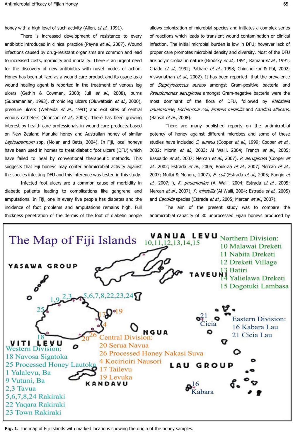
111 97
112 98
113 99
114 100
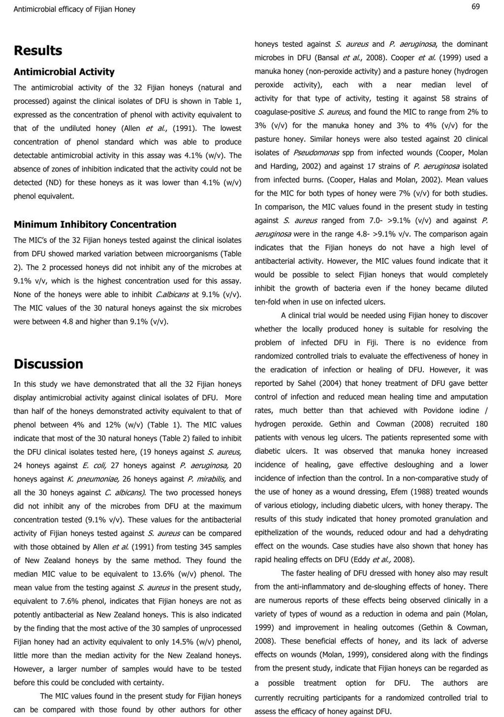
115 101
116 102
117 103
118 104
The medicinal use of honey has been known since ancient
 THE JOURNAL OF ALTERNATIVE AND COMPLEMENTARY MEDICINE Volume 13, Number 4, 2007, pp. 439 441 Mary Ann Liebert, Inc. DOI: 10.1089/acm.2007.6366 Bactericidal Activity of Different Types of Honey Against
Topical Honey A Cost Effective Option For Wound Management
 ISPUB.COM The Internet Journal of Third World Medicine Volume 8 Number 2 Topical Honey A Cost Effective Option For Wound Management S Salati, A Rather Citation S Salati, A Rather. Topical Honey A Cost
Journal of Research in Biology
 Journal of Research in Biology Original Research paper An International Online Open Access Publication group Assessment of the in vitro antibacterial activity of honey on some common human pathogens Journal
Topical antimicrobial agents in wound care. Professor Val Edwards-Jones Manchester Metropolitan University UK
 Topical antimicrobial agents in wound care Professor Val Edwards-Jones Manchester Metropolitan University UK Antimicrobial agents Antibacterial agents Antifungal agents Antiviral agents Antiparasitic agents?others
CHAPTER 2 THE ANTIMICROBIAL ACTIVITY OF HONEY
 CHAPTER 2 THE ANTIMICROBIAL ACTIVITY OF HONEY Rose Cooper It is now widely accepted that honey has antimicrobial activity and that this is dependent upon a variety of different modes of action (Molan,
CHAPTER 8 ANTIBACTERIAL ACTIVITY OF THE CRUDE ETHANOLIC EXTRACT AND THE ISOLATED COMPOUNDS FROM THE STEM OF COSTUS IGNEUS
 CHAPTER 8 ANTIBACTERIAL ACTIVITY OF THE CRUDE ETHANOLIC EXTRACT AND THE ISOLATED COMPOUNDS FROM THE STEM OF COSTUS IGNEUS 8.1 INTRODUCTION Medicinal plants are the backbone of traditional medicine and
Growth inhibition of foodborne pathogens and food spoilage organisms by select raw honeys
 International Journal of Food Microbiology 97 (2004) 1 8 www.elsevier.com/locate/ijfoodmicro Growth inhibition of foodborne pathogens and food spoilage organisms by select raw honeys Melissa A. Mundo,
Honey as nutrient and functional. Prof: Maha M. Saber Head of Complementary Medicine Department National Research Centre, Egypt
 Honey as nutrient and functional food Prof: Maha M. Saber Head of Complementary Medicine Department National Research Centre, Egypt Nutrient And Functional Food Recent years have seen growing interest
A new selective blood agar medium for Streptococcus pyogenes and other haemolytic streptococci
 J. clin. Path. (1964), 17, 231 A new selective blood agar medium for Streptococcus pyogenes and other haemolytic streptococci E. J. L. LOWBURY, A. KIDSON, AND H. A. LILLY From the Medical Research Council
Honey has been used in wound care since the
 Honey as a dressing for chronic wounds in adults Carolyn Fox Carolyn Fox is Clinical Nurse Specialist - Tissue Viability, University Hospital Lewisham, London Series editor: Peter Griffiths Honey has been
Department of Employment, Economic Development and Innovation
 Department of Employment, Economic Development and Innovation Berringa bioactive honey Comparison of honey vs silver for antibacterial activity January 2012 This project was commissioned by: Peter Woodward
Healing Properties of Honey. From honeyo.com
 Healing Properties of Honey From honeyo.com American Apitherapy Society - A non-profit membership organization devoted to advancing the investigation and promoting the use of honey bee products to further
Antibacterial, antifungal and antioxidant activities of honey collected from Timergara (Dir, Pakistan)
") Antibacterial, antifungal and antioxidant activities of honey collected from Timergara (Dir, Pakistan) Muhammad Zahoor, Sumaira Naz and Muhammad Sangeen Department of Chemistry University of Malakand,
Antibacterial, antifungal and antioxidant activities of honey collected from Timergara (Dir, Pakistan) Muhammad Zahoor, Sumaira Naz and Muhammad Sangeen Department of Chemistry University of Malakand,
Arglaes provides a seven-day, non-cytotoxic barrier against infection
 Arglaes provides a seven-day, non-cytotoxic barrier against infection Arglaes Controlled-Release Silver Technology Reduce bioburden with Arglaes Silver Barrier Dressings Antimicrobial Arglaes began the
Chapter 4 Inflammation and Infection
 Chapter 4 Inflammation and Infection Defense Mechanisms Three lines of defense protect the body against foreign invasion: Physical or surface barriers Inflammation Immune response Inflammation Non-specific
Honey has a potent antibacterial activity and is very effective
 THE JOURNAL OF ALTERNATIVE AND COMPLEMENTARY MEDICINE Volume 14, Number 2, 2008, pp. 181 184 Mary Ann Liebert, Inc. DOI: 10.1089/acm.2007.0589 Synergistic Action of Starch and Honey Against Pseudomonas
Normal Flora. CLS 212: Medical Microbiology
 Normal Flora CLS 212: Medical Microbiology Relationships between Organisms Symbiosis Permanent association between two different organisms. Neutralism Two organisms living together, and neither is affected
Asian Journal of Pharmaceutical Analysis and Medicinal Chemistry Journal home page:
 Research Article CODEN: AJPAD7 ISSN: 2321-0923 Asian Journal of Pharmaceutical Analysis and Medicinal Chemistry Journal home page: www.ajpamc.com FORMULATION AND EVALUATION OF HERBAL WASH FOR NASAL HEALTH
The effect of probiotics on animal health: a focus on host s natural intestinal defenses
 The effect of probiotics on animal health: a focus on host s natural intestinal defenses Guillaume Tabouret Animal Health Dept. Joint Unit 1225 Host Pathogens Interactions History of probiotics and definition
PROLONGED USE OF COUGH FORMULATIONS AND THE HEALTH RISK FROM THEIR ANTIMICROBIAL ACTIVITY ON SOME NORMAL BACTERIAL FLORA
 ORIGINAL ARTICLE AFRICAN JOURNAL OF CLINICAL AND EXPERIMENTAL MICROBIOLOGY JANUARY 2010 ISBN 155-68X VOL 11(1) AJCEM/2008140/21007 -http://www.ajol.info/journals/ajcem COPYRIGHT 2010 AFR. J. CLN. EXPER.
Higher plants produced hundreds to thousands of diverse chemical compounds with different biological activities (Hamburger and Hostettmann, 1991).
.") 4. ANTIMICROBIAL ACTIVITY OF PHYSALIS MINIMA L. 4.1. Introduction Use of herbal medicines in Asia represents a long history of human interactions with the environment. Plants used for traditional medicine
4. ANTIMICROBIAL ACTIVITY OF PHYSALIS MINIMA L. 4.1. Introduction Use of herbal medicines in Asia represents a long history of human interactions with the environment. Plants used for traditional medicine
Chapter 4. Anti-bacterial studies of PUFA extracts from Sardinella longiceps and Sardinella fimbriata. 4.1 Introduction
 Anti-bacterial studies of PUFA extracts from Sardinella longiceps and Sardinella fimbriata C o n t e n t s 4.1 Introduction 4.2 Materials and Methods 4.2.1 Extract Preparation and Determination of PUFA
SCREENING THE BIOACTIVE POTENTIAL OF PROTEIN ISOLATED FROM CYPRINUS CARPIO. Iyyanuchamy, S.K and A. Periyanayagasamy*
 Research Paper: Iyyanuchamy and Periyanayagasamy., 2011: Pp.6-10 SCREENING THE BIOACTIVE POTENTIAL OF PROTEIN ISOLATED FROM CYPRINUS CARPIO Iyyanuchamy, K and A. Periyanayagasamy* Department of Microbiology,
The antibacterial activity of honey and its role in treating diseases
 The antibacterial activity of honey and its role in treating diseases Disclaimer: The content of this document is for general information purposes only and does not constitute, nor does it intend to constitute,
ANTIBACTERIAL ACTIVITY OF GYMNEMA SYLVESTRE HYDROALCOHOLIC LEAF EXTRACT.
 International Journal of Advanced Research and Review www.ijarr.in ANTIBACTERIAL ACTIVITY OF GYMNEMA SYLVESTRE HYDROALCOHOLIC LEAF EXTRACT. Dr.Mayuri Thanwar 1, Dr.Dhananjay Dwivedi 2 1. Scientific Officer,
FACTORS AFFECTING THE GROWTH OF MICRO-ORGANISMS IN FOODS
 FACTORS AFFECTING THE GROWTH OF MICRO-ORGANISMS IN FOODS FACTORS AFFECTING THE GROWH OF MICRO-ORGANISMS IN FOODS Several factors related to the environment and the conditions in which food is stored influence
Antimicrobial activity of Karuveppilai vadagam against Enteric pathogens
 International Journal of Current Research in Medical Sciences ISSN: 2454-5716 P-ISJN: A4372-3064, E -ISJN: A4372-3061 www.ijcrims.com Original Research Article Volume 4, Issue 6-2018 Antimicrobial activity
Bioprospecting of Neem for Antimicrobial Activity against Soil Microbes
 ISSN: 2454-132X Impact factor: 4.295 (Volume3, Issue1) Available online at: www.ijariit.com Bioprospecting of Neem for Antimicrobial Activity against Soil Microbes R. Prasanna PRIST University, Tamilnadu
URINARY TRACT INFECTIONS 3 rd Y Med Students. Prof. Dr. Asem Shehabi Faculty of Medicine, University of Jordan
 URINARY TRACT INFECTIONS 3 rd Y Med Students Prof. Dr. Asem Shehabi Faculty of Medicine, University of Jordan Urinary Tract Infections-1 Normal urine is sterile.. It contains fluids, salts, and waste products,
In vitro antimicrobial activity of leaves and bark extracts of Ficus religiosa (Linn.)
") INDIAN JOURNAL OF PHARMACEUTICAL & BIOLOGICAL RESEARCH (IJPBR). Vol. 1 (1), March., 13 In vitro antimicrobial activity of leaves and bark extracts of Ficus religiosa (Linn.) G.Ramakrishnaiah * 1, T. Hariprasad
INDIAN JOURNAL OF PHARMACEUTICAL & BIOLOGICAL RESEARCH (IJPBR). Vol. 1 (1), March., 13 In vitro antimicrobial activity of leaves and bark extracts of Ficus religiosa (Linn.) G.Ramakrishnaiah * 1, T. Hariprasad
Evaluation of antibacterial potential of some Indian honey samples against throat and skin infective pathogens
 ISSN: 2319-7706 Volume 3 Number 1 (2014) pp. 362-369 http://www.ijcmas.com Original Research Article Evaluation of antibacterial potential of some Indian honey samples against throat and skin infective
Kamaruddin MY.,Anwar S., Zainabe SA and Mohd Yassim MY Faculty of Medicine University of Malaya Kuala Lumpur MALAYSIA
 Kamaruddin MY.,Anwar S., Zainabe SA and Mohd Yassim MY Faculty of Medicine University of Malaya Kuala Lumpur MALAYSIA ANTIBACTERIAL SYSTEM IN HONEY THE ANTIBACTERIAL SYSTEM 1993 HIGH SUGAR CONCENTRATION
ASEAN GUIDELINES ON LIMITS OF CONTAMINANTS FOR TRADITIONAL MEDICINES (TM) AND HEALTH SUPPLEMENTS (HS)
 AND HEALTH SUPPLEMENTS (HS)") Association of South East Asian Nations (ASEAN) ASEAN GUIDELINES ON LIMITS OF CONTAMINANTS FOR TRADITIONAL MEDICINES (TM) AND HEALTH SUPPLEMENTS (HS) Version 3.0 CONTENTS 1. Introduction...2 2. Objective...
Association of South East Asian Nations (ASEAN) ASEAN GUIDELINES ON LIMITS OF CONTAMINANTS FOR TRADITIONAL MEDICINES (TM) AND HEALTH SUPPLEMENTS (HS) Version 3.0 CONTENTS 1. Introduction...2 2. Objective...
NOVASTREAK. Microbial Contamination Monitoring Device TYPICAL CULTURAL MORPHOLOGY Baird Parker Agar. S. aureus growth on Baird Parker Agar
 NOVASTREAK Microbial Contamination Monitoring Device TYPICAL CULTURAL MORPHOLOGY Baird Parker Agar S. aureus growth on Baird Parker Agar Baird Parker Agar is used for the selective isolation and enumeration
HONEY 1
 1 Is made by bees from nectar from flowers (Floral) and from tree saps (Honey dew). Man is indebted to the honeybee for searching earths fields and forests for their treasured sweetness Prepared by the
International Journal of Scientific & Engineering Research, Volume 7, Issue 8, August ISSN
 International Journal of Scientific & Engineering Research, Volume 7, Issue 8, August-2016 105 Antimicrobial activity of Andrographis paniculata stem extracts. S.Gurupriya 1 and Dr.L.Cathrine 2 1 M.phil
COMMUNICATION A NEW ANTIMICROBIAL AGENT FROM ALMOND (PRUNUS AMYGDALIS) SHELLS. Department of Surgery, Medical College, Amritsar
 SHELLS. Department of Surgery, Medical College, Amritsar") SHORT COMMUNICATION A NEW ANTIMICROBIAL AGENT FROM ALMOND (PRUNUS AMYGDALIS) SHELLS By YUDHVEER SACHDEV Department of Surgery, Medical College, Amritsar A constant search for a perfect antibacterial agent
SHORT COMMUNICATION A NEW ANTIMICROBIAL AGENT FROM ALMOND (PRUNUS AMYGDALIS) SHELLS By YUDHVEER SACHDEV Department of Surgery, Medical College, Amritsar A constant search for a perfect antibacterial agent
Manuka honey as a medicine P. C. Molan, Honey Research Unit, University of Waikato, Hamilton, New Zealand
 Manuka honey as a medicine P. C. Molan, Honey Research Unit, University of Waikato, Hamilton, New Zealand E-mail: p.molan@waikato.ac.nz Honey - an ancient remedy "rediscovered" The usage of honey as a
Influence of Different Prebiotics and Probiotics on Selective Intestinal Pathogens
 ISSN: 2319-7706 Volume 3 Number 10 (2014) pp. 657-663 http://www.ijcmas.com Original Research Article Influence of Different Prebiotics and Probiotics on Selective Intestinal Pathogens Anayata Sharma 1*
COMPARATIVE ANTI MICROBIAL STUDY OF SHUDDHA KASISA AND KASISA BHASMA
 Research Article International Ayurvedic Medical Journal ISSN:2320 5091 COMPARATIVE ANTI MICROBIAL STUDY OF SHUDDHA KASISA AND KASISA BHASMA Dr. Nisha Kumari.P. R *Dr. Dinesh Nayak J **, Dr. Sathyanarayana
Preservation Power of Honey
 Preservation Power of Honey Name Food Preservation Methods How do people prevent the following foods from spoiling? Beef jerky Bread Milk Pickles Raw steak Cucumber Cup cake Croutons Twinkie My prediction
RN Cathy Hammond. Specialist Wound Management Service at Nurse Maude Christchurch
 RN Cathy Hammond Specialist Wound Management Service at Nurse Maude Christchurch 14:00-14:55 WS #141: Wound Infection - What You Need to Know 15:05-16:00 WS #153: Wound Infection - What You Need to Know
Microbiology Lab Microbial Growth: Environmental Factors
 Microbiology Lab Microbial Growth: Environmental Factors Ex. 2-7: Oxygen Requirements: Effects on Growth A. Bacteria may use oxygen as part of the cellular respiration. Cellular respiration is the process
The antimicrobial activity of honeys produced in the Czech Republic
 Original Paper Czech J. Anim. Sci., 50, 05 (8): 376 384 The antimicrobial activity of honeys produced in the Czech Republic L. VORLOVÁ 1, R. KARPÍŠKOVÁ 2, I. CHABINIOKOVÁ 3, K. KALÁBOVÁ 1, Z. BRÁZDOVÁ
KILLS FOUR. Watch Informative Video
 Watch Informative Video 1 KILLS FOUR Virus Bacteria Mold Fungus After a decade of testing Inspired TEC s Cluster Ion Technology emerges as a formidable strategy to combat many of the biological, chemical
Comparative Efficacy Of Endodontic Medicaments Against Enterococcus Faecalis Biofilms
 Comparative Efficacy Of Endodontic Medicaments Against Enterococcus Faecalis Biofilms A thesis submitted to the University of Adelaide in partial fulfilment of the requirements for the Degree of Doctor
Management of traumatic wounds by honey dressing at pravara rural hospital, Loni: A prospective study
 2018; 4(4): 566-570 ISSN: 2395-1958 IJOS 2018; 4(4): 566-570 2018 IJOS www.orthopaper.com Received: 01-08-2018 Accepted: 05-09-2018 Dr. DV Prasad Professor and HOD, Dr. Nikhil Suri Assistant Professor,
ANNEX III ASEAN GUIDELINES ON LIMITS OF CONTAMINANTS FOR TRADITIONAL MEDICINES
 Association of South East Asian Nations (ASEAN) ANNEX III ASEAN GUIDELINES ON LIMITS OF CONTAMINANTS FOR TRADITIONAL MEDICINES Disclaimer: This document is provided for information purpose only and subject
Process Optimization for Preparation of an Antibacterial Agent Using a Combined Extract of Tulsi and Marigold Leaves
 IOSR Journal of Environmental Science, Toxicology and Food Technology (IOSR-JESTFT) e-issn: 319-40,p- ISSN: 319-399.Volume 11, Issue 5 Ver. I (May. 017), PP 31-35 www.iosrjournals.org Process Optimization
Introduction. Crowded environments where the air is re-circulated can often be heavily infected with unseen germs and viruses.
 Introduction The continued increase in urbanisation,, population growth and global travel means germs and viruses can spread faster and further than ever before. Air, sea, rail and road travel have never
Lab 4. Blood Culture (Media) MIC AMAL-NORA-ALJAWHARA 1
 MIC AMAL-NORA-ALJAWHARA 1") Lab 4. Blood Culture (Media) 2018 320 MIC AMAL-NORA-ALJAWHARA 1 Blood Culture 2018 320 MIC AMAL-NORA-ALJAWHARA 2 What is a blood culture? A blood culture is a laboratory test in which blood is injected
Lab 4. Blood Culture (Media) 2018 320 MIC AMAL-NORA-ALJAWHARA 1 Blood Culture 2018 320 MIC AMAL-NORA-ALJAWHARA 2 What is a blood culture? A blood culture is a laboratory test in which blood is injected
THE "REVERSAL," NEUTRALIZATION, AND SELECTIVITY
 THE "REVERSAL," NEUTRALIZATION, AND SELECTIVITY OF GERMICIDAL CATIONIC DETERGENTS' MORTON KLEIN AND ZELMA G. KARDON Department of Bacteriology, School of Medicine, University of Pennsylvania, Philadelphia
Evaluation of Antibacterial Effect of Odor Eliminating Compounds
 Evaluation of Antibacterial Effect of Odor Eliminating Compounds Yuan Zeng, Bingyu Li, Anwar Kalalah, Sang-Jin Suh, and S.S. Ditchkoff Summary Antibiotic activity of ten commercially available odor eliminating
Burton's Microbiology for the Health Sciences
 Burton's Microbiology for the Health Sciences Section VII. Pathogenesis and Host Defense Mechanisms Burton's Microbiology for the Health Sciences Chapter 14. Pathogenesis of Infectious Diseases 1 Chapter
HARMONISED PHARMACOPOEIA DEHYDRATED CULTURE MEDIA FOR SUPPORTING REGULATORY COMPLIANCE AVAILABLE NOW P O RTF O LIO.
 DEHYDRATED CULTURE MEDIA FOR ENHANCED P O RTF O LIO AVAILABLE NOW HARMONISED PHARMACOPOEIA SUPPORTING REGULATORY COMPLIANCE A Neogen Company THE GATEWAY TO MICROBIOLOGY INTRODUCTION Harmonised Pharmacopoeia;
MECHANISMS. Dr. WILLIAM J. BENJAMIN. Eye Physiology & Ocular Prosthetics Laboratory. School of Optometry
 OCULAR DEFENSE MECHANISMS Dr. WILLIAM J. BENJAMIN Eye Physiology & Ocular Prosthetics Laboratory University of Alabama at Birmingham School of Optometry Presented at the UAB School of Optometry as Part
. Chemistry Department, Faculty of Science, Chulalongkom University, Bangkok 10330, Thailand. Antibacterial Activity of Some Essential Oils
 Antibacterial Activity of Some Essential Oils Sophon Roengsumran*, Amorn Petsom*, Suthep Thaniyavarn**, Surachai Pompakakul*, and Suntree Khantahiran*. Chemistry Department, Faculty of Science, Chulalongkom
*MIAN SHAHZADA ZIA AHMAD & ZAHEER-UD-DIN KHAN. Department of Botany, GC University, Lahore. ABSTRACT INTRODUCTION
 BIOLOGIA (PAKISTAN) 01, 8 (1&), 17-17 PK ISSN 000-09 Antibacterial activity of crude s of different parts of Butea monosperma (Lamk.) Taub. *MIAN SHAHZADA ZIA AHMAD & ZAHEER-UD-DIN KHAN Department of Botany,
Relation of the Heat Resistance of Salmonellae to
 APPLED MICROBIOLOGY, Mar. 1970, p. 9- Copyright 1970 American Society for Microbiology Vol. 19, No. Printed in U.S.A. Relation of the Heat Resistance of Salmonellae to the Water Activity of the Environment1
Ch 15. Microbial Mechanisms of Pathogenicity
 Ch 15 Microbial Mechanisms of Pathogenicity Student Learning Outcomes Identify the principal portals of entry and exit. Using examples, explain how microbes adhere to host cells. Explain how capsules and
6. SUMMARY AND CONCLUSION
 6. SUMMARY AND CONCLUSION Infectious diseases are one of man s oldest enemies. They continue to be a serious burden around the world, in developing and industrialised countries alike. It is said that every
MANAGEMENT OF LEG WOUNDS/ULCERS WITH HONEY. Ms. Irny Hidaya Binte Muhammad Raffles Hospital Nurse Clinician (Wound Care)
") MANAGEMENT OF LEG WOUNDS/ULCERS WITH HONEY Ms. Irny Hidaya Binte Muhammad Raffles Hospital Nurse Clinician (Wound Care) Outline: Normal Phases of Wound Healing Wound Infection Continuum Non-healing wounds
MANAGEMENT OF LEG WOUNDS/ULCERS WITH HONEY Ms. Irny Hidaya Binte Muhammad Raffles Hospital Nurse Clinician (Wound Care) Outline: Normal Phases of Wound Healing Wound Infection Continuum Non-healing wounds
Phytochemical screening and antibacterial properties of Garcinia kola
 2013; 2(3): 34-38 Online at: www.phytopharmajournal.com Research Article ISSN 2230-480X JPHYTO 2013; 2(3): 34-38 2013, All rights reserved Phytochemical screening and antibacterial properties of Garcinia
Aerobic bacteria isolated from diabetic septic wounds
 Aerobic bacteria isolated from diabetic septic wounds Eithar Mohammed Mahgoub*, Mohammed Elfatih A. Omer Faculty of Pharmacy, Omdurman Islamic University Department of Pharmaceutical Microbiology, Omdurman
The relative importance of the SANUKEHL remedies within SANUM therapy Extending regulation therapy by biological methods
 The relative importance of the SANUKEHL remedies within SANUM therapy Extending regulation therapy by biological methods by Dr. Dr. Peter Schneider Verlag für experimentelle Onkologie GmbH 27316 Hoya Germany
Additive Activity of Royal Jelly and Honey Against Pseudomonas aeruginosa
 Additive Activity of Royal Jelly and Against Pseudomonas aeruginosa Laid Boukraa, DVM, MSc, PhD Abstract As natural products garner attention in the medical field, the emergence of antibiotic-resistant
SUSCEPTIBILITY OF MYCOBACTERIUM ULCERANS TO HERBAL PREPARATIONS
 IN-VITRO SUSCEPTIBILITY OF MYCOBACTERIUM ULCERANS TO HERBAL PREPARATIONS Phyllis G. A. Addo MSc.. (Vet. Med), Ph.D Noguchi Memorial Institute for Medical Research INTRODUCTION Surgery is the main treatment
Antibiotic Activity of Various Types of Honey of Apis Species
 World Journal of Agricultural Sciences 9 (4): 309-315, 2013 ISSN 1817-3047 IDOSI Publications, 2013 DOI: 10.5829/idosi.wjas.2013.9.4.1607 Antibiotic Activity of Various Types of Honey of Apis Species 1
Re-introducing Honey in the Management of Wounds and Ulcers Theory and Practice
 Ostomy/Wound Management 48 (11) 28-40 (2002) Re-introducing Honey in the Management of Wounds and Ulcers Theory and Practice Peter C. Molan, BSc(Hons), PhD Dr. Molan is Associate Professor of Biochemistry
Silverzanit antibacterial ceramics for a healthy foundation.
 Silverzanit antibacterial ceramics for a healthy foundation. Die Fliesen der Sonderklasse. Silver for health, tiles of a class of its own for architecture. Silverzanit antibacterial ceramics is active
MICROBIOLOGICAL PROFILE
 Evans Vanodine International plc G L O B A L H Y G I E N E S O L U T I O N S HANDSAN MICROBIOLOGICAL PROFILE 2 INTRODUCTION HANDSAN is a ready to use, quick acting and highly effective, alcohol based hygienic
Positioning of new CMH quality mark as it relates to Certification of Medical-grade Manuka honey used in LMP wound care dressings versus other brands.
 Positioning of new CMH quality mark as it relates to Certification of Medical-grade Manuka honey used in LMP wound care dressings versus other brands. Overview Links Medical Products Inc. (LMP) wound care
There is restriction of free rotation ( freedom) across. for vicinal hydrogen & OH to take trans position.
 across. for vicinal hydrogen & OH to take trans position.") 5. SUMMARY & CONCLUSION. 5.1. Synthesis of Novel dihydro methyl Chalcones and their biological activity. The starting materials for the synthesis of various substituted dihydro 3-methyl chalcones were
5. SUMMARY & CONCLUSION. 5.1. Synthesis of Novel dihydro methyl Chalcones and their biological activity. The starting materials for the synthesis of various substituted dihydro 3-methyl chalcones were
ANTIBACTERIAL ACTIVITY OF LOCAL MALAYSIAN HONEY
 Malaysian Journal of Pharmaceutical Sciences, Vol. 3, No. 2, 1 10 (2005) ANTIBACTERIAL ACTIVITY OF LOCAL MALAYSIAN HONEY NURIZA TUMIN 1, N. ARSYIAH A. HALIM 1, M. SHAHJAHAN 2*, NOOR IZANI N. J., MUNAVVAR
International Journal of Pharma and Bio Sciences A COMPARITIVE STUDY OF ANTIMICROBIAL ACTIVITY OF SOME HERBS AND THEIR SYNERGISTIC EFFECT ABSTRACT
 Research Article Microbiology International Journal of Pharma and Bio Sciences ISSN 0975-6299 A COMPARITIVE STUDY OF ANTIMICROBIAL ACTIVITY OF SOME HERBS AND THEIR SYNERGISTIC EFFECT SNEHA GOGTE*¹ AND
4b. Innate (nonspecific) Immunity
 Immunity") 4b. Innate (nonspecific) Immunity Chapter 16: Innate (nonspecific) Immunity! Some terms:! Susceptibility: Lack of immunity to a disease.! Immunity: Ability to ward off disease.! Innate immunity: Defenses
4b. Innate (nonspecific) Immunity Chapter 16: Innate (nonspecific) Immunity! Some terms:! Susceptibility: Lack of immunity to a disease.! Immunity: Ability to ward off disease.! Innate immunity: Defenses
(NATO STANAG 2122, CENTO STANAG 2122, SEATO STANAG 2122)
") (NATO STANAG 2122, CENTO STANAG 2122, SEATO STANAG 2122) Bacteria Bacteria are microscopic, single-celled forms of plant life, containing no chlorophyll. They live on the skin, on the surface of the stratum
(NATO STANAG 2122, CENTO STANAG 2122, SEATO STANAG 2122) Bacteria Bacteria are microscopic, single-celled forms of plant life, containing no chlorophyll. They live on the skin, on the surface of the stratum
Lab 4: Enzymes ANSWER KEY. 1. How could you test to see if an enzyme was completely saturated during an experiment? (2 pts)
") Lab 4: Enzymes ANSWER KEY Pre-Lab Questions 1. How could you test to see if an enzyme was completely saturated during an experiment? (2 pts) To test this, you d simply see if the reaction was still taking
Lab 4: Enzymes ANSWER KEY Pre-Lab Questions 1. How could you test to see if an enzyme was completely saturated during an experiment? (2 pts) To test this, you d simply see if the reaction was still taking
320 MBIO Microbial Diagnosis. Aljawharah F. Alabbad Noorah A. Alkubaisi 2017
 320 MBIO Microbial Diagnosis Aljawharah F. Alabbad Noorah A. Alkubaisi 2017 Blood Culture What is a blood culture? A blood culture is a laboratory test in which blood is injected into bottles with culture
EVALUATION OF THE ANTIBACTERIAL, ANTIBIOFILM, ANTIOXIDANT, AND CYTOTOXIC EFFECTS OF SOME TURKISH HONEYS
 EVALUATION OF THE ANTIBACTERIAL, ANTIBIOFILM, ANTIOXIDANT, AND CYTOTOXIC EFFECTS OF SOME TURKISH HONEYS OZGUR CEYLAN a*, AYSEL UGUR b, MUSTAFA ISILOGLU c, FILIZ OZCAN c a Apiculture Program, Ula Ali Kocman
Understanding probiotics and health
 Understanding probiotics and health Gemma Laws MSc Student Microbiology and Immunology Department The gut microbiota The name given to the total microbial population living in our intestine Bacteria, fungi,
Honey compared with silver sulphadiazine in the treatment of superficial partial-thickness burns
 ORIGINAL ARTICLE Honey compared with silver sulphadiazine in the treatment of superficial partial-thickness burns Kamran Ishaque Malik, MA Nasir Malik, Azhar Aslam Malik KI, Malik MAN, Aslam A. Honey compared
ESSENTIAL OILS & The MICROBIOME
 ESSENTIAL OILS & The MICROBIOME BAHAR MAHMOUDI CONTENTS - The Human Microbiome - Benefits of the Normal Flora - Microbiome Improvement - Essential Oils 1 What is Human Microbiome Microbe: Tiny living organism,
For Fresh4Sure we use 10% Manuka 400+ (400 ppm MGO) together with the anti-bacterial components Erythritol and Zinccitrate.
 together with the anti-bacterial components Erythritol and Zinccitrate.") Peroxide activity: The major contributor to the antimicrobial activity of honey is hydrogen peroxide which is formed from the oxidation of glucose by glucose oxidase to gluconic acid and hydrogen peroxide
Peroxide activity: The major contributor to the antimicrobial activity of honey is hydrogen peroxide which is formed from the oxidation of glucose by glucose oxidase to gluconic acid and hydrogen peroxide
Chandan Prasad.et.al. Int. Journal of Engineering Research and Application ISSN : , Vol. 7, Issue 9, ( Part -6) September 2017, pp.
 September 2017, pp.") RESEARCH ARTICLE OPEN ACCESS Antibacterial properties of Unripened Fruits of Trapa natans, Annona squamosa, Ficus carica and Anthocephalus cadamba against Pathogenic Gram Positive and Negative Bacteria.
RESEARCH ARTICLE OPEN ACCESS Antibacterial properties of Unripened Fruits of Trapa natans, Annona squamosa, Ficus carica and Anthocephalus cadamba against Pathogenic Gram Positive and Negative Bacteria.
1. Effect of Temperature on Antibacterial Activity of Honey
 ~'} ~" The Antimicrobial Action of Honey Original Article 1. Effect of Temperature on Antibacterial Activity of Honey Nongluksna Sriubolmas M.Sc. (Pharm)* Areerat Laorpaksa M.Sc. (Pharm)* Saree Virunhaphol
MICROBIOLOGY - An Overview
 MICROBIOLOGY - An Overview Hieucam Phan, MD Pediatrics St. Luke s Hospital San Francisco, CA Microbiology 6/01 1 Introduction Major Achievements of Medical Sciences in the 20th Century Microbiology DNA
Evaluation of Antibacterial Activity of Two Different Honeys against Clinical Isolates of β-hemolytic Streptococci Group A 1
 Evaluation of Antibacterial Activity of Two Different Honeys against Clinical Isolates of β-hemolytic Streptococci Group A 1 Ivan Kotris 1, Jasminka Talapko 2, Domagoj Drenjančević 2,3 1 General Hospital
SCREENING OF ANTIMICROBIAL ACTIVITY OF OILS FROM DIFFERENT SPECIES OF OSCIMUM AGAINST PATHOGENIC BACTERIAL STRAINS
 I: 1077-1085 ISSN: 2277 4998 SCREENING OF ANTIMICROBIAL ACTIVITY OF OILS FROM DIFFERENT SPECIES OF OSCIMUM AGAINST PATHOGENIC BACTERIAL STRAINS PRAJAPATHI DN, SAROJA K* AND NAROLKAR SN Ashok & Rita Patel
SUBMISSION OF THE FINAL REPORT OF THE WORK DONE ON THE PROJECT
 SUBMISSION OF THE FINAL REPORT OF THE WORK DONE ON THE PROJECT NAME OF THE PRINCIPAL INVESTIGATOR : Dr.V.M.Barot, NAME AND ADDRESS OF THE INSTITUTION : Smt.S.M.Panchal Science College, -383215,Gujarat
The Adaptive Immune Responses
 The Adaptive Immune Responses The two arms of the immune responses are; 1) the cell mediated, and 2) the humoral responses. In this chapter we will discuss the two responses in detail and we will start
Physical, Chemical, and Microbiological Characteristics of Healthy Drink that Contains Honey and Duck Egg Yolk in Difference Age
 Physical, Chemical, and Microbiological Characteristics of Healthy Drink that Contains Honey and Duck Egg Yolk in Difference Age Z. Wulandari 1) *, R.R.A. Maheswari 1), & S.M. Anggraini 2) 1 Department
Manuka Honey: A Potent Cariostatic Agent An in vitro Study
 ORIGINAL ARTICLE Manuka 10.5005/jp-journals-10005-1494 Honey: A Potent Cariostatic Agent Manuka Honey: A Potent Cariostatic Agent An in vitro Study 1 Javaregowda P Beena, 2 Punyatoya Sahoo, 3 Sapna Konde,
In vitro study of antibacterial activity of Carissa carandas leaf extracts
 Available online at www.pelagiaresearchlibrary.com Asian Journal of Plant Science and Research, 2012, 2 (1):36-40 ISSN : 2249-7412 CODEN (USA): AJPSKY In vitro study of antibacterial activity of Carissa
Research Article Antibacterial Efficacy of Raw and Processed Honey
 SAGE-Hindawi Access to Research Biotechnology Research International Volume 2011, Article ID 917505, 6 pages doi:10.4061/2011/917505 Research Article Antibacterial Efficacy of Raw and Processed Honey D.
SUPPORTING INFORMATION. Studies on antimicrobial evaluation of some 1-((1-(1H-benzo[d]imidazol-2-
![SUPPORTING INFORMATION. Studies on antimicrobial evaluation of some 1-((1-(1H-benzo[d]imidazol-2-](/thumbs/79/79646314.jpg "SUPPORTING INFORMATION. Studies on antimicrobial evaluation of some 1-((1-(1H-benzo[d]imidazol-2-") SUPPORTIG IFORMATIO Studies on antimicrobial evaluation of some 1-((1-(1H-benzo[d]imidazol-2- yl)ethylidene)amino)-6-((arylidene)amino)-2-oxo-4-phenyl-1,2-dihydropyridine-3,5- dicarbonitriles C Desai*,
SUPPORTIG IFORMATIO Studies on antimicrobial evaluation of some 1-((1-(1H-benzo[d]imidazol-2- yl)ethylidene)amino)-6-((arylidene)amino)-2-oxo-4-phenyl-1,2-dihydropyridine-3,5- dicarbonitriles C Desai*,
PRESCRIBING INFORMATION. BaciJect. Bacitracin for injection U.S.P. Powder for Solution. 50,000 Units/Vial. Antibiotic
 PRESCRIBING INFORMATION BaciJect Bacitracin for injection U.S.P. Powder for Solution 50,000 Units/Vial Antibiotic SteriMax Inc. 2770 Portland Drive, Oakville, ON L6H 6R4 Date of Revision: July 30, 2018
(a) (i) Which type of cells ingest and kill invading microbes? (lines 3-4) ... (1)
 (i) Which type of cells ingest and kill invading microbes? (lines 3-4) ... (1)") Q1. Read the following passage. The immune system is the body s defence force. It protects against infections which might enter the body. The potential invaders include bacteria and viruses. The two basic
Q1. Read the following passage. The immune system is the body s defence force. It protects against infections which might enter the body. The potential invaders include bacteria and viruses. The two basic
Effect of various solvents on bacterial growth in context of determining MIC of various antimicrobials
 ISPUB.COM The Internet Journal of Microbiology Volume 7 Number 1 Effect of various solvents on bacterial growth in context of determining MIC of various antimicrobials T Wadhwani, K Desai, D Patel, D Lawani,
St. Joseph's Journal of Humanities and Science ISSN:
 M. Parimala Celia et al. / St. Joseph s Journal of Humanities and Science (Volume 4 Issue 1 January 2017) 23-28 33 St. Joseph s Journal of Humanities and Science (Volume 4 Issue 2 August 2015) 33-38 St.