Nature Methods: doi: /nmeth.3177
|
|
|
- Felix Gallagher
- 5 years ago
- Views:
Transcription
1
2 Supplementary Figure 1 Characterization of LysargiNase, trypsin and LysN missed cleavages. (a) Proportion of peptides identified in LysargiNase and trypsin digests of MDA-MB-231 cell lysates carrying 0, 1, 2 or 3 missed cleavage sites using enzyme-specific settings for peptide-to-sequence matching. IceLogos showing (b) unique cleavage sites, inferred from both ends of peptides identified in LysargiNase or trypsin-digested MDA-MB-231 proteomes (this study) or from peptides identified in Lys-N digested HEK293 cell lysates reported by Raijmakers et al. 12 and (c, d) the sequence context surrounding missed LysargiNase or trypsin cleavage sites at Arg or Lys and missed Lys-N cleavages sites at Lys. Shown are significant differences in occurrence (P < 0.05) as compared to (b,c) the natural amino acid abundance in the human proteome and (d) specific cleavage sites identified for each enzyme.
 at 37 C in 50 mm HEPES, 10 mm CaCl 2, ph 7.")
3 Supplementary Figure 2 LysargiNase activity in common proteomics conditions and solutions and at different temperatures. LysargiNase activity was assayed after 30 min incubation with resorufin-labeled casein (enzyme:substrate ratio 1:400 w/w) at 37 C in 50 mm HEPES, 10 mm CaCl 2, ph 7.5 with added indicated concentrations of (a) NaCl and reducing agents DTT and TCEP, (b) organic solvents methanol (MeOH) and acetonitrile (ACN), (c) the detergent RapiGest TM and denaturing concentrations of urea and (d)
4 denaturing chaotropic agents urea with thiourea and guanidine alone. Significance of difference to incubation at control conditions was tested with two-tailed Student s t-test (n = 3, * p < 0.05). (e) Incomplete LysargiNase digestion of human MDA-MB-231 cell lysate at 37 C under enzyme-limited conditions (enzyme:proteome 1:100 w/w) so as to semi-quantitatively monitor differences in proteome digest in the presence of detergents, organic solvents and reducing agents. Note that enzymatic activity in the presence of reducing agents is restored by quenching with the alkylating reagent iodoacetamide (IAM) added after reduction. (g) LysargiNase activity assayed after 30 min incubation with resorufin-labeled casein substrate (enzyme:substrate ratio 1:400 w/w) in 50 mm Hepes, 10 mm CaCl 2, ph 7.5 at the indicated temperatures. Significance of difference to incubation at 25 C was tested with two-tailed Student s t-test (n = 6, * p < 0.05). (h) Comparison of human MDA-MB-231 cell lysates digestion with LysargiNase at different enzyme:proteome ratios at 37 C and 50 C.
5 Supplementary Figure 3 MALDI-TOF based exopeptidase activity assay with synthetic peptides. The synthetic peptides (a) RGPANL KGR, (b) RGISEVNLDAEF RH, and (c) RGPANLIG R were used to assay tri-, di- and monocarboxypeptidase activities of LysargiNase, respectively. Peptides were analyzed by MALDI-TOF mass spectrometry after over-night incubation with (w/w 50:1; right panels) and without (left panels) LysargiNase. The cleavage product of RGPANLIG R was subsequently analyzed using a MALDI TOF/TOF tandem mass spectrometer, revealing a fragmentation spectrum dominated by b- series ions with a prominent arginine b1 ion (m/z 157) and no arginine y1 ion (m/z 175), ruling out aminopeptidase activity (not shown).
6 Supplementary Figure 4 MALDI-TOF based exopeptidase activity assay. (a) A tryptic BSA digest was used to (b) assay LysargiNase for amino- and carboxypeptidase activities. No aminopeptidase activity was observed, but two peptides (m/z and , indicated in blue) out of 10 tryptic BSA peptides were C-terminally processed indicating minor arginyl-carboxypeptidase activity. A LysargiNase autolytic peptide was observed (REMPMPR, m/z ; indicated in black).
7
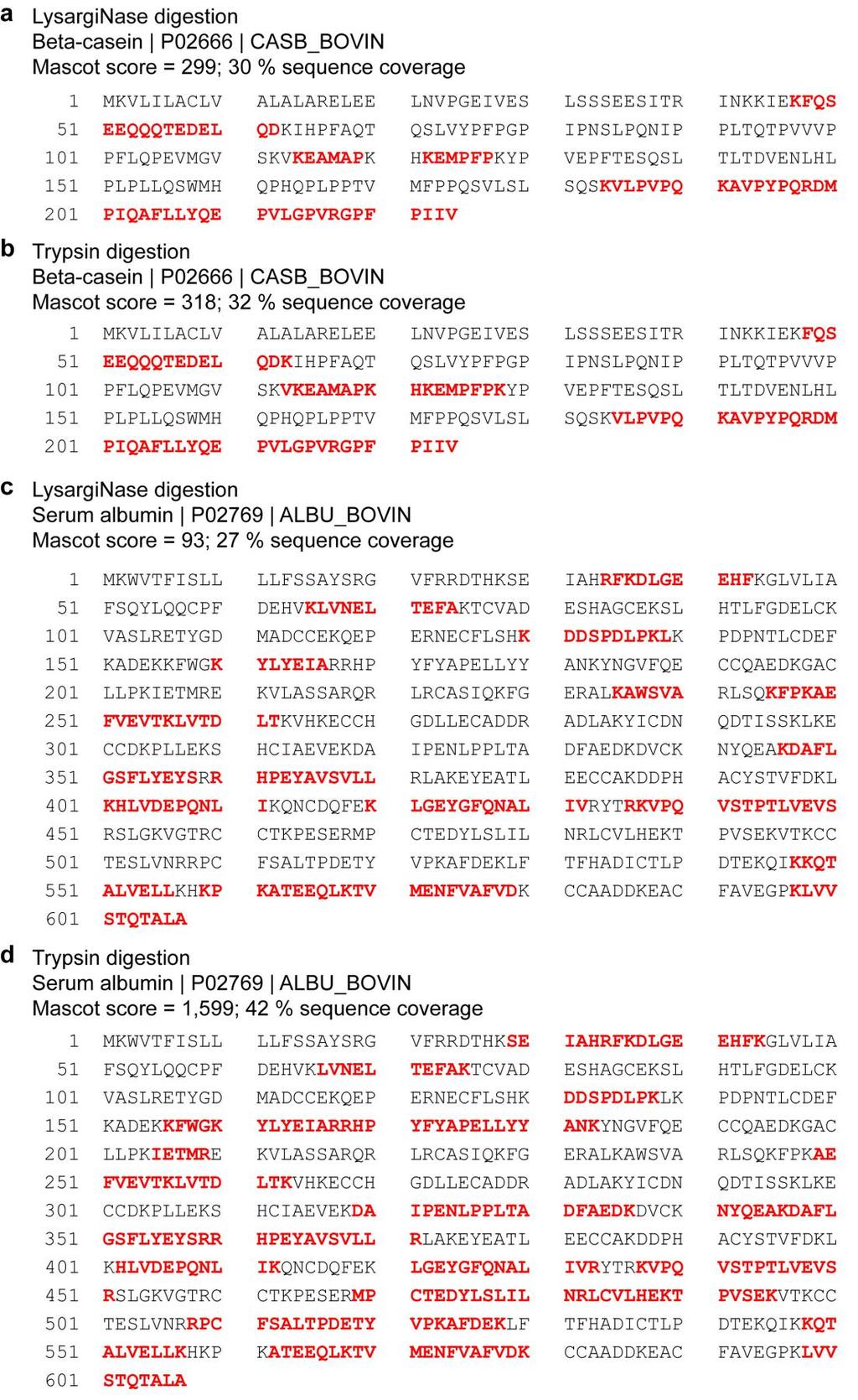
8 Supplementary Figure 5 In-gel digests of bovine β-casein and BSA. Peptides identified by LC-MS/MS using beam-type CID after in-gel digestion of bovine beta-casein with (a) LysargiNase or (b) trypsin and bovine serum albumin with (c) LysargiNase or (d) trypsin. Peptides identified by Mascot database searches are highlighted in red, Mascot protein identification score and sequence coverage are indicated.
9 Supplementary Figure 6 Complementarity of LysargiNase and trypsin in shotgun proteomics. (a) Box plots of the ratios of b-type to y-type fragment ions observed in 8,463 ion trap CID peptide spectrum matches for trypsin and 5,474 for LysargiNase (FDR < 0.01). The centerlines show the medians, box limits indicate the 25th and 75th percentiles, whiskers extend to the 5th and 95th percentiles, and outliers are represented by dots. Significance of the difference was tested using the twotailed Student s t-test (*** P < 0.001). (b) Overlap of peptide sequences identified after trypsin or LysargiNase cleavage of proteomes. For comparison, N- or C-terminal basic residues were removed from the peptide sequences identified from LysargiNase and trypsin datasets, respectively.
10
11 Supplementary Figure 7 Examples of peptide fragmentation spectra with overlapping sequences but differential placement of identical basic residues. (a-d) Peptide ion trap CID fragmentation spectra from LysargiNase-digested MDA-MB-231 cell lysates peptides (left panels) and corresponding peptide fragmentation spectra from trypsin-digested MDA-MB-231 cell lysates covering the same protein sequence (right panels). Spectra are shown as matched to peptide sequences and annotated by MaxQuant v
12
13 Supplementary Figure 8 Examples of peptide fragmentation spectra with overlapping sequences but differential placement of varying basic residues. (a-d) Peptide ion trap CID fragmentation spectra from LysargiNase-digested MDA-MB-231 cell lysates peptides (left panels) and corresponding peptide fragmentation spectra from trypsin-digested MDA-MB-231 cell lysates covering the same protein sequence (right panels). Spectra are shown as matched to peptide sequences and annotated by MaxQuant v
14 Supplementary Figure 9 Effect of a-ion fragments on spectral quality and Comet XCORR scores. (a) Beam-type CID MS/MS spectra of the LysargiNase peptide RLLVDCYSPVE (top panel) and corresponding tryptic peptide LLVDCYSPVER (bottom panel). The tryptic peptide exhibits strong and extensive y-type fragment ions whereas the LysargiNasederived peptide exhibits strong b-type and a-type fragment ions. (b) Increase in Comet XCORR score by inclusion of a-type fragment ions during peptide-to-sequence matching of beam-type CID fragmentation spectra, plotted as % increase. Inclusion of a-type fragment ions increased scores for LysargiNase-derived peptides by 33% (left panel), significantly stronger than for tryptic peptides (right panel), where scores increased only by 15% (P < , two sided Student s t-test, one of two replicates shown). Notably, inclusion of a-type fragment ions did not affect the number of identifications, solely their confidence.
15 Supplementary Figure 10 Protein C-terminal peptides identified from LysargiNase-digested MDA-MB-231 cell lysates. (a-f) Selected exemplary ion trap CID peptide fragmentation spectra matched to protein C-terminal peptide sequences and annotated by MaxQuant v
 Phosphorylation motifs identified by motif-x and their prevalence among high confidence phosphopeptides, with a localization probability > 0.75 identified by MaxQuant at an FDR < 0.")
16 Supplementary Figure 11 LysargiNase and trypsin facilitate identification of different phosphorylation motifs in phosphoproteomics. (a) Phosphorylation motifs identified by motif-x and their prevalence among high confidence phosphopeptides, with a localization probability > 0.75 identified by MaxQuant at an FDR < 0.01 from trypsin and LysargiNase-digested human MDA-MB-231 cell lysates (*P < 0.05, **P < 0.01, ***P < 0.001, two-tailed Student s t-test). (b) Match of kinase specificity with identified phosphorylation motifs as extracted from the human protein reference database (HRDB). (c) Relative change in abundance of phosphorylation motifs following stimulation by 12-O-tetradecanoylphorbol-13-acetate (PMA) for 5 and 20 min.
17
18 Supplementary Figure 12 Examples of corresponding phosphosites identified both in LysargiNase and tryptic digests of MDA-MB-231 cell lysates. (a-e) Selected ion-trap CID spectra from LysargiNase generated peptides (left panels) show stronger b-type fragment ions series and generated higher Andromeda scores and higher phosphosite localization probabilities than corresponding spectra from tryptic peptides (right panels). Within each peptide sequence phosphosite localization probabilities are enclosed within brackets to the right of each modified residue. Spectra are shown as matched to peptide sequences and annotated by MaxQuant v
19 Supplementary Figure 13 Monomethylated peptides identified from LysargiNase-digested MDA-MB-231 cell lysates. Exemplary ion trap CID fragmentation spectra matching peptides containing (a-c) mono-methylated Lys or (d-f) mono-methylated Arg, identified and annotated by MaxQuant v
20 Supplementary Figure 14 Dimethylated peptides identified from LysargiNase-digested MDA-MB-231 cell lysates. Exemplary ion trap CID fragmentation spectra matching peptides containing (a, b) di-methylated Lys or (c-f) di-methylated Arg, identified and annotated by MaxQuant v
 Structural model depicting a possible interaction between a dimethylated lysine side chain and the LysargiNase S 1' pocket.")
21 Supplementary Figure 15 LysargiNase facilitates identification of peptides with methylated and dimethylated arginine and lysine residues. (a) Structural model depicting a possible interaction between a dimethylated lysine side chain and the LysargiNase S 1' pocket. This model is based on the experimental crystal structure of the enzyme with an arginine-valine dipeptide occupying the S 1 and S 2 positions (pdb access code 2CKI 13 ). (b) Peptides with methylated or dimethylated arginine and lysine as a proportion of all peptides identified in shotgun proteomics analysis of human MDA-MB-231 cell lysates using ion trap CID fragmentation (n = 4). White, tryptic digests; grey, LysargiNase digests. Center lines show the medians, box limits indicate the 25th and 75th percentiles, whiskers extend 1.5 times the interquartile range from the 25th and 75th percentiles, and outliers are represented by dots. Significance of the difference between LysargiNase and trypsin datasets was tested with two-tailed Student s t-test (* p < 0.05, *** p < 0.001).
Protein Identification and Phosphorylation Site Determination by de novo sequencing using PepFrag TM MALDI-Sequencing kit
 Application Note Tel: +82-54-223-2463 Fax : +82-54-223-2460 http://www.genomine.com venture ldg 306 Pohang techno park Pohang, kyungbuk, Korea(ROK) Protein Identification and Phosphorylation Site Determination
Improve Protein Analysis with the New, Mass Spectrometry- Compatible ProteasMAX Surfactant
 Improve Protein Analysis with the New, Mass Spectrometry- Compatible Surfactant ABSTRACT Incomplete solubilization and digestion and poor peptide recovery are frequent limitations in protein sample preparation
Supporting Information. Lysine Propionylation to Boost Proteome Sequence. Coverage and Enable a Silent SILAC Strategy for
 Supporting Information Lysine Propionylation to Boost Proteome Sequence Coverage and Enable a Silent SILAC Strategy for Relative Protein Quantification Christoph U. Schräder 1, Shaun Moore 1,2, Aaron A.
Multiplex Protein Quantitation using itraq Reagents in a Gel-Based Workflow
 Multiplex Protein Quantitation using itraq Reagents in a Gel-Based Workflow Purpose Described herein is a workflow that combines the isobaric tagging reagents, itraq Reagents, with the separation power
The distribution of log 2 ratio (H/L) for quantified peptides. cleavage sites in each bin of log 2 ratio of quantified. peptides
 for quantified peptides. cleavage sites in each bin of log 2 ratio of quantified. peptides") Journal: Nature Methods Article Title: Corresponding Author: Protein digestion priority is independent of their abundances Mingliang Ye and Hanfa Zou Supplementary Figure 1 Supplementary Figure 2 The distribution
Journal: Nature Methods Article Title: Corresponding Author: Protein digestion priority is independent of their abundances Mingliang Ye and Hanfa Zou Supplementary Figure 1 Supplementary Figure 2 The distribution
Trypsin Mass Spectrometry Grade
 058PR-03 G-Biosciences 1-800-628-7730 1-314-991-6034 technical@gbiosciences.com A Geno Technology, Inc. (USA) brand name Trypsin Mass Spectrometry Grade A Chemically Modified, TPCK treated, Affinity Purified
Biomolecular Mass Spectrometry
 Lipids ot different than other organic small molecules Carbohydrates Polymers of monosaccharides linked via glycosidic bonds (acetals/ ketals) many different combinationsvery interesting no time ucleic
LysargiNase mirrors trypsin for protein C-termini and methylation site identification
 LysargiNase mirrors trypsin for protein C-termini and methylation site identification Pitter F. Huesgen 1-4, Philipp F. Lange 1-3, Lindsay D. Rogers 1-3, Nestor Solis 1-3, Ulrich Eckhard 1-3, Oded Kleifeld
Mass Spectrometry and Proteomics - Lecture 4 - Matthias Trost Newcastle University
 Mass Spectrometry and Proteomics - Lecture 4 - Matthias Trost Newcastle University matthias.trost@ncl.ac.uk previously Peptide fragmentation Hybrid instruments 117 The Building Blocks of Life DNA RNA Proteins
Nature Biotechnology: doi: /nbt Supplementary Figure 1
 Supplementary Figure 1 The timeline of the NGAG method for extraction of N-linked glycans and glycosite-containing peptides. The timeline can be changed based on the number of samples. Supplementary Figure
Protein sequence mapping is commonly used to
 Reproducible Microwave-Assisted Acid Hydrolysis of Proteins Using a Household Microwave Oven and Its Combination with LC-ESI MS/MS for Mapping Protein Sequences and Modifications Nan Wang and Liang Li
Characterization of Disulfide Linkages in Proteins by 193 nm Ultraviolet Photodissociation (UVPD) Mass Spectrometry. Supporting Information
 Mass Spectrometry. Supporting Information") Characterization of Disulfide Linkages in Proteins by 193 nm Ultraviolet Photodissociation (UVPD) Mass Spectrometry M. Montana Quick, Christopher M. Crittenden, Jake A. Rosenberg, and Jennifer S. Brodbelt
Characterization of Disulfide Linkages in Proteins by 193 nm Ultraviolet Photodissociation (UVPD) Mass Spectrometry M. Montana Quick, Christopher M. Crittenden, Jake A. Rosenberg, and Jennifer S. Brodbelt
The N-terminal loop of IRAK-4 death domain regulates ordered assembly of the Myddosome signalling scaffold
 The N-terminal loop of IRAK-4 death domain regulates ordered assembly of the Myddosome signalling scaffold Anthony C.G. Dossang * 1, Precious G. Motshwene * 1, Yang Yang* 1, Martyn F. Symmons*, Clare E.
Supplementary Materials for
 www.sciencesignaling.org/cgi/content/full/8/398/rs12/dc1 Supplementary Materials for Quantitative phosphoproteomics reveals new roles for the protein phosphatase PP6 in mitotic cells Scott F. Rusin, Kate
Figure S6. A-J) Annotated UVPD mass spectra for top ten peptides found among the peptides identified by Byonic but not SEQUEST + Percolator.
 Annotated UVPD mass spectra for top ten peptides found among the peptides identified by Byonic but not SEQUEST + Percolator.") Extending Proteome Coverage by Combining MS/MS Methods and a Modified Bioinformatics Platform adapted for Database Searching of Positive and Negative Polarity 193 nm Ultraviolet Photodissociation Mass
Extending Proteome Coverage by Combining MS/MS Methods and a Modified Bioinformatics Platform adapted for Database Searching of Positive and Negative Polarity 193 nm Ultraviolet Photodissociation Mass
Application Note # ET-17 / MT-99 Characterization of the N-glycosylation Pattern of Antibodies by ESI - and MALDI mass spectrometry
 Bruker Daltonics Application Note # ET-17 / MT-99 Characterization of the N-glycosylation Pattern of Antibodies by ESI - and MALDI mass spectrometry Abstract Analysis of the N-glycosylation pattern on
Increased Efficiency of Biomolecule Identification by Optimization of Trypsin Digestion Buffers
 Increased Efficiency of Biomolecule Identification by Optimization of Trypsin Digestion Buffers Phillip Humphryes, Valeria Barattini; Thermo Fisher Scientific, Runcorn, UK Application Note 274 Key Words
Protein Reports CPTAC Common Data Analysis Pipeline (CDAP)
") Protein Reports CPTAC Common Data Analysis Pipeline (CDAP) v. 05/03/2016 Summary The purpose of this document is to describe the protein reports generated as part of the CPTAC Common Data Analysis Pipeline
Protein Reports CPTAC Common Data Analysis Pipeline (CDAP) v. 05/03/2016 Summary The purpose of this document is to describe the protein reports generated as part of the CPTAC Common Data Analysis Pipeline
Biological Mass spectrometry in Protein Chemistry
 Biological Mass spectrometry in Protein Chemistry Tuula Nyman Institute of Biotechnology tuula.nyman@helsinki.fi MASS SPECTROMETRY is an analytical technique that identifies the chemical composition of
Lecture 3. Tandem MS & Protein Sequencing
 Lecture 3 Tandem MS & Protein Sequencing Nancy Allbritton, M.D., Ph.D. Department of Physiology & Biophysics 824-9137 (office) nlallbri@uci.edu Office- Rm D349 Medical Science D Bldg. Tandem MS Steps:
Universal sample preparation method for proteome analysis
 nature methods Universal sample preparation method for proteome analysis Jacek R Wi niewski, Alexandre Zougman, Nagarjuna Nagaraj & Matthias Mann Supplementary figures and text: Supplementary Figure 1
REDOX PROTEOMICS. Roman Zubarev.
 REDOX PROTEOMICS Roman Zubarev Roman.Zubarev@ki.se Physiological Chemistry I, Department for Medical Biochemistry & Biophysics, Karolinska Institutet, Stockholm What is (RedOx) Proteomics? Proteomics -
Nature Methods: doi: /nmeth Supplementary Figure 1
 Supplementary Figure 1 Subtiligase-catalyzed ligations with ubiquitin thioesters and 10-mer biotinylated peptides. (a) General scheme for ligations between ubiquitin thioesters and 10-mer, biotinylated
Lys-C/Arg-C, a More Specific and Efficient Digestion Approach for
 Lys-C/Arg-C, a More Specific and Efficient Digestion Approach for Proteomics Studies Zhen Wu, Jichang Huang, Jingnan Huang, Qingqing Li, Xumin Zhang *, State Key Laboratory of Genetic Engineering, Department
LABORATÓRIUMI GYAKORLAT SILLABUSZ SYLLABUS OF A PRACTICAL DEMONSTRATION. financed by the program
 TÁMOP-4.1.1.C-13/1/KONV-2014-0001 projekt Az élettudományi-klinikai felsőoktatás gyakorlatorientált és hallgatóbarát korszerűsítése a vidéki képzőhelyek nemzetközi versenyképességének erősítésére program
2. Ionization Sources 3. Mass Analyzers 4. Tandem Mass Spectrometry
 Dr. Sanjeeva Srivastava 1. Fundamental of Mass Spectrometry Role of MS and basic concepts 2. Ionization Sources 3. Mass Analyzers 4. Tandem Mass Spectrometry 2 1 MS basic concepts Mass spectrometry - technique
Supplementary Materials for
 www.sciencesignaling.org/cgi/content/full/6/278/rs11/dc1 Supplementary Materials for In Vivo Phosphoproteomics Analysis Reveals the Cardiac Targets of β-adrenergic Receptor Signaling Alicia Lundby,* Martin
Molecular Cell, Volume 46. Supplemental Information
 Molecular Cell, Volume 46 Supplemental Information Mapping N-Glycosylation Sites across Seven Evolutionary Distant Species Reveals a Divergent Substrate Proteome Despite a Common Core Machinery Dorota
Learning Objectives. Overview of topics to be discussed 10/25/2013 HIGH RESOLUTION MASS SPECTROMETRY (HRMS) IN DISCOVERY PROTEOMICS
 IN DISCOVERY PROTEOMICS") HIGH RESOLUTION MASS SPECTROMETRY (HRMS) IN DISCOVERY PROTEOMICS A clinical proteomics perspective Michael L. Merchant, PhD School of Medicine, University of Louisville Louisville, KY Learning Objectives
HIGH RESOLUTION MASS SPECTROMETRY (HRMS) IN DISCOVERY PROTEOMICS A clinical proteomics perspective Michael L. Merchant, PhD School of Medicine, University of Louisville Louisville, KY Learning Objectives
SUPPORTING INFORMATION. Lysine Carbonylation is a Previously Unrecognized Contributor. to Peroxidase Activation of Cytochrome c by Chloramine-T
 Electronic Supplementary Material (ESI) for Chemical Science. This journal is The Royal Society of Chemistry 2019 SUPPORTING INFORMATION Lysine Carbonylation is a Previously Unrecognized Contributor to
AccuMAP Low ph Protein Digestion Kits
 TECHNICAL MANUAL AccuMAP Low ph Protein Digestion Kits Instruc ons for Use of Products VA1040 and VA1050 5/17 TM504 AccuMAP Low ph Protein Digestion Kits All technical literature is available at: www.promega.com/protocols/
Quantification with Proteome Discoverer. Bernard Delanghe
 Quantification with Proteome Discoverer Bernard Delanghe Overview: Which approach to use? Proteome Discoverer Quantification Method What When to use Metabolic labeling SILAC Cell culture systems Small
Enhancing Sequence Coverage in Proteomics Studies by Using a Combination of Proteolytic Enzymes
 Enhancing Sequence Coverage in Proteomics Studies by Using a Combination of Proteolytic Enzymes Dominic Baeumlisberger 2, Christopher Kurz 3, Tabiwang N. Arrey, Marion Rohmer 2, Carola Schiller 3, Thomas
Quantitative chromatin proteomics reveals a dynamic histone. post-translational modification landscape that defines asexual
 Quantitative chromatin proteomics reveals a dynamic histone post-translational modification landscape that defines asexual and sexual Plasmodium falciparum parasites Nanika Coetzee 1, Simone Sidoli 2,
Sequence Identification And Spatial Distribution of Rat Brain Tryptic Peptides Using MALDI Mass Spectrometric Imaging
 Sequence Identification And Spatial Distribution of Rat Brain Tryptic Peptides Using MALDI Mass Spectrometric Imaging AB SCIEX MALDI TOF/TOF* Systems Patrick Pribil AB SCIEX, Canada MALDI mass spectrometric
Supplementary Figure 1 (previous page). EM analysis of full-length GCGR. (a) Exemplary tilt pair images of the GCGR mab23 complex acquired for Random
. EM analysis of full-length GCGR. (a) Exemplary tilt pair images of the GCGR mab23 complex acquired for Random") S1 Supplementary Figure 1 (previous page). EM analysis of full-length GCGR. (a) Exemplary tilt pair images of the GCGR mab23 complex acquired for Random Conical Tilt (RCT) reconstruction (left: -50,right:
S1 Supplementary Figure 1 (previous page). EM analysis of full-length GCGR. (a) Exemplary tilt pair images of the GCGR mab23 complex acquired for Random Conical Tilt (RCT) reconstruction (left: -50,right:
for the Identification of Phosphorylated Peptides
 Application of a Data Dependent Neutral-Loss Experiment on the Finnigan LTQ for the Identification of Phosphorylated Peptides Gargi Choudhary Diane Cho Thermo Electron, San Jose, CA Abstracted from posters
PTM Discovery Method for Automated Identification and Sequencing of Phosphopeptides Using the Q TRAP LC/MS/MS System
 Application Note LC/MS PTM Discovery Method for Automated Identification and Sequencing of Phosphopeptides Using the Q TRAP LC/MS/MS System Purpose This application note describes an automated workflow
Trypsin Digestion Mix
 G-Biosciences 1-800-628-7730 1-314-991-6034 technical@gbiosciences.com A Geno Technology, Inc. (USA) brand name 239PR Trypsin Digestion Mix Provides optimal buffered conditions for in gel trypsin digestion
Comparison of mass spectrometers performances
 Comparison of mass spectrometers performances Instrument Mass Mass Sensitivity resolution accuracy Quadrupole 1 x 10 3 0.1 Da* 0.5-1.0 pmol DE-MALDI 2 x 10 4 20 ppm 1-10 fmol peptide 1-5 pmol protein Ion
Proteomics Grade. Protocol. Catalog # Agilent Technologies. Research Use Only. Not for use in Diagnostic Procedures. Version A, January 2010
 Proteomics Grade Trypsin Catalog #204310 Protocol Version A, January 2010 Research Use Only. Not for use in Diagnostic Procedures. Agilent Technologies Notices Agilent Technologies, Inc. 2010 No part of
5 Identification of Binding Partners of the Annexin A2 / P11 Complex by Chemical Cross-Linking
 5 Identification of Binding Partners of the Annexin A2 / P11 Complex by Chemical Cross-Linking In the quest of the omics sciences for holistic schemes, the identification of binding partners of proteins
Supplementary Figures and Notes for Digestion and depletion of abundant
 Supplementary Figures and Notes for Digestion and depletion of abundant proteins improves proteomic coverage Bryan R. Fonslow, Benjamin D. Stein, Kristofor J. Webb, Tao Xu, Jeong Choi, Sung Kyu Park, and
Unsupervised Identification of Isotope-Labeled Peptides
 Unsupervised Identification of Isotope-Labeled Peptides Joshua E Goldford 13 and Igor GL Libourel 124 1 Biotechnology institute, University of Minnesota, Saint Paul, MN 55108 2 Department of Plant Biology,
Introduction to Peptide Sequencing
 Introduction to Peptide equencing Quadrupole Ion Traps tructural Biophysics Course December 3, 2014 12/8/14 Introduction to Peptide equencing - athan Yates 1 Why are ion traps used to sequence peptides?
Automating Mass Spectrometry-Based Quantitative Glycomics using Tandem Mass Tag (TMT) Reagents with SimGlycan
 Reagents with SimGlycan") PREMIER Biosoft Automating Mass Spectrometry-Based Quantitative Glycomics using Tandem Mass Tag (TMT) Reagents with SimGlycan Ne uaca2-3galb1-4glc NAcb1 6 Gal NAca -Thr 3 Ne uaca2-3galb1 Ningombam Sanjib
PREMIER Biosoft Automating Mass Spectrometry-Based Quantitative Glycomics using Tandem Mass Tag (TMT) Reagents with SimGlycan Ne uaca2-3galb1-4glc NAcb1 6 Gal NAca -Thr 3 Ne uaca2-3galb1 Ningombam Sanjib
Proteins: Proteomics & Protein-Protein Interactions Part I
 Proteins: Proteomics & Protein-Protein Interactions Part I Jesse Rinehart, PhD Department of Cellular & Molecular Physiology Systems Biology Institute DNA RNA PROTEIN DNA RNA PROTEIN Proteins: Proteomics
(III) MALDI instrumentation
 MALDI instrumentation") Dr. Sanjeeva Srivastava (I) Basics of MALDI-TF (II) Sample preparation In-gel digestion Zip-tip sample clean-up Matrix and sample plating (III) MALDI instrumentation 2 1 (I) Basics of MALDI-TF Analyte
Dr. Sanjeeva Srivastava (I) Basics of MALDI-TF (II) Sample preparation In-gel digestion Zip-tip sample clean-up Matrix and sample plating (III) MALDI instrumentation 2 1 (I) Basics of MALDI-TF Analyte
Babu Antharavally, Ryan Bomgarden, and John Rogers Thermo Fisher Scientific, Rockford, IL
 A Versatile High-Recovery Method for Removing Detergents from Low-Concentration Protein or Peptide Samples for Mass Spectrometry Sample Preparation and Analysis Babu Antharavally, Ryan Bomgarden, and John
Agilent Protein In-Gel Tryptic Digestion Kit
 Agilent 5188-2749 Protein In-Gel Tryptic Digestion Kit Agilent Protein In-Gel Tryptic Digestion Kit Instructions Kit Contents The Protein In-Gel Tryptic Digestion Kit includes sufficient reagents for approximately
Mass Spectrometry. - Introduction - Ion sources & sample introduction - Mass analyzers - Basics of biomolecule MS - Applications
 - Introduction - Ion sources & sample introduction - Mass analyzers - Basics of biomolecule MS - Applications Adapted from Mass Spectrometry in Biotechnology Gary Siuzdak,, Academic Press 1996 1 Introduction
Supporting information
 Electronic Supplementary Material (ESI) for ChemComm. This journal is The Royal Society of Chemistry 2014 Supporting information Glycan Reductive Isotope-coded Amino Acid Labeling (GRIAL) for Mass Spectrometry-based
In-Gel Tryptic Digestion Kit
 INSTRUCTIONS In-Gel Tryptic Digestion Kit 3747 N. Meridian Road P.O. Box 117 Rockford, IL 61105 89871 1468.2 Number Description 89871 In-Gel Tryptic Digestion Kit, sufficient reagents for approximately
SUPPLEMENTAL INFORMATION
 SUPPLEMENTAL INFORMATION EXPERIMENTAL PROCEDURES Tryptic digestion protection experiments - PCSK9 with Ab-3D5 (1:1 molar ratio) in 50 mm Tris, ph 8.0, 150 mm NaCl was incubated overnight at 4 o C. The
MALDI Imaging Mass Spectrometry
 MALDI Imaging Mass Spectrometry Nan Kleinholz Mass Spectrometry and Proteomics Facility The Ohio State University Mass Spectrometry and Proteomics Workshop What is MALDI Imaging? MALDI: Matrix Assisted
NIH Public Access Author Manuscript J Proteome Res. Author manuscript; available in PMC 2014 July 05.
 NIH Public Access Author Manuscript Published in final edited form as: J Proteome Res. 2013 July 5; 12(7): 3071 3086. doi:10.1021/pr3011588. Evaluation and Optimization of Mass Spectrometric Settings during
PHOSPHOPEPTIDE ANALYSIS USING IMAC SAMPLE PREPARATION FOLLOWED BY MALDI-MS and MALDI PSD MX
 PHOSPHOPEPTIDE ANALYSIS USING IMAC SAMPLE PREPARATION FOLLOWED BY MALDI-MS and MALDI PSD MX E. Claude 1, E. Emekwue 2, M. Snel 1, T. McKenna 1 and J. Langridge 1 1: Waters Corporation, Manchester, UK 2:
4th Multidimensional Chromatography Workshop Toronto (January, 2013) Herman C. Lam, Ph.D. Calibration & Validation Group
 Herman C. Lam, Ph.D. Calibration & Validation Group") 4th Multidimensional Chromatography Workshop Toronto (January, 2013) Herman C. Lam, Ph.D. Calibration & Validation Group MDLC for Shotgun Proteomics Introduction General concepts Advantages Challenges
4th Multidimensional Chromatography Workshop Toronto (January, 2013) Herman C. Lam, Ph.D. Calibration & Validation Group MDLC for Shotgun Proteomics Introduction General concepts Advantages Challenges
Peptide sequencing using chemically assisted fragmentation (CAF) and Ettan MALDI-ToF Pro mass spectrometry
 and Ettan MALDI-ToF Pro mass spectrometry") application note Ett MALDI-ToF Pro Peptide sequencing using chemically assisted fragmentation (CAF) d Ett MALDI-ToF Pro mass spectrometry Key words: MALDI-ToF, PSD, peptide sequencing, chemically assisted
application note Ett MALDI-ToF Pro Peptide sequencing using chemically assisted fragmentation (CAF) d Ett MALDI-ToF Pro mass spectrometry Key words: MALDI-ToF, PSD, peptide sequencing, chemically assisted
Advances in Hybrid Mass Spectrometry
 The world leader in serving science Advances in Hybrid Mass Spectrometry ESAC 2008 Claire Dauly Field Marketing Specialist, Proteomics New hybrids instruments LTQ Orbitrap XL with ETD MALDI LTQ Orbitrap
Mass Spectrometry. Mass spectrometer MALDI-TOF ESI/MS/MS. Basic components. Ionization source Mass analyzer Detector
 Mass Spectrometry MALDI-TOF ESI/MS/MS Mass spectrometer Basic components Ionization source Mass analyzer Detector 1 Principles of Mass Spectrometry Proteins are separated by mass to charge ratio (limit
One Gene, Many Proteins. Applications of Mass Spectrometry to Proteomics. Why Proteomics? Raghothama Chaerkady, Ph.D.
 Applications of Mass Spectrometry to Proteomics Raghothama Chaerkady, Ph.D. McKusick-Nathans Institute of Genetic Medicine and the Department of Biological Chemistry Why Proteomics? One Gene, Many Proteins
Supporting Information MALDI versus ESI the impact of the ion source on peptide identification
 Supporting Information MALDI versus ESI the impact of the ion source on peptide identification Wiebke Maria Nadler, 1 Dietmar Waidelich, 2 Alexander Kerner, 1 Sabrina Hanke, 1 Regina Berg, 3 Andreas Trumpp
Department of Chemistry, Université de Montréal, C.P. 6128, Succursale centre-ville, Montréal, Québec, H3C 3J7, Canada.
 Phosphoproteome dynamics of Saccharomyces cerevisiae under heat shock and cold stress Evgeny Kanshin 1,5, Peter Kubiniok 1,2,5, Yogitha Thattikota 1,3, Damien D Amours 1,3 and Pierre Thibault 1,2,4 * 1
SUPPORTING INFORMATION. Multimodal Mass Spectrometry Imaging of N-glycans and Proteins from the Same
 SUPPORTING INFORMATION Multimodal Mass Spectrometry Imaging of N-glycans and Proteins from the Same Tissue Section. Bram Heijs 1, Stephanie Holst 1, Inge H. Briaire-de Bruijn 2, Gabi W. van Pelt 3, Arnoud
Double charge of 33kD peak A1 A2 B1 B2 M2+ M/z. ABRF Proteomics Research Group - Qualitative Proteomics Study Identifier Number 14146
 Abstract The 2008 ABRF Proteomics Research Group Study offers participants the chance to participate in an anonymous study to identify qualitative differences between two protein preparations. We used
Systematic analysis of protein-detergent complexes applying dynamic light scattering to optimize solutions for crystallization trials
 Supporting information 1 2 3 Volume 71 (2015) Supporting information for article: 4 5 6 7 8 Systematic analysis of protein-detergent complexes applying dynamic light scattering to optimize solutions for
TiO 2 Mag Sepharose. SepharoseTM. GE Healthcare. Simple handling
 GE Healthcare Data file 28-9539-44 AA Protein sample preparation TiO 2 Mag Sepharose TiO 2 Mag Sepharose magnetic beads use titanium dioxide (TiO 2 )-based chromatography to simplify capture and enrichment
Quantitation of Protein Phosphorylation Using Multiple Reaction Monitoring
 Quantitation of Protein Phosphorylation Using Multiple Reaction Monitoring Application Note Authors Ning Tang, Christine A. Miller and Keith Waddell Agilent Technologies, Inc. Santa Clara, CA USA This
Faster Mass Spec: Same-Day Sample Prep Now a Reality. Michael M. Rosenblatt, Ph.D.
 Faster Mass Spec: Same-Day Sample Prep Now a Reality Michael M. Rosenblatt, Ph.D. Applications of Mass Spec in Biology Biomarker Discovery Protein Interactions Protein Expression Drug Discovery Subcellular
Shotgun Proteomics MS/MS. Protein Mixture. proteolysis. Peptide Mixture. Time. Abundance. Abundance. m/z. Abundance. m/z 2. Abundance.
 Abundance Abundance Abundance Abundance Abundance Shotgun Proteomics Protein Mixture 1 2 3 MS/MS proteolysis m/z 2 3 Time µlc m/z MS 1 m/z Peptide Mixture m/z Block Diagram of a Mass Spectrometer Sample
Supplementary Figure 1
 Supplementary Figure 1 Isolation of mt-trnas and RNA-MS analysis of mt-trna Asn from M. nudus (a)m. nudus mt-trnas were isolated by RCC and resolved by 10% denaturing PAGE. The gel was stained with SYBR
SUPPLEMENTARY INFORMATION
 SUPPLEMENTARY INFORMATION doi:10.1038/nature13418 Supplementary Results: USP30 opposes autophagic flux In HEK-293 cells, USP30 overexpression increased basal LC3-II levels, dependent on enzymatic activity,
Supplementary Figure-1. SDS PAGE analysis of purified designed carbonic anhydrase enzymes. M1-M4 shown in lanes 1-4, respectively, with molecular
 Supplementary Figure-1. SDS PAGE analysis of purified designed carbonic anhydrase enzymes. M1-M4 shown in lanes 1-4, respectively, with molecular weight markers (M). Supplementary Figure-2. Overlay of
Nature Structural & Molecular Biology: doi: /nsmb.3218
 Supplementary Figure 1 Endogenous EGFR trafficking and responses depend on biased ligands. (a) Lysates from HeLa cells stimulated for 2 min. with increasing concentration of ligands were immunoblotted
PREPARED FOR: U.S. Army Medical Research and Materiel Command Fort Detrick, Maryland
 AD Award Number: W81XWH-12-1-0248 TITLE: Targeted Inhibition of Tyrosine Kinase-Mediated Epigenetic Alterations to Prevent Resurgence of Castration-Resistant Prostate Cancer PRINCIPAL INVESTIGATOR: Dr.
Biotransformation-induced toxicity. Challenges in drug design M. Matilde Marques
 Biotransformation-induced toxicity. Challenges in drug design M. Matilde Marques Drug Discovery Session July 4, 2016 Drug Discovery Toxicology The Road to Safe Drugs Attrition in drug development remains
Development of a Human Cell-Free Expression System to Generate Stable-Isotope-Labeled Protein Standards for Quantitative Mass Spectrometry
 Development of a Human Cell-Free Expression System to Generate Stable-Isotope-Labeled Protein Standards for Quantitative Mass Spectrometry Ryan D. omgarden 1, Derek aerenwald 2, Eric Hommema 1, Scott Peterman
Flow-Through Electron Capture Dissociation in a novel Branched RF Ion Trap
 Flow-Through Electron Capture Dissociation in a novel Branched RF Ion Trap Takashi Baba, J. Larry Campbell, Yves Le Blanc, Jim. W. Hager and Bruce A. Thomson ASMS, June 18 / 2014 1 2014 AB SCIEX Trapping
Enrichment and Separation of Mono- and Multiply Phosphorylated Peptides Using Sequential Elution from IMAC Prior to Mass Spectrometric Analysis
 Chapter 6 Enrichment and Separation of Mono- and Multiply Phosphorylated Peptides Using Sequential Elution from IMAC Prior to Mass Spectrometric Analysis Tine E. Thingholm, Ole N. Jensen, and Martin R.
Supplementary Information. Top-down/bottom-up mass spectrometry workflow using dissolvable polyacrylamide gels
 Supplementary Information Top-down/bottom-up mass spectrometry workflow using dissolvable polyacrylamide gels Nobuaki Takemori,,* Ayako Takemori,, Piriya Wongkongkathep, Michael Nshanian, Rachel R. Ogorzalek
MALDI-TOF. Introduction. Schematic and Theory of MALDI
 MALDI-TOF Proteins and peptides have been characterized by high pressure liquid chromatography (HPLC) or SDS PAGE by generating peptide maps. These peptide maps have been used as fingerprints of protein
Technical Note # TN-31 Redefining MALDI-TOF/TOF Performance
 Bruker Daltonics Technical Note # TN-31 Redefining MALDI-TOF/TOF Performance The new ultraflextreme exceeds all current expectations of MALDI-TOF/TOF technology: A proprietary khz smartbeam-ii TM MALDI
Fig.S1 ESI-MS spectrum of reaction of ApA and THPTb after 16 h.
 Electronic Supplementary Material (ESI) for RSC Advances. This journal is The Royal Society of Chemistry 2014 Experiment Cleavage of dinucleotides Dinucleotides (ApA, CpC, GpG, UpU) were purchased from
Supplementary Information. Addressing proteolytic efficiency in enzymatic degradation therapy for celiac. disease
 Supplementary Information Addressing proteolytic efficiency in enzymatic degradation therapy for celiac disease Martial Rey, Menglin Yang, Linda Lee, Ye Zhang, Joey G. Sheff, Christoph W. Sensen, Hynek
Bench Protocol to Perform TAILS v4
 Bench Protocol to Perform TAILS v4 OVERALL Lab Protocols May 2016 We have been performing TAILS for 12 years and this is now a very streamlined and highly optimized approach. The one request we have is
SMART Digest Kit Facilitating perfect digestion
 Questions Answers SMART Digest Kit Facilitating perfect digestion The modern biopharmaceutical and protein research laboratory is tasked with providing high quality analytical results, often in high-throughput,
Supporting Information. Post translational Modifications of Serotonin Type 4 Receptor Heterologously Expressed in. Mouse Rod Cells
 Supporting Information Post translational Modifications of Serotonin Type 4 Receptor Heterologously Expressed in Mouse Rod Cells David Salom,, Benlian Wang,, Zhiqian Dong, Wenyu Sun, Pius Padayatti, Steven
Quantification by Mass Spectrometry
 Quantification by Mass Spectrometry PC219 Lecture 5 30 April, 2010 sguan@cgl.ucsf.edu 1 Applications of Quantitative Proteomics Qualitative protein identification is NOT sufficient to describe a biological
Proteomics of body liquids as a source for potential methods for medical diagnostics Prof. Dr. Evgeny Nikolaev
 Proteomics of body liquids as a source for potential methods for medical diagnostics Prof. Dr. Evgeny Nikolaev Institute for Biochemical Physics, Rus. Acad. Sci., Moscow, Russia. Institute for Energy Problems
Types of Modifications
 Modifications 1 Types of Modifications Post-translational Phosphorylation, acetylation Artefacts Oxidation, acetylation Derivatisation Alkylation of cysteine, ICAT, SILAC Sequence variants Errors, SNP
Supplementary Figure 1. Method development.
 Supplementary Figure 1 Method development. Titration experiments to determine standard antibody:lysate concentration. Lysates (~2 mg of total proteins) were prepared from cells expressing FLAG- tagged
Phosphorylation of proteins Steve Barnes Feb 19th, 2002 in some cases, proteins are found in a stable, hyperphosphorylated state, e.g.
 Phosphorylation of proteins Steve Barnes Feb 19th, 2002 in some cases, proteins are found in a stable, hyperphosphorylated state, e.g., casein more interestingly, in most other cases, it is a transient
TECHNICAL BULLETIN. R 2 GlcNAcβ1 4GlcNAcβ1 Asn
 GlycoProfile II Enzymatic In-Solution N-Deglycosylation Kit Product Code PP0201 Storage Temperature 2 8 C TECHNICAL BULLETIN Product Description Glycosylation is one of the most common posttranslational
The peptide samples of C. elegans (15 mg) and S. cerevisiae (20 mg) were prepared as
 and S. cerevisiae (20 mg) were prepared as") Supplementary material and methods The peptide samples of C. elegans (15 mg) and S. cerevisiae (20 mg) were prepared as described recently 1. The dried down peptides were resolubilized to a final concentration
Supplementary material and methods The peptide samples of C. elegans (15 mg) and S. cerevisiae (20 mg) were prepared as described recently 1. The dried down peptides were resolubilized to a final concentration
Bruker Daltonics. autoflex III smartbeam. The Standard in MALDI-TOF Performance MALDI-TOF/TOF. think forward
 Bruker Daltonics autoflex III smartbeam The Standard in MALDI-TOF Performance think forward MALDI-TOF/TOF Designed for a Routine High Level of Performance The autoflex III smartbeam brings the power of
MS1 and MS2 crosstalk in label free quantitation of mass spectrometry data independent acquisitions
 MS1 and MS2 crosstalk in label free quantitation of mass spectrometry data independent acquisitions MS1 528.18 +++ m/z 568.98 ++ m/z 678.34 ++ m/z MS2/SWATH June 9th, 2013 Matthew J. Rardin SIRT3 regulated
Profiling the Distribution of N-Glycosylation in Therapeutic Antibodies using the QTRAP 6500 System
 Profiling the Distribution of N-Glycosylation in Therapeutic Antibodies using the QTRAP 6500 System Scheduled MRM Pro Algorithm for Increased Efficiency of Targeted Detection Jenny Albanese 1, Christie
Research Article A Colorimetric Method for Monitoring Tryptic Digestion Prior to Shotgun Proteomics
 International Journal of Proteomics Volume 24, Article ID 25482, 9 pages http://dx.doi.org/.55/24/25482 Research Article A Colorimetric Method for Monitoring Tryptic Digestion Prior to Shotgun Proteomics
Optimization of Mass Spectrometry-Compatible Surfactants for Shotgun Proteomics
 Optimization of Mass Spectrometry-Compatible Surfactants for Shotgun Proteomics Emily I. Chen, Daniel Cociorva, Jeremy L. Norris, and John R. Yates, III*, Department of Cell Biology, 10550 North Torrey