locomotice system Plastinated specimensⅠ: Silicone specimens Regional specimens and organs
|
|
|
- Cordelia Tate
- 5 years ago
- Views:
Transcription
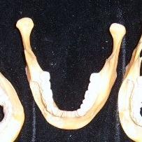
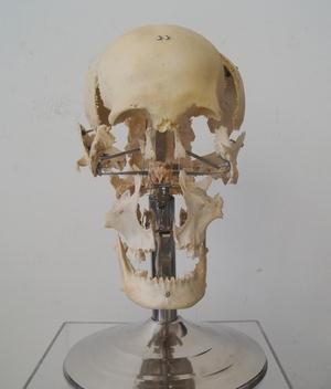
1 locomotice system Plastinated specimensⅠ: Silicone specimens Regional specimens and organs Art-No. Name Description The locomotor system SL001 Two hundred pieces of plastinated bones (without six The bones of the whole-body auditory ossicles). SL002 The whole skull Without the hyoid bone. SL003 Separated skull Separated twenty-three bones, including the temporal bone, sphenoid bone, ethmoid bone etc, which are set up in situ with stainless steel frame. SL004 Horizontal section of the Displaying: foramen, groove, orifice, lamina, skull protuberance, impression etc. SL005 Median-sagittal section of the Displaying: groove, orifice, lamina, sinus etc; may remove the zygomatic arch to show the pterygopalatine skull fossa. SL006 Coronary section of the skull Display: groove, orifice, lamina, sinus etc. SL007 The frontal bone Showing the arch, foramen, process, groove, crest, and impression etc. SL008 The parietal bone Showing the angle, line, foramen, groove, tubercle, and diploic veins etc. SL009 The sphenoid bone Showing the sinus, foramen, canal, groove, and fissure etc clearly. SL010 The temporal bone Showing the groove, border, canal, orifice, foramen, and fossa etc clearly. SL011 The maxilla Showing the groove, crest, process, orifice, foramen, and fossa etc clearly. SL012 The zygomatic bone and nasal bone Showing the structures outside. SL013 The ethmoid bone Keeping the important structures intact, for example the lamina, foramen, groove, and wing. SL014 The palatine bone Keeping the important structures intact, for example the groove, process, notch, and crest. SL015 The auditory ossicles 3 in number,putting in situ. SL016 Tympanic cavity Showing the facial nerve, cells, tympanic cavity(with temporal bone opened), vestibular fossa of carotid etc. SL017 Semicircular canal Showing the intact semicircular canals and spiral canal of cochlea. SL018 The mandible SL019 Structure of bone SL020 The clavicle SL021 The scapula SL022 The humerus the opened long bone,showing the structures of the bone.
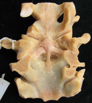
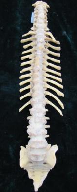
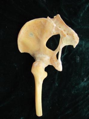
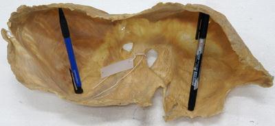
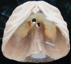
2 SL023 The radius and ulna SL024 The bones of the hand 27 in number. SL025 The hip bone SL026 The femur SL027 The tibia and fibula SL028 The patella SL029 Bones of the foot 26 in number. SL030 The atlas and axis SL031 The cervical vertebrae One vertebrae with adjacent intervertebral disc. SL032 The thoracic vertebrae One vertebrae with adjacent intervertebral disc. SL033 The lumbar vertebrae One vertebrae with adjacent intervertebral disc. SL034 The sacrum SL035 The sternum With xiphoid process. SL036 The costal bones SL037 The skull of infant Showing the cranial fontanelles. SL038 The temporomandibular joint Showing the articular disc. SL039 The intervertebral joints Showing the intervertebral disc (fibrous ring and pulpiform nucleus), ligaments. SL040 The atlantoaxial joint Showing the cruciform ligament of atlas. SL041 One half of female pelvis Cut sagitally, showing the ligaments of pelvic cavity. SL042 One half of male pelvis Cut sagitally, showing the ligaments of pelvic cavity. SL043 Full female pelvis Showing the ligaments of pelvic cavity. SL044 Full male pelvis Showing the ligaments of pelvic cavity. SL045 The spinal column Showing the correlative joints, ligaments, physiological curves, etc. SL046 The shoulder joint Reserve the correlative ligaments, open articular capsule to show the cavity. SL047 The elbow joint With an intact articular capsule. SL048 The elbow joint Showing the three ligaments and articular surface. SL049 The joints of hand Ligaments dissected, the wrist joint is coronally cut to show the articular disc and cavity. SL050 The hip joint With sacrotuberal and sacrospinal ligaments. SL051 SL052 SL053 SL054 The knee joint Sagittal section of the knee joint Meniscus The joints of the foot With the joint capsule removed, showing ligaments and articular cavity. With the patella and the patellar ligament reserved, showing the meniscus, cruciate ligament, transverse ligament etc. Ligaments dissected, coronally cut to show the intertarsal joints and the anatomy of. SL055 Half head specimen Superficial dissection SL056 Half head specimen deep dissection SL057 Full head specimen Keeping the pharynx closed,showing the posterior aspect of the pharyngeal muscles.
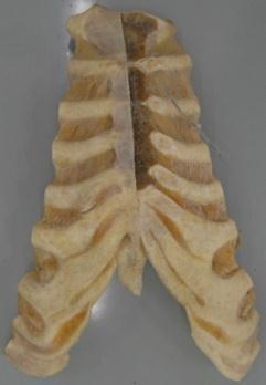
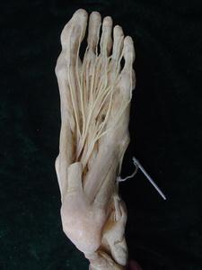
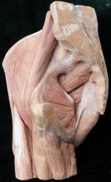
3 SL058 Full head specimen With stylohyoid muscles, pharynx opened, view upon larynx from rearside. SL059 The upper limb Superficial muscle dissection, with arteries and nerves. SL060 The upper limb Deep muscle dissection, with arteries and nerves. SL061 The upper limb Combined superficial and deep muscle dissection. SL062 Muscles of hand Superficial muscle dissection. SL063 Muscles of hand Deep muscle dissection. SL064 The diaphragm With part of spinal column, stomach, liver, spleen and pancreas. SL065 The diaphragm Without the viscera. SL066 Muscles of trunk Superficial muscle dissection. SL067 Muscles of trunk Deep muscle dissection. SL068 The male perineal muscles Superficial muscle dissection, with arteries and nerves. SL069 The female perineal muscles Superficial muscle dissection, with arteries and nerves. SL070 The lower limb With half pelvis attached, superficial muscle dissection. SL071 The lower limb With half pelvis attached, deep muscle dissection. SL072 The lower limb With half of pelvis attached, combined superficial and deep muscle dissection. SL073 Muscles of hip and thigh Bilateral, deep muscle dissection on one side, superficial muscle dissection on the other side. SL074 Muscles of leg Superficial muscle dissection SL075 Muscles of leg Deep muscle dissection. SL076 Muscles of foot Superficial dissection of sole. SL077 Muscles of foot Deep dissection of sole.


4 Art No / S L 004 Horizontal section of the skul Art No / S L005 Median-sagittal section of the skull Art No / S L011 The maxilla Showing the groove, crest, process, orifice, foramen, and fossa etc clearly Art No / S L015 The auditory ossicles 3 in number,putting in situ


5 Art No / S L 017 Semicircular canal Showing the intact semicircular canals and spiral canal of cochlea Art No / S L019 Structure of bone the opened long bone,showing the structures of the bone Art No / S L021 The scapula Art No / S L024 The bones of the hand 27 in number


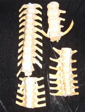
6 Art No / S L 027 The tibia and fibula Art No / S L029 Bones of the foot 26 in number Art No / S L 030 The atlas and axis Art No / S L 031 The cervical vertebrae One vertebrae with adjacent intervertebral disc


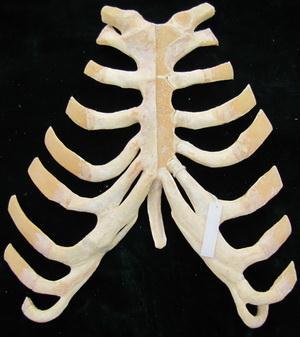
7 Art No / S L 033 The lumbar vertebrae One vertebrae with adjacent intervertebral disc Art No / S L034 The sacrum Art No / S L 035 The sternum with xiphoid process Art No / S L 37 The skull of infant Showing the cranial fontanelles.


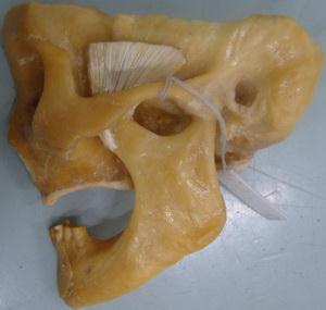
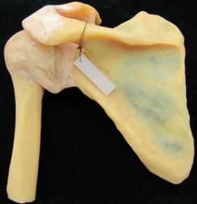
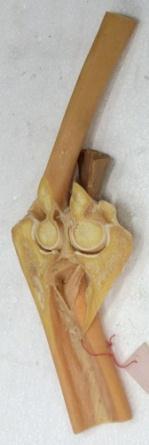
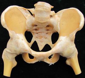
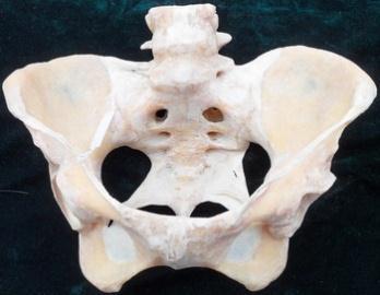
8 Art No / S L 39 The intervertebral joints Showing the intervertebral disc (fibrous ring and pulpiform nucleus), ligaments Art No / S L042 One half of male pelvis Cut sagitally, showing the ligaments of pelvic cavity Art No / S L 045 The spinal column Showing the correlative joints, ligaments, physiological curves, etc Art No / S L046 The shoulder joint reserve the correlative ligaments, open articular capsule to show the cavity


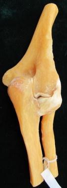
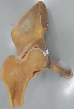
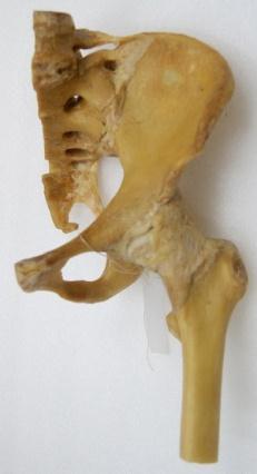
9 Art No / S L048 The elbow joint showing the three ligaments and articular surface Art No / S L049 The joints of hand Ligaments dissected, the wrist joint is coronally cut to show the articular disc and cavity Art No / S L050 The hip joint With sacrotuberal and sacrospinal ligaments Art No / S L051 The knee joint with the joint capsule removed, showing ligaments and articular cavity


10 Art No / S L052 Sagittal section of the knee joint Art No / S L 053 Meniscus with the patella and the patellar ligament reserved, showing the meniscus, cruciate ligament, transverse ligament etc Art No / S L054 The joints of the foot Art No / S L055 Half head specimen Superficial dissection


11 Art No / S L056 Half head specimen deep dissection Art No / S L059 The upper limb Superficial muscle dissection, with arteries and nerves Art No / S L 062 Muscles of hand superficial muscle dissection Art No / S L065 The diaphragm without the viscera


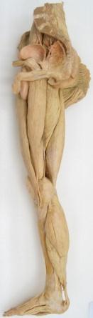
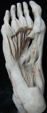
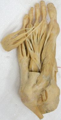
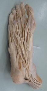
12 Art No / S L068 The male perineal muscles superficial muscle dissection, with arteries and nerves Art No / S L070 The lower limb with half pelvis attached, superficial muscle dissection Art No / S L 076 Muscles of foot Superficial dissection of sole


13 Separated skull Mandible Scapula Intervertebral joints Temporomandibular joint Sternocostal joint and sternoclavicular joint Vertebral column Atlanto-occipital and atlanto-axial joints Hip joint (coronal section) Elbow joint (sagittal section) Elbow joint Shoulder joint Female pelvic ligaments (half) Male pelvic ligaments (half) Female pelvic ligaments (whole) Male pelvic ligaments (whole)


14 Combined superficial dissection of whole head Combined deep dissection of head and neck Combined superficial dissection of head and neck Muscles of mastication Superficial muscles of the head Medial and lateral pterygoids


15 Deep dissection of upper limb muscles Superficial dissection of upper limb muscles Deep muscles of leg Superficial muscles of leg Deep muscles of the lower limb Superficial muscles of the lower limb
16 Articulationes sternocostales Anterior longitudinal ligament Deep muscles of the foot Middle muscles of the foot Combined middle dissection of foot muscles Combined superficial dissection of foot muscles Muscles of hip Diaphragm


")


17 Inguinal canal Diaphragm and muscles of the posterior abdominal wall Muscles of the trunk (Child) Muscles of the trunk (Adult) Superficial dissection of face Deep structure of face inferior to parotid gland


")


18 Deep blood vessels and nerves of facial lateral aspect(superficial layer) Deep blood vessels and nerves of facial lateral aspect (deep layer) Tentorium of cerebellum and sinuses of dura mater Superficial structure of neck Submandibular triangle Carotid triangle


 Dissection")


19 Internal and external carotid arteries and cranial nerves Anterior region of neck (superficial layer) Dissection of head, neck, upper limb and thoracic cavity Blood vessels of stomach Duodenum and pancreas Distribution and relations of pancreas Structure of retroperitoneal space


20 The adjacent organs of kidneys Muscles of male pelvic diaphragm Female superficial perineal space Dorsal artery and nerve of penis Anterior wall of axillary fossa Axillary lymph nodes
General Anatomy p. 1 Organization of the Human Body p. 1 Skeleton of the Human Body p. 4 Ossification of the Bones p. 6 Bone Structure p. 8 Joints p.
 General Anatomy p. 1 Organization of the Human Body p. 1 Skeleton of the Human Body p. 4 Ossification of the Bones p. 6 Bone Structure p. 8 Joints p. 10 Principal Joints (Immovable) p. 12 Synovial Joints
APPENDICULAR SKELETON 126 AXIAL SKELETON SKELETAL SYSTEM. Cranium. Skull. Face. Skull and associated bones. Auditory ossicles. Associated bones.
 SKELETAL SYSTEM 206 AXIAL SKELETON 80 APPENDICULAR SKELETON 26 Skull Skull and associated s 29 Cranium Face Auditory ossicles 8 4 6 Associated s Hyoid Thoracic cage 25 Sternum Ribs 24 Vertebrae 24 column
Cranium Facial bones. Sternum Rib
 Figure 7.1 The human skeleton. Skull Thoracic cage (ribs and sternum) Cranium Facial bones Sternum Rib Bones of pectoral girdle Vertebral column Sacrum Vertebra Bones of pelvic girdle (a) Anterior view
Chapter 7: Skeletal System: Gross Anatomy
 Chapter 7: Skeletal System: Gross Anatomy I. General Considerations A. How many bones in an average adult skeleton? B. Anatomic features of bones are based on II. Axial Skeleton A. Skull 1. Functionally
Biology 2401 The Skeletal System
 Biology 2401 The Skeletal System Purpose: The lab will describe the microscopic and gross anatomy of bone, identify bones of the body, and identify important bone markings. I. Overview of the Skeleton
Lab Exercise #04 The Skeletal System Student Performance Objectives
 Lab Exercise #04 The Skeletal System Student Performance Objectives The material that you are required to learn in this exercise can be found in either the lecture text or the supplemental materials provided
11/25/2012. Chapter 7 Part 2: Bones! Skeletal Organization. The Skull. Skull Bones to Know Cranium
 Chapter 7 Part 2: Bones! 5) Distinguish between the axial and appendicular skeletons and name the major parts of each 6) Locate and identify the bones and the major features of the bones that compose the
Skeletal system. Prof. Abdulameer Al-Nuaimi. E. mail:
 Skeletal system Prof. Abdulameer Al-Nuaimi E-mail: a.al-nuaimi@sheffield.ac.uk E. mail: abdulameerh@yahoo.com Functions of Bone and The Skeletal System Support: The skeleton serves as the structural framework
Nervous & Skeletal Systems. Virtual Science University
 Nervous & Skeletal Systems Virtual Science University 1 Nervous & Skeletal Systems Texas TEK B.10(A) The student will interpret the function of systems in organisms (humans) including the nervous and skeletal
CHAPTER 7, PART II (BONES)
") Anatomy Name: CHAPTER 7, PART II (BONES) Entry #: INSTRUCTIONS: 1) READ Chapter 7, pg. 140-161. 2) Using the outline, make a note card for each underlined bone name or phrase. 3) On each note card, put
Anatomy Name: CHAPTER 7, PART II (BONES) Entry #: INSTRUCTIONS: 1) READ Chapter 7, pg. 140-161. 2) Using the outline, make a note card for each underlined bone name or phrase. 3) On each note card, put
Biology 218 Human Anatomy. Adapted from Martini Human Anatomy 7th ed. Chapter 6 The Skeletal System: Axial Division
 Adapted from Martini Human Anatomy 7th ed. Chapter 6 The Skeletal System: Axial Division Introduction The axial skeleton: Composed of bones along the central axis of the body Divided into three regions:
BLUE SKY SCHOOL OF PROFESSIONAL MASSAGE AND THERAPEUTIC BODYWORK. Musculoskeletal Anatomy & Kinesiology I TERMINOLOGY, STRUCTURES, & SKELETAL OVERVIEW
 BLUE SKY SCHOOL OF PROFESSIONAL MASSAGE AND THERAPEUTIC BODYWORK Musculoskeletal Anatomy & Kinesiology I TERMINOLOGY, STRUCTURES, & SKELETAL OVERVIEW MSAK101-I Session 1 Learning Objectives: 1. Define
The Skeletal System: Axial Skeleton
 The Skeletal System: Axial Skeleton The Big Idea The Axial Skeleton & Homeostasis The bones of the axial skeleton contribute to homeostasis by protecting many of the body s organs such as the brain, spinal
Bone Flashcards for 10a
 Bone Flashcards for 0a CLAVICLE (collar bone). Sternal extremity (end) flat end. Acromial extremity (end) rounded end. SCAPULA (shoulder blade). Right or left scapula?. Superior border (superior margin).
Human Anatomy and Physiology - Problem Drill 07: The Skeletal System Axial Skeleton
 Human Anatomy and Physiology - Problem Drill 07: The Skeletal System Axial Skeleton Question No. 1 of 10 Which of the following statements about the axial skeleton is correct? Question #01 A. The axial
External Acoustic Meatus. Mastoid Process. Zygomatic Process. Temporal Bone
 Bone lab review 1. Frontal Bone 2. Supra-Orbital Foramen 3. Orbit (Orbital Cavity) 4. Superior Orbital Fissure 5. Inferior Orbital Fissure 6. Zygomatic Bone 7. Infra-Orbital Foramen 8. Maxilla 9. Mandible
Chapter 8A. The Skeletal System: The Axial Skeleton. The Skeletal System: The Axial Skeleton. Types of Bones. Types of Bones
 Chapter 8A The Skeletal System: The Axial Skeleton The Skeletal System: The Axial Skeleton 206 named bones Axial Skeleton 80 bones lie along longitudinal axis skull, hyoid, vertebrae, ribs, sternum, ear
Copyright 2010 Pearson Education, Inc.
 E. VERTEBRAL COLUMN 1. The vertebral column extends from the skull to the pelvis and forms the vertical axis of the skeleton. 2. The vertebral column is composed of vertebrae that are separated by intervertebral
Chapter 7. Skeletal System
 Chapter 7 Skeletal System 1 Skull A. The skull is made up of 22 bones: 8 cranial bones, 13 facial bones, and the mandible. B. The Cranium encloses and protects the brain, provides attachments for muscles,
Human Anatomy - Problem Drill 06: The Skeletal System Axial Skeleton & Articualtions
 Human Anatomy - Problem Drill 06: The Skeletal System Axial Skeleton & Articualtions Question No. 1 of 10 Instructions: (1) Read the problem and answer choices carefully, (2) Work the problems on paper
TEST YOURSELF- Chapter 7
 TEST YOURSELF- Chapter 7 Cranial Bones 1. Give the name of the bone for each of the following markings. Some of the markings are found on more than one bone. List all that apply. Cranium a. Frontal squama:
BIO 137 AXIAL SKELETON BONE STUDY THE HUMAN SKELETON
 BIO 137 THE AXIAL SKELETON MARY CATHERINE FLATH, Ph.D. THE HUMAN SKELETON AXIAL SKULL HYOID THORACIC CAGE VERTEBRAL COLUMN APPENDICULAR PECTORAL GIRDLE UPPER LIMBS PELVIC GIRDLE LOWER LIMBS AXIAL SKELETON
BONE CHALLENGE DANIL HAMMOUDI.MD
 BONE CHALLENGE DANIL HAMMOUDI.MD Bone Basic functions? A. support B. protection C. movement assistance in D. RBC formation-hemopoiesis E. mineral homeostasis +importance of calcium F. energy supply -yellow
Axial skeleton bones and markings
 Axial skeleton bones and markings Skull Cranial bones Frontal x 1 Supraorbital foramen Occipital x 1 Foramen magnum Occipital condyles Superior nuchal line Inferior nuchal line Anterior cranial fossa External
Bone List Anatomy
 1 Frontal Bone Skull 2 Parietal Bone Skull 3 Occipital Bone Skull 4 Temporal Bone Skull 5 Coronal Suture Skull 6 Sagittal Suture Skull 7 Squamous suture Skull 8 Lambdoid Suture Skull 9 Surpaorbital Ridge
Important Parts of Bones
 Important Parts of Bones For 2015 Know: Humerus (posterior) Clavical Femur (Anterior) Foot Hand Mandible Os Coxa Scapula Skull (Anterior, Inferior, Lateral) Sternum Humerus (posterior) A. olecranon fossa
Lab-1. Miss. Lina Al-Onazy & samar Al-Wgeet =)
") Lab-1 Introduction The human skeleton is composed of 300 bones at birth and by the time adulthood is reached, some bones have fused together to give a total of 206 bones in the body. The human skeleton
Lab-1 Introduction The human skeleton is composed of 300 bones at birth and by the time adulthood is reached, some bones have fused together to give a total of 206 bones in the body. The human skeleton
Anatomy and Physiology II. Review Spine and Neck
 Anatomy and Physiology II Review Spine and Neck Spine regions How many cervical vertibrae are there? 7 The curvature is the cervical region posterior? Concave posterior How many thoracic? And curvature?
Biology 210 Chapter 8: Skeletal Tissues Supplement 1
 Biology 210 Chapter 8: Skeletal Tissues Supplement 1 By John McGill Material contributed by Beth Wyatt & Jack Bagwell DIVISIONS OF THE SKELETAL SYSTEM AXIAL SKELETON (80 BONES) Bones of the Head, Neck,
Spring Written By: J. E. Sutton. Contents: I. Overview of the Skeleton: II. Appendicular Skeleton III. Axial Skeleton IV.
 Spring 2012 Written By: J. E. Sutton Contents: I. Overview of the Skeleton: II. Appendicular Skeleton III. Axial Skeleton IV. Articulations Overview of the Skeleton: I. Orientation to Human Skeleton: a.
Chapter 7: Skeletal System
 Chapter 7: Skeletal System The Skeletal System Introduction P. 182 Bone is an organ made up of tissues: It is made up of the following components. Cartilage Blood Nerves Bone Connective Bone Classification
The Musculoskeletal System
 The Musculoskeletal System Introduction The skeletal system and muscular system are often considered together because they are close in terms of structure and function. The two systems are referred to
topographical anatomy
 Kaan Yücel M.D., Ph.D. 30. September 2014 Tuesday topographical anatomy organization of the human body as major parts or segments Head Neck Trunk thorax, abdomen, back, & pelvis/perineum Upper limbs &
bio4165 lab quiz 1 Posterior View Anterior View Lateral View Anterior View bio fall.quarter lab.quiz.1...page.1 of 6
 B A Posterior View D C E Lateral View bio.4165...fall.quarter.2005...lab.quiz.1...page.1 of 6 F I G 35 Posterior View H bio.4165...fall.quarter.2005...lab.quiz.1...page.2 of 6 J Posterior View L K Inferior
Skull-2. Norma Basalis Interna Norma Basalis Externa. Dr. Heba Kalbouneh Associate Professor of Anatomy and Histology
 Skull-2 Norma Basalis Interna Norma Basalis Externa Dr. Heba Kalbouneh Associate Professor of Anatomy and Histology Norma basalis interna Base of the skull- superior view The interior of the base of the
Acknowledgments Figure Credits List of Clinical Blue Boxes Introduction to Clinically Oriented Anatomy Approaches to Studying Anatomy p.
 Preface p. ix Acknowledgments p. xi Figure Credits p. xv List of Clinical Blue Boxes p. xix Introduction to Clinically Oriented Anatomy Approaches to Studying Anatomy p. 2 Regional Anatomy p. 2 Systemic
YOU MUST BRING YOUR OWN GLOVES FOR THIS ACTIVITY.
 ACTIVITY 3: AXIAL SKELETON AND LONG BONE DISSECTION Objectives: 1) How to get ready: Read Chapter 7, McKinley et al., Human Anatomy, 5e. All text references are for this textbook. Learning the meanings
Perpendicular Plate Zygomatic Bone. Mental Foramen Mandible
 Glabella Frontal Middle Nasal Concha Nasal Lacrimal Perpendicular Plate Zygomatic Inferior Nasal Concha Maxilla Mental Mandible Skull (anterior view) Squamosal Suture Coronal Suture Frontal Parietal Nasal
Chapter 7 The Skeletal System:The Axial Skeleton
 Chapter 7 The Skeletal System:The Axial Skeleton Axial Skeleton 80 bones lie along longitudinal axis skull, hyoid, vertebrae, ribs, sternum, ear ossicles Appendicular Skeleton 126 bones upper & lower limbs
Riverside Community College Anatomy & Physiology 2B SPRING 2012 EXAM #1-ABC (Nervous System)
") Riverside Community College Anatomy & Physiology 2B SPRING 2012 EXAM #1-ABC (Nervous System) Name: 1) This vertebra is an example of a(n). 1) A) thoracic B) axis C) atlas D) lumbar E) sacral 1 2) W hich
Riverside Community College Anatomy & Physiology 2B SPRING 2012 EXAM #1-ABC (Nervous System) Name: 1) This vertebra is an example of a(n). 1) A) thoracic B) axis C) atlas D) lumbar E) sacral 1 2) W hich
Structure Location Function
 Frontal Bone Cranium forms the forehead and roof of the orbits Occipital Bone Cranium forms posterior and inferior portions of the cranium Temporal Bone Cranium inferior to the parietal bone forms the
ACTIVITY 3: AXIAL SKELETON AND LONG BONE DISSECTION COW BONE DISSECTION
 ACTIVITY 3: AXIAL SKELETON AND LONG BONE DISSECTION Objectives: 1) How to get ready: Read Chapter 7, McKinley et al., Human Anatomy, 4e. All text references are for this textbook. Learning the meanings
THE SKELETAL SYSTEM. Focus on the Skull
 THE SKELETAL SYSTEM Focus on the Skull Review Anatomical Terms Anterior/Posterior Dorsal/Ventral Medial/Lateral Superior/Inferior Bone Markings - Review Projections for attachment of muscles, ligaments
ANATOMY & PHYSIOLOGY I Laboratory Version B Name Section. REVIEW SHEET Exercise 10 Axial Skeleton
 ANATOMY & PHYSIOLOGY I Laboratory Version B Name Section REVIEW SHEET Exercise 10 Axial Skeleton 1 POINT EACH. THE SKULL MULTIPLE CHOICE 1. The major components of the axial skeleton include the 7. The
The Axial Skeleton Hyoid Bone. Lecture Overview. Marieb s Human Anatomy and Physiology. Chapter 7 The Axial and Appendicular Skeleton Lecture 14
 Marieb s Human Anatomy and Physiology Marieb Hoehn Chapter 7 The Axial and Appendicular Skeleton Lecture 14 1 Axial Skeleton Hyoid bone Bones of the orbit Paranasal sinuses Infantile skull Vertebral column
Bone Composition. Bone is very strong for its relatively light weight The major components of bone are:
 Human Bones Bone Composition Bone is very strong for its relatively light weight The major components of bone are: Calcium carbonate Calcium phosphate Collagen Water Cortical Bone Spongy Bone Medullary
The Axial Skeleton. C h a p t e r. PowerPoint Lecture Slides prepared by Jason LaPres Lone Star College - North Harris
 C h a p t e r 7 The Axial Skeleton PowerPoint Lecture Slides prepared by Jason LaPres Lone Star College - North Harris Copyright 2009 Pearson Education, Inc., publishing as Pearson Benjamin Cummings An
Anatomy and Physiology 1 Chapter 7 self quiz Pro, Dima Darwish,MD.
 Anatomy and Physiology 1 Chapter 7 self quiz Pro, Dima Darwish,MD. 1) How many bones make up the axial skeleton? A) 50 B) 60 C) 70 D) 80 E) 90 2) Which of the following is a function of the axial skeleton?
the Skeletal System provided by Academic Web Services Grand Canyon University
 Anatomy Resource Center Study Guides the Skeletal System HEAD & NECK REGIONAL VIEW SKULL BONES CRANIUM FACE SKULL LANDMARKS ANTERIOR SIDE SUPERIOR/INFERIOR VERTEBRAL COLUMN VERTEBRAL REGIONS CERVICAL C1
Classification of bones
 Classification of bones compact intramembranous axial histology development regional spongy Intra cartilaginous appendicular flat Irregular shape Sesamoid Long Short Wormian pneumatic Classification
Bio 5/6 5 The Skeletal System Study Guide
 Name: THE SKELETAL SYSTEM: 5 The Skeletal System Study Guide Period: The skeleton is constructed of two of the most supportive tissues found in the human body - cartilage and bone. Besides supporting and
Exercise 10. The Axial Skeleton
 Exercise 10 The Axial Skeleton The Axial Skeleton Consists of the skeletal structures found along the midline of the body. Includes the skull, hyoid, vertebrae, ribs, sternum, and sacrum. The cartilages
Ch. 5 - Skeletal System
 Ch. 5 - Skeletal System Bones are living, ever-changing structures. This allows them grow and adapt to new situations that the body encounters. The functions of the skeletal system: 1) support bones are
Skeletal System. It s all about the bones!!!
 Skeletal System It s all about the bones!!! The Skeletal System in Action!! The Skeletal System in Action! https://www.youtube.com/watch?v=icwllrqkv cg&list=plzile25upgebvru0jneppcabh0fhktgt Q 1. FYI 5
Labs 9 and 10. Classification of Bones. Bone Shapes 1/05/13. Skeletal system overview. Bone are identified by:
 Labs 9 and 10 Skeletal system overview Classification of Bones Bone are identified by: shape internal tissues bone markings 1. Flat bones 2. Long bones 3. Short bones 4. Irregular bones 5. Sutural bones
1TRUNK: BODY WALL AND SPINE
 TRUNK: BODY WALL AND SPINE SURFACE ANATOMY SKELETON JOINTS & LIGAMENTS MUSCLES VASCULATURE NERVES SPINAL CORD & VERTEBRAL CANAL ANTERIOR BODY WALL & MAMMARY GLAND LATERAL BODY WALL INGUINAL REGION SUPERFICIAL
Anatomy. Anatomy deals with the structure of the human body, and includes a precise language on body positions and relationships between body parts.
 Anatomy deals with the structure of the human body, and includes a precise language on body positions and relationships between body parts. Proper instruction on safe and efficient exercise technique requires
Chapter 7 Skeletal System. Skeletal System: Bone Functions: Describe the role the skeletal system plays in each of the following functions.
 Chapter 7 Skeletal System Skeletal System: Bone Functions: Describe the role the skeletal system plays in each of the following functions. support protection muscle attachment - movement blood production
Bones of Thorax (Rib Cage)
") Musculoskeletal System (Part A-2) Module 7 -Chapter 10 Overview Muscles Attachments Bones Bone types Surface features of bones Divisions of the skeletal system Joints or Articulations Susie Turner, M.D.
Musculoskeletal System (Part A-2) Module 7 -Chapter 10 Overview Muscles Attachments Bones Bone types Surface features of bones Divisions of the skeletal system Joints or Articulations Susie Turner, M.D.
Axial Skeleton: Vertebrae and Thorax
 Axial Skeleton: Vertebrae and Thorax Function of the vertebral column (spine or backbone): 1) 2) 3) Composition of Vertebral column The vertebral column is formed by 33 individual vertebrae (some of which
Vertebral Column. Backbone consists of 26 vertebrae. Five vertebral regions. Cervical
 Vertebral Column Backbone consists of 26 vertebrae. Five vertebral regions Cervical vertebrae (7) in the neck. Thoracic vertebrae (12) in the thorax. Lumbar vertebrae (5) in the lower back. Sacrum (5,
in compact bone, large vertical canals carrying blood vessels and nerves. in compact bone, large horizontal canals carrying blood vessels and nerves.
 Carl Christensen, PhD Skeletal System (Bones`) Bio. 2304 Human Anatomy 1. Identify a term for each of the following: shaft of a long bone ends of a long bone ossified remnant of the "growth plate" connective
Rachana Sareeram. : Classes for 6 months, including examination & result. Study leave + Universrity Exam for 2 months
 Rachana Sareeram It is proposed to divide the 1-½ years of professional course into three terms as envisaged by DAME. The three terms, study leave & periodical exams can be as follows: TTerm 1 Term 2 Term
Skeletal system overview. Classification of Bones
 Skeletal system overview BIOL241 Lab #9 Classification of Bones Bone are identified by: shape internal tissues bone markings 1 1. Flat bones 2. Long bones 3. Short bones 4. Irregular bones 5. Sutural bones
SYLLABUS BDS I PROFESSIONAL GENERAL HUMAN ANATOMY INCLUDING EMBRYOLOGY AND HISTOLOGY
 GENERAL HUMAN ANATOMY INCLUDING EMBRYOLOGY AND HISTOLOGY I. General Anatomy 1. Anatomical terms 2. Skin, superficial fascia & deep fascia 3. Cardiovascular system, portal system, collateral circulation
The skeletal system is the framework for the muscular system to attach to so we can move.
 Skeletal System The skeletal system is the framework for the muscular system to attach to so we can move. BONE: A rigid connective tissue Helps to move & support the body Protect the organs (skull, ribs)
BIOLOGY 113 LABORATORY Skeletal System
 BIOLOGY 113 LABORATORY Skeletal System Objectives Distinguish between the axial and appendicular skeleton. Distinguish between the cranium and facial skeleton. Locate and name the bones of the skull and
Chest cavity, vertebral column and back muscles. Respiratory muscles. Sándor Katz M.D., Ph.D.
 Chest cavity, vertebral column and back muscles. Respiratory muscles. Sándor Katz M.D., Ph.D. Chest cavity - bony structures Chest cavity- bony structures Sternum Ribs True ribs: first seven pairs connect
Dr.Noor Hashem Mohammad Lecture (5)
") Dr.Noor Hashem Mohammad Lecture (5) 2016-2017 If the mandible is discarded, the anterior part of this aspect of the skull is seen to be formed by the hard palate. The palatal processes of the maxillae
Dr.Noor Hashem Mohammad Lecture (5) 2016-2017 If the mandible is discarded, the anterior part of this aspect of the skull is seen to be formed by the hard palate. The palatal processes of the maxillae
Crafton Hills College Human Anatomy & Physiology Axial Skeleton
 A. Major Divisions Crafton Hills College Human Anatomy & Physiology Axial keleton 1. Axial: Part of skeleton lies along long axis of body 2. Appendicular: Bones & features of the appendages B. AXIAL KELETON
The Skeletal System. Parts of the skeletal system. Bones (Skeleton) Joints Cartilages Ligaments
 Joints Cartilages Ligaments") The Skeletal System Parts of the skeletal system Bones (Skeleton) Joints Cartilages Ligaments Functions of the Bones Support Internal framework of the body Protection Skull and vertebrae protect brain
The Skeletal System Parts of the skeletal system Bones (Skeleton) Joints Cartilages Ligaments Functions of the Bones Support Internal framework of the body Protection Skull and vertebrae protect brain
LANGUAGE OF ANATOMY PART 1
 1 LANGUAGE OF ANATOMY PART 1 Courtesy of Dr. Susan Maskel Western Connecticut State University 2 ANATOMICAL POSITION In the anatomical position, the human body is erect, with the feet only slightly apart,
Anatomy and Physiology. Bones, Sutures, Teeth, Processes and Foramina of the Human Skull
 Anatomy and Physiology Chapter 6 DRO Bones, Sutures, Teeth, Processes and Foramina of the Human Skull Name: Period: Bones of the Human Skull Bones of the Cranium: Frontal bone: forms the forehead and the
Anatomy images for MSS practical exam- 2019
 Anatomy images for MSS practical exam- 2019 Ilium Ischium Pubis Acetabulaum Iliac crest Iliac tubercle ASIS (muscle and ligament attached) AIIS (muscle attached) PSIS PIIS Ischial spine Ischial tuberosity
Bones of the wrist and ankle Bones that form within tendons (e.g., patella)
") Skeletal System Review Surface Anatomy Dr. Gary Mumaugh Function of Bones Support form the framework that supports the body and cradles soft organs Protection provide a protective case for the brain, spinal
Skeletal System Review Surface Anatomy Dr. Gary Mumaugh Function of Bones Support form the framework that supports the body and cradles soft organs Protection provide a protective case for the brain, spinal
Overview of the Skeleton: Bone Markings
 Name Overview of the Skeleton: Bone Markings Match the terms in column B with the appropriate description in column A. Column A 1. sharp, slender process* 2. small rounded projection* 3. narrow ridge of
NOTES SKELETAL SYSTEM
 NOTES for the SKELETAL SYSTEM Anatomy & Physiology 2016 Johnson The Skeletal System I. System includes 4 basic parts: A. Bones (206 of em) B. Joints C. Cartilages D. Ligaments II. Bones have 5 basic functions:
Temporal fossa Infratemporal fossa Pterygopalatine fossa Terminal branches of external carotid artery Pterygoid venous plexus
 Outline of content Temporal fossa Infratemporal fossa Pterygopalatine fossa Terminal branches of external carotid artery Pterygoid venous plexus Boundary Content Communication Mandibular division of trigeminal
University of Palestine. Midterm Exam 2013/2014 Total Grade:
 Course No: DNTS2208 Course Title: Head and Neck Anatomy Date: 09/11/2013 No. of Questions: (50) Time: 1hour Using Calculator (No) University of Palestine Midterm Exam 2013/2014 Total Grade: Instructor
Lab 6, 7, 8: Skeletal System
 107 Lab 6, 7, 8: Skeletal System Adult Skull Bony orbit (FLEZMS) Frontal bone supraorbital foramen frontal sinus Lacrimal bone Ethmoid bone perpendicular plate of ethmoid middle nasal conchae cribriform
2 skull, vertebral column, thoracic cage
 CHAPTER 7-SKELTON FILL-IN NOTES 2 skull, vertebral column, thoracic cage 3 Fig. 7.1 pg. 199 4 I. Skull: A. : Encloses and the brain - 8 bones B. : 14 bones Cranium A. Forehead (brain) Anterior part of
Hole s Human Anatomy and Physiology Eleventh Edition. Mrs. Hummer. Chapter 7 Skeletal System
 Hole s Human Anatomy and Physiology Eleventh Edition Mrs. Hummer Chapter 7 Skeletal System 1 Chapter 7 Skeletal System Bone Classification Long Bones Short Bones Flat Bones Irregular Bones Sesamoid (Round)
Musculoskeletal System (Part A-1) Module 7 -Chapter 10 Overview. Functions
 Module 7 -Chapter 10 Overview. Functions") Musculoskeletal System (Part A-1) Module 7 -Chapter 10 Overview Susie Turner, M.D. 1/8/13 Muscles Attachments Bones Bone types Surface features of bones Divisions of the skeletal system Joints or Articulations
Musculoskeletal System (Part A-1) Module 7 -Chapter 10 Overview Susie Turner, M.D. 1/8/13 Muscles Attachments Bones Bone types Surface features of bones Divisions of the skeletal system Joints or Articulations
Anatomy & Physiology Skeletal System Worksheet
 1. Name the five functions of the skeleton. c) d) e) Anatomy & Physiology Skeletal System Worksheet 2. The term for the shaft of a bone is:. 3. The bony struts found in spongy bone are called. 4. In ossification,
Bio 103 Skeletal System 45
 45 Lecture Outline: SKELETAL SYSTEM [Chapters 7, 8] Introduction A. Components B. Functions 1. 2. 3. 4. Classification and Parts A. Bone Shapes 1. Long: 2. Short: 3. Flat: 4. Irregular: 5. Sesamoid: B.
Anatomy. Contents Brain (Questions)
") Anatomy 12 Contents 12.1 Brain (Questions).................................................... 683 12.2 Head and Neck (Questions)............................................. 685 12.3 Thorax (Questions)...................................................
Anatomy 12 Contents 12.1 Brain (Questions).................................................... 683 12.2 Head and Neck (Questions)............................................. 685 12.3 Thorax (Questions)...................................................
Chapter 7 Part A The Skeleton
 Chapter 7 Part A The Skeleton Why This Matters Understanding the anatomy of the skeleton enables you to anticipate problems such as pelvic dimensions that may affect labor and delivery The Skeleton The
UNIT 4 - SKELETAL SYSTEM LECTURE NOTES
 UNIT 4 - SKELETAL SYSTEM LECTURE NOTES 4.01 FUNCTIONS OF THE SKELETAL SYSTEM A. Support 1. Provides a framework for the body 2. Supports soft tissue 3. Serves as a point of attachment for ligaments, tendons,
Chapter 7 /8 pgs SKELETAL TISSUES AND THE SKELETAL SYSTEM
 Chapter 7 /8 pgs. 189-250 SKELETAL TISSUES AND THE SKELETAL SYSTEM Skeletal Tissue Introduction Bone and cartilage are a specialized types of connective tissue Individual Bones are considered separate
SKULL AS A WHOLE + ANTERIOR CRANIAL FOSSA
 SKULL AS A WHOLE + ANTERIOR CRANIAL FOSSA LEARNING OBJECTIVES At the end of this lecture, the student should be able to know: Parts of skeleton (axial and appendicular) Parts of skull Sutures of skull
Skeletal System - Prelab 1
 Skeletal System - Prelab 1 1. Which bones contain the paranasal sinuses? What function do the sinuses serve? 2. What two areas are separated from each other by the hard palate? Name the two bones that
Human Anatomy & Physiology I Dr. Sullivan Unit VIIIa The Axial Skeleton Chapter 8 (Sections )
") Human Anatomy & Physiology I Dr. Sullivan Unit VIIIa The Axial Skeleton Chapter 8 (Sections 8.1-8.3) I. Divisions of the skeletal system a) An adult human skeleton has 206 named bones b) Most are paired
Human Anatomy & Physiology I Dr. Sullivan Unit VIIIa The Axial Skeleton Chapter 8 (Sections 8.1-8.3) I. Divisions of the skeletal system a) An adult human skeleton has 206 named bones b) Most are paired
AXIAL SKELETON SKULL
 AXIAL SKELETON SKULL CRANIAL BONES (8 total flat bones w/ 2 paired) 1. Frontal forms forehead & upper portion of eyesocket (orbital) 2. Parietal paired bones; form superior & lateral walls of cranium 3.
Skeletal System. Skeleton. Support. Function of Bones. Movement. Protection 10/15/12
 Skeleton Skeletal System 1 Axial Skeleton-Bones that form the longitudinal axis of the body (skull and spinal column). Appendicular Skeleton-Bones of the limbs and girdles. Also include joints, ligaments
ANATOMY. Schedule for 2014/2015 academic school year (2x15 weeks)
") ANATOMY Schedule for 2014/2015 academic school year (2x15 weeks) SEMESTER LECTURES LAB CLASSES SEMINARS TOTAL FIRST 4 hours (2+2) 4 hours (2+2) 1 hour 135 hours SECOND 3 hours 4 hours (2+2) 2 hours 135
ANATOMY Schedule for 2014/2015 academic school year (2x15 weeks) SEMESTER LECTURES LAB CLASSES SEMINARS TOTAL FIRST 4 hours (2+2) 4 hours (2+2) 1 hour 135 hours SECOND 3 hours 4 hours (2+2) 2 hours 135
Skeletal System -Axial System. Chapter 7 Part A
 Skeletal System -Axial System Chapter 7 Part A Skeleton Learn: Names of the s. Identify specific landmarks that allow: Bones to fit into each other, Organs to fit into the cavities, Muscles to attach,
Exercise Science Section 2: The Skeletal System
 Exercise Science Section 2: The Skeletal System An Introduction to Health and Physical Education Ted Temertzoglou Paul Challen ISBN 1-55077-132-9 Role of the Skeleton Protection Framework Attachments for
SHORT ANSWER. Write the word or phrase that best completes each statement or answers the question.
 Exam Name SHORT ANSWER. Write the word or phrase that best completes each statement or answers the question. Figure 7.1 Using Figure 7.1, match the following: 1) Articulates with hip bones of the pelvis.
VERTEBRAL COLUMN VERTEBRAL COLUMN
 VERTEBRAL COLUMN FUNCTIONS: 1) Support weight - transmits weight to pelvis and lower limbs 2) Houses and protects spinal cord - spinal nerves leave cord between vertebrae 3) Permits movements - *clinical