MULTIPLE APPLICATIONS OF THE MINIRAIL
|
|
|
- Elvin Richardson
- 5 years ago
- Views:
Transcription
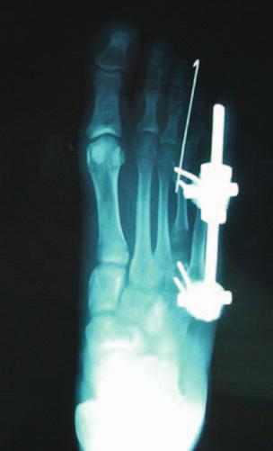
1 C H A P T E R 2 1 MULTIPLE APPLICATIONS OF THE MINIRAIL Thomas J. Merrill, DPM James M. Losito, DPM Mario Cala, DPM Victor Herrera, DPM Alan E. Sotelo, DPM INTRODUCTION The unilateral MiniRail External Fixation System is a device used in the treatment of bone conditions and deformities of the foot. In the last decade this application has evolved and become more popular with the development of systems that allow a greater understanding of this technique. External fixation is divided into two main categories: circular frames and unilateral rails (1, 2); however the use of Illizarov-type circular frames is reserved for more complex deformities in the foot and ankle as well as the distal leg. MiniRail external fixators have been described in the use of a variety of procedures, more commonly used in forefoot surgery (1, 3, 4). In this paper we put together a total of nine different surgical procedures used to treat fifteen different foot conditions and deformities as well as trauma. All conditions were treated with a unilateral external fixation system with excellent results. These procedures include arthrodesis of the first metatarsocuneiform joint (with two revisions of this procedure not previously using a MiniRail), medial column fusion, open and closed reduction of Lisfranc fracture dislocation injuries, metatarsal callus distraction, correction of first and fifth metatarsal fractures, sliding calcaneal osteotomy, first metatarsal-cuneiform fusion, and first metatarsophalangeal joint fusion. External fixation systems have been shown to be advantageous over internal fixation for various reasons. Placement of an external fixator is done percutaneously thus eliminating any unnecessary incisions and risk of infection (1). Dehiscence and the need for wound care are thus prevented. Associated treatments, such as dressing changes, skin grafting, bone grafting, and irrigation are possible without disturbing the correction or fixation. Any postoperative condition that may arise such as ulcerations or pin-track infections may be easily accessed and cared for; a luxury not enjoyed by a plate or screw. External fixators can also be applied in the presence of a bone infection. The percutaneous placement of pins eliminates guesswork involved in deciding how to correct the deformity since the pins can be placed at a safe distance from the infection. External fixators can also be safely used in the presence of comorbidities such as diabetes mellitus, smokers, osteomyelitis, and avascular necrosis (1-3). As with any external fixation system, early weight bearing is not only allowable but encouraged as this expedites bone healing. Immediate motion of the proximal and distal joints is also allowed aiding in the reduction of edema and preventing capsular fibrosis, joint stiffening, muscle atrophy, and osteoporosis. When it comes to multiplanar deformities, external fixators provide neutralization and stabilization with adjustable amounts of compression or distraction. This allows correction (compression or distraction) throughout the postoperative period through a minimally invasive procedure and a multifunctional correction (1, 2). Finally, once the desired correction has been achieved, the pins are removed and the patient is left with no internal hardware that may cause pain in the future. Disadvantages of the unilateral MiniRail system are mainly due to the complexity of the system and difficulty in application and manipulation. This difficulty can be overcome, as with any technical and mechanical difficulty, through surgeon education, training, and experience. Another disadvantage of external fixator systems is the cost of the equipment, including the tools needed for application and removal. Although major incisions are avoided as well as placement of internal hardware, the risk of pin track infection and possible neurovascular damage continue to be realistic (1-3). As stated earlier however, most of these problems can be solved by early detection, quick action, and by surgeon education and experience. MATERIALS AND METHODS A total of 29 Orthofix MiniRail external fixators were placed on 26 patients with 15 different diagnoses who underwent 9 different surgical procedures. The patients ranged in age
2 102 CHAPTER 21 from 23 to 79 years with 8 male (30%) and 18 female (70%). Each patient was educated at length about both internal and external fixation. All advantages and disadvantages including complications as well as recovery time and weight bearing status after surgery were discussed with the patients in detail. All patients who opted for the MiniRail external fixator received preoperative and postoperative instructions for careful management of the MiniRail. All patients received prophylactic intravenous antibiotic therapy 30 minutes preoperative and postoperative weekly pin care (cleansing and dressing changes). In the study, compression-stabilization techniques were used in 26 of the 29 procedures, within these cases 20 were arthrodesis and 5 were fracture management techniques. One sliding osteotomy with fixation and 3 distractionstabilization procedures were also performed (Table 1). All patients had weekly postoperative adjustments of the mini-rail except for the callus distraction patient who performed his own adjustments daily. Intra-operative radiographs were performed to confirm position and stabilization with follow-up radiographs performed at 3 weeks and 8 weeks postoperative. The average postoperative period with the MiniRail was 8 weeks with weightbearing beginning as early as one week postoperative with the aid of a surgical shoe and crutches. Ten Lapidus fusions were performed with 4 pins placed perpendicular to the long axis of the bone: 2 in the medial cuneiform and 2 in the shaft of the first metatarsal. Prior to pin insertion, the first cuneiform-metatarsal joint was prepared under fluoroscopy with temporary fixation through the use of a 0.45 Kirshner wire. After placement of the Mini- Rail, compression was then achieved with an Allen wrench. Two revisional Lapidus fusions were performed after failed procedures with internal fixation resulted in non-union. The screws were removed and the joint was prepared once again for the Lapidus procedure described above. Three medial column fusions were performed with a talo-navicular joint fusion involving the use of 2 pins in each bone and compression through the rail with early weight bearing after 1 week and postoperative adjustments made every other week (Table 1). Three Lisfranc s fracture-dislocations, two fifth metatarsal fractures, and one first metatarsal fracturedislocation were reduced with a total of 6 MiniRails with compression through the fracture defect. Five first metatarsophalangeal joint fusions were performed with 4 pin compression at the joint through the neck of the first of the metatarsal and the first proximal phalanx. Two brachymetatarsia callus distraction procedures were performed with MiniRail placement along the metatarsal shaft. Daily adjustments of the MiniRail were performed for callus distraction by the patient at home. One sliding calcaneal osteotomy procedure was performed and a MiniRail external fixator was used for compression and stabilization of the osteotomy. Table 1 DIAGNOSIS PROCEDURE SURGERIES 1 Hallux Rigidus 1st MPJ Fusion 2 2 Hallux Varus 1st MPJ Fusion 1 3 Charcot Arthropathy 1st MPJ Fusion 1 4 Osteomyelitis 1st MPJ Fusion 1 5 HAV + Hypermobility Lapidus 10 6 Non-Union Medial Column Fusion Revision Lapidus 2 7 Avascular Necrosis Medial Cuneiform Medial Column Fusion 1 8 Severe Pes Planus Medial Column Fusion 1 9 Severe Osteoarthritis Medial Column Fusion 1 10 Calcaneal Varus Calcaneal Sliding Osteotomy 1 11 Brachymetarsia Callus Distraction th Metatarsal Fracture Repair 5th Met Fracture 1 13 Non-Union 5th Metatarsal Fracture Repair 5th Met Fracture 1 14 First Ray Fracture Dislocation Repair 1st Met Fracture/Dislocation 1 15 LisFranc s Fracture Dislocation Repair LisFranc s Fracture/Dislocation 3
3 CHAPTER RESULTS Of the 29 procedures, all patients went on to full recovery with no complications or recurrence. The average time of duration with the MiniRail external fixator was 8 weeks, with removal at that time +/- one week. After removal of the MiniRail external fixator, all patients had an average recovery period of approximately 3 weeks at which time patients were allowed to transition out of their postoperative shoes and into athletic shoes. By one month following removal, patients were cleared to return to all normal preoperative activity without restrictions. Physical therapy was highly recommended to all patients to regain muscle strength and balance and averaged 3 weekly physical therapy sessions for 3 weeks. Most patients were allowed to begin physical therapy a week after removal of the external fixator. To date there have been no recurrences and patient satisfaction has been overall positive with results. There was no need for further corrective procedures and all patients went on to full recovery. DISCUSSION The use of external fixation devices has been in practice for many years. Today the use of external fixators has become a popular methodology for treating a great variety of conditions with minimally invasive procedures. While the larger ring fixators are reserved for more complex conditions (ankle fractures, limb lengthening, Charcot reconstructions), MiniRail external fixators have been a staple for the minimally invasive surgical correction of various forefoot and midfoot conditions as well as some calcaneal and rear foot conditions. In this study, 26 patients underwent a total of 9 different surgical procedures with application of 29 MiniRail external fixators to correct conditions in 15 different diagnostic categories. All patients received preoperative education and weekly postoperative adjustments and pin care with follow up radiographs at 3 and 8 weeks. The patient population ranged in age from years of age with females outweighing males 18 to 8, respectively. Since 2009 we have found MiniRail external fixators to be superior over internal fixation for the various reasons listed above. The success rate is exceptionally high with patients able to ambulate very early after surgery, and the ability to perform any necessary adjustments postoperatively make the MiniRail system a very useful device (4, 5). It should also be noted that satisfaction is overall very positive considering they are able to ambulate early on and they have the peace of mind knowing that any correction needed can be easily adjusted at any time. Patient complaints are minimal and are generally geared toward the bulky dressings and uncomfortable postoperative shoe gear however any complaints of pain or discomfort are virtually non-existent. Finally, it is of importance to note once again that this device can be safely and successfully used on patients with comorbidities that would otherwise lead to failure with internal fixation such as patients who are smokers, have bone infections, or have chronic illnesses such as diabetes mellitus (5, 6). We will continue to use MiniRail external fixators for future cases and hope to broaden the scope of indication for the device. Although this study has a very small sample population, the degree of success we have experienced thus far will propel us to continue. REFERENCES 1. LaBianco GL,Vito GR, Rush SM. External fixation. In: Banks AS, Downey MS, Martin DE, Miller SJ, eds. McGlamry s comprehensive textbook of foot and ankle surgery, vol. 1. 3rd edition. Philadelphia: Lippincott, Williams & Wilkins; p Seibert FJ, Et al., External fixation in trauma of the foot and ankle. Clinics in Podiatric Medicine and Surgery 2003;20: Treadwell JR. Rail external fixation for stabilization of closing base wedge osteotomies and Lapidus procedures: a retrospective analysis of sixteen cases. J Foot Ankle Surg 2005;44: Hamilton GA, Mullins S, Schuberth JM, Rush SM, Ford L. Revision Lapidus arthrodesis: rate of union in 17 cases. J Foot Ankle Surg 2007;46: Gamble J, Decker S, Abrams RC. Short first ray as a complication of multiple metatarsal Osteotomies. Clin Orthop 1982;164: Levine SE, Davidson RS, Dormans JP, et al. Distraction osteogenesis for congenitally short lesser metatarsals. Foot Ankle Int 1925;16:


4 104 CHAPTER 21 Figure 1. Figure 2. Figure 3. Figure 4.


5 CHAPTER Figure 5. Figure 6. Figure 7. Figure 8.


6 106 CHAPTER 21 Figure 9. Figure 10. Figure 11. Figure 12.


7 CHAPTER Figure 13. Figure 14. Figure 15. Figure 16.


8 108 CHAPTER 21 Figure 17. Figure 18. Figure 19. Figure 20.


9 CHAPTER Figure 21. Figure 22. Figure 23. Figure 24.

10 110 CHAPTER 21 Figure 25.
Use of the 20 Memory Staple in Osteotomies of Fusions of the Forefoot
 168 Forefoot Reconstruction Use of the 20 Memory Staple in Osteotomies of Fusions of the Forefoot Definition, History, Generalities This staple first provides a permanent compression both in the prongs
Minimally Invasive Bunionectomy: The Lam Modification of the Traditional Distal First Metatarsal Osteotomy Bunionectomy
 CHAPTER 2 Minimally Invasive Bunionectomy: The Lam Modification of the Traditional Distal First Metatarsal Osteotomy Bunionectomy Kevin Lam, DPM Rikhil Patel, DPM Thomas Merrill, DPM Hallux abducto valgus
Workshop Outline. Pre-operative planning
 Workshop Objective To build and apply the True/Lok TM circular external fixator frame for correction of the Charcot forefoot deformity (Lisfranc fracture dislocation) Workshop Outline Pre-operative planning
QUICK REFERENCE GUIDE. MiniRail System. Part B: Foot Applications. By Dr. B. Magnan, Dr. E. Rodriguez and Dr. G. Vito ALWAYS INNOVATING
 14 MiniRail System Part B: Foot Applications By Dr. B. Magnan, Dr. E. Rodriguez and Dr. G. Vito ALWAYS INNOVATING ORDERING INFORMATION Sterilization box, empty M190 Can accommodate: M101 Standard MiniRail
Correction of Traumatic Ankle Valgus and Procurvatum using the Taylor Spatial Frame: A Case Report
 The Foot and Ankle Online Journal Official publication of the International Foot & Ankle Foundation Correction of Traumatic Ankle Valgus and Procurvatum using the Taylor Spatial Frame: A Case Report by
RETROSPECTIVE ANALYSIS OF END-TO-END DIGITAL ARTHRODESIS
 C H A P T E R 1 7 RETROSPECTIVE ANALYSIS OF END-TO-END DIGITAL ARTHRODESIS Michelle L. Butterworth, DPM Michael S. Downey, DPM Digital deformities are one of the most common entities we face as foot and
MiniRail System. Part B: Foot Applications. By Dr. B. Magnan, Dr. E. Rodriguez and Dr. G. Vito
 Q U I C K R E F E R E N C E G U I D E 14 MiniRail System Part B: Foot Applications By Dr. B. Magnan, Dr. E. Rodriguez and Dr. G. Vito ORDERING INFORMATION MiniRail System Kit, M190C Contents: M 101 Standard
Index. Clin Podiatr Med Surg 22 (2005) Note: Page numbers of article titles are in boldface type.
 Note: Page numbers of article titles are in boldface type.") Clin Podiatr Med Surg 22 (2005) 309 314 Index Note: Page numbers of article titles are in boldface type. A Abductor digiti minimi muscle, myectomy of, for tailor s bunionette, 243 Achilles tendon, lengthening
Clin Podiatr Med Surg 22 (2005) 309 314 Index Note: Page numbers of article titles are in boldface type. A Abductor digiti minimi muscle, myectomy of, for tailor s bunionette, 243 Achilles tendon, lengthening
Low Profile Medial Locking plate augmentation Lapidus Arthrodesis with an early weight bearing protocol: Clinical and Radiographic Analysis
 Low Profile Medial Locking plate augmentation Lapidus Arthrodesis with an early weight bearing protocol: Clinical and Radiographic Analysis James Cottom, DPM Anand Vora, MD Low Profile Medial Locking plate
REPAIR OF THE DISPLACED AUSTIN OSTEOTOMY
 C H A P T E R 2 1 REPAIR OF THE DISPLACED AUSTIN OSTEOTOMY John V. Vanore, DPM INTRODUCTION Bunion surgery is frequently performed by foot and ankle surgeons. Generally, bunion surgery is quite predictable,
Retrospective Study of Surgical Outcomes for Combined Ankle and Subtalar Joint Arthrodesis, Cavovarus Deformity Correction and Ankle Fractures
 FOOT/ ANKLE RETROSPECTIVE STUDYIC S Retrospective Study of Surgical Outcomes for Combined Ankle and Subtalar Joint Arthrodesis, Cavovarus Deformity Correction and Ankle Fractures Adult & Pediatric Deformity
Increased pressures at
 Surgical Off-loading of Plantar Hallux Ulcerations These approaches can be used to treat DFUs. By Adam R. Johnson, DPM Increased pressures at the plantar aspect of the hallux leading to chronic hyperkeratosis
Re+Line Bunion Correction System for Correction of Hallux Abducto Valgus Deformity
 Re+Line Bunion Correction System for Correction of Hallux Abducto Valgus Deformity Amber M. Shane, DPM, FACFAS 1, Christopher L. Reeves, DPM, FACFAS 1 1. Orlando Foot & Ankle Clinic, Orlando, FL Abstract
The American Academy of Foot & Ankle Osteosynthesis. presents
 The American Academy of Foot & Ankle Osteosynthesis presents Comprehensive Course of Internal Fixation for Reconstructive Surgery and Trauma of the Foot & Ankle September 4-6, 2014 Goodlett Farms Innovation
Medincenter GlavUpDK by the Ministry of Foreign Affairs of Russia, Moscow.
 Medincenter GlavUpDK by the Ministry of Foreign Affairs of Russia, Moscow. Berezhnoy Sergey. Percutaneous First Metatarsocuneiform Joint Arthrodesis in a Treatment of Metatarsus Primus Varus: a Prospective
DARCO. Bow 2 Plate SURGIC AL TECHNIQUE
 DARCO Bow 2 Plate SURGIC AL TECHNIQUE Contents 2 Preface 3 Chapter 1 4 Chapter 2 5 6 7 8 9 Appendix 10 10 11 Intended Use Indications/Contraindications Design Rationale Preoperative Planning Surgical Technique
19 Arthrodesis of the First Metatarsocuneiform Joint
 19 Arthrodesis of the First Metatarsocuneiform Joint CHARLES GUDAS Abduction of the first metatarsal to correct metatarsus primus varus and hallux valgus was first described by Albrecht in 1911. 1 Lapidus
American Board of Foot and Ankle Surgery
 American Board of Foot and Ankle Surgery Logging Guidelines Revisions ABFAS Task Force Mindy Benton, DPM Randall Dei, DPM Charles Lombardi, DPM John Thomas Marcoux, DPM Roya Mirmiran, DPM Michael Vaardahl,
ORTHOLOC 3Di. Foot Reconstruction System SURGIC AL TECHNIQUE
 ORTHOLOC 3Di Foot Reconstruction System S C R E W TA R G E T I N G G U I D E SURGIC AL TECHNIQUE SURGEON DESIGN TEAM The ORTHOLOC 3Di Foot Reconstruction System was developed in conjuction with: ORTHOLOC
Variable Angle LCP Forefoot/Midfoot System 2.4/2.7. Procedure specific plates for osteotomies, arthrodeses and fractures of the foot.
 Variable Angle LCP Forefoot/Midfoot System 2.4/2.7. Procedure specific plates for osteotomies, arthrodeses and fractures of the foot. Compression technology Variable angle locking technology Anatomic and
COMPLICATIONS OF BRACHYMETATARSIA REPAIR WITH CALLUS DISTRACTION
 C H A P T E R 4 1 COMPLICATIONS OF BRACHYMETATARSIA REPAIR WITH CALLUS DISTRACTION Michelle L. Butterworth, DPM INTRODUCTION Brachymetatarsia represents one of the best and most useful indications for
Technique Guide. 6.5 mm Midfoot Fusion Bolt. For intramedullary fixation of the medial column of the foot.
 Technique Guide 6.5 mm Midfoot Fusion Bolt. For intramedullary fixation of the medial column of the foot. Table of Contents Introduction 6.5 mm Midfoot Fusion Bolt 2 AO Principles 4 Indications 5 Surgical
Proper Logging of Surgical Procedures (Effective July 1, 2018)
") Proper Logging of Surgical Procedures (Effective July 1, 2018) GENERAL GUIDELINES: 1) For the procedure codes listed below, the program director must review each entry to determine proper usage. The following
Proper Logging of Surgical Procedures (Effective July 1, 2018) GENERAL GUIDELINES: 1) For the procedure codes listed below, the program director must review each entry to determine proper usage. The following
Surgical Off-loading. Reiber et al Goals of Diabetic Foot Surgery 4/28/2012. The most common causal pathway to a diabetic foot ulceration
 Reiber et al. 1999 Surgical Off-loading The most common causal pathway to a diabetic foot ulceration Alex Reyzelman DPM Associate Professor California School of Podiatric Medicine at Samuel Merritt University
Digital Surgery Complications
 Annual Surgical Conference 2018 Digital Surgery Complications Zeeshan S. Husain, DPM, FACFAS, FASPS Great Lakes Foot and Ankle Institute September 21, 2018 None Disclosures Presentation Outline Differentials
Alberta Health Care Insurance Plan. Schedule Of Anaesthetic Rates Applicable To Podiatry. Procedure List. As Of. 01 April Government of Alberta
 Alberta Health Care Insurance Plan Procedure List As Of 01 April 2017 Alberta Health Care Insurance Plan Page i Generated 2017/03/14 TABLE OF CONTENTS As of 2017/04/01 II. OPERATIONS ON THE NERVOUS SYSTEM.......................
Section 6: Preoperative Planning
 Clinical Relevance of the PedCat Study: In many ways the PedCat study confirmed radiographic findings. With the measuring tools embedded in the DICOM viewing software it was possible to gauge the thickness
The American Academy of Foot & Ankle Osteosynthesis. presents
 The American Academy of Foot & Ankle Osteosynthesis presents Comprehensive Course of Internal Fixation for Reconstructive Surgery and Trauma of the Foot & Ankle Hyatt DFW Airport Dallas, Texas SCIENTIFIC
Case Study: David. Conditions Treated Femoral Neck Fracture with Avascular Necrosis of the Hip. Age Range During Treatment 16 Years
 Case Study: David Conditions Treated Femoral Neck Fracture with Avascular Necrosis of the Hip Age Range During Treatment 16 Years David S. Feldman, MD Chief of Pediatric Orthopedic Surgery Professor of
CORRECTING HAMMERTOE DEFORMITIES UTILIZING AN INTRAMEDULLARY DEVICE: Case Reports
 C H A P T E R 1 1 CORRECTING HAMMERTOE DEFORMITIES UTILIZING AN INTRAMEDULLARY DEVICE: Case Reports B. Cory Brown, DPM Robert K. Cohen, DPM Jason R. Miller, DPM Scott R. Roman, DPM INTRODUCTION The most
How to avoid complications of distraction osteogenesis for first brachymetatarsia
 220 Acta Orthopaedica 2009; 80 (2): 220 225 How to avoid complications of distraction osteogenesis for first brachymetatarsia Keun-Bae Lee, Hyun-Kee Yang, Jae-Yoon Chung, Eun-Sun Moon, and Sung-Taek Jung
Reconstructing Limb Deformities using the VCAM TM External Fixator: A series of 3 cases
 The Foot and nkle Online Journal Open ccess Publishing Reconstructing Limb Deformities using the VCM TM External Fixator: series of 3 cases by Michael P. DellaCorte, DPM, FCFS 1, Panagiotis Panagakos,
Minimally Invasive Bunion Surgery: Methods and Outcomes
 Minimally Invasive Bunion Surgery: Methods and Outcomes Hummira H. Abawi, DPM Diplomate ABPM, AACFAS Instructor of Orthopedics, University of Maryland Medical Center Director of Education, Maryland VA
CASE REPORT. Bone transport utilizing the PRECICE Intramedullary Nail for an infected nonunion in the distal femur
 PRODUCTS CASE REPORT Bone transport utilizing the PRECICE Intramedullary Nail for an infected nonunion in the distal femur Robert D. Fitch, M.D. Duke University Health System 1 1 CONDITION Infected nonunion
Merete PlantarMAX Lapidus Plate Surgical Technique. Description of Plate
 Merete PlantarMAX Lapidus Plate Surgical Technique Description of Plate Merete Medical has designed the PlantarMax; a special Plantar/Medial Locking Lapidus plate which places the plate in the most biomechanically
Metatarsal Lengthening By Callus Distraction For Brachymetatarsia: Case Report and Review of the Literature
 ISPUB.COM The Internet Journal of Third World Medicine Volume 1 Number 2 Metatarsal Lengthening By Callus Distraction For Brachymetatarsia: Case Report and Review of the R Rose Citation R Rose. Metatarsal
Use of internal callus distraction in the treatment of congenital brachymetatarsia
 British Journal of Plastic Surgery (2005) 58, 1014 1019 CASE REPORT Use of internal callus distraction in the treatment of congenital brachymetatarsia Naoto Yamada*, Yoshihiro Yasuda, Nobuko Hashimoto,
6.5 mm midfoot fusion bolt
 6.5 mm midfoot fusion bolt For intramedullary fixation of the medial column of the foot SurgIcal technique Table of Contents Introduction 6.5 mm Midfoot Fusion Bolt 2 AO Principles 4 Indications 5 Surgical
HEMI IMPLANT ARTHROPLASTY FOR THE SECOND METATARSOPHALANGEAL JOINT
 C H A P T E R 1 5 HEMI IMPLANT ARTHROPLASTY FOR THE SECOND METATARSOPHALANGEAL JOINT Joe T. Southerland, DPM Mickey D. Stapp, DPM INTRODUCTION Hemi-implant arthroplasty of the first metatarsophalangeal
Proximal metatarsal osteotomy and distal soft tissue reconstruction as treatment for hallux valgus deformity
 REVIEW Proximal metatarsal osteotomy and distal soft tissue reconstruction as treatment for hallux valgus deformity Michael J. Coughlin and J. Speight Grimes Boise, Idaho, USA (Received for publication
Merete BLP. Surgical Technique. - Bunion Locking Plate - Low Profile Locking Bone Plate System
 Merete BLP - Bunion Locking Plate - Low Profile Locking Bone Plate System Surgical Technique Merete Medical, Inc. 49 Purchase Street Rye, N.Y. 10580 Phone: 914 967-1532 www.merete-medical.com - Surgical
SUBTALAR ARTHROEREISIS IN THE OLDER PATIENT
 C H A P T E R 1 7 SUBTALAR ARTHROEREISIS IN THE OLDER PATIENT William D. Fishco, DPM, MS INTRODUCTION Arthroereisis is a surgical procedure designed to limit the motion of a joint. Subtalar joint arthroereisis
Matthew Beuchel MD 2 C. Luke Rust MD 3 Jessica Hooper MD 3
 A Prospective, Randomized,Controlled Trial Comparing Early- Weightbearing vs. Non-Weightbearing Following Modified Lapidus Arthrodesis Intermediate Results - Donald R. Bohay, MD, FACS Professor, Michigan
Combination of First Metatarsophalangeal Joint Arthrodesis and Proximal Correction for Severe Hallux Valgus Deformity
 FOOT &ANKLE INTERNATIONAL DOI: 10.3113/FAI.2012.0400 Combination of First Metatarsophalangeal Joint Arthrodesis and Proximal Correction for Severe Hallux Valgus Deformity Pascal F. Rippstein, MD; Young-Uk
CHARLOTTE Lisfranc. Recontruction System SURGICAL TECHNIQUE
 CHARLOTTE Lisfranc Recontruction System SURGICAL TECHNIQUE Contents Preface 3 Chapter 1 4 Chapter 2 4 4 4 4 4 Chapter 3 5 5 5 6 6 6 7 7 8 Chapter 4 8 8 9 10 Appendix 10 Introduction Indications and Contraindications
2017 SAFSA CONGRESS PROGRAMME
 2017 SAFSA CONGRESS PROGRAMME THURSDAY, MAY 25 07h45 07h55: WELCOME & INTRODUCTIONS Forefoot I: Hallux Valgus and Lesser Toes (08h00-10h00 Lectures) 08h00 08h30: Surgical Management of Hallux Valgus Rippstein,
Locking Forefoot/Midfoot Plating System
 Locking Forefoot/Midfoot Plating System Locking Forefoot/Midfoot Plating System Acumed is an industry leader in innovative solutions for extremities and trauma. We are dedicated to pioneering solutions
ORTHOLOC 3Di. Foot Reconstruction System Midfoot Fusion Plate SURGICAL TECHNIQUE
 ORTHOLOC 3Di Foot Reconstruction System Midfoot Fusion Plate SURGICAL TECHNIQUE ORTHOLOC Foot Reconstruction System Midfoot Fusion Plate SURGICAL TECHNIQUE Surgeon Design Team The ORTHOLOC 3Di Foot Reconstruction
DRACO FOOT & ANKLE SYSTEM PRODUCT INFORMATION
 DRACO FOOT & ANKLE SYSTEM PRODUCT INFORMATION 2 DRACO FOOT & ANKLE SYSTEM www.hnmtotal.com DRACO Foot & Ankle System CONTENTS MetaFuse L Plate 3 MetaFuse BLP Plate 4 MetaFuse Midfoot/Lisfranc Plate 5 MetaFuse
The Latest Breakthrough in TTC Fusion Technology
 The Latest Breakthrough in TTC Fusion Technology Treatment of Hindfoot Non-Union with DynaNail TTC Fusion System A CASE REPORT Dr. L. Daniel Latt, MD, PhD Background The DynaNail TTC Fusion System is intended
The American Academy of Foot & Ankle Osteosynthesis. presents ADVANCED TECHNIQUES IN SKELETAL FIXATION OF THE FOOT AND ANKLE
 The American Academy of Foot & Ankle Osteosynthesis presents ADVANCED TECHNIQUES IN SKELETAL FIXATION OF THE FOOT AND ANKLE Hyatt DFW Airport Dallas, Texas SCIENTIFIC CHAIRMAN John M. Schuberth, DPM San
The American Academy of Foot & Ankle Osteosynthesis. presents ADVANCED TECHNIQUES IN SKELETAL FIXATION OF THE FOOT AND ANKLE
 The American Academy of Foot & Ankle Osteosynthesis presents ADVANCED TECHNIQUES IN SKELETAL FIXATION OF THE FOOT AND ANKLE Goodlett Farms Innovation Centre Memphis, Tennessee SCIENTIFIC CHAIRMAN John
Kirienko Alexander Peccati Andrea, Portinaro Nicola Istituto Clinico Humanitas, Milano, Italy
 METATARSAL BONES LENGTHENING WITH LAKI MINI EXTERNAL FIXATOR Kirienko Alexander Peccati Andrea, Portinaro Nicola Istituto Clinico Humanitas, Milano, Italy alexander@kirienko.com Alexander KIRIENKO, MD
Index. Note: Page numbers of article titles are in boldface type.
 Note: Page numbers of article titles are in boldface type. A Abscess, in puncture wounds, 531 Absorbable fixation, for Lisfranc joint injuries, 556 Advanced glycosylation end products, complications due
Preservation of the First Ray in Patients with Diabetes
 Preservation of the First Ray in Patients with Diabetes Surgical approaches are often necessary to off-load excessive pressure. By Derek Ley, DPM, and Barry Rosenblum, DPM Introduction In approaching diabetic
Temporary bridge plating of the medial column in Chopart and Lisfranc injuries
 Temporary bridge plating of the medial column in Chopart and Lisfranc injuries by Alaa Mansour DPM 1*, Lawrence Fallat DPM, FACFAS 2 The Foot and Ankle Online Journal 10 (1): 5 Severe traumatic injuries
LAPIDUS What is Old is New
 LAPIDUS What is Old is New Alan Jay Block, DPM, MS, FASPS, FACFAS Fellowship trained in Advanced Ankle Techniques Adjunct Professor Dept Of Orthopeadics The Ohio State University Board Member The Ohio
PL ATING SYSTEM. Pre-contoured implants Versatile fixation system Hexalobe screw recess design Locked fixation
 I N N O VA T I O N ME A NS M OT I O N FOOTMOTION PL ATING SYSTEM Pre-contoured implants Versatile fixation system Hexalobe screw recess design Locked fixation FOOTMOTION PLATING SYSTEM Indications: The
Modified Proximal Scarf Osteotomy for Hallux Valgus
 Original Article Clinics in Orthopedic Surgery 2018;10:479-483 https://doi.org/10.4055/cios.2018.10.4.479 Modified Proximal Scarf Osteotomy for Hallux Valgus Ki Won Young, MD, Hong Seop Lee, MD, Seong
Midfoot - Reduction & Fixation - ORIF - screw fixation - AO Surgery Reference. ORIF - screw fixation
 Midfoot - TMT (Lisfranc) injury 1. Diagnosis ORIF - screw fixation Authors Mechanism of the injury Tarso-metatarsal (Lisfranc) injuries may be caused by direct or indirect forces. Direct forces include
Biomet. Vision Pin-To-Bar System. Surgical Technique. Calcaneal Reduction Frame
 Biomet Vision Pin-To-Bar System Surgical Technique Calcaneal Reduction Frame One Surgeon. One Patient. Over 1 million times per year, Biomet helps one surgeon provide personalized care to one patient.
Forefoot/Midfoot Plating System. Surgical Technique
 Forefoot/Midfoot Plating System Surgical Technique Acumed is a global leader of innovative orthopaedic and medical solutions. We are dedicated to developing products, service methods, and approaches that
Hallux Malleus develops after Flexible Hallux Varus correction with Tensioned Suture Device: A Case Report
 The Foot and nkle Online Journal Official publication of the International Foot & nkle Foundation Hallux Malleus develops after Flexible Hallux Varus correction with Tensioned Suture Device: Case Report
Foot Reconstruction System SURGICAL TECHNIQUE
 ORTHOLOC 3Di Foot Reconstruction System SURGICAL TECHNIQUE SURGEON DESIGN TEAM The ORTHOLOC 3Di Foot Reconstruction System was developed in conjuction with: Robert B. Anderson, MD OrthoCarolina Charlotte,
EXPERT TIBIAL NAIL PROTECT
 EXPERT TIBIAL NAIL PROTECT Enhance your first line of defense This publication is not intended for distribution in the USA. CLINICAL EVIDENCE CONTENT AUTHOR TITLE OF CHAPTER PAGE ETN PROtect clinical evidence
Provision of Rotational Stability: Prevention of Collapse: Closed Fracture Reduction: Minimally Invasive Surgery with no Exposure of the Fracture:
 INTRODUCTION Percutaneous Compression Plating was developed by considering each of the stages in the surgical procedure for pertrochanteric fractures and the ways in which these might be improved. Primary
Metatarsal Lengthening By Callus Distraction For Brachymetatarsia. Case Report and Review of the Literature
 The Internet Journal of Third World Medicine TM ISSN: 1539-4646 Home Current Issue Archives Instructions for Authors Disclaimer Printable Version Metatarsal Lengthening By Callus Distraction For Brachymetatarsia:
Distraction Osteogenesis and Fusion for Failed First Metatarsophalangeal Joint Replacement: Case Series
 737481FAIXXX10.1177/1071100717737481Foot & Ankle InternationalDa Cunha et al research-article2017 Case Report Distraction Osteogenesis and Fusion for Failed First Metatarsophalangeal Joint Replacement:
Alberta Health Care Insurance Plan. Schedule Of Anaesthetic Rates Applicable To Podiatric Surgery. Procedure List. As Of.
 Alberta Health Care Insurance Plan Procedure List As Of 01 April 2016 Alberta Health Care Insurance Plan Page i Generated 2016/03/22 TABLE OF CONTENTS As of 2016/04/01 07 PHYSICAL MEDICINE, REHABILITATION,
Joint Preserving Surgery in Severe Forefoot Disorders
 Joint Preserving Surgery in Severe Forefoot Disorders J ORTHOP TRAUMA SURG REL RES 4 (12) 2008 Review article LOUIS S. BAROUK*, PIERRE BAROUK** * 39, Chemin de la Roche, 33370, Yvrac, France ** Clinique
Clinical. Solutions. Synthes Solutions. Foot and Ankle.
 Clinical Solutions Foot and Ankle. Foot and Ankle. Fractures of the tibial shaft Fractures of the distal fibula Fractures of the distal tibia Fractures and osteotomies of the calcaneus Arthrodesis Fractures,
Surgical Technique. Customer Service:
 Patent and Patent Pending CAUTION: Federal Law (USA) restricts this device to sale by or on the order of a physician. INDICATIONS FOR USE The Axis Charcot Fixation System in diameters of 5.5, 6.5 and 7.5mm
THE MEMORY STAPLE SURGICAL TECHNIQUE
 THE MEMORY STAPLE SURGICAL TECHNIQUE TABLE OF CONTENTS Introduction PAGE 1 Features and Benefits PAGE 1 Ordering Information PAGE 2-3 Indications PAGE 4 Contra-indications PAGE 4 Surgical Technique PAGE
*Rippstein, Trnka, Saragas, Hoffman
 THURS 25th MAY 07:00 07:10 Welcome and Introductions Paulo Ferrao Lecture 1: 07:10 09:45 Forefoot I: Hallux Valgus and Lesser Toes Mark Easley 40 mins 07:10 07:50 Surgical Management of Hallux Valgus 30
PROstep Minimally Invasive Surgery HALLUX VALGUS CORRECTION USING PROSTEP MICA MINIMALLY INVASIVE FOOT SURGERY: TWO CASE STUDIES
 PROstep Minimally Invasive Surgery HALLUX VALGUS CORRECTION USING PROSTEP MICA MINIMALLY INVASIVE FOOT SURGERY: TWO CASE STUDIES AS PRESENTED BY: JOEL VERNOIS M.D. 016798A Case Study 1 PROstep Minimally
1. Orthoapedic Associates of Michigan, PC, Grand Rapids, MI 2. Michigan State University College of Human Medicine, Grand Rapids, MI
 Second Metatarsal Osteotomy Shortening with Tarsometatarsal Arthrodesis: Comparison of Outcomes Between MSP TM Metatarsal Shortening System and Plates and Screws Donald R. Bohay, MD, FACS 1 ; John G. Anderson,
Forefoot/Midfoot Plating System
 Surgical Technique Forefoot/Midfoot Plating System Acumed is a global leader of innovative orthopaedic and medical solutions. We are dedicated to developing products, service methods, and approaches that
Index. Note: Page numbers of article titles are in bold face type.
 Index Note: Page numbers of article titles are in bold face type. A Achilles tendon, Zadek osteotomy effects on, 430 Adult acquired flatfoot disorder, 387 403 calcaneal Z osteotomy for, 397 399 historical
Foot and Ankle Technique Guide Metatarsal Shortening Osteotomy
 Surgical Technique Foot and Ankle Technique Guide Metatarsal Shortening Osteotomy Prepared in consultation with: Phinit Phisitkul, MD Department of Orthopedics and Rehabilitation University of Iowa Iowa
Hallux Valgus Deformity: Preoperative Radiologic Assessment
 119 Pictorial Essay H............ - Hallux Valgus Deformity: Preoperative Radiologic Assessment David Karasick1 and Keith L. Wapner An estimated 40% of the American adult population experiences foot problems,
Rippstein, Trnka, Saragas, Narramore
 THURS 25th MAY 07:45 07:55 Welcome and Introductions Paulo Ferrao Lecture 1: 08:00 10:20 Forefoot I: Hallux Valgus and Lesser Toes Mark Easley 30 mins 08:00 08:30 Surgical Management of Hallux Valgus Saragas,
pedcat Clinical Case Studies
 pedcat Clinical Case Studies C u r v e B e a m 1 7 5 T i t u s A v e, S u i t e 3 0 0 W a r r i n g t o n, P A 1 8 9 7 6 267-4 8 3-8081 w w w. c u r v e b e a m. c o m PedCAT: Clinical Evidence of diagnostic
MIDFOOT INJURIES-ARE WE UNDERTREATING IT? Mr Rajiv Limaye Mr Prasad Karpe University Hospital of North Tees 3 rd Foot and Ankle Symposium
 MIDFOOT INJURIES-ARE WE UNDERTREATING IT? Mr Rajiv Limaye Mr Prasad Karpe University Hospital of North Tees 3 rd Foot and Ankle Symposium Introduction Increasing sports injuries RTA and traumatic injuries
ORTHOLOC CROSSCHECK TECHNOLOGY OVERVIEW FOR HOSPITAL VALUE ANALYSIS COMMITTEE
 ORTHOLOC CROSSCHECK TECHNOLOGY OVERVIEW FOR HOSPITAL VALUE ANALYSIS COMMITTEE Indications for Use The ORTHOLOC 3Di Foot Reconstruction System is intended for use in stabilization of fresh fractures, revision
QUICK REFERENCE GUIDE. Arthrodesis. Joint Fusion. By Dr. S. Agostini and Dr. F. Lavini
 QUICK REFERENCE GUIDE 5 Arthrodesis Joint Fusion By Dr. S. Agostini and Dr. F. Lavini QUICK REFERENCE GUIDE ARTHRODESIS OF THE HIP Apply the ProCallus Fixator to the lateral aspect of the thigh. Insert
PAINFUL SESAMOID OF THE GREAT TOE Dr Vasu Pai ANATOMICAL CONSIDERATION. At the big toe MTP joint: Tibial sesamoid (medial) & fibular (lateral)
 & fibular (lateral)") PAINFUL SESAMOID OF THE GREAT TOE Dr Vasu Pai ANATOMICAL CONSIDERATION At the big toe MTP joint: Tibial sesamoid (medial) & fibular (lateral) They are contained within the tendons of Flexor Hallucis Brevis
PAINFUL SESAMOID OF THE GREAT TOE Dr Vasu Pai ANATOMICAL CONSIDERATION At the big toe MTP joint: Tibial sesamoid (medial) & fibular (lateral) They are contained within the tendons of Flexor Hallucis Brevis
CLAD Error Key. Error Levels: Definite, Possible. Error Procedure Scope. Validation Scope. Location Scope. Violation/Information Text
 CLAD Key s: Definite, Possible Procedure 1 2 3 4 5 6 7 8 9 10 Two or more category 1 procedures Digit Definite 1.6 plus one or more of the following: 2.1.3, 2.1.7, 2.2.2, 2.2.6, and 2.3.4 Side Definite
Mr. Siva Chandrasekaran Orthopaedic Surgeon MBBS MSpMed MPhil (surg) FRACS
 FRACS") Bunion Surgery Most people with bunions find pain relief with simple treatments to reduce pressure on the big toe, such as wearing wider shoes or using pads in their shoes. However, if these measures do
Bunion Surgery Most people with bunions find pain relief with simple treatments to reduce pressure on the big toe, such as wearing wider shoes or using pads in their shoes. However, if these measures do
Foot and Ankle Technique Guide Proximal Inter-Phalangeal (PIP) Fusion
 Fusion") Surgical Technique Foot and Ankle Technique Guide Proximal Inter-Phalangeal (PIP) Fusion Prepared in consultation with: Phinit Phisitkul, MD Department of Orthopedics and Rehabilitation University of Iowa
Surgical Technique Foot and Ankle Technique Guide Proximal Inter-Phalangeal (PIP) Fusion Prepared in consultation with: Phinit Phisitkul, MD Department of Orthopedics and Rehabilitation University of Iowa
Patient Guide. Intramedullary Skeletal Kinetic Distractor For Tibial and Femoral Lengthening
 Patient Guide Intramedullary Skeletal Kinetic Distractor For Tibial and Femoral Lengthening Introduction You have decided to have a limb lengthening operation. The surgery you have chosen uses a device
M I N I R A I L S Y S T E M. MINI Fixators... MAXIMUM Results
 M I N I R A I L S Y S T E M MINI Fixators... MAXIMUM Results MiniRail Lengtheners Standard - M0 Long - M0 Short - M03 Optional T-Clamp attachment for insertion of screws close to the joint, in a plane
Technical Tip: A Simple Method for Proper Placement of an Intramedullary Nail Entry Point for Tibiotalocalcaneal or Tibiocalcaneal Arthrodesis
 Open Access Publication Technical Tip: A Simple Method for Proper Placement of an Intramedullary Nail Entry Point for Tibiotalocalcaneal or Tibiocalcaneal Arthrodesis by Ronald Belczyk, DPM 1, Wenjay Sung,
Diseases of the Musculoskeletal System and Connective Tissue M00 M50 (Part 2)
") Diseases of the Musculoskeletal System and Connective Tissue M00 M50 (Part 2) Presented by Lawrence Santi, DPM, FASPS Webinar 4b: Thursday, March 6, 2014 1 APMA Educational Information: ICD-10 Webinars
Diseases of the Musculoskeletal System and Connective Tissue M00 M50 (Part 2) Presented by Lawrence Santi, DPM, FASPS Webinar 4b: Thursday, March 6, 2014 1 APMA Educational Information: ICD-10 Webinars
InCoreTM Lapidus System
 InCoreTM Lapidus System Precision Guided Correction Surgical Technique InCore TM Lapidus System Precision Guided Correction Tri-Planar Correction Targeting Guide is intended to aid and stabilize angular/rotational
Memory Staple. Surgical Technique
 Memory Staple Surgical Technique Contents Product The Memory Staple is an ideal form of fixation for arthrodesis, osteotomy and fracture fixation. Made from Nitinol memory alloy, the BioPro Memory Staple
Diseases of the Musculoskeletal System and Connective Tissue M00 M50. Painful Hammered Toes on the Left Foot. Lawrence A.
 Diseases of the Musculoskeletal System and Connective Tissue M00 M50 Painful Hammered Toes on the Left Foot Lawrence A.San,, DPM, FASPS GUIDELINES AND INSTRUCTIONS FOR CHAPTER 13 (M00 M50) Speci8ic Coding
Assessment of percutaneous V osteotomy of the calcaneus with Ilizarov application for correction of complex foot deformities
 Acta Orthop. Belg., 2004, 70, 586-590 ORIGINAL STUDY Assessment of percutaneous V osteotomy of the calcaneus with Ilizarov application for correction of complex foot deformities Hani EL-MOWAFI From Mansoura
Forefoot Procedures to Heal and Prevent Recurrence. Watermark. Diabetic Foot Update 2015 San Antonio, Texas
 Forefoot Procedures to Heal and Prevent Recurrence Diabetic Foot Update 2015 San Antonio, Texas J. Randolph Clements, DPM Assistant Professor of Orthopaedics Virginia Tech- Carilion School of Medicine