Nerves of the upper limb Prof. Abdulameer Al-Nuaimi. E. mail:
|
|
|
- Helena Carter
- 5 years ago
- Views:
Transcription

1 Nerves of the upper limb Prof. Abdulameer Al-Nuaimi E. mail:

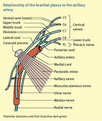
2 Brachial plexus
3
4
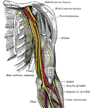
5 Median nerve After originating from the brachial plexus in the axilla, the median nerve descends down the arm, initially lateral to the brachial artery. Halfway down the arm, the nerve crosses over the brachial artery, and becomes situated medially. The median nerve enters the anterior compartment of the forearm between the two heads of pronator teres muscle via the cubital fossa. In the forearm, the nerve travels between the flexor digitorum profundus and flexor digitorum superficialis muscles. Major branches in the forearm: 1- Anterior interosseous nerve supplies the deep muscles in the anterior forearm. 2- Palmar cutaneous nerve innervates the skin of the lateral palm


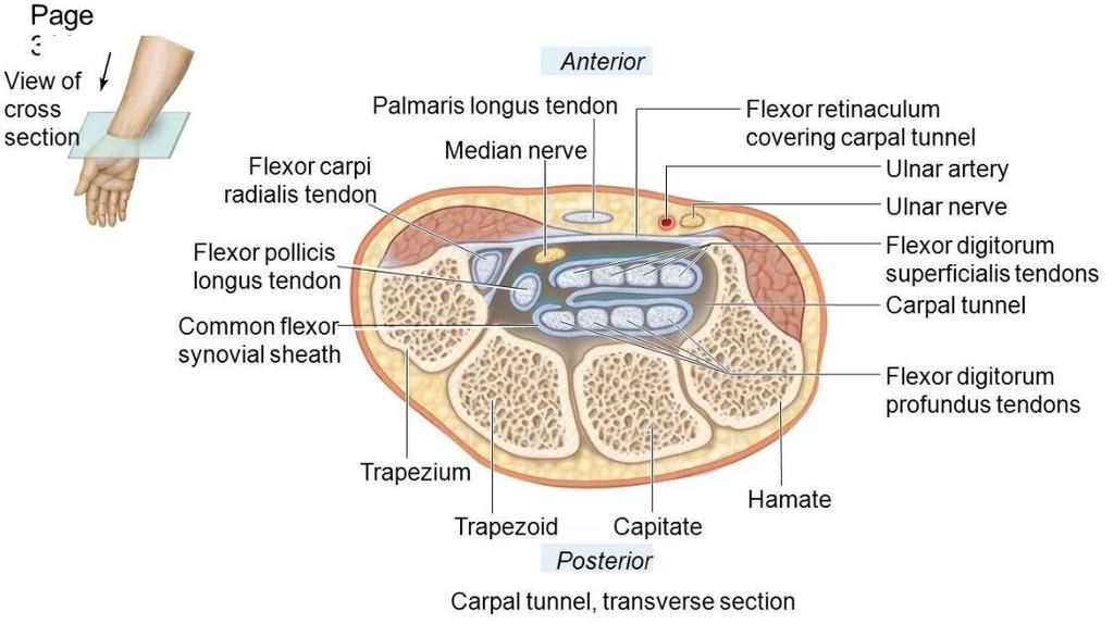
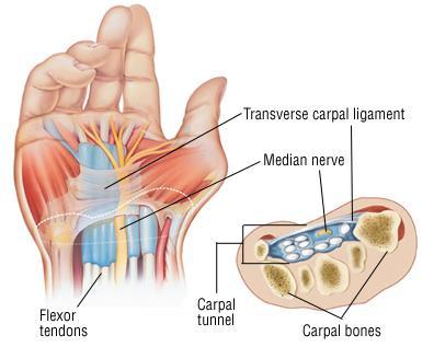
6 The median nerve enters the hand via the carpal tunnel where it terminates by dividing into two branches: 1- Recurrent branch innervates the thenar muscles. 2- Palmar digital branch innervates the palmar surface and fingertips of the lateral three and half digits. Also innervates the lateral two lumbrical muscles. Recrrent branch Palmar digital branches
7
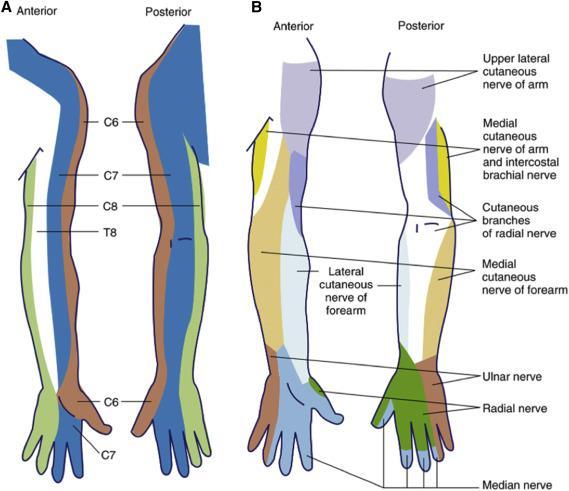
8 Median nerve Sensory functions: Gives rise to the palmar cutaneous branch, which innervates the lateral aspect of the palm, and the digital cutaneous branch, which innervates the lateral three and a half fingers on the anterior (palmar) surface of the hand Nerve roots: C6 T1 (also contains fibres from C5 in some individuals). Motor functions: Innervates the flexor and pronator muscles in the anterior compartment of the forearm (except the flexor carpi ulnaris and part of the flexor digitorum profundus, innervated by the ulnar nerve). Also supplies innervation to the thenar muscles and lateral two lumbricals in the hand.

9 Median cutaneous nerve supply of the hand
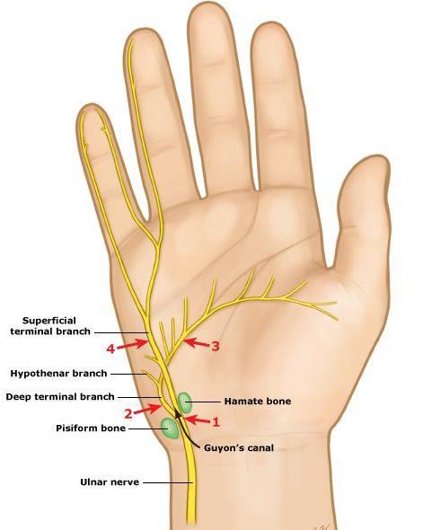
10 Sensory functions: Innervates the anterior and posterior surfaces of the medial one and half fingers, and the associated palm area Anatomical Course After arising from the brachial plexus, the ulnar nerve descends down the medial aspect of the upper arm. At the elbow, it passes posterior to the medial epicondyle of the humerus and gives rise to an articular branch to the elbow joint. Ulnar nerve is palpable at the medial epicondyle Ulnar nerve Spinal roots: C8-T1. Motor functions: Innervates the intrinsic muscles of the hand (hypothenar muscles and two medial lumbricals), and two muscles in the forearm; flexor carpi ulnaris and medial half of flexor digitorum profundus.
11
12 In the forearm, the ulnar nerve pierces the two heads of the flexor carpi ulnaris, and travels deep to the muscle, alongside the ulna. Three main branches arise in the forearm: 1- Muscular branch innervates two muscles in the anterior compartment of the forearm. 2- Palmar cutaneous branch innervates the medial half of the palm. 3- Dorsal cutaneous branch innervates the dorsal surface of the medial one and a half fingers, and the associated dorsal hand area At the wrist, the ulnar nerve travels superficially to the flexor retinaculum, and is medial to the ulnar artery. It enters the hand via the ulnar canal. In the hand, the nerve terminates by giving rise to superficial and deep branches


13 Palmer cutan branch of Ul N Palmar cutaneous branch Of ulnar nerve

14 Ulnar nerve
15 Motor Functions The ulnar nerve innervates In the anterior compartment of the forearm Flexor carpi ulnaris. Flexor digitorum profundus (medial half) In the hand. The majority of the intrinsic hand muscles are innervated by the deep branch of the ulnar nerve: Hypothenar muscles Medial two lumbricals Adductor pollicis Palmar and dorsal interossei of the hand
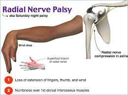
16 Radial nerve Anatomical Course: The radial nerve is the terminal continuation of the posterior cord of the brachial plexus. It therefore contains fibres from nerve roots C5 T1. The nerve arises in the axilla region, where it is situated posteriorly to the axillary artery. It exits the axilla inferiorly (via the triangular interval), and supplies branches to the long and lateral heads of the triceps brachii. The radial nerve then descends down the arm, travelling in a shallow depression within the surface of the humerus, known as the radial groove. As it descends, the radial nerve wraps around the humerus laterally, and supplies a branch to the medial head of the triceps brachii. During much of its course within the arm, it is accompanied by the profunda brachii branch of the brachial artery
17 To enter the forearm, the radial nerve travels anterior to the lateral epicondyle of the humerus (Radial nerve has motor and sensory parts).the radial nerve then divides into two branches, superficial and deep branches. The deep branch of the radial nerve winds to the back of the forearm around the lateral side of the radius between the two planes of fibers of the Supinator, and is prolonged downward between the superficial and deep layers of muscles, to the middle of the forearm. The deep branch continues as posterior interosseous nerve provides motor supply to the muscles in the posterior compartment of the forearm, which is mostly the extensor muscles of the hand. The radial nerve also innervates the triceps brachii
18
19 Superficial branch (sensory) The superficial branch of the radial nerve passes along the front of the radial side of the forearm. It lies at slightly lateral to the radial artery, beneath the Brachioradialis. It leaves the artery about 7 cm. above the wrist, passes beneath the tendon of the Brachioradialis, and, piercing the deep fascia, divides into two branches: lateral and medial. Innervates most of the skin of the posterior side of forearm, the dorsal surface of the lateral side of the palm, and dorsal surface of the lateral three and a half fingers.

20 Brachioradialis muscle Superficial branch of radial nerve

21 Radial nerve

22 Thank You
Key Relationships in the Upper Limb
 Key Relationships in the Upper Limb This list contains some of the key relationships that will help you identify structures in the lab. They are organized by dissection assignment as defined in the syllabus.
MCQWeek2. All arise from the common flexor origin. The posterior aspect of the medial epicondyle is the common flexor origin.
 MCQWeek2. 1. Regarding superficial muscles of anterior compartment of the forearm: All arise from the common flexor origin. The posterior aspect of the medial epicondyle is the common flexor origin. Flexor
The arm: *For images refer back to the slides
 The arm: *For images refer back to the slides Muscles of the arm: deltoid, triceps (which is located at the back of the arm), biceps and brachialis (it lies under the biceps), brachioradialis (it lies
Nerves of Upper limb. Dr. Brijendra Singh Professor & Head Department of Anatomy AIIMS Rishikesh
 Nerves of Upper limb Dr. Brijendra Singh Professor & Head Department of Anatomy AIIMS Rishikesh 1 Objectives Origin, course & relation of median & ulnar nerves. Motor & sensory distribution Carpal tunnel
Fascial Compartments of the Upper Arm
 Fascial Compartments of the Upper Arm The upper arm is enclosed in a sheath of deep fascia and has two fascial septa: 1- Medial fascial septum (medial intermuscular septum): attached to the medial supracondylar
region of the upper limb between the shoulder and the elbow Superiorly communicates with the axilla.
 1 region of the upper limb between the shoulder and the elbow Superiorly communicates with the axilla. Inferiorly, a number of important structures pass between arm & forearm through cubital fossa. 2 medial
divided by the bones ( redius and ulna ) and interosseous membrane into :
 and interosseous membrane into :") fossa Cubital Has: * floor. * roof : - Skin - superficial fasica - deep fascia ( include bicipital aponeurosis ) Structures within the roof : -cephalic and basilic veins -and between them median cubital
fossa Cubital Has: * floor. * roof : - Skin - superficial fasica - deep fascia ( include bicipital aponeurosis ) Structures within the roof : -cephalic and basilic veins -and between them median cubital
Nerve Injury. 1) Upper Lesions of the Brachial Plexus called Erb- Duchene Palsy or syndrome.
 Upper Lesions of the Brachial Plexus called Erb- Duchene Palsy or syndrome.") Nerve Injury - Every nerve goes to muscle or skin so if the nerve is injured this will cause paralysis in the muscle supplied from that nerve (paralysis means loss of function) then other muscles and other
Nerve Injury - Every nerve goes to muscle or skin so if the nerve is injured this will cause paralysis in the muscle supplied from that nerve (paralysis means loss of function) then other muscles and other
The Muscular System. Chapter 10 Part C. PowerPoint Lecture Slides prepared by Karen Dunbar Kareiva Ivy Tech Community College
 Chapter 10 Part C The Muscular System Annie Leibovitz/Contact Press Images PowerPoint Lecture Slides prepared by Karen Dunbar Kareiva Ivy Tech Community College Table 10.9: Muscles Crossing the Shoulder
Al-Balqa Applied University
 Al-Balqa Applied University Faculty Of Medicine *You can use this checklist as a guide to you for the lab. the items on this checklist represent the main features of the models that you have to know for
BRACHIAL PLEXUS. DORSAL SCAPULAR NERVE (C5) supraclavicular branch innervates rhomboids (major and minor) and levator scapulae
 supraclavicular branch innervates rhomboids (major and minor) and levator scapulae") THE BRACHIAL PLEXUS DORSAL SCAPULAR NERVE (C5) supraclavicular branch innervates rhomboids (major and minor) and levator scapulae SCHEMA OF THE BRACHIAL PLEXUS THE BRACHIAL PLEXUS PHRENIC NERVE supraclavicular
THE BRACHIAL PLEXUS DORSAL SCAPULAR NERVE (C5) supraclavicular branch innervates rhomboids (major and minor) and levator scapulae SCHEMA OF THE BRACHIAL PLEXUS THE BRACHIAL PLEXUS PHRENIC NERVE supraclavicular
Supplied in part by the musculocutaneous nerve. Forms the axis of rotation in movements of pronation and supination
 Anatomy: Upper limb (15 questions) 1. Latissimus Dorsi: Is innervated by the dorsal scapular nerve Lies above feres major muscle Medially rotates the humerus All of the above 2. Supinator muscle is: Deep
The hand is full with sweat glands, activated at times of stress. In Slide #2 there was a mistake where the doctor mentioned lateral septum twice.
 We should only know: Name, action & nerve supply Layers - Skin - Superficial fascia - Deep fascia The hand is full with sweat glands, activated at times of stress. Deep fascia In Slide #2 there was a mistake
ARM Brachium Musculature
 ARM Brachium Musculature Coracobrachialis coracoid process of the scapula medial shaft of the humerus at about its middle 1. flexes the humerus 2. assists to adduct the humerus Blood: muscular branches
Netter's Anatomy Flash Cards Section 6 List 4 th Edition
 Netter's Anatomy Flash Cards Section 6 List 4 th Edition https://www.memrise.com/course/1577581/ Section 6 Upper Limb (66 cards) Plate 6-1 Humerus and Scapula: Anterior View 1.1 Acromion 1.2 Greater tubercle
Dr. Mahir Alhadidi Anatomy Lecture #9 Feb,28 th 2012
 Quick Revision: Upper arm is divided into two compartments: 1. Anterior Compartment: Contains three muscles (Biceps brachii, Coracobrachialis, Brachialis). Innervated by Musculocutaneous nerve. 2. Posterior
STRUCTURAL BASIS OF MEDICAL PRACTICE EXAMINATION 5 October 6, 2006
 STRUCTURAL BASIS OF MEDICAL PRACTICE EXAMINATION 5 October 6, 2006 PART l. Answer in the space provided. (8 pts) 1. Identify the structures. (2 pts) B C A. _pisiform B. _ulnar artery A C. _flexor carpi
compartments of the forearm
 " forearm posterior compartment " compartments of the forearm Posterior Fascial compartment Muscles: ** The superficial group 1. Extensor carpi radialis brevis 2. Ex. digitorum 3. Ex. digiti minimi 4.
Levels of the anatomical cuts of the upper extremity RADIUS AND ULNA right
 11 CHAPTER 2 Levels of the anatomical cuts of the upper extremity AND right CUT 1 CUT 4 1 2 3 4 5 6 Isolated fixation of the radius is difficult at this level because of the anterolateral vessels and the
Lecture 9: Forearm bones and muscles
 Lecture 9: Forearm bones and muscles Remember, the region between the shoulder and the elbow = brachium/arm, between elbow and wrist = antebrachium/forearm. Forearm bones : Humerus (distal ends) Radius
Muscles of the hand Prof. Abdulameer Al-Nuaimi
 Muscles of the hand Prof. Abdulameer Al-Nuaimi a.alnuaimi@sheffield.ac.uk abdulameerh@yahoo.com Thenar Muscles Thenar muscles are three short muscles located at base of the thumb. All are innervated by
Slides of Anatomy. Spring Dr. Maher Hadidi, University of Jordan
 Slides of Anatomy Please note : These slides are Dr. Maher Hadidi s slides of spring 2016 and were edited by the Premed Academic Team to fit the slides of spring 2019. Spring 2019 Dr. Maher Hadidi, University
Cubital fossa and forearm
 Cubital fossa and forearm Cubital fossa is the triangular space in front of elbow joint. - The Cubital fossa has boundaries: apex, base, roof and floor and it has contents. The base: an imaginary horizontal
Joints of the upper limb II
 Joints of the upper limb II Prof. Abdulameer Al-Nuaimi E-mail: a.al-nuaimi@sheffield.ac.uk E. mail: abdulameerh@yahoo.com Elbow joint The elbow joint is connecting the upper arm to the forearm. It is classed
Forearm and Wrist Regions Neumann Chapter 7
 Forearm and Wrist Regions Neumann Chapter 7 REVIEW AND HIGHLIGHTS OF OSTEOLOGY & ARTHROLOGY Radius dorsal radial tubercle radial styloid process Ulna ulnar styloid process ulnar head Carpals Proximal Row
The Elbow and the cubital fossa. Prof Oluwadiya Kehinde
 The Elbow and the cubital fossa Prof Oluwadiya Kehinde www.oluwadiya.com Elbow and Forearm Anatomy The elbow joint is formed by the humerus, radius, and the ulna Bony anatomy of the elbow Distal Humerus
forearm posterior compartment
 Quick revision: The anterior compartment of the forearm contains of 8 muscles... -4 superficial -1 intermediate -3 deep *All supplied by median nerve except 1 and 1/2 muscle (by ulnar N.) forearm posterior
Anatomy Workshop Upper Extremity David Ebaugh, PT, PhD Workshop Leader. Lab Leaders: STATION I BRACHIAL PLEXUS
 Anatomy Workshop Upper Extremity David Ebaugh, PT, PhD Workshop Leader Lab Leaders: STATION I BRACHIAL PLEXUS A. Posterior cervical triangle and axilla B. Formation of plexus 1. Ventral rami C5-T1 2. Trunks
REFERENCE DIAGRAMS OF UPPER LIMB MUSCLES: NAMES, LOCATIONS, ATTACHMENTS, FUNCTIONS MUSCLES CONNECTING THE UPPER LIMB TO THE AXIAL SKELETON
 REFERENCE DIAGRAMS OF UPPER LIMB MUSCLES: NAMES, LOCATIONS, ATTACHMENTS, FUNCTIONS MUSCLES CONNECTING THE UPPER LIMB TO THE AXIAL SKELETON A25LAB EXERCISES: UPPER LIMB MUSCLES Page 1 MUSCLES CONNECTING
Biceps Brachii. Muscles of the Arm and Hand 4/4/2017 MR. S. KELLY
 Muscles of the Arm and Hand PSK 4U MR. S. KELLY NORTH GRENVILLE DHS Biceps Brachii Origin: scapula Insertion: radius, fascia of forearm (bicipital aponeurosis) Action: supination and elbow flexion Innervation:
MUSCLES OF THE ELBOW REGION
 MUSCLES OF THE ELBOW REGION Dr Bronwen Ackermann COMMONWEALTH OF AUSTRALIA Copyright Regulation WARNING This material has been reproduced and communicated to you by or on behalf of the University of Sydney
MSK Imaging Conference. 07/22/2016 Eman Alqahtani, MD, MPH R3/PGY4 UCSD Radiology
 MSK Imaging Conference 07/22/2016 Eman Alqahtani, MD, MPH R3/PGY4 UCSD Radiology A 51 years old female with chronic thumb pain, and inability to actively flex the thumb interphalyngeal joint Possible trigger
The Clavicle Right clavicle Deltoid tubercle: Conoid tubercle, conoid ligamen Impression for the
 The Clavicle Muscle Attachment Sites in the Upper Limb Pectoralis major Right clavicle Smooth superior surface of the shaft, under the platysma muscle tubercle: attachment of the deltoid Acromial facet
*the Arm* -the arm extends from the shoulder joint (proximal), to the elbow joint (distal) - it has one bone ; the humerus which is a long bone
, to the elbow joint (distal) - it has one bone ; the humerus which is a long bone") *the Arm* -the arm extends from the shoulder joint (proximal), to the elbow joint (distal) - it has one bone ; the humerus which is a long bone - muscles in the arm : *brachialis muscle *Biceps brachii
*the Arm* -the arm extends from the shoulder joint (proximal), to the elbow joint (distal) - it has one bone ; the humerus which is a long bone - muscles in the arm : *brachialis muscle *Biceps brachii
Kinesiology of The Wrist and Hand. Cuneyt Mirzanli Istanbul Gelisim University
 Kinesiology of The Wrist and Hand Cuneyt Mirzanli Istanbul Gelisim University Bones The wrist and hand contain 29 bones including the radius and ulna. There are eight carpal bones in two rows of four to
The Arm and Cubital Fossa
 The Arm and Cubital Fossa Dr. Andrew Gallagher School of Anatomical Sciences University of the Witwatersrand Introduction The ARM (BRACHIUM) is the most proximal segment of the upper limb musculoskeletal
Abduction of arm until your hand rich your head. Flexion of forearm at elbow joint. Extension of arm at elbow joint. Flexion of fingers 10.
 Num. answer 1. Medialy With the manubrium ( sternum ), and laterally with the acromion of the scapula 2. 1. Trapezius 2. Levator scapulae 3. Rhomboids 3. 1. Pectoralis major 2. Pectoralis minor 3. Latissiumus
Lab Activity 11: Group II
 Lab Activity 11: Group II Muscles Martini Chapter 11 Portland Community College BI 231 Origin and Insertion Origin: The place where the fixed end attaches to a bone, cartilage, or connective tissue. Insertion:
Human Anatomy Biology 351
 1 Human Anatomy Biology 351 Upper Limb Exam Please place your name on the back of the last page of this exam. You must answer all questions on this exam. Because statistics demonstrate that, on average,
The Forearm 2. Extensor & lateral Compartments of the Forearm
 The Forearm 2 Extensor & lateral Compartments of the Forearm 1-Lateral Fascial Compartment (at the lateral side of the forearm ) *Some books mention the lateral compartment contain just the Brachioradialis
Peripheral Nervous Sytem: Upper Body
 Peripheral Nervous Sytem: Upper Body MSTN121 - Neurophysiology Session 10 Department of Myotherapy Cervical Plexus Accessory nerve (CN11 + C1-5) Motor: trapezius and sternocleidomastoid Greater auricular
Systematic Anatomy (For international students)
") Systematic Anatomy (For international students) Department of Anatomy,Fudan University Teaching contents Muscles of abdomen & upper limbs Dr.Hongqi Zhang ( 张红旗 ) Email: zhanghq58@126.com 1 Muscles of abdomen
Systematic Anatomy (For international students) Department of Anatomy,Fudan University Teaching contents Muscles of abdomen & upper limbs Dr.Hongqi Zhang ( 张红旗 ) Email: zhanghq58@126.com 1 Muscles of abdomen
[[Sally Leaning Towards Peter To Take Cold Hand]]
![[[Sally Leaning Towards Peter To Take Cold Hand]]](/thumbs/84/91174469.jpg "[[Sally Leaning Towards Peter To Take Cold Hand]]") In this lecture we will talk about the bones of the hand, and the muscles and contents of the forearm. *The hand bones are: - Carpal bones. -Metacarpals. -Phalanges. *The carpal bones (wrist bones): They
In this lecture we will talk about the bones of the hand, and the muscles and contents of the forearm. *The hand bones are: - Carpal bones. -Metacarpals. -Phalanges. *The carpal bones (wrist bones): They
ANATOMY. Subject : Lecturer : Maher Hadidi Done by: lecture # : 11 Date :
 ANATOMY Subject : Lecturer : Maher Hadidi Done by: lecture # : 11 Date : Median nerve Meaian nerve to Pronator teres Course: Enter cubital fossa. Pierce Pronat. teres Between to FDS, FOP Below and lateral
Misc Anatomy. Upper Limb! 2. Lower Limb! 5. Venous Drainage! Head & neck! 8
 Misc Anatomy Upper Limb! 2 Arteries!... 2 Veins!... 2 Spaces!... 4 Lower Limb! 5 Arteries!... 5 Venous Drainage!... 6 Spaces!... 7 Head & neck! 8 Artery!... 8 Ultrasound View for IJ CVL!... 8 Arteries
1/13/2013. Anatomy Guy Dissection Sheet Extensor Forearm and Hand. Eastern Virginia Medical School
 Dr. Craig Goodmurphy Anatomy Guy Superficial Extensor Muscles Complete skin removal if necessary then remove the antebrachial fascia starting at the extensor retinaculum and working proximally. Define
Main Menu. Wrist and Hand Joints click here. The Power is in Your Hands
 1 The Wrist and Hand Joints click here Main Menu K.5 http://www.handsonlineeducation.com/classes/k5/k5entry.htm[3/23/18, 1:40:40 PM] Bones 29 bones, including radius and ulna 8 carpal bones in 2 rows of
Anatomy of the Upper Limb
 Anatomy of the Upper Limb Figure 53: The thenar & midpalmar spaces. The synovial (tendon) sheaths of the long flexors [Figure.54] These sheaths surround the tendons of the long flexors; flexor digitorum
Functional Anatomy of the Elbow
 Functional Anatomy of the Elbow Orthopedic Institute Daryl C. Osbahr, M.D. Chief of Sports Medicine, Orlando Health Chief Medical Officer, Orlando City Soccer Club Orthopedic Consultant, Washington Nationals
medial half of clavicle; Sternum; upper six costal cartilages External surfaces of ribs 3-5
 MUSCLE ORIGIN INSERTION ACTION NERVE Pectoralis Major medial half of clavicle; Sternum; upper six costal cartilages Lateral lip of intertubercular groove of horizontal adduction Medial and lateral pectoral
Year 2004 Paper one: Questions supplied by Megan
 QUESTION 47 A 58yo man is noted to have a right foot drop three days following a right total hip replacement. On examination there is weakness of right ankle dorsiflexion and toe extension (grade 4/5).
Muscular Nomenclature and Kinesiology - One
 Chapter 16 Muscular Nomenclature and Kinesiology - One Lessons 1-3 (with lesson 4) 1 Introduction 122 major muscles covered in this chapter Chapter divided into nine lessons Kinesiology study of human
STRUCTURAL BASIS OF MEDICAL PRACTICE EXAMINATION 5. September 30, 2011
 STRUCTURAL BASIS OF MEDICAL PRACTICE EXAMINATION 5 September 30, 2011 PART l. Answer in the space provided. (12 pts) 1. Identify the structures. (2 pts) EXAM NUMBER A. Suprascapular nerve B. Axillary nerve
Hand and Wrist Editing file. Color Code Important Doctors Notes Notes/Extra explanation
 Hand and Wrist Editing file Color Code Important Doctors Notes Notes/Extra explanation Objectives Describe the anatomy of the deep fascia of the wrist & hand (flexor & extensor retinacula & palmar aponeurosis).
The Elbow and Radioulnar Joints Kinesiology. Dr Cüneyt Mirzanli Istanbul Gelisim University
 The Elbow and Radioulnar Joints Kinesiology Dr Cüneyt Mirzanli Istanbul Gelisim University 1 The Elbow & Radioulnar Joints Most upper extremity movements involve the elbow & radioulnar joints. Usually
Muscles of the Upper Limb
 Muscles of the Upper Limb anterior surface of ribs 3 5 coracoid process Pectoralis minor pectoral nerves protracts / depresses scapula Serratus anterior Subclavius ribs 1-8 long thoracic nerve rib 1 ----------------
Neurovascular Variations in Upper Limb
 Case report : Neurovascular Variations in Upper Limb 1Dr. Dinendra Kumar Saha, 2 Dr. Jayeeta Burman, 3 Dr. Sudeshna Majumdar, 4 Dr. Manotosh Banerjee, 5 Dr. Sharmistha Chakraborty, 6 Dr. Sushmita Sen,
Practical 2 Worksheet
 Practical 2 Worksheet Upper Extremity BONES 1. Which end of the clavicle is on the lateral side (acromial or sternal)? 2. Describe the difference in the appearance of the acromial and sternal ends of the
Lecture 10 Arteries and veins of the upper limb
 Lecture 10 Arteries and veins of the upper limb 1. Identify the Subclavian, axillary, brachial (deep and superficial), radial and ulnar arteries and superficial/deep palmar arches 2. Describe the major
Axilla and Brachial Region
 L 4 A B O R A T O R Y Axilla and Brachial Region BRACHIAL PLEXUS 5 Roots/Rami (ventral rami C5 T1) 3 Trunks Superior (C5, C6) Middle (C7) Inferior (C8, T1) 3 Cords Lateral Cord (Anterior Superior and Anterior
LIST OF STRUCTURES TO BE IDENTIFIED IN LAB: UPPER EXTREMITY REVIEW 2016
 LIST OF STRUCTURES TO BE IDENTIFIED IN LAB: UPPER EXTREMITY REVIEW 2016 BONES Ribs, sternum, clavicle Humerus: Head, greater tubercle, lesser tubercle, intertubercular sulcus, surgical neck, anatomical
Figure 27: The synovial membrane of the shoulder joint (anterior view)
") The coracoacromial ligament; is an accessory ligament that protects the superior aspect of the joint extending from the coracoid process to the acromion over the tendon of supraspinatus. The synovial membrane
The coracoacromial ligament; is an accessory ligament that protects the superior aspect of the joint extending from the coracoid process to the acromion over the tendon of supraspinatus. The synovial membrane
# Anatomy. Upper Extremities Muscles and anatomy of axilla. Tiba Al-Ani 9/10/2015 Nabil. Page 0 of 16
 #10 25 Anatomy Upper Extremities Muscles and anatomy of axilla Tiba Al-Ani 9/10/2015 Nabil Page 0 of 16 Salam AWN Today s lecture is divided into two parts, the first part is the continuation of the upper
Multiple variations involving all the terminal branches of the brachial plexus and the axillary artery a case report
 SHORT REPORT Eur J Anat, 10 (3): 61-66 (2006) Multiple variations involving all the terminal branches of the brachial plexus and the axillary artery a case report K. Ramachandran, I. Kanakasabapathy and
BRACHIAL PLEXUS 11/12/2014 كيف تتكون الضفيرة FORMATION ENLARGEMENT (INTUMESCENCE) OF THE SPINAL CORD. Grey matter. Cervical intumescence - C 6 - T 2
 OF THE SPINAL CORD. Grey matter. Cervical intumescence - C 6 - T 2") BRACHIAL PLEXUS Prof. Fawzy Elnady ENLARGEMENT (INTUMESCENCE) OF THE SPINAL CORD Grey matter Cervical intumescence - C 6 - T 2 Lumbar intumescence - L 4 S 2 كيف تتكون الضفيرة FORMATION The ventral rami
BRACHIAL PLEXUS Prof. Fawzy Elnady ENLARGEMENT (INTUMESCENCE) OF THE SPINAL CORD Grey matter Cervical intumescence - C 6 - T 2 Lumbar intumescence - L 4 S 2 كيف تتكون الضفيرة FORMATION The ventral rami
Upper limb Arm & Cubital region 黃敏銓
 Upper limb Arm & Cubital region 黃敏銓 1 Arm Lateral intermuscular septum Anterior (flexor) compartment: stronger Medial intermuscular septum Posterior (extensor) compartment 2 Coracobrachialis Origin: coracoid
LECTURE 8 HANDS: BONES AND MUSCLES
 LECTURE 8 HANDS: BONES AND MUSCLES WRIST AND HAND - Human hand can do power grip and precision grip - Thumb is 90 to the rest of the hand can do fine actions - Often able to do power actions o Take tools
In the name of Allah, Most gracious, Most merciful
 In the name of Allah, Most gracious, Most merciful This lecture includes the following: The Palmer Oponeurosis. The Carpel tunnel. The palmaris brevis muscle. The anatomical snuffbox. The Fibrous flexor
Ultrasonography of Peripheral Nerve -upper extremity
 Ultrasonography of Peripheral Nerve -upper extremity Department of Physical Medicine and Rehabilitation Korea University Guro Hospital Korea University College of Medicine Yoon Joon Shik Normal median
10/10/2014. Structure and Function of the Hand. The Hand. Osteology of the Hand
 Structure and Function of the Hand 19 bones and 19 joints are necessary to produce all the motions of the hand The Hand Dorsal aspect Palmar aspect The digits are numbered 1-5 Thumb = #1 Little finger
MUSCLES. Anconeus Muscle
 LAB 7 UPPER LIMBS MUSCLES Anconeus Muscle anconeus origin: distal end of dorsal surface of humerus insertion: lateral surface of ulna from distal margin of the semilunar notch to proximal end of the olecranon
The Elbow 3/5/2015. The Elbow Scanning Sequence. * Anterior Joint (The anterior Pyramid ) * Lateral Epicondyle * Medial Epicondyle * Posterior Joint
 * Lateral Epicondyle * Medial Epicondyle * Posterior Joint") Scanning Sequence * Anterior Joint (The anterior Pyramid ) * Lateral Epicondyle * Medial Epicondyle * Posterior Joint Anterior Elbow Pyramid Courtesy of Jay Smith, MD. Vice chair PMR Mayo Clinic Rochester,
Scanning Sequence * Anterior Joint (The anterior Pyramid ) * Lateral Epicondyle * Medial Epicondyle * Posterior Joint Anterior Elbow Pyramid Courtesy of Jay Smith, MD. Vice chair PMR Mayo Clinic Rochester,
Peripheral Nerve Injuries of the Upper Limb.
 Peripheral Nerve Injuries of the Upper Limb www.fisiokinesiterapia.biz Definitions Radiculopathy Process affecting the nerve root, most commonly by a herniated disc Weakness in muscles supplied by the
BOGOMOLETS NATIONAL MEDICAL UNIVERSITY. Department of Human Anatomy GUIDELINES. The theme of the lesson The vessels of the upper limb.
 BOGOMOLETS NATIONAL MEDICAL UNIVERSITY Department of Human Anatomy GUIDELINES Academic discipline HUMAN ANATOMY Module 2 The theme of the lesson The vessels of the upper limb. Course Faculties І Medical
Done By : Isra a Aweidah
 Anatomy Made Easy MSS هذا الملف يشمل تفريغ المحاضرة 10 المحاضرة 12 ابتداء من صفحة 13 وتفريغ part #9 Done By : Isra a Aweidah Edited by: AWN Academic Team Everything is included ( the drs slides in addition
13 13/3/2012. Adel Muhanna
 13 13/3/2012 Adel Muhanna بسم هللا الرحمن الرحيم The Hand Extensor retinaculum: Deep fascia of anterior compartment of the wrist is thickened to form flexor retinaculum : a bridge that have 6 structures
MLT Muscle(s) Patient Position Therapist position Stabilization Limb Position Picture Put biceps on slack by bending elbow.
 Patient Position Therapist position Stabilization Limb Position Picture Put biceps on slack by bending elbow.") MLT Muscle(s) Patient Position Therapist position Stabilization Limb Position Picture Put biceps on slack by bending elbow. Pectoralis Minor Supine, arm at side, elbows extended, supinated Head of Table
MLT Muscle(s) Patient Position Therapist position Stabilization Limb Position Picture Put biceps on slack by bending elbow. Pectoralis Minor Supine, arm at side, elbows extended, supinated Head of Table
Elbow, Wrist & Hand Evaluation.
 Elbow, Wrist & Hand Evaluation www.fisiokinesiterapia.biz Common Injuries to the Elbow, Wrist, Hand & Fingers Lateral epicondylitis tennis elbow Medial epicondylitis golfer s s elbow, little league elbow
Module 7 - The Muscular System Muscles of the Arm and Trunk
 Module 7 - The Muscular System Muscles of the Arm and Trunk This Module will cover the muscle anatomy of the arms and trunk. We have already seen the muscles that move the humerus, so this module will
Copy Right- Hongqi ZHANG-Department of Anatomy-Fudan University. Systematic Anatomy. Locomotor system - Part 6
 Systematic Anatomy Locomotor system - Part 6 Muscles of abdomen Muscles of the upper limb Dr.Hongqi Zhang ( 张红旗 ) Email: zhanghq58@126.com 1 Muscles of abdomen Muscles of the upper limb Muscles of abdomen
David G. Simpson, Ph.D.
 David G. Simpson, Ph.D. ARM & CUBITAL FOSSA Revised 7/08 Text References Moores 3 rd ed., p402 408, 436 439, 439 443, 478, 481 LEARNING OBJECTIVES: 1. Describe the humerus, indicating the sites of muscle
Ulnar Neuropathy in the Distal Ulnar Tunnel
 Ulnar Neuropathy in the Distal Ulnar Tunnel DAVID W. SHUPE, PT, ATC' Journal of Orthopaedic & Sports Physical Therapy A brief anatomical review of the ulnar nerve and areas of ulnar nerve entrapment is
Candidate s instructions Look at this cross-section taken at the level of C5. Answer the following questions.
 Section 1 Anatomy Chapter 1. Trachea 1 Candidate s instructions Look at this cross-section taken at the level of C5. Answer the following questions. Pretracheal fascia 1 2 5 3 4 Questions 1. Label the
Traditional Thai Acupressure Points. The anterior aspect of the body THE ANATOMICAL ATLAS
 Traditional Thai Acupressure Points The anterior aspect of the body THE ANATOMICAL ATLAS lines of the SHOULDER BLADES AND POSTERIOR ARM Scapula Line This line runs through landmarks: 1. Above the midpoint
Anatomy of the Forearm
 Anatomy of the Forearm Musculoskeletal block- Anatomy-lecture 8 Editing file Objectives List the names of the Flexors Group of Forearm (superficial & deep muscles). Identify the common flexor origin of
Neurovascular variations in upper limb
 Case report : Neurovascular variations in upper limb 1Dr. Dinendra Kumar Saha, 2 Dr. Jayeeta Burman, 3 Dr. Sudeshna Majumdar, 4 Dr. Manotosh Banerjee, 5 Dr. Sharmistha Chakraborty, 6 Dr. Sushmita Sen,
Deep dry needling of the arm and hand muscles
 Deep dry needling of the arm and hand s 8 César Fernández-de-las-Peñas Javier González Iglesias Christian Gröbli Ricky Weissmann CHAPTER CONTENT Introduction................... 107 Clinical relevance of
VARIANT ARTERIAL PATTERN IN THE FOREARM WITH ITS EMBRYOLOGICAL BASIS. Vaishnavi Joshi and Dr. Shaheen Sajid Rizvi
 Volume-8, Issue-3 July-Sept-2018 Coden:IJPAJX-CAS-USA, Copyrights@2018 ISSN-2231-4490 Received: 8 th June-2018 Revised: 15 th July-2018 Accepted: 16 th July-2018 DOI: 10.21276/Ijpaes http://dx.doi.org/10.21276/ijpaes
Connects arm to thorax 3 joints. Glenohumeral joint Acromioclavicular joint Sternoclavicular joint
 Connects arm to thorax 3 joints Glenohumeral joint Acromioclavicular joint Sternoclavicular joint Scapula Elevation Depression Protraction (abduction) Retraction (adduction) Downward Rotation Upward Rotation
Ligaments of Elbow hinge: sagittal plane so need lateral and medial ligaments
 Ligaments of Elbow hinge: sagittal plane so need lateral and medial ligaments Ulnar Collateral ligament on medial side; arising from medial epicondyle and stops excess valgus movement (lateral movement)
e- Lateral pectoral nerve
 1. All of the following muscles have double innervations except: a- Brachialis b- Flexor digitorum profundus c- Trapezius d- Pectoralis major e- Subscapularis 2. You can t put your hand over your head
Anatomage Table Instructors Guide- Upper Limb
 The Upper Limb Anatomage Table Instructors Guide- Upper Limb Table of Contents Upper Limb 1- The Skeletal System...3 1: Clavicle...3 2: Scapula...5 3: Shoulder (Glenohumeral) and Proximal Humerus...7 4:
Wrist & Hand Assessment and General View
 Wrist & Hand Assessment and General View Done by; Mshari S. Alghadier BSc Physical Therapy RHPT 366 m.alghadier@sau.edu.sa http://faculty.sau.edu.sa/m.alghadier/ Functional anatomy The hand can be divided
Muscle Anatomy Review Chart
 Muscle Anatomy Review Chart BACK Superficial (5) Trapezius Transverse cervical a. Latissimus dorsi Thoracodorsal a. Rhomboideus major Dorsal scapular a. Rhomboideus minor Levator scapulae Intermediate
Wrist and Hand Anatomy/Biomechanics
 Wrist and Hand Anatomy/Biomechanics Kristin Kelley, DPT, OCS, FAAOMPT Orthopaedic Manual Physical Therapy Series Charlottesville 2017-2018 Orthopaedic Manual Physical Therapy Series 2017-2018 Anatomy -
Anatomy - Hand. Wrist and Hand Anatomy/Biomechanics. Osteology. Carpal Arch. Property of VOMPTI, LLC
 Wrist and Hand Anatomy/Biomechanics Kristin Kelley, DPT, OCS, FAAOMPT The wrist The metacarpals The Phalanges Digit 1 thumb Digit 5 digiti minimi Anatomy - Hand Orthopaedic Manual Physical Therapy Series
Elbow Elbow Anatomy. Flexion extension. Pronation Supination. Anatomy. Anatomy. Romina Astifidis, MS., PT., CHT
 Elbow Elbow Anatomy Romina Astifidis, MS., PT., CHT Curtis National Hand Center Baltimore, MD October 6-8, 2017 Link between the arm and forearm to position the hand in space Not just a hinge Elbow = 70%
Clinical examination of the wrist, thumb and hand
 Clinical examination of the wrist, thumb and hand 20 CHAPTER CONTENTS Referred pain 319 History 319 Inspection 320 Functional examination 320 The distal radioulnar joint.............. 320 The wrist.......................
Viorel Nacu. The clinical anatomy of the Hand
 Viorel Nacu The clinical anatomy of the Hand The distal part of the upper limb is divided in to three regions: 1. The wrist (carpus) 2. The hand (metacarpus) 3. The digits (fingers) The landmarks of this
The Role of Muscles in Movement
 The Role of Muscles in Movement Muscles can t push, they can only pull as they contract, so most often body movements are the result of the activity of pairs or teams of muscles acting together or against