Functional anatomy and variability of the blood vessels of the upper and lower limbs. Anastasia Bendelic Human Anatomy Departament
|
|
|
- Loren Parsons
- 5 years ago
- Views:
Transcription
1 Functional anatomy and variability of the blood vessels of the upper and lower limbs Anastasia Bendelic Human Anatomy Departament
2 Plan: 1. Variations of the branching pattern of the aortic arch 2. Arterial anastomoses of the upper extremity 3. Determining the arterial pulse on the upper limbs 4. Variations of the arterial patterns in the upper limb 5. Arterial anastomoses of the lower extremity 6. Determining the arterial pulse on the lower limbs 7. Variations of the arterial patterns in the lower limb
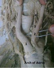
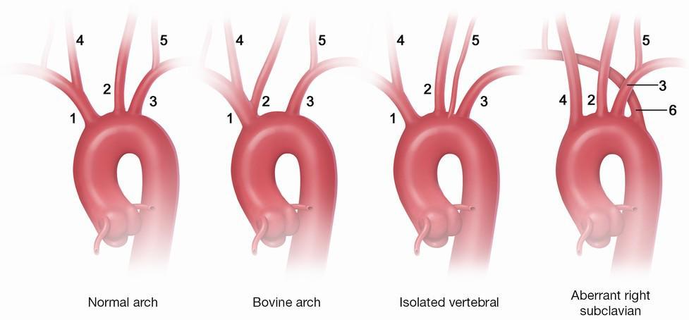
3 Variations of the branching pattern of the aortic arch Variations of the branching pattern of the aortic arch are not rare. Most of them are asymptomatic and mostly found as incidental finding during routine diagnostic procedures. However, head and neck surgeons and interventional radiologists should be aware of aortic arch variation. The normal three-branch pattern of the aortic arch is found with an incidence of 64,9-94,3% according to the literature.


4 The normal three-branch pattern of the aortic arch (64,9-94,3%)
5 Variations of the branching pattern of the aortic arch The most common variation of the aortic arch with two branches (common brachiocephalic trunk with left common carotid artery and left subclavian artery) is found with an incidence of 10-22% in literature. Origination of the left vertebral artery from the aortic arch is not uncommon and the reported prevalence is between 2,4 and 8%.


6 Origination of the left vertebral artery from the aortic arch (2,4-8%)
7 Variations of the branching pattern of the aortic arch Two common brachiocephalic trunks. The brachiocephalic trunk is absent and right subclavian artery, right common carotid artery, left common carotid artery and left subclavian artery leave the aortic arch separately. The right subclavian artery is the last branch of the aortic arch in ~1% of individuals. It courses to the right behind of the esophagus. A retroesophageal course may be the cause of dysphagia.
8 Two brachiocephalic trunks


9 The brachiocephalic trunk is absent (retroesophageal course of the right subclavian artery)

10 Double aortic arch Double aortic arch is a rare anomaly caused by the persistence of the fetal double aortic system. It is a form of complete vascular ring that may compress the trachea and esophagus.

11 The arteries of the upper limb The brachial artery usually begins as a continuation of the axillary artery at the inferior border of the teres major muscle. It ends at about a centimeter distal to the elbow joint at the level of the neck of radius by dividing into radial and ulnar arteries.
12 The arteries of the upper limb
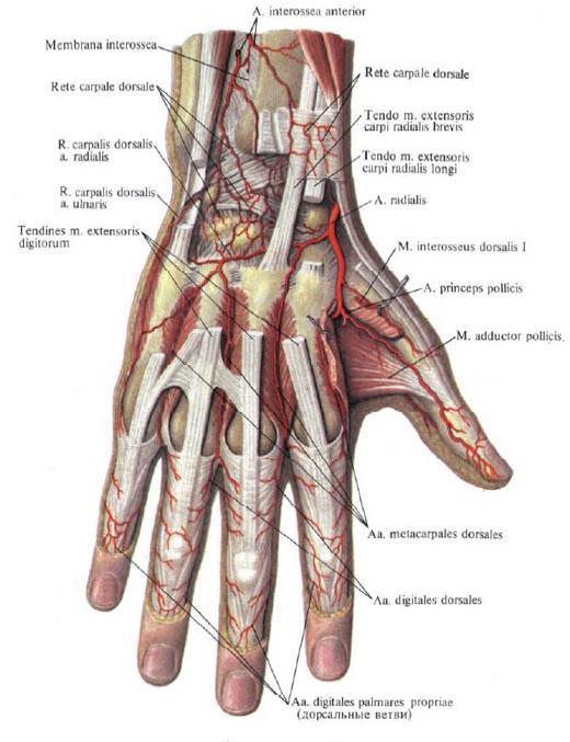
13 Arterial anastomoses of the upper extremity 1. Scapular and shoulder anastomoses 2. Elbow anastomosis 3. Wrist and hand anastomoses
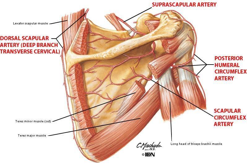
14 Scapular and shoulder anastomoses Around the scapula (scapular anastomosis): a) Suprascapular artery (from the thyrocervical trunk); b) Dorsal scapular artery (from the transverse cervical artery); c) Circumflex scapular artery (from the subscapular artery). Around the surgical neck of the humerus: a) Anterior humeral circumflex artery (from the axillary artery); b) Posterior humeral circumflex artery (from the artery). Around the acromioclavicular joint (acromial anastomosis): a) Acromial branch of the thoracoacromial artery; b) Acromial branches of the circumflex humeral arteries; c) Suprascapular artery (from the thyrocervical trunk).

15 Scapular and shoulder anastomoses

16 Scapular and shoulder anastomoses
17 Elbow anastomosis (rete articulare cubiti) Four collateral arteries: a) Radial collateral artery (from the profunda brachii artery); b) Medial collateral artery (from the profunda brachii artery); c) Superior ulnar collateral artery (from the brachial artery); d) Inferior ulnar collateral artery (from the brachial artery). Four recurrent arteries: a) Radial recurrent artery (from the radial artery); b) Interosseous recurrent artery (from the common interosseous artery); c) Posterior branch of the ulnar recurrent artery (from the ulnar artery); d) Anterior branch of the ulnar recurrent artery (from the ulnar artery).

18 Elbow anastomosis (rete articulare cubiti)
19 Elbow anastomosis (rete articulare cubiti)
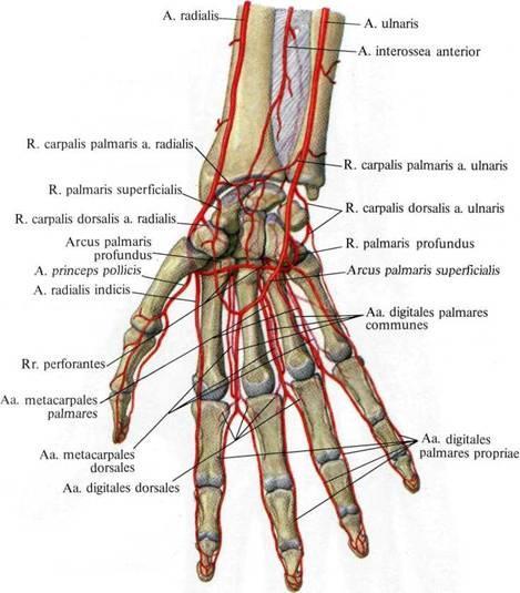
20 Wrist anastomoses Rete carpale dorsale: a) Dorsal carpal branch, ramus carpalis dorsalis, (from the ulnar artery); b) Dorsal carpal branch, ramus carpalis dorsalis, (from the radial artery); c) Anterior and posterior interosseous arteries (from the common interosseous artery). Rete carpale palmare: a) Palmar carpal branch, ramus carpalis palmaris, (from the ulnar artery); b) Palmar carpal branch, ramus carpalis palmaris, (from the radial artery); c) Anterior interosseous artery (from the common interosseous artery).
21 Wrist anastomoses
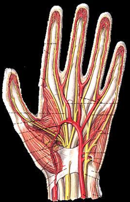
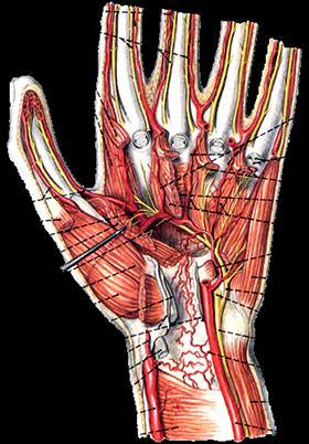
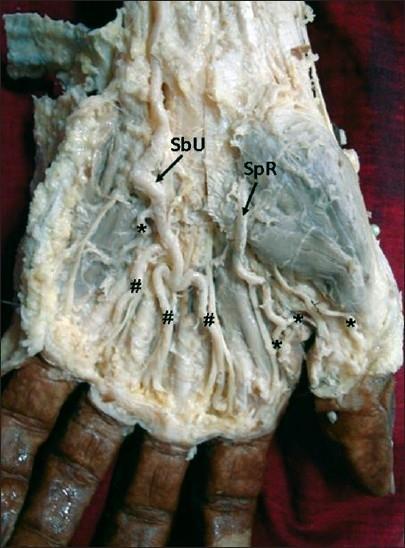
22 Hand anastomoses (palmar arches) Superficial palmar arch (arcus palmaris superficialis) is formed by: a) Ulnar artery (its terminal part); b) Superficial palmar branch, ramus palmaris superficialis, (from the radial artery). The superficial palmar arch gives rise to three common palmar digital arteries, each then divides into two proper palmar digital arteries. Deep palmar arch (arcus palmaris profundus) is formed by: a) Radial artery (its terminal part); b) Deep palmar branch, ramus palmaris profundus, (from the ulnar artery). The deep palmar arch gives rise to three palmar metacarpal arteries.


23 Hand anastomoses (palmar arches)
24 Determining the arterial pulse on the upper limbs Axillary pulse is palpated on the lateral wall of the axillary cavity (axillary artery). Brachial pulse is determined from the brachial artery in the distal third of the medial bicipital sulcus or near the elbow joint. Radial pulse is palpated on the lateral side of the wrist and in the anatomical snuffbox (radial artery). Ulnar pulse is defined on the medial side of the wrist (ulnar artery).

25 Determining the arterial pulse on the upper limbs
26 Variability of the blood vessels The variations of the branching patterns of the arteries of the limbs have clinical and surgical significance. The knowledge of variations of the arteries of the limbs is important in procedures like the cardiac catheterization, arterial grafting and other angiographic procedures. The variations may cause a misinterpretation of the angiographic images. Accidental punctures of the superficially placed arteries may occur while venipunctures are attempted. The superficially located artery brings an elevated risk of bleeding complications in unexpected situations.
27 Development of the arteries of the upper limb The anomalies of various blood vessels of upper extremity can be explained on the basis of the embryological development of the vascular plexus of limb buds. The lateral branch of the 7 th cervical intersegmental artery gets enlarged to form the axial artery of the upper limb, which ends in the terminal plexus (axial arterial plexus). The axial artery of the upper limb persist as axillary, brachial and anterior interosseous arteries. The terminal plexus takes part in the formation of the deep palmar arch.
28 Development of the arteries of the upper limb
29 Development of the arteries of the upper limb The axial artery initially splits into the posterior interosseous artery and the median artery (which is reduced to un unnamed vessel in adult). The axial artery later splits into the radial and ulnar arteries. Embryologically, the median artery is the dominant blood supply to the hand. It normally regresses in the second month of the intrauterine life. The persistent median artery has a range of incidence from 17-20%.
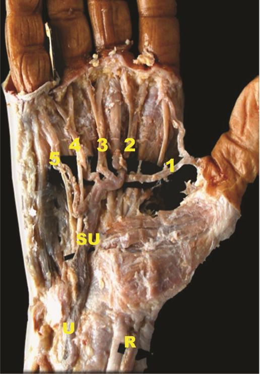
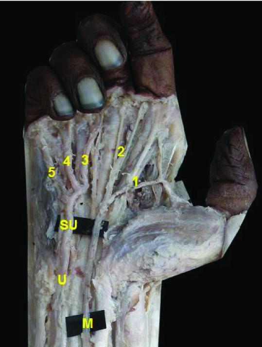
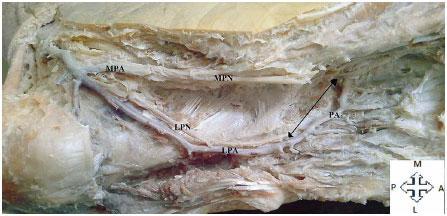
30 The persistent median artery The median artery (a. comitans nervi mediani) is an artery that occasionally found in humans. a) When present, it is found in the forearm, between the radial and ulnar arteries. b) It is a persistent embryological remnant that usually regresses by the 8 th week of gestation. c) It runs with the median nerve and supplies the same structures as that nerve. d) It passes deep to the flexor retinaculum and may terminate at one or more of the palmar arches.

31 The persistent median artery

32 The persistent median artery
33 Common trunk from the axillary artery Anomalous branching of the third part of the axillary artery: a) Common trunk, which gives rise to: Subscapular artery, Anterior circumflex humeral artery, Posterior circumflex humeral artery, Radial collateral artery, Middle collateral artery, Superior ulnar collateral artery.

34 A common trunk from axillary artery
35 Variations of the arterial patterns in the upper limb The profunda brachii artery is the largest branch of the brachial artery and it shows considerable variations in its origin. a) It may arise from the axillary artery (22% cases); as a common trunk with superior ulnar collateral artery (22% cases), as a branch of circumflex humeral artery (in 7% cases).

36 Common trunk of the profunda brachii artery with superior ulnar collateral artery
37 Variations of the arterial patterns in the upper limb Superficial brachial artery a brachial artery coursing in front of rather than behind the median nerve. Accessory brachial artery originates from upper third of the brachial artery and rejoins proximal to the elbow with the brachial artery. Brachioradial artery is define as a radial artery with a high origin coexisting with brachial artery that branches into ulnar and common interosseous trunk. Superficial brachioradial artery is define as a high origin of the radial artery coursing over the brachioradialis muscle or tendons, which define the snuffbox and coexisting with a brachial artery that usually branches into ulnar and interosseous arteries.

38 Accessory brachial artery

")
39 Brachioradial artery (a high origin of the radial artery)
40 Variations of the arterial patterns in the upper limb Brachioulnar artery is define as an ulnar artery with a high origin and a normal course along the forearm and hand and coexisting with a brachial artery which branches into the radial and common interosseous trunk. Superficial brachioulnar artery is define as an ulnar artery with a high origin and which courses over the superficial forearm flexor muscles and coexisting with a brachial artery which branches into the radial and common interosseous trunk.
41 Variations of the arterial patterns in the upper limb Superficial radial artery is a radial artery coursing over the tendons defining the snuffbox. Duplication of the radial artery. Absence of the radial artery. In this case, the radial blood supply territory is provided by the anterior interosseous or the median artery. Duplication of the ulnar artery. Absence of the ulnar artery. This absence is compensated by the radial and interosseous rather than by the median artery.
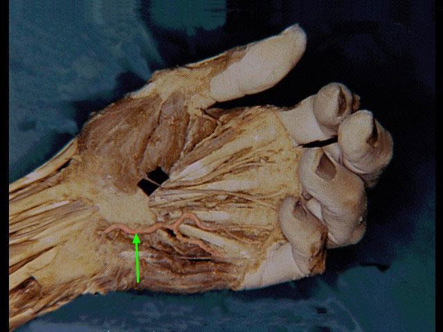
42 The superficial palmar arch The superficial palmar arch mainly is fed by the ulnar artery passing superficial to the flexor retinaculum, then curving laterally to form an arch, lying just deep to the palmar aponeurosis. a) About one third of the superficial palmar arch is formed by ulnar artery alone. b) A further third is completed by the superficial palmar branch of the radial artery. c) A third is completed by the a. radialis indicis, or a branch of the a. princeps pollicis, or by the median artery.
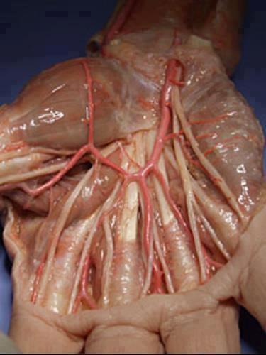
43 Superficial palmar arch (arch type)
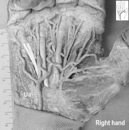
44 Superficial palmar arch (non-arch type)

45 Superficial palmar arch
46 The deep palmar arch The deep palmar arch is found to be less variable as compared to superficial palmar arch. It is formed by the radial artery which anastomoses with the deep palmar branch of the ulnar artery. Incomplete arch is found in 3% of cases.

47 The arteries of the lower limb The main artery of the lower limb is femoral artery. It is a continuation of the external iliac artery. In the popliteal fossa it is known as popliteal artery which terminates by dividing into anterior and posterior tibial arteries.
48 Arterial anastomoses of the lower extremity Hip arterial anastomoses (trochanteric and cruciate anastomoses); Anastomosis around the knee joint (genicular anastomosis); Ankle and foot anastomoses.
49 The hip arterial anastomoses The trochanteric anastomosis provides the main source of blood supply of the head of femur. It lies near the trochanteric fossa (at the greater trochanter) hence its name. It is formed by: a) Descending branch of the superior gluteal artery (from the internal iliac artery); b) Inferior gluteal artery (from the internal iliac artery); c) Ascending branch of the lateral circumflex femoral artery (from the profunda femoris artery); d) Ascending branch of the medial circumflex femoral artery (from the profunda femoris artery).
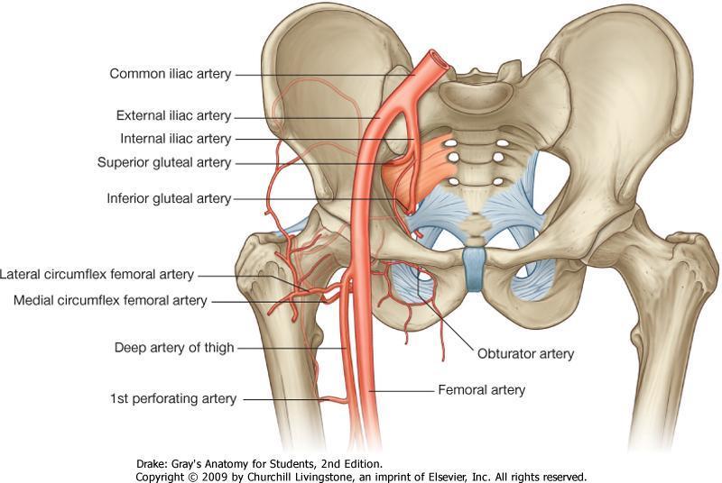
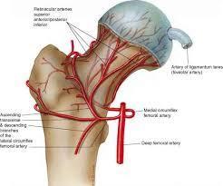
50 The trochanteric anastomosis

51 The trochanteric anastomosis
52 The cruciate anastomosis The cruciate anastomosis is located at the level of the lesser trochanter (cruciform in shape hence its name). It is formed by: a) Transverse branch of the medial circumflex femoral artery (from the profunda femoris artery); b) Transverse branch of the lateral circumflex femoral artery (from the profunda femoris artery); c) Descending branch of the inferior gluteal artery (from the internal iliac artery); d) Ascending branch of the first perforating artery (from the profunda femoris artery).

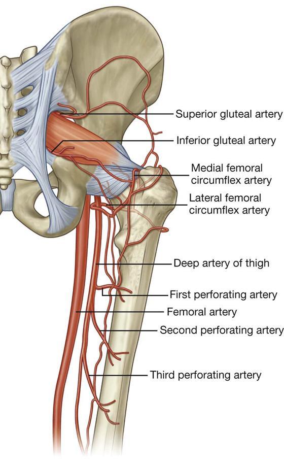
53 The hip arterial anastomoses
54 Anastomosis around the knee joint (genicular anastomosis) The genicular arteries participate in the formation of the important genicular anastomosis around the knee. (It compensates for the narrowing of the popliteal artery during the flexion of the knee.) It is formed by: a) Descending genicular branch (from the femoral artery); b) Descending branch of the lateral circumflex femoral artery (from the deep femoral artery); c) Genicular arteries (from the popliteal artery); d) Anterior and posterior recurrent tibial arteries (from the anterior tibial artery); e) Circumflex fibular branch (from the posterior tibial artery).

55 Genicular anastomosis (rete articulare genus)
56 Anastomoses around the ankle joint Rete malleolare mediale (medial malleolar network): a) Anterior medial malleolar artery (from the anterior tibial artery); b) Medial malleolar branches (from the posterior tibial artery). Rete malleolare laterale (lateral malleolar network): a) Anterior lateral malleolar artery (from the anterior tibial artery); b) Lateral malleolar branches (from the peroneal artery); c) Perforating branch of the peroneal artery; d) Lateral tarsal artery (from the dorsalis pedis artery). e) Rete calcaneum: f) Calcaneal branches of the peroneal artery; g) Calcaneal branches of the posterior tibial artery.

57 Anastomoses around the ankle joint
58 Plantar arches The posterior tibial artery is the larger of the terminal branches of the popliteal artery that terminates by bifurcating into medial and lateral plantar arteries. The lateral plantar artery is the larger terminal branch of the posterior tibial artery. At the base of the 5 th metatarsal bone it curves medially to form the (deep) plantar arch. The plantar arch is completed by the deep plantar branch from the dorsalis pedis artery. The plantar arch gives rise to the plantar metatarsal arteries, plantar digital arteries and three perforating arteries (which anastomoses with the dorsalis pedis artery).

59 Plantar arch
60 Determining the arterial pulse on the lower limbs Femoral pulse can be palpated just inferior to the inguinal ligament and midway between the anterior superior iliac spine and pubic tubercle. Popliteal pulse is best felt in the inferior part of the popliteal fossa, where the artery is related to the tibia. (Weakening or loss of the popliteal pulse is a sign of femoral artery obstruction.) Posterior tibial pulse is taken postero-inferior to the medial malleolus. (Palpation of the posterior tibial pulse is essential for examining patients with occlusive peripheral arterial diseases.) Dorsalis pedis pulse is easy to be felt (being subcutaneous) over the tarsal bones between the tendons of the extensor hallucis longus and the extensor digitorum longus. (A diminished or absent dorsalis pedis pulse usually suggests vascular insufficiency resulting from arterial disease.)

61 Determining the arterial pulse on the lower limbs
62 Development of the arteries of the lower limbs During the embryological development, the lateral branch of the 5 th lumbar intersegmental artery forms the axial artery of the lower limbs, named sciatic artery. The most proximal segment of the sciatic artery usually disappear; however the medium and distal segments persist and form the definitive popliteal and peroneal arteries. By the 14-mm embryonic stage (7 th week), the external iliac artery and its continuation the femoral artery grows towards the thigh and joins the part of the sciatic artery, which lies in the popliteal fossa. The anterior and posterior tibial arteries originate from the popliteal artery.
63 Development of the arteries of the lower limbs
64 Variations of the arterial patterns in the lower limb The primary artery of the thigh, in mammals, was the ischiadic (sciatic) artery accompanying the ischiadic (sciatic) nerve and that the femoral artery later took on that function. In case of persistence of the embryonal axial artery (ischiadic or sciatic artery) its representative, the inferior gluteal artery continuous downward to supply the leg and foot, and the femoral artery ends at the knee.
65 Persistent sciatic artery A persistent sciatic artery is a rare vascular anomaly where there is a continuation of the internal iliac artery into the thigh through the greater sciatic foramen. It may be the dominant artery supplying the leg, in which case the superficial femoral artery may be small.

66 Persistent sciatic artery
67 Variations of the arterial patterns in the lower limb A doubling of the femoral artery may occur below the origin of the deep femoral artery; the doublet vessels may reunite in the distal thigh. The great saphenous artery courses between adductor magnus and vastus medialis, pierces the crural fascia below the knee, and runs with the saphenous vein to the medial malleolus or end at the knee. The fourth perforating artery sometimes splits distally into an anterior and posterior tibial arteries.
68 Variations of the arterial patterns in the lower limb Tibial artery anomalies are present in about 3% to 10% of the population. The most frequent are `high` bifurcation or true trifurcation of the popliteal artery; common origin of the anterior tibial and peroneal arteries; and hypoplasia or absence of the anterior or posterior tibial arteries. The continuation of the peroneal (fibular) artery as dorsalis pedis artery is a rare finding. However, the anterior tibial artery is hypoplastic in this case.

 artery continous")
69 Variations of the arterial patterns in the lower limb The hypoplastic anterior tibial artery. The peroneal (fibular) artery continous as dorsalis pedis artery.
70 Variations of the arterial patterns in the lower limb The posterior tibial artery may be absent, rudimentary or replaced by the peroneal (fibular) artery. The plantar arch may be double or absent in which case the plantar tissues of the foot are supplied by a single artery, posterior tibial artery. Rarely, in the absence of the posterior tibial artery, the peroneal artery develops into the medial and lateral plantar arteries.

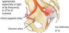
71 `Corona mortis` or `crown of death` `Corona mortis`, Latin for `crown of death` is a common variant vascular anastomosis between the external iliac artery or inferior epigastric artery with the obturator artery. It is important for femoral hernia anatomy and surgery.
72 `Corona mortis` In some cases, the `corona mortis` is the actual obturator artery that arises from the inferior epigastric artery instead of the internal iliac artery. It can also arise from the external iliac artery. In both cases it has been called an `aberrant obturator artery`. This anatomical variation can be present in up to 25% of cases. When present it can be injured when a surgeon looks to enlarge the femoral ring by opening the lacunar ligament.

73 Aberrant obturator artery

74 Aberrant obturator artery
75 Thank you!
Femoral Artery. Its entrance to the thigh Position Midway between ASIS and pubic symphysis
 Lower Limb Vessels Lecture Objectives Describe the major arteries of the lower limb. Describe the deep and superficial veins of the lower limb. Describe the topographical relationships of the arteries
Gross Anatomy Coloring Book Series. Lower Extremity Arteries
 Gross Anatomy Coloring Book Series Lower Extremity Arteries 1 Femoral Artery and Associated Branches For the life of the flesh is in the blood. Leviticus 17:11 Femoral Artery and Associated Branches After
BRACHIAL PLEXUS. DORSAL SCAPULAR NERVE (C5) supraclavicular branch innervates rhomboids (major and minor) and levator scapulae
 supraclavicular branch innervates rhomboids (major and minor) and levator scapulae") THE BRACHIAL PLEXUS DORSAL SCAPULAR NERVE (C5) supraclavicular branch innervates rhomboids (major and minor) and levator scapulae SCHEMA OF THE BRACHIAL PLEXUS THE BRACHIAL PLEXUS PHRENIC NERVE supraclavicular
THE BRACHIAL PLEXUS DORSAL SCAPULAR NERVE (C5) supraclavicular branch innervates rhomboids (major and minor) and levator scapulae SCHEMA OF THE BRACHIAL PLEXUS THE BRACHIAL PLEXUS PHRENIC NERVE supraclavicular
Al-Balqa Applied University
 Al-Balqa Applied University Faculty Of Medicine *You can use this checklist as a guide to you for the lab. the items on this checklist represent the main features of the models that you have to know for
Key Relationships in the Upper Limb
 Key Relationships in the Upper Limb This list contains some of the key relationships that will help you identify structures in the lab. They are organized by dissection assignment as defined in the syllabus.
Netter's Anatomy Flash Cards Section 6 List 4 th Edition
 Netter's Anatomy Flash Cards Section 6 List 4 th Edition https://www.memrise.com/course/1577581/ Section 6 Upper Limb (66 cards) Plate 6-1 Humerus and Scapula: Anterior View 1.1 Acromion 1.2 Greater tubercle
Misc Anatomy. Upper Limb! 2. Lower Limb! 5. Venous Drainage! Head & neck! 8
 Misc Anatomy Upper Limb! 2 Arteries!... 2 Veins!... 2 Spaces!... 4 Lower Limb! 5 Arteries!... 5 Venous Drainage!... 6 Spaces!... 7 Head & neck! 8 Artery!... 8 Ultrasound View for IJ CVL!... 8 Arteries
Muscle Anatomy Review Chart
 Muscle Anatomy Review Chart BACK Superficial (5) Trapezius Transverse cervical a. Latissimus dorsi Thoracodorsal a. Rhomboideus major Dorsal scapular a. Rhomboideus minor Levator scapulae Intermediate
musculoskeletal system <lower limb vessle> <1> done by:renad abu ruman &rama alawamleh
 musculoskeletal system done by:renad abu ruman &rama alawamleh The entrance to the anterior compartment of the leg is through lingual lig. & superior ramus, & to the posterior compartment
Year 2004 Paper one: Questions supplied by Megan
 QUESTION 47 A 58yo man is noted to have a right foot drop three days following a right total hip replacement. On examination there is weakness of right ankle dorsiflexion and toe extension (grade 4/5).
Artery 1 Head and Thoracic Arteries. Arrange the parts in the order blood flows through them.
 Artery 1 Head and Thoracic Arteries 1. Given the following parts of the aorta: 1. abdominal aorta 2. aortic arch 3. ascending aorta 4. thoracic aorta Arrange the parts in the order blood flows through
DISSECTION SCHEDULE. Session I - Hip (Front) & Thigh (Superficial)
 & Thigh (Superficial)") DISSECTION SCHEDULE Session I - Hip (Front) & Thigh (Superficial) Surface anatomy Inguinal region Gluteal region Thigh Leg Foot bones Hip bone Femur Superficial fascia Great saphenous vein Superficial
DISSECTION SCHEDULE Session I - Hip (Front) & Thigh (Superficial) Surface anatomy Inguinal region Gluteal region Thigh Leg Foot bones Hip bone Femur Superficial fascia Great saphenous vein Superficial
HUMAN BODY COURSE LOWER LIMB NERVES AND VESSELS
 HUMAN BODY COURSE LOWER LIMB NERVES AND VESSELS October 22, 2010 D. LOWER LIMB MUSCLES 2. Lower limb compartments ANTERIOR THIGH COMPARTMENT General lfunction: Hip flexion, knee extension, other motions
divided by the bones ( redius and ulna ) and interosseous membrane into :
 and interosseous membrane into :") fossa Cubital Has: * floor. * roof : - Skin - superficial fasica - deep fascia ( include bicipital aponeurosis ) Structures within the roof : -cephalic and basilic veins -and between them median cubital
fossa Cubital Has: * floor. * roof : - Skin - superficial fasica - deep fascia ( include bicipital aponeurosis ) Structures within the roof : -cephalic and basilic veins -and between them median cubital
MUSCULOSKELETAL LOWER LIMB
 MUSCULOSKELETAL LOWER LIMB Spinal Cord Lumbar and Sacral Regions Spinal cord Dorsal root ganglion Conus medullaris Cauda equina Dorsal root ganglion of the fifth lumbar nerve End of subarachnoid space
Ali Yaghi. Omar Eyad. Ahmad Salman. 1 P a g e
 5 Ali Yaghi Omar Eyad Ahmad Salman 1 P a g e **There are two types of groin hernia; the femoral hernia and the inguinal hernia. But how can we differentiate between the inguinal hernia and the femoral
THE LOWER LIMB NERVES VESSELS
 THE LOWER LIMB NERVES VESSELS LOWER LIMB: FEMORAL TRIANGLE FEMORAL TRIANGLE LOWER LIMB: FEMORAL TRIANGLE FEMORAL TRIANGLE is a triangular landmark useful in dissection and in understanding relationships
ANATYOMY OF The thigh
 ANATYOMY OF The thigh 1- Lateral cutaneous nerve of the thigh Ι) Skin of the thigh Anterior view 2- Femoral branch of the genitofemoral nerve 5- Intermediate cutaneous nerve of the thigh 1, 2 and 3 are
Nerves of the upper limb Prof. Abdulameer Al-Nuaimi. E. mail:
 Nerves of the upper limb Prof. Abdulameer Al-Nuaimi E-mail: a.al-nuaimi@sheffield.ac.uk E. mail: abdulameerh@yahoo.com Brachial plexus Median nerve After originating from the brachial plexus in the axilla,
The thigh. Prof. Oluwadiya KS
 The thigh Prof. Oluwadiya KS www.oluwadiya.com The Thigh: Boundaries The thigh is the region of the lower limb that is approximately between the hip and knee joints Anteriorly, it is separated from the
Gross Anatomy: Upper Extremity Arteries
 Gross Anatomy: Upper Extremity Arteries By: Trevor Lohman DPT Illustrated by: Dennis Breese 1 Subclavian and Axillary arteries Hardening of the heart ages people more quickly than hardening of the arteries
The arm: *For images refer back to the slides
 The arm: *For images refer back to the slides Muscles of the arm: deltoid, triceps (which is located at the back of the arm), biceps and brachialis (it lies under the biceps), brachioradialis (it lies
Lower Limb Nerves. Clinical Anatomy
 Lower Limb Nerves Clinical Anatomy Lumbar Plexus Ventral rami L1 L4 Supplies: Abdominal wall External genitalia Anteromedial thigh Major nerves.. Lumbar Plexus Nerves relation to psoas m. : Obturator n.
Where should you palpate the pulse of different arteries in the lower limb?
 Where should you palpate the pulse of different arteries in the lower limb? The femoral artery In the femoral triangle, its pulse is easily felt just inferior to the inguinal ligament midway between the
rotation of the hip Flexion of the knee Iliac fossa of iliac Lesser trochanter Femoral nerve Flexion of the thigh at the hip shaft of tibia
 Anatomy of the lower limb Anterior & medial compartments of the thigh Dr. Hayder The fascia lata encloses the entire thigh like a sleeve/stocking. Three intramuscular fascial septa (lateral, medial, and
ANATYOMY OF The thigh
 ANATYOMY OF The thigh 1- Lateral cutaneous nerve of the thigh Ι) Skin of the thigh Anterior view 2- Femoral branch of the genitofemoral nerve 5- Intermediate cutaneous nerve of the thigh 1, 2 and 3 are
BOGOMOLETS NATIONAL MEDICAL UNIVERSITY. Department of Human Anatomy GUIDELINES. The theme of the lesson The vessels of the lower limb.
 BOGOMOLETS NATIONAL MEDICAL UNIVERSITY Department of Human Anatomy GUIDELINES Academic discipline HUMAN ANATOMY Module 2 The theme of the lesson The vessels of the lower limb. Course Faculties І Medical
Contents of the Posterior Fascial Compartment of the Thigh
 Contents of the Posterior Fascial Compartment of the Thigh 1-Muscles: B i c e p s f e m o r i s S e m i t e n d i n o s u s S e m i m e m b r a n o s u s a small part of the adductor magnus (h a m s t
Fascial Compartments of the Upper Arm
 Fascial Compartments of the Upper Arm The upper arm is enclosed in a sheath of deep fascia and has two fascial septa: 1- Medial fascial septum (medial intermuscular septum): attached to the medial supracondylar
Anatomy of the Musculoskeletal System
 Anatomy of the Musculoskeletal System Kyle E. Rarey, Ph.D. Department of Anatomy & Cell Biology and Otolaryngology University of Florida College of Medicine Outline of Presentation Vertebral Column Upper
Latin. VASA REGIONIS PELVINAE 72 A. et V. iliaca communis 73 A. et V. iliaca externa 74 A. sacralis mediana 75 A. et V.
 G30 Latin VASA CAPITIS et CERVICIS 1 V. frontalis 2 V. temporalis superficialis 3 A. temporalis superficialis 3 a A. maxillaris 4 A. occipitalis 5 A. supratrochlearis 6 A. et V. angularis 7 A. et V. facialis
Lecture 08 THIGH MUSCLES ANTERIOR COMPARTMENT. Dr Farooq Khan Aurakzai. Dated:
 Lecture 08 THIGH MUSCLES ANTERIOR COMPARTMENT BY Dr Farooq Khan Aurakzai Dated: 11.02.2017 INTRODUCTION to the thigh Muscles. The musculature of the thigh can be split into three sections by intermuscular
BOGOMOLETS NATIONAL MEDICAL UNIVERSITY. Department of Human Anatomy GUIDELINES. The theme of the lesson The vessels of the upper limb.
 BOGOMOLETS NATIONAL MEDICAL UNIVERSITY Department of Human Anatomy GUIDELINES Academic discipline HUMAN ANATOMY Module 2 The theme of the lesson The vessels of the upper limb. Course Faculties І Medical
Gluteal region DR. GITANJALI KHORWAL
 Gluteal region DR. GITANJALI KHORWAL Gluteal region The transitional area between the trunk and the lower extremity. The gluteal region includes the rounded, posterior buttocks and the laterally placed
Axilla and Brachial Region
 L 4 A B O R A T O R Y Axilla and Brachial Region BRACHIAL PLEXUS 5 Roots/Rami (ventral rami C5 T1) 3 Trunks Superior (C5, C6) Middle (C7) Inferior (C8, T1) 3 Cords Lateral Cord (Anterior Superior and Anterior
1-Muscles: 2-Blood supply: Branches of the profunda femoris artery. 3-Nerve supply: Sciatic nerve
 1-Muscles: B i c e p s f e m o r i s S e m i t e n d i n o s u s S e m i m e m b r a n o s u s a small part of the adductor magnus (h a m s t r i n g p a r t o r i s c h i a l p a r t ) 2-Blood supply:
MCQWeek2. All arise from the common flexor origin. The posterior aspect of the medial epicondyle is the common flexor origin.
 MCQWeek2. 1. Regarding superficial muscles of anterior compartment of the forearm: All arise from the common flexor origin. The posterior aspect of the medial epicondyle is the common flexor origin. Flexor
CARDIOVASCULAR DANIL HAMMOUDI.MD
 CARDIOVASCULAR DANIL HAMMOUDI.MD 18 Systemic Circulation Figure 19.19 Pulmonary Circulation Figure 19.18b 1. Thyroid gland 2. Trachea 3. Brachiocephalic 4. Common carotid 5. Internal jugular 6. Superior
Femoral Triangle and Adductor Canal. Dr. Heba Kalbouneh Associate Professor of Anatomy and Histology
 Femoral Triangle and Adductor Canal Dr. Heba Kalbouneh Associate Professor of Anatomy and Histology Femoral Triangle and Adductor Canal Femoral triangle Is a triangular depressed area located in the upper
Part One Anatomy Practice MeQ's
 Part One natomy Practice MeQ's 1. Regarding the flexor retinaculum, which of the following statements is FLS?: o It is crossed by palmaris longus, the palmar branch of the median nerve, and the superficial
musculoskeletal system anatomy nerves of the lower limb 2 done by: Dina sawadha & mohammad abukabeer
 musculoskeletal system anatomy nerves of the lower limb 2 done by: Dina sawadha & mohammad abukabeer #Sacral plexus : emerges from the ventral rami of the spinal segments L4 - S4 and provides motor and
Region of upper limb attachment to the trunk Proximal segment of limb overlaps parts of the trunk (thorax and back) and lower lateral neck.
 and lower lateral neck.") Region of upper limb attachment to the trunk Proximal segment of limb overlaps parts of the trunk (thorax and back) and lower lateral neck. includes Pectoral Scapular Deltoid regions of the upper limb
Region of upper limb attachment to the trunk Proximal segment of limb overlaps parts of the trunk (thorax and back) and lower lateral neck. includes Pectoral Scapular Deltoid regions of the upper limb
Abduction of arm until your hand rich your head. Flexion of forearm at elbow joint. Extension of arm at elbow joint. Flexion of fingers 10.
 Num. answer 1. Medialy With the manubrium ( sternum ), and laterally with the acromion of the scapula 2. 1. Trapezius 2. Levator scapulae 3. Rhomboids 3. 1. Pectoralis major 2. Pectoralis minor 3. Latissiumus
lower limb Anterior Compartment: lecture 3 The deep fascia ( fascia lata) divides the thigh into 3 compartments:
 divides the thigh into 3 compartments:") lower limb lecture 3 The deep fascia ( fascia lata) divides the thigh into 3 compartments: 1. Anterior Extensor compartment 2. Medial Adductor compartment 3. Posterior Flexor compartment Anterior Compartment:
lower limb lecture 3 The deep fascia ( fascia lata) divides the thigh into 3 compartments: 1. Anterior Extensor compartment 2. Medial Adductor compartment 3. Posterior Flexor compartment Anterior Compartment:
Adductor canal (Subsartorial) or Hunter s canal
 or Hunter s canal") Adductor canal (Subsartorial) or Hunter s canal John Hunter described the exposure and ligation of the femoral artery in this canal for aneurysm of the popliteal artery; this method has the advantage that
Adductor canal (Subsartorial) or Hunter s canal John Hunter described the exposure and ligation of the femoral artery in this canal for aneurysm of the popliteal artery; this method has the advantage that
region of the upper limb between the shoulder and the elbow Superiorly communicates with the axilla.
 1 region of the upper limb between the shoulder and the elbow Superiorly communicates with the axilla. Inferiorly, a number of important structures pass between arm & forearm through cubital fossa. 2 medial
Anatomage Table Instructors Guide- Lower Limb
 The Lower Limb Anatomage Table Instructors Guide- Lower Limb Table of Contents Lower Limb 1- The Skeletal System...3 1: Hip Bone...3 2: Hip Joint and Femur...4 3: Patella and Knee Joint...7 4: Tibia, Fibula,
Anterior and Medial compartments of the thigh. Dr. Heba Kalbouneh Associate Professor of Anatomy and Histology
 Anterior and Medial compartments of the thigh Dr. Heba Kalbouneh Associate Professor of Anatomy and Histology Terms Related to Movements Movement Flexion Extension Abduction Adduction Medial (internal)
Lumbar Plexus. Ventral rami L1 L4 Supplies: Major nerves.. Abdominal wall External genitalia Anteromedial thigh
 Lower Limb Nerves Lectures Objectives Describe the structure and relationships of the plexuses of the lower limb. Describe the course, relationships and structures supplied for the major nerves of the
Muscles of the lower extremities. Dr. Nabil khouri MD, MSc, Ph.D
 Muscles of the lower extremities Dr. Nabil khouri MD, MSc, Ph.D Posterior leg Popliteal fossa Boundaries Biceps femoris (superior-lateral) Semitendinosis and semimembranosis (superior-medial) Gastrocnemius
ANATYOMY OF The thigh
 ANATYOMY OF The thigh 1- Lateral cutaneous nerve of the thigh Ι) Skin of the thigh Anterior view 2- Femoral branch of the genitofemoral nerve 1, 2 and 3 are From the lumber plexus 5- Intermediate cutaneous
Lecture 9: Forearm bones and muscles
 Lecture 9: Forearm bones and muscles Remember, the region between the shoulder and the elbow = brachium/arm, between elbow and wrist = antebrachium/forearm. Forearm bones : Humerus (distal ends) Radius
ARM Brachium Musculature
 ARM Brachium Musculature Coracobrachialis coracoid process of the scapula medial shaft of the humerus at about its middle 1. flexes the humerus 2. assists to adduct the humerus Blood: muscular branches
3 Circulatory Pathways
 40 Chapter 3 Circulatory Pathways Systemic Arteries -Arteries carry blood away from the heart to the various organs of the body. -The aorta is the longest artery in the body; it branches to give rise to
STRUCTURAL BASIS OF MEDICAL PRACTICE EXAMINATION 5 October 6, 2006
 STRUCTURAL BASIS OF MEDICAL PRACTICE EXAMINATION 5 October 6, 2006 PART l. Answer in the space provided. (8 pts) 1. Identify the structures. (2 pts) B C A. _pisiform B. _ulnar artery A C. _flexor carpi
Leg. Dr. Heba Kalbouneh Associate Professor of Anatomy and Histology
 Leg Dr. Heba Kalbouneh Associate Professor of Anatomy and Histology Skin of the Leg Cutaneous Nerves Medially: The saphenous nerve, a branch of the femoral nerve supplies the skin on the medial surface
MUSCLES. Anconeus Muscle
 LAB 7 UPPER LIMBS MUSCLES Anconeus Muscle anconeus origin: distal end of dorsal surface of humerus insertion: lateral surface of ulna from distal margin of the semilunar notch to proximal end of the olecranon
VARIANT ARTERIAL PATTERN IN THE FOREARM WITH ITS EMBRYOLOGICAL BASIS. Vaishnavi Joshi and Dr. Shaheen Sajid Rizvi
 Volume-8, Issue-3 July-Sept-2018 Coden:IJPAJX-CAS-USA, Copyrights@2018 ISSN-2231-4490 Received: 8 th June-2018 Revised: 15 th July-2018 Accepted: 16 th July-2018 DOI: 10.21276/Ijpaes http://dx.doi.org/10.21276/ijpaes
Upper limb Arm & Cubital region 黃敏銓
 Upper limb Arm & Cubital region 黃敏銓 1 Arm Lateral intermuscular septum Anterior (flexor) compartment: stronger Medial intermuscular septum Posterior (extensor) compartment 2 Coracobrachialis Origin: coracoid
Supplied in part by the musculocutaneous nerve. Forms the axis of rotation in movements of pronation and supination
 Anatomy: Upper limb (15 questions) 1. Latissimus Dorsi: Is innervated by the dorsal scapular nerve Lies above feres major muscle Medially rotates the humerus All of the above 2. Supinator muscle is: Deep
Anatomy MCQs Week 13
 Anatomy MCQs Week 13 1. Posterior to the medial malleolus of the ankle: The neurovascular bundle lies between Tibialis Posterior and Flexor Digitorum Longus The tendon of Tibialis Posterior inserts into
Bones of Lower Limb. Dr. Heba Kalbouneh Associate Professor of Anatomy and Histology
 Bones of Lower Limb Dr. Heba Kalbouneh Associate Professor of Anatomy and Histology Bones of the lower limb Hip Bone Made up of 3 bones: 1) Ilium (flat), superior in position 2) Ischium (L), postero-inferior
The Leg. Prof. Oluwadiya KS
 The Leg Prof. Oluwadiya KS www.oluwadiya.sitesled.com Compartments of the leg 4 Four Compartments: 1. Anterior compartment Deep fibular nerve Dorsiflexes the foot and toes 2. Lateral Compartment Superficial
Biology 323 Human Anatomy for Biology Majors Lecture 11 Dr. Stuart S. Sumida. Peripheral Circulation
 Biology 323 Human Anatomy for Biology Majors Lecture 11 Dr. Stuart S. Sumida Peripheral Circulation Structures of the Splanchnopleure: receive unpaired vessels of the abdominal aorta. Structures of the
Lower limb summary. Anterior compartment of the thigh. Done By: Laith Qashou. Doctor_2016
 Lower limb summary Done By: Laith Qashou Doctor_2016 Anterior compartment of the thigh Sartorius Anterior superior iliac spine Upper medial surface of shaft of tibia 1. Flexes, abducts, laterally rotates
Biceps Brachii. Muscles of the Arm and Hand 4/4/2017 MR. S. KELLY
 Muscles of the Arm and Hand PSK 4U MR. S. KELLY NORTH GRENVILLE DHS Biceps Brachii Origin: scapula Insertion: radius, fascia of forearm (bicipital aponeurosis) Action: supination and elbow flexion Innervation:
Anatomy Workshop Upper Extremity David Ebaugh, PT, PhD Workshop Leader. Lab Leaders: STATION I BRACHIAL PLEXUS
 Anatomy Workshop Upper Extremity David Ebaugh, PT, PhD Workshop Leader Lab Leaders: STATION I BRACHIAL PLEXUS A. Posterior cervical triangle and axilla B. Formation of plexus 1. Ventral rami C5-T1 2. Trunks
The Hip (Iliofemoral) Joint. Presented by: Rob, Rachel, Alina and Lisa
 Joint. Presented by: Rob, Rachel, Alina and Lisa") The Hip (Iliofemoral) Joint Presented by: Rob, Rachel, Alina and Lisa Surface Anatomy: Posterior Surface Anatomy: Anterior Bones: Os Coxae Consists of 3 Portions: Ilium Ischium Pubis Bones: Pubis Portion
The Hip (Iliofemoral) Joint Presented by: Rob, Rachel, Alina and Lisa Surface Anatomy: Posterior Surface Anatomy: Anterior Bones: Os Coxae Consists of 3 Portions: Ilium Ischium Pubis Bones: Pubis Portion
Muscular Nomenclature and Kinesiology - One
 Chapter 16 Muscular Nomenclature and Kinesiology - One Lessons 1-3 (with lesson 4) 1 Introduction 122 major muscles covered in this chapter Chapter divided into nine lessons Kinesiology study of human
For the following questions, indicate the letter that corresponds to the SINGLE MOST APPROPRIATE ANSWER.
 NAME GROSS ANATOMY EXAMINATION March 13, 2000 For the following questions, indicate the letter that corresponds to the SINGLE MOST APPROPRIATE ANSWER. 1. A patient in the emergency room has a penetrating
Muscles of the Upper Limb
 Muscles of the Upper Limb anterior surface of ribs 3 5 coracoid process Pectoralis minor pectoral nerves protracts / depresses scapula Serratus anterior Subclavius ribs 1-8 long thoracic nerve rib 1 ----------------
Skeletal System. Bones & Joints
 Skeletal System Bones & Joints Vertebral Column Upper Limb Lower Limb OUTLINE Clinical Related Features Arrangements Features of the Joints Vertebral Column (Overview) Costal Element Regional Features
Nerves of Upper limb. Dr. Brijendra Singh Professor & Head Department of Anatomy AIIMS Rishikesh
 Nerves of Upper limb Dr. Brijendra Singh Professor & Head Department of Anatomy AIIMS Rishikesh 1 Objectives Origin, course & relation of median & ulnar nerves. Motor & sensory distribution Carpal tunnel
The Hay is in the Barn
 Anatomy 1 Practical 1 Review Made by Forrest Allen (nerd) Edited by TJ Williamson (not nerd) The Hay is in the Barn 2019 Thunderbringers Too much to handle https://www.youtube.com/watch?v=glii-kaza d8
The Clavicle Right clavicle Deltoid tubercle: Conoid tubercle, conoid ligamen Impression for the
 The Clavicle Muscle Attachment Sites in the Upper Limb Pectoralis major Right clavicle Smooth superior surface of the shaft, under the platysma muscle tubercle: attachment of the deltoid Acromial facet
213: HUMAN FUNCTIONAL ANATOMY: PRACTICAL CLASS 1: Proximal bones, plexuses and patterns
 213: HUMAN FUNCTIONAL ANATOMY: PRACTICAL CLASS 1: Proximal bones, plexuses and patterns CLAVICLE Examine an isolated clavicle and compare it with a clavicle on an articulated skeleton. Viewed from above,
TABLES OF MUSCLE ACTIONS, INNERVATIONS, AND ATTACHMENTS
 TABLES OF MUSCLE ACTIONS, INNERVATIONS, AND ATTACHMENTS Table 1-1 ERECTOR SPINAE MUSCLES Intrinsic muscles producing extension and/or lateral of the spine Muscle Joint and Action Innervation Inferior Attachment
REFERENCE DIAGRAMS OF UPPER LIMB MUSCLES: NAMES, LOCATIONS, ATTACHMENTS, FUNCTIONS MUSCLES CONNECTING THE UPPER LIMB TO THE AXIAL SKELETON
 REFERENCE DIAGRAMS OF UPPER LIMB MUSCLES: NAMES, LOCATIONS, ATTACHMENTS, FUNCTIONS MUSCLES CONNECTING THE UPPER LIMB TO THE AXIAL SKELETON A25LAB EXERCISES: UPPER LIMB MUSCLES Page 1 MUSCLES CONNECTING
musculoskeletal system anatomy nerves of the lower limb 1 done by: dina sawadha & mohammad abukabeer
 musculoskeletal system anatomy nerves of the lower limb 1 done by: dina sawadha & mohammad abukabeer What is the importance of plexuses? plexuses provides us the advantage of a phenomenon called convergence
Human Anatomy Biology 351
 Human Anatomy Biology 351 Lower Limb Please place your name on the back of the last page of this exam. You must answer all questions on this exam. Because statistics demonstrate that, on average, between
The Appendicular Skeleton
 8 The Appendicular Skeleton PowerPoint Lecture Presentations prepared by Jason LaPres Lone Star College North Harris 8-1 The Pectoral Girdle The Pectoral Girdle Also called shoulder girdle Connects the
VENOUS DRAINAGE OF THE LOWER LIMB
 Anatomy of the lower limb Superficial veins & nerve injuries Dr. Hayder VENOUS DRAINAGE OF THE LOWER LIMB The venous drainage of the lower limb is of huge clinical & surgical importance. Since the venous
The Arm and Cubital Fossa
 The Arm and Cubital Fossa Dr. Andrew Gallagher School of Anatomical Sciences University of the Witwatersrand Introduction The ARM (BRACHIUM) is the most proximal segment of the upper limb musculoskeletal
Mohammad Ashraf. Abdulrahman Al-Hanbali. Ahmad Salman. 1 P a g e
 - 7 Mohammad Ashraf Abdulrahman Al-Hanbali Ahmad Salman 1 P a g e Structures under the cover of Gluteus Maximus: 1-Bones: Ileum, Femur (Head, greater trochanter and gluteal tuberosity), Ischium (ischial
5.1 Identify, describe the attachments of and deduce the actions of the muscles of the thigh:
 5.1 Identify, describe the attachments of and deduce the actions of the muscles of the thigh: Anterior group Proximal attachment Distal attachment Sartorius ASIS» Upper part of shaft tibia (middle surface)»
Lecture 09. Popliteal Fossa. BY Dr Farooq Khan Aurakzai
 Lecture 09 Popliteal Fossa BY Dr Farooq Khan Aurakzai Dated: 14.02.2018 What is popliteus? Introduction Anything relating to, or near the part of the leg behind the knee. From New Latin popliteus the muscle
Copyright 2003 Pearson Education, Inc. publishing as Benjamin Cummings. Dr. Nabil khouri
 Dr. Nabil khouri Appendicular Skeleton The appendicular skeleton is made up of the bones of the upper and lower limbs and their girdles Two girdles: Pectoral girdles attach the upper limbs to the body
Appendix. Useful Anatomical Data of Clinical Significance
 Appendix Useful Anatomical Data of Clinical Significance Appendix Outline Respiratory System 426 Table I. Important Airway Distances (Adult) 426 Table II. Important Data Concerning the Trachea 426 Musculoskeletal
inerve Guide to Nerves 2009
 inerve Guide to Nerves 2009 A guide to self learning and self assessment Context: The following guide is intended to help interpret the sono-anatomy and follow a systematic stepwise approach to the practice
Human Anatomy Biology 351
 1 Human Anatomy Biology 351 Upper Limb Exam Please place your name on the back of the last page of this exam. You must answer all questions on this exam. Because statistics demonstrate that, on average,
Functional anatomy of the muscles of the limbs. Lecturer DR. OKSANA PETRICHKO Department of Human Anatomy
 Functional anatomy of the muscles of the limbs Lecturer DR. OKSANA PETRICHKO Department of Human Anatomy Functional anatomy of the muscles of the limbs 1. 2. 3. 4. Development of the limb muscles Peculiarities
Anatomy of the lower limb
 Anatomy of the lower limb 1. Bones of the lower limb Pelvis Hip bone/coxal bone Acetabulum o Acetabular margin o Acetabular fossa o Acetabular notch o Lunate surface Ischiopubic ramus Obturator foramen
The arterial system of upper limb begins with the
 Kathmandu University Medical Journal (2009), Vol. 7, No. 3, Issue 27 Case Note Multiple arterial anomalies in upper limb Baral P 1, Vijayabhaskar P 2, Roy S 1, Kumar S 2, Ghimire S 3, Shrestha U 3 1 Lecturer,
SUBJECTS 1st year, 1st semester
 SUBJECTS 1st year, 1st semester I. Embryology 1. Spermatogenesis and blood testes barrier 2. Spermiogenesis. Sperm characteristics 3. Oogenesis and arrested prophase 4. Follicular cycle 5. Ovulation. Corpus
The Elbow and the cubital fossa. Prof Oluwadiya Kehinde
 The Elbow and the cubital fossa Prof Oluwadiya Kehinde www.oluwadiya.com Elbow and Forearm Anatomy The elbow joint is formed by the humerus, radius, and the ulna Bony anatomy of the elbow Distal Humerus
Demonstrate the bony features of Cl and C2 vertebrae evident on this Xray
 SUBJECT: ANATOMY 7 September 2007 am. TOPIC: X-ray: Lateral C spine NUMBER: JL Demonstrate the bony features of Cl and C2 vertebrae evident on this Xray 1 Odontoid peg (dens) 2 Bodies of Cl andc2 3 anterior
Day 5 Respiratory & Cardiovascular: Respiratory System
 Day 5 Respiratory & Cardiovascular: Respiratory System Be very careful not to damage the heart and lungs while separating the ribs! Analysis Questions-Respiratory & Cardiovascular Log into QUIA using your
Practical 2 Worksheet
 Practical 2 Worksheet Upper Extremity BONES 1. Which end of the clavicle is on the lateral side (acromial or sternal)? 2. Describe the difference in the appearance of the acromial and sternal ends of the
Human Anatomy Biology 351
 Human Anatomy Biology 351 Lower Limb Please place your name on the back of the last page of this exam. You must answer all questions on this exam. Because statistics demonstrate that, on average, between
Lab Activity 9. Appendicular Skeleton Martini Chapter 8. Portland Community College BI 231
 Lab Activity 9 Appendicular Skeleton Martini Chapter 8 Portland Community College BI 231 Appendicular Skeleton Upper & Lower extremities Shoulder Girdle Pelvic Girdle 2 Humerus 3 Humerus: Proximal End