HUMERUS GREATER TUBERCLE OF HUMERUS MEDIAL (INNER) EPICONDYLE OF HUMERUS LATERAL (OUTER) EPICONDYLE OF HUMERUS STYLOID PROCESS OF RADIUS
|
|
|
- Chastity Andrews
- 5 years ago
- Views:
Transcription
1

, including the shoulder girdle and pelvic girdle.")

 and posterior (back) views of the figure.")
2 The Main Bones of the Human Figure The human skeleton consists of two basic parts the axial skeleton and the appendicular skeleton. The axial skeleton is the group of bones that forms the central axis of the body; its primary function is to support and protect the internal organs. The bones of the axial skeleton include the cranium, the bones BASIC BONES OF THE SKELETON, ANTERIOR VIEW CRANIUM of the vertebral column (including the sacrum and coccyx), and the rib cage. The appendicular skeleton is the group of bones forming the appendages of the body (upper and lower limbs), including the shoulder girdle and pelvic girdle. The primary functions of the appendicular skeleton are to support and move the axial skeleton and to allow movement of the limbs. Appendicular BASIC BONES OF THE SKELETON, POSTERIOR VIEW CRANIUM bones include the humerus, ulna, and radius of the upper limbs; the femur, tibia, and fibula of the lower limbs; the bones of the hands and feet; the scapula and clavicle; and the two hip bones (os coxae) of the pelvis. The two drawings opposite show the basic bones and their locations, as seen from both anterior (front) and posterior (back) views of the figure. Learning the placement of the bones will help you identify the bony landmarks you may see as a live model takes various poses. Bony Landmarks When drawing the human figure, artists look for various surface landmarks. These can be soft surface forms, such as muscles, tendons, ligaments, and fatty tissue, or hard surface forms, which are places where evidence of the skeleton can be seen on the surface of the body. Hard surface forms are also known as bony landmarks parts of bones (ridges, bumps, depressions) that are positioned close to the skin, creating visual landmarks on the surface form. The drawings below provide a general guide to where the main bony landmarks are, as seen from both the anterior and posterior views. Whether you can see these skeletal clues on a live model will depend on both the pose and the model s body type. On some figures, fatty tissue may obscure much of the evidence of the bones, while on leaner figures it will be easier to see bone definition at several places on their bodies. CERVICAL VERTEBRAE CLAVICLE CERVICAL VERTEBRAE BONY LANDMARKS OF THE SKELETON, ANTERIOR VIEW BONY LANDMARKS OF THE SKELETON, POSTERIOR VIEW SCAPULA CLAVICLE SCAPULA STERNUM RIB CAGE LUMBAR VERTEBRAE PELVIS FEMUR PATELLA CARPALS METACARPALS CARPALS METACARPALS THORACIC VERTEBRAE LUMBAR VERTEBRAE SACRUM PELVIS FEMUR ZYGOMATIC ARCH/BONE (CHEEKBONES) MANDIBLE (LOWER JAW) ACROMION PROCESS (TIP OF SHOULDER) GREATER TUBERCLE OF MEDIAL (INNER) LATERAL (OUTER) STYLOID PROCESS OF METACARPAL OF THUMB HEAD OF GREATER TROCHANTER OF FEMUR LATERAL CONDYLE OF FEMUR MEDIAL CONDYLE OF FEMUR FRONTAL BONE (FOREHEAD) NASAL BONE CLAVICLE (COLLARBONE) STERNUM (BREASTBONE) ILIAC CREST OF PELVIS ASIS THORACIC ARCH (RIB CAGE ARCH) LATERAL (OUTER) MEDIAL (INNER) STYLOID PROCESS OF METACARPAL BONES METACARPAL OF THUMB OF FINGERS (FINGER BONES) PATELLA (KNEECAP) ZYGOMATIC ARCH/BONE (CHEEKBONES) MANDIBLE (LOWER JAW) ACROMION PROCESS (TIP OF SHOULDER) SPINOUS PROCESSES OF VERTEBRAE LATERAL (OUTER) EPICONDYLE OF POSTERIOR BORDER OF HEAD OF ILIAC CREST PSIS SACRUM GREATER TROCHANTER HEAD OF FIBULA CRANIUM MASS (IF THERE IS NO HAIR) SEVENTH CERVICAL VERTEBRA (NECK VERTEBRA) SPINE OF SCAPULA VERTEBRAL BORDER OF SCAPULA (INNER EDGE OF SHOULDER BLADE) MEDIAL (INNER) OLECRANON PROCESS OF (ELBOW) STYLOID PROCESS OF CARPAL BONES (WRIST BONES) METACARPALS METACARPAL HEADS (KNUCKLES) OF FINGERS (FINGER BONES) TARSALS TIBIA FIBULA TIBIA FIBULA HEAD OF FIBULA MEDIAL MALLEOLUS (INNER ANKLE) LATERAL MALLEOLUS (OUTER ANKLE) TIBIAL TUBEROSITY ANTERIOR BORDER OF TIBIA LATERAL MALLEOLUS (OUTER ANKLE) LATERAL AND MEDIAL CONDYLE OF FEMUR MEDIAL CONDYLE OF TIBIA MEDIAL MALLEOLUS (INNER ANKLE) METATARSALS CALCANEUS (HEEL BONE) TALUS FIRST METATARSAL BASE (TUBEROSITY) OF FIFTH METATARSAL CALCANEUS (HEEL BONE) 6 Classic Human Anatomy in Motion Bones and Surface Landmarks 7
 CALCANEUS (HEEL) HEAD OF MCP")

 THENAR MUSCLE GROUP")


3 PORTRAIT STUDY OF CLAUDIO, WITH HEAD TURNED STUDY OF A HAND, SHOWING TENDONS STUDY OF FEET TENDONS OF EXTENSOR DIGITORUM MUSCLE ACHILLES TENDON PIP JOINTS LATERAL MALLEOLUS (OUTER ANKLE BONE) CALCANEUS (HEEL) HEAD OF MCP JOINTS (KNUCKLES) LATERAL MUSCULAR RIDGE OF FOOT Sanguine and brown pastel pencils, charcoal, Graphite pencil, ballpoint pen, colored pencil, STUDY OF A TIGHTLY CLENCHED FIST STUDY OF THE BACK OF THE LEGS STERNOCLEIDOMASTOID MCP JOINTS (KNUCKLES) PIP JOINTS TENDON OF BICEPS FEMORIS THYROID CARTILAGE (ADAM S APPLE) POSTERIOR TRIANGLE HAMSTRING MUSCLE GROUP (SEMITENDINOSUS, SEMIMEMBRANOSUS, BICEPS FEMORIS) THENAR MUSCLE GROUP TENDONS OF SEMITENDINOSUS AND SEMIMEMBRANOSUS GASTROCNEMIUS TENDON OF BICEPS FEMORIS POPLITEAL FOSSA FAT PAD TRAPEZIUS HYPOTHENAR MUSCLE GROUP POPLITEAL FOSSA HOLLOW CLAVICLE ACHILLES TENDON TENDONS OF FLEXOR MUSCLES OF LOWER ARM Sanguine and brown pastel pencils TENDONS OF STERNAL HEAD (STERNOCLEIDOMASTOID) Sanguine and brown pastel pencils, charcoal, Charcoal pencil, sanguine and brown pastel pencils, 74 Classic Human Anatomy in Motion Muscle and Tendon Characteristics 75


4 Method #4: The Organic Line Approach Poses of 1 to 2 minutes In the organic line approach to gesture drawing, you lay down your lines in a flowing, loose manner, but more selectively than in the searching line approach. If you wish, you can use different values (light and dark lines) to accentuate the rhythm of forms, to convey tension, or to show that forms are advancing or receding though in a highly edited way. Very often, the lines in an organic-line gesture drawing have a calligraphic quality, changing from thick to thin or dark to light in a single stroke. You can also add tones to your drawing. Charcoal, Conté crayons, graphite pencils, and colored pencils are all suitable for this approach, as are calligraphy pens that can produce both thick and thin strokes. The organic line approach is great for relatively short poses 1 to 2 minutes. When doing an organic line study, look for any interesting tilting or twisting action within the pose that you can exaggerate to create greater dynamic tension. You may emphasize certain anatomical forms, but draw them rapidly because of the time constraint. GESTURE DRAWING OF A FIGURE IN A CONTRAPPOSTO POSE Organic line approach GESTURE STUDY OF TWO MALE FIGURES IN DANCE POSES Organic line approach 2-MINUTE STUDIES OF ACTION POSES Organic line approach Black crayon on newsprint. Black Conté crayon on newsprint. Black Conté crayon on newsprint. 224 Classic Human Anatomy in Motion Gesture and Action Drawing 225


 GAIT CYCLE WALKING The arms tend to swing in opposition to the swing of the legs: When the right leg swings forward, the right arm moves back, and when the left leg swings forward, the left arm")
5 When beginning to study the gait cycle, it s easier to focus on just one leg. The drawing below emphasizes the stance and swing phases of the right leg referred to, technically, as the right stance phase and the right swing phase. The small footprints show where on the sole of the foot the weight of the body is concentrated during the heel strike and toe-off. As the right leg moves through the gait cycle, so does the left leg, with the phases in reverse order. At one point during the stride, both feet are touching the ground a stage known as double support. (This does not occur in the running gait cycle.) GAIT CYCLE WALKING The arms tend to swing in opposition to the swing of the legs: When the right leg swings forward, the right arm moves back, and when the left leg swings forward, the left arm moves back. In ordinary walking, arms are generally held in a relaxed position; in a power walk, however, the arms are intentionally bent at the elbow and pump like pistons during the stride. The drawing at bottom is a simple study of a basic walking movement. I based this study on a video, pressing the pause button at sequential stages and drawing the figure from these key frames. I roughed in the figures using a basic manikin structure, then replayed the video, adding more visual information. The Running Gait Cycle Like the walking gait cycle, the running gait cycle has two phases: stance phase and swing phase. One big difference from the walking cycle, however, occurs in the swing phase of the running cycle when at one point both feet are off the ground the flight phase (a subphase within the swing phase). To practice depicting running, you can study videos of people on treadmills or running along a track and freeze various frames to sketch the basic positions. As you study, you will see subtle differences between different runners. Some people run with their torsos upright, some lean slightly forward, and others especially sprinters have a strong diagonal tilt. Runners arms generally move like pumping pistons, with the elbows held close to the torso, though some people keep their arms more relaxed and loose. Joggers hands are usually relaxed and slightly cupped, but sprinters generally clench their hands into fists. Some runner-athletes are trained to flex the knee so far that the heel almost hits the glutes ( heel to butt ), while other runners do not lift the foot nearly so dramatically. A runner s foot usually lands on the ball of the foot or the mid-foot, although some runners land on the heel. Strides also vary in length, depending on the individual and the speed of the run: Some runners have strides so long they look like leaps, while others have shorter, more compact strides. I have purposely kept the running movements depicted here very simple. They re just your first step toward understanding the principles of running how the figure s weight shifts during the running gait, how the legs and arms alternate, and so on. The drawing below shows a simple breakdown of a manikin figure running one complete gait cycle; the right leg is emphasized. The stance and swing phases are identified, as are the foot strike, foot flat, mid-stance, toe-off (acceleration), flight, mid-swing, and foot descent (deceleration) stages. GAIT CYCLE RUNNING HEEL STRIKE FOOT FLAT MID-STANCE TOE-OFF MID-SWING HEEL STRIKE Stance phase Swing phase STUDY OF A WALKING GAIT CYCLE FOOT STRIKE FOOT FLAT MID-STANCE TOE-OFF (ACCELERATION) FLIGHT PHASE Stance phase Swing phase HEEL STRIKE FOOT FLAT MID-STANCE HEEL-OFF TOE-OFF EARLY SWING (ACCELERATION) Focusing on the right leg MID-SWING LATE SWING (DECELERATION) MID-SWING FLIGHT PHASE FOOT DESCENT (DECELERATION) 270 Classic Human Anatomy in Motion Sequential Movement 271

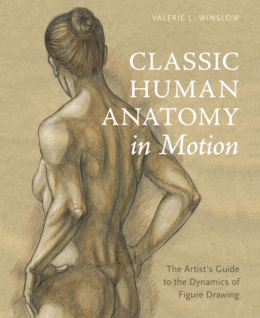
6 CLASSI CHUMANANATOMYI NMOTI ON
Dr.Israa H. Mohsen. Lecture 5. The vertebral column
 Anatomy Lecture 5 Dr.Israa H. Mohsen The vertebral column The vertebral column a flexible structure consisting of 33 vertebrae holds the head and torso upright, serves as an attachment point for the legs,
11/25/2012. Chapter 7 Part 2: Bones! Skeletal Organization. The Skull. Skull Bones to Know Cranium
 Chapter 7 Part 2: Bones! 5) Distinguish between the axial and appendicular skeletons and name the major parts of each 6) Locate and identify the bones and the major features of the bones that compose the
CHAPTER 7, PART II (BONES)
") Anatomy Name: CHAPTER 7, PART II (BONES) Entry #: INSTRUCTIONS: 1) READ Chapter 7, pg. 140-161. 2) Using the outline, make a note card for each underlined bone name or phrase. 3) On each note card, put
Anatomy Name: CHAPTER 7, PART II (BONES) Entry #: INSTRUCTIONS: 1) READ Chapter 7, pg. 140-161. 2) Using the outline, make a note card for each underlined bone name or phrase. 3) On each note card, put
TRAINING LAB SKELETAL REMAINS: IDENTIFYING BONES NAME
 TRAINING LAB SKELETAL REMAINS: IDENTIFYING BONES NAME Background: Skeletal remains are important pieces of evidence. The flesh, muscle, and organs of a victim rapidly decompose; however, the victim s skeleton
Lab-1. Miss. Lina Al-Onazy & samar Al-Wgeet =)
") Lab-1 Introduction The human skeleton is composed of 300 bones at birth and by the time adulthood is reached, some bones have fused together to give a total of 206 bones in the body. The human skeleton
Lab-1 Introduction The human skeleton is composed of 300 bones at birth and by the time adulthood is reached, some bones have fused together to give a total of 206 bones in the body. The human skeleton
The Skeletal System THE APPENDICULAR SKELETON
 The Skeletal System THE APPENDICULAR SKELETON The appendicular skeleton consists of the girdles and the skeleton of the limbs. The upper (anterior) limbs are attached to the pectoral (shoulder) girdle
Biology 210 Chapter 8: Skeletal Tissues Supplement 1
 Biology 210 Chapter 8: Skeletal Tissues Supplement 1 By John McGill Material contributed by Beth Wyatt & Jack Bagwell DIVISIONS OF THE SKELETAL SYSTEM AXIAL SKELETON (80 BONES) Bones of the Head, Neck,
The Appendicular Skeleton
 8 The Appendicular Skeleton PowerPoint Lecture Presentations prepared by Jason LaPres Lone Star College North Harris 8-1 The Pectoral Girdle The Pectoral Girdle Also called shoulder girdle Connects the
Chapter 7: Skeletal System: Gross Anatomy
 Chapter 7: Skeletal System: Gross Anatomy I. General Considerations A. How many bones in an average adult skeleton? B. Anatomic features of bones are based on II. Axial Skeleton A. Skull 1. Functionally
The Skeletal System in Action!! The Skeletal System in Action!
 Skeletal System The Skeletal System in Action!! The Skeletal System in Action! 5 Functions of the Skeletal System 1. Movement: Skeletal system provides points of attachment for muscles. Your legs and arms
Pectoral (Shoulder) Girdle
 Girdle") Chapter 8 Skeletal System: Appendicular Skeleton Pectoral girdle Pelvic girdle Upper limbs Lower limbs 8-1 Pectoral (Shoulder) Girdle Consists of scapula and clavicle Clavicle articulates with sternum
Chapter 8 Skeletal System: Appendicular Skeleton Pectoral girdle Pelvic girdle Upper limbs Lower limbs 8-1 Pectoral (Shoulder) Girdle Consists of scapula and clavicle Clavicle articulates with sternum
10/12/2010. Upper Extremity. Pectoral (Shoulder) Girdle. Clavicle (collarbone) Skeletal System: Appendicular Skeleton
 Girdle. Clavicle (collarbone) Skeletal System: Appendicular Skeleton") Skeletal System: Appendicular Skeleton Pectoral girdle Pelvic girdle Upper limbs Lower limbs 8-1 Pectoral (Shoulder) Girdle Consists of scapula and clavicle Clavicle articulates with sternum (Sternoclavicular
Skeletal System: Appendicular Skeleton Pectoral girdle Pelvic girdle Upper limbs Lower limbs 8-1 Pectoral (Shoulder) Girdle Consists of scapula and clavicle Clavicle articulates with sternum (Sternoclavicular
Country Health SA Medical Imaging
 Country Health SA Medical Imaging REMOTE OPERATORS POSITIONING GUIDE Contents Image Evaluation Page 4 Positioning Guides Section 1 - THORAX 1.1 Chest Page 5 1.2 Bedside Chest Page 7 1.3 Ribs Page 8 Section
Bone Composition. Bone is very strong for its relatively light weight The major components of bone are:
 Human Bones Bone Composition Bone is very strong for its relatively light weight The major components of bone are: Calcium carbonate Calcium phosphate Collagen Water Cortical Bone Spongy Bone Medullary
Chapter 8B. The Skeletal System: Appendicular Skeleton. The Appendicular Skeleton. Clavicle. Pectoral (Shoulder) Girdle
 Girdle") The Appendicular Skeleton Chapter 8B The Skeletal System: Appendicular Skeleton 126 bones Pectoral (shoulder) girdle Pelvic (hip) girdle Upper limbs Lower limbs Functions primarily to facilitate movement
The Appendicular Skeleton Chapter 8B The Skeletal System: Appendicular Skeleton 126 bones Pectoral (shoulder) girdle Pelvic (hip) girdle Upper limbs Lower limbs Functions primarily to facilitate movement
Anatomy & Physiology Skeletal System Worksheet
 1. Name the five functions of the skeleton. c) d) e) Anatomy & Physiology Skeletal System Worksheet 2. The term for the shaft of a bone is:. 3. The bony struts found in spongy bone are called. 4. In ossification,
TERMINOLOGY AS IT APPLIES TO TICA BREED STANDARDS. Interpretation by Marge Hanna
 TERMINOLOGY AS IT APPLIES TO TICA BREED STANDARDS Interpretation by Marge Hanna 1. Nose: The area, with its underlying cartilage, from the top edge of the nose leather up to the bottom of the bridge of
Biology 218 Human Anatomy. Adapted from Martini Human Anatomy 7th ed. Chapter 7 The Skeletal System Appendicular Division
 Adapted from Martini Human Anatomy 7th ed. Chapter 7 The Skeletal System Appendicular Division Introduction The appendicular skeleton includes: Pectoral girdle Shoulder bones Upper limbs Pelvic girdle
Exercise 11. The Appendicular Skeleton
 Exercise 11 The Appendicular Skeleton The Appendicular Skeleton The appendicular skeleton contains 126 bones. Consists of the upper and lower limbs, the pectoral girdles, and the pelvic girdles. The pectoral
Copyright 2003 Pearson Education, Inc. publishing as Benjamin Cummings. Dr. Nabil khouri
 Dr. Nabil khouri Appendicular Skeleton The appendicular skeleton is made up of the bones of the upper and lower limbs and their girdles Two girdles: Pectoral girdles attach the upper limbs to the body
Anatomy and Physiology 2016
 Anatomy and Physiology 2016 O = Temporal line I = coronoid process (Mandible) A = elevates mandible (chewing) O = galea aponeurotica (layer of dense fibrous tissue which covers the upper part of the cranium)
BLUE SKY SCHOOL OF PROFESSIONAL MASSAGE AND THERAPEUTIC BODYWORK. Musculoskeletal Anatomy & Kinesiology I TERMINOLOGY, STRUCTURES, & SKELETAL OVERVIEW
 BLUE SKY SCHOOL OF PROFESSIONAL MASSAGE AND THERAPEUTIC BODYWORK Musculoskeletal Anatomy & Kinesiology I TERMINOLOGY, STRUCTURES, & SKELETAL OVERVIEW MSAK101-I Session 1 Learning Objectives: 1. Define
Principles of Anatomy and Physiology
 Principles of Anatomy and Physiology 14 th Edition CHAPTER 8 The Skeletal System: The Appendicular Skeleton The Appendicular Skeleton The 126 bones of the appendicular skeleton are primarily concerned
YOGA ANATOMY. Part Three - Bones. Yoga Teacher Training Robin Bennett 200 RYT
 YOGA ANATOMY Yoga Teacher Training Part Three - Bones 2015 Robin Bennett 200 RYT THE HUMAN SKELETON BONE COMPOSITION A femur head with a cortex of compact bone and medulla of trabecular (spongy) bone OSTEOBLASTS
The Musculoskeletal System
 The Musculoskeletal System Introduction The skeletal system and muscular system are often considered together because they are close in terms of structure and function. The two systems are referred to
Exercise Science Section 2: The Skeletal System
 Exercise Science Section 2: The Skeletal System An Introduction to Health and Physical Education Ted Temertzoglou Paul Challen ISBN 1-55077-132-9 Role of the Skeleton Protection Framework Attachments for
UNIT 4. BONES AND MUSCLES
 UNIT 4. BONES AND MUSCLES 1. Skeletal System 1. Functions of the Skeletal System The cervical vertebrae make up the neck. You have 7 of these and the top one is connected to the base of the skull. They
Bones of Thorax (Rib Cage)
") Musculoskeletal System (Part A-2) Module 7 -Chapter 10 Overview Muscles Attachments Bones Bone types Surface features of bones Divisions of the skeletal system Joints or Articulations Susie Turner, M.D.
Musculoskeletal System (Part A-2) Module 7 -Chapter 10 Overview Muscles Attachments Bones Bone types Surface features of bones Divisions of the skeletal system Joints or Articulations Susie Turner, M.D.
Important Parts of Bones
 Important Parts of Bones For 2015 Know: Humerus (posterior) Clavical Femur (Anterior) Foot Hand Mandible Os Coxa Scapula Skull (Anterior, Inferior, Lateral) Sternum Humerus (posterior) A. olecranon fossa
The skeletal system is the framework for the muscular system to attach to so we can move.
 Skeletal System The skeletal system is the framework for the muscular system to attach to so we can move. BONE: A rigid connective tissue Helps to move & support the body Protect the organs (skull, ribs)
In-Depth Foundations: Anatomy Terms to Know
 Be familiar with / able to identify and define all the following parts. The Spine Cranium Vertebrae Cervical, Thoracic, Lumbar Sacrum Coccyx Bones of Upper Body Cranium Mastoid process; Occipital condyle,
Ch. 5 - Skeletal System
 Ch. 5 - Skeletal System Bones are living, ever-changing structures. This allows them grow and adapt to new situations that the body encounters. The functions of the skeletal system: 1) support bones are
Chapter 8 The Skeletal System: The Appendicular Skeleton. Copyright 2009 John Wiley & Sons, Inc.
 Chapter 8 The Skeletal System: The Appendicular Skeleton Appendicular Skeleton It includes bones of the upper and lower limbs Girdles attach the limbs to the axial skeleton The pectoral girdle consists
Chapter 8. The Appendicular Skeleton. Lecture Presentation by Lee Ann Frederick University of Texas at Arlington Pearson Education, Inc.
 Chapter 8 The Appendicular Skeleton Lecture Presentation by Lee Ann Frederick University of Texas at Arlington An Introduction to the Appendicular Skeleton The Appendicular Skeleton 126 bones Allows us
Human Skeletal System Glossary
 Acromegaly Apatite Acromegaly - is a condition which involves excessive growth of the jaw, hands, and feet. It results from overproduction of somatotropin in adults (after fusion of the ossification centres
Human Anatomy, First Edition McKinley & O'Loughlin
 Human Anatomy, First Edition McKinley & O'Loughlin Chapter 8 : Appendicular Skeleton 8-1 Appendicular Skeleton Includes the bones of the upper and lower limbs. The girdles of bones that attach the upper
S.A.F.E. Elements of Technique. S.A.F.E. is an acronym for strength, alignment, flexibility, and STRENGTH ALIGNMENT FLEXIBILITY ENDURANCE
 Elements of Technique S.A.F.E. S.A.F.E. is an acronym for strength, alignment, flexibility, and endurance. These are all elements that are innate in the study of dance. STRENGTH The amount of control and
A. Incorrect! The appendicular skeleton includes bones of the shoulder, arm, hand, pelvis, leg and foot.
 Anatomy and Physiology - Problem Drill 08: The Skeletal System III No. 1 of 10 1. Which of the following statements about the appendicular skeleton is correct? A. The appendicular skeleton includes bones
Chapter 6 & 7 The Skeleton
 Chapter 6 & 7 The Skeleton Try this Make clockwise circles with your RIGHT foot, while doing this, draw the number 6 in the air with you RIGHT hand what happens to your foot???? Bony Background Adult body
36.3 The Integumentary System The Skin. KEY CONCEPT The integumentary system has many tissues that protect the body.
 36.3 The Integumentary System The Skin KEY CONCEPT The integumentary system has many tissues that protect the body. 36.3 The Integumentary System The Skin The integument is the body system that surrounds
BIO 137 AXIAL SKELETON BONE STUDY THE HUMAN SKELETON
 BIO 137 THE AXIAL SKELETON MARY CATHERINE FLATH, Ph.D. THE HUMAN SKELETON AXIAL SKULL HYOID THORACIC CAGE VERTEBRAL COLUMN APPENDICULAR PECTORAL GIRDLE UPPER LIMBS PELVIC GIRDLE LOWER LIMBS AXIAL SKELETON
SKELETAL SYSTEM 206. AXIAL SKELETON 80 APPENDICULAR SKELETON 126 (see Figure 6.1) Clavicle. Clavicle. Pectoral girdles. Scapula. Scapula.
 Clavicle. Clavicle. Pectoral girdles. Scapula. Scapula.") SKELETAL SYSTEM 206 AXIAL SKELETON 80 APPENDICULAR SKELETON 126 (see Figure 6.1) Pectoral girdles 4 Clavicle Scapula 2 2 Clavicle Scapula Humerus 2 Humerus Upper limbs 60 Radius 2 Ulna Carpal bones Metacarpal
SKELETAL SYSTEM 206 AXIAL SKELETON 80 APPENDICULAR SKELETON 126 (see Figure 6.1) Pectoral girdles 4 Clavicle Scapula 2 2 Clavicle Scapula Humerus 2 Humerus Upper limbs 60 Radius 2 Ulna Carpal bones Metacarpal
Practical 1 Worksheet
 Practical 1 Worksheet ANATOMICAL TERMS 1. Use the word bank to fill in the missing words. reference side stand body arms palms anatomical forward All anatomical terms have a(n) point which is called the
Chapter 7: Skeletal System
 Chapter 7: Skeletal System The Skeletal System Introduction P. 182 Bone is an organ made up of tissues: It is made up of the following components. Cartilage Blood Nerves Bone Connective Bone Classification
PRE-LAB EXERCISES. Before we get started, look up the definitions of these common bone marking terms: Canal: Condyle: Facet: Fissure:
 1 PRE-LAB EXERCISES When studying the skeletal system, the bones are often sorted into two broad categories: the axial skeleton and the appendicular skeleton. This lab focuses on the appendicular skeleton,
Biology 2401 The Skeletal System
 Biology 2401 The Skeletal System Purpose: The lab will describe the microscopic and gross anatomy of bone, identify bones of the body, and identify important bone markings. I. Overview of the Skeleton
Skeletal System. Std. VIII
 Skeletal System Std. VIII The skeleton in our body serves following functions : 1. Support and shape : The skeleton provides a support or framework to all the soft parts and gives the body and its parts
Chapter 7 /8 pgs SKELETAL TISSUES AND THE SKELETAL SYSTEM
 Chapter 7 /8 pgs. 189-250 SKELETAL TISSUES AND THE SKELETAL SYSTEM Skeletal Tissue Introduction Bone and cartilage are a specialized types of connective tissue Individual Bones are considered separate
Class Outline: Posterior Anatomy
 Class Outline: Posterior Anatomy 5 minutes Breath of Arrival and Attendance 5 minutes Howdy Partner 35 minutes Posterior Anatomy using Power Point Presentation 5 minutes Overview of skeletal segments 5
Anatomy. Anatomy deals with the structure of the human body, and includes a precise language on body positions and relationships between body parts.
 Anatomy deals with the structure of the human body, and includes a precise language on body positions and relationships between body parts. Proper instruction on safe and efficient exercise technique requires
SD School Anatomy Program 1: Bones QuikNotes. Student Notes
 QuikNotes The transverse plane runs from right to left and divides the body into superior (upper) and inferior (lower) sections. Student Notes The frontal plane lies vertically along the body from head
THE SKELETAL SYSTEM 7 TH GRADE SCIENCE
 THE SKELETAL SYSTEM 7 TH GRADE SCIENCE INTRODUCTION Skeletal system is made up of your bones, ligaments, and tendons. It determines : the shape and symmetry of the body acts as protection for your organs
NOTES SKELETAL SYSTEM
 NOTES for the SKELETAL SYSTEM Anatomy & Physiology 2016 Johnson The Skeletal System I. System includes 4 basic parts: A. Bones (206 of em) B. Joints C. Cartilages D. Ligaments II. Bones have 5 basic functions:
BIOLOGY 113 LABORATORY Skeletal System
 BIOLOGY 113 LABORATORY Skeletal System Objectives Distinguish between the axial and appendicular skeleton. Distinguish between the cranium and facial skeleton. Locate and name the bones of the skull and
Overview of Anatomy and Physiology Crash Course Questions (2 pts/piece 30 In Class Points on 1 st grad in 2 nd quarter)
") Overview of Anatomy and Physiology Crash Course Questions (2 pts/piece 30 In Class Points on 1 st grad in 2 nd quarter) Objective: Review material from unit #1-3 and preview unit #5 on the skeletal system
Overview of Anatomy and Physiology Crash Course Questions (2 pts/piece 30 In Class Points on 1 st grad in 2 nd quarter) Objective: Review material from unit #1-3 and preview unit #5 on the skeletal system
Bone Flashcards for 10a
 Bone Flashcards for 0a CLAVICLE (collar bone). Sternal extremity (end) flat end. Acromial extremity (end) rounded end. SCAPULA (shoulder blade). Right or left scapula?. Superior border (superior margin).
Bone List Anatomy
 1 Frontal Bone Skull 2 Parietal Bone Skull 3 Occipital Bone Skull 4 Temporal Bone Skull 5 Coronal Suture Skull 6 Sagittal Suture Skull 7 Squamous suture Skull 8 Lambdoid Suture Skull 9 Surpaorbital Ridge
CHAPTER 8 LECTURE OUTLINE
 CHAPTER 8 LECTURE OUTLINE I. INTRODUCTION A. The appendicular skeleton includes the bones of the upper and lower extremities and the shoulder and hip girdles. B. The appendicular skeleton functions primarily
A&P 1 Muscle In-Lab Guide
 A&P 1 Muscle In-Lab Guide This lab guide includes a table with all the muscles you need to ID, along with their origins, insertions and actions Dashed lines means ignore. If several actions are listed,
Lab Exercise #04 The Skeletal System Student Performance Objectives
 Lab Exercise #04 The Skeletal System Student Performance Objectives The material that you are required to learn in this exercise can be found in either the lecture text or the supplemental materials provided
SKELETAL MUSCLE ANATOMY
 SKELETAL MUSCLE ANATOMY OUTLINE I. Anatomical Terms of Motion II. Head, Face & Neck Muscles III. Anterior Torso Muscles IV. Posterior Torso Muscles V. Arm & Shoulder Muscles VI. Leg & Hip Muscles 2 ANATOMICAL
Human Body. Bones, Joints and Muscles
 Human Body Bones, Joints and Muscles 1 Bone cranium mandible clavicle sternum humerus ribs pelvis ulna femur patella phalanges phalanges vertebrae Also known as skull lower jaw collarbone breastplate upper
Session 6 Bony Bits in Detail
 Session 6 Bony Bits in Detail Australian Institute of Fitness 1 / 38 SETTING THE SCENE Building on the knowledge gained in the previous session where we introduced the skeletal system, we now address the
Biology 152 Appendicular Skeleton Anatomy Objectives
 Biology 152 Appendicular Skeleton Anatomy Objectives We will learn proper bone names, left/right/medial, and the parts of bones in this exercise. Start by learning the names of the bones. As you gain comfort
VCE PHYSICAL EDUCATION WORKBOOK UNIT 1 BODIES IN MOTION NAME:
 VCE PHYSICAL EDUCATION WORKBOOK UNIT 1 BODIES IN MOTION NAME: SKELETAL SYSTEM List the 5 functions of the skeletal system and complete the following table. FUNCTION DESCRIPTION Label the following features
Radiographic Positioning Summary (Basic Projections RAD 222)
") Lower Extremity Radiographic Positioning Summary (Basic Projections RAD 222) AP Pelvis AP Hip (Unilateral) (L or R) AP Femur Mid and distal AP Knee Lateral Knee Pt lies supine on table Align MSP to Center
Lower Extremity Radiographic Positioning Summary (Basic Projections RAD 222) AP Pelvis AP Hip (Unilateral) (L or R) AP Femur Mid and distal AP Knee Lateral Knee Pt lies supine on table Align MSP to Center
Biology 218 Human Anatomy
 Chapter 8 Adapted from Tortora 10 th ed. LECTURE OUTLINE A. Introduction (p. 203) 1. The appendicular skeleton contains 126 bones that form: i. two pectoral (shoulder) girdles two upper limbs i one pelvic
5.1 BONES: AN OVERVIEW
 Unit 5 Skeletal System 5.1 BONES: AN OVERVIEW Section Objectives Identify the major structures and functions of the skeletal system. Differentiate between the two divisions (axial and appendicular) of
VCE PHYSICAL EDUCATION WORKBOOK UNIT 1 BODIES IN MOTION NAME:
 VCE PHYSICAL EDUCATION WORKBOOK UNIT 1 BODIES IN MOTION NAME: SKELETAL SYSTEM List the 5 functions of the skeletal system and complete the following table. FUNCTION DESCRIPTION Label the following features
PRELIMINARY HSC PDHPE. CQ1 How do the musculoskeletal and cardiorespiratory systems of the body influence and respond to movement?
 PRELIMINARY HSC PDHPE CQ1 How do the musculoskeletal and cardiorespiratory systems of the body influence and respond to movement? How do the musculoskeletal and cardiorespiratory systems of the body influence
Bio 113 Anatomy and Physiology The Muscles. Muscles of the Head and Neck. Masseter. Orbicularis occuli. Orbicularis oris. Sternocleidomastoid
 Bio 113 Anatomy and Physiology The Muscles Muscles of the Head and Neck Masseter Orbicularis occuli Orbicularis oris Sternocleidomastoid Temporalis BIO 113 Fall 2011 Muscles Page 1 of 5 Muscles of the
Classification of bones
 Classification of bones compact intramembranous axial histology development regional spongy Intra cartilaginous appendicular flat Irregular shape Sesamoid Long Short Wormian pneumatic Classification
Electrode Placement. Skin Preparation. Frontalis (FRL) (Specific) Temporalis Anterior (TA) (Specific) Sternocleidomastoid (SCM) (Specific)
 (Specific) Temporalis Anterior (TA) (Specific) Sternocleidomastoid (SCM) (Specific)") Electrode Placement Skin Preparation 1) Removing the hair: Shave if necessary 2) Clean the skin: Use a towel or abrasive pad with conductive cleaning paste or alcohol to remove dead skin cells (high impedance)
Electrode Placement Skin Preparation 1) Removing the hair: Shave if necessary 2) Clean the skin: Use a towel or abrasive pad with conductive cleaning paste or alcohol to remove dead skin cells (high impedance)
Perpendicular Plate Zygomatic Bone. Mental Foramen Mandible
 Glabella Frontal Middle Nasal Concha Nasal Lacrimal Perpendicular Plate Zygomatic Inferior Nasal Concha Maxilla Mental Mandible Skull (anterior view) Squamosal Suture Coronal Suture Frontal Parietal Nasal
Axial Skeleton BONE TERMINOLOGY FEATURES
 Axial Skeleton BONE TERMINOLOGY FEATURES Tuberosity Rounded area on bone often roughened for muscle attachment. Tubercle Rounded projection on bone. This is called a tuberosity on the femur. Crest Ridgeline
Balanced Body Movement Principles
 Balanced Body Movement Principles How the Body Works and How to Train it. Module 3: Lower Body Strength and Power Developing Strength, Endurance and Power The lower body is our primary source of strength,
Healing Hands School of Holistic Health. Advanced Circulatory & Sports Massage Class Handouts
 Class Handouts 1 Posterior Trepidations Torso Rock Torso Rock half-step Torso Rock both sides Torso Rock down body Torso Side Stretch Erector Rock Spinal Rock Lumbo Rock Cha Cha Leg Clay Snake Flop Leg
Skeletal System. Skeleton. Support. Function of Bones. Movement. Protection 10/15/12
 Skeleton Skeletal System 1 Axial Skeleton-Bones that form the longitudinal axis of the body (skull and spinal column). Appendicular Skeleton-Bones of the limbs and girdles. Also include joints, ligaments
Spring Written By: J. E. Sutton. Contents: I. Overview of the Skeleton: II. Appendicular Skeleton III. Axial Skeleton IV.
 Spring 2012 Written By: J. E. Sutton Contents: I. Overview of the Skeleton: II. Appendicular Skeleton III. Axial Skeleton IV. Articulations Overview of the Skeleton: I. Orientation to Human Skeleton: a.
The skeleton supports the body. The skeleton protects soft body parts The skeleton produces blood cells. The skeleton stores minerals and fat.
 SKELETAL SYSTEM SKELETAL SYSTEM The skeleton supports the body. The bones of the legs support the entire body when we are standing, and the coxal bones of the pelvic girdle support the abdominal cavity.
Muscular Nomenclature and Kinesiology - One
 Chapter 16 Muscular Nomenclature and Kinesiology - One Lessons 1-3 (with lesson 4) 1 Introduction 122 major muscles covered in this chapter Chapter divided into nine lessons Kinesiology study of human
Lab Exercise #5 The Muscular System Student Performance Objectives
 Student Performance Objectives The material that you are required to learn in this exercise can be found in either the lecture text or the supplemental materials provided in lab. Prior to coming to class,
1.1 The structure and functions of the musculo-skeletal system Learning objectives
 1.1 The structure and functions of the musculo-skeletal system Learning objectives To be able to describe the functions of the skeleton. To understand different bone classifications and functioning. To
In which arm muscle are intramuscular injections most often given? (not in text)
") AP1 Lab 9 - Muscles of the Arms and Legs Locate the following muscles on the models and on yourself. Recall anatomical position. Directional terms such as anterior, posterior, lateral, etc. all assume
AP1 Lab 9 - Muscles of the Arms and Legs Locate the following muscles on the models and on yourself. Recall anatomical position. Directional terms such as anterior, posterior, lateral, etc. all assume
Bones of the wrist and ankle Bones that form within tendons (e.g., patella)
") Skeletal System Review Surface Anatomy Dr. Gary Mumaugh Function of Bones Support form the framework that supports the body and cradles soft organs Protection provide a protective case for the brain, spinal
Skeletal System Review Surface Anatomy Dr. Gary Mumaugh Function of Bones Support form the framework that supports the body and cradles soft organs Protection provide a protective case for the brain, spinal
Skeletal System. Supplementary Information
 Skeletal System Supplementary Information COMMON ANATOMICAL TERMS Planes run through the body side to side and front to back eg. median plane Surfaces of the body are also named eg. anterior surface This
The Skeletal System. Mosby items and derived items 2010, 2006, 2002, 1997, 1992 by Mosby, Inc., an affiliate of Elsevier Inc.
 The Skeletal System Functions of Skeletal System Provides internal framework that supports the body Protects internal organs Helps fight disease by producing white blood cells 2 Functions of Skeletal System
OHIO STATE UNIVERSITY EXTENSION
 Cloverbud Investigators: Career Detectives October Background: Today we are going to learn about our bones and how they join together to hold up our body, all the way from our head to our toes. Did you
THE SKELETAL SYSTEM. Focus on the Pectoral Girdle
 THE SKELETAL SYSTEM Focus on the Pectoral Girdle Appendicular Skeleton 126 bones Includes bones of the limbs (arms and legs) Pectoral girdle (shoulder) Pelvic girdle (hip) Pectoral Girdle (the shoulder)
Scapula Spine Lateral edge of clavicle. Medial border Scapula. Medial border of Scapula, between superior angle and root of spine. Scapula.
 Muscle attachments and actions answer sheet Muscle Origins insertions Movements Joints crossed Trapezius Base of skull Spinous process of C7 Thoracic Spine Lateral edge of clavicle Elevation Retraction
Prime movers provide the major force for producing a specific movement Antagonists oppose or reverse a particular movement Synergists
 Dr. Gary Mumaugh Prime movers provide the major force for producing a specific movement Antagonists oppose or reverse a particular movement Synergists Add force to a movement Reduce undesirable or unnecessary
The Skeletal System. Chapter 7a. Skeletal System Introduction Functions of the skeleton Framework of bones The skeleton through life
 The Skeletal System Skeletal System Introduction Functions of the skeleton Framework of bones The skeleton through life Chapter 7a Support Protection Movement Storage areas Minerals Lipids Hemopoiesis
Nervous & Skeletal Systems. Virtual Science University
 Nervous & Skeletal Systems Virtual Science University 1 Nervous & Skeletal Systems Texas TEK B.10(A) The student will interpret the function of systems in organisms (humans) including the nervous and skeletal
TEST BANK FOR THE HUMAN BODY IN HEALTH AND ILLNESS 5TH EDITION BY BARBARA HERLIHY Chapter 8: Skeletal System
 Link download Full : http://testbankair.com/download/test-bank-for-thehuman-body-in-health-and-illness-5th-edition-by-barbara-herlihy/ TEST BANK FOR THE HUMAN BODY IN HEALTH AND ILLNESS 5TH EDITION BY
The Appendicular Skeleton
 8 The Appendicular Skeleton PowerPoint Lecture Presentations prepared by Jason LaPres Lone Star College North Harris An Introduction to the Appendicular Skeleton Learning Outcomes 8-1 Identify the bones
Lab Activity 9. Appendicular Skeleton Martini Chapter 8. Portland Community College BI 231
 Lab Activity 9 Appendicular Skeleton Martini Chapter 8 Portland Community College BI 231 Appendicular Skeleton Upper & Lower extremities Shoulder Girdle Pelvic Girdle 2 Humerus 3 Humerus: Proximal End
Chapter 8 The Skeletal System: The Appendicular Skeleton. Copyright 2009 John Wiley & Sons, Inc.
 Chapter 8 The Skeletal System: The Appendicular Skeleton Appendicular Skeleton The primary function is movement It includes bones of the upper and lower limbs Girdles attach the limbs to the axial skeleton
Warm-Up Activity. Fill in the names of the bones in the skeleton diagram.
 Warm-Up Activity Fill in the names of the bones in the skeleton diagram. Warm-Up 1. What are the 4 types of bones? Give an example of each. 2. Give 3 ways you can tell a female skeleton from a male skeleton.
Amy Warenda Czura, Ph.D. 1 SCCC BIO130 Lab 7 Appendicular Skeleton & Articulations
 The Skeletal System II: Appendicular Skeleton and Articulations Exercises 11, 13 (begins: page 145 in 9 th and 10 th editions) Exercises 10, 11 (begins: page 147 in 11 th edition, page 149 in 12 th edition)
TEST YOURSELF- Chapter 7
 TEST YOURSELF- Chapter 7 Cranial Bones 1. Give the name of the bone for each of the following markings. Some of the markings are found on more than one bone. List all that apply. Cranium a. Frontal squama: