Integra. Proximal Humeral Fracture Plate System SURGICAL TECHNIQUE
|
|
|
- Denis Crawford
- 5 years ago
- Views:
Transcription


1 Integra Proximal Humeral Fracture Plate System SURGICAL TECHNIQUE
2 Table of Contents Introduction Product Description... 2 Indications... 3 Contraindications... 3 Warnings and Precautions... 4 Design Rationale LP (Low Profile) Plate... 5 (Greater Tuberosity) Plate... 6 LP Plate Surgical Technique Step 1: Patient Positioning... 7 Step 2: Exposure... 7 Step 3: Fracture Reduction and Debridement... 7 Step 4: Plate Positioning... 8 Step 5: Proximal 3.5mm Screw Insertion... 9 Step 6: 3.5mm Shaft Screw Insertion Step 7: Final Verification Plate Surgical Technique Step 1: Patient Positioning Step 2: Exposure Step 3: Fracture Reduction and Debridement Step 4: Plate Positioning Step 5: Proximal 3.5mm Screw Insertion Step 6: 2.7mm Proximal Holes ( Plate Only) Step 7: 3.5mm Shaft Screw Insertion Step 8: Final Verification Product Information Instrumentation... 19
3 Surgical Technique As the manufacturer of this device, Integra does not practice medicine and does not recommend this or any other surgical technique for use on a specific patient. The surgeon who performs any implant procedure is responsible for determining and using the appropriate techniques for implanting the device in each patient. Introduction Joseph Abboud, MD Philip Duke, MB.BS, FRACS, FA(ORTH)A William Geissler, MD Anand Murthi, MD Mark Ross, MB.BS, FRACS, FA (ORTH)A Caution: Federal law restricts this device to sale by or on the order of a physician or practitioner. Product Description The Integra Proximal Humeral Fracture Plate System is composed of left and right Humeral reconstruction plate implants in two options, Plate and LP Plate. The plate is designed to cover the greater tuberosity and is available in four-hole (91mm) and seven-hole (115mm), and ten-hole (138mm) lengths or sizes. The LP plate is designed to sit lower on the greater tuberosity and is available in three-hole (88mm), six-hole (112mm), and nine-hole (135mm) lengths or sizes. The system will feature 3.5mm locking screws (15-75mm lengths), non-locking compression screws (15-40mm lengths) and non-locking partial thread lag screws (40-75mm lengths)., In addition to, 2.7mm locking screws (10-25mm lengths) are also available. The Integra Proximal Humeral Fracture Plate is a single component made from stainless steel (SS 316L). The non-locking compression, locking and non-locking partial thread lag screws are made from stainless steel (SS 316L). 2
4 Indications The Integra Proximal Humeral Fracture Plate system is designed for fractures and fracture dislocations, osteotomies and non-unions of the proximal humerus. Indications Include: Dislocated two-, three-, and four-fragment fractures of the proximal humerus, including fractures involving osteopenic bone Pseudoarthroses in the proximal humerus Osteotomies in the proximal humerus Contraindications Not indicated for use with acute infections or for children during the growth phase Screws are contraindicated in: active infection, conditions which tend to retard healing such as blood supply limitations, previous infections, insufficient quantity or quality of bone to permit stabilization of the fracture complex, conditions that restrict the patient s ability or willingness to follow postoperative instructions during the healing process and foreign body sensitivity. Cases with malignant primary or metastatic tumors which preclude adequate bone support or screw fixations, unless supplemental fixation or stabilization methods are utilized. Foreign body sensitivity where material sensitivity is suspected, appropriate tests should be made and sensitivity ruled out prior to implementations. These implants are intended as a guide to normal healing, and are NOT intended to replace normal body structure or bear the weight of the body in the presence of incomplete bone healing. Delayed unions or non-unions in the presence of load bearing or weight bearing might eventually cause the implant to break due to metal fatigue. All metal surgical implants are subjected to repeated stress in use, which can result in metal fatigue. 3
5 Warnings and Precautions No metallic surgical implant should be reused. Any metal implant, once used, should be discarded. Even though it appears undamaged, it may already have small defects and internal stress patterns which may lead to fatigue failure. Correct handling of implant is extremely important. Avoid contouring metallic implants whenever possible. If necessary, or allowed by design, the device should not be bent sharply, reverse bent, notched or scratched. All of these operations can produce defects in the surface finish and internal stress concentrations, which may become the focal point for eventual failure of the appliance. If metal plates or other metallic devices are to be used together with the Integra Proximal Humeral Fracture Plates, all such devices should be manufactured from a metal that has a similar composition to avert possibility of galvanic corrosion or other metallic reactions. Correct selection of the implant is extremely important. The potential for success in fracture fixation is increased by the selection of the proper size of the implants. The patient s anatomy and indication will determine the size of the Integra Proximal Humeral Fracture Plate to be used. The size of the human bones presents limiting restrictions on the size and strength of implants. Postoperative care is extremely important. The patient must be warned that noncompliance with postoperative instructions could lead to breakage of the implant requiring revision surgery to remove the device. The use of Integra Proximal Humeral Fracture Plates provides the surgeon a means of bone fixation and helps generally in the management of fractures and reconstructive surgeries. The implants are intended as a guide to normal healing and are NOT intended to replace normal body structure. All metal surgical implants are subject to repeated stress in use which can result in metal fatigue. Failure to immobilize a delayed union or nonunion of bone will result in excessive and repeated stresses which are transmitted by the body to any temporary internal fixation device prior to the healing of the fracture. Due to normal metal fatigue, these stresses can cause eventual bending or breakage of the device. Therefore, it is important that immobilization of the fracture site is maintained until firm bony union (confirmed by clinical and roentgenographic examination) is established. Detailed written instructions on the use and limitations of the device should be given to the patient. While the surgeon must make the final decision on implant removal, whenever possible and practical for the individual patient, fixation devices should be removed once their service as an aid to healing is accomplished, particularly in younger more active patients. The MR environment presents risks to patients with metal implant. Review of the available literature documents that metal implants may heat resulting in tissue damage and may migrate out of position. They may also cause artifact affecting image quality. Physicians should take these risks into consideration when recommending MRI imaging for patients with metal implants. Note: The Integra Proximal Humeral Fracture Plate System has not been evaluated for safety and compatibility in the MR environment. The Integra Proximal Humeral Fracture Plate System has not been tested for heating or migration in the MR environment. 4

 6-hole (112mm) 9-hole (135mm) 6 suture holes for")

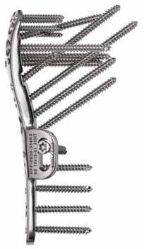
6 Design Rationale LP (Low Profile) Plate Designed to sit lower on the greater tuberosity to minimize subacromial impingement 9 proximal screw options 4 calcar screws to provide inferior medial support Slotted screw option for initial plate positioning Anatomic fit with right and left plates and different length plates 3-hole (89mm) 6-hole (112mm) 9-hole (135mm) 6 suture holes for tuberosity fixation Easy in-situ needle passing through angled and scalloped holes Also used as wire holes for initial plate fixation Multiple Screw Options 3.5mm Locking, Non-Locking and Lag Screw Options 5

 7-hole (116mm) 10-hole (140mm) 5 suture holes for soft tuberosity fixation")

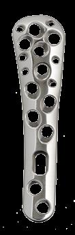
7 Design Rationale (Greater Tuberosity) Plate Maximizes tuberosity coverage and humeral head bone purchase with 12 proximal screw options 4 calcar screws to provide inferior medial support Anterior tab for initial plate positioning and buttress option Anatomic fit with right and left plates and different length plate 4-hole (91mm) 7-hole (116mm) 10-hole (140mm) 5 suture holes for soft tuberosity fixation Easy in-situ needle passing through angled and scalloped holes Also used as wire holes for initial plate fixation Multiple Screw Options 2.7mm Proximal Locking Screws 3.5mm Locking, Non-Locking and Lag Screw Options 6
8 Surgical Technique: LP (Low Profile) Plate Step 1: Patient Positioning 1-1 Place the patient in beach chair position (1-1). Ensure that the involved shoulder extends laterally over the top corner of the table so that the arm can be brought into extension and abduction (this is essential for good exposure of the proximal humerus). 1-1 Step 2: Exposure 2-1 Deltopectoral Approach Exposure is achieved through a 10-12cm incision that starts at the clavicle and proceeds over the coracoid toward the deltoid insertion. The incision should be in line with the deltoid-pectoral interval. Once the initial incision is made, develop the subcutaneous flap, locate and identify the cephalic vein. Gently retract the cephalic vein either laterally with the deltoid muscle or medially with the pectoralis major. Self-retaining retractors can be utilized to achieve this exposure. Release the fascia along the lateral border of the coracobrachialis and gently retract it medially. To help improve visualization and reduction find the pectoralis insertion and release the proximal third of the pectoralis tendon to expose the biceps. Take care to protect the biceps tendon which lies directly underneath. Step 3: Fracture Reduction and Debridement 3-1 Debride the fracture and keep any bone pieces to aid in the repair. After debridement of the fracture, reduction can be achieved by manual traction and indirect manipulation. Reduction clamps are also available in the tray. In cases of severe comminution reduction of the tuberosities can be achieved by suturing the rotator cuff together. To aid in healing, bone graft should be considered to fill any voids. 7
9 Step 4: Plate Positioning 4-1 Select the appropriate Integra Proximal Humeral plate, left or right, LP plate (4-1) in the appropriate length to stabilize the fracture. The LP plate is designed to sit lower on the proximal humerus which can help to prevent acromial impingement but does not allow full buttress of the greater tuberosity. 4-1 The LP plate is placed onto the humerus approximately 2cm distal to the rotator cuff attachment on the upper part of the greater tuberosity and 2-3mm lateral to the bicipital groove. Temporarily fix the plate with 2.0mm guide wires. 4-2 Place a PF Pin in the center of the slot on the plate(4-2). This will allow the plate to be translated up or down to achieve desired plate position. Once desired plate positioning has been achieved proceed with proximal screw insertion. 4-2 LP LP 8

10 Step 5: Proximal 3.5mm Screw Insertion Note: It is recommended to start with the screws that angle up into the head to secure the reduced humeral head fragments. 5-1 Place the appropriate LP Proximal Hole Targeting Guide onto the plate. Lock the targeting guide into place by inserting the 2.4mm Locking Drill Guide into the most proximal hole on the guide (5-1). 5-1 LP 5-2 Using the 2.4mm Drill Sleeve inside the 3.5mm Screw Guide place the guide into one of the four proximally angled holes on the targeting guide (5-2). Drill using the 2.4mm drill until resistance is felt. Use fluoroscopy to verify the tip of the drill is close to the subchondral bone. 5-2 Note: Care should be taken to ensure the subchondral bone is not penetrated. LP 5-3 Use the depth readings on the drill to determine screw length (close up of drill in drill guide showing laser marks on drill). The Standard Depth Gauge can be used at this point if necessary (5-3). Remove the drill and 2.4mm Drill Sleeve leaving the 3.5mm Screw Guide in place. 5-3 LP 9

11 5-4 Insert the determined length locking, non-locking compression, or non-locking partial thread 3.5mm screw through the Screw Guide using the TX15 Driver Shaft and Ratcheting Driver Handle (5-4). Repeat the above steps for the remaining angled screw holes on the targeting guide before proceeding to the additional proximal screw holes. 5-4 LP Step 6: 3.5mm Shaft Screw Insertion The shaft of the plate can be secured by either locking screws or multidirectional non-locking screws. 6-1 Locking Screws Thread the 2.4mm Locking Drill Guide into the most proximal shaft hole. Drill using the 2.4mm Drill until the far cortex is penetrated). Use the markings on the drill to determine screw length or remove the drill guide and use the Standard Depth Gauge (6-1a). 6-1a If power is used to insert the screw, use of a torque limiter is recommended. Start the power tool slowly, increase the speed and then reduce it again before the screw is fully tightened. Final tightening of Locking Screws should always be performed manually using a two finger tighten technique to prevent over-tightening of Locking Screws. Insert the corresponding 3.5mm locking screw using the TX15 Driver Shaft and Ratcheting Driver Handle (6-1b). Repeat for the remaining shaft holes ensuring the plate stays aligned with the humeral shaft as you work distally. 6-1b LP Note: Use of a torque limiter is required if inserting screws under power. Do not lock the screws under power as this risks damaging the screw recess, making implant removal difficult. LP 10
. Use the Standard Depth Gauge to determine screw length (6-2b). 6-2a Insert the corresponding 3.")

12 6-2 Non-Locking Compression Screws Place the 2.4mm drill sleeve into the 3.5mm Screw Guide, insert in the desired screw orientation and drill using the 2.4mm drill until the desired depth is achieved (6-2a). Use the Standard Depth Gauge to determine screw length (6-2b). 6-2a Insert the corresponding 3.5mm non-locking compression screw using the TX15 Driver Shaft and Ratcheting Driver Handle (6-2c). Repeat for the remaining shaft holes ensuring the plate stays aligned with the humeral shaft as you work distally. 6-2b LP LP 6-2c LP Step 7: Final Verification 7-1 Using Fluoroscopy evaluate the humerus to verify proper reduction and screw placement. 11

13 Surgical Technique: (Greater Tuberosity) Plate Step 1: Patient Positioning 1-1 Place the patient in beach chair position (1-1). Ensure that the involved shoulder extends laterally over the top corner of the table so that the arm can be brought into extension and abduction (this is essential for good exposure of the proximal humerus). 1-1 Step 2: Exposure 2-1 Deltopectoral approach Exposure is achieved through a 10-12cm incision that starts at the clavicle and proceeds over the coracoid toward the deltoid insertion. The incision should be in line with the deltoid-pectoral interval. Once the initial incision is made, develop the subcutaneous flap, locate and identify the cephalic vein. Gently retract the cephalic vein either laterally with the deltoid muscle or medially with the pectoralis major. Self-retaining retractors can be utilized to achieve this exposure. Release the fascia along the lateral border of the coracobrachialis and gently retract it medially. To help improve visualization and reduction find the pectoralis insertion and release the proximal third of the pectoralis tendon to expose the biceps. Take care to protect the biceps tendon which lies directly underneath. Step 3: Fracture Reduction and Debridement 3-1 Debride the fracture and keep any bone pieces to aid in the repair. After debridement of the fracture, reduction can be achieved by manual traction and indirect manipulation. Reduction clamps are also available in the tray. In cases of severe comminution reduction of the tuberosities can be achieved by suturing the rotator cuff together. To aid in healing, bone graft should be considered to fill any voids. 12
14 Step 4: Plate Positioning 4-1 Select the appropriate Integra Proximal Humeral plate, left or right, plate (4-1) in the appropriate length to stabilize the fracture. The Plate is designed to cover the greater tuberosity and should be utilized in fractures that require additional support or buttress of the greater tuberosity fragments. The plate is placed onto the humerus approximately 6-10mm distal to the rotator cuff attachment on the upper part of the greater tuberosity and 2-3mm lateral to the bicipital groove. Temporarily fix the plate with 2.0mm guide wires Place a PF Pin in the center of the slot on the plate(4-2). This will allow the plate to be translated up or down to achieve desired plate position. Once desired plate positioning has been achieved proceed with proximal screw insertion
.")
15 Step 5: Proximal 3.5mm Screw Insertion Note: It is recommended to start with the screws that angle up into the head to secure the reduced humeral head fragments. 5-1 Place the appropriate Proximal Hole Targeting Guide onto the plate. Lock the targeting guide into place by inserting the 2.4mm Locking Drill Guide into the most proximal hole on the guide (5-1) Using the 2.4mm Drill Sleeve inside the 3.5mm Screw Guide place the guide into one of the four proximally angled holes on the targeting guide (5-2). 5-2 Drill using the 2.4mm drill until resistance is felt. Use fluoroscopy to verify the tip of the drill is close to the subchondral bone. Note: Care should be taken to ensure the subchondral bone is not penetrated Use the depth readings on the drill to determine screw length (close up of drill in drill guide showing laser marks on drill). The Standard Depth Gauge can be used at this point if necessary (5-3). Remove the drill and 2.4mm Drill Sleeve leaving the 3.5mm Screw Guide in place. 5-4 Insert the determined length locking, non-locking compression, or non-locking partial thread 3.5mm screw through the Screw Guide using the TX15 Driver Shaft and Mini Ratchet Driver Handle (5-4). Repeat the above steps for the remaining angled screw holes on the targeting guide before proceeding to the additional proximal screw holes
.")

16 Step 6: 2.7mm Proximal holes ( Plate Only) These four most proximal holes can either be used for suture holes or 2.7mm locking screws or a combination of both to repair the tuberosity. 6-1 Suture Technique Place a heavy non absorbable suture through the tendon at the attachment to the tuberosity and then through the plate (6-1). There is a recess cut out of the back of the plate to allow the passing of suture after the plate is secured. 6-1 Note: Sutures may also be passed prior to the plate being secured for ease of insertion. Tighten the suture until appropriate tension is achieved mm Screw Technique Thread the 1.9mm Locking Drill Guide into the plate. Drill with the 1.9mm Drill until desired depth is achieved (6-2). Use either the markings on the drill to determine screw length or remove the drill guide and use the Standard Depth Gauge (6-3). Insert the corresponding 2.7mm locking screw using the TX8 Driver Shaft and Mini Ratchet Driver Handle (6-4). Repeat the above steps for the remaining 2.7mm screw holes. 6-3 If power is used to insert the screw, use of a torque limiter is recommended. Start the power tool slowly, increase the speed and then reduce it again before the screw is fully tightened. Final tightening of Locking Screws should always be performed manually using a two finger tighten technique to prevent over-tightening of Locking Screws. Note: Use of a torque limiter is required if inserting screws under power. Do not lock the screws under power as this risks damaging the screw recess, making implant removal difficult
.")
17 Step 7: 3.5mm Shaft Screw Insertion The shaft of the plate can be secured by either locking screws or multidirectional non-locking screws. 7-1 Locking Screws Thread the 2.4mm Locking Drill Guide into the most proximal shaft hole. Drill using the 2.4mm Drill until the far cortex is penetrated). Use the markings on the drill to determine screw length or remove the drill guide and use the Standard Depth Gauge (7-1a). 7-1a If power is used to insert the screw, use of a torque limiter is recommended. Start the power tool slowly, increase the speed and then reduce it again before the screw is fully tightened. Final tightening of Locking Screws should always be performed manually using a two finger tighten technique to prevent over-tightening of Locking Screws. Insert the corresponding 3.5mm locking screw using the TX15 Driver Shaft and Mini Ratchet Driver Handle (7-1b). Repeat for the remaining shaft holes ensuring the plate stays aligned with the humeral shaft as you work distally. 7-1b Note: Use of a torque limiter is required if inserting screws under power. Do not lock the screws under power as this risks damaging the screw recess, making implant removal difficult. 16
. Use the Standard Depth Gauge to determine screw length (7-2b). 7-2a Insert the corresponding 3.")
18 7-2 Non-Locking Compression Screws Place the 2.4mm drill sleeve into the 3.5mm Screw Guide, insert in the desired screw orientation and drill using the 2.4mm drill until the desired depth is achieved (7-2a). Use the Standard Depth Gauge to determine screw length (7-2b). 7-2a Insert the corresponding 3.5mm non-locking compression screw using the TX15 Driver Shaft and Mini Ratchet Driver Handle (7-2c). Repeat for the remaining shaft holes ensuring the plate stays aligned with the humeral shaft as you work distally. 7-2b 7-2c Step 8: Final Verification 8-1 Using Fluoroscopy evaluate the humerus to verify proper reduction and screw placement. 17
19 Product Information Screws Plates Catalog Number Description Catalog Number Description 2.7mm Locking Screws HFP RS Humeral Fracture Plate 4-Hole, Right PSS2710LS 2.7mm Locking Screw 10mm HFP LS Humeral Fracture Plate 4-Hole, Left PSS2712LS 2.7mm Locking Screw 12.5mm HFP RS Humeral Fracture Plate 7-Hole, Right PSS2715LS 2.7mm Locking Screw 15mm HFP LS Humeral Fracture Plate 7-Hole, Left PSS2717LS 2.7mm Locking Screw 17.5mm HFP RS Humeral Fracture Plate 10-Hole, Right PSS2720LS 2.7mm Locking Screw 20mm HFP LS Humeral Fracture Plate 10-Hole, Left PSS2722LS 2.7mm Locking Screw 22.5mm PSS2725LS 2.7mm Locking Screw 25mm HFP RS Low Profile Humeral Fracture Plate 3-Hole, Right HFP LS Low Profile Humeral Fracture Plate 3-Hole, Left 3.5mm Locking Screws HFP RS Low Profile Humeral Fracture Plate 6-Hole, Right PSS3515LS 3.5mm Locking Screw 15mm HFP LS Low Profile Humeral Fracture Plate 6-Hole, Left PSS3517LS 3.5mm Locking Screw 17.5mm HFP RS Low Profile Humeral Fracture Plate 9-Hole, Right PSS3520LS 3.5mm Locking Screw 20mm HFP LS Low Profile Humeral Fracture Plate 9-Hole, Left PSS3522LS 3.5mm Locking Screw 22.5mm PSS3525LS 3.5mm Locking Screw 25mm PSS3527LS 3.5mm Locking Screw 27.5mm Single Use PSS3530LS 3.5mm Locking Screw 30mm Catalog Number Description PSS3532LS 3.5mm Locking Screw 32.5mm PSS3535LS 3.5mm Locking Screw 35mm DRL mm Drill Bit PSS3537LS 3.5mm Locking Screw 37.5mm DRL mm Drill Bit PSS3540LS 3.5mm Locking Screw 40mm DRL mm Drill Bit PSS3545LS 3.5mm Locking Screw 45mm WIRE S 2.0mm K-wire Smooth PSS3550LS 3.5mm Locking Screw 50mm WIRE T 2.0mm K-wire Threaded PSS3555LS 3.5mm Locking Screw 55mm PFP mm Provisional Fixation Pin PSS3560LS 3.5mm Locking Screw 60mm 225TXSX002 Disposable Torque Limiter for 3.5mm screws PSS3565LS 3.5mm Locking Screw 65mm 225TXSX001 Disposable Torque Limiter for 2.7mm screws PSS3570LS 3.5mm Locking Screw 70mm PSS3575LS 3.5mm Locking Screw 75mm 3.5mm Compression Screws Instruments PSS3515CS 3.5mm Compression Screw 15mm Catalog Number Description PSS3517CS 3.5mm Compression Screw 17.5mm LDG mm Locking Drill Guide Green PSS3520CS 3.5mm Compression Screw 20mm LDG mm Locking Drill Guide White PSS3522CS 3.5mm Compression Screw 22.5mm DDG mm Drill Guide White PSS3525CS 3.5mm Compression Screw 25mm DDG mm Screw Guide Blue PSS3527CS 3.5mm Compression Screw 27.5mm DDG mm Drill Guide PSS3530CS 3.5mm Compression Screw 30mm Torx 15 Drive Shaft PSS3532CS 3.5mm Compression Screw 32.5mm Torx 8 Drive Shaft PSS3535CS 3.5mm Compression Screw 35mm PDG Plate Depth Gauge PSS3537CS 3.5mm Compression Screw 37.5mm PDG Drill Guide Depth Gauge PSS3540CS 3.5mm Compression Screw 40mm RM1011S03 Mini Ratchet Driver Handle 3.5mm Partially Threaded Screws AO to Trinkle adapter PSS3540PS 3.5mm Partially Threaded Screw 40mm A Periosteal Elevator PSS3545PS 3.5mm Partially Threaded Screw 45mm Bone Reduction Forceps Straight 9 PSS3550PS 3.5mm Partially Threaded Screw 50mm Bone Reduction Forceps Curved 8 PSS3555PS 3.5mm Partially Threaded Screw 55mm HTG093003L Left Proximal Hole Targeting Guide Plate PSS3560PS 3.5mm Partially Threaded Screw 60mm HTG093003R Right Proximal Hole Targeting Guide Plate PSS3565PS 3.5mm Partially Threaded Screw 65mm HTG093001L Left Proximal Hole Targeting Guide LP Plate PSS3570PS 3.5mm Partially Threaded Screw 70mm HTG093001R Right Proximal Hole Targeting Guide LP Plate PSS3575PS 3.5mm Partially Threaded Screw 75mm CSA00014 Tray Lid CSA Tray Base CSA Tray Upper CSA Screw Caddy Base CSA Screw Caddy Lid 18

20 Instrumentation Bone Reduction Forceps 2. Left/Right Plates 3. Left/Right LP Plates 4. Bone Reduction Forceps 19

21 Smooth/Threaded K-Wires mm Drill Guide mm Provisional Fixation Pin mm/2.4mm Locking and Non-Locking Drill Guides 5. Mini Ratchet Driver Handle 6. Left/Right Hole Targeting Guides 7. Drill Guide Depth Gauge 8. Plate Depth Gauge mm/2.4mm/3.5mm Drills 10. TORX 8/15 Drive Shafts 11. AO Trinkle Adaptor 12. Elevator 13. Screw Caddy 20
22 21
23 22
24 Integra Proximal Humeral Fracture Plate System Availability of these products might vary from a given country or region to another, as a result of specific local regulatory approval or clearance requirements for sale in such country or region. n Non contractual document. The manufacturer reserves the right, without prior notice, to modify the products in order to improve their quality. n Warning: Applicable laws restrict these products to sale by or on the order of a physician. n Consult product labels and inserts for any indication, contraindications, hazards, warnings, precautions, and instructions for use. For more information or to place an order, please contact: United States, Canada, Asia, Pacific, Latin America USA fax n International fax n integralife.com/contact Manufacturer: Ascension Orthopedics, Inc Metric Blvd Austin, TX n USA Integra and the Integra logo are registered trademarks of Integra LifeSciences Corporation or its subsidiaries in the United States and/or other countries. Patent Pending Integra LifeSciences Corporation. All rights reserved. Printed in USA. 0M LC Rev. D
Surgical Technique. Proximal Humerus Locking Plate
 Surgical Technique Proximal Humerus Locking Plate PERI-LOC Upper Extremity Locked Plating System 3.5mm & 4.5mm Proximal Humerus Locking PlatesCatalog Infor Table of Contents Introduction.........................................................2
Integra. DigiFuse Cannulated Intramedullary Fusion System SURGICAL TECHNIQUE
 Integra DigiFuse Cannulated Intramedullary Fusion System SURGICAL TECHNIQUE Table of Contents Design Rationale... 2 System Features... 2 Indications... 2 Contraindications... 2 Surgical Technique...3 Step
AcUMEDr. Locking Proximal Humeral Plate. PoLARUSr PHPt
 AcUMEDr Locking Proximal Humeral Plate PoLARUSr PHPt PoLARUSr PHPt LOCKING PROXIMAL HUMERAL PLATE Since 1988 Acumed has been designing solutions to the demanding situations facing orthopedic surgeons,
Proximal Humerus Plating System. Surgical Technique TRAUMA
 Proximal Humerus Plating System Surgical Technique TRAUMA S 3 Proximal Humerus Plating System Contents Introduction... 3 S 3 Proximal Humerus Plating System... 4 Deltopectoral Approach... 6 Surgical Technique...
Technique Guide. 3.5 mm LCP Periarticular Proximal Humerus Plate. Part of the Synthes locking compression plate (LCP) system.
 system.") Technique Guide 3.5 mm LCP Periarticular Proximal Humerus Plate. Part of the Synthes locking compression plate (LCP) system. Table of Contents Introduction 3.5 mm LCP Proximal Humerus Plate 2 AO Principles
Technique Guide 3.5 mm LCP Periarticular Proximal Humerus Plate. Part of the Synthes locking compression plate (LCP) system. Table of Contents Introduction 3.5 mm LCP Proximal Humerus Plate 2 AO Principles
Technique Guide Small Bone Fusion System
 Technique Guide Small Bone Fusion System The Pinit Plate Small Bone Fusion System is a super low profile, modular bone plate and screw system designed to stabilize a bunionectomy with a medial to lateral
Integra SURGICAL TECHNIQUE. MemoFix Super Elastic Nitinol Staple System. Super Elastic Nitinol Staple System
 Integra MemoFix Super Elastic Nitinol Staple System SURGICAL TECHNIQUE Super Elastic Nitinol Staple System Table of Contents Design Rationale...2 System Features...2 Indications...2 Contraindications...2
Conventus CAGE PH Surgical Techniques
 Conventus CAGE PH Surgical Techniques Conventus Orthopaedics The Conventus CAGE PH (PH Cage) is a permanent implant comprised of an expandable scaffold, made from nitinol and titanium, which is deployed
Lapidus Arthrodesis System Instructions for Use
 Lapidus Arthrodesis System Instructions for Use Description The AlignMATE Lapidus Arthrodesis System consists of bone plates and bone screws (locking, non-locking and interfragmentary), which are intended
Surgical Technique Guide PANTERA. Proximal Humerus Fracture Fixation Plate System
 Surgical Technique Guide PANTERA Proximal Humerus Fracture Fixation Plate System Installing the PANTERA is a 4-Step Process: The following technique is designed to optimize the surgical exercise. Step
TABLE OF CONTENTS. 2 (8144 Rev 2)
") 1 (8144 Rev 2) TABLE OF CONTENTS Introduction Conventus CAGE TM - Proximal Humerus...3 Indications and Contraindications...4 Surgical Summary...5 Patient Positioning & Approach...6 Surgical Technique Plate
1 (8144 Rev 2) TABLE OF CONTENTS Introduction Conventus CAGE TM - Proximal Humerus...3 Indications and Contraindications...4 Surgical Summary...5 Patient Positioning & Approach...6 Surgical Technique Plate
Pinit Plate Small Bone Fusion System Bone Plate & Screw System
 Pinit Plate Small Bone Fusion System Bone Plate & Screw System Description The Pinit Plate Small Bone Fusion System consists of 2-hole bone plates made available in three length options and two thickness
Zimmer Periarticular Proximal Humeral Locking Plate
 Zimmer Periarticular Proximal Humeral Locking Plate Surgical Technique The Science of the Landscape Zimmer Periarticular Proximal Humeral Locking Plate 1 Surgical Technique Table of Contents Introduction
Surgical Technique. Clavicle Locking Plate
 Surgical Technique Clavicle Locking Plate PERI-LOC Locked Plating System Clavicle Locking Plate Surgical Technique Table of Contents Introduction...2 Indications...3 Plate Features...3 Patient Positioning...4
LCP Medial Distal Tibia Plate, without Tab. The Low Profile Anatomic Fixation System with Angular Stability and Optimal Screw Orientation.
 LCP Medial Distal Tibia Plate, without Tab. The Low Profile Anatomic Fixation System with Angular Stability and Optimal Screw Orientation. Technique Guide LCP Small Fragment System Table of Contents Introduction
SMV Scientific Bone Plate and Screw System Surgical Technique
 SMV Scientific Bone Plate and Screw System Surgical Technique Description: The SMV Scientific Bone Plate and Screw System consists of non-locking plates and bone screw fasteners in a variety of lengths,
Humeral SuturePlate. Surgical Technique
 Humeral SuturePlate Surgical Technique The humeral SuturePlate is an anatomically designed, low profile, titanium polyaxial locking plate and screw system. Multiple chamfered suture eyelets along the margin
Surgical Technique. 3.5mm and 4.5mm Lateral Proximal Tibia Locking Plates
 Surgical Technique 3.5mm and 4.5mm Lateral Proximal Tibia Locking Plates PERI-LOC Periarticular Locked Plating System 3.5mm and 4.5mm Lateral Proximal Tibia Locking Plate Surgical Technique Contents Product
Surgical Technique. Anterolateral and Medial Distal Tibia Locking Plates
 Surgical Technique Anterolateral and Medial Distal Tibia Locking Plates PERI-LOC Periarticular Locked Plating System Anterolateral and Medial Distal Tibia Locking Plates Surgical Technique Contents Product
Flexible Fragment Fixation. Surgical Technique
 Flexible Fragment Fixation Surgical Technique 2 F 3 Flexible Fragment Fixation The F 3 Fragment Plating System offers low profile, yet strong fixation in a locked plating construct that can be contoured
Surgical Technique. Olecranon Locking Plate
 Surgical Technique Olecranon Locking Plate PERI-LOC Locked Plating System Olecranon Locking Plate Surgical Techniquealog Infor Table of Contents Introduction...2 Indications...3 Plate Features...3 Patient
Technique Guide. LCP Proximal Femoral Hook Plate 4.5/5.0. Part of the LCP Periarticular Plating System.
 Technique Guide LCP Proximal Femoral Hook Plate 4.5/5.0. Part of the LCP Periarticular Plating System. Table of Contents Introduction Features and Benefits 2 AO ASIF Principles 4 Indications 5 Surgical
AcUMEDr. LoCKING CLAVICLE PLATE SYSTEM
 AcUMEDr LoCKING CLAVICLE PLATE SYSTEM LoCKING CLAVICLE PLATE SYSTEM Since 1988 Acumed has been designing solutions to the demanding situations facing orthopedic surgeons, hospitals and their patients.
Surgical Technique. Locking Small Fragment Overview
 Surgical Technique Locking Small Fragment Overview PERI-LOC Locked Plating System Locking Small Fragment Overview Surgical Technique Table of contents Product overview... 2 Introduction... 2 Indications...
3. PATIENT POSITIONING & FRACTURE REDUCTION 3 8. DISTAL GUIDED LOCKING FOR PROXIMAL NAIL PROXIMAL LOCKING FOR LONG NAIL 13
 Contents IMPLANT FEATURES 2 1. INDICATIONS 3 2. PRE-OPERATIVE PLANNING 3 3. PATIENT POSITIONING & FRACTURE REDUCTION 3 4. INCISION 4 5. ENTRY POINT 4-6 6. PROXIMAL NAIL INSERTION 6-7 7. PROXIMAL LOCKING
Surgical Technique. CONQUEST FN Femoral Neck Fracture System
 Surgical Technique CONQUEST FN Femoral Neck Fracture System Table of Contents Introduction... 3 Indications... 3 Product Overview... 4 Surgical Technique... 5 Patient Positioning... 5 Reduce the Fracture...
System. Humeral Nail. Surgical Technique
 System Humeral Nail Surgical Technique Contents IMPLANT FEATURES 2 1. INDICATIONS 3 2. PRE-OPERATIVE PLANNING 3 3. PATIENT POSITIONING & FRACTURE REDUCTION 3 4. INCISION 4 5. ENTRY POINT 4-6 6. PROXIMAL
PediLoc 3.5mm and 4.5mm Contour Femur Plate Surgical Technique
 PediLoc 3.5mm and 4.5mm Contour Femur Plate Surgical Technique Surgical Technique Contour Femur Plate The technique description herein is made available to the healthcare professional to illustrate the
Surgical Technique. Calcaneal Locking Plate
 Surgical Technique Calcaneal Locking Plate PERI-LOC Locked Plating System Calcaneal Locking Plate Surgical TechniqueCatalog Infor Table of Contents Introduction...2 Indications...3 Plate Features...3 Patient
Surgical Technique International Version. Clavicle Locking Plate
 Surgical Technique International Version Clavicle Locking Plate PERI-LOC Upper Extremity Locked Plating System Clavicle Surgical Techniquefor Table of Contents Introduction........................................................2
Surgical Technique. Distal Humerus Locking Plate
 Surgical Technique Distal Humerus Locking Plate PERI-LOC Locked Plating System Distal Humerus Locking Plate Surgical Technique Table of Contents Introduction...2 Indications...3 Plate Features...3 Patient
3.5 mm LCP Clavicle Hook Plates
 Part of the Synthes Locking Compression Plate (LCP ) System 3.5 mm LCP Clavicle Hook Plates Surgical Technique Table of Contents Introduction 3.5 mm LCP Clavicle Hook Plates 2 AO Principles 4 Indications
Open reduction; plate fixation 1 Principles
 Executive Editor: Peter Trafton Authors: Martin Jaeger, Frankie Leung, Wilson Li Proximal humerus 11-A2 Open reduction, plate fixation Search search... Shortcuts All Preparations All Approaches All Reductions
Integra. ADVANSYS Plating System SURGICAL TECHNIQUE
 Integra ADVANSYS Plating System SURGICAL TECHNIQUE Table of Contents Dorsal Lisfranc Plate Indications...2 Contraindications...2 Description...2 Surgical Technique...3 Surgical Site Preparation...3 Step
Technique Guide. 3.5 mm LCP Low Bend Medial Distal Tibia Plates. Part of the Synthes locking compression plate (LCP) system.
 system.") Technique Guide 3.5 mm LCP Low Bend Medial Distal Tibia Plates. Part of the Synthes locking compression plate (LCP) system. Table of Contents Introduction 3.5 mm LCP Low Bend Medial Distal Tibia Plates
Technique Guide 3.5 mm LCP Low Bend Medial Distal Tibia Plates. Part of the Synthes locking compression plate (LCP) system. Table of Contents Introduction 3.5 mm LCP Low Bend Medial Distal Tibia Plates
WINSTA-C. Clavicle Plating System
 Clavicle Plating System Clinical Advisor Michael Kurer FRCS FRCS (Orth) Consultant Orthopaedic and Shoulder Surgeon North Middlesex University Hospital NHS Trust Table of Contents Introduction Indication
Surgical Technique. Targeter Systems Overview
 Surgical Technique Targeter Systems Overview PERI-LOC Locked Plating System Targeter Systems Overview Table of contents Product overview... 2 Introduction... 2 Indications... 2 Design features and benefits...
Integra. Silicone PIP Implant SURGICAL TECHNIQUE
 Integra Silicone PIP Implant SURGICAL TECHNIQUE Table of Contents Indications For Use...2 Contraindications...2 Warnings and Precautions...2 Surgical Technique Preoperative Assessment... 3 Step 1: Initial
Surgical Technique. Lower Extremity Plates and Straight Plates
 Surgical Technique Lower Extremity Plates and Straight Plates 2 Table of contents Overview...4 Indications... 4 Contraindications... 4 Screw Options... 5 Straight Plate Options... 6 Proximal Tibia Plate
Technique Guide. PHILOS and PHILOS Long. The anatomic fixation system for the proximal humerus.
 Technique Guide PHILOS and PHILOS Long. The anatomic fixation system for the proximal humerus. Table of Contents Introduction PHILOS and PHILOS Long 2 AO Principles 4 Indications 5 Surgical Technique
3.5 mm LCP Olecranon Plates
 Part of the DePuy Synthes Locking Compression Plate (LCP ) System 3.5 mm LCP Olecranon Plates Surgical Technique Table of Contents Introduction 3.5 mm LCP Olecranon Plates 2 AO Principles 3 Indications
Technique Guide. 3.5 mm LCP Low Bend Medial Distal Tibia Plate Aiming Instruments. Part of the 3.5 mm LCP Percutaneous Instrument System.
 Technique Guide 3.5 mm LCP Low Bend Medial Distal Tibia Plate Aiming Instruments. Part of the 3.5 mm LCP Percutaneous Instrument System. Table of Contents Introduction 3.5 mm LCP Low Bend Medial Distal
Technique Guide. 3.5 mm LCP Olecranon Plates. Part of the Synthes locking compression plate (LCP) system.
 system.") Technique Guide 3.5 mm LCP Olecranon Plates. Part of the Synthes locking compression plate (LCP) system. Table of Contents Introduction 3.5 mm LCP Olecranon Plates 2 AO Principles 3 Indications 3 Clinical
Technique Guide 3.5 mm LCP Olecranon Plates. Part of the Synthes locking compression plate (LCP) system. Table of Contents Introduction 3.5 mm LCP Olecranon Plates 2 AO Principles 3 Indications 3 Clinical
BICEPTOR Tenodesis System
 BICEPTOR Tenodesis System Sub-Pectoral Biceps Tenodesis A Shoulder Series Technique Guide As described by: Nikhil N. Verma, MD As described by: Nikhil N. Verma, MD Midwest Orthopedics at Rush Chicago,
Mandible External Fixator II. Provides treatment for fractures of the maxillofacial area.
 Mandible External Fixator II. Provides treatment for fractures of the maxillofacial area. Technique Guide This publication is not intended for distribution in the USA. Instruments and implants approved
Low Bend Distal Tibia Plates
 Part of the DePuy Synthes Locking Compression Plate (LCP ) System 3.5 mm LCP Low Bend Medial Distal Tibia Plates Surgical Technique Table of Contents Introduction 3.5 mm LCP Low Bend Medial Distal Tibia
Correction System. Surgical Technique
 Re+Line Bunion Correction System Surgical Technique Bunion Correction System Easy insertion and medial placement accuracy using Landmark Guide technology 1 mm compression slot and fixed tines to encourage
Surgical Technique. Cannulated Angled Blade Plate 3.5 and 4.5, 90
 Surgical Technique Cannulated Angled Blade Plate 3.5 and 4.5, 90 Cannulated Angled Blade Plate 3.5 and 4.5, 90 Table of contents Indications/Contraindications 2 Implants 3 Surgical technique 5 Implant
Integra. Titan Modular Shoulder System, 2.5
 Titan Modular Shoulder System, 2.5 Limit uncertainty with a shoulder implant system that redefines modularity, addresses multiple indications, and allows for reproducible results. Titan Modular Shoulder
3.5 mm LCP Extra-articular Distal Humerus Plate
 Part of the DePuy Synthes Locking Compression Plate (LCP ) System 3.5 mm LCP Extra-articular Distal Humerus Plate Surgical Technique Table of Contents Introduction 3.5 mm LCP Extra-articular Distal Humerus
For the Attention of the Operating Surgeon: IMPORTANT INFORMATION ON THE MATRIXRIB FIXATION SYSTEM
 For the Attention of the Operating Surgeon: IMPORTANT INFORMATION ON THE MATRIXRIB FIXATION SYSTEM 10/16 GP2685-E-CAN DESCRIPTION The MatrixRIB Fixation System consists of locking plates, locking screws,
3.5 mm Locking Attachment Plate
 For Treatment of Periprosthetic Fractures 3.5 mm Locking Attachment Plate Surgical Technique Table of Contents Introduction 3.5 mm Locking Attachment Plate 2 Indications 4 Surgical Technique Preparation
wave Calcaneal Fracture Plate
 wave Calcaneal Fracture Plate s u r g i c a l t e c h n i q u e Tornier WAVE Calcaneal fracture plate system surgical procedure Indications for Use: The Tornier Calcaneal Fracture Plate System is indicated
The Locking Calcaneal Plate Instrument and Implant Sets
 Part of the DePuy Synthes Locking Compression Plate (LCP ) System The Locking Calcaneal Plate Instrument and Implant Sets Surgical Technique Table of Contents Introduction Locking Calcaneal Plate 2 AO
MetaFix Ludloff Plate
 Merete MetaFix Ludloff Plate Low Profile Locking Bone Plate System Surgical Technique and Ordering Information - Content - Content 1. Description.................................................. 3 2.
OBSOLETED. LCP Medial Distal Tibia Plate, without Tab. The Low Profile Anatomic Fixation System with Angular Stability and Optimal Screw Orientation.
 LCP Medial Distal Tibia Plate, without Tab. The Low Profile Anatomic Fixation System with Angular Stability and Optimal Screw Orientation. Surgical Technique LCP Small Fragment System This publication
PediLoc 3.5mm and 4.5mm Bowed Femur Plate Surgical Technique
 PediLoc 3.5mm and 4.5mm Bowed Femur Plate Surgical Technique 2957 Bow Broch_REV_B.indd 1 2/10/11 12:47 PM Surgical Technique Bowed Femur Plate The technique description herein is made available to the
3.5 mm LCP Low Bend Medial Distal Tibia Plate Aiming Instruments
 Part of the 3.5 mm LCP 3.5 mm LCP Low Bend Medial Distal Tibia Plate Aiming Instruments Surgical Technique TABLE OF CONTENTS INTRODUCTION 3.5 mm LCP Low Bend Medial Distal Tibia Plate 2 Aiming Instruments
Small Fragment Plating System. Securing optimal fixation through locked and compression plating technology
 Small Fragment Plating System Securing optimal fixation through locked and compression plating technology Contents Design Rationale Introduction Interfragmentary Fixation Insertion of a 3.5 mm Cortical
Distal Radius Plate Instrument and Implant Set. Discontinued December 2017 DSUS/TRM/0916/1063(1)
") Distal Radius Plate Instrument and Implant Set Surgical Technique Discontinued December 2017 DSUS/TRM/0916/1063(1) The Distal Radius Plates Indications For fixation of fractures and osteotomies, including
Distal Radius Plate Instrument and Implant Set Surgical Technique Discontinued December 2017 DSUS/TRM/0916/1063(1) The Distal Radius Plates Indications For fixation of fractures and osteotomies, including
Small Fragment Plating System
 Small Fragment Plating System Securing optimal fixation through locked and compression plating technology SURGICAL TECHNIQUE RECOVERY FUNCTION SURVIVORSHIP DePuy believes in an approach to trauma surgery
Technique Guide Lapidus Arthrodesis System
 Technique Guide Lapidus Arthrodesis System HVA Angle IMA Angle The AlignMate Lapidus Arthrodesis System features low-profile, anatomically pre-contoured Bone Plates with either a combination of locking
3.5 MM VA-LCP PROXIMAL TIBIA PLATE SYSTEM
 3.5 MM VA-LCP PROXIMAL TIBIA PLATE SYSTEM Part of the DePuy Synthes Variable Angle Periarticular Plating System SURGICAL TECHNIQUE TABLE OF CONTENTS INTRODUCTION 3.5 mm VA-LCP Proximal Tibial Plate 2 AO
3.5 mm Clavicle Hook Plates
 A Single Solution for Lateral Clavicle Fractures and Acromioclavicular Joint Dislocations 3.5 mm Clavicle Hook Plates Surgical Technique Discontinued December 2017 DSUS/TRM/1016/1126(1) Table of Contents
Surgical Technique International Version
 Surgical Technique International Version PERI-LOC VLP Variable-Angle Locked Plating System Surgical Technique Table of Contents Product Overview...2 Introduction...2 Indications and Contraindications...3
Cannulated Angled Blade Plate 3.5 and 4.5, 90.
 Cannulated Angled Blade Plate 3.5 and 4.5, 90. Technique Guide This publication is not intended for distribution in the USA. Instruments and implants approved by the AO Foundation. Table of Contents Introduction
Olecranon Osteotomy Nail. For simple fractures and osteotomies of the olecranon.
 Olecranon Osteotomy Nail. For simple fractures and osteotomies of the olecranon. Technique Guide Discontinued June 2016; AVAILABLE FOR IMPLANT REMOVAL PURPOSES ONLY DSEM/TRM/0517/0843 Table of Contents
AxSOS Locking Plate System
 AxSOS Locking Plate System Operative Technique Small Fragment Basic Fragment 1 2 Contents Page 1. Introduction 4 2. Features & Benefits 5 4 and 5mm Compression Plates 5 Reconstruction and 1/3 Tubular Locking
Technique Guide KISSloc Suture System
 Technique Guide KISSloc Suture System The KISSloc Suture System consists of a strong self-cinching suture assembly, a unique load-dispersing Arrow Plate and procedure specific instrumentation. Simple,
Surgical Technique Carpal Fusion
 Carpal Fusion Patent and Patent Pending CAUTION: Federal Law (USA) restricts this device to sale by or on the order of a physician. INDICATIONS FOR USE The Extremity Medical Lag Screw and X-Post System
LCP Medial Proximal Tibial Plate 3.5. Part of the Synthes small fragment Locking Compression Plate (LCP) system.
 system.") LCP Medial Proximal Tibial Plate 3.5. Part of the Synthes small fragment Locking Compression Plate (LCP) system. Technique Guide This publication is not intended for distribution in the USA. Instruments
LCP Medial Proximal Tibial Plate 3.5. Part of the Synthes small fragment Locking Compression Plate (LCP) system. Technique Guide This publication is not intended for distribution in the USA. Instruments
TM TM Surgical Technique
 TM TM Surgical Technique TABLE OF CONTENTS Reli SP Spinous Plating System Overview Device Description Implant Features Indications Instruments Access Instruments Preparation Instruments Insertion Instruments
OptiLock Periarticular Plating System For Distal Tibial Fractures. Surgical Technique
 OptiLock Periarticular Plating System For Distal Tibial Fractures Surgical Technique Contents Introduction... Page 1 Indications & Contraindications... Page 6 System Features... Page 7 Surgical Technique...
LCP Distal Humerus Plates
 The anatomic fixation system for the distal humerus with angular stability Surgical technique LCP Locking Compression Plate Contents Indications and contraindications 2 Implants 3 Instruments 5 Preparation
Instrument and Implant for wrist fracture
 Instrument and Implant for wrist fracture Jansri Janpanya Product specialist The Bangkok Unitrade Co,.ltd. Objectives Type of LCP for distal radius Fx. The new LCP design for distal radius Fx. Have knowledge
Zimmer Anterior Buttress Plate System. Surgical Technique
 Zimmer Anterior Buttress Plate System Surgical Technique 2 Zimmer Anterior Buttress Plate System Surgical Technique Zimmer Anterior Buttress Plate System Surgical Technique Description, Indications & Contraindications...
Technique Guide. 3.5 mm LCP Proximal Tibia Plate. Part of the Synthes Small Fragment LCP System.
 Technique Guide 3.5 mm LCP Proximal Tibia Plate. Part of the Synthes Small Fragment LCP System. Table of Contents AO ASIF Principles of Internal Fixation 4 Indications/Contraindications 5 Surgical Technique
Low Profile Neuro Plating System. Surgical Technique
 Low Profile Neuro Plating System Surgical Technique TABLE OF CONTENTS INTRODUCTION Low Profile Neuro Plating System 2 SURGICAL TECHNIQUE Technique 5 PRODUCT INFORMATION Low Profile Neuro Plates 10 Low
DYNAMIC, TRANSVERSE COMPRESSION. Low Profile, Anatomic Design, Type II Anodized. Mechanical Compression Designed to Stimulate the Fusion Process
 CoLink_XP_ST_080218.pdf 1 8/2/18 8:36 AM SURGICAL TECHNIQUE CoLink XP Plates DYNAMIC, TRANSVERSE COMPRESSION C M Y CM MY CY CMY K MTP Std. Plate MTP Revision Plate Lapidus Standard +1mm and +2mm Y Plate
OPERATIVE TECHNIQUE GALAXY FIXATION SHOULDER
 OPERATIVE TECHNIQUE GALAXY FIXATION SHOULDER cop2 OPERATIVE TECHNIQUE INTRODUCTION 1 FEATURES OF SHOULDER COMPONENTS 2 EQUIPMENT REQUIRED 5 PREOPERATIVE PLANNING 6 SURGICAL PROCEDURE 8 POST OPERATIVE MANAGEMENT
P.R.C.T II FIXATION PLATE FOR ARTICULAR FRACTURE OF THE PROXIMAL HUMERUS SURGICAL TECHNIQUE
 P.R.C.T II FIXATION PLATE FOR ARTICULAR FRACTURE OF THE PROXIMAL HUMERUS SURGICAL TECHNIQUE INDICATIONS - Surgical technique...page 0 - Mini invasive approach...page 04 - Diaphyseal plates...page 05 -
Technique Guide. 2.7 mm/3.5 mm LCP Distal Fibula Plates. Part of the Synthes locking compression plate (LCP) system.
 system.") Technique Guide 2.7 mm/3.5 mm LCP Distal Fibula Plates. Part of the Synthes locking compression plate (LCP) system. Table of Contents Introduction 2.7 mm/3.5 mm LCP Distal Fibula Plates 2 AO Principles
Technique Guide 2.7 mm/3.5 mm LCP Distal Fibula Plates. Part of the Synthes locking compression plate (LCP) system. Table of Contents Introduction 2.7 mm/3.5 mm LCP Distal Fibula Plates 2 AO Principles
Zimmer Small Fragment Universal Locking System. Surgical Technique
 Zimmer Small Fragment Universal Locking System Surgical Technique Zimmer Small Fragment Universal Locking System 1 Zimmer Small Fragment Universal Locking System Surgical Technique Table of Contents Introduction
Distal Ulnar Locking Plate
 INDEX Indications Patient Position Surgical Technique - Step 1 Approach - Step 2 Plate Contouring - Step 3 Fracture Reduction - Step 4 Distal Plate Fixation - Step 5 Confirm Proper Reconstruction - Step
Locking Ankle Plating System. Surgical Technique
 Locking Ankle Plating System Surgical Technique Acumed is a global leader of innovative orthopaedic and medical solutions. We are dedicated to developing products, service methods, and approaches that
TORNIER BIO-RSA. Bony Increased Offset - Reversed Shoulder Arthroplasty SURGICAL TECHNIQUE
 TORNIER BIO-RSA Bony Increased Offset - Reversed Shoulder Arthroplasty SURGICAL TECHNIQUE 2 Table of Contents: Concept...4 Bony Increased Offset Reversed Shoulder Arthroplasty (BIO-RSA ) Concept...4 Surgical
Polarus 3 Solution Plates and Nails
 Surgical Technique Polarus 3 Solution Plates and Nails Acumed is a global leader of innovative orthopaedic and medical solutions. We are dedicated to developing products, service methods, and approaches
Integra. surgical technique. Advansys Midfoot Plating System. eng. D.L.P. Dorsal Lisfranc Plate. M.L.P. Medial Lisfranc Plate
 eng Integra Advansys Midfoot Plating System surgical technique D.L.P. Dorsal Lisfranc Plate M.L.P. Medial Lisfranc Plate Table of Contents Advansys Medial Lisfranc Plate (DLP)... 4 Indications... 4 Contraindications...
Locking Proximal Humerus Plate. For complex and unstable fractures.
 Locking Proximal Humerus Plate. For complex and unstable fractures. Features and Benefits Anatomic design & low profile (2.2mm) 95 No bending required Minimised soft tissue irritation Low risk of subacromial
NCB Proximal Humerus Plating System
 NCB Proximal Humerus Plating System Surgical Technique The right locking option for tough fractures Disclaimer This document is intended exclusively for experts in the field, i.e. physicians in particular,
3.5 mm LCP Superior Anterior Clavicle Plates
 Part of the DePuy Synthes Locking Compression Plate (LCP ) System 3.5 mm LCP Superior Anterior Clavicle Plates Surgical Technique Table of Contents Introduction 3.5 mm LCP Superior Anterior Clavicle Plates
DEVELOPED BY MEDSHAPE, INC. IN CONJUNCTION WITH PATRICK ST. PIERRE, M.D. BICEPS TENODESIS ARTHROSCOPIC AND SUBPECTORAL SURGICAL TECHNIQUE
 ! SURGICAL TECHNIQUE! DEVELOPED BY MEDSHAPE, INC. IN CONJUNCTION WITH PATRICK ST. PIERRE, M.D. ARTHROSCOPIC AND SUBPECTORAL BICEPS TENODESIS SURGICAL TECHNIQUE BICEPS TENODESIS Indications Tenodesis of
LCP Distal Tibia Plate
 Surgical Technique LCP Locking Compression Plate Original Instruments and Implants of the Association for the Study of Internal Fixation AO/ASIF Table of contents Indications 3 Implants/Instruments 5 Surgical
Integra. Spider and Mini Spider Limited Wrist Fusion System SURGICAL TECHNIQUE
 Integra Spider and Mini Spider Limited Wrist Fusion System SURGICAL TECHNIQUE Table of contents Description... 02 Indications... 02 Contraindications... 02 Surgical Technique... 03 Spider Introduction-Four
Zimmer MIS Periarticular 3.5mm Proximal Tibial Locking Plate
 Zimmer MIS Periarticular 3.5mm Proximal Tibial Locking Plate Surgical Technique The Science of the Landscape Zimmer MIS Periarticular 3.5mm Proximal Tibial Locking Plate Surgical Technique 1 Zimmer MIS
BIOKNOTLESSRC ROTATOR CUFF REPAIR SUTURE ANCHOR SURGICAL TECHNIQUE. Surgical Technique for Arthroscopic Rotator Cuff Repair. Raymond Thal, M.D.
 SURGICAL TECHNIQUE ROTATOR CUFF REPAIR BIOKNOTLESSRC SUTURE ANCHOR Surgical Technique for Arthroscopic Rotator Cuff Repair Raymond Thal, M.D. Town Center Orthopaedic Associates Reston, Virginia Surgical
LCP Low Bend Medial Distal Tibia Plates 3.5 mm. Anatomic plates with low profile head for intra- and extraarticular fractures.
 LCP Low Bend Medial Distal Tibia Plates 3.5 mm. Anatomic plates with low profile head for intra- and extraarticular fractures. Surgical Technique This publication is not intended for distribution in the
Surgical Technique Guide
 Surgical Technique Guide Minimally Invasive, Intramedullary Device For Distal Radius Fragility Fractures The Sonoma WRx Wrist Fracture Repair Device is flexible, inserting easily through a small incision