BIOMECHANICAL EXAMINATION OF THE PEDIATRIC LOWER EXTREMITY 2017
|
|
|
- Arthur Wilson
- 5 years ago
- Views:
Transcription

1 BIOMECHANICAL EXAMINATION OF THE PEDIATRIC LOWER EXTREMITY 2017 B. RESSEQUE, D.P.M., D.A.B.P.O. Professor, N.Y. College of Podiatric Medicine
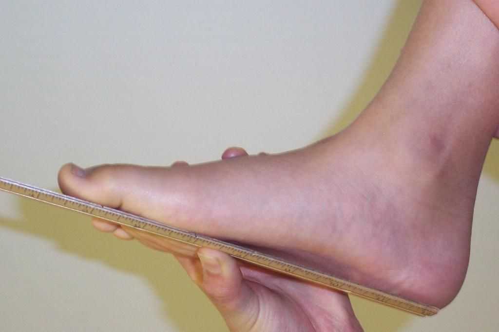
2 ARCH HEIGHT OFF WEIGHTBEARING Evaluate arch height by placing a ruler from the heel to the first metatarsal head Compare arch height of one foot to the other Any digital deformities?
3
4 Low Arch

5 Cavus Foot High Arch

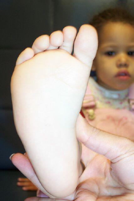
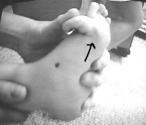
6 Bleck s Test Bisect the plantar heel Where does the ruler bisect the forefoot?
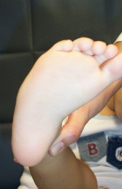
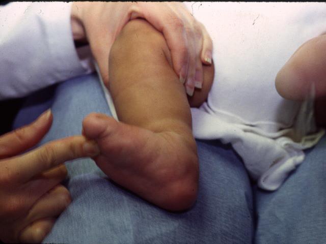
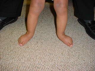
7 Metatarsus Adductus
8

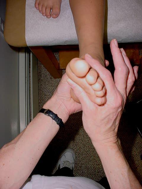
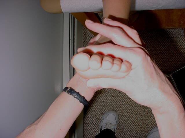
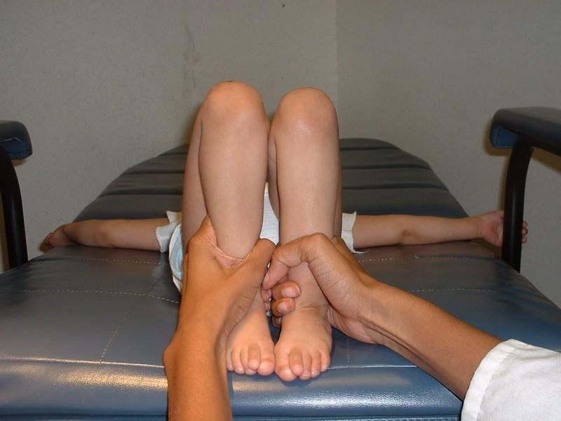
9 HALLUX RANGE OF MOTION OFF WEIGHT BEARING Hallux range of motion off weight bearing Grasp the proximal phalanx and dorsiflex at 1 st MPJ
10 First Ray Range of Motion Grasp forefoot so that thumb is under the metatarsal heads With your other hand grasp 1 st metatarsal head
11 First ray range of motion-dorsiflexion

12 First Ray Motion- Plantarflexion

13 Plantarflexed First Metatarsal

14 PLANTARFLEXED 1 ST METATARSAL

15 Midtarsal Joint Range of Motion- oblique axis Supinate STJ Grasp foot just distal to the MTJ and put it through a range of motion (dorsiflex-abduct, plantarflex adduct)
16 MTJ Longitudinal Axis
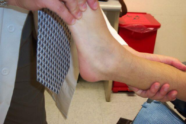
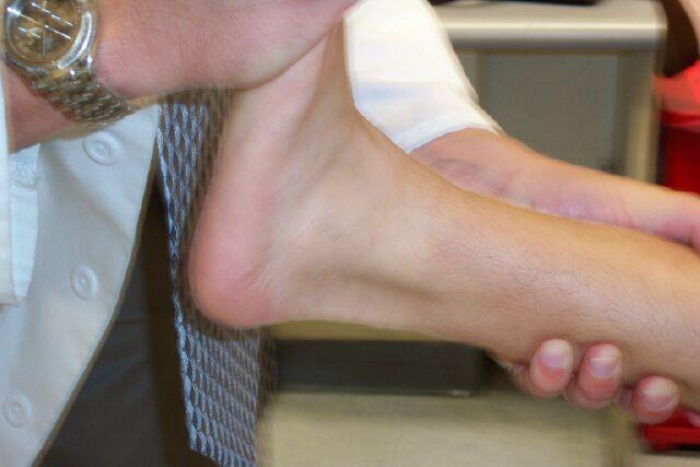
17 Ankle Joint Range of Motion Bisect the lower lateral 1/3 of the leg and the lateral plantar aspect of the rearfoot Dorsiflex the ankle, keeping the STJ neutral and MTJ supinated

18 Ankle Range of Motion-Knee Extended
19 Ankle Dorsiflexion neutral versus pronated

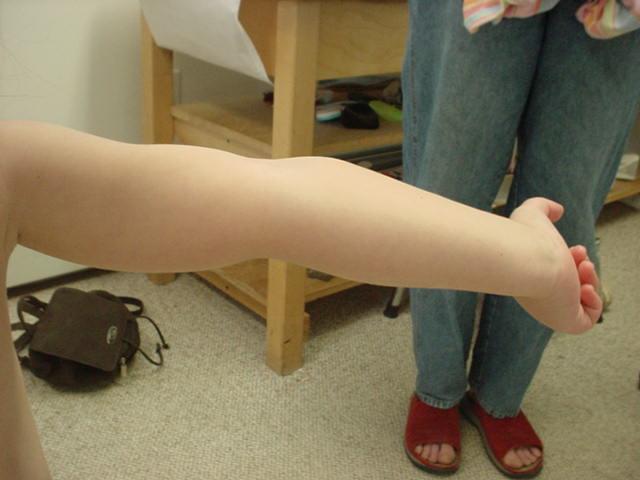
20 Increased ankle joint dorsiflexion

21 Leg Length Evaluation Patient is supine Place hands on ASIS to make sure that pelvis is level

22 Leg Length Evaluation Compare the level of one malleolus to the other

23 Leg Length Evaluation Flex both hips and knees so that both heels are parallel with each other Compare knee heights
24


25 CHECK FOR SCOLIOSIS ADAM S FORWARD BEND TEST

26 Lumbosacral Area Patient had an underlying spinal tumor and unilateral cavus foot deformity Courtesy of Dr. Jordan

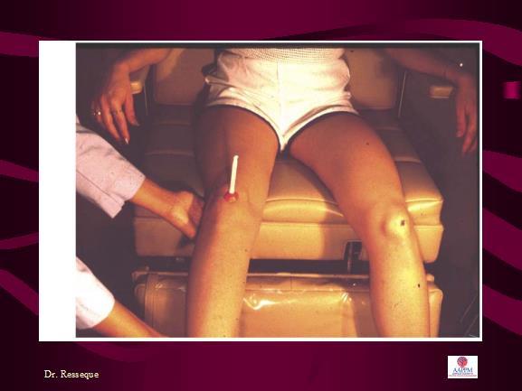
27 Netter s Method of Assessing Femoral Anteversion Ruwe et al JBJS Patient is prone with knee flexed 90 degrees Palpate greater trochanter with one hand while opposite hand internally rotates hip At the point of maximum greater trochanter prominence, neck is parallel to the table 2017

28 Clinical Measurement of Femoral Anteversion Angle formed between the tibia and true vertical represents the femoral anteversion 2017


29 HIP ROTATION

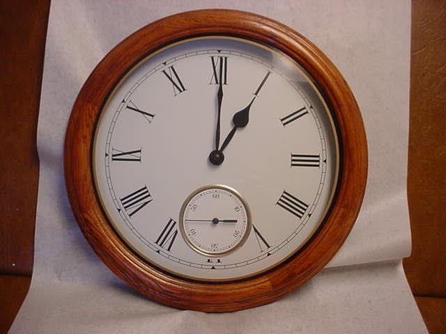
30 EACH HOUR ON THE CLOCK REPRESENTS 30 DEGREES OF FEMORAL ROTATION I 2 9 3

31 Hip Rotation - Extended Grasp leg behind knee and medially and laterally rotate from the hip

32

33 INCREASED MEDIAL FEMORAL ROTATION
34 Femoral Rotation Infants -60 degrees lateral; 0-30 degrees medial 6 months of age- 50 degrees lateral; 30 degrees medial 1-4 years of age degrees lateral; degrees medial

35 HIP ABDUCTION (hip extended)

36 HIP ABDUCTION (hip flexed)

37 Tibial Torsion Place knee on the frontal plane Compare the medial malleolus to the lateral malleolus
38 Tibial Torsion Infant- 0-5 degrees 18 months- 9 degrees external 3 years -12 degrees external 6 years degrees external

39 Tibial Torsion


40 MALLEOLAR POSITION


41 What side is more severe? LEFT LEG

42 MEASUREMENT OF TRANSMALLEOLAR AXIS

43 Measuring malleolar position using tractograph

44 High Malleolar Position

45 High External Tibial Torsion
46 Hamstring Flexibility Hip and knee are extended Leg is raised to resistance 70 degrees of hip flexion should be obtained
47 POPLITEAL ANGLE With hip flexed 90 degrees, extend knee until firm resistance is met, acute angle between lower leg & imaginary line extending up from flexed femur Birth- 2 yrs. 0-6 degrees 5 and older 0-25 degrees full ROM
48 POPLITEAL ANGLE If popliteal angle close to 90 degrees (very tight), suspect pathology of nerve roots (spondylolesthesis, tumor, diskitis, syrinx)

49 Hip Flexors Patient is supine Flex both hips. While maintaining one hip flexed, extend the other hip. The extended leg should touch the supporting surface.
50 Subtalar Joint Neutral Position Bisect lower 1/3 of leg Bisect posterior calcaneus Place STJ in neutral position
51 Subtalar Joint Neutral Position Observe relationship between heel and lower leg

52 Subtalar Varus

53 Subtalar Joint Range of Motion Patient is prone. Rotate heel into maximum supination

54 Subtalar Joint Range of Motion Rotate heel into maximum pronation There should be a 2:1 relationship between supination and pronation. A young child may have a 3:1 or 4:1 ratio

55 Forefoot to Rearfoot Relationship Place STJ in neutral position Place thumb on 4 th and 5 th metatarsal heads and dorsiflex to resistance

56 Forefoot to Rearfoot Relationship Compare plane of metatarsals to heel bisection The plane of the metatarsals should be perpendicular to the heel bisection

57 Forefoot Varus Loading 4 th & 5 th metatarsal heads Note low arch contour

58 Forefoot Varus- left foot

59 Forefoot valgus- left foot
60 Stance Measurements
61 Genu Varum or Valgum Place patient s feet so that ankles are as close together as possible
62 Genu Varum If ankles touch and knees can t, a genu varum

63 Genu Valgum If knees touch and ankles can t, a genu valgum
64 Normal Ranges Birth 1 ½ genu varum 1 ½ - 3 straight 3 6 genu valgum 6 12 straight
65 Ligamentous Laxity
66 Ligamentous Laxity
67 Ligamentous Laxity
68 Ligamentous Laxity
69 Ligamentous Laxity
70 Ligamentous Laxity
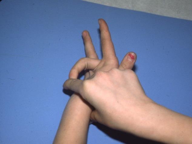
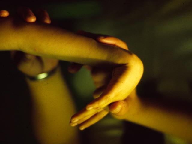
71 Beighton Scale 4 or > out of 9 2 pts. dorsiflexion of 5 th MCP joint > or equal to 90 degrees 2 pts. Opposition of thumb to volar surface of forearm 2 pts. Hyperextension of elbows > or equal to 10 degrees 2 pts. Hyperextension of knee > or equal to 10 degrees 1 pt. Place hands flat on floor without bending knees

72 Lumbar Lordosis

73 Arch Height On Weight Bearing Tell patient to take a few steps in place and then stop. This is their angle and base of stance Compare arch height of one foot to the other

74 Arch Height

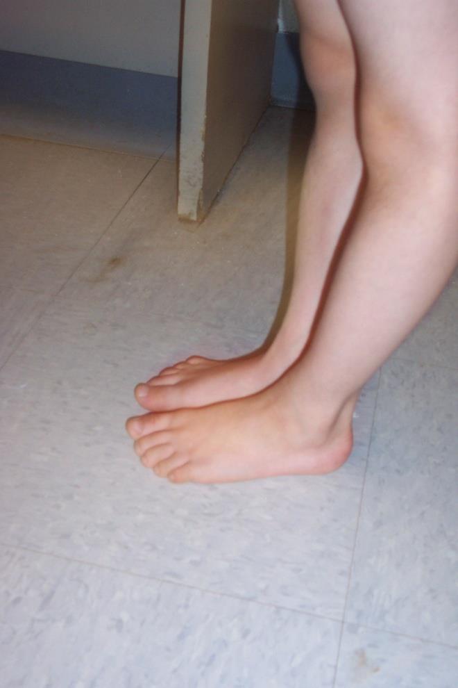
75 Wide Base of Stance 3 year old hypotonic child

76 Hallux Range of Motion On Weight Bearing Place patient in angle and base of gait Dorsiflex hallux degrees dorsiflexion
77 Relaxed Calcaneal Stance Position Have the patient take a few steps in place and then stop Compare the heel bisection to an imaginary perpendicular to the ground

78 Relaxed Calcaneal Stance Position - Valgus

79 Relaxed Calcaneal Stance Position - Varus

80 MIDTARSAL JOINT SUBLUXATION
81 Neutral Calcaneal Stance Position Patient is placed in angle and base of stance Place patient in neutral Compare heel bisection to imaginary perpendicular line


82 RELAXED CALCANEAL STANCE POSITION

83 Tibial Varum or Valgum Place subtalar joint in neutral Compare lower leg bisection to an imaginary perpendicular to the ground

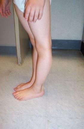
84 Tibial Varum 1 year old
85 Malleolar Heights Have the patient take a few steps in place and then stop Compare the level of one malleolus to the other
86 Malleolar Heights The left foot is more pronated than the right because the left malleolus is lower
87 CONCLUSION
BIOMECHANICAL EXAMINATION OF THE PEDIATRIC LOWER EXTREMITY
 BIOMECHANICAL EXAMINATION OF THE PEDIATRIC LOWER EXTREMITY B.Resseque, D.P.M. ARCH HEIGHT OFF WEIGHTBEARING Evaluate arch height by placing a ruler from the heel to the first metatarsal head Compare arch
BORGinsole Measurement devices
 BORGinsole Measurement devices BORGinsole Angle-Finder Dorsal Flexion of the first Metatarsophalangeal joint - P. is sitting up on the examination table, with legs straight. - T. is sitting at the end
THE FOOT S CONNECTED TOO... Evaluation Procedures for Orthotic Therapy Prescription 2005
 THE FOOT S CONNECTED TOO... Evaluation Procedures for Orthotic Therapy Prescription 2005 Unpublished Copyright Biomechanical Services, Inc. 2003 Biomechanical Services, Inc. 1050 Central Ave., Suite D
Balanced Body Movement Principles
 Balanced Body Movement Principles How the Body Works and How to Train it. Module 3: Lower Body Strength and Power Developing Strength, Endurance and Power The lower body is our primary source of strength,
Evaluation of Gait Mechanics Using Computerized Plantar Surface Pressure Analysis and it s Relation to Common Musculoskeletal Problems
 Evaluation of Gait Mechanics Using Computerized Plantar Surface Pressure Analysis and it s Relation to Common Musculoskeletal Problems Laws of Physics effecting gait Ground Reaction Forces Friction Stored
Copyright 2004, Yoshiyuki Shiratori. All right reserved.
 Ankle and Leg Evaluation 1. History Chief Complaint: A. What happened? B. Is it a sharp or dull pain? C. How long have you had the pain? D. Can you pinpoint the pain? E. Do you have any numbness or tingling?
Evidence-Based Examination of the Foot Presented by Alexis Wright, PT, PhD, DPT, FAAOMPT Practice Sessions/Skill Check-offs
 Evidence-Based Examination of the Foot Presented by Alexis Wright, PT, PhD, DPT, FAAOMPT Practice Sessions/Skill Check-offs Module Five: Movement Assessment of the Foot/Ankle (1 hour CEU Time) Skilled
Dorsal surface-the upper area or top of the foot. Terminology
 It is important to learn the terminology as it relates to feet to properly communicate with referring physicians when necessary and to identify the relationship between the anatomical structure of the
Therapeutic Foot Care Certificate Program Part I: Online Home Study Program
 Therapeutic Foot Care Certificate Program Part I: Online Home Study Program 1 Anatomy And Terminology Of The Lower Extremity Joan E. Edelstein, MA, PT, FISPO Associate Professor of Clinical Physical Therapy
Scar Engorged veins. Size of the foot [In clubfoot, small foot]
![Scar Engorged veins. Size of the foot [In clubfoot, small foot]](/thumbs/78/77722241.jpg "Scar Engorged veins. Size of the foot [In clubfoot, small foot]") 6. FOOT HISTORY Pain: Walking, Running Foot wear problem Swelling; tingly feeling Deformity Stiffness Disability: At work; recreation; night; walk; ADL, Sports Previous Rx Comorbidities Smoke, Sugar, Steroid
6. FOOT HISTORY Pain: Walking, Running Foot wear problem Swelling; tingly feeling Deformity Stiffness Disability: At work; recreation; night; walk; ADL, Sports Previous Rx Comorbidities Smoke, Sugar, Steroid
ANKLE PLANTAR FLEXION
 ANKLE PLANTAR FLEXION Evaluation and Measurements By Isabelle Devreux 1 Ankle Plantar Flexion: Gastrocnemius and Soleus ROM: 0 to 40-45 A. Soleus: Origin: Posterior of head of fibula and proximal1/3 of
CHAPTER 8: THE BIOMECHANICS OF THE HUMAN LOWER EXTREMITY
 CHAPTER 8: THE BIOMECHANICS OF THE HUMAN LOWER EXTREMITY _ 1. The hip joint is the articulation between the and the. A. femur, acetabulum B. femur, spine C. femur, tibia _ 2. Which of the following is
Musculoskeletal Examination
 Musculoskeletal Examination Statement of Goals Know how to perform a complete musculoskeletal examination. Learning Objectives A. Describe the anatomy of the musculoskeletal system including the bony structures,
ChiroCredit.com Presents Biomechanics: Focus on
 ChiroCredit.com Presents Biomechanics: Focus on the Knee Presented by: Ivo Waerlop, DC Shawn Allen, DC 1 Focus on The Knee 2 Pertinent Anatomy Femur Tibia Fibula Patella Prepatellar bursa Infrapatellar
Functional Movement Screen (Cook, 2001)
") Functional Movement Screen (Cook, 2001) TEST 1 DEEP SQUAT Purpose - The Deep Squat is used to assess bilateral, symmetrical, mobility of the hips, knees, and ankles. The dowel held overhead assesses bilateral,
Functional Movement Screen (Cook, 2001) TEST 1 DEEP SQUAT Purpose - The Deep Squat is used to assess bilateral, symmetrical, mobility of the hips, knees, and ankles. The dowel held overhead assesses bilateral,
Physical Examination of the Foot & Ankle
 Inspection Standing, feet straight forward facing toward examiner Swelling Deformity Flatfoot (pes planus and hindfoot valgus) High arch (pes cavus and hindfoot varus) Peek-a-boo heel Varus Too many toes
Redirect GRF to Affect Mobility, Stability or Load? Increase/Decrease Joint Moments to Reduce Stress Strain Relationships?
 5-1 SECTION 5 CRITICAL DECISION MAKING IN ORTHOTIC THERAPY QUESTIONS Answering the some critical (as in choosing between criteria) questions should help as a guide to selecting an appropriate orthosis,
RN(EC) ENC(C) GNC(C) MN ACNP *** MECHANISM OF INJURY.. MOST IMPORTANT ***
 ENC(C) GNC(C) MN ACNP *** MECHANISM OF INJURY.. MOST IMPORTANT ***") HISTORY *** MECHANISM OF INJURY.. MOST IMPORTANT *** Age of patient - Certain conditions are more prevalent in particular age groups (Hip pain in children may refer to the knee from Legg-Calve-Perthes
HISTORY *** MECHANISM OF INJURY.. MOST IMPORTANT *** Age of patient - Certain conditions are more prevalent in particular age groups (Hip pain in children may refer to the knee from Legg-Calve-Perthes
SMALL GROUP SESSION 16 January 8 th or 10 th Shoulder pain case/ Touch workshop/ Upper and Lower Extremity Examination
 SMALL GROUP SESSION 16 January 8 th or 10 th Shoulder pain case/ Touch workshop/ Upper and Lower Extremity Examination Suggested Readings: Opatrny L. The Healing Touch. Ann Int Med 2002; 137:1003. http://www.annals.org/cgi/reprint/137/12/1003.pdf
OTM Lecture Gait and Somatic Dysfunction of the Lower Extremity
 OTM Lecture Gait and Somatic Dysfunction of the Lower Extremity Somatic Dysfunction Tenderness Asymmetry Range of Motion Tissue Texture Changes Any one of which must be present to diagnosis somatic dysfunction.
Podo-Pediatrics in Private Practice. Elisabeth Hibbert B.Sc. D.Ch. November 11, 2016
 Podo-Pediatrics in Private Practice Elisabeth Hibbert B.Sc. D.Ch. November 11, 2016 My background Private Practice since 1998 Began promoting children s foot care in 2007 In 2016-35% of new patients are
Functional Hallux Limitus Orthotic Therapy for Hallux Valgus and Hallux Rigidus
 Pathology Specific Orthoses Evidence Based Orthotic Therapy: Functional Hallux Limitus Orthotic Therapy for Hallux Valgus and Hallux Rigidus Lawrence Z. Huppin, DPM California School of Podiatric Medicine
بسم هللا الرحمن الرحيم
 بسم هللا الرحمن الرحيم Laboratory RHS 221 Manual Muscle Testing Theory 1 hour practical 2 hours Dr. Ali Aldali, MS, PT Department of Physical Therapy King Saud University Talocrural and Subtalar Joint
Functional Movement Test. Deep Squat
 Functional Movement Test Put simply, the FMS is a ranking and grading system that documents movement patterns that are key to normal function. By screening these patterns, the FMS readily identifies functional
Let's Talk about the Terms
 Page 1 of 15 Let's Talk about the Terms Hello, readers. I guess if you are stopping in at this site, you share either my interest in or concern for the issue of nomenclature in our professional publications
Functional biomechanics of the lower limb
 Functional biomechanics of the lower limb Ben and Matt. 24th July 2011 Principles of function Gravity Ground reaction Eco-concentric eccentric loading (preload) of a muscle (or group) is essential for
ORTHOSCAN MOBILE DI POSITIONING GUIDE
 ORTHOSCAN MOBILE DI POSITIONING GUIDE Table of Contents SHOULDER A/P of Shoulder... 4 Tangential (Y-View) of Shoulder... 5 Lateral of Proximal Humerus... 6 ELBOW A/P of Elbow... 7 Extended Elbow... 8 Lateral
What Happens to the Paediatric Flat Foot? Peter J Briggs Freeman Hospital Newcastle upon Tyne
 What Happens to the Paediatric Flat Foot? Peter J Briggs Freeman Hospital Newcastle upon Tyne We don t know!! Population Studies 2300 children aged 4-13 years Shoe wearers Flat foot 8.6% Non-shoe wearers
Could this Research Change the Way You Treat Hallux Limitus?
 Could this Research Change the Way You Treat Hallux Limitus? Lawrence Z. Huppin, D.P.M. Assistant Clinical Professor, Western University of Health Sciences, College of Podiatric Medicine Disclosure: Medical
Clarification of Terms
 Clarification of Terms The plantar aspect of the foot refers to the role or its bottom The dorsal aspect refers to the top or its superior portion The ankle and foot perform three main functions: 1. shock
Module Three: Interventions of the Foot/Ankle
 Evidence-Based Treatment of the Foot Presented by Alexis Wright, PT, PhD, DPT, FAAOMPT Practice Sessions/Skill Check-offs Module Three: Interventions of the Foot/Ankle (75 minutes) Skilled Process a rearfoot
Intoeing: When to Worry? Sukhdeep K. Dulai SPORC 2018
 Intoeing: When to Worry? Sukhdeep K. Dulai SPORC 2018 What is it? Intoeing: When to worry? Why isn t it always cause for worry? What are the benign causes of intoeing? What are the pathologic causes of
TERTIARY DANCE COUNCIL: PHYSIOTHERAPY EXAMINATION
 TERTIARY DANCE COUNCIL: PHYSIOTHERAPY EXAMINATION SEX: Female Male Transgender/Intersex/Other NAME: ADDRESS: PHONE: ( ) DOB (AGE): GENERAL MEDICAL HISTORY Height: cms Weight: kgs Do you have any current
RN(EC) ENC(C) GNC(C) MN ACNP *** MECHANISM OF INJURY.. MOST IMPORTANT *** - Useful in determining mechanism of injury / overuse
 ENC(C) GNC(C) MN ACNP *** MECHANISM OF INJURY.. MOST IMPORTANT *** - Useful in determining mechanism of injury / overuse") HISTORY *** MECHANISM OF INJURY.. MOST IMPORTANT *** Age of patient Sport / Occupation - Certain conditions are more prevalent in particular age groups (Osgood Schlaters in youth / Degenerative Joint Disease
HISTORY *** MECHANISM OF INJURY.. MOST IMPORTANT *** Age of patient Sport / Occupation - Certain conditions are more prevalent in particular age groups (Osgood Schlaters in youth / Degenerative Joint Disease
The Language of Anatomy. (Anatomical Terminology)
") The Language of Anatomy (Anatomical Terminology) Terms of Position The anatomical position is a fixed position of the body (cadaver) taken as if the body is standing (erect) looking forward with the upper
The Language of Anatomy (Anatomical Terminology) Terms of Position The anatomical position is a fixed position of the body (cadaver) taken as if the body is standing (erect) looking forward with the upper
Terms of Movements by Prof. Dr. Muhammad Imran Qureshi
 Terms of Movements by Prof. Dr. Muhammad Imran Qureshi Three systems of the body work in coordination to perform various movements of the body. These are: A System of Bones (Osteology), A System of Muscles
Solving Today s Pain and Injury Puzzle with Erik Dalton An Online Workshop for ABMP Members Session 4 Handout
 Solving Today s Pain and Injury Puzzle with Erik Dalton An Online Workshop for ABMP Members Session 4 Handout Please Note: Erik Dalton teaches his Myoskeletal Alignment Techniques with the expectation
What This Is! What This Isn t! Insights Into Functional Training 5/27/15. #ideaworld. Chuck Wolf, MS, FAFS Thank you for coming!!!
 Insights Into Functional Training Insights Into Functional Training 2015 IDEA Health & Fitness Association. All Rights Reserved. www.ideafit.com/world P R E S E N T E D B Y Chuck Wolf, MS, FAFS Human Motion
Musculoskeletal Examination Benchmarks
 Musculoskeletal Examination Benchmarks _ The approach to examining the musculoskeletal system is the same no matter what joint or limb is being examined. The affected and contralateral region should both
Feet First. Michael K. Cooper, DO FACOFP Family Practice/OMM St John Clinic - Claremore OOA 2018 Annual Convention
 Feet First Michael K. Cooper, DO FACOFP Family Practice/OMM St John Clinic - Claremore OOA 2018 Annual Convention Disclaimer I have no conflict of interest. I am not on any pharmaceutical company payroll
Radiology Positioning Practical Test #2 Table (By Jung Park):
:") Radiology Positioning Practical Test #2 Table (By Jung Park): (Lower Extremity): patient is fully gowned / no artifacts / properly shielded (exposure for femur and below : hold still, don t move ) (exposure
Radiology Positioning Practical Test #2 Table (By Jung Park): (Lower Extremity): patient is fully gowned / no artifacts / properly shielded (exposure for femur and below : hold still, don t move ) (exposure
Introduction. The primary function of the ankle and foot is to absorb shock and impart thrust to the body during walking.
 The ankle 1 Introduction The primary function of the ankle and foot is to absorb shock and impart thrust to the body during walking. OSTEOLOGRY The term ankle refers primarily to the talocrural joint,
The Leg. Prof. Oluwadiya KS
 The Leg Prof. Oluwadiya KS www.oluwadiya.sitesled.com Compartments of the leg 4 Four Compartments: 1. Anterior compartment Deep fibular nerve Dorsiflexes the foot and toes 2. Lateral Compartment Superficial
Movement Terminology. The language of movement is designed to allow us to describe how the body moves through space.
 Movement Terminology The language of movement is designed to allow us to describe how the body moves through space. In exercise it allows us to communicate with other movement professionals so we can describe
My Technique for Adjusting the Excessively Pronated Foot
 My Technique for Adjusting the Excessively Pronated Foot by Mark N. Charrette, DC One can think of Chiropractic in terms of science, art, and philosophy. The art or application of Chiropractic technique
Radiographic Positioning Summary (Basic Projections RAD 222)
") Lower Extremity Radiographic Positioning Summary (Basic Projections RAD 222) AP Pelvis AP Hip (Unilateral) (L or R) AP Femur Mid and distal AP Knee Lateral Knee Pt lies supine on table Align MSP to Center
Lower Extremity Radiographic Positioning Summary (Basic Projections RAD 222) AP Pelvis AP Hip (Unilateral) (L or R) AP Femur Mid and distal AP Knee Lateral Knee Pt lies supine on table Align MSP to Center
Subtalar Joint Neutral Positions and Drop Test
 A R C H S T U D Y.. Reliability of Open and Closed Kinetic Chain Subtalar Joint Neutral Positions and Drop Test Ann Marie Picciano, MS, PT, ATC' Megan S. Rowlands, MS, PT2 Teddy Worrell, EdD, PT, SCS,
Flexibility. STRETCH: Kneeling gastrocnemius. STRETCH: Standing gastrocnemius. STRETCH: Standing soleus. Adopt a press up position
 STRETCH: Kneeling gastrocnemius Adopt a press up position Rest one knee on mat with the opposite leg straight Maintain a neutral spine position Push through arms to lever ankle into increased dorsiflexion
Body Organizations Flashcards
 1. What are the two main regions of the body? 2. What three structures are in the Axial Region? 1. Axial Region (Goes down midline of the body) 2. Appendicular Region (limbs) 3. Axial Region (Goes down
Managing Tibialis Posterior Tendon Injuries
 Managing Tibialis Posterior Tendon Injuries by Thomas C. Michaud, DC Published April 1, 2015 by Dynamic Chiropractic Magazine Tibialis posterior is the deepest, strongest, and most central muscle of the
 17.2 A-P Lower Leg Measure: A-P at mid-lower leg Protection: Apron draped over pelvis SID: 40 Table top No Tube Angle Film: 7 x17 I.D. down or diagonal 14 x 17 www.fisiokinesiterapia.biz A-P Lower Leg
Instructional Course Lecture 2011
 Instructional Course Lecture 2011 Yoon Hae Kwak Dept. of Orthopaedic Surgery Hallym University Sacred Heart Hospital Hallym University Medical Center Rotational and Angular variations of the lower extremities
copyrighted material by PRO-ED, Inc.
 Contents Preparation for Functional Sitting Partial Pull to Sit.......................................................... 2 Pull to Sit................................................................ 3
Other Congenital and Developmental Diseases of the Foot. Department of Orthopedic Surgery St. Vincent s s Hospital, The Catholic University
 Other Congenital and Developmental Diseases of the Foot Department of Orthopedic Surgery St. Vincent s s Hospital, The Catholic University Contents Metatarsus Adductus Skewfoot Hallux Valgus Hallux Valgus
Trainers. Anne-Marie O Connor Musculoskeletal Podiatrist
 Trainers Anne-Marie O Connor Musculoskeletal Podiatrist Agenda Background Tarso-navicular stress fractures Case Study Interventions and research Further Research Anatomy Anatomically, wedged between the
Nicky Schmidt PT, C/NDT 1
 Preparing the foot for third rocker and initial contact Nicky Schmidt PT, C/NDT copyright 2012 References Laboratory Strategies developed and taught by Nicky Schmidt, P.T. in the NDTA Approved Advanced
The Valgus Foot in Cerebral Palsy Equinovalgus not Plano-Valgus. Alfred D. Grant, M.D. David Feldman, M.D.
 The Valgus Foot in Cerebral Palsy Equinovalgus not Plano-Valgus Alfred D. Grant, M.D. David Feldman, M.D. Norman Otsuka, MD M.D. THE PURPOSE OF THIS PRESENTATION IS TO STATE CLEARLY THAT THE VALGUS FOOT
9/26/2012. Osteokinematics (how the bones move) & Arthrokinematics (how the joints move) Planes & Axes. Planes & Axes continued
 & Arthrokinematics (how the joints move) Planes & Axes. Planes & Axes continued") Osteokinematics (how the bones move) & (how the joints move) Planes & Axes Planes of Action = Three fixed lines of reference along which the body is divided. Each plane is at right angles (or perpendicular)
Osteokinematics (how the bones move) & (how the joints move) Planes & Axes Planes of Action = Three fixed lines of reference along which the body is divided. Each plane is at right angles (or perpendicular)
Biomechanical Explanations for Selective Sport Injuries of the Lower Extremity
 Biomechanical Explanations for Selective Sport Injuries of the Lower Extremity American Osteopathic Academy of Sports Medicine Presentation April 23, 2015 Understanding Normalcy What is Normal? Rearfoot/heel
Goniometry. Wrist Flexion: Pt seated with forearm resting on table (use olecranon process & midline of ulna as reference for stationary arm)
") Goniometry Wrist Flexion: Pt seated with forearm resting on table (use olecranon process & midline of ulna as reference for stationary arm) Wrist Extension: Pt seated with forearm resting on table (Goniometer
Goniometry Wrist Flexion: Pt seated with forearm resting on table (use olecranon process & midline of ulna as reference for stationary arm) Wrist Extension: Pt seated with forearm resting on table (Goniometer
the muscle that opposes the action of a joint about an axis
 Adams forward bend test Aetiology Agonist Ambulation Anisomelia Antagonist Antagonistic pelvic torsion the patient bends forward to emphasise any asymmetry in the rib cage or loin on the back for the clinical
Introduction -
 Introduction - http://www.irdpq.qc.ca/communication/publications/pdf/preliminaries.pdf Part 1 Gait : http://www.irdpq.qc.ca/communication/publications/pdf/part_1_gait.pdf Part 2 Muscle Strength and Physical
Richie Brace Treatment Guide: Tips for Evaluation, Casting, Prescription, Modifications and Troubleshooting
 Richie Brace Treatment Guide: Tips for Evaluation, Casting, Prescription, Modifications and Troubleshooting TABLE OF CONTENTS PAGES General Considerations 1-2 Conditions Adult Acquired Flatfoot (PTTD)
Types of Body Movements
 Types of Body Movements Bởi: OpenStaxCollege Synovial joints allow the body a tremendous range of movements. Each movement at a synovial joint results from the contraction or relaxation of the muscles
Maximal isokinetic and isometric muscle strength of major muscle groups related to age, body weight, height, and sex in 178 healthy subjects
 Maximal isokinetic and isometric muscle strength of major muscle groups related to age, body weight, height, and sex in 178 healthy subjects Test protocol Muscle test procedures. Prior to each test participants
IFAST Assessment. Name: Date: Sport: Review Health Risk Assessment on initial consult form. List Client Goals (what brings you here?
 IFAST Assessment Name: Date: Sport: Review Health Risk Assessment on initial consult form List Client Goals (what brings you here?) Cardiovascular Measurements Blood Pressure Resting Heart Rate Body Composition
Medical Terminology. Anatomical Position, Directional Terms and Movements
 Medical Terminology Anatomical Position, Directional Terms and Movements What we will cover... Content Objectives Students will be able to gain a better understanding and application of medical terminology
Chapter 6 part 2. Skeletal Muscles of the Body
 Chapter 6 part 2 Skeletal Muscles of the Body Basic Principles 600 + muscles in the human body (you are required to learn 45, lucky kids)! Skeletal Muscles pull on bones Origin of a muscle = point of attachment
31b Passive Stretches:! Technique Demo and Practice - Lower Body
 31b Passive Stretches:! Technique Demo and Practice - Lower Body 31b Passive Stretches:! Technique Demo and Practice - Lower Body! Class Outline" 5 minutes" "Attendance, Breath of Arrival, and Reminders
PHYSICAL TRAINING INSTRUCTORS MANUAL TABLE OF CONTENT PART 5. Exercise No 31: Reverse Crunch 1. Exercise No 32: Single Hip Flexion 3
 TABLE OF CONTENT PART 5 Exercise No 31: Reverse Crunch 1 Exercise No 32: Single Hip Flexion 3 Exercise No 33: Bicycle Crunch 4 Exercise No 34: Straight Leg U Crunches 5 Exercise No 35: Bent Knee U Crunch
ACE Personal Trainer Manual, 4 th edition
 ACE Personal Trainer Manual, 4 th edition Chapter 7: Functional Assessments: Posture, Movement, Core, Balance, and Flexibility 1 Learning Objectives This session, which is based on Chapter 7 of the ACE
Static Flexibility/Stretching
 Static Flexibility/Stretching Points of Emphasis Always stretch before and after workouts. Stretching post-exercise will prevent soreness and accelerate recovery. Always perform a general warm-up prior
CHAPTER 3 What Is Anatomy?
 CHAPTER 3 What Is Anatomy? Kinesiology Books Publisher 1 TABLE OF CONTENTS The Language of Anatomy Anatomical Position Directional Terms Body Planes Movements Musculoskeletal System Human Skeleton Types
BIOMECHANICS OF ANKLE FRACTURES
 BIOMECHANICS OF ANKLE FRACTURES William R Reinus, MD MBA FACR Significance of Ankle Fractures Most common weight-bearing Fx 70% of all Fxs Incidence is increasing Bimodal distribution Men 15-24 Women over
BLUE SKY SCHOOL OF PROFESSIONAL MASSAGE AND THERAPEUTIC BODYWORK Musculoskeletal Anatomy & Kinesiology KNEE & ANKLE MUSCLES
 BLUE SKY SCHOOL OF PROFESSIONAL MASSAGE AND THERAPEUTIC BODYWORK Musculoskeletal Anatomy & Kinesiology KNEE & ANKLE MUSCLES MSAK201-I Session 3 1) REVIEW a) THIGH, LEG, ANKLE & FOOT i) Tibia Medial Malleolus
Biology 325 Fall 2003
 Name: pre-lab exercise due at beginning of your lab session Matching a. fibrous joints b. cartilaginous joints c. synovial joints 1. exhibit a joint cavity 2. types are sutures and syndesmoses 3. bones
Radiographic Assessment of Pediatric Foot Alignment: Self-Assessment Module
 1.5 CME AJR Integrative Imaging LIFELONG LEARNING FOR RADIOLOGY Radiographic Assessment of Pediatric Foot Alignment: Self-Assessment Module Mahesh M. Thapa 1,2, Sumit Pruthi 1,2, Felix S. Chew 2 ABSTRACT
Occupational Therapy Assessment Guidelines
 Occupational Therapy Assessment Guidelines DATA BASE Patient's Main Concerns and Goals Patient's problems. - pq!p"*q., Vancouver " i,'" ~;a ;i 5 r,:!. H e a Lt h Prontof~trg u~cllness Rnsurrng car? Mary
Leonid Solomin, Elena Schepkina, Pavel Kulesh, Viktor Vilensky, Konstantin Korchagin, Peter Skomoroshko Reference Lines and Angles
 Leonid Solomin, Elena Schepkina, Pavel Kulesh, Viktor Vilensky, Konstantin Korchagin, Peter Skomoroshko Reference Lines and Angles 2 For each of bones reference lines are offered. The angles at which these
Arthritic history is similar to that of the hip. Add history of give way and locking, swelling
 KNEE VASU PAI Arthritic history is similar to that of the hip. Add history of give way and locking, swelling INJURY MECHANISM When How Sequence Progress Disability IKDC Activity I - Strenuous activity
General Procedure and Rules
 General Procedure and Rules PROCEDURE Description: This assessment is a measure of upper extremity (UE) and lower extremity (LE) motor and sensory impairment. Equipment: A chair, bedside table, reflex
Contraindicated and High-Risk Exercises
 Contraindicated and High-Risk Exercises Young sub Kwon, Ph.D. ACSM RCEP, NSCA CSCS,*D Exercise Physiology Laboratory The University of New Mexico Albuquerque, NM, USA Introduction Any activity selected
Section Three: The Leg, Ankle, and Foot Lecture: Review of Clinical Anatomy, Patterns of Dysfunction and Injury, and
 Section Three: The Leg, Ankle, and Foot Lecture: Review of Clinical Anatomy, Patterns of Dysfunction and Injury, and Treatment Implications for the Leg, Ankle, and Foot Levels I and II Demonstration and
MUSCLES OF THE LOWER LIMBS
 MUSCLES OF THE LOWER LIMBS Naming, location and general function Dr. Nabil khouri ROLES THAT SHOULD NOT BE FORGOTTEN Most anterior compartment muscles of the hip and thigh Flexor of the femur at the hip
Case Report: Metatarsalgia (by first ray insufficiency)
") Sergio Puigcerver (1) ; Juan Carlos González (1) ; Roser Part (1) ; Eduardo Brau (1) ; Felip Salinas (2) (1) Instituto de Biomecánica de Valencia, UPV. Valencia, España; ibv@ibv.upv.es ; www.ibv.org (2)
Sergio Puigcerver (1) ; Juan Carlos González (1) ; Roser Part (1) ; Eduardo Brau (1) ; Felip Salinas (2) (1) Instituto de Biomecánica de Valencia, UPV. Valencia, España; ibv@ibv.upv.es ; www.ibv.org (2)
Understanding Leg Anatomy and Function THE UPPER LEG
 Understanding Leg Anatomy and Function THE UPPER LEG The long thigh bone is the femur. It connects to the pelvis to form the hip joint and then extends down to meet the tibia (shin bone) at the knee joint.
The Pelvic Equilibrium Theory Part 2
 The Pelvic Equilibrium Theory Part 2 Understanding the abnormal motion patterns associated with The Pelvic Equilibrium Theory and Leg length Inequality. Aims of this section! To discuss the abnormal motion
Country Health SA Medical Imaging
 Country Health SA Medical Imaging REMOTE OPERATORS POSITIONING GUIDE Contents Image Evaluation Page 4 Positioning Guides Section 1 - THORAX 1.1 Chest Page 5 1.2 Bedside Chest Page 7 1.3 Ribs Page 8 Section
Relationship of Foot Type to Callus Location in Healthy Subjects
 Relationship of Foot Type to Callus Location in Healthy Subjects Do-young Jung, M.Sc., P.T. Dept. of Prosthetics and Orthotics, Suncheon First College Moon-hwan Kim, B.H.Sc., P.T. Dept. of Rehabilitation
DR. (PROF.) ANIL ARORA MS
 ANIL ARORA MS") Hip Examination DR. (PROF.) ANIL ARORA MS (Ortho) DNB (Ortho) Dip SIROT (USA) FAPOA (Korea), FIGOF (Germany), FJOA (Japan) Commonwealth Fellow Joint Replacement (Royal National Orthopaedic Hospital, London,
Hip Examination DR. (PROF.) ANIL ARORA MS (Ortho) DNB (Ortho) Dip SIROT (USA) FAPOA (Korea), FIGOF (Germany), FJOA (Japan) Commonwealth Fellow Joint Replacement (Royal National Orthopaedic Hospital, London,
NEWBORN NURSES POLICY AND PROCEDURES. PURPOSE: Varying positions helps to stimulate physiological functioning and provides rest.
 NEWBORN NURSES POLICY AND PROCEDURES SUBJECT: POSITIONING EFFECTIVE DATE: 6/91 PURPOSE: Varying positions helps to stimulate physiological functioning and provides rest. POLICY: 1. The nurse will vary
Dropfoot - Video Gait Analysis - Craig A. Camasta, DPM, FACFAS Atlanta, Georgia, USA
 Equinus, Pes Cavus and Dropfoot - Video Gait Analysis - Craig A. Camasta, DPM, FACFAS Atlanta, Georgia, USA Equinus = Toe Walker Soft Tissue Static fixed contracture Dynamic spastic, hypertonic Bone Procurvatum,,
right Initial examination established that you have 'flat feet'. Additional information left Left foot is more supinated possibly due to LLD
 Motion analysis report for Feet In Focus at 25/01/2013 Personal data: Mathew Vaughan DEMO REPORT, 20 Churchill Way CF10 2DY Cardiff - United Kingdom Birthday: 03/01/1979 Telephone: 02920 644900 Email:
Muscle Testing of Knee Extensors. Yasser Moh. Aneis, PhD, MSc., PT. Lecturer of Physical Therapy Basic Sciences Department
 Muscle Testing of Knee Extensors Yasser Moh. Aneis, PhD, MSc., PT. Lecturer of Physical Therapy Basic Sciences Department Muscle Testing of Knee Extensors othe Primary muscle Quadriceps Femoris -Rectus
Muscles of the Hip 1. Tensor Fasciae Latae O: iliac crest I: lateral femoral condyle Action: abducts the thigh Nerve: gluteal nerve
 Muscles of the Hip 1. Tensor Fasciae Latae O: iliac crest I: lateral femoral condyle Action: abducts the thigh Nerve: gluteal nerve 2. Gluteus Maximus O: ilium I: femur Action: abduct the thigh Nerve:
Body Planes & Positions
 Learning Objectives Objective 1: Identify and utilize anatomical positions, planes, and directional terms. Demonstrate what anatomical position is and how it is used to reference the body. Distinguish
Quads (machines) Cable Lunge
 Cable Lunge") Cable Lunge Cable Lunge 1) Stand with feet hip width apart and a cable attached around your waist. Take left leg and step back approximately 2 feet standing on the ball of the foot. 2) Start position:
Cable Lunge Cable Lunge 1) Stand with feet hip width apart and a cable attached around your waist. Take left leg and step back approximately 2 feet standing on the ball of the foot. 2) Start position:
THE LOWER EXTREMITY EXAM FOR THE FAMILY PRACTITIONER
 THE LOWER EXTREMITY EXAM FOR THE FAMILY PRACTITIONER Melinda A. Scott, D.O. Orthopedic Associates of Dayton Board Certified in Primary Care Sports Medicine GOALS Identify landmarks necessary for exam of
Leg. Dr. Heba Kalbouneh Associate Professor of Anatomy and Histology
 Leg Dr. Heba Kalbouneh Associate Professor of Anatomy and Histology Skin of the Leg Cutaneous Nerves Medially: The saphenous nerve, a branch of the femoral nerve supplies the skin on the medial surface
Applied Functional Science Chain Reaction Skeletal: Real creates Relative. Presented By David Tiberio, Ph.D., PT, FAFS Dean, Gray Institute
 Applied Functional Science Chain Reaction Skeletal: Real creates Relative Presented By David Tiberio, Ph.D., PT, FAFS Dean, Gray Institute 1 How Movement is Created Total body movements are made up of