Sensory nerve conduction studies
|
|
|
- Patrick Skinner
- 5 years ago
- Views:
Transcription
1 Genomläst /meg Sensory nerve conduction studies Department of clinical neurophysiology University hospital Uppsala, Sweden Björn Falck, Erik Stålberg and Lena Eriksson Department of clinical neurophysiology University hospital Uppsala, Sweden
2 METHODS FOR INDIVIDUAL NERVES In the following section a detailed description of the methods for sensory nerves studied in the department. Surface electrodes. The surface electrodes used both for stimulation and consist of two felt pads with a diameter of 7 mm. The electrodes are fixed on a plastic bar. The distance between the electrodes is 23 mm. These electrodes are available form Medtronic (Medtronic 9013L0361) and Nihon Kohden xx. Near nerve electrodes The electrodes used for recording in the department are Medtronic 9013L0601 (25mm), 9013L0611 (35mm), 9013L0621 (45mm) and 9013L0631 (60mm). The lengt of the recording electrodes depends on the amount of subcutaneous tissue. The identical electrodes can be used for stimulation, butn often thinner electrodes are used (Medtronic 9013L0641) N. AURICULARIS POSTERIOR Position of head: Slightly rotated to the contralateral side of the measured nerve. Type of recording electrodes: Plate electrodes. Placement of recording electrodes: Behind the earlobe. Stimulation sites: On the posterior border of m.sternocleidomastoideus in the middle of the muscle. N. RADIALIS Position of limb: Subject supine, elbow extended, and forearm midway between pronation and supination. Placement of recording electrodes: Active electrode over the middle of I metacarpal bone overlying the tendon of the extensor pollicis muscles. Reference electrode placed distally.
3 Stimulation sites: Lateral side of the forearm 140 mm proximal to the recording electrode. Elbow, medial to m.brachioradialis and lateral to the biceps tendon Upper arm in the radial groove Picture of SCS n Radialis N. MEDIANUS Type of measurement: Orthodromic. Position of limb: Subject supine, elbow extended, and forearm supinated. Placement of recording electrodes: Above the wrist around 140mm proximal to the stimulating electrode, usually at the site where n.medianus is stimulated at for motor nerve conduction studies. Reference electrode placed proximally. Stimulation sites: Base of digit I Base of digit II Base of digit III Base of digit IV Palm Picture of orthodromic SCS n Medianus N. MEDIANUS Position of limb: Subject supine, elbow extended, and forearm supinated. Placement of recording electrodes: The active electrode is placed on palmar surface of the digit over the middle of the proximal phalanx of digits I-IV. Reference electrode placed distally.
4 Stimulation sites: Palm, around 70 mm (50-80 mm) proximal to the recording electrode Wrist 140 mm proximal to the recording electrode in digits II, III and IV. The distance is mm for digit I. Elbow, immediately medial to the tendon of the biceps. N. ULNARIS Type of measurement: Orthodromic. Position of limb: Subject supine, elbow extended. Forearm supinated. Placement of recording electrodes: Above the wrist around 140mm proximal to the stimulating electrode, usually at the site where n.ulnaris is stimulated at for motor nerve conduction studies Stimulation sites: Base of digit IV Base of digit V palm Picture of orthodromic SCS n Ulnaris N. ULNARIS Position of limb: Subject supine, elbow in degree flexion if the segment across the elbow is measured, otherwise extended. Forearm supinated. Placement of recording electrodes: The active electrode is placed on palmar surface of the digit over the middle of the proximal phalanx of digits IV or V. Reference electrode placed distally. Stimulation points: Palm, around 70 mm proximal to the recording electrode Wrist 140 mm proximal to the recording electrode.
5 Below elbow, cathode about mm distal to the medial epicondylus Above elbow, cathode about mm above the medial epicondylus. N. ULNARIS RAMUS DORSALIS Position of limb: Subject supine, elbow extended, and forearm pronated. Placement of recording electrodes: Active electrode between metacarpal bones IV and V in the middle of the metacarpal bones. Reference electrode placed distally. Type of stimulating electrode: Surface electrodes on a fixed bar. Stimulation site: Distal part of forearm, mm proximal to the recording electrode. Picture of SCS n Ulnaris ramus dorsalis N. CUTANEUS ANTEBRACHII LATERALIS Position of limb: Subject supine, elbow extended, and forearm supinated. Placement of recording electrodes: Active electrode over the anterior-radial side of the forearm between the distal and middle third of the forearm, 140 mm distal to the tendon of m.biceps brachii. Reference electrode placed distally. Stimulation point: Elbow, lateral to the tendon of the biceps muscle. Picture os SCS n Cutaneus antebrachii lateralis N. CUTANEUS ANTEBRACHII MEDIALIS Position of limb: Subject supine, elbow extended, and forearm supinated. Placement of recording electrodes: Active electrode over the anterior-ulnar side of the forearm at between the distal and middle third of the forearm (140 mm). Reference electrode placed distally.
6 Stimulation site: Over the volar side of the forearm, mm in anterior and lateral to the medial epicondyle. Picture of SCS n Cutaneus antebrachii medialis N. CUTANEUS ANTEBRACHII POSTERIOR Position of limb: Subject supine, elbow extended, and forearm pronated. Placement of recording electrodes: Active electrode over the central part of the dorsal side of the forearm 140 mm distal to the lateral epicondyle. Reference electrode placed distally. Stimulation site: Slightly medial to lateral epicondyle. Picture of SCS n Cutaneus antebrachii posterior N. SURALIS Position of limb: Subject prone. Knee extended, ankle in a neutral position. Placement of recording electrodes: Active electrode behind the lateral malleolus. Reference electrode placed distally. Stimulation sites: On the dorsal aspect of the calf, 140 mm proximal to the recording electrode. Behind the knee. Picture of SCS n Suralis N. PERONEUS PROFUNDUS Type of measurement: Orthodromic.
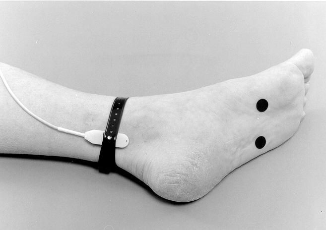
7 Position of limb: Subject supine, knee extended. Ankle in neutral position. Placement of recording electrodes: Active electrode placed over n.peroneus profundus at the site used for distal stimulation of the motor nerve conduction velocity. The exact location may vary slightly from one subject to another. Usually 1-2 cm lateral to the m.tibailis anterior tendon. Stimulation site: Stimulating cathode in the interspace between the I and II metatarsal bones just proximal to the MCP joint. Picture of SCS n Peroneus profundus N. PERONEUS SUPERFICIALIS RAMUS MEDIALIS (n.cutaneus medialis dorsalis) Position of limb: Subject supine, knee extended. Ankle in neutral position. Placement of recording electrodes: Active electrode placed over the middle of the first metatarsal bone. Reference electrode placed distally. Stimulation site: Stimulating cathode 140 mm proximal to the recording electrode on the lateral side of the leg. Picture of SCS n Peroneus superficialis ramus medialis N. PERONEUS SUPERFICIALIS RAMUS LATERALIS (n.cutaneus intermedius dorsalis) Position of limb: Subject supine. Knee extended and ankle in a neutral position. Placement of recording electrodes: The active electrode is placed over the middle of the third metatarsal bone. Often the nerve can be seen when the ankle is slightly inverted. Reference electrode placed distally. Stimulation site: Stimulating cathode 140 mm proximal to the recording electrode on the lateral side of the leg. Picture os SCS n Peroneus ramus lateralis
8 N. SAPHENUS Position of limb: Subject supine, knee extended. Placement of recording electrodes: Over the anterior-medial surface of the distal part of the tibia, 150 mm above the medial malleolus. Reference electrode placed distally. Stimulation sites: Over the medial side of the tibia 140 mm proximal to the recording electrode. (On the medial side of the knee just below the medial epicondyle.) Picture of SCS n Saphenus N. CUTANEUS FEMORIS LATERALIS Position of limb: Subject supine, hip extended. Electrode has to be attached with adhesive tape. Placement of recording electrodes: 140 mm distal to spina iliaca anterior superior over the thigh. Reference electrode placed distally. Stimulation site: Just proximal to the spina iliaca anterior superior. Note: With this method it is not possible to measure the conduction velocity across the entrapment site in "meralgia paresthetica". Orthodromic measurement with near nerve electrodes is recommendable in most patients. N.CUTANEUS FEMORIS LATERALIS Type of measurement: Orthodromic. Position of patient: Patient supine. Type of recording electrodes: Near nerve needle electrodes.
9 Placement of recording electrode: 5-10 mm above spina ilica anterior superior. Depth of electrode placement depends on the amount of subcutaneous fat. In lean persons the depth is around mm and in obese persons it may be mm (when you place the electrode deeper than 15 mm try to direct it slightly lateral to avoid penetration of the peritoneum). Often it is possible to place the recording electrode blindly and find a good recording site. Sometimes it is helpful to stimulate with the recording electrode to ascertain that the electrode is close to the nerve. If the patient has a paresthetic sensation in radiating into the lateral side of the thigh with a stimulus intensity of 1-2 ma (0,2 ms duration) the electrode is very close to the nerve. Often it is difficult to obtain this and acceptable responses may be recorded with 3-5 ma stimulus thresholds for the sensation. Placement of reference electrode: Subcutaneously mm proximal to the recording electrode. A surface reference electrode may be used instead of a needle. Stimulation site: Over the anterior-lateral portion of the thigh around mm distal to the recording electrode. Often it is necessary to search for the site. Picture od SCS n Cutaneus femoris lateralis N. CUTANEUS FEMORIS POSTERIOR Position of limb: Subject prone. Placement of recording electrodes: Active electrode over the middle of the posterior side of the thigh. Reference electrode placed distally. Stimulation site: Just below the buttock 140 mm proximal to the recording electrode. Picture of SCS n Cutaneus femoris posterior N. PLANTARIS MEDIALIS Type of measurement: Orthodromic. Position of limb: Subject supine, knee extended, and ankle in a neutral position. Placement of recording electrodes: Active electrode above the medial malleolus. Reference electrode placed proximally.
10 Stimulation point: On the medial side of the sole of the foot, the cathode mm from the recording electrode. Picture of SCS n Plantaris medialis/ lateralis N PLANTARIS MEDIALIS Type of measurement: Orthodromic. Position of patient: Patient supine or sitting. Type of recording electrodes: Near nerve needle electrodes. Placement of recording electrode: mm above the medial malleolus. To place the electrode close to the tibial nerve the recording electrode is used to stimulate and the motor response is picked up from m.abductor hallucis with surface electrodes. When the threshold for a motor response is around 1mA (stimulus duration 0.2 ms) the position of the recording electrode is acceptable. Thresholds of more than 3 ma rarely give satisfactory recording. Placement of reference electrode: Subcutaneously mm proximal to the recording electrode. A surface reference electrode may be used instead of a needle. Type of stimulating electrodes: Surface electrodes. Stimulation site: On the medial side of the sole (over the I or II metatarsal bone) of the foot mm distal to the recording electrode N. PLANTARIS LATERALIS Type of measurement: Orthodromic. Position of limb: Subject supine, elbow extended, and forearm midway between pronation and supination. Placement of recording electrodes: Active electrode above the medial malleolus. Reference electrode placed proximally. Stimulation site: On the lateral side of the sole, the cathode mm from the recording electrode. Picture of SCS n Plantaris lateralis/ medialis
11 N PLANTARIS LATERALIS Type of measurement: Orthodromic Position of patient: Patient supine or sitting Type of recording electrodes: Near nerve needle electrodes. Placement of recording electrode: 20 mm above the medial malleolus. To place the electrode close to the tibial nerve the recording electrode is used to stimulate and the motor response is picked up from m.abductor digiti minimi with surface electrodes. When the threshold for a motor response is around 1mA (stimulus duration 0.2 ms) the position of the recording electrode is acceptable. Thresholds of more than 3 ma rarely give satisfactory recording. Placement of reference electrode: Subcutaneously mm proximal to the recording electrode. A surface reference electrode may be used instead of a needle. Stimulation site: On the lateral side (over the IV metatarsal bone) of the sole of the foot mm distal to the recording electrode. Picture of SCS Plantaris lateralis N.ILIOINGUINALIS This is not a standard method that is commonly used. Type of measurement: Orthodromic. Position of patient: Patient supine. Type of recording electrodes: Near nerve needle electrodes. Placement of recording electrode: 5-10 mm above spina ilica anterior superior. Depth of electrode placement depends on the amount of subcutaneous fat. In lean persons the depth is around mm and in obese persons it may be mm (when you place the electrode deeper than 15 mm try to direct it slightly lateral to avoid penetration of the peritoneum). Placement of reference electrode: Subcutaneously mm proximal to the recording electrode. A surface reference electrode may be used instead of a needle. Stimulation site: Over the inguinal ligament mm from the medial to the recording electrode. N DIGITALIS I-V PLANTARIS MEDIALIS Type of measurement: Orthodromic. Position of patient: Patient supine or sitting.
12 Type of recording electrodes: Near nerve needle electrodes. Placement of recording electrode: mm above the medial malleolus. To place the electrode close to the tibial nerve the recording electrode is used to stimulate and the motor response is picked up from m.abductor hallucis with surface electrodes. When the threshold for a motor response is around 1mA (stimulus duration 0.2 ms) the position of the recording electrode is excellent. Thresholds of more than 3 ma rarely give satisfactory recordings. Placement of reference electrode: Subcutaneously mm proximal to the recording electrode. A surface reference electrode may be used instead of a needle. Type of stimulating electrodes: Near nerve needle electrodes. Stimulation site: The cathode is placed on the medial side close to the base of the toe. Each toe, I-V, is studied separately. The cathode is inserted trough the skin from the dorsal side around midway of the toe. The needle is advanced until the tip of the electrode can be felt just under the skin on the plantar side of the toe. The anode is placed distal to the cathode on the same side of the toe. Stimulus intensity: Optimal stimulus intensity is 3-4 ma (duration 0.2 ms). With high stimulus intensities, > 6-10 ma there is the possibility that the plantar digital nerve on the lateral side of the toe is stimulated. Averaging: Averaging is required in most subjects. In a young person, less than 30 years of age the responses can usually be seen in the unaveraged trace. Mostly stimuli need to be averaged for satisfactory recordings. Sometimes 1000 stimuli are required in older subjects. Picture of SCS n digitalis I-V Plantaris medialis N DIGITALIS I-IV PLANTARIS LATERALIS Type of measurement: Orthodromic. Position of patient: Patient supine or sitting. Type of recording electrodes: Near nerve needle electrodes. Placement of recording electrode: mm above the medial malleolus. To place the electrode close to the tibial nerve the recording electrode is used to stimulate and the motor response is picked up from m.abductor hallucis with surface electrodes. When the threshold for a motor response is around 1mA (stimulus duration 0.2 ms) the position of the recording electrode is excellent. Thresholds of more than 3 ma rarely give satisfactory recording. Placement of reference electrode: Subcutaneously mm proximal to the recording electrode. A surface reference electrode may be used instead of a needle. Type of stimulating electrodes: Near nerve needle electrodes. Stimulation site: The cathode is placed on the lateral side close to the base of the toe. Each toe I-V is studied separately. The cathode is inserted trough the skin from the dorsal side around midway of the toe.
.")
13 The needle is advanced until the tip of the electrode can be felt just under the skin on the plantar side of the toe. The anode is placed distal to the cathode on the same side of the toe. Stimulus intensity: Optimal stimulus intensity is 3-4 ma (duration 0.2 ms). With high stimulus intensities, > 6-10 ma there is the possibility that the plantar digital nerve on the medial side of the toe is stimulated. Note: Averaging is required in most subjects. In a young person, less than 30 years of age the responses can usually be seen in the unaveraged trace. Mostly stimuli need to be averaged for satisfactory recordings. Sometimes 1000 stimuli are required in older subjects. SCS n Radialis

14 SCS n Medianus (orthodromic)
")
15 SCS n Ulnaris (orthodromic)

16 SCS Ulnaris ramus dorsalis


17 SCS n cutaneus Antebrachii lateralis SCS n cutaneus Antebrachii medialis


18 SCS n cutaneus Antebrachii posterior SCS n Suralis


19 SCS n Peroneus profundus SCS n Peroneus superficialis ramus medialis


20 SCS n Peroneus superficialis ramus lateralis SCS n Saphenus

21 SCS n cutaneus Femoris lateralis

22 SCS n cutaneus Femoris posterior
23 SCS n Plantaris medialis/lateralis

24 SCS n Plantaris lateralis (nerar-nerve)

25 SCS n digitalis I-V plantaris medialis
METHODS FOR SENSORY NERVE CONDUCTION STUDIES WITH NEAR NERVE NEEDLE ELECTRODES
 Klin. neurofys.lab1 METHODS FOR SENSORY NERVE CONDUCTION STUDIES WITH NEAR NERVE NEEDLE ELECTRODES N.cutaneus femoris lateralis 1 N.ilioinguinalis 2 N digitalis I-V plantaris medialis 3 N digitalis I-IV
Hands on Nerve Conduction Studies
 Hands on Nerve Conduction Studies N. CUTANEUS ANTEBRACHII LATERALIS Type of measurement: Antidromic. Position of limb: Subject supine, elbow extended, and forearm supinated. Type of recording electrodes:
In which arm muscle are intramuscular injections most often given? (not in text)
") AP1 Lab 9 - Muscles of the Arms and Legs Locate the following muscles on the models and on yourself. Recall anatomical position. Directional terms such as anterior, posterior, lateral, etc. all assume
AP1 Lab 9 - Muscles of the Arms and Legs Locate the following muscles on the models and on yourself. Recall anatomical position. Directional terms such as anterior, posterior, lateral, etc. all assume
Year 2004 Paper one: Questions supplied by Megan
 QUESTION 47 A 58yo man is noted to have a right foot drop three days following a right total hip replacement. On examination there is weakness of right ankle dorsiflexion and toe extension (grade 4/5).
ARM Brachium Musculature
 ARM Brachium Musculature Coracobrachialis coracoid process of the scapula medial shaft of the humerus at about its middle 1. flexes the humerus 2. assists to adduct the humerus Blood: muscular branches
Lab Activity 11: Group II
 Lab Activity 11: Group II Muscles Martini Chapter 11 Portland Community College BI 231 Origin and Insertion Origin: The place where the fixed end attaches to a bone, cartilage, or connective tissue. Insertion:
Lever system. Rigid bar. Fulcrum. Force (effort) Resistance (load)
 Resistance (load)") Lever system lever is any elongated, rigid (bar) object that move or rotates around a fixed point called the fulcrum when force is applied to overcome resistance. Force (effort) Resistance (load) R Rigid
Lever system lever is any elongated, rigid (bar) object that move or rotates around a fixed point called the fulcrum when force is applied to overcome resistance. Force (effort) Resistance (load) R Rigid
Main Menu. Wrist and Hand Joints click here. The Power is in Your Hands
 1 The Wrist and Hand Joints click here Main Menu K.5 http://www.handsonlineeducation.com/classes/k5/k5entry.htm[3/23/18, 1:40:40 PM] Bones 29 bones, including radius and ulna 8 carpal bones in 2 rows of
Key Relationships in the Upper Limb
 Key Relationships in the Upper Limb This list contains some of the key relationships that will help you identify structures in the lab. They are organized by dissection assignment as defined in the syllabus.
Radiographic Positioning Summary (Basic Projections RAD 222)
") Lower Extremity Radiographic Positioning Summary (Basic Projections RAD 222) AP Pelvis AP Hip (Unilateral) (L or R) AP Femur Mid and distal AP Knee Lateral Knee Pt lies supine on table Align MSP to Center
Lower Extremity Radiographic Positioning Summary (Basic Projections RAD 222) AP Pelvis AP Hip (Unilateral) (L or R) AP Femur Mid and distal AP Knee Lateral Knee Pt lies supine on table Align MSP to Center
Human Anatomy Biology 351
 Human Anatomy Biology 351 Lower Limb Please place your name on the back of the last page of this exam. You must answer all questions on this exam. Because statistics demonstrate that, on average, between
Muscle Anatomy Review Chart
 Muscle Anatomy Review Chart BACK Superficial (5) Trapezius Transverse cervical a. Latissimus dorsi Thoracodorsal a. Rhomboideus major Dorsal scapular a. Rhomboideus minor Levator scapulae Intermediate
Muscles of the Hip 1. Tensor Fasciae Latae O: iliac crest I: lateral femoral condyle Action: abducts the thigh Nerve: gluteal nerve
 Muscles of the Hip 1. Tensor Fasciae Latae O: iliac crest I: lateral femoral condyle Action: abducts the thigh Nerve: gluteal nerve 2. Gluteus Maximus O: ilium I: femur Action: abduct the thigh Nerve:
MLT Muscle(s) Patient Position Therapist position Stabilization Limb Position Picture Put biceps on slack by bending elbow.
 Patient Position Therapist position Stabilization Limb Position Picture Put biceps on slack by bending elbow.") MLT Muscle(s) Patient Position Therapist position Stabilization Limb Position Picture Put biceps on slack by bending elbow. Pectoralis Minor Supine, arm at side, elbows extended, supinated Head of Table
MLT Muscle(s) Patient Position Therapist position Stabilization Limb Position Picture Put biceps on slack by bending elbow. Pectoralis Minor Supine, arm at side, elbows extended, supinated Head of Table
Anatomy & Physiology. Muscles of the Lower Limbs.
 Anatomy & Physiology Muscles of the Lower Limbs http://www.ishapeup.com/musclecharts.html Muscles of the Lower Limbs Among the strongest muscles in the body. Because pelvic girdle is composed of heavy,
Lower limb summary. Anterior compartment of the thigh. Done By: Laith Qashou. Doctor_2016
 Lower limb summary Done By: Laith Qashou Doctor_2016 Anterior compartment of the thigh Sartorius Anterior superior iliac spine Upper medial surface of shaft of tibia 1. Flexes, abducts, laterally rotates
Human Anatomy Lab #7: Muscles of the Cadaver
 Human Anatomy Lab #7: Muscles of the Cadaver Table of Contents: Expected Learning Outcomes.... 1 Introduction...... 1 Identifying Muscles on Yourself.... 2 Muscles of the Anterior Trunk and Arm.. 2 Muscles
5.1 Identify, describe the attachments of and deduce the actions of the muscles of the thigh:
 5.1 Identify, describe the attachments of and deduce the actions of the muscles of the thigh: Anterior group Proximal attachment Distal attachment Sartorius ASIS» Upper part of shaft tibia (middle surface)»
BLUE SKY SCHOOL OF PROFESSIONAL MASSAGE AND THERAPEUTIC BODYWORK Musculoskeletal Anatomy & Kinesiology KNEE & ANKLE MUSCLES
 BLUE SKY SCHOOL OF PROFESSIONAL MASSAGE AND THERAPEUTIC BODYWORK Musculoskeletal Anatomy & Kinesiology KNEE & ANKLE MUSCLES MSAK201-I Session 3 1) REVIEW a) THIGH, LEG, ANKLE & FOOT i) Tibia Medial Malleolus
The Muscular System. Chapter 10 Part C. PowerPoint Lecture Slides prepared by Karen Dunbar Kareiva Ivy Tech Community College
 Chapter 10 Part C The Muscular System Annie Leibovitz/Contact Press Images PowerPoint Lecture Slides prepared by Karen Dunbar Kareiva Ivy Tech Community College Table 10.9: Muscles Crossing the Shoulder
11/15/2018. Temporalis Elevates & retracts mandible. Masseter = Prime mover of jaw closure. Levator scapulae Supraspinatus Clavicle.
 Due in Lab 10 Lab 8 MUSCLES 2 weeks because of Thanksgiving Prelab #10 Both sides! Homework #8 Both sides! Refer to Muscles 22-23 Examples of Origin & Insertion Naming of muscles Origin Site of muscle
Human Anatomy Biology 351
 Human Anatomy Biology 351 Lower Limb Please place your name on the back of the last page of this exam. You must answer all questions on this exam. Because statistics demonstrate that, on average, between
Anatomy MCQs Week 13
 Anatomy MCQs Week 13 1. Posterior to the medial malleolus of the ankle: The neurovascular bundle lies between Tibialis Posterior and Flexor Digitorum Longus The tendon of Tibialis Posterior inserts into
Muscular Nomenclature and Kinesiology - One
 Chapter 16 Muscular Nomenclature and Kinesiology - One Lessons 1-3 (with lesson 4) 1 Introduction 122 major muscles covered in this chapter Chapter divided into nine lessons Kinesiology study of human
200 meter or yard event. However, it is also the lead off event in the individual medley,
 Butterfly is one of four competitive strokes in swimming. Usually it is raced, in a 100 or 200 meter or yard event. However, it is also the lead off event in the individual medley, meaning it is part of
Muscles of the Upper Limb
 Muscles of the Upper Limb anterior surface of ribs 3 5 coracoid process Pectoralis minor pectoral nerves protracts / depresses scapula Serratus anterior Subclavius ribs 1-8 long thoracic nerve rib 1 ----------------
lesser trochanter of femur lesser trochanter of femur iliotibial tract (connective tissue) medial surface of proximal tibia
 medial surface of proximal tibia") LOWER LIMB MUSCLES OF THE APPENDICULAR SKELETON The muscles that act on the lower limb fall into three groups: those that move the thigh, those that move the lower leg, and those that move the ankle, foot,
LOWER LIMB MUSCLES OF THE APPENDICULAR SKELETON The muscles that act on the lower limb fall into three groups: those that move the thigh, those that move the lower leg, and those that move the ankle, foot,
The Human Muscular System Required reading before beginning this lab: Saladin, KS: Human Anatomy 5th ed (2017) Chapters 10, 11, 12 INTRODUCTION
 Chapters 10, 11, 12 INTRODUCTION") Biology 322: Human Anatomy The Human Muscular System Required reading before beginning this lab: Saladin, KS: Human Anatomy 5 th ed (2017) Chapters 10, 11, 12 INTRODUCTION We will use a number of lab periods
Biology 322: Human Anatomy The Human Muscular System Required reading before beginning this lab: Saladin, KS: Human Anatomy 5 th ed (2017) Chapters 10, 11, 12 INTRODUCTION We will use a number of lab periods
Nerves of the upper limb Prof. Abdulameer Al-Nuaimi. E. mail:
 Nerves of the upper limb Prof. Abdulameer Al-Nuaimi E-mail: a.al-nuaimi@sheffield.ac.uk E. mail: abdulameerh@yahoo.com Brachial plexus Median nerve After originating from the brachial plexus in the axilla,
Muscles of the hand Prof. Abdulameer Al-Nuaimi
 Muscles of the hand Prof. Abdulameer Al-Nuaimi a.alnuaimi@sheffield.ac.uk abdulameerh@yahoo.com Thenar Muscles Thenar muscles are three short muscles located at base of the thumb. All are innervated by
The Lower Limb II. Anatomy RHS 241 Lecture 3 Dr. Einas Al-Eisa
 The Lower Limb II Anatomy RHS 241 Lecture 3 Dr. Einas Al-Eisa Tibia The larger & medial bone of the leg Functions: Attachment of muscles Transfer of weight from femur to skeleton of the foot Articulations
Functional anatomy and variability of the blood vessels of the upper and lower limbs. Anastasia Bendelic Human Anatomy Departament
 Functional anatomy and variability of the blood vessels of the upper and lower limbs Anastasia Bendelic Human Anatomy Departament Plan: 1. Variations of the branching pattern of the aortic arch 2. Arterial
Clinical examination of the wrist, thumb and hand
 Clinical examination of the wrist, thumb and hand 20 CHAPTER CONTENTS Referred pain 319 History 319 Inspection 320 Functional examination 320 The distal radioulnar joint.............. 320 The wrist.......................
Lower Limb Nerves. Clinical Anatomy
 Lower Limb Nerves Clinical Anatomy Lumbar Plexus Ventral rami L1 L4 Supplies: Abdominal wall External genitalia Anteromedial thigh Major nerves.. Lumbar Plexus Nerves relation to psoas m. : Obturator n.
Neuro Exam Workshop. AAO Convocation, 2018 Drew Lewis, DO, FAAO, FAOCPMR Associate Professor, OMM Department Des Moines University
 Neuro Exam Workshop AAO Convocation, 2018 Drew Lewis, DO, FAAO, FAOCPMR Associate Professor, OMM Department Des Moines University Table of Contents I. Neuro Exam Screen... 2 A. Inspection... 2 B. Reflexes...
musculoskeletal system anatomy muscles of foot sheet done by: dina sawadha & mohammad abukabeer
 musculoskeletal system anatomy muscles of foot sheet done by: dina sawadha & mohammad abukabeer Extensor retinaculum : A- superior extensor retinaculum (SER) : originates from the distal ends of the tibia
Practical 1 Worksheet
 Practical 1 Worksheet ANATOMICAL TERMS 1. Use the word bank to fill in the missing words. reference side stand body arms palms anatomical forward All anatomical terms have a(n) point which is called the
~, /' ~::'~ EXTENSOR HALLUCIS LONGUS. Leg-anterolateral :.:~ / ~\,
 TIBIALIS ANTERIOR Lateral condyle of tibia, upper half of lateral surface of tibia, interosseous membrane Medial side and plantar surface of medial cuneiform bone, and base of first metatarsal bone Dorsiflexes
Biceps Brachii. Muscles of the Arm and Hand 4/4/2017 MR. S. KELLY
 Muscles of the Arm and Hand PSK 4U MR. S. KELLY NORTH GRENVILLE DHS Biceps Brachii Origin: scapula Insertion: radius, fascia of forearm (bicipital aponeurosis) Action: supination and elbow flexion Innervation:
Module 7 - The Muscular System Muscles of the Arm and Trunk
 Module 7 - The Muscular System Muscles of the Arm and Trunk This Module will cover the muscle anatomy of the arms and trunk. We have already seen the muscles that move the humerus, so this module will
Human Anatomy Biology 351
 1 Human Anatomy Biology 351 Upper Limb Exam Please place your name on the back of the last page of this exam. You must answer all questions on this exam. Because statistics demonstrate that, on average,
Muscles of the Gluteal Region
 Muscles of the Gluteal Region 1 Some of the most powerful in the body Extend the thigh during forceful extension Stabilize the iliotibial band and thoracolumbar fascia Related to shoulders and arms because
Levels of the anatomical cuts of the upper extremity RADIUS AND ULNA right
 11 CHAPTER 2 Levels of the anatomical cuts of the upper extremity AND right CUT 1 CUT 4 1 2 3 4 5 6 Isolated fixation of the radius is difficult at this level because of the anterolateral vessels and the
Due in Lab weeks because of Thanksgiving Prelab #10. Homework #8. Both sides! Both sides!
 Lab 8 MUSCLES Due in Lab 10 2 weeks because of Thanksgiving Prelab #10 Both sides! Homework #8 Both sides! Refer to Muscles 22-23 Naming of muscles Origin Site of muscle attachment that doesn t move during
forearm posterior compartment
 Quick revision: The anterior compartment of the forearm contains of 8 muscles... -4 superficial -1 intermediate -3 deep *All supplied by median nerve except 1 and 1/2 muscle (by ulnar N.) forearm posterior
Ligaments of Elbow hinge: sagittal plane so need lateral and medial ligaments
 Ligaments of Elbow hinge: sagittal plane so need lateral and medial ligaments Ulnar Collateral ligament on medial side; arising from medial epicondyle and stops excess valgus movement (lateral movement)
HUMAN BODY COURSE LOWER LIMB NERVES AND VESSELS
 HUMAN BODY COURSE LOWER LIMB NERVES AND VESSELS October 22, 2010 D. LOWER LIMB MUSCLES 2. Lower limb compartments ANTERIOR THIGH COMPARTMENT General lfunction: Hip flexion, knee extension, other motions
REFERENCE DIAGRAMS OF UPPER LIMB MUSCLES: NAMES, LOCATIONS, ATTACHMENTS, FUNCTIONS MUSCLES CONNECTING THE UPPER LIMB TO THE AXIAL SKELETON
 REFERENCE DIAGRAMS OF UPPER LIMB MUSCLES: NAMES, LOCATIONS, ATTACHMENTS, FUNCTIONS MUSCLES CONNECTING THE UPPER LIMB TO THE AXIAL SKELETON A25LAB EXERCISES: UPPER LIMB MUSCLES Page 1 MUSCLES CONNECTING
Practical 2 Worksheet
 Practical 2 Worksheet Upper Extremity BONES 1. Which end of the clavicle is on the lateral side (acromial or sternal)? 2. Describe the difference in the appearance of the acromial and sternal ends of the
musculoskeletal system anatomy nerves of the lower limb 2 done by: Dina sawadha & mohammad abukabeer
 musculoskeletal system anatomy nerves of the lower limb 2 done by: Dina sawadha & mohammad abukabeer #Sacral plexus : emerges from the ventral rami of the spinal segments L4 - S4 and provides motor and
Lecture 9: Forearm bones and muscles
 Lecture 9: Forearm bones and muscles Remember, the region between the shoulder and the elbow = brachium/arm, between elbow and wrist = antebrachium/forearm. Forearm bones : Humerus (distal ends) Radius
Where should you palpate the pulse of different arteries in the lower limb?
 Where should you palpate the pulse of different arteries in the lower limb? The femoral artery In the femoral triangle, its pulse is easily felt just inferior to the inguinal ligament midway between the
STRUCTURAL BASIS OF MEDICAL PRACTICE EXAMINATION 5 October 6, 2006
 STRUCTURAL BASIS OF MEDICAL PRACTICE EXAMINATION 5 October 6, 2006 PART l. Answer in the space provided. (8 pts) 1. Identify the structures. (2 pts) B C A. _pisiform B. _ulnar artery A C. _flexor carpi
The Muscular System. Chapter 10 Part D. PowerPoint Lecture Slides prepared by Karen Dunbar Kareiva Ivy Tech Community College
 Chapter 10 Part D The Muscular System Annie Leibovitz/Contact Press Images PowerPoint Lecture Slides prepared by Karen Dunbar Kareiva Ivy Tech Community College Table 10.14: Muscles Crossing the Hip and
MCQWeek2. All arise from the common flexor origin. The posterior aspect of the medial epicondyle is the common flexor origin.
 MCQWeek2. 1. Regarding superficial muscles of anterior compartment of the forearm: All arise from the common flexor origin. The posterior aspect of the medial epicondyle is the common flexor origin. Flexor
Epicranius (frontal belly) Zygomaticus minor. Zygomaticus major Buccinator
 Zygomaticus minor. Zygomaticus major Buccinator") Epicranius (frontal belly) Zygomaticus minor Zygomaticus major Buccinator Masseter Digastric (posterior belly) Stylohyoid Sternocleidomastoid Trapezius Scalenus Omohyoid (inferior belly) Orbicularis oris
Epicranius (frontal belly) Zygomaticus minor Zygomaticus major Buccinator Masseter Digastric (posterior belly) Stylohyoid Sternocleidomastoid Trapezius Scalenus Omohyoid (inferior belly) Orbicularis oris
The Foot. Dr. Wegdan Moh.Mustafa Medicine Faculty Assistant Professor Mob:
 The Foot Dr. Wegdan Moh.Mustafa Medicine Faculty Assistant Professor Mob: 0127155717 The skeleton of the foot Cutaneous innervations Sole of foot layers of muscles First layer -Abductor hallucis -Flexor
Copyright 2004, Yoshiyuki Shiratori. All right reserved.
 Ankle and Leg Evaluation 1. History Chief Complaint: A. What happened? B. Is it a sharp or dull pain? C. How long have you had the pain? D. Can you pinpoint the pain? E. Do you have any numbness or tingling?
Muscles of the lower extremities. Dr. Nabil khouri MD, MSc, Ph.D
 Muscles of the lower extremities Dr. Nabil khouri MD, MSc, Ph.D Posterior leg Popliteal fossa Boundaries Biceps femoris (superior-lateral) Semitendinosis and semimembranosis (superior-medial) Gastrocnemius
Location Terms. Anterior and posterior. Proximal and Distal The term proximal (Latin proximus; nearest) describes where the appendage joins the body.
 describes where the appendage joins the body.") HUMAN ANAT OMY Location Terms Anterior and posterior In human anatomical usage, anterior refers to the front of the individual. Similarly, posterior refers to the back of the subject. In standard anatomical
HUMAN ANAT OMY Location Terms Anterior and posterior In human anatomical usage, anterior refers to the front of the individual. Similarly, posterior refers to the back of the subject. In standard anatomical
ORTHOSCAN MOBILE DI POSITIONING GUIDE
 ORTHOSCAN MOBILE DI POSITIONING GUIDE Table of Contents SHOULDER A/P of Shoulder... 4 Tangential (Y-View) of Shoulder... 5 Lateral of Proximal Humerus... 6 ELBOW A/P of Elbow... 7 Extended Elbow... 8 Lateral
Myologia Part II Objective: Students will examine the muscles of a canine in order to identify the musculature of the body.
 Okay Anatomy Anatomy I: Lesson 11 Myologia Part II Objective: Students will examine the muscles of a canine in order to identify the musculature of the body. Practical Tasks: 6) carpal flexors, pronators
Leg. Dr. Heba Kalbouneh Associate Professor of Anatomy and Histology
 Leg Dr. Heba Kalbouneh Associate Professor of Anatomy and Histology Skin of the Leg Cutaneous Nerves Medially: The saphenous nerve, a branch of the femoral nerve supplies the skin on the medial surface
Forearm and Wrist Regions Neumann Chapter 7
 Forearm and Wrist Regions Neumann Chapter 7 REVIEW AND HIGHLIGHTS OF OSTEOLOGY & ARTHROLOGY Radius dorsal radial tubercle radial styloid process Ulna ulnar styloid process ulnar head Carpals Proximal Row
Netter's Anatomy Flash Cards Section 6 List 4 th Edition
 Netter's Anatomy Flash Cards Section 6 List 4 th Edition https://www.memrise.com/course/1577581/ Section 6 Upper Limb (66 cards) Plate 6-1 Humerus and Scapula: Anterior View 1.1 Acromion 1.2 Greater tubercle
Human Anatomy Biology 255
 Human Anatomy Biology 255 Exam #4 Please place your name and I.D. number on the back of the last page of this exam. You must answer all questions on this exam. Because statistics demonstrate that, on average,
MUSCLES OF THE LOWER LIMBS
 MUSCLES OF THE LOWER LIMBS Naming, location and general function Dr. Nabil khouri ROLES THAT SHOULD NOT BE FORGOTTEN Most anterior compartment muscles of the hip and thigh Flexor of the femur at the hip
Introduction to Anatomy. Dr. Maher Hadidi. Laith Al-Hawajreh. Mar/25 th /2013
 Introduction to Anatomy Dr. Maher Hadidi Laith Al-Hawajreh 22 Mar/25 th /2013 Lower limb - The leg The skeleton of the leg is formed by two bones: 1) Medial: Tibia 2) Lateral: Fibula The two bones are
region of the upper limb between the shoulder and the elbow Superiorly communicates with the axilla.
 1 region of the upper limb between the shoulder and the elbow Superiorly communicates with the axilla. Inferiorly, a number of important structures pass between arm & forearm through cubital fossa. 2 medial
Nerve Injury. 1) Upper Lesions of the Brachial Plexus called Erb- Duchene Palsy or syndrome.
 Upper Lesions of the Brachial Plexus called Erb- Duchene Palsy or syndrome.") Nerve Injury - Every nerve goes to muscle or skin so if the nerve is injured this will cause paralysis in the muscle supplied from that nerve (paralysis means loss of function) then other muscles and other
Nerve Injury - Every nerve goes to muscle or skin so if the nerve is injured this will cause paralysis in the muscle supplied from that nerve (paralysis means loss of function) then other muscles and other
Nerves of Upper limb. Dr. Brijendra Singh Professor & Head Department of Anatomy AIIMS Rishikesh
 Nerves of Upper limb Dr. Brijendra Singh Professor & Head Department of Anatomy AIIMS Rishikesh 1 Objectives Origin, course & relation of median & ulnar nerves. Motor & sensory distribution Carpal tunnel
The Forearm 2. Extensor & lateral Compartments of the Forearm
 The Forearm 2 Extensor & lateral Compartments of the Forearm 1-Lateral Fascial Compartment (at the lateral side of the forearm ) *Some books mention the lateral compartment contain just the Brachioradialis
Peripheral Vascular Examination. Dr. Gary Mumaugh Western Physical Assessment
 Peripheral Vascular Examination Dr. Gary Mumaugh Western Physical Assessment Competencies 1. Inspection of upper extremity for: size symmetry swelling venous pattern color Texture nail beds Competencies
Muscle Testing of Knee Extensors. Yasser Moh. Aneis, PhD, MSc., PT. Lecturer of Physical Therapy Basic Sciences Department
 Muscle Testing of Knee Extensors Yasser Moh. Aneis, PhD, MSc., PT. Lecturer of Physical Therapy Basic Sciences Department Muscle Testing of Knee Extensors othe Primary muscle Quadriceps Femoris -Rectus
General Procedure and Rules
 General Procedure and Rules PROCEDURE Description: This assessment is a measure of upper extremity (UE) and lower extremity (LE) motor and sensory impairment. Equipment: A chair, bedside table, reflex
The Leg. Prof. Oluwadiya KS
 The Leg Prof. Oluwadiya KS www.oluwadiya.sitesled.com Compartments of the leg 4 Four Compartments: 1. Anterior compartment Deep fibular nerve Dorsiflexes the foot and toes 2. Lateral Compartment Superficial
Kinesiology of The Wrist and Hand. Cuneyt Mirzanli Istanbul Gelisim University
 Kinesiology of The Wrist and Hand Cuneyt Mirzanli Istanbul Gelisim University Bones The wrist and hand contain 29 bones including the radius and ulna. There are eight carpal bones in two rows of four to
Goniometry. Wrist Flexion: Pt seated with forearm resting on table (use olecranon process & midline of ulna as reference for stationary arm)
") Goniometry Wrist Flexion: Pt seated with forearm resting on table (use olecranon process & midline of ulna as reference for stationary arm) Wrist Extension: Pt seated with forearm resting on table (Goniometer
Goniometry Wrist Flexion: Pt seated with forearm resting on table (use olecranon process & midline of ulna as reference for stationary arm) Wrist Extension: Pt seated with forearm resting on table (Goniometer
Contents. Preface xv. SECTION 1: Introduction to the Bodynamic System 1. SECTION 2: The Bodynamic Psycho-Motor Anatomy 29
 Contents Preface xv SECTION 1: Introduction to the Bodynamic System 1 Definitions in the Bodynamic System 3 Ego Formation through the Coding Elements 9 Examples of Formation of Coding 17 Using This Book
Lab 9: Learn origin and insertion for each of the listed muscles. For Exercise 15, do Activities 1-6 in 9 th edition, Activities 1-4 in 10 th edition
 The Muscular System Exercises 14, 15, and 16 (begins: page 187 in 9 th and 10 th editions) Exercises 12, 13, and 14 (begins: page 185 in 11 th edition, page 189 in 12 th edition) Lab 8 and 9 Objectives
divided by the bones ( redius and ulna ) and interosseous membrane into :
 and interosseous membrane into :") fossa Cubital Has: * floor. * roof : - Skin - superficial fasica - deep fascia ( include bicipital aponeurosis ) Structures within the roof : -cephalic and basilic veins -and between them median cubital
fossa Cubital Has: * floor. * roof : - Skin - superficial fasica - deep fascia ( include bicipital aponeurosis ) Structures within the roof : -cephalic and basilic veins -and between them median cubital
Muscles of Lesson Five. Muscular Nomenclature and Kinesiology - Two. Muscles of Lesson Five, cont. Chapter 16
 Chapter 16 Muscular Nomenclature and Kinesiology - Two Lessons 5-6 Muscles of Lesson Five Iliopsoas (psoas major, iliacus) Hip outward rotators (piriformis, gemellus superior, gemellus inferior, obturator
Understanding Leg Anatomy and Function THE UPPER LEG
 Understanding Leg Anatomy and Function THE UPPER LEG The long thigh bone is the femur. It connects to the pelvis to form the hip joint and then extends down to meet the tibia (shin bone) at the knee joint.
Muscles to know. Lab 21. Muscles of the Pelvis and Lower Limbs. Muscles that Position the Lower Limbs. Generally. Muscles that Move the Thigh
 Muscles to know Lab 21 Muscles of the Pelvis, Leg and Foot psoas major iliacus gluteus maximus gluteus medius sartorius quadriceps femoris (4) gracilus adductor longus biceps femoris semitendinosis semimembranosus
MUSCLES OF THE ELBOW REGION
 MUSCLES OF THE ELBOW REGION Dr Bronwen Ackermann COMMONWEALTH OF AUSTRALIA Copyright Regulation WARNING This material has been reproduced and communicated to you by or on behalf of the University of Sydney
Muscles of the Thigh. 6.1 Identify, describe the attachments of and deduce the actions of the muscles of the thigh: Anterior group
 Muscles of the Thigh 6.1 Identify, describe the attachments of and deduce the actions of the muscles of the thigh: Anterior group Sartorius: This is a long strap like muscle with flattened tendons at each
Contents of the Posterior Fascial Compartment of the Thigh
 Contents of the Posterior Fascial Compartment of the Thigh 1-Muscles: B i c e p s f e m o r i s S e m i t e n d i n o s u s S e m i m e m b r a n o s u s a small part of the adductor magnus (h a m s t
For convenience values outside the normal range are bolded. Normal values for the specified patient are stated below the tables.
 Case tudy 8 or convenience values outside the normal range are bolded. Normal values for the specified patient are stated below the tables. History: 60 year-ol man with a history of left hand weakness
The Lower Limb VI: The Leg. Anatomy RHS 241 Lecture 6 Dr. Einas Al-Eisa
 The Lower Limb VI: The Leg Anatomy RHS 241 Lecture 6 Dr. Einas Al-Eisa Muscles of the leg Posterior compartment (superficial & deep): primary plantar flexors of the foot flexors of the toes Anterior compartment:
1. A worker falls from a height and lands on his feet. Radiographs reveal a fracture of the sustentaculum tali. The muscle passing immediately
 1. A worker falls from a height and lands on his feet. Radiographs reveal a fracture of the sustentaculum tali. The muscle passing immediately beneath it that would be adversely affected is the: fibularis
ANATYOMY OF The thigh
 ANATYOMY OF The thigh 1- Lateral cutaneous nerve of the thigh Ι) Skin of the thigh Anterior view 2- Femoral branch of the genitofemoral nerve 5- Intermediate cutaneous nerve of the thigh 1, 2 and 3 are
The hand is full with sweat glands, activated at times of stress. In Slide #2 there was a mistake where the doctor mentioned lateral septum twice.
 We should only know: Name, action & nerve supply Layers - Skin - Superficial fascia - Deep fascia The hand is full with sweat glands, activated at times of stress. Deep fascia In Slide #2 there was a mistake
TABLES OF MUSCLE ACTIONS, INNERVATIONS, AND ATTACHMENTS
 TABLES OF MUSCLE ACTIONS, INNERVATIONS, AND ATTACHMENTS Table 1-1 ERECTOR SPINAE MUSCLES Intrinsic muscles producing extension and/or lateral of the spine Muscle Joint and Action Innervation Inferior Attachment
Functional Anatomy of the Elbow
 Functional Anatomy of the Elbow Orthopedic Institute Daryl C. Osbahr, M.D. Chief of Sports Medicine, Orlando Health Chief Medical Officer, Orlando City Soccer Club Orthopedic Consultant, Washington Nationals
Al-Balqa Applied University
 Al-Balqa Applied University Faculty Of Medicine *You can use this checklist as a guide to you for the lab. the items on this checklist represent the main features of the models that you have to know for
Prime movers provide the major force for producing a specific movement Antagonists oppose or reverse a particular movement Synergists
 Dr. Gary Mumaugh Prime movers provide the major force for producing a specific movement Antagonists oppose or reverse a particular movement Synergists Add force to a movement Reduce undesirable or unnecessary
Human Anatomy, First Edition McKinley & O'Loughlin
 Human Anatomy, First Edition McKinley & O'Loughlin Chapter 8 : Appendicular Skeleton 8-1 Appendicular Skeleton Includes the bones of the upper and lower limbs. The girdles of bones that attach the upper
Synergist Muscles. Shoulder (glenohumeral joint) Flexion Deltoid (anterior fibers) Pectoralis major (upper fibers) Biceps Brachii Coracobrachialis
 Flexion Deltoid (anterior fibers) Pectoralis major (upper fibers) Biceps Brachii Coracobrachialis") Synergist Muscles Dr Gene Desepoli DrGeneLMT@gmail.com Shoulder (glenohumeral joint) Deltoid (anterior fibers) Pectoralis major (upper fibers) Biceps Brachii Coracobrachialis Deltoid (posterior fibers)
Synergist Muscles Dr Gene Desepoli DrGeneLMT@gmail.com Shoulder (glenohumeral joint) Deltoid (anterior fibers) Pectoralis major (upper fibers) Biceps Brachii Coracobrachialis Deltoid (posterior fibers)
Lumbar Plexus. Ventral rami L1 L4 Supplies: Major nerves.. Abdominal wall External genitalia Anteromedial thigh
 Lower Limb Nerves Lectures Objectives Describe the structure and relationships of the plexuses of the lower limb. Describe the course, relationships and structures supplied for the major nerves of the
Myofascial sequence of EXTRAmotion. Course. Dr Antonio Stecco M.D. 16/06/2014
 The Fascial Manipulation Association has been formed to serve as a point of reference for research concerning the fascia, myofascial pain, and visceral dysfunctions related to the physiology of the fascia.