PRE-LAB EXERCISES. Before we get started, look up the definitions of these common bone marking terms: Canal: Condyle: Facet: Fissure:
|
|
|
- Jocelin Hamilton
- 5 years ago
- Views:
Transcription
1 1
2 PRE-LAB EXERCISES When studying the skeletal system, the bones are often sorted into two broad categories: the axial skeleton and the appendicular skeleton. This lab focuses on the appendicular skeleton, which is formed from the pectoral and pelvic girdles and the upper and lower limbs. In addition to learning about all the bones of the appendicular skeleton, it is also important to identify some significant bone markings. Bone markings can have many shapes, including holes, round or sharp projections, and shallow or deep valleys, among others. These markings on the bones serve many purposes, including forming attachments to other bones or muscles and allowing passage of a blood vessel or nerve. It is helpful to understand the meanings of some of the more common bone marking terms. Before we get started, look up the definitions of these common bone marking terms: Canal: Condyle: Facet: Fissure: Foramen: (see Module Foramina of Skull) Fossa: Margin: Process: Proximal: Trochanter: Tubercle: Tuberosity: Throughout this exercise, you will notice bold terms. This is meant to focus your attention on these important words. Make sure you pay attention to any bold words and know how to explain their definitions and/or where they are located. Use the following modules to guide your exploration of the appendicular skeleton. As you explore these bones in Visible Body s app, also locate the bones and bone markings on any available charts, models, or specimens. You may also find it helpful to palpate bones on yourself or make drawings of the bones with the bone markings labeled. The drawings don t have to be perfect; just make sure the different bone markings are in the correct locations, relative to each other. If you have trouble finding a bone or bone marking, you can always type its name into the search bar to get a list of 3D anatomical views where that bone or bone marking is highlighted for you. To access disarticulated bones with color-coded bone markings, select a bone, and then, in the content box, choose the landmark icon, which shows a bone with pink, yellow, and blue ends. 2

3 IN-LAB EXERCISES Open the Atlas app. From the Views menu, go to System Views to view the Skeletal System Views at the top of the screen. You are responsible for the identification of all bold terms. A. Pectoral Girdle In the Skeletal System Views, select View 15. Shoulder Girdle to identify the clavicles and scapulae. The clavicles and scapulae make up the pectoral girdle and are responsible for attaching the upper limbs to the skeleton as well as providing attachment points for the shoulder muscles. In the Skeletal System Views, select View 16. Axillary Region to observe how muscles attach to the clavicles and scapulae. You can select the systems icons on the left side of the screen to hide blood vessels, lymphatic vessels, and nerves. The shoulder has the largest range of motion of any joint in the body, and the many muscles that attach here stabilize the pectoral girdle to allow for that movement. After identifying the bones and how they function in muscle stabilization, find their bone markings and answer the questions. Clavicle Scapula Humerus 3
4 Clavicle Supraspinatus muscle Deltoid muscle (anterior head) Scapula Infraspinatus muscle Deltoid muscle (posterior head) Deltoid muscle (middle head) 4
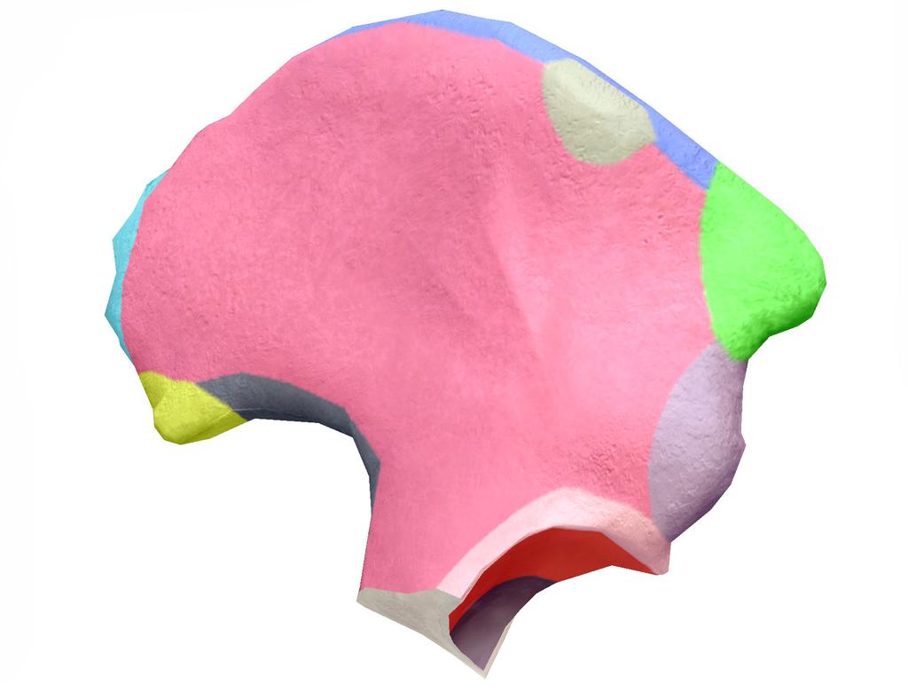
5 1. Scapula Suprascapular notch Coracoid process Superior angle Glenoid cavity Neck Medial border Subscapular fossa Lateral border Inferior angle Superior angle Supraspinous fossa Acromion Scapular spine Neck Infraspinous fossa Medial border Lateral border Inferior angle 5
6 a. Identify the following bone markings: i. Glenoid cavity ii. Spine iii. Acromion iv. Coracoid process v. Infraspinous fossa vi. Supraspinous fossa vii. Subscapular fossa viii. Inferior angle ix. Superior angle x. Lateral border xi. Medial border xii. Neck xiii. Acromial angle b. Describe how to determine a right scapula from a left scapula. 6
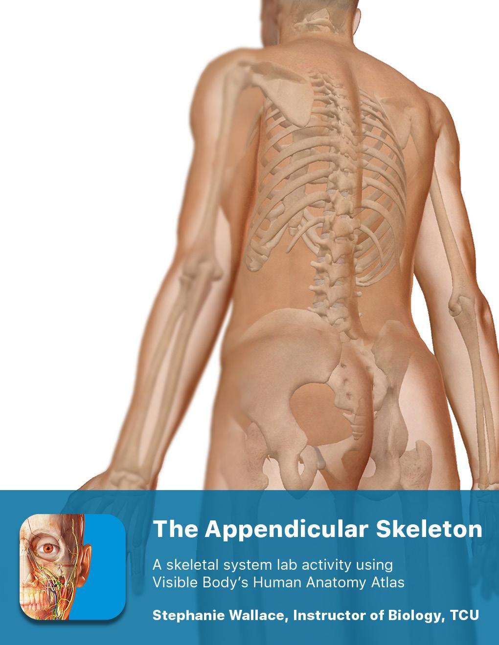
7 2. Clavicle Acromial end Conoid tubercle Shaft Sternal end a. Identify the following bone markings: i. Acromial end ii. Sternal end iii. Shaft iv. Conoid tubercle b. Describe how the clavicle curves and articulates with other bones. 7
8 B. Upper Limb In the Skeletal System Views, select View 1. Full Skeleton to identify the bones of the upper limb. The upper limb consists of the bones of the arm, forearm, wrist, and hand. Zoom in on the arm and identify the following bones and bone markings, and then zoom out again to look at the full arm and observe how the individual bones fit together. Note how processes often fit into the fossae of the same name. Clavicle Humerus Scapula Radius Ulna Carpal bones (wrist) Bones of the hand 8
9 1. Humerus Greater tubercle Lesser tubercle Neck Head Surgical neck Intertubercular groove Deltoid tuberosity Shaft Capitulum Lateral epicondyle Trochlea Coronoid fossa Medial epicondyle a. Identify the following bone markings: i. Head ii. Anatomical neck iii. Surgical neck iv. Shaft v. Greater tubercle vi. Lesser tubercle 9
10 vii. Intertubercular groove viii. Radial groove ix. Deltoid tuberosity x. Trochlea xi. Capitulum xii. Coronoid fossa xiii. Olecranon fossa xiv. Medial epicondyle xv. Lateral epicondyle xvi. Radial fossa b. Describe the difference in position of the anatomical neck and the surgical neck. 10
11 2. Radius Head Tubercle Shaft Styloid process Ulnar notch a. Identify the following bone markings: i. Head ii. Neck iii. Shaft iv. Tubercle v. Styloid process vi. Ulnar notch 11
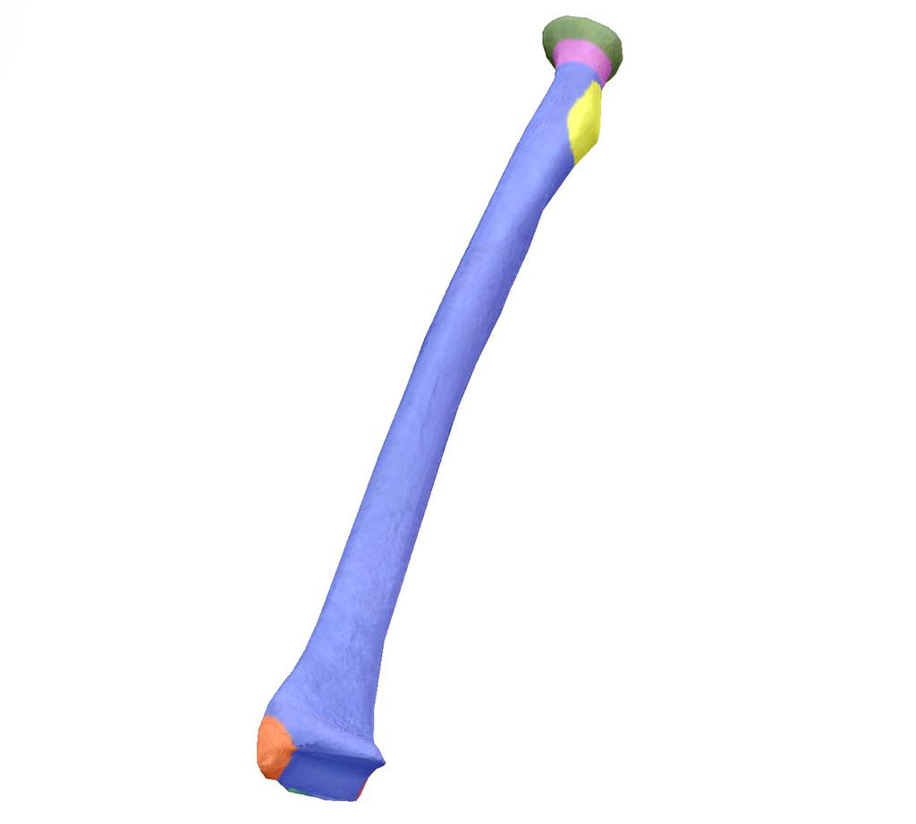
12 3. Ulna Olecranon Trochlear notch Radial notch Coronoid process Tuberosity Shaft Head Styloid process a. Identify the following bone markings: i. Coronoid process ii. Olecranon iii. Radial notch iv. Trochlear notch v. Styloid process vi. Head vii. Shaft 12
13 Humerus Ulna Radius b. How do the bones of the radius and ulna attach to each other? c. Describe how the radius and ulna attach to and rotate around the humerus when the elbow flexes. 13
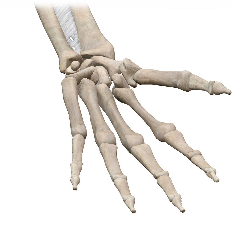
14 4. Carpus To see the carpals more clearly, hide some or all of the ligaments on the wrist after you zoom in. Carpus (Posterior) Scaphoid Trapezium Trapezoid Lunate Triquetral Hamate Capitate 14
15 Carpus (Anterior) Scaphoid Triquetral Pisiform Hamate Lunate Trapezium Capitate Trapezoid a. Identify the following carpal bones: i. Lunate ii. Scaphoid iii. Trapezium iv. Trapezoid v. Capitate vi. Hamate vii. Triquetral viii. Pisiform 15
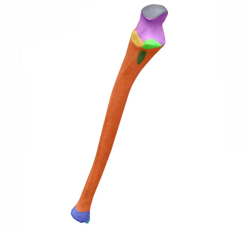
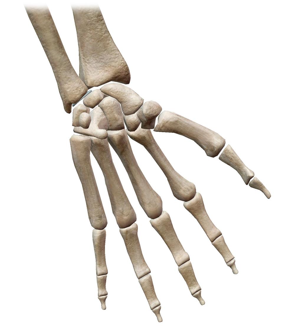
16 5. Hand Note the nomenclature for the bones of the hand. The thumb is considered digit I, while the pinky finger is digit V. The metacarpals and phalanges are numbered accordingly. Metacarpals Proximal phalanges Middle phalanges Distal phalanges a. Identify the following hand bones: i. Metacarpals ii. Proximal phalanges iii. Middle phalanges iv. Distal phalanges b. List all the bones in your hand on your thumb from proximal to distal. Do the same for your pinky finger. What is the difference? 16
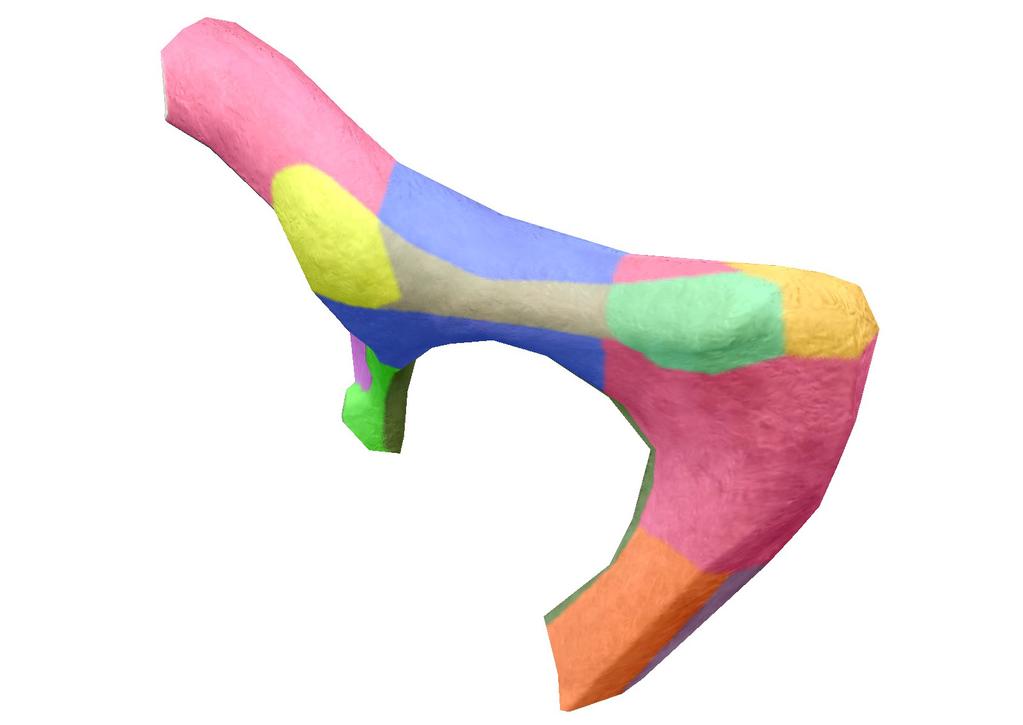
17 C. Pelvic Girdle In the Skeletal System Views, select View 11. Pelvic Girdle to identify the ilium, ischium, and pubis. Just like the pectoral girdle attaches the upper limbs to the skeleton, the pelvic girdle attaches the lower limbs to the skeleton. The pelvic girdle consists of the right and left hip bones and the sacrum that connects the hip bones on the posterior side. You may remember the sacrum from studying the axial skeleton, because it is part of the vertebral column. Each of the hip bones begins as three separate bones: the ilium, ischium, and pubis. These bones eventually fuse together, but bone markings are still considered to belong to one of these three distinct regions. Observe the following bone markings on the ilium, ischium, and pubis, and then try to find the same bone markings on the entire hip bone. Ilium Sacrum Hip bone Pubis Pubic symphysis Ischium 17
18 1. Ilium Iliac crest Posterior superior iliac spine Anterior superior iliac spine Posterior inferior iliac spine Greater sciatic notch Anterior interior iliac spine a. Identify the following bone markings: i. Body ii. Iliac crest iii. Iliac fossa iv. Acetabulum v. Anterior inferior iliac spine vi. Anterior superior iliac spine vii. Posterior inferior iliac spine viii. Posterior superior iliac spine ix. Auricular surface x. Greater sciatic notch 18
19 2. Ischium Acetabular rim Ischial spine Body Ischial tuberosity Obturator foramen (ischial surface) Ramus a. Identify the following bone markings: i. Body ii. Ramus iii. Acetabulum iv. Lesser sciatic notch v. Ischial tuberosity vi. Ischial spine vii. Pubic arch viii. Obturator foramen (ischial surface) 19
20 3. Pubis Body Iliopubic eminence Superior ramus Obturator crest Pubic tubercle Acetabular notch Pubic crest Obturator foramen, pubis surface Pubic arch Inferior ramus a. Identify the following bone markings: i. Body ii. Acetabulum iii. Superior ramus iv. Inferior ramus v. Pubic arch vi. Pubic crest vii. Pubic tubercle viii. Obturator crest ix. Obturator foramen (pubis surface) 4. Where does the lower limb attach to the hip bone? Which part of the hip bone is this? 5. The male and female pelvises have a few differences due to childbearing adaptations. Observe the pelvises by selecting Tours: Pelvis Comparison and answer the following questions. 20
21 Pelvic brim Pubic arch Pelvic brim Pubic arch 21
22 a. Does the male or female pelvis have more space inside? b. Describe what the pelvic brim is. c. How is the pelvic brim different in males vs. females? d. Describe what the pubic arch is. e. How is the pubic arch different in males vs. females? 22
23 D. Lower Limb In the Skeletal System Views, select View 1. Full Skeleton to identify the bones of the lower limb. In addition to their importance in movement, the lower limb bones support the weight of the rest of the body. As a result, they are generally larger and heavier than the bones of the upper limb. Hip bone Head of femur Femur Patella Tibia Fibula 23
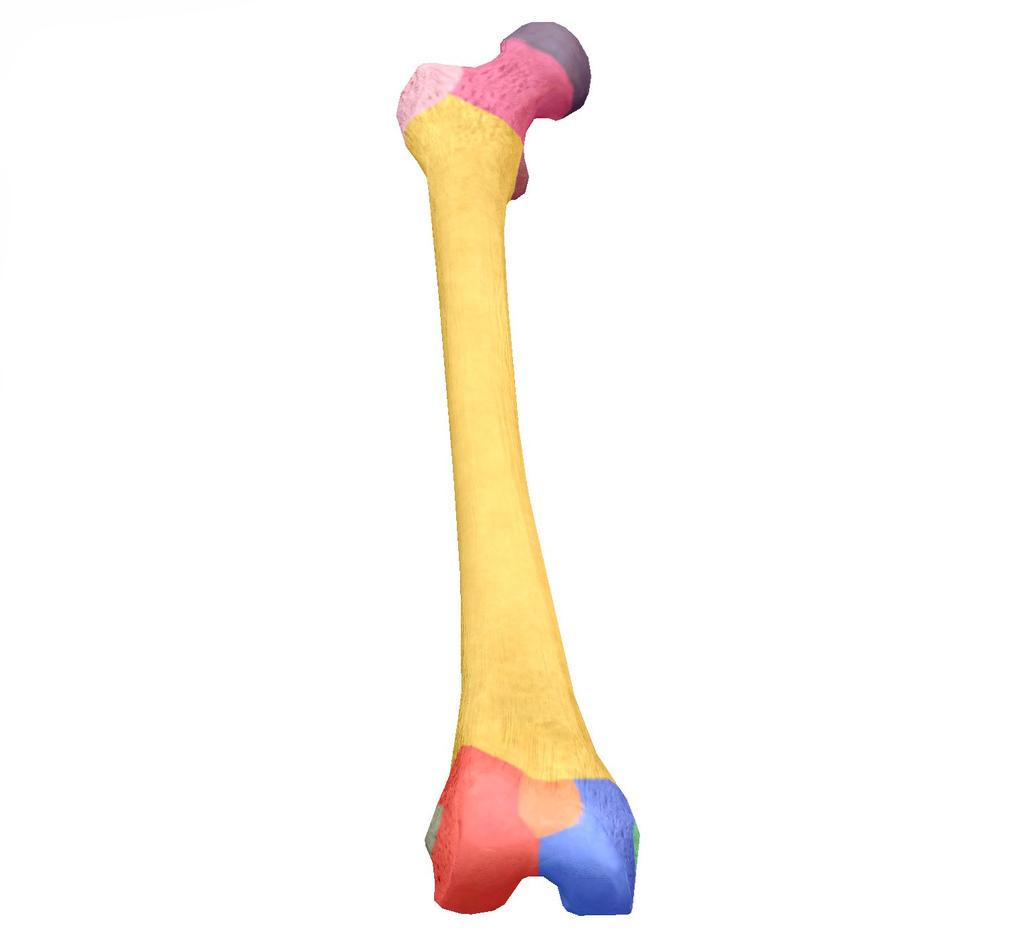
24 1. Femur Head Greater trochanter Neck Lesser trochanter Shaft Lateral condyle Lateral epicondyle Patellar surface Medial epicondyle Medial condyle a. Identify the following bone markings: i. Head ii. Shaft iii. Greater trochanter iv. Lesser trochanter v. Neck vi. Lateral condyle vii. Lateral epicondyle viii. Medial condyle 24
25 ix. Medial epicondyle x. Intercondylar fossa xi. Gluteal tuberosity xii. Linea aspera xiii. Patellar surface xiv. Popliteal surface b. Describe how you would differentiate between a right femur and a left femur. 25
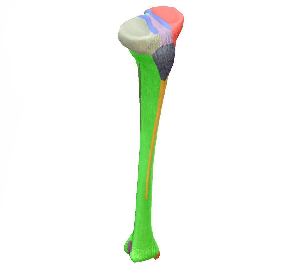
26 2. Tibia Tubercles of intercondylar eminence Lateral condyle Medial condyle Intercondylar area Tibial tuberosity Anterior border Shaft Fibular notch Medial malleolus a. Identify the following bone markings: i. Shaft ii. Medial condyle iii. Lateral condyle iv. Intercondylar area v. Tibial tuberosity vi. Anterior border vii. Medial malleolus 26
27 3. Fibula Head Shaft Lateral malleolus a. Identify the following bone markings: i. Head ii. Shaft iii. Lateral malleolus 27
28 4. Patella Femur Patella Medial condyle of femur Patellar ligament Medial condyle of tibia Lateral condyle of femur Lateral condyle of tibia Fibula Tibia a. Which bones compose the knee joint? b. Where do the different bones attach to each other? 28
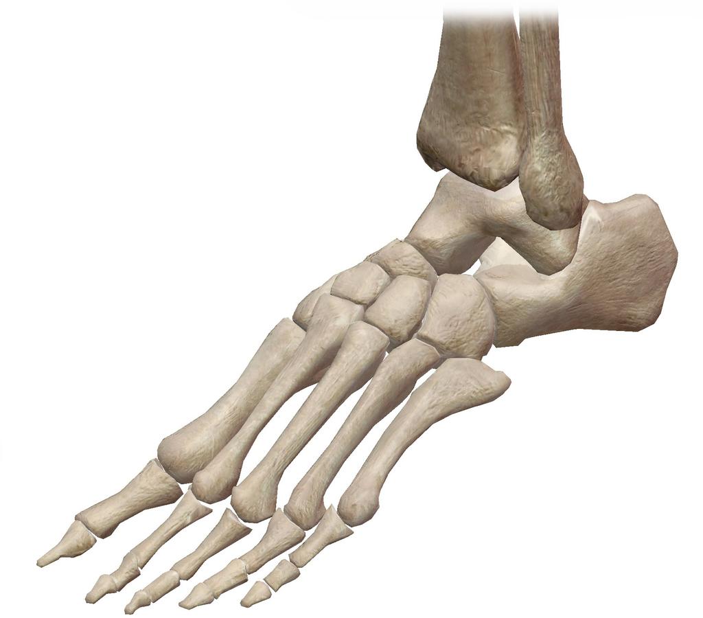
29 5. Tarsus To see the tarsals more clearly, hide some or all of the ligaments on the wrist after you zoom in. Tibia Fibula Talus Navicular Intermediate cuneiform Medial cuneiform Calcaneus Cuboid Lateral cuneiform a. Identify the following tarsal bones: i. Calcaneus ii. Talus iii. Medial cuneiform iv. Intermediate cuneiform v. Lateral cuneiform vi. Cuboid vii. Navicular b. Which bone forms the heel of the foot? 29
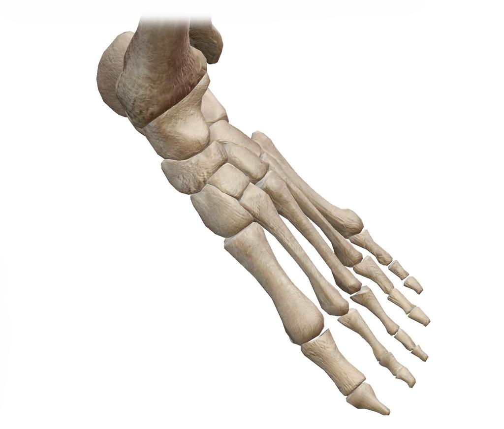
30 6. Foot Note that the nomenclature for the toes is the same as for the hand. The big toe is considered digit I, and the pinky toe is digit V. Metatarsals Middle phalanges Proximal phalanges Distal phalanges a. Identify the following foot bones: i. Metatarsals ii. Proximal Phalanges iii. Middle Phalanges iv. Distal Phalanges 30
31 PUTTING IT ALL TOGETHER 1. How is the shape of the glenoid cavity of the scapula different from that of the acetabulum in the hip bone? How do the shapes of these bone markings affect the range of motion at their respective joints? 2. How do the distinct characteristics of the female pelvis make childbirth easier? 3. Which bone markings are responsible for making the protrusions of the ankles, just above the feet? 4. When someone falls with an outstretched arm, a broken clavicle often results. Physically, how would that person appear afterward? What is the function of the clavicle? TIME TO PRACTICE! GO TO THE SKELETAL SYSTEM QUIZZES AND TAKE QUIZ 7 GIRDLES, QUIZ 8 UPPER LIMB, QUIZ 9 HAND, QUIZ 10 LOWER LIMB, AND QUIZ 11 FOOT 31
32 32

33 Source: Skeletal System Views: View 15. Shoulder Girdle 33

34 Source: Skeletal System Views: View 16. Axillary Region 34

35 Scapula (Anterior) Source: Skeletal System Views: View 16: Axillary Region 35

36 Scapula (Posterior) Source: Skeletal System Views: View 16: Axillary Region 36

37 Clavicle 37

38 Source: Skeletal System Views: View 1. Full Skeleton 38
39 Humerus 39
40 Radius 40
41 Ulna 41

42 Radius and Ulna 42
43 Carpus (Posterior) 43
44 Carpus (Anterior) 44
45 Hand 45

46 Source: Skeletal System Views: View 11. Pelvic Girdle 46
47 Ilium 47

48 Ischium 48
49 Pubis 49
50 Source: Tours: Pelvis Comparison 50

51 Source: Tours: Pelvis Comparison 51

52 Source: Skeletal System Views: View 1. Lower Limb 52
53 Femur 53
54 Tibia 54
55 Fibula 55

56 Patella 56
57 Tarsus 57
58 Foot 58
SKELETAL SYSTEM 206. AXIAL SKELETON 80 APPENDICULAR SKELETON 126 (see Figure 6.1) Clavicle. Clavicle. Pectoral girdles. Scapula. Scapula.
 Clavicle. Clavicle. Pectoral girdles. Scapula. Scapula.") SKELETAL SYSTEM 206 AXIAL SKELETON 80 APPENDICULAR SKELETON 126 (see Figure 6.1) Pectoral girdles 4 Clavicle Scapula 2 2 Clavicle Scapula Humerus 2 Humerus Upper limbs 60 Radius 2 Ulna Carpal bones Metacarpal
SKELETAL SYSTEM 206 AXIAL SKELETON 80 APPENDICULAR SKELETON 126 (see Figure 6.1) Pectoral girdles 4 Clavicle Scapula 2 2 Clavicle Scapula Humerus 2 Humerus Upper limbs 60 Radius 2 Ulna Carpal bones Metacarpal
Biology 218 Human Anatomy. Adapted from Martini Human Anatomy 7th ed. Chapter 7 The Skeletal System Appendicular Division
 Adapted from Martini Human Anatomy 7th ed. Chapter 7 The Skeletal System Appendicular Division Introduction The appendicular skeleton includes: Pectoral girdle Shoulder bones Upper limbs Pelvic girdle
The Appendicular Skeleton
 8 The Appendicular Skeleton PowerPoint Lecture Presentations prepared by Jason LaPres Lone Star College North Harris 8-1 The Pectoral Girdle The Pectoral Girdle Also called shoulder girdle Connects the
Biology 152 Appendicular Skeleton Anatomy Objectives
 Biology 152 Appendicular Skeleton Anatomy Objectives We will learn proper bone names, left/right/medial, and the parts of bones in this exercise. Start by learning the names of the bones. As you gain comfort
Biology 218 Human Anatomy
 Chapter 8 Adapted from Tortora 10 th ed. LECTURE OUTLINE A. Introduction (p. 203) 1. The appendicular skeleton contains 126 bones that form: i. two pectoral (shoulder) girdles two upper limbs i one pelvic
Chapter 8. The Appendicular Skeleton. Lecture Presentation by Lee Ann Frederick University of Texas at Arlington Pearson Education, Inc.
 Chapter 8 The Appendicular Skeleton Lecture Presentation by Lee Ann Frederick University of Texas at Arlington An Introduction to the Appendicular Skeleton The Appendicular Skeleton 126 bones Allows us
Lab Activity 9. Appendicular Skeleton Martini Chapter 8. Portland Community College BI 231
 Lab Activity 9 Appendicular Skeleton Martini Chapter 8 Portland Community College BI 231 Appendicular Skeleton Upper & Lower extremities Shoulder Girdle Pelvic Girdle 2 Humerus 3 Humerus: Proximal End
Amy Warenda Czura, Ph.D. 1 SCCC BIO130 Lab 7 Appendicular Skeleton & Articulations
 The Skeletal System II: Appendicular Skeleton and Articulations Exercises 11, 13 (begins: page 145 in 9 th and 10 th editions) Exercises 10, 11 (begins: page 147 in 11 th edition, page 149 in 12 th edition)
10/12/2010. Upper Extremity. Pectoral (Shoulder) Girdle. Clavicle (collarbone) Skeletal System: Appendicular Skeleton
 Girdle. Clavicle (collarbone) Skeletal System: Appendicular Skeleton") Skeletal System: Appendicular Skeleton Pectoral girdle Pelvic girdle Upper limbs Lower limbs 8-1 Pectoral (Shoulder) Girdle Consists of scapula and clavicle Clavicle articulates with sternum (Sternoclavicular
Skeletal System: Appendicular Skeleton Pectoral girdle Pelvic girdle Upper limbs Lower limbs 8-1 Pectoral (Shoulder) Girdle Consists of scapula and clavicle Clavicle articulates with sternum (Sternoclavicular
Pectoral (Shoulder) Girdle
 Girdle") Chapter 8 Skeletal System: Appendicular Skeleton Pectoral girdle Pelvic girdle Upper limbs Lower limbs 8-1 Pectoral (Shoulder) Girdle Consists of scapula and clavicle Clavicle articulates with sternum
Chapter 8 Skeletal System: Appendicular Skeleton Pectoral girdle Pelvic girdle Upper limbs Lower limbs 8-1 Pectoral (Shoulder) Girdle Consists of scapula and clavicle Clavicle articulates with sternum
bio4165 lab quiz 1 Posterior View Anterior View Lateral View Anterior View bio fall.quarter lab.quiz.1...page.1 of 6
 B A Posterior View D C E Lateral View bio.4165...fall.quarter.2005...lab.quiz.1...page.1 of 6 F I G 35 Posterior View H bio.4165...fall.quarter.2005...lab.quiz.1...page.2 of 6 J Posterior View L K Inferior
Chapter 8B. The Skeletal System: Appendicular Skeleton. The Appendicular Skeleton. Clavicle. Pectoral (Shoulder) Girdle
 Girdle") The Appendicular Skeleton Chapter 8B The Skeletal System: Appendicular Skeleton 126 bones Pectoral (shoulder) girdle Pelvic (hip) girdle Upper limbs Lower limbs Functions primarily to facilitate movement
The Appendicular Skeleton Chapter 8B The Skeletal System: Appendicular Skeleton 126 bones Pectoral (shoulder) girdle Pelvic (hip) girdle Upper limbs Lower limbs Functions primarily to facilitate movement
Bone Flashcards for 10a
 Bone Flashcards for 0a CLAVICLE (collar bone). Sternal extremity (end) flat end. Acromial extremity (end) rounded end. SCAPULA (shoulder blade). Right or left scapula?. Superior border (superior margin).
The Appendicular Skeleton
 8 The Appendicular Skeleton PowerPoint Lecture Presentations prepared by Jason LaPres Lone Star College North Harris An Introduction to the Appendicular Skeleton Learning Outcomes 8-1 Identify the bones
Copyright 2003 Pearson Education, Inc. publishing as Benjamin Cummings. Dr. Nabil khouri
 Dr. Nabil khouri Appendicular Skeleton The appendicular skeleton is made up of the bones of the upper and lower limbs and their girdles Two girdles: Pectoral girdles attach the upper limbs to the body
Chapter 8 The Skeletal System: The Appendicular Skeleton. Copyright 2009 John Wiley & Sons, Inc.
 Chapter 8 The Skeletal System: The Appendicular Skeleton Appendicular Skeleton It includes bones of the upper and lower limbs Girdles attach the limbs to the axial skeleton The pectoral girdle consists
Bone List Anatomy
 1 Frontal Bone Skull 2 Parietal Bone Skull 3 Occipital Bone Skull 4 Temporal Bone Skull 5 Coronal Suture Skull 6 Sagittal Suture Skull 7 Squamous suture Skull 8 Lambdoid Suture Skull 9 Surpaorbital Ridge
Chapter 8 The Skeletal System: The Appendicular Skeleton. Copyright 2009 John Wiley & Sons, Inc.
 Chapter 8 The Skeletal System: The Appendicular Skeleton Appendicular Skeleton The primary function is movement It includes bones of the upper and lower limbs Girdles attach the limbs to the axial skeleton
BIOLOGY 113 LABORATORY Skeletal System
 BIOLOGY 113 LABORATORY Skeletal System Objectives Distinguish between the axial and appendicular skeleton. Distinguish between the cranium and facial skeleton. Locate and name the bones of the skull and
Exercise Science Section 2: The Skeletal System
 Exercise Science Section 2: The Skeletal System An Introduction to Health and Physical Education Ted Temertzoglou Paul Challen ISBN 1-55077-132-9 Role of the Skeleton Protection Framework Attachments for
Exercise 11. The Appendicular Skeleton
 Exercise 11 The Appendicular Skeleton The Appendicular Skeleton The appendicular skeleton contains 126 bones. Consists of the upper and lower limbs, the pectoral girdles, and the pelvic girdles. The pectoral
Perpendicular Plate Zygomatic Bone. Mental Foramen Mandible
 Glabella Frontal Middle Nasal Concha Nasal Lacrimal Perpendicular Plate Zygomatic Inferior Nasal Concha Maxilla Mental Mandible Skull (anterior view) Squamosal Suture Coronal Suture Frontal Parietal Nasal
Principles of Anatomy and Physiology
 Principles of Anatomy and Physiology 14 th Edition CHAPTER 8 The Skeletal System: The Appendicular Skeleton The Appendicular Skeleton The 126 bones of the appendicular skeleton are primarily concerned
THE SKELETAL SYSTEM. Focus on the Pectoral Girdle
 THE SKELETAL SYSTEM Focus on the Pectoral Girdle Appendicular Skeleton 126 bones Includes bones of the limbs (arms and legs) Pectoral girdle (shoulder) Pelvic girdle (hip) Pectoral Girdle (the shoulder)
Chapter 7: Skeletal System: Gross Anatomy
 Chapter 7: Skeletal System: Gross Anatomy I. General Considerations A. How many bones in an average adult skeleton? B. Anatomic features of bones are based on II. Axial Skeleton A. Skull 1. Functionally
Figure 1: Bones of the upper limb
 BONES OF THE APPENDICULAR SKELETON The appendicular skeleton is composed of the 126 bones of the appendages and the pectoral and pelvic girdles, which attach the limbs to the axial skeleton. Although the
Chapter 8. The Pectoral Girdle & Upper Limb
 Chapter 8 The Pectoral Girdle & Upper Limb Pectoral Girdle pectoral girdle (shoulder girdle) supports the arm consists of two on each side of the body // clavicle (collarbone) and scapula (shoulder blade)
Appendicular Skeleton. Prof. Abdulameer Al-Nuaimi
 Appendicular Skeleton Prof. Abdulameer Al-Nuaimi a.alnuaimi@sheffield.ac.uk abdulameerh@yahoo.com Hi Prof, It is great to hear from you, I really enjoyed your teaching last year. You taught me the hardest
A&P 1 Skeletal Lab Guide Week 2 - Appendicular Skeleton and Joints Lab Exercises: Pectoral Girdle
 A&P 1 Skeletal Lab Guide Week 2 - Appendicular Skeleton and Joints Lab Exercises: Pectoral Girdle PLEASE NOTE: Your group will need an articulated skeleton, a disarticulated skeleton, and the joint models
the Skeletal System provided by Academic Web Services Grand Canyon University
 Anatomy Resource Center Study Guides the Skeletal System HEAD & NECK REGIONAL VIEW SKULL BONES CRANIUM FACE SKULL LANDMARKS ANTERIOR SIDE SUPERIOR/INFERIOR VERTEBRAL COLUMN VERTEBRAL REGIONS CERVICAL C1
Spring Written By: J. E. Sutton. Contents: I. Overview of the Skeleton: II. Appendicular Skeleton III. Axial Skeleton IV.
 Spring 2012 Written By: J. E. Sutton Contents: I. Overview of the Skeleton: II. Appendicular Skeleton III. Axial Skeleton IV. Articulations Overview of the Skeleton: I. Orientation to Human Skeleton: a.
Biology 2401 The Skeletal System
 Biology 2401 The Skeletal System Purpose: The lab will describe the microscopic and gross anatomy of bone, identify bones of the body, and identify important bone markings. I. Overview of the Skeleton
Axial skeleton bones and markings
 Axial skeleton bones and markings Skull Cranial bones Frontal x 1 Supraorbital foramen Occipital x 1 Foramen magnum Occipital condyles Superior nuchal line Inferior nuchal line Anterior cranial fossa External
Chapter 8 Outline. Pectoral Girdle Upper Limb Pelvic Girdle Lower Limb Aging of the Appendicular Skeleton Development of the Appendicular Skeleton
 Chapter 8 Outline Pectoral Girdle Upper Limb Pelvic Girdle Lower Limb Aging of the Appendicular Skeleton Development of the Appendicular Skeleton Figure 8.1 Appendicular Skeleton Pectoral Girdle Clavicle
An Introduction to the Appendicular Skeleton
 An Introduction to the Appendicular Skeleton The Appendicular Skeleton is composed of the 126 bones of the appendages (limbs) and the pectoral and pelvic girdles, which attach to the axial skeleton. Each
Important Parts of Bones
 Important Parts of Bones For 2015 Know: Humerus (posterior) Clavical Femur (Anterior) Foot Hand Mandible Os Coxa Scapula Skull (Anterior, Inferior, Lateral) Sternum Humerus (posterior) A. olecranon fossa
Chapter 7 Part C The Skeleton
 Chapter 7 Part C The Skeleton Part 2 The Appendicular Skeleton Consists of bones of the limbs and their girdles Pectoral girdle Attaches upper limbs to body trunk Pelvic girdle Attaches lower limbs to
Figure 7: Bones of the lower limb
 BONES OF THE APPENDICULAR SKELETON The appendicular skeleton is composed of the 126 bones of the appendages and the pectoral and pelvic girdles, which attach the limbs to the axial skeleton. Although the
Axial Skeleton BONE TERMINOLOGY FEATURES
 Axial Skeleton BONE TERMINOLOGY FEATURES Tuberosity Rounded area on bone often roughened for muscle attachment. Tubercle Rounded projection on bone. This is called a tuberosity on the femur. Crest Ridgeline
TEST YOURSELF- Chapter 7
 TEST YOURSELF- Chapter 7 Cranial Bones 1. Give the name of the bone for each of the following markings. Some of the markings are found on more than one bone. List all that apply. Cranium a. Frontal squama:
A. Incorrect! The appendicular skeleton includes bones of the shoulder, arm, hand, pelvis, leg and foot.
 Anatomy and Physiology - Problem Drill 08: The Skeletal System III No. 1 of 10 1. Which of the following statements about the appendicular skeleton is correct? A. The appendicular skeleton includes bones
Riverside Community College Anatomy & Physiology 2B SPRING 2012 EXAM #1-ABC (Nervous System)
") Riverside Community College Anatomy & Physiology 2B SPRING 2012 EXAM #1-ABC (Nervous System) Name: 1) This vertebra is an example of a(n). 1) A) thoracic B) axis C) atlas D) lumbar E) sacral 1 2) W hich
Riverside Community College Anatomy & Physiology 2B SPRING 2012 EXAM #1-ABC (Nervous System) Name: 1) This vertebra is an example of a(n). 1) A) thoracic B) axis C) atlas D) lumbar E) sacral 1 2) W hich
Appendicular Skeleton. Dr. Carmen E. Rexach Anatomy 35 Mt. San Antonio College
 Appendicular Skeleton Dr. Carmen E. Rexach Anatomy 35 Mt. San Antonio College Pectoral girdle clavicle scapula Upper limb brachium antebrachium carpus manus Pelvic girdle oscoxae Lower limb femoral region
AP1 Lab 4 - Appendicular Skeleton
 Project 1 Learn the Bone Names AP1 Lab 4 - Appendicular Skeleton Use Figure 7.1 and the hanging skeletons to learn the whole bones of the arms and legs. Don t learn the features of each bone yet just be
The Skeletal System THE APPENDICULAR SKELETON
 The Skeletal System THE APPENDICULAR SKELETON The appendicular skeleton consists of the girdles and the skeleton of the limbs. The upper (anterior) limbs are attached to the pectoral (shoulder) girdle
Lab Unit One Flashcards
 CLAVICLE (collar bone). Sternal extremity (end) flat end. Acromial extremity (end) rounded end.. Conoid tubercle near round end SCAPULA (shoulder blade). Right or left scapula?. Superior border (superior
Copyright 2003 Pearson Education, Inc. publishing as Benjamin Cummings. Dr. Nabil Khouri MD, MSc, Ph.D
 Dr. Nabil Khouri MD, MSc, Ph.D Pelvic Girdle (Hip) Organization of the Lower Limb It is divided into: The Gluteal region The thigh The knee The leg The ankle The foot The thigh and the leg have compartments
Introduction. Skeletal Nomenclature. Lesson One: Bones of the Upper Extremity. Shoulder Girdle: Clavicle. Scapula: Bony Markings
 Chapter 14 Skeletal Nomenclature Susan G. Salvo Bones help locate muscles Can also be areas to avoid Chapter divided into 5 lessons covering bones, bony markings, and joints Introduction 1 2 Lesson One:
Anatomy and Physiology 2016
 Anatomy and Physiology 2016 O = Temporal line I = coronoid process (Mandible) A = elevates mandible (chewing) O = galea aponeurotica (layer of dense fibrous tissue which covers the upper part of the cranium)
Skeletal System - Prelab 1
 Skeletal System - Prelab 1 1. Which bones contain the paranasal sinuses? What function do the sinuses serve? 2. What two areas are separated from each other by the hard palate? Name the two bones that
Dr.Israa H. Mohsen. Lecture 5. The vertebral column
 Anatomy Lecture 5 Dr.Israa H. Mohsen The vertebral column The vertebral column a flexible structure consisting of 33 vertebrae holds the head and torso upright, serves as an attachment point for the legs,
BLUE SKY SCHOOL OF PROFESSIONAL MASSAGE AND THERAPEUTIC BODYWORK. Musculoskeletal Anatomy & Kinesiology I TERMINOLOGY, STRUCTURES, & SKELETAL OVERVIEW
 BLUE SKY SCHOOL OF PROFESSIONAL MASSAGE AND THERAPEUTIC BODYWORK Musculoskeletal Anatomy & Kinesiology I TERMINOLOGY, STRUCTURES, & SKELETAL OVERVIEW MSAK101-I Session 1 Learning Objectives: 1. Define
It is formed by fusion of 3 bones: I. Ilium (superior bone). II. Pubis (antero-inferior bone). III. Ischium (postero-inferior bone).
. II. Pubis (antero-inferior bone). III. Ischium (postero-inferior bone).") It is formed by fusion of 3 bones: I. Ilium (superior bone). II. Pubis (antero-inferior bone). III. Ischium (postero-inferior bone). Pubis Acetabulum Ana (242 ) The three constituent of bones of the hip
It is formed by fusion of 3 bones: I. Ilium (superior bone). II. Pubis (antero-inferior bone). III. Ischium (postero-inferior bone). Pubis Acetabulum Ana (242 ) The three constituent of bones of the hip
Nervous/Vascular/Skeletal Systems
 L 2 A B O R A T O R Y Nervous/Vascular/Skeletal Systems NERVOUS SYSTEM Identify the structures associated with the central nervous system and the peripheral nervous system. Identify the location of gray
The skeleton consists of: Bones: special connective tissue, hard. Cartilage: special connective tissue, less hard than bones. Joints: joint is the
 The skeleton consists of: Bones: special connective tissue, hard. Cartilage: special connective tissue, less hard than bones. Joints: joint is the location at witch two bones make contact, whereas ligaments
Pearson Education Limited Edinburgh Gate Harlow Essex CM20 2JE England and Associated Companies throughout the world
 Pearson Education Limited Edinburgh Gate Harlow Essex CM20 2JE England and Associated Companies throughout the world Visit us on the World Wide Web at: www.pearsoned.co.uk Pearson Education Limited 204
Hole s Human Anatomy and Physiology
 Hole s Human Anatomy and Physiology 1 Chapter 7 Skeletal System Bone Classification Long Bones Short Bones Flat Bones Irregular Bones Sesamoid (Round) Bones 2 Parts of a Long Bone epiphysis distal proximal
Pectoral girdle, SUPERIEUR ARM AND HAND. Danil Hammoudi.MD
 Pectoral girdle, SUPERIEUR ARM AND HAND Danil Hammoudi.MD The pectoral girdle is the set of bones which connect the upper limb to the axial skeleton on each side. It consists of the clavicle scapula in
Lab 6, 7, 8: Skeletal System
 107 Lab 6, 7, 8: Skeletal System Adult Skull Bony orbit (FLEZMS) Frontal bone supraorbital foramen frontal sinus Lacrimal bone Ethmoid bone perpendicular plate of ethmoid middle nasal conchae cribriform
Bones of Lower Limb. Dr. Heba Kalbouneh Associate Professor of Anatomy and Histology
 Bones of Lower Limb Dr. Heba Kalbouneh Associate Professor of Anatomy and Histology Bones of the lower limb Hip Bone Made up of 3 bones: 1) Ilium (flat), superior in position 2) Ischium (L), postero-inferior
Axilla and Brachial Region
 L 4 A B O R A T O R Y Axilla and Brachial Region BRACHIAL PLEXUS 5 Roots/Rami (ventral rami C5 T1) 3 Trunks Superior (C5, C6) Middle (C7) Inferior (C8, T1) 3 Cords Lateral Cord (Anterior Superior and Anterior
Muscles of the Upper Limb
 Muscles of the Upper Limb anterior surface of ribs 3 5 coracoid process Pectoralis minor pectoral nerves protracts / depresses scapula Serratus anterior Subclavius ribs 1-8 long thoracic nerve rib 1 ----------------
SUPERIEUR ARM AND HAND
 Pectoral girdle, SUPERIEUR ARM AND HAND Danil Hammoudi.MD The pectoral girdle is the set of bones which connect the upper limb to the axial skeleton on each side. It consists of the clavicle scapula in
Overview of the Skeleton: Bone Markings
 Name Overview of the Skeleton: Bone Markings Match the terms in column B with the appropriate description in column A. Column A 1. sharp, slender process* 2. small rounded projection* 3. narrow ridge of
Human Anatomy, First Edition McKinley & O'Loughlin
 Human Anatomy, First Edition McKinley & O'Loughlin Chapter 8 : Appendicular Skeleton 8-1 Appendicular Skeleton Includes the bones of the upper and lower limbs. The girdles of bones that attach the upper
11/25/2012. Chapter 7 Part 2: Bones! Skeletal Organization. The Skull. Skull Bones to Know Cranium
 Chapter 7 Part 2: Bones! 5) Distinguish between the axial and appendicular skeletons and name the major parts of each 6) Locate and identify the bones and the major features of the bones that compose the
Radiographic Positioning Summary (Basic Projections RAD 222)
") Lower Extremity Radiographic Positioning Summary (Basic Projections RAD 222) AP Pelvis AP Hip (Unilateral) (L or R) AP Femur Mid and distal AP Knee Lateral Knee Pt lies supine on table Align MSP to Center
Lower Extremity Radiographic Positioning Summary (Basic Projections RAD 222) AP Pelvis AP Hip (Unilateral) (L or R) AP Femur Mid and distal AP Knee Lateral Knee Pt lies supine on table Align MSP to Center
Located more distal and anterior together with Trapezoid, anterior to scaphoid Trapezium rarely to be fractured.
 The hand The hand consists of the 3 groups: Proximal part: carpals bones (8) Middle part: metacarpal bones (5) Distal part: fingers or phalanges bones (3 for each finger except for the thumb just 2 bones).
Anatomy Lab: The skeletal system. Part I: Vertebrae and Thoracic cage
 ANA Lab: Bone 1 Anatomy Lab: The skeletal system Part I: Vertebrae and Thoracic cage Spine (Vertebrae) Body Vertebral arch Vertebral canal Pedicle Lamina Spinous process Transverse process Sup. articular
Hole s Human Anatomy and Physiology Tenth Edition. Chapter 7
 PowerPoint Lecture Outlines to accompany Hole s Human Anatomy and Physiology Tenth Edition Shier Butler Lewis Chapter 7 Copyright The McGraw-Hill Companies, Inc. Permission required for reproduction or
8 THE APPENDICULAR SKELETON
 CHAPTER 8 THE APPENDICULAR SKELETON 293 8 THE APPENDICULAR SKELETON Figure 8.1 Dancer The appendicular skeleton consists of the upper and lower limb bones, the bones of the hands and feet, and the bones
Anatomy and Physiology II. Review Shoulder Girdle New Material Upper Extremities - Bones
 Anatomy and Physiology II Review Shoulder Girdle New Material Upper Extremities - Bones Anatomy and Physiology II Shoulder Girdle Review Questions From Last Lecture Can you identify the following muscles?
Muscular Nomenclature and Kinesiology - One
 Chapter 16 Muscular Nomenclature and Kinesiology - One Lessons 1-3 (with lesson 4) 1 Introduction 122 major muscles covered in this chapter Chapter divided into nine lessons Kinesiology study of human
C. Bones of the Pelvic Girdle
 C. Bones of the Pelvic Girdle 1. 2 coxal bones (a.k.a hip bones): -bony pelvis is made up of hip bones, sacrum, & coccyx -pelvic bones are large & heavy & attach to the axial skeleton via sacrum/coccyx
External Acoustic Meatus. Mastoid Process. Zygomatic Process. Temporal Bone
 Bone lab review 1. Frontal Bone 2. Supra-Orbital Foramen 3. Orbit (Orbital Cavity) 4. Superior Orbital Fissure 5. Inferior Orbital Fissure 6. Zygomatic Bone 7. Infra-Orbital Foramen 8. Maxilla 9. Mandible
CHAPTER 8 LECTURE OUTLINE
 CHAPTER 8 LECTURE OUTLINE I. INTRODUCTION A. The appendicular skeleton includes the bones of the upper and lower extremities and the shoulder and hip girdles. B. The appendicular skeleton functions primarily
Bones of the Upper and Lower limb
 Bones of the Upper and Lower limb Musculoskeletal block- Anatomy-lecture 1 Editing file Objectives Classify the bones of the three regions of the lower limb (thigh, leg and foot). Color guide : important
BONE CHALLENGE DANIL HAMMOUDI.MD
 BONE CHALLENGE DANIL HAMMOUDI.MD Bone Basic functions? A. support B. protection C. movement assistance in D. RBC formation-hemopoiesis E. mineral homeostasis +importance of calcium F. energy supply -yellow
Labs 6, 7, 8: Skeletal System
 153 Labs 6, 7, 8: Skeletal System Unit 6: Skeletal System: Bone tissue, Bones and Joints (p. 105-152) Ex. 6-1: Histology of Osseous Tissue, p. 113 Model: Osteon Tiss Lamella Osteocyte Lacunae Canaliculi
Anatomy & Physiology Skeletal System Worksheet
 1. Name the five functions of the skeleton. c) d) e) Anatomy & Physiology Skeletal System Worksheet 2. The term for the shaft of a bone is:. 3. The bony struts found in spongy bone are called. 4. In ossification,
The Lower Limb. Sevda LAFCI FAHRİOĞLU, MD.PhD.
 The Lower Limb Sevda LAFCI FAHRİOĞLU, MD.PhD. The Lower Limb The bones of the lower limb form the inferior part of the appendicular skeleton the organ of locomotion for bearing the weight of body stronger
Lab 1: body, skeletal and axial systems
 Lab 1: body, skeletal and axial systems Types of tissue Epithelia tissue - Sheets of tissue covering other tissues or linin cavities - Protective tissue e.g. skin - Cell types: squamous, cuboid, columnar
Chapter 7: Skeletal System
 Chapter 7: Skeletal System The Skeletal System Introduction P. 182 Bone is an organ made up of tissues: It is made up of the following components. Cartilage Blood Nerves Bone Connective Bone Classification
Practical 2 Worksheet
 Practical 2 Worksheet Upper Extremity BONES 1. Which end of the clavicle is on the lateral side (acromial or sternal)? 2. Describe the difference in the appearance of the acromial and sternal ends of the
Ch. 5 - Skeletal System
 Ch. 5 - Skeletal System Bones are living, ever-changing structures. This allows them grow and adapt to new situations that the body encounters. The functions of the skeletal system: 1) support bones are
Introduction to Human Osteology Chapter 3: Hands and Feet
 Introduction to Human Osteology Chapter 3: Hands and Feet Roberta Hall Kenneth Beals Holm Neumann Georg Neumann Gwyn Madden Revised in 1978, 1984, and 2008 Bones of the Hand Eight carpal bones, in two
Lab Exercise #04 The Skeletal System Student Performance Objectives
 Lab Exercise #04 The Skeletal System Student Performance Objectives The material that you are required to learn in this exercise can be found in either the lecture text or the supplemental materials provided
Nervous & Skeletal Systems. Virtual Science University
 Nervous & Skeletal Systems Virtual Science University 1 Nervous & Skeletal Systems Texas TEK B.10(A) The student will interpret the function of systems in organisms (humans) including the nervous and skeletal
SKELETON FUNCTIONS OF BONE:
 SKELETON FUNCTIONS OF BONE: SKELETON: 1. Performs a mechanical function in forming the skeletal support of the body and in forming a leverage system whereby work and movement are possible. 2. Serves as
Netter's Anatomy Flash Cards Section 6 List 4 th Edition
 Netter's Anatomy Flash Cards Section 6 List 4 th Edition https://www.memrise.com/course/1577581/ Section 6 Upper Limb (66 cards) Plate 6-1 Humerus and Scapula: Anterior View 1.1 Acromion 1.2 Greater tubercle
Lab Activity 11: Group II
 Lab Activity 11: Group II Muscles Martini Chapter 11 Portland Community College BI 231 Origin and Insertion Origin: The place where the fixed end attaches to a bone, cartilage, or connective tissue. Insertion:
Sports Medicine Part I : ANATOMY OF THE SPINE, ABDOMEN AND SHOULDER COMPLEX
 Sports Medicine 25 1.1 Part I : ANATOMY OF THE SPINE, ABDOMEN AND SHOULDER COMPLEX c.w.p. Wagner High School, Sports Medicine, A. Morgan, T. Morgan 2008 Anatomy of the Upper Body In this section of the
TABLES OF MUSCLE ACTIONS, INNERVATIONS, AND ATTACHMENTS
 TABLES OF MUSCLE ACTIONS, INNERVATIONS, AND ATTACHMENTS Table 1-1 ERECTOR SPINAE MUSCLES Intrinsic muscles producing extension and/or lateral of the spine Muscle Joint and Action Innervation Inferior Attachment
Anatomage Table Instructors Guide- Lower Limb
 The Lower Limb Anatomage Table Instructors Guide- Lower Limb Table of Contents Lower Limb 1- The Skeletal System...3 1: Hip Bone...3 2: Hip Joint and Femur...4 3: Patella and Knee Joint...7 4: Tibia, Fibula,
First practical session. Bones of the gluteal region
 First practical session 2017 Bones of the gluteal region The Hip bone The hip bone is made of: 1 The ilium: superior in position 2 The ischium:postero-inferior in position 3 The pubis: antero-inferior
LAB Notes#1. Ahmad Ar'ar. Eslam
 LAB Notes#1 Ahmad Ar'ar Eslam 1 P a g e Anatomy lab Notes Lower limb bones :- Pelvic girdle: It's the connection between the axial skeleton and the lower limb; it's made up of one bone called the HIP BONE
Lab 9: Learn origin and insertion for each of the listed muscles. For Exercise 15, do Activities 1-6 in 9 th edition, Activities 1-4 in 10 th edition
 The Muscular System Exercises 14, 15, and 16 (begins: page 187 in 9 th and 10 th editions) Exercises 12, 13, and 14 (begins: page 185 in 11 th edition, page 189 in 12 th edition) Lab 8 and 9 Objectives
Bones of the Lower Limb Bone Structure Description Notes. border of the superior ramus. inferolaterally from the pubic symphysis
 Bones of the Lower Limb Bone Structure Description Notes pubis an angulated bone the forms the anterior part of the pelvis one of three bones that form the os coxae: ilium, ischium, pubis; its forms 1/5
TRAINING LAB SKELETAL REMAINS: IDENTIFYING BONES NAME
 TRAINING LAB SKELETAL REMAINS: IDENTIFYING BONES NAME Background: Skeletal remains are important pieces of evidence. The flesh, muscle, and organs of a victim rapidly decompose; however, the victim s skeleton
Bones of Thorax (Rib Cage)
") Musculoskeletal System (Part A-2) Module 7 -Chapter 10 Overview Muscles Attachments Bones Bone types Surface features of bones Divisions of the skeletal system Joints or Articulations Susie Turner, M.D.
Musculoskeletal System (Part A-2) Module 7 -Chapter 10 Overview Muscles Attachments Bones Bone types Surface features of bones Divisions of the skeletal system Joints or Articulations Susie Turner, M.D.