|
|
|
- Tobias Quinn
- 5 years ago
- Views:
Transcription

1

2 Mmmmmm Mmmmmm mmmmmmmmmmmmm mmmmmmm mmmmmmm
3 MRI of Equine Stifle Injuries: A Review of the first 100 clinical cases Martin Waselau, Dr.med.vet., MS Diplomate ACVS, Diplomate ECVS Equine Hospital Aschheim, Equine Diagnostic Center Munich
4 Declaration No sources of funding No conflict of interest No any other conflicts
5 Purpose of this talk Describe a routine technique for equine stifle MRI Report on pathologic MRI-Findings Compare MRI-Findings to traditional diagnostics Outline therapeutic consequences based on MRI
6 INTRODUCTION


7 Introduction Anatomy Equine stifle diseases are common Complex joint bone and soft tissue pathology
8 Introduction Diagnostics Traditional diagnostics Radiographs 1 Ultrasonography 2 Scintigraphy 1, CT 3 Diagnostic arthroscopy 2... Despite that exact diagnosis can be frustrating... 1 Auer J, Equine Surgery 4th ed. 2011; 2 Barrett B, EVJ 2012; 3 Bergman E, Equitana 2015;
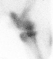
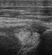
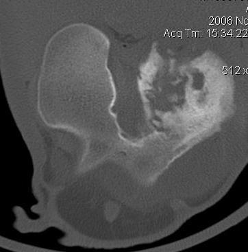
9 Introduction Diagnostics
10 Introduction Problem Frequently, problem was localized by intraarticular block But negative on current imaging techniques... Current solution Arthroscopic exploration as Gold Standard...
11 Introduction However... Arthroscopy was also fruitless... So what now...? No diagnosis Which treatment...? What s the prognosis? Current solution Human medicine = MRI Equine surgery = mainly arthroscopy 1 Equine Hospital Aschheim = MRI Barrett B, EVJ 2012; 2 Waselau M, ECVS 2014; 3 Waselau M, ACVS 2014; 4 Waselau M, Equitana 2015 ; 5 Waselau M, ECVS 2015

12 Introduction Equine Hospital Aschheim Equine Diagnostic Center Munich Munich, Germany MRI-Scans since 2011 Head, +/- Neck Distal foot Carpi, shoulder Tarsi and stifles...
13 MATERIALS & METHODS

14 MRI Technique Custom-made MRI-Table with rotating MRI-Scanner

15 MRI Examination GA, dorsal recumbency, rotating MRI-Scanner
16 MRI Scanning Protocol MRI Sequences Equine Hospital Aschheim T1 (1mm) = Cartilage, subchondral bone T2 (4mm) = Ligament, tendons, menisci PD (4mm) = Ligament, tendons, menisci, cartilage SHARC (1,3mm) = Cartilage STIR (4mm) = Bone X-Bone (4mm) = Subchondral bone 3 planes Frontal, sagittal and axial; T1 / SHARC 3D-Rekonstruktion
17 MRI Scanning Protocol MRI Sequences Equine Hospital Aschheim T1 (1mm) = Cartilage, subchondral bone T2 (4mm) = Ligament, tendons, menisci PD (4mm) = Ligament, tendons, menisci, cartilage SHARC (1,3mm) = Cartilage STIR (4mm) = Bone X-Bone (4mm) = Subchondral bone 3 planes Frontal, sagittal and axial; T1 / SHARC 3D-Rekonstruktion


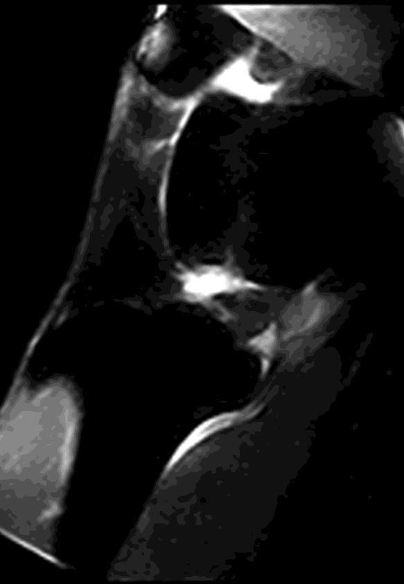
18 MRI 3 planes STIR Sagittal PD - Axial T1 - Dorsal
19 MRI Review of Images MRI Scans were reviewed by Diplomate ACVR and/or Diplomate ACVS MRI findings retrospectively compared to Ultrasonography, radiographs +/- A-Copy
20 MRI What horses were scanned? Inclusion criteria Lameness was localized to stifle joint Negative on ultrasonography and/or Negative on radiographs and/or A-Copy
21 MRI What parameters recorded? General parameters Age, breed, gender, anesthesia time Saved in medical records Pathology Bone pathology Soft tissue pathology (menisci, cruciates and cartilage) Concurrent lesions
22 MRI Classification of parameters Pathology classification Meniscopathy > Tear / Degeneration Cruciate ligaments > Tear / Degeneration Chondropathy > Erosion Concurrent lesions > Frequency
23 RESULTS
24 Results General parameters Patients Number:100 horses Age: 10,8 years (1-27) Gender: 37% Mares, 50% Geldings, 13% Stallions Breeds: different breeds representative hospital popul. Bilateral stifle scans: 5% Anesthesia time 65min (40-87min)
25 Results Overview on Pathology Bone lesions Bone edema/bruises Fissures (Subchondral) bone cysts Soft tissue lesions Synovitis / Fibrinous adhesions Articular cartilage defects Cruciate / collateral / mensicotibial desmitis Meniscal tears / degenerations
26 BONE PATHOLOGY

27 MRI Bone Edema STIR STIR STIR STIR


28 MRI Bone Fissures T1 STIR T1

29 MRI Bone Fissures T1


30 MRI Tibial Cysts T1 STIR STIR
31 MRI Femoral Condyle Cysts Tieraerztliche Klinik fuer Pferde G-ScanVet Tieraerztliche Klinik fuer Pferde G-ScanVet Ex: SE PD-T2 dorsal Aschheim Se: 6 Im: 42 / x 256 OTHER L F Dres. Lu Abel M Img Tm: A H STIR PD ET: 90 TR: 4730 TE: 1 PD W: L: :15 / Nr.: / Se: 6 / Im: 42 R H


32 MRI Femoral Condyle Cysts Tieraerztliche Klinik fuer Pferde G-ScanVet Tieraerztliche Klinik fuer Pferde G-ScanVet Ex: SE PD-T2 dorsal Aschheim Se: 6 Im: 42 / x 256 OTHER L F Dres. Lu Abel M Img Tm: A H STIR PD ET: 90 TR: 4730 TE: 1 PD W: L: :15 / Nr.: / Se: 6 / Im: 42 R H
33 SOFT TISSUE PATHOLOGY
34 MRT CRUCIATE DESMOPATHIES

35 MRI Normal Cranial Cruciate PD sagittal


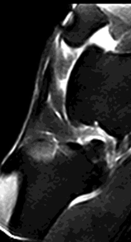
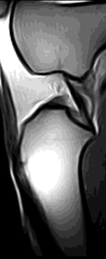
36 MRI Cranial Cruciate Desmopathy SE PD SE PD T2 SE PD Degeneration & Tear Tear Partial Tear Degeneration

37 MRI Normal Caudal Cruciate


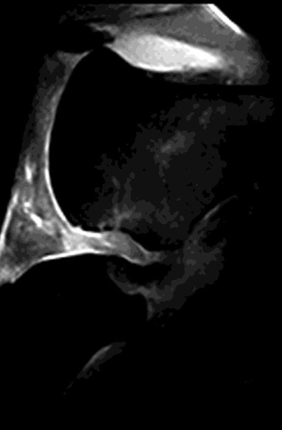
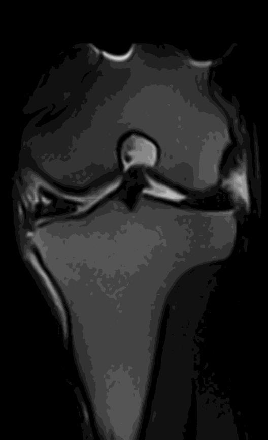
38 MRI Caudal Cruciate Desmopathy PD PD PD PD Tear Degeneration Tear Tear
39 MRI CHONDROPATHIES


40 MRI Normal Cartilage


41 MRI Cartilage Defects T 3D 1 - Dorsal T 3D 1 - Sagittal SE T 2 - Dorsal T 3D 1 - Dorsal Multiple cartilage defects, Subchondral sclerosis and remodelling
42 MRI MENISCOPATHIES

43 MRI Normal Menisci PD Signal Intensity
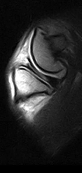
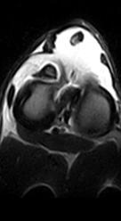
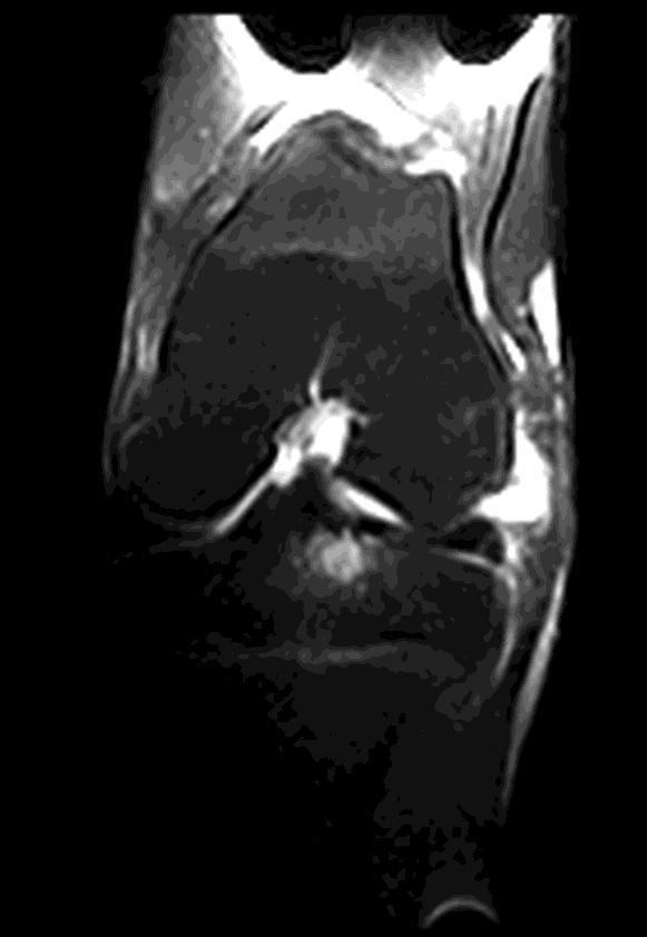
44 MRI Meniscopathy PD PD PD PD Tear Tear Tear lateral menisci
45 Results Meniscopathies Incidence 95% Location 47% medial 45% lateral 19,5% medial and lateral
46 Results Meniscopathies Medial Tears = 58% Degenerations = 39% Lateral Tears = 14% Degenerations = 86% Medial & lateral Tears = 7% Degenerations = 13%
47 Results Concurrent Pathology Menisco- and Cruciate Desmopathy = 43% Medial meniscopathy = 47% Cranial Cruciate Desmopathy = 31% Caudal Cruciate Desmopathy Lateral meniscopathy = 51% Cranial Cruciate Desmopathy = 29% Caudal Cruciate Desmopathy
48 Results Concurrent Pathology Menisco- and Chondropathy = 66% Medial femoral condyle > lateral femoral condyle Femoral chondropathy >>> tibia >> patella
49 Results Retrospective evaluation of Radiographs Bone edema not / Fissure unreliably detected Rads = 50% change in bone density... Ultrasonography, A-Copy Incidence and extent of menisco-, cruciate ligament desmo- and chondropathy were underestimated
50 Results MRI and treatment Conservative Therapy 61% of all lesions surgically inaccessible I.A. & system. Medication Mild-moderate Orthobiologics >>>>> Anti-inflammatories Orthobiologics (PRP >>> stem cells; Pentosansulphate; PSGAG etc.) Severe Anti-inflammatory >> Orthobiologics Bisphosphonates (TILDREN )
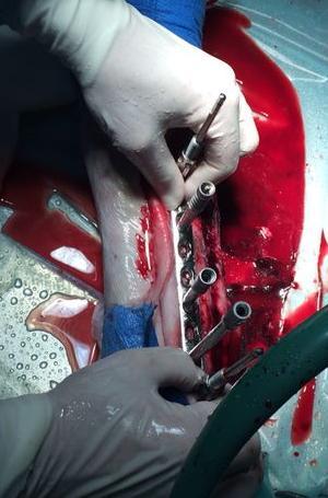
51 Results MRI and treatment Surgical A-Copy 39% of all lesions surgically accessible Surgical revision & lavage Concurrent I.A. / I.L. orthobiologics PRP >>>> stem cells Superficial menisco- & cruciate pathology confirmed Meniscus degeneration and extent of cruciate desmopathy unreliably confirmed


52 Results MRI based A-Copy bbbb


53 Results MRI based A-Copy Cranial Cruciate Desmopathy Waselau, M. Arthroscopic exploration of the equine stifle advantages and limitations in respect to MRI-findings. The 7th Edition of MRI in Veterinary Medicine, Tuscany, November 2014; Waselau, M. Magnetic resonance Imaging of meniscal, cruciate ligament and cartilage pathology - 76 clinical cases. ECVS, Berlin, July 2015


54 Results MRI and Rehabilitation Rehabilitation in counter-current swimming pool
55 DISCUSSION
56 Discussion Technique Scanning procedure Straight forward and save protocol Can be routinely performed on all horses independent on age, breed and gender Bone structures and soft tissue pathology Completely/toroughly portrayed Higher sensitivity than traditional imaging modalities Similar to reports from human literature 1 1 Hayashi D. Curr Rheumatol Rep Jan;16(1):391
57 Discussion Pathology Location Central, submeniscal, plantar, extraarticular... Intrastructural, insertional desmopathies etc. Relevance Poorly detectable on traditional imaging Frequently inaccessible and complex pathology May explain overall low therapeutic success rates in past
58 Discussion Pathology Bone pathology acute lesions... Bone bruises >>> other pathology Acute pain i.a. anesthesia less specific... Different pathophysiology...? Soft tissue pathology chronic lesions... Wide variety Complex pathology 1 Hayashi D. Curr Rheumatol Rep Jan;16(1):391
59 Discussion Soft tissue pathology Meniscopathy higher than previously reported Medial = tear...; Lateral = degeneration... Complex joint biomechanics? -> ex vivo studies... Cruciates Cranial desmopathy higher than caudal Similar in humans 1 Complex joint biomechanics? -> ex vivo studies... 1 Sri-Ram K. Bone Joint J Jan;95-B(1):59-64
60 Discussion Concurrent pathology Concurrent pathology high Menisco-, cruciate desmo- and chondropathy Similar to reports in other species 1 May reflect chronicity of pathology May reflect complex joint biomechanics? -> requires ex vivo studies... Indication for a Snowball effect...? 1 Hulse D. Vet Surg Apr;39(3):350-4


61 Discussion Concurrent pathology Repetative Trauma Cartilage Damage Abnormal weight bearing Snowball Effect? Bone Edema Pain and Inflammation Meniscal lesions / cruciate stress
62 Discussion MRI and Tx > 50% of lesions inaccessible > conservative Tx... May explain frustrating A-Copy results in the past A-Copy avoided = change in Tx < than 50% of lesions accessible > A-Copy Modified surgical approach = influence on Tx
63 Discussion MRI and Costs Advantages More detailed and global view on stifle pathology Hit 5 birds with one stone Financial considerations Information gained within 60min less expensive than months of diagnostic therapy Diagnosed-based treatment
64 Conclusion Stifle MRI... (1) save & routine for all horses (2) deliniated all bone and soft tissue pathology (3) confirmed co-existence of pathology (4) revealed that soft tissue pathology is highly underestimated
65 Conclusion Stifle MRI... (5) has a prognostic value (6) is an excellent pre-op diagnostic tool (7) influenced / changed treatment (5) may develop in Gold Standard for diagnosis

66 Finally Some practical aspects... I.A. Anesthesia 60% improvement considered positive... +/- dorsal pouches blocked... Plantar lesions? Bone edema? A-Copy Positive = suspect concurrent lesions... Negativ = consider MRI for further evaluation = Three Counties Equine Hospital MRI + A-Copy = Global view on stifle pathology May be Goldstandard for Dx & Tx
67 Questions and/or suggestions? Mmmmmm Mmmmmm mmmmmmmmmmmmm mmmmmmm mmmmmmm
Magnetic Resonance Imaging of Equine Tarsal Disorders
 Equine Diagnostic Center Munich Magnetic Resonance Imaging of Equine Tarsal Disorders 34 Clinical Cases Martin Waselau, Dr. med. vet., MS Diplomate ACVS, Diplomate ECVS, FTA für Pferdechirurgie Equine
Proceedings of the 59th Annual Convention of the American Association of Equine Practitioners - AAEP -
 http://www.ivis.org Proceedings of the 59th Annual Convention of the American Association of Equine Practitioners - AAEP - December 7-11, 2013 Nashville, TN, USA Next Meeting : Dec. 6-10, 2014 - Salt Lake
HOW DO WE DIAGNOSE LAMENESS IN YOUR HORSE?
 HOW DO WE DIAGNOSE LAMENESS IN YOUR HORSE? To help horse owners better understand the tools we routinely use at VetweRx to evaluate their horse s soundness, the following section of this website reviews
GENERAL PRINCIPLES FOR TREATMENT OF EQUINE JOINT DISEASES
 GENERAL PRINCIPLES FOR TREATMENT OF EQUINE JOINT DISEASES Martin Waselau, Dr.med.vet, MS Diplomate ACVS, Diplomate ECVS - University of Helsinki - Eläinlääketieteellinen tiedekunta / Faculty of Veterinary
How to Perform and Interpret Navicular Bursography
 How to Perform and Interpret Navicular Bursography Tracy A. Turner, DVM, MS, Diplomate ACVS, ACVSMR The injection of contrast material into the navicular bursa, accompanied by a subsequent radiographic
What MRI has taught us about ultrasound. W. Michael Karlin DVM, MS, Dipl ACVS Mid-Atlantic Equine Medical Center
 What MRI has taught us about ultrasound W. Michael Karlin DVM, MS, Dipl ACVS Mid-Atlantic Equine Medical Center Overview Ultrasound uses MRI Experimentally induced injury How does understanding MRI help
Case Studies. A. Kent Allen, DVM LAMENESS AND IMAGING IN THE SPORT HORSE
 Case Studies A. Kent Allen, DVM Author s address: Virginia Equine Imaging, 2716 Landmark School Road, The Plains, VA 20198; e-mail: vaequine@aol.com. 2007 AAEP. 1. Case Study #1: Medial Collateral Desmitis
Diagnostic Stifle Joint Arthroscopy Using a Needle Arthroscope in Standing Horses
 Using a Needle Arthroscope in Standing Horses David D. Frisbie, DVM, PhD, Diplomate ACVS & ACVSMR, Myra F. Barrett, DVM, MS, Diplomate ACVR, C. Wayne McIlwraith, BVSc, PhD, DSc, FRCVS, Diplomate ACVS &
Zoran Lončar. CONGRESS AMVAC/RoSAVA September, 2014
 Zoran Lončar Veterinary Clinic www.vetnovak.com loncarzor@yahoo.co.uk Belgrade, Serbia CONGRESS AMVAC/RoSAVA 11-13 September, 2014 ALL ABOUT THE KNEEE To become familiar with the EXAM To recognize most
Why Talk About Technique? MRI of the Knee:
 Why Talk About Technique? MRI of the Knee: Part 1 - Imaging Techniques Mark Anderson, M.D. University of Virginia Health Sciences Center Charlottesville, Virginia Always had an interest teach our fellows
Proceedings of the 56th Annual Convention of the American Association of Equine Practitioners - AAEP -
 http://www.ivis.org Proceedings of the 56th Annual Convention of the American Association of Equine Practitioners - AAEP - December 4-8, 2010 Baltimore, Maryland, USA Next Meeting : Nov. 18-22, 2011 -
The Role of Select Imaging Studies in the Lameness Examination
 The Role of Select Imaging Studies in the Lameness Examination M. J. Martinelli, DVM, PhD and Norm Rantanen, DVM, MS The physical examination and gait analysis are the most important aspects of a lameness
Proceedings of the 55th Annual Convention of the American Association of Equine Practitioners
 Close this window to return to IVIS www.ivis.org Proceedings of the 55th Annual Convention of the American Association of Equine Practitioners December 5 9, 2009, Las Vegas, Nevada Program Chair : Nathaniel
ORIGINAL ARTICLE. ROLE OF MRI IN EVALUATION OF TRAUMATIC KNEE INJURIES Saurabh Chaudhuri, Priscilla Joshi, Mohit Goel
 ROLE OF MRI IN EVALUATION OF TRAUMATIC KNEE INJURIES Saurabh Chaudhuri, Priscilla Joshi, Mohit Goel 1. Associate Professor, Department of Radiodiagnosis & imaging, Bharati Vidyapeeth Medical College and
What is the most effective MRI specific findings for lateral meniscus posterior root tear in ACL injuries
 What is the most effective MRI specific findings for lateral meniscus posterior root tear in ACL injuries Kazuki Asai 1), Junsuke Nakase 1), Kengo Shimozaki 1), Kazu Toyooka 1), Hiroyuki Tsuchiya 1) 1)
MRI KNEE WHAT TO SEE. Dr. SHEKHAR SRIVASTAV. Sr.Consultant KNEE & SHOULDER ARTHROSCOPY
 MRI KNEE WHAT TO SEE Dr. SHEKHAR SRIVASTAV Sr.Consultant KNEE & SHOULDER ARTHROSCOPY MRI KNEE - WHAT TO SEE MRI is the most accurate and frequently used diagnostic tool for evaluation of internal derangement
As for the forelimb, treatment of condition of the hindlimb may be treated by both localised therapy, applying the laser
 MLS Master Class - Veterinary Imaging Presented by CelticSMR Ltd Free Phone (UK): 0800 279 9050 International: +44 (0) 1646 603150 AUTHOR DETAILS Carl Gorman BVSc MRCVS PUBLISHER DETAILS Mike Howe B Vet
May 2011, Issue 31. In addition to our regular ER hours, AMVS is providing emergency and critical care services to your patients: Fridays, all day
 Page 1 of 5 Having Trouble Viewing this Email? Click Here You're receiving this email because of your relationship with Aspen Meadow Veterinary Specialists. Please confirm your continued interest in receiving
LATERAL MENISCUS SLOPE AND ITS CLINICAL RELEVANCE IN PATIENTS WITH A COMBINED ACL TEAR AND POSTERIOR TIBIA COMPRESSION
 LATERAL MENISCUS SLOPE AND ITS CLINICAL RELEVANCE IN PATIENTS WITH A COMBINED ACL TEAR AND POSTERIOR TIBIA COMPRESSION R. ŚMIGIELSKI, B. DOMINIK, U, ZDANOWICZ, Z. GAJEWSKI, K. SKIERBISZEWSKA, K. SIEWRUK,
Priorities Forum Statement GUIDANCE
 Priorities Forum Statement Number 21 Subject Knee Arthroscopy including arthroscopic knee washouts Date of decision November 2016 Date refreshed March 2017 Date of review November 2018 Osteoarthritis of
CLINICAL EXAM IMAGING AND TREATMENT OF THE NECK
 Arbeitsgruppe Pferd Advanced applications of ultrasound imaging in horses Bonn Germany sept.2010 CLINICAL EXAM IMAGING AND TREATMENT OF THE NECK Philippe HEILES DVM MSc Les Bréviaires France philippeheiles@equineclinic.fr
Proceedings of the 56th Annual Convention of the American Association of Equine Practitioners - AAEP -
 http://www.ivis.org Proceedings of the 56th Annual Convention of the American Association of Equine Practitioners - AAEP - December 4-8, 2010 Baltimore, Maryland, USA Next Meeting : Nov. 18-22, 2011 -
Role of magnetic resonance imaging in the evaluation of traumatic knee joint injuries
 Original Research Article Role of magnetic resonance imaging in the evaluation of traumatic knee joint injuries Dudhe Mahesh 1*, Rathi Varsha 2 1 Resident, 2 Professor, Department of Radio-Diagnosis, Grant
MR imaging of the knee in marathon runners before and after competition
 Skeletal Radiol (2001) 30:72 76 International Skeletal Society 2001 ARTICLE W. Krampla R. Mayrhofer J. Malcher K.H. Kristen M. Urban W. Hruby MR imaging of the knee in marathon runners before and after
This presentation is the intellectual property of the author. Contact them for permission to reprint and/or distribute.
 MRI of the Knee Jennifer Swart, M.D. Musculoskeletal Radiology South Texas Radiology Group Outline Coils, Patient Positioning Acquisition Parameters, Planes and Pulse Sequences Knee Arthrography Normal
Diagnosing Forelimb Lameness in Canine Patients
 OCTOBER 2018 Diagnosing Forelimb Lameness in Canine Patients DR. SEVIMA AKTAY, VMD, DACVS Diagnosing and treating forelimb lameness in dogs can often be challenging. Our patients rarely demonstrate overt
This presentation is the intellectual property of the author. Contact them at for permission to reprint and/or distribute.
 MRI of the Knee Jennifer Swart, M.D. Musculoskeletal Radiology South Texas Radiology Group Financial Disclosure Dr. Jennifer Swart has no relevant financial relationships with commercial interests to disclose.
Equine Lameness & Imaging Techniques
 Equine Lameness & Imaging Techniques Peter Heidmann DVM MPH Specialist in Equine Internal Medicine Montana Equine Medical & Surgical Center www.montanaequine.com 406-285-0123 Types of lameness Skeletal
Navicular Bursa (fluid sack at back of bone) Pedal (coffin) bone Navicular bone
 Pedal (coffin) bone Navicular bone") NAVICULAR SYNDROME Navicular syndrome is one of the most common causes of intermittent forelimb lameness in horses. It is the inflammation or degeneration of the navicular bone and its surrounding structures
NAVICULAR SYNDROME Navicular syndrome is one of the most common causes of intermittent forelimb lameness in horses. It is the inflammation or degeneration of the navicular bone and its surrounding structures
Lameness & Non- Surgical Therapies of the Equine Athlete
 Lameness & Non- Surgical Therapies of the Equine Athlete Mark T. Reilly, DVM, Dipl. ABVP (Equine) Linda J. Cimetti, DVM South Shore Equine Clinic & Diagnostic Center Lameness & Non-Surgical Therapy of
UNUSUAL ACL CASE: Tibial Eminence Fracture in a Female Collegiate Basketball Player
 UNUSUAL ACL CASE: Tibial Eminence Fracture in a Female Collegiate Basketball Player Cheri Drysdale, MEd,, ATC Margot Putukian,, MD Jeffery Bechler,, MD Princeton University How many of you have done an
ACL AND PCL INJURIES OF THE KNEE JOINT
 ACL AND PCL INJURIES OF THE KNEE JOINT Dr.KN Subramanian M.Ch Orth., FRCS (Tr & Orth), CCT Orth(UK) Consultant Orthopaedic Surgeon, Special interest: Orthopaedic Sports Injury, Shoulder and Knee Surgery,
JMSCR Vol 05 Issue 01 Page January
 www.jmscr.igmpublication.org Impact Factor 5.244 Index Copernicus Value: 83.27 ISSN (e)-2347-176x ISSN (p) 2455-0450 DOI: https://dx.doi.org/10.18535/jmscr/v5i1.28 Diagnostic Accuracy of Magnetic Resonance
SIMITRI STABLE IN STRIDE SURGICAL PROCEDURE
 Copyright 2016 NGD. All rights reserved Neil Embleton, B.Sc., DVM and Veronica Barkowski, DVM Helivet Mobile Surgical Services, Sundre, AB, Canada July 2016 SIMITRI STABLE IN STRIDE SURGICAL PROCEDURE
MY PATIENT HAS KNEE PAIN. David Levi, MD Chief, Division of Musculoskeletal l limaging Atlantic Medical Imaging
 MY PATIENT HAS KNEE PAIN David Levi, MD Chief, Division of Musculoskeletal l limaging Atlantic Medical Imaging Causes of knee pain Non traumatic Trauma Osteoarthritis Patellofemoral pain Menisci or ligaments
Cranial Cruciate disease
 Cranial Cruciate disease Anatomy The Cranial cruciate ligament is located in the stifle joint (or knee). It is a thick fibrous band that runs from the distal femur to the proximal tibia. It is designed
Treatment and Rehabilit Treatment and ation Plans for Various Musculoske Musculosk letal Conditions
 Treatment and Rehabilitation Plans for Various Musculoskeletal Conditions Indiana Association of Equine Practitioners November 2012 Duncan Peters DVM, MS Equine Lameness and Sports Medicine Michigan State
Magnetic Resonance Imaging Findings in Horses With Recent and Chronic Bilateral Forelimb Lameness Diagnosed as Navicular Syndrome
 Magnetic Resonance Imaging Findings in Horses With Recent and Chronic Bilateral Forelimb Lameness Diagnosed as Navicular Syndrome Sarah N. Sampson, DVM; Robert K. Schneider, DVM, MS, Diplomate ACVS; and
Cranial cruciate ligament rupture in Dogs
 Clinical sheet - Surgery Cranial cruciate ligament rupture in Dogs Cranial cruciate ligament rupture is one of the most common orthopedic conditions in dogs. Rupture of the cranial cruciate ligament is
Proceedings of the 12th International Congress of the World Equine Veterinary Association WEVA
 www.ivis.org Proceedings of the 12th International Congress of the World Equine Veterinary Association WEVA November 2-5, 2011 Hyderabad, India Reprinted in IVIS with the Permission of WEVA Organizers
Pre-operative evaluation
 Pre-operative evaluation Andrea Meyer-Lindenberg Clinic of Small Animal Surgery and eproduction Ludwig-Maximilians-University Munich Importance of pre-operative planning Evaluate patient before selecting
Arthrographic study of the rheumatoid knee.
 Annals of the Rheumatic Diseases, 1981, 40, 344-349 Arthrographic study of the rheumatoid knee. Part 2. Articular cartilage and menisci KYOSUKE FUJIKAWA, YOSHINORI TANAKA, TSUNEYO MATSUBAYASHI, AND FUJIO
Patellofemoral Instability
 Disclaimer This movie is an educational resource only and should not be used to manage Patellofemoral Instability. All decisions about the management of Patellofemoral Instability must be made in conjunction
Case Report A Rare Case of Traumatic Bilateral Fibular Head Fractures
 Case Reports in Medicine Volume 2010, Article ID 920568, 4 pages doi:10.1155/2010/920568 Case Report A Rare Case of Traumatic Bilateral Fibular Head Fractures Anastasios Chytas, Antonios Spyridakis, John
Original Report. The Reverse Segond Fracture: Association with a Tear of the Posterior Cruciate Ligament and Medial Meniscus
 Eva M. Escobedo 1 William J. Mills 2 John. Hunter 1 Received July 10, 2001; accepted after revision October 1, 2001. 1 Department of Radiology, University of Washington Harborview Medical enter, 325 Ninth
Proceedings of the 10th International Congress of World Equine Veterinary Association
 www.ivis.org Proceedings of the 10th International Congress of World Equine Veterinary Association Jan. 28 Feb. 1, 2008 - Moscow, Russia Next Congress: Reprinted in IVIS with the permission of the Conference
Focus on Hindlimb Lameness Oklahoma City, OK, USA 2012
 www.ivis.org Proceedings of the American Association of Equine Practitioners - Focus Meeting Focus on Hindlimb Lameness Oklahoma City, OK, USA 2012 Next Focus Meetings: August 4-6, 2013 - Focus on Dentistry
Iso-toggle LigaFiba. Academy Step by Step. Issue 3
 Issue 3 t +44 (0)114 258 8530 info@vetinst.com www.vetinst.com Academy Step by Step Iso-toggle LigaFiba Fig. 1 Introduction Stabilisation of the canine stifle using extra-articular sutures is a well established
REPORTING SERVICE: XR
 REPORTING SERVICE: XR Report number: VETCT-70603 Report date: 04/04/2017 Referring Veterinarian: xxxxx Referring Practice: xxxxx Email address: xxxxx Owner: xxxx Patient: xxxx Species: Equine Breed: Belgian
and K n e e J o i n t Is the most complicated joint in the body!!!!
 K n e e J o i n t K n e e J o i n t Is the most complicated joint in the body!!!! 1-Consists of two condylar joints between: A-The medial and lateral condyles of the femur and The condyles of the tibia
Proceedings of the 12th International Congress of the World Equine Veterinary Association WEVA
 www.ivis.org Proceedings of the 12th International Congress of the World Equine Veterinary Association WEVA November 2-5, 2011 Hyderabad, India Reprinted in IVIS with the Permission of WEVA Organizers
Thoracic Limb Lameness. Jason Eisele, DVM, CCRP, DACVS
 Thoracic Limb Lameness Jason Eisele, DVM, CCRP, DACVS Difficulties with Thoracic Limb Lameness Can be difficult to know which limb is affected Owners often do not know which limb Patient is rarely non-weight
TTA. Common Tangent Method
 TTA Common Tangent Method This document is derived from a presentation by Dr. Randy Boudrieau DVM, Dipl. ACVS, ECVS, Prof. of Surgery, Cummings School of Veterinary Medicine, Tufts University IVET DESIG
Medical Practice for Sports Injuries and Disorders of the Knee
 Sports-Related Injuries and Disorders Medical Practice for Sports Injuries and Disorders of the Knee JMAJ 48(1): 20 24, 2005 Hirotsugu MURATSU*, Masahiro KUROSAKA**, Tetsuji YAMAMOTO***, and Shinichi YOSHIDA****
Available online at
 Original Research Article Evaluation of knee joint by MRI in 65 patients Gulamus sibtain asad *, Himanshu Singla, Ankit vasoya, P. J. Jhala 2 2 nd year Resident, 2 Professor Radiology Department, SBKS
CORRECTIVE OSTEOTOMY BRINGING THE PLAN TO THE BONE (TRIGONOMETERY, GUIDE WIRES, SLA MODELING AND ART)
") CORRECTIVE OSTEOTOMY BRINGING THE PLAN TO THE BONE (TRIGONOMETERY, GUIDE WIRES, SLA MODELING AND ART) Randy J. Boudrieau, DVM, DACVS, DECVS Cummings School of Veterinary Medicine at Tufts University, North
CORRECTIVE OSTEOTOMY BRINGING THE PLAN TO THE BONE (TRIGONOMETERY, GUIDE WIRES, SLA MODELING AND ART) Randy J. Boudrieau, DVM, DACVS, DECVS Cummings School of Veterinary Medicine at Tufts University, North
Ultrasonographic Examination of Joints in Horses
 Ultrasonographic Examination of Joints in Horses J.-M. Denoix, DVM, PhD, Agrégé; F. Audigié, DVM, PhD The diagnosis of lesions can be made with ultrasonography in many painful joints with no abnormal radiographic
OTHER IMAGING TECHNIQUES AND THEIR ADDED VALUE TO DIAGNOSE ELBOW DYSPLASIA. I. Gielen, H. van Bree
 OTHER IMAGING TECHNIQUES AND THEIR ADDED VALUE TO DIAGNOSE ELBOW DYSPLASIA. I. Gielen, H. van Bree Department of Medical Imaging & Small Animal Orthopaedics. Faculty of Veterinary Medicine, Ghent University,
Take Pride in Performance
 2017 Take Pride in Performance Knee: Meniscal Tear FSE PD - Sagittal FSE PD - Coronal FSTIR - Coronal Knee: ACL Tibial Avulsion 3D SHARC ISO - Sagittal FSE PD - Sagittal FSTIR - Coronal Knee: Subchondral
Case study #12 Left knee
 The patient is a 55 year old female who presents with bilateral knee pain. Patient is a collegiate softball coach and has a very active lifestyle and career that is hampered by her chronic knee pain. She
This page is intentionally blank
 This page is intentionally blank 1 Focus on Canine Sports Medicine Cranial Cruciate Ligament Injury in Agility Dogs Part 1 By Sherman O. Canapp, Jr., DVM, MS, Diplomate ACVS TIEN TRAN PHOTOGRAPHY Kili,
Cruciate ligament injury
 Cruciate ligament injury This is an extremely common injury in dogs, less so in cats. Let s start by looking at the anatomy of the stifle (knee) joint of the dog. The important differences between the
Torn ACL - Anatomic Footprint ACL Reconstruction
 Torn ACL - Anatomic Footprint ACL Reconstruction The anterior cruciate ligament (ACL) is one of four ligaments that are crucial to the stability of your knee. It is a strong fibrous tissue that connects
Dr. Chris Bell BSc, DVM, MVetSc, Dip ACVS* *Elders Equine Veterinary Service, Winnipeg, MB, Canada
 Use of a nutraceutical supplement (EXCEED 6 WAY ) Med Vet Pharmaceuticals (MVP) for daily joint support in horses with osteoarthritis Minimal risk field trial and survey. Introduction: Dr. Chris Bell BSc,
Indications: - Anatomical Deformities:
 Indications: - Anatomical Deformities: - The ligaments present an echogenic aspect, however there are some who have helicoidal fibers whose image can give anechogenic areas that should not be confused
Imaging the Knee 17/10/2017. Friction syndrome Common in runners or cyclists Fluid between ITB and Lateral femoral condyle
 17/10/2017 Imaging the Knee Alicia M. Yochum RN, DC, DACBR, RMSK Iliotibial Band Syndrome Ligamentous Tears (ACL, PCL, MCL, LCL) Meniscal Tears Cartilage Degeneration Quadriceps/Patellar tendinosis Osteochondral
Message of the Month for GPs June 2013
 Message of the Month for GPs June 2013 Dr Winn : Consultant Musculoskeletal Radiologist, Manchester Royal Infirmary Imaging of the musculoskeletal system Musculoskeletal pain is a common problem in the
Evaluation of partial cranial cruciate ligament rupture with positive contrast computed tomographic arthrography in dogs
 J. Vet. Sci. (2008), 9(4), 395 400 JOURNAL OF Veterinary Science Evaluation of partial cranial cruciate ligament rupture with positive contrast computed tomographic arthrography in dogs Sungyoung Han 1,
W. Dilworth Cannon, M.D. Professor of Clinical Orthopaedic Surgery University of California San Francisco
 Knee Pain And Injuries In Adults W. Dilworth Cannon, M.D. Professor of Clinical Orthopaedic Surgery University of California San Francisco Pain Control Overview Narcotics rarely necessary after 1 st 1-2
MRI grading of postero-lateral corner and anterior cruciate ligament injuries
 MRI grading of postero-lateral corner and anterior cruciate ligament injuries Poster No.: C-2533 Congress: ECR 2012 Type: Educational Exhibit Authors: J. Lopes Dias, J. A. Sousa Pereira, L. Fernandes,
Role of Magnetic Resonance Imaging in Patients with Knee Trauma
 Original Research Article Role of Magnetic Resonance Imaging in Patients with Knee Trauma Bhautik Kapadia 1, Bhumika Suthar 2* 1 Associate Professor, 2 Assistant Professor, Department of Radiodiagnosis,
Proceedings of the American Association of Equine Practitioners - Focus Meeting. First Year of Life Austin, Texas, USA 2008
 www.ivis.org Proceedings of the American Association of Equine Practitioners - Focus Meeting First Year of Life Austin, Texas, USA 2008 Next AAEP Focus Meeting : Focus on the Foot Jul. 19-21, 2009 Columbus,
Ligamentous and Meniscal Injuries: Diagnosis and Management
 Ligamentous and Meniscal Injuries: Diagnosis and Management Daniel K Williams, MD Franciscan Physician Network Orthopedic Specialists September 29, 2017 No Financial Disclosures INTRODUCTION Overview of
Navicular Syndrome/Heel Pain
 Navicular Syndrome/Heel Pain Navicular Syndrome/Heel Pain Clinical signs: Forelimb lameness, intermittent, progressive and insidious onset, usually bilateral. Stumbling Pointing toes to relieve pressure
Utility of Instrumented Knee Laxity Testing in Diagnosis of Partial Anterior Cruciate Ligament Tears
 Utility of Instrumented Knee Laxity Testing in Diagnosis of Partial Anterior Cruciate Ligament Tears Ata M. Kiapour, Ph.D. 1, Ali Kiapour, Ph.D. 2, Timothy E. Hewett, Ph.D. 3, Vijay K. Goel, Ph.D. 2. 1
Canine Juvenile Orthopedic Disease
 STEP 1: Comprehensive Overview Canine Juvenile Orthopedic Disease Jonathan Miller, DVM, MS, DACVS Oradell Animal Hospital Paramus, New Jersey Most juvenile orthopedic disease is developmental in nature,
Lateral knee injuries
 Created as a free resource by Clinical Edge Based on Physio Edge podcast episode 051 with Matt Konopinski Get your free trial of online Physio education at Orthopaedic timeframes Traditionally Orthopaedic
MRI of the Knee: Part 4 - normal variants that may simulate disease. Mark Anderson, M.D. University of Virginia
 MRI of the Knee: Part 4 - normal variants that may simulate disease Mark Anderson, M.D. University of Virginia discuss the most common normal variants in the pediatric knee that may simulate pathology
Casey House Hospice Case Study for Rehabilitation in the Context of HIV Workshop January 24, Presented by: D. McGuire, RMT/Aromatherapist 1
 Casey House Hospice Case Study for Rehabilitation in the Context of HIV Workshop January 24, 2003 Presented by: D. McGuire, RMT/Aromatherapist 1 CWGHR Definitions Impairment Refers to loss of physiological
Osteoarthritis. RA Hughes
 Osteoarthritis RA Hughes Osteoarthritis (OA) OA is the most common form of arthritis and the most common joint disease Most of the people who have OA are older than age 45, and women are more commonly
What s your diagnosis?
 Case Study 58 A 61-year-old truck driver man presented with a valgus injury to the left knee joint when involved in a truck accident. What s your diagnosis? Diagnosis : Avulsion of Deep MCL The medial
Dukes Vet Practice
 www.dukesvets.com Getting the best out of your horse Jim Dukes, BVM&S, MRCVS Response to treatment for lameness What do we know about lameness in horses? Our approach to treatment and effectiveness What
Evidence Process for Knee Pain Guideline Research 3/27/3018
 Evidence Process for Knee Pain Guideline Research 3/27/3018 Guideline Review using ADAPTE method and AGREE II instrument 41 Potentially relevant guidelines identified in various resources* Searches done
Lameness in the Rodeo Horse
 Lameness in the Rodeo Horse Robert D. Lewis, DVM Author s address: Elgin Veterinary Hospital, Inc., P.O. Box 629, Elgin, Texas 78621. 2001 AAEP. The ever-increasing popularity of the rodeo has created
Non Surgical Management of Soft Tissue Injuries. Megan LeFave, DVM cvma
 Non Surgical Management of Soft Tissue Injuries Megan LeFave, DVM cvma Non Surgical Management of Soft Tissue Injuries Biomechanical Principles Common front limb and hind limb injuries In hospital treatments
CLINICAL PRESENTATION AND RADIOLOGY QUIZ QUESTION
 Donald L. Renfrew, MD Radiology Associates of the Fox Valley, 333 N. Commercial Street, Suite 100, Neenah, WI 54956 12/01/2012 Radiology Quiz of the Week # 101 Page 1 CLINICAL PRESENTATION AND RADIOLOGY
Proceeding of the NAVC North American Veterinary Conference Jan. 8-12, 2005, Orlando, Florida
 Proceeding of the NAVC North American Veterinary Conference Jan. 8-12, 2005, Orlando, Florida Reprinted in the IVIS website with the permission of the NAVC http:/// The North American Veterinary Conference
On Field Assessment and Management of Acute Knee Injuries: A Physiotherapist s Perspective
 On Field Assessment and Management of Acute Knee Injuries: A Physiotherapist s Perspective Jessica Condliffe Physiotherapist / Clinic Manager TBI Health Wellington Presentation Outline Knee anatomy review
Multi-ligamentous knee injuries - MRI injury patterns at a glance
 Multi-ligamentous knee injuries - MRI injury patterns at a glance Poster No.: P-0068 Congress: ESSR 2015 Type: Educational Poster Authors: A. Rastogi, D. Whelan, R. Martin, W. Mak, D. Pearce ; 1 1 1 2
CLASSIFICATION OF JOINTS STRUCTURAL VS FUNCTIONAL
 CHAPTER 8 JOINTS CLASSIFICATION OF JOINTS STRUCTURAL VS FUNCTIONAL The most moveable type of joint is a 1) Synarthrosis 2) Amphiarthrosis 3) Diarthrosis FIBROUS JOINTS Figure 8.1 Fibrous joints. (a) Suture
ELENI ANDIPA General Hospital of Athens G. Gennimatas
 ELENI ANDIPA General Hospital of Athens G. Gennimatas Technological advances over the last years have caused a dramatic improvement in ultrasound quality and resolution An established imaging modality
Overview Ligament Injuries. Anatomy. Epidemiology Very commonly injured joint. ACL Injury 20/06/2016. Meniscus Tears. Patellofemoral Problems
 Overview Ligament Injuries Meniscus Tears Pankaj Sharma MBBS, FRCS (Tr & Orth) Consultant Orthopaedic Surgeon Manchester Royal Infirmary Patellofemoral Problems Knee Examination Anatomy Epidemiology Very
Small Animal radiography Stifle Joint and CruS
 Peer reviewed ImagIng EssEnTIals Small Animal radiography Stifle Joint and CruS Danielle Mauragis, CVT, and Clifford R. erry, DVM, Diplomate ACVR This is the fourth article in our Imaging Essentials series,
How to Interpret Radiographs of the Stifle Joint of the Young Performance Horse
 How to Interpret Radiographs of the Stifle Joint of the Young Horse Elizabeth M. Santschi, DVM, Diplomate ACVS Author s address: Department of Veterinary Clinical Sciences, The Ohio State University, 601
Musculoskeletal MR Protocols
 Musculoskeletal MR Protocols Joint-based protocols MSK 1: Shoulder MRI MSK 1A: Shoulder MR arthrogram MSK 1AB: Shoulder MR arthrogram (instability protocol) MSK 2: Elbow MRI MSK 2A: Elbow MR arthrogram
Meniscus Tears. Three bones meet to form your knee joint: your thighbone (femur), shinbone (tibia), and kneecap (patella).
, shinbone (tibia), and kneecap (patella).") Meniscus Tears Information on meniscus tears is also available in Spanish: Desgarros de los meniscus (topic.cfm?topic=a00470) and Portuguese: Rupturas do menisco (topic.cfm?topic=a00754). Meniscus tears
Meniscus Tears Information on meniscus tears is also available in Spanish: Desgarros de los meniscus (topic.cfm?topic=a00470) and Portuguese: Rupturas do menisco (topic.cfm?topic=a00754). Meniscus tears
CLINICAL PRESENTATION AND RADIOLOGY QUIZ QUESTION
 Donald L. Renfrew, MD Radiology Associates of the Fox Valley, 333 N. Commercial Street, Suite 100, Neenah, WI 54956 11/24/2012 Radiology Quiz of the Week # 100 Page 1 CLINICAL PRESENTATION AND RADIOLOGY
The examination of the painful knee. Maja K Artandi, MD, FACP Clinical Associate Professor of Medicine Stanford University
 The examination of the painful knee Maja K Artandi, MD, FACP Clinical Associate Professor of Medicine Stanford University Objectives of the talk By the end of this talk you will know The important anatomy
A Guide to Common Ankle Injuries
 A Guide to Common Ankle Injuries Learn About: Common ankle injuries Feet and Ankle Diagnosis and Treatment Ankle exercises Beginning your recovery Frequently asked questions Do s and Don t s Arthroscopy
Musculoskeletal Ultrasonography
 Imaging Is Believing Musculoskeletal Ultrasonography Column Editor Anthony Pease, DVM, MS, DACVR North Carolina State University Digital imaging has revolutionized how veterinarians evaluate musculoskeletal