2
|
|
|
- Debra Estella Armstrong
- 6 years ago
- Views:
Transcription
1 1
2 2
3 3
4 4
5 5
6 6
7 7
8 8
9 9
10 10
11 11
12 12
13 13

14 Cine loop of tomosynthesis slice images through the chest. 14
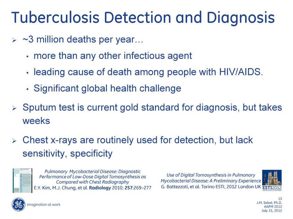
15 Standard PA chest radiograph (left) and single slice from the tomosynthesis image dataset (right) of a patient presenting with a lesion in the lower left lung. Although the lesion can be seen in the conventional image, it can be better visualized and characterized in the tomosynthesis slice. In particular it can be seen to be cavitated, characteristic of a tuberculosis infection. 15

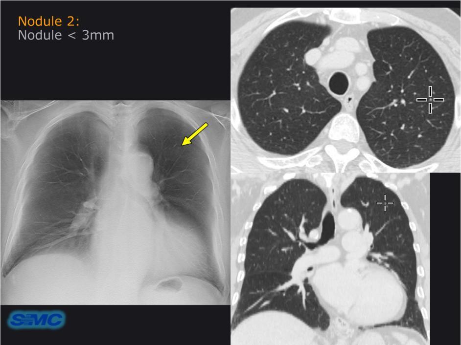
16 Tuberculosis with Small cavity Conventional PA x-ray (Left) and tomosynthesis slice image (right) of a tuberculosis patient with a small cavitated nodule. Image courtesy of Dr. M.J. Chung, Samsung Medical Center, Seoul Korea. 16

17 Accidental swallowing of a pill in a press-through package. Chest conventional radiograph (left) and tomosynthesis image (right). The tomosynthesis image enables improved visualization of the object as a donut-like lucent area in the esophagus, whereas the foreign object is not visible on the conventional radiograph. 17
18 18

19 Tomosynthesis image reveals a subtle fracture of the acetabulum, not seen on conventional hip radiograph. 19
.")
20 Patient imaged for routine follow-up of treatment for a fracture of the scaphoid. Conventional radiograph (left) appears to shown good evidence of healing (increased bone density at fracture site). However, examination of tomosynthesis images shows that despite increased bone density, the fracture still remains indicated by the gap in the bone (arrow in right image). This is a clear example of how overlying structures (in this case increased bone growth) can obscure underlying pathology. 20

21 Following a snowmobile accident, a patient was imaged at a rural trauma clinic. The AP radiograph on the left clearly shows a black line or shadow across the odontoid process raising the possibility of a fracture. A single slice from the tomosynthesis dataset (right image) removed the overlying shadow and the odontoid process was readily cleared for fracture. Had the ER physician not been able to clear the spine he would have had to send this patient, via ambulance, to the nearest trauma hospital, over 4 hours away. 21

22 13 year old with dislocation Of epiphyseal (growth) plate of the femur in extreme hip pain following surgery. Left: A post surgical projection image to determine if the implanted screw had invaded the joint space. Center and Right: Tomosynthesis imaging allows for complete assessment of the joint without overlying structures with minimal metal artefacts. The tomosynthesis images revealed that the screw had not invaded the joint space. 22

23 Left: Tomosynthesis images of a trauma patient imaged in the ER following a motor vehicle accident. Extensive trauma to lower leg and ankle can be visualized in the tomosynthesis data set, despite the presence of a metal fixation splint. Sufficient information about the extent of the fractures and the location of the bone fragments enabled the surgeon to plan intervention without the need for CT imaging. Right: Postoperative follow-up after total hip replacement: (Left) tomosynthesis slice image (Right) CT MPR coronal image. The cranio-caudal tomosynthesis sweep provides detailed information on potential loosening or peri-prosthetic fracture around the femoral stem prosthesis, compared to the conventional radiograph and with much less metallic artifact than CT. In the tomosynthesis image the minor metallic artifact appears as a low signal level or undershooting only along the sweep direction at the edges of the metallic prosthesis. 23
24 24

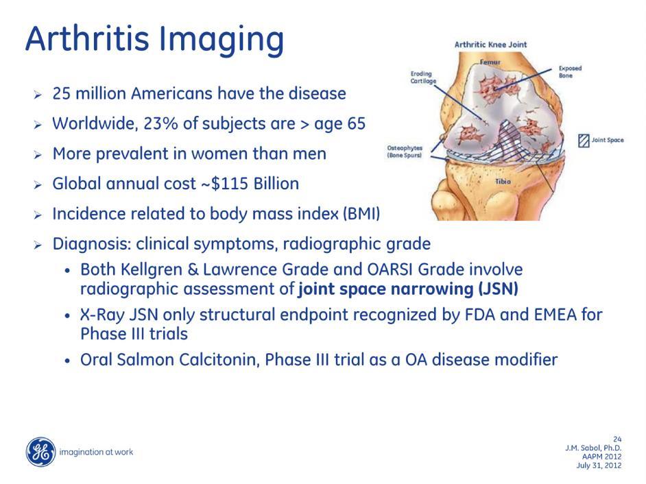
25 Upper: Examples of improved visualization and characterization of bone erosions in a patient with rheumatoid arthritis. Lower Left: Example of a conventional PA radiograph and a slice from a tomosynthesis dataset of a patient being image for assessment of knee osteoarthritis. As seen on the tomosynthesis image, The patient has a bone chip in the knee joint space. The bone chip is very difficult to detect on the conventional image. Lower Right: Conventional PA radiograph and a slice from a tomosynthesis dataset of a patient presenting with osteoarthritis. The extent of narrowing of the joint space is readily apparent on the tomosynthesis images. 25

26 With tomosynthesis imaging, it is possible to extract quantitative information about the joint space. In this case, a semi-automatic algorithm has been developed by Kalinosky et al to segment the edges of the femur and tibia and define the extent of the joint space in the knee. An example tomosynthesis slice image (Left) extracted joint space width profile (center) and reconstructed 2-D map of the joint space (right) are shown for both PA (Upper) and Lateral (Lower) image acquisitions. The extracted joint space profile are compared to values extracted from a CT dataset (center). 26
27 27

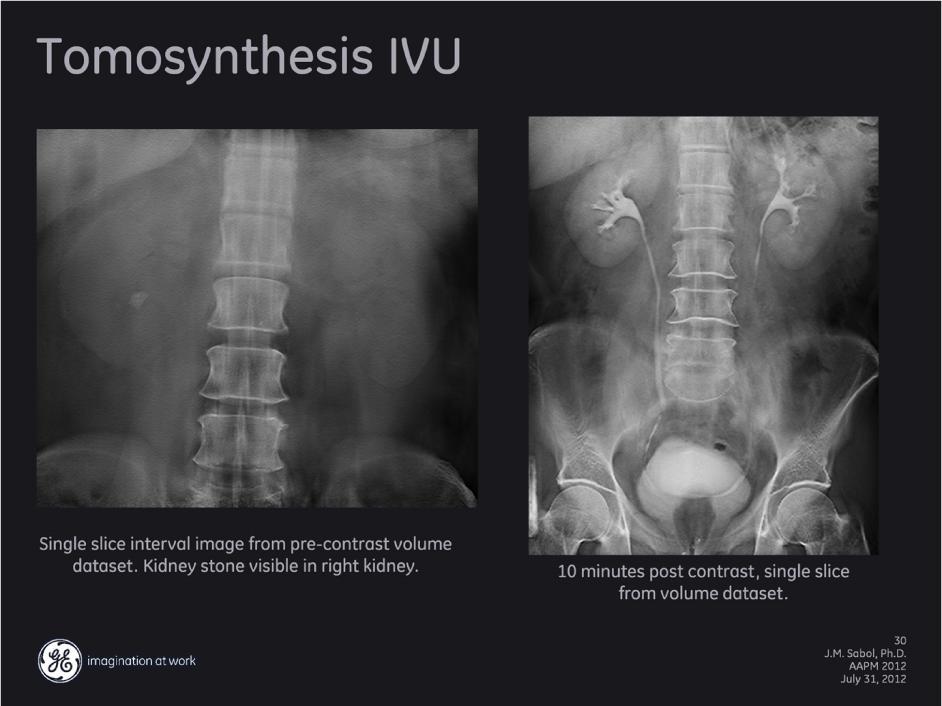
28 Globally, IVP procedures with traditional x-ray is still a common procedure. Tomosynthesis replaces the need for linear tomography which is prone to fulcrum errors and increased dose. Linear tomography is performed to determine size, location and shape of the kidneys and generally limited to a field of view of 10x12. Tomosynthesis offers an increased FOV when needed, at the same dose as coned FOV s. This patient has multiple areas of concern that were visualized in the full field FOV mode. 28

29 Individual slice images from previously shown patient. Slice 9 shows a kidney stone formation within the kidney. Slice 15 shows a Greenfield filter in the venacava used to trap blood clots from migrating to the lungs. Slice 18 shows calcification in the Iliac arteries and distal aorta. Slice 22 shows gall stones within the gall bladder. 29
30 30
31 31
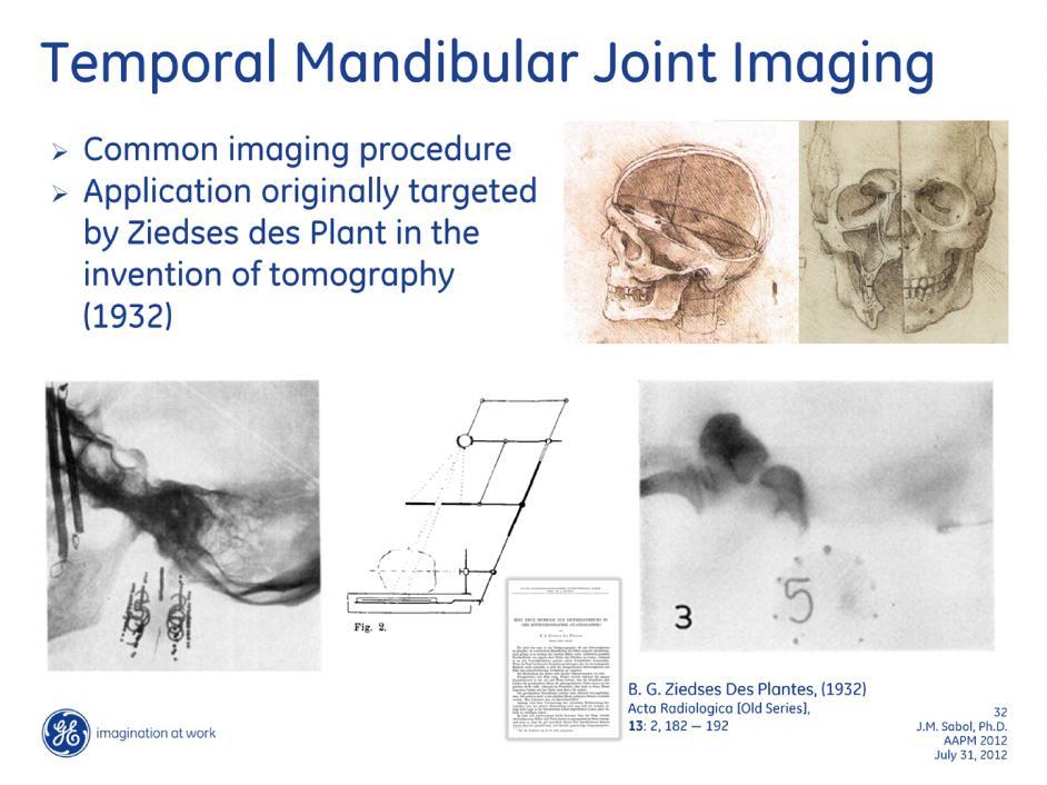
32 32

33 Modern digital tomosynthesis enables high resolution reconstruction of slice images through both TMJ joints after a single acquisition sweep. 33

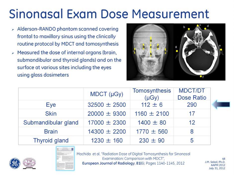
34 Chronic maxillary sinusitis with obstruction of the natural ostia clearly visible on the tomosynthesis images (left). The complete obstruction of the patient s right maxillary sinus can be seen, in comparison with the patent left sinus. Similar results can be seen on the much higher dose MDCT image (right). 34
 well delineates the air-fluid level in the left maxillary sinus more easily and with less radiation dose than CT.")
35 Acute maxillary sinusitis as demonstrated on sinus tomosynthesis and MDCT MPR coronal images. The tomosynthesis images (left) well delineates the air-fluid level in the left maxillary sinus more easily and with less radiation dose than CT. Note that tomosynthesis enables imaging in both the upright and supine positions which changes the appearance of the air-fluid level, unlike in CT. 35
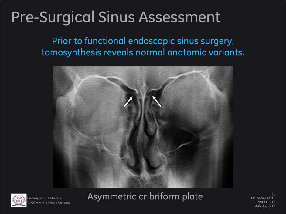
36 36
 as well as the mandibular canal than the conventional radiograph.")
37 Mandibular cyst imaged with both conventional mandibular radiography (left) and tomosynthesis (right). The tomosynthesis radiograph better delineates the cyst (arrows) as well as the mandibular canal than the conventional radiograph. Note that there is minimal metallic artifact produced as a result of the dental fillings. 37

38 Example of a blow-out fracture on both tomosynthesis and CT MPR coronal images. The tomosynthesis image clearly delineates the fracture of the right orbital floor. Tomosynthesis offers easier imaging at less cost and radiation exposure than with CT. However, due to relatively lower contrast sensitivity, it is more difficult to differentiate the content prolapsing into the maxillary sinus with tomosynthesis than it is with CT. Note that the air-fluid level (arrowhead) resulting from the hemorrhage is visible in the right maxillary sinus in the tomosynthesis image due to the upright position acquisition. 38
39 39

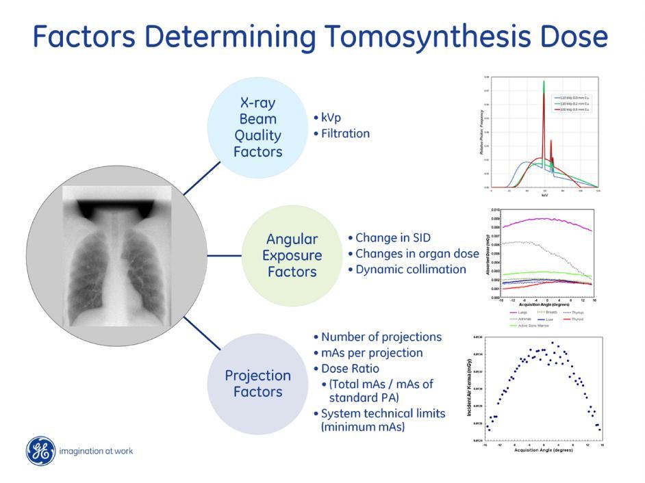
40 Left: Linear path of x-ray source changes SID, decreases relative exposure to the patient at extremes of the sweep Middle: Collimation is adjusted with projection angle. Dose area changes, excess dose is not delivered to the patient. Relevant to possible organ-specific dose effects at different views Right: mas or dose may be changed as a function of view angle. Total tomosynthesis mas is often expressed as a ratio of the mas for the standard projection view. 40
41 84 different techniques investigated 41
42 42
43 43
44 The minimum exposure duration and finite mas increments can prevent the tomosynthesis system from delivering the desired exposure for all desired exposure techniques. This results in a lower dose to the patient for most techniques. However, for some techniques, specifically those of high kvp and minimal filtration, the tomosynthesis systems can deliver higher exposures than desired. Similarly, the desired exposures corresponding to very low dose ratios will not routinely be delivered by the system. 44
 were acquired with different techniques demonstrating significant changes in dose possible with tomosynthesis.")
45 Examples of images from thoracic tuberculosis cases shown previously with estimated effective dose for each acquisition. The tomosynthesis images (center and right) were acquired with different techniques demonstrating significant changes in dose possible with tomosynthesis. Further research into the optimization is required to determine the dose required for different tomosynthesis exams. 45
46 46
47 47
48 48
49 49
50 50
51 51
52 52
Diagnostic Imaging
 www.fisiokinesiterapia.biz Diagnostic Imaging Diagnostic Imaging is no longer limited to radiography. Major technological advancements have lead to the use of new and improved imaging technologies. The
Shoulder Position: Supine arm in the neutral position. Collateral arm above head Indication: fracture humerus, fracture scapula
 Shoulder Position: Supine arm in the neutral position. Collateral arm above head Indication: fracture humerus, fracture scapula No instrumentation With metal or cast KV/ Effective mas/rotation time 140/300/1.0
CT Imaging at the Point-of-Care
 ENGLISH True Dedication The new Planmed Verity Extremity CT Scanner revolutionizes extremity CT imaging. The compact unit brings 3D imaging at emergency departments, orthopedic clinics or trauma centers
CLINICAL AND OPERATIVE APPROACH FOR TOTAL KNEE REPLACEMENT DR.VINMAIE ORTHOPAEDICS PG 2 ND YEAR
 CLINICAL AND OPERATIVE APPROACH FOR TOTAL KNEE REPLACEMENT DR.VINMAIE ORTHOPAEDICS PG 2 ND YEAR Evolution of TKR In 1860, Verneuil proposed interposition arthroplasty, involving the insertion of soft tissue
Routine Guide EXAMINATION PROJECTION CASSETTE SIZE NOTES PRINT ORIENTATION. 14x17 CW* 14x17LW 14x17LW. 14x17LW 14x17LW 14x17LW
 EXAMINATION PROJECTION CASSETTE SIZE NOTES PRINT ORIENTATION A-C Joints without weights with weights 14x17 CW* One 14x17 divided; both shoulders on one exposure. *If part does not fit, do 10x12s CW. Both
R/F. Use of Tomosynthesis at the Aizawa Hospital. 1. Introduction. 2. Use of Tomosynthesis at the Aizawa Hospital
 R/F Use of Tomosynthesis at the Aizawa Hospital Diagnostic Imaging Center, Aizawa Hospital Masahiro Iwama, Kei Takehara, Kouichi Anraku Mr. Masahiro Iwama 1. Introduction The Aizawa Hospital is located
CT of the Head, Spine, and Cerebral Vessels
 CT of the Head, Spine, and Cerebral Vessels Objectives Determine specific imaging plane used to acquire or reformat CT scan, i.e. sagittal, coronal, transverse, and offaxis or oblique. Assess and evaluate
R/F. Clinical Experience Using the SONIALVISION safire II Utility of Tomosynthesis in Orthopedic Surgery
 R/F Clinical Experience Using the SONIALVISION safire II Utility of Tomosynthesis in Orthopedic Surgery Iwate Medical University Hospital, Central Department of Radiology Shouta Miura Mr. Shouta Miura
Digital tomosynthesis (DT) has been well described as a
 has been well described as a") Case Report The Usefulness of Digital Tomosynthesis (DT) in Assisting in Cases of Doubtful Routine Radiography and/or Computed Tomography (CT) Image. Abstract Digital tomosynthesis is useful in assisting
Case Report The Usefulness of Digital Tomosynthesis (DT) in Assisting in Cases of Doubtful Routine Radiography and/or Computed Tomography (CT) Image. Abstract Digital tomosynthesis is useful in assisting
Complex Fractures and Hip Dislocations
 IMAGING OF HIP PAIN Patients may present with acute (< 2 weeks) or chronic hip pain. Acute pain may be related or not related to an acute traumatic event such as fall or trauma from a motor vehicle accident.
Low Dose Excellent Image Quality Rapid Reconstruction
 Low Dose Excellent Image Quality Rapid Reconstruction Efficient 3 in 1 Dental X-ray System CBCT > Precise 3-D Anatomical structures - Accurate diagnosis for doctors - Safe implant for patients > Significant
R/F. Can T-smart Tomosynthesis Improve Diagnostic Accuracy on THA Component Stability? 1. Abstract
 R/F Can T-smart Tomosynthesis Improve Diagnostic Accuracy on THA Component Stability? Professor and Chair Dept. of Adult Reconstructive Surgery Beijing Jishuitan Hospital, the 4th Clinical College of PKU
Flexible Easy Competitive. SCANORA 3Dx - The in-office large field-of-view Cone Beam CT system for Head and Neck imaging
 Flexible Easy Competitive SCANORA 3Dx - The in-office large field-of-view Cone Beam CT system for Head and Neck imaging SCANORA 3Dx. The solution. SCANORA 3Dx makes advanced 3D imaging easy in the head
7/23/2018 DESCRIBING THE FRACTURE. Pattern Open vs closed Location BASIC PRINCIPLES OF FRACTURE MANAGEMENT. Anjan R. Shah MD July 21, 2018.
 BASIC PRINCIPLES OF FRACTURE MANAGEMENT Anjan R. Shah MD July 21, 2018 DESCRIBING THE FRACTURE Pattern Open vs closed Location POLL OPEN HOW WOULD YOU DESCRIBE THIS FRACTURE PATTERN? 1 Spiral 2 Transverse
Metal Artefact Reduction in CT
 Metal Artefact Reduction in CT DANIEL MARRINER Metal Artefact Reduction in CT Metal Artefact Clinical Indications for MAR SEMAR and How It Works Technical Considerations Case Studies utilising SEMAR Metal
Proteus XR/f Patient positioning guide
 Proteus XR/f Patient positioning guide PROTEUS XR/F Now a single digital x-ray room accommodates nearly all your radiographic studies. With extended tube coverage and wireless detectors, Proteus XR/f gives
X-Ray & CT Physics / Clinical CT
 Computed Tomography-Basic Principles and Good Practice X-Ray & CT Physics / Clinical CT INSTRUCTORS: Dane Franklin, MBA, RT (R) (CT) Office hours will be Tuesdays from 5pm to 6pm CLASSROOM: TIME: REQUIRED
CT SCAN PROTOCOL. Shoulder
 CT SCAN PROTOCOL Shoulder Purpose and Summary CT images made with this protocol are used to provide the orthopedic surgeon with a detailed 3D anatomical reconstruction of the patient s scapula and proximal
3D Cone beam CT & Digital Radiography Dedicated to Otorhinolaryngology
 3D Cone beam CT & Digital Radiography Dedicated to Otorhinolaryngology Multi-functional imaging solution3 RAYSCAN m is an unique 2-in-1 imaging solution, combining Cone Beam CT and Digital Radiography,
Basics of MR Imaging. Dynamic MRI. MRI Closed. The bed rotates from Upright to Recumbent, stopping at any angle in between.
 Basics of MR Imaging Dynamic MRI MRI Closed The bed rotates from Upright to Recumbent, stopping at any angle in between MRI Open Patient with Low Back Pain After Surgery Extremity MRI Sagittal T2 WI of
X-ray (Radiography) - Bone
 - Bone") Scan for mobile link. X-ray (Radiography) - Bone Bone x-ray uses a very small dose of ionizing radiation to produce pictures of any bone in the body. It is commonly used to diagnose fractured bones or
Scan for mobile link. X-ray (Radiography) - Bone Bone x-ray uses a very small dose of ionizing radiation to produce pictures of any bone in the body. It is commonly used to diagnose fractured bones or
Emerging Applications in Musculoskeletal CT Imaging
 Emerging pplications in Musculoskeletal CT Imaging y K Murali MD(RD), PDCC, Director of Interventional Radiology, G. Francis DMRD, DN (RD), Consultant Radiologist, and R. Madan, MS, MD, Consultant Radiologist,
*smith&nephew. BIRMINGHAM HIP Resurfacing (BHR ) System Patient Information Revision 0
 System Patient Information Revision 0") *smith&nephew BIRMINGHAM HIP Resurfacing (BHR ) System Patient Information 0120 0021666 - Revision 0 Table of Contents 1.0 What is the BHR Device? 2.0 What is the Purpose of the BHR Device? 3.0 When Should
*smith&nephew BIRMINGHAM HIP Resurfacing (BHR ) System Patient Information 0120 0021666 - Revision 0 Table of Contents 1.0 What is the BHR Device? 2.0 What is the Purpose of the BHR Device? 3.0 When Should
CAUTION Federal law (USA) restricts this device to sale, by or on the order of a physician.
 restricts this device to sale, by or on the order of a physician.") CAUTION Federal law (USA) restricts this device to sale, by or on the order of a physician. ENGLISH Mpact 3D Metal Implants and Augments 3D Metal INSTRUCTION FOR USE Important notice: the device(s) can
CAUTION Federal law (USA) restricts this device to sale, by or on the order of a physician. ENGLISH Mpact 3D Metal Implants and Augments 3D Metal INSTRUCTION FOR USE Important notice: the device(s) can
HI-Res Extremity Sensation 16
 Page 1 Routine Extremity - (2/14/2013) CTDI: ~20 mgy per acquisition Used for evaluation of: Humerus Forearm Femur Knee Tib/Fib Billing: 1. CT Upper/Lower Extremity of concern without contrast, with contrast,
Metal Artifact Reduction by Dual Energy CT
 Metal Artifact Reduction by Dual Energy CT Poster No.: C-0108 Congress: ECR 2011 Type: Authors: Keywords: DOI: Scientific Paper T. Johnson, F. Bamberg, A. Dierks, H.-C. Becker, M. F. Reiser; Munich/DE
Cone Beam 3D Imaging
 Cone Beam 3D Imaging NewTom Sets the Standard in 3D Maxillofacial Imaging Cone Beam 3D Imaging The Global Market Leader The Inventors n of Cone Beam 3D In 1996, QR srl developed the first generation of
Bone Densitometry Radiation dose: what you need to know
 Bone Densitometry Radiation dose: what you need to know John Damilakis, PhD Associate Professor and Chairman University of Crete, Iraklion, Crete, GREECE Estimation of bone status using X-rays Assessment
Specialist Referral Service Willows Information Sheets. Limb deformity
 Specialist Referral Service Willows Information Sheets Limb deformity Limb deformity Why do limbs become deformed? The limbs of dogs and cats, like the legs of people, are meant to be relatively straight.
Ethan M. Braunstein, M.D. 1, Steven A. Goldstein, Ph.D. 2, Janet Ku, M.S. 2, Patrick Smith, M.D. 2, and Larry S. Matthews, M.D. 2
 Skeletal Radiol (1986) 15:27-31 Skeletal Radiology Computed tomography and plain radiography in experimental fracture healing Ethan M. Braunstein, M.D. 1, Steven A. Goldstein, Ph.D. 2, Janet Ku, M.S. 2,
5/31/2018. Ipsilateral Femoral Neck And Shaft Fractures. Ipsilateral Neck-Shaft Fractures Introduction. Ipsilateral Neck-Shaft Fractures Introduction
 Ipsilateral Femoral Neck And Shaft Fractures Exchange Nailing For Non- Union Donald Wiss MD Cedars-Sinai Medical Center Los Angeles, California Introduction Uncommon Injury Invariably High Energy Trauma
Extraoral Imaging. Chapter 42. Copyright 2018, Elsevier Inc. All Rights Reserved. 1
 Extraoral Imaging Chapter 42 Copyright 2018, Elsevier Inc. All Rights Reserved. 1 Learning Objectives Lesson 42.1: Panoramic Imaging 1. Pronounce, define, and spell the key terms. 2. Discuss panoramic
4/28/2010. Fractures. Normal Bone and Normal Ossification Bone Terms. Epiphysis Epiphyseal Plate (physis) Metaphysis
 Metaphysis") Fractures Normal Bone and Normal Ossification Bone Terms Epiphysis Epiphyseal Plate (physis) Metaphysis Diaphysis 1 Fracture Classifications A. Longitudinal B. Transverse C. Oblique D. Spiral E. Incomplete
Fractures Normal Bone and Normal Ossification Bone Terms Epiphysis Epiphyseal Plate (physis) Metaphysis Diaphysis 1 Fracture Classifications A. Longitudinal B. Transverse C. Oblique D. Spiral E. Incomplete
PACS: ERGONOMIC CONSIDERATIONS 1
 RADIOLOGY RESEARCH Radiographic Tomosynthesis: Acquisition Parameters Michael J. Flynn, PhD Henry Ford Health System Detroit, MI Learning Objectives Learn.. 1. Appreciate the importance of scan direction,
The diagnostic value of Computed Tomography in evaluation of maxillofacial Trauma
 The diagnostic value of Computed Tomography in evaluation of maxillofacial Trauma Qais H. Muassa FICMS College of Dentistry, Babylon University Ibrahim S. Gataa, BDS, FICMS College of Dentistry, Sulaimania
28 Surgical Technique
 Surgical Technique 10 12 14 16 18 20 22 24 28 26 Technique described by James L. Guyton, MD Campbell Clinic Memphis, Tennessee James W. Harkess, MD Campbell Clinic Memphis, Tennessee David G. LaVelle,
Clinical Image Gallery Next Generation Volume 1
 Clinical Image Gallery Next Generation Volume 1 Dr. Russell Bull Royal Bournemouth Hospital, Bournemouth, United Kingdom After long experience with the first generation, a next generation Aquilion ONE
CT Scanning Protocol For V2R Guided Surgery Solutions
 CT Scanning Protocol For V2R Guided Surgery Solutions 2 V2R CT Scanning Protocol \\ Contents Contents General requirements... 3 V2R Dual Scan Protocol... 5 V2R Single Scan Protocol... 8 Overview... 10
 www.oralradiologists.com CONE BEAM CT REPORT CASE ---- Case Information Referring Doctor: - Patient Name: - Scan Date: December 1, 2015 Patient DOB: - Reason for Exam: - Study Details: icat Flex, 160x160x112
CPT CODES. Ph: (307) Fax: (307) CATSCAN IV Contrast: 87.00
 Fax: (307) CATSCAN IV Contrast: 87.00") Ph: (307) 382-4282 Fax: (307) 382-4291 CPT CODES CATSCAN IV Contrast: 87.00 74150 Abdomen w/o contrast $ 809.00 74160 Abdomen w/ contrast $1175.00 w/ contrast: $1262.00 74170 Abdomen w_w/o contrast $1324.00
Ph: (307) 382-4282 Fax: (307) 382-4291 CPT CODES CATSCAN IV Contrast: 87.00 74150 Abdomen w/o contrast $ 809.00 74160 Abdomen w/ contrast $1175.00 w/ contrast: $1262.00 74170 Abdomen w_w/o contrast $1324.00
5G XL - R EN ENGLISH
 5G XL - R15.0 - EN ENGLISH Sede legale ed amministrativa - Headquarters QR srl - Via Selice Provinciale, 23/a - 40026 Imola - Bo (Italy) Stabilimento - Plant Via Fermi, 40-37136 Verona (Italy) Tel. +39
3D titanium interbody fusion cages sharx. White Paper
 3D titanium interbody fusion cages sharx (SLM selective laser melted) Goal of the study: Does the sharx intervertebral cage due to innovative material, new design, and lordotic shape solve some problems
CBCT Specific Guidelines for South African Practice as Indicated by Current Literature:
 CBCT Specific Guidelines for South African Practice as Indicated by Current Literature: CF Hoogendijk Maxillo- facial and Oral surgery: Trauma: 1. Facial trauma for the confirmation or exclusion of fractures
FOR CMS (MEDICARE) MEMBERS ONLY NATIONAL COVERAGE DETERMINATION (NCD) FOR MAGNETIC RESONANCE IMAGING:
 MEMBERS ONLY NATIONAL COVERAGE DETERMINATION (NCD) FOR MAGNETIC RESONANCE IMAGING:") National Imaging Associates, Inc. Clinical guidelines TEMPOROMANDIBULAR JOINT (TMJ) MRI Original Date: May 23, 2003 Page 1 of 5 CPT Code: 70336 Last Review Date: May 2016 NCD 220.2 MRI Last Effective Date:
National Imaging Associates, Inc. Clinical guidelines TEMPOROMANDIBULAR JOINT (TMJ) MRI Original Date: May 23, 2003 Page 1 of 5 CPT Code: 70336 Last Review Date: May 2016 NCD 220.2 MRI Last Effective Date:
DIRECT ANTERIOR APPROACH
 DIRECT ANTERIOR APPROACH JOINT REPLACEMENT PROGRAM 2301 25TH STREET SOUTH FARGO ND 58103 CENTER FOR MINIMAL INVASIVE JOINT SURGERY (p) 701-241-9300 (tf) 866-887-9300 www.jointpain.md FARGO FERGUS FALLS
Orthopedic Trauma. I have nothing to disclose. Objectives 3/7/2018. What is Orthopedic Trauma? What is Orthopedic Trauma? Trauma
 Orthopedic Trauma I have nothing to disclose. David Miller Memorial Trauma Symposium 2017 James Black, MD Mercy Orthopedic Trauma - Springfield 10/27/2017 Objectives What is Orthopedic Trauma? What is
RADIOGRAPHY OF THE KNEE, PATELLA, and FEMUR
 RADIOGRAPHY OF THE KNEE, PATELLA, and FEMUR KNEE AP Projection Patient Position: Part Position: Leg in Center Femoral condyles Central Ray: - Asthenic patient - if ASIS to tabletop is < 19 cm Sthenic patient
Intraoperative case studies. Portable full body 32-slice CT scanner
 Intraoperative case studies Portable full body 32-slice CT scanner Point-of-care CT imaging Your multi-departmental imaging solution Orthopedic surgery Arthroplasty Musculoskeletal disorders Hip replacement
Country Health SA Medical Imaging
 Country Health SA Medical Imaging REMOTE OPERATORS POSITIONING GUIDE Contents Image Evaluation Page 4 Positioning Guides Section 1 - THORAX 1.1 Chest Page 5 1.2 Bedside Chest Page 7 1.3 Ribs Page 8 Section
FOLLICULAR / OVULATION STUDY USG HIP JOINT (LEFT) USG HIP JOINT (RIGHT) USG KNEE JOINT (LEFT) USG KNEE JOINT (RIGHT) USG KUB USG MUSKULOSKELETAL USG
 USG HIP JOINT (RIGHT) USG KNEE JOINT (LEFT) USG KNEE JOINT (RIGHT) USG KUB USG MUSKULOSKELETAL USG") RADIOLOGY TESTS SONOGRAPHY,COLOR DOPPLER 3D/4D ANAMOLY SCAN 3D/4D ANAMOLY SCAN TWINS 3D/4D USG PELVIS ABDOMEN & PELVIS USG ABDOMEN UPPER USG ANKLE JOINT (LEFT) USG ANKLE JOINT (RIGHT) USG B SCAN BREAST
RADIOLOGY TESTS SONOGRAPHY,COLOR DOPPLER 3D/4D ANAMOLY SCAN 3D/4D ANAMOLY SCAN TWINS 3D/4D USG PELVIS ABDOMEN & PELVIS USG ABDOMEN UPPER USG ANKLE JOINT (LEFT) USG ANKLE JOINT (RIGHT) USG B SCAN BREAST
Semiotics in Radiology
 Adelino Santos Health Technology College Coimbra, Portugal Collaboration of António Agudo Student of Radiology College of Health Technology Coimbra, Portugal What are the most important points to evaluate
DISLOCATION AND FRACTURES OF THE HIP. Dr Károly Fekete
 DISLOCATION AND FRACTURES OF THE HIP Dr Károly Fekete 1 OUTLINE Epidemiology Incidence Anatomy Patient s examination, clinical symptons Diagnosis Classification Management Special complications 2 EPIDEMIOLOGY,
revised originals as separate pages on I://DX(all folders)/trauma X Manual and project
/trauma X Manual and project") 06-25-14 revised originals as separate pages on I://DX(all folders)/trauma X Manual and project Procedure for Suspected Child Abuse Imaging Trauma X GUIDELINES: Every effort should be made to request studies
06-25-14 revised originals as separate pages on I://DX(all folders)/trauma X Manual and project Procedure for Suspected Child Abuse Imaging Trauma X GUIDELINES: Every effort should be made to request studies
T h e D e n t a l C o m p a n y FROM DIAGNOSTIC SCAN TO SURGERY, WE SHAPE THE FUTURE OF DENTISTRY.
 T h e D e n t a l C o m p a n y FROM DIAGNOSTIC SCAN TO SURGERY, WE SHAPE THE FUTURE OF DENTISTRY. SIDEXIS SOFTWARE ORTHOPHOS SL D/D SEAMLESS THE NEW STANDARD IN CLINICAL DIAGNOSIS AND PATIENT COMMUNICATION
Hand and wrist emergencies
 Chapter1 Hand and wrist emergencies Carl A. Germann Distal radius and ulnar injuries PEARL: Fractures of the distal radius and ulna are the most common type of fractures in patients younger than 75 years.
AUA Guidelines for Imaging Known or Suspected Ureteral Calculi. Michael Ferrandino, MD Assoc Professor of Urology Duke University Medical Center
 AUA Guidelines for Imaging Known or Suspected Ureteral Calculi Michael Ferrandino, MD Assoc Professor of Urology Duke University Medical Center Imaging for Urolithiasis Justification for the Guidelines
Utility of Dual-Energy CT to Evaluate Patients with Hip and Pelvis Pain in the ER Setting
 Utility of Dual-Energy CT to Evaluate Patients with Hip and Pelvis Pain in the ER Setting Johnson, T., Moran, E., Glazebrook, K., Leng, S., Fletcher, J., and McCollough, C. An educational review ER011
3D-MODEL CUSTOM-MADE MODELS SEGMENTATION AND PRODUCTION SERVICE OF BONE MODELS WITH HIGHEST 3D PRINTING RESOLUTION
 CUSTOM-MADE MODELS 3D-MODEL SEGMENTATION AND PRODUCTION SERVICE OF BONE MODELS WITH HIGHEST 3D PRINTING RESOLUTION FOLLOW US ON CUSTOM-MADE MODELS 3D-MODEL From a CT or CBCT scan, 3D-model service provides
Practical Reduction Techniques: Diaphyseal Reduction. Philip Wolinsky University of California at Davis Medical Center
 OTA Specialty Day 2016 Practical Reduction Techniques: Diaphyseal Reduction Philip Wolinsky University of California at Davis Medical Center 8:55 am 9:55 am Tips and Tricks: Practical Reduction Techniques
Diagnostic Tools: Equine Dentistry. Dr. Chris Blevins Equine Field Service Clinician
 Diagnostic Tools: Equine Dentistry Dr. Chris Blevins Equine Field Service Clinician Objectives Know 3 useful diagnostic tools. What is most important aspect about dental radiology? Know 3 standard radiographs
Technique Manual Technique Manual Rev. A 01/11/2011
 Technique Manual Technique Manual Table of Contents Introduction 4 Indications 5 Instrumentation 6 Frame Assembly 7 Position Guide 8-9 Tips and Tricks 10-13 Cleaning 14 Sterilization 14 Storage Instructions
Robotic-Arm Assisted Surgery
 Mako TM Robotic-Arm Assisted Surgery for Total Hip Replacement A Patient s Guide Causes of Your Hip Pain Your joints are involved in almost every activity you do. Movements such as walking, bending and
Optimizing function Maximizing survivorship Accelerating recovery
 Surgical Technique Optimizing Function Maximizing Survivorship Accelerating Recovery The company believes in an approach to patient treatment that places equal importance on: Optimizing function Maximizing
The PinTrace system. medical robotics
 The PinTrace system The demand for minimal invasive surgery (MIS) with more rapid patient recovery creates an increasing need of high-performance technology, based on computer and robot assisted surgery.
PINTRACE method. Navigation with or without robot assistance. The PinTrace method is based entirely on robotassisted
 The TM PINTRACE method The demand for minimal invasive surgery (MIS) with more rapid patient recovery creates an increasing need of high-performance technology, based on computer and robot assisted surgery.
CLINICAL PAPER / ORTHOPEDIC
 HIP LEG LENGTH AND OFFSET Kelley T.C. and Swank M.L. (2009) Using CAS leads to more accurate positioning within the safe zone (inclination between 30 and 50, anteversion between 5 and 25 ) CAS improves
Diagnostic Tools: Equine Dentistry. Dr. Chris Blevins Equine Field Service Clinician
 Diagnostic Tools: Equine Dentistry Dr. Chris Blevins Equine Field Service Clinician Objectives Know 3 useful diagnostic tools. What is most important aspect about dental radiology? Know 3 standard radiographs
B. CT protocols for the spine
 B. CT protocols for the spine Poster No.: A-003 Congress: ECR 2010 Type: Invited Speaker Topic: Neuro Authors: B. Tins; Oswestry/UK Keywords: CT, spine, diagnostic imaging protocol DOI: 10.1594/ecr2010/A-003
Original Date: December 2015 Page 1 of 8 FOR CMS (MEDICARE) MEMBERS ONLY
 MEMBERS ONLY") National Imaging Associates, Inc. Clinical guidelines TOTAL JOINT ARTHROPLASTY -Total Hip Arthroplasty -Total Knee Arthroplasty -Replacement/Revision Hip or Knee Arthroplasty CPT4 Codes: Please refer to
National Imaging Associates, Inc. Clinical guidelines TOTAL JOINT ARTHROPLASTY -Total Hip Arthroplasty -Total Knee Arthroplasty -Replacement/Revision Hip or Knee Arthroplasty CPT4 Codes: Please refer to
Commonly Missed Injuries of the Extremities
 Commonly Missed Injuries of the Extremities Dr. Tudor H. Hughes M.D., FRCR Department of Radiology University of California School of Medicine San Diego, California 1. Base of skull 2. Odontoid process
Head to new heights with your imaging SCANORA 3D
 SCANORA 3D Head to new heights with your imaging Benefits at a glance The solution for dentomaxillofacial and ENT imaging Easy Patient seated for added stability during exposure. Clear, self-explinatory
Overview. Acceptance criteria for all protocols
 X-Ray protocol Overview The Smith & Nephew VISIONAIRE X-Ray protocol is essentially an AP leg length image. The images are preferred to be done erect, but can be done supine if necessary due to the type
Conventional radiograph verses CT for evaluation of sagittal fracture of mandibular condyle
 Case Report: Conventional radiograph verses CT for evaluation of sagittal fracture of mandibular condyle Dr Anjali Wadhwa, Dr Gaurav Shah, Dr Shweta Sharma, Dr Anand Bhatnagar, Dr Pallavi Malaviya NIMS
Fundamentals, Techniques, Pitfalls, and Limitations of MDCT Interpretation and Measurement
 Fundamentals, Techniques, Pitfalls, and Limitations of MDCT Interpretation and Measurement 3 rd Annual Imaging & Physiology Summit November 20-21, 21, 2009 Seoul, Korea Wm. Guy Weigold, MD, FACC Cardiovascular
Indications and limits of digital tomosynthesis in trauma imaging.
 Indications and limits of digital tomosynthesis in trauma imaging. Poster No.: C-1788 Congress: ECR 2015 Type: Educational Exhibit Authors: P. Gollini, G. Battezzati, G. Cortese; torino/it Keywords: Digital
Introduction to Radiology
 Introduction - Lecture 1 436 Teams Introduction to Radiology Objectives Introduce the various Medical Imaging Modalities. Understand the basics of image generation. Relate imaging to gross anatomy. Appreciate
NewTom 5G XL EXTRA.VISION
 CEFLA s.c. Via Selice Provinciale 23/a 40026 Imola Italy t. +39 045 8202727 045 583500 info@newtom.it newtom.it 06/2018 N5GXGB181S00 According to the standards in force, in extra-eu areas the availability
A 42-year-old patient presenting with femoral
 Kanda et al. Journal of Medical Case Reports 2015, 9:17 JOURNAL OF MEDICAL CASE REPORTS CASE REPORT Open Access A 42-year-old patient presenting with femoral head migration after hemiarthroplasty performed
Pediatric CT Protocols (18 years old or less)
") Pediatric CT Protocols (18 years old or less) Ped1: Head CT Ped2: Cervical spine CT Ped3: Sinus CT Ped4: Neck CT Ped5: Chest CT Ped6: Abdomen and pelvis CT Ped7: Thoracic or lumbar spine CT Ped8: Extremity
Pediatric CT Protocols (18 years old or less) Ped1: Head CT Ped2: Cervical spine CT Ped3: Sinus CT Ped4: Neck CT Ped5: Chest CT Ped6: Abdomen and pelvis CT Ped7: Thoracic or lumbar spine CT Ped8: Extremity
RADIOGRAPHY OF THE ANKLE and LOWER LEG
 RADIOGRAPHY OF THE ANKLE and LOWER LEG Patient Position: ANKLE AP Projection Part Position: True Slight to place foot s long axis Center to Central Ray: to IR Midway Note: Ankle joint is to tips of malleoli
2016 SIGN Conference: Treatment of Difficult Fractures Around The World
 2016 SIGN Conference: Treatment of Difficult Fractures Around The World September 21-24, 2016 Long Bone Osteosynthesis Failure DUONG BUNN Trauma-Orthopedique ward Preah Kossamak Preah Sihaknouk Ville Beach
Digital Imaging from a new perspective
 TREATMENT CENTRES HANDPIECES HYGIENE SYSTEMS X-RAY SYSTEMS CEREC TREATMENT CENTRES HANDPIECES HYGIENE SYSTEMS X-RAY SYSTEMS CEREC SIRONA CREATING AND MAINTAINING VALUE. You are right to expect a great
Osteonecrosis - Spectrum of imaging findings
 Osteonecrosis - Spectrum of imaging findings Poster No.: C-1861 Congress: ECR 2016 Type: Educational Exhibit Authors: P. Ninitas, A. L. Amado Costa, A. Duarte, I. Távora ; Lisbon/ 1 1 2 1 1 2 PT, Costa
PROPHECY. Preoperative Navigation Guides ANKLE CT SCAN PROTOCOL
 PROPHECY Preoperative Navigation Guides ANKLE CT SCAN PROTOCOL 90 FIGURE 1 Examples FIGURE 1 Examples of neutral ankle positioning. PROPHECY Ankle CT Scan Protocol PROPHECY INBONE and PROPHECY INFINITY
LIMB LENGTH DISCREPANCIES
 LIMB LENGTH DISCREPANCIES Jill C Flanagan, MD OBJECTIVES Evaluate the patient with a possible limb length difference (LLD) Understand general treatment principles when managing limb length differences
MRI PEDIATRIC PROTOCOLS (Updated 6/19/2018)
") MRI PEDIATRIC PROTOCOLS (Updated 6/19/2018) *Please get or let us know where radiologist can review plain films. *For Texas Orthopedics and other Docs requesting only MSK section read for their pediatric
MRI PEDIATRIC PROTOCOLS (Updated 6/19/2018) *Please get or let us know where radiologist can review plain films. *For Texas Orthopedics and other Docs requesting only MSK section read for their pediatric
CS 9300 Family. The power of flexibility
 CS 9300 Family The power of flexibility The new CS 9300 digital imaging system from Carestream Dental take the guesswork out of examinations The all-in-one CS 9300 is the most versatile multimodality imaging
INVISION Total Ankle Replacement System with PROPHECY Preoperative Navigation Revision of a Failed Agility Total Ankle Replacement
 016625 REVISION R INVISION Total Ankle Replacement System with PROPHECY Preoperative Navigation Revision of a Failed Agility Total Ankle Replacement CASE STUDY Patient History The patient was a 65-year-old
Optimum implant geometry
 Surgical Technique Optimum implant geometry Extending proven Tri-Lock heritage The original Tri-Lock was introduced in 1981. This implant was the first proximally coated tapered-wedge hip stem available
Complications of Treatment: Nonsurgical and Surgical
 Complications of Treatment: Nonsurgical and Surgical Whenever orthopedic surgeons discuss a treatment with patients we must always consider the risks and complications of any treatment we recommend. Part
Disclosure. FAI: Imaging Modalities and Dynamic Imaging Software. Acceptance of Hip Arthroscopy & FAI. Public. Payors. Orthopaedic Community
 2015 Chicago Sports Medicine Symposium Chicago, Illinois USA FAI: Imaging Modalities and Dynamic Imaging Software Allston J. Stubbs, M.D., M.B.A. Medical Director Hip Arthroscopy & Associate Professor
TOTAL HIP ARTHROPLASTY (Total Hip Replacement)
") (Total Hip Replacement) The Hip Joint The hip is a ball and socket joint. The joint is formed by the head of the femur (thighbone) and the acetabulum (pelvis). The bones are coated in cartilage, which
(Total Hip Replacement) The Hip Joint The hip is a ball and socket joint. The joint is formed by the head of the femur (thighbone) and the acetabulum (pelvis). The bones are coated in cartilage, which
Anabolic Therapy With Teriparatide Indications Beyond Osteoporosis
 Anabolic Therapy With Teriparatide Indications Beyond Osteoporosis Andreas Panagopoulos MD, PhD Upper Limb & Sports Medicine Orthopaedic Surgeon Assistant Professor, University of Patras Outline Teriparatide
Distal Femur Fractures in The Elderly The Ideal Construct
 Distal Femur Fractures in The Elderly The Ideal Construct Tak-Wing Lau Department of Orthopaedics and Traumatology Queen Mary Hospital The University of Hong Kong Singapore Trauma 2015 Trauma Through the
HELIOS h i p s y s t e m
 HELIOS h i p s y s t e m Design The Helios stem is a highly polished, High Nitrogen Stainless Steel (ISO5832-9) dual tapered cemented stem. The design of the stem is based on the clinically lly successful
Original Research THE USE OF REFORMATTED CONE BEAM CT IMAGES IN ASSESSING MID-FACE TRAUMA, WITH A FOCUS ON THE ORBITAL FLOOR FRACTURES
 DOI: 10.15386/cjmed-601 Original Research THE USE OF REFORMATTED CONE BEAM CT IMAGES IN ASSESSING MID-FACE TRAUMA, WITH A FOCUS ON THE ORBITAL FLOOR FRACTURES RALUCA ROMAN 1, MIHAELA HEDEȘIU 1, FLOAREA
Post test for O&P 2 Hrs CE. The Exam
 Post test for O&P 2 Hrs CE The Exam This examination is taken in "open book" format. That means you are free to answer the questions after research or discussion with your fellow workers. We feel this
R/F. Use of Tomosynthesis Radiography for Total Hip Arthroplasty (THA) at Iida Hospital. 1. Introduction. 2. Present Situation.
 at Iida Hospital. 1. Introduction. 2. Present Situation.") R/F Use of Tomosynthesis Radiography for Total Hip Arthroplasty (THA) at Iida Hospital Department of Radiology, Iida Hospital Shota Horitake Mr. Shota Horitake 1. Introduction Iida Hospital (Fig. 1) was
R/F Use of Tomosynthesis Radiography for Total Hip Arthroplasty (THA) at Iida Hospital Department of Radiology, Iida Hospital Shota Horitake Mr. Shota Horitake 1. Introduction Iida Hospital (Fig. 1) was
MANAGEMENT OF FRACTURE. Sudi maiteh (seminar 2 )
") MANAGEMENT OF FRACTURE Sudi maiteh (seminar 2 ) Management of fracture Subjects : _ general management of fractures & Orthopedic patient evaluation _ Closed and open fractures management (Conservative,
MANAGEMENT OF FRACTURE Sudi maiteh (seminar 2 ) Management of fracture Subjects : _ general management of fractures & Orthopedic patient evaluation _ Closed and open fractures management (Conservative,
Fractures Healing & Management. Traumatology RHS 231 Dr. Einas Al-Eisa Lecture 4
 Fractures Healing & Management Traumatology RHS 231 Dr. Einas Al-Eisa Lecture 4 Fractures Despite their strength, bones are susceptible to fractures. In young people, most fractures result from trauma