Understanding Leg Anatomy and Function THE UPPER LEG
|
|
|
- Aubrey Skinner
- 6 years ago
- Views:
Transcription
 at the knee joint.")

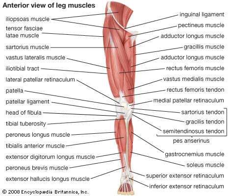
1 Understanding Leg Anatomy and Function THE UPPER LEG The long thigh bone is the femur. It connects to the pelvis to form the hip joint and then extends down to meet the tibia (shin bone) at the knee joint. The hip is a ball-and-socket joint where the head of the femur (ball) fits deeply and snugly into the acetabulum (socket) of the pelvis. This perfect fit gives the hip joint a great degree of stability. Because it is such a large and stable joint, it helps dissipate the forces that come from weight-bearing activities. The hip muscles can flex (bring the leg up toward the body), extend (move the leg backward away from the body), abduct (move the leg out to the side of the body), adduct (move the leg toward the midline of the body), and internally and externally rotate the leg. The knee joint consists of the femur, the tibia, and the patella (kneecap). Patellofemoral refers to the union between the patella and the femur. The primary muscles surrounding the knee joint are the quadriceps muscles on the front of the thigh, the adductor muscles on the inside of the thigh, the hamstring muscles on the back of the thigh, and the tensor fasciae latae and iliotibial band on the outside of the thigh. The patella is located inside the tendon of the rectus femoris muscle, which connects the femur to the tibia. The femur has a concavity or groove where the patella rides up during extension (knee straightening) and down during flexion (knee bending). The underside of the patella has many ridges that are covered with protective cartilage. The patella's function is to give the quadriceps muscles increased efficiency and to protect the front of the femur. Muscles of the upper leg will bring the other parts of the lower leg upward. The skeletal components of the knee joint are the protective patella, or kneecap, the femur, or thigh bone, connected at the joint to the tibia, the shin bone, and the fibula, which are the long bones of the lower leg. The integrity of the knee joint is secured by the sets of ligaments connecting the three bones, as well as through the stabilizing effect of knee cartilage.
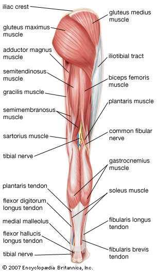
2 THE LOWER LEG The lower-leg bones, the tibia and fibula, also help to form the ankle joint. The tibia sits on top of the talus (an ankle bone), which fits into the calcaneus (heel bone). The fibula forms the lateral malleolus (the bony projection on the outside of your ankle). The lower-leg muscles can act on the ankle and either dorsiflex (bring the toes towards the nose) or plantarflex (point the toes down). They can also act at the foot and either invert (move the foot toward midline) or evert (move the foot away from the body). The foot's bony and muscular anatomy is very similar to that of the hand. There are tarsals, metatarsals, and phalanges, and the muscles can flex, extend, abduct, and adduct the toes. The lower leg is a remarkable structure, where each of its sophisticated components must work in harmony with the adjacent mechanisms to achieve support for the body or movement. No portion of the lower leg anatomy is capable of independent physical action. The lower leg anatomy is composed of five distinct parts: the knee joint, the shin, the calf, the ankle, and the foot. In terms of the general functions of the lower leg, all movement is initiated by either a flexion or an extension of the knee joint. Either movement will stimulate a corresponding action on the part of the calf muscles and the attached Achilles tendon. These structures are themselves attached to the flexor and extension muscles of the ankle and the foot, which govern how the foot will be moved. The entire process of knee action to foot position is not a continuum, progressing down the lower leg. It is an integrated, system-wide response to a stimulus transmitted by the brain to the central nervous system and simultaneously received at the nerve endings in the muscles of the lower leg. When the lower leg components respond in harmony to the direction of the nervous system to achieve the desired physical movement, all components must be functioning properly. When one of the lower leg anatomical parts is not capable of a proper response, the entire structure is compromised. The knee joint is the hinge mechanism that initiates the propulsion of the lower leg. A flex of the hinge, powered by the hamstring and quadriceps, when one of the lower leg anatomical parts is not capable of a proper response, the entire structure is compromised. The tibia and the fibula are commonly treated as a single skeletal structure. While neither bone is capable of independent movement, the chief function of these bones is in the formation of the knee and the various ankle joints, as well as providing support over a significant anatomical distance the tibia (the shin bone), relative to the overall body height, can range in length from approximately 10 in to over 20 in (25-50 cm) in healthy adults. The shin is covered with a very thin tissue that represents the limited cushion between the surface of the tibia and the skin. The most common ailment involving the shin is medial tibial stress syndrome, or shin splints, caused by the stresses of either poor running mechanics or overuse directed into the tissue adjacent to the tibia. The tibia and the fibula provide support for both the calf muscles and the Achilles tendon. The calf muscles are a two-part structure, the larger gastrocnemius and the underlying soleus muscle. These are connected to the knee joint at one end, and through the Achilles tendon are joined to the calcaneus, the heel bone. The calf muscles and the Achilles working in concert link the flexing and extending motions of the knee to the movements of the ankle and the foot.
3


4 THE ANKLE AND THE FOOT The ankle joint is created at the junction of the tibia, fibula, and the talus, the ankle bone. There are three separate joints formed by these three bones, all of which are secured by a protective structure known as the synovial capsule, which encloses the joint in a fluid that both protects and lubricates the joint. The three bones are connected by way of three separate sets of ankle ligaments. The structure of the joint and the manner in which its ligaments are arranged permits the ankle to be rotated, flexed, and extended in all directions. The ankle is attached to the bones of the foot at the talus, which is positioned above the calcaneus, the largest of the bones of the foot. It is the heel that absorbs a significant degree of force in every movement made through the lower leg. The ankle and the foot skeleton are comprised of 26 different bones, many of which are small, but are secured through the sophisticated structure of the foot anatomy. In addition to its bone structure, the arch of the foot is secured through the plantar, which extends from the heel to the forefoot, often referred to as the ball of the foot. The metatarsal bones are the five structures extending from the ankle to the toes, or phalanges, which extend from the base of the metatarsalphalangeal joint. Each toe is secured by its own set of ligaments; movement of the toes in relation to the rest of the structure of the foot is achieved through a complex system of tendons and small muscles on the top, the sole, and the sides of each foot. Tendons and Muscles in the Foot and Ankle In the foot diagram to the right, the main tendons on the top of the foot are labeled. In the foot diagram at the bottom, the tendons on the lateral aspect of the foot are labeled. A. Extensor hallucis longus: dorsi flexes the big toe and dorsiflexes the foot (pulls up the big toe and the foot). B. Extensor digitorum longus: dorsi flexes the small toes and dorsiflexes the foot (pulls the little toes up and pulls the foot up). C. Extensor digitorum brevis: dorsi flexes the small toes (pulls the small toes up). Peroneus longus: Everts and plantar flexes the foot (rotates the foot out and down). Stabilizes the arch when walking. Peroneus brevis: Everts and plantar flexes the foot and stabilizes the foot when walking. The brevis tendon is a more powerful everter than the peroneus longus tendon.
. B.")
. D.")


5 In the foot diagram to the right, the main tendons on the bottom of the foot are labeled. A. Flexor hallucis longus: plantar flexes the hallux and the foot (brings the big toe toe and flexes the foot down). B. Flexor hallucis brevis: plantar flexes the hallux (brings the big toe down). C. Flexor digitorum longus: plantar flexes the toes and the foot (brings the little toes down and flexes the foot down). D. Posterior tibialis: inverts and plantar flexes the foot (brings the foot in and down). The images on the left and below show the tendons, muscles and plantar fascia on the inside, back and bottom of the foot. Posterior tibialis: inverts and plantar flexes the foot (brings the foot in and down). Posterior tibial tendonitis Achilles tendon: plantar flexes the foot (brings the foot down and assists in "push off" when walking). The largest and strongest tendon in the body. Plantar fascia: long ligament-type structure which supports the longitudinal arch when walking. There are three bands, the medial, central and lateral band. Ankle Ligaments


6 Ankle Ligaments In the ankle image on the right, the medial ankle ligaments are shown. There are four ligaments which make up the deltoid ligament: posterior tibiotalar ligament tibiocalcaneal ligament tibionavicular ligament anterior tibiotalar ligament These ligaments give support to the inside of the ankle (medial ankle joint). In the ankle image on the left is a diagram of the lateral ankle ligaments. There are three ligaments on the lateral aspect of the ankle joint. anterior talofibular ligament calcaneofibular ligament posterior talofibular ligament The posterior talofibular ligament is not visible in this diagram. The ligament sits under the peroneal tendons. It attaches to the fibula and extends to the back of the ankle and attaches to the talus. Neurovascular Structures in the Foot "Neuro" refers to nerve and "vascular" refers to the veins and arteries, hence the term neurovascular. In the foot diagram on the right the nerves are represented by the yellow structures, the arteries are represented by the red structures and the veins are represented by the blue structures. Nerves carry sensory and movement information to and from the brain. Arteries carry blood with oxygen and nutrients from the heart to the feet. Veins carry blood without oxygen or nutrients and with CO2 and byproducts from the feet back to the heart.

7 The foot diagram to the left shows the sensory nerves in the foot. The main nerves which control movement in the foot, branch much higher in the leg. The three nerves shown here are: superficial peroneal nerve dorsal intermediate cutaneous nerve dorsal medial cutaneous nerve The superficial peroneal nerve branches off the common peroneal nerve which wraps around the knee. The common peroneal nerve is a branch off the sciatic nerve.
Muscles of the Hip 1. Tensor Fasciae Latae O: iliac crest I: lateral femoral condyle Action: abducts the thigh Nerve: gluteal nerve
 Muscles of the Hip 1. Tensor Fasciae Latae O: iliac crest I: lateral femoral condyle Action: abducts the thigh Nerve: gluteal nerve 2. Gluteus Maximus O: ilium I: femur Action: abduct the thigh Nerve:
MUSCLES OF THE LOWER LIMBS
 MUSCLES OF THE LOWER LIMBS Naming, location and general function Dr. Nabil khouri ROLES THAT SHOULD NOT BE FORGOTTEN Most anterior compartment muscles of the hip and thigh Flexor of the femur at the hip
BLUE SKY SCHOOL OF PROFESSIONAL MASSAGE AND THERAPEUTIC BODYWORK Musculoskeletal Anatomy & Kinesiology KNEE & ANKLE MUSCLES
 BLUE SKY SCHOOL OF PROFESSIONAL MASSAGE AND THERAPEUTIC BODYWORK Musculoskeletal Anatomy & Kinesiology KNEE & ANKLE MUSCLES MSAK201-I Session 3 1) REVIEW a) THIGH, LEG, ANKLE & FOOT i) Tibia Medial Malleolus
Clarification of Terms
 Clarification of Terms The plantar aspect of the foot refers to the role or its bottom The dorsal aspect refers to the top or its superior portion The ankle and foot perform three main functions: 1. shock
Leg. Dr. Heba Kalbouneh Associate Professor of Anatomy and Histology
 Leg Dr. Heba Kalbouneh Associate Professor of Anatomy and Histology Skin of the Leg Cutaneous Nerves Medially: The saphenous nerve, a branch of the femoral nerve supplies the skin on the medial surface
Anatomy & Physiology. Muscles of the Lower Limbs.
 Anatomy & Physiology Muscles of the Lower Limbs http://www.ishapeup.com/musclecharts.html Muscles of the Lower Limbs Among the strongest muscles in the body. Because pelvic girdle is composed of heavy,
Muscles of the lower extremities. Dr. Nabil khouri MD, MSc, Ph.D
 Muscles of the lower extremities Dr. Nabil khouri MD, MSc, Ph.D Posterior leg Popliteal fossa Boundaries Biceps femoris (superior-lateral) Semitendinosis and semimembranosis (superior-medial) Gastrocnemius
The Leg. Prof. Oluwadiya KS
 The Leg Prof. Oluwadiya KS www.oluwadiya.sitesled.com Compartments of the leg 4 Four Compartments: 1. Anterior compartment Deep fibular nerve Dorsiflexes the foot and toes 2. Lateral Compartment Superficial
Copyright 2004, Yoshiyuki Shiratori. All right reserved.
 Ankle and Leg Evaluation 1. History Chief Complaint: A. What happened? B. Is it a sharp or dull pain? C. How long have you had the pain? D. Can you pinpoint the pain? E. Do you have any numbness or tingling?
ANKLE PLANTAR FLEXION
 ANKLE PLANTAR FLEXION Evaluation and Measurements By Isabelle Devreux 1 Ankle Plantar Flexion: Gastrocnemius and Soleus ROM: 0 to 40-45 A. Soleus: Origin: Posterior of head of fibula and proximal1/3 of
5.1 Identify, describe the attachments of and deduce the actions of the muscles of the thigh:
 5.1 Identify, describe the attachments of and deduce the actions of the muscles of the thigh: Anterior group Proximal attachment Distal attachment Sartorius ASIS» Upper part of shaft tibia (middle surface)»
Practical 1 Worksheet
 Practical 1 Worksheet ANATOMICAL TERMS 1. Use the word bank to fill in the missing words. reference side stand body arms palms anatomical forward All anatomical terms have a(n) point which is called the
The Muscular System. Chapter 10 Part D. PowerPoint Lecture Slides prepared by Karen Dunbar Kareiva Ivy Tech Community College
 Chapter 10 Part D The Muscular System Annie Leibovitz/Contact Press Images PowerPoint Lecture Slides prepared by Karen Dunbar Kareiva Ivy Tech Community College Table 10.14: Muscles Crossing the Hip and
Human Anatomy Biology 351
 Human Anatomy Biology 351 Lower Limb Please place your name on the back of the last page of this exam. You must answer all questions on this exam. Because statistics demonstrate that, on average, between
Lower limb summary. Anterior compartment of the thigh. Done By: Laith Qashou. Doctor_2016
 Lower limb summary Done By: Laith Qashou Doctor_2016 Anterior compartment of the thigh Sartorius Anterior superior iliac spine Upper medial surface of shaft of tibia 1. Flexes, abducts, laterally rotates
lesser trochanter of femur lesser trochanter of femur iliotibial tract (connective tissue) medial surface of proximal tibia
 medial surface of proximal tibia") LOWER LIMB MUSCLES OF THE APPENDICULAR SKELETON The muscles that act on the lower limb fall into three groups: those that move the thigh, those that move the lower leg, and those that move the ankle, foot,
LOWER LIMB MUSCLES OF THE APPENDICULAR SKELETON The muscles that act on the lower limb fall into three groups: those that move the thigh, those that move the lower leg, and those that move the ankle, foot,
Anatomy MCQs Week 13
 Anatomy MCQs Week 13 1. Posterior to the medial malleolus of the ankle: The neurovascular bundle lies between Tibialis Posterior and Flexor Digitorum Longus The tendon of Tibialis Posterior inserts into
Human Anatomy Biology 351
 Human Anatomy Biology 351 Lower Limb Please place your name on the back of the last page of this exam. You must answer all questions on this exam. Because statistics demonstrate that, on average, between
~, /' ~::'~ EXTENSOR HALLUCIS LONGUS. Leg-anterolateral :.:~ / ~\,
 TIBIALIS ANTERIOR Lateral condyle of tibia, upper half of lateral surface of tibia, interosseous membrane Medial side and plantar surface of medial cuneiform bone, and base of first metatarsal bone Dorsiflexes
CHAPTER 8: THE BIOMECHANICS OF THE HUMAN LOWER EXTREMITY
 CHAPTER 8: THE BIOMECHANICS OF THE HUMAN LOWER EXTREMITY _ 1. The hip joint is the articulation between the and the. A. femur, acetabulum B. femur, spine C. femur, tibia _ 2. Which of the following is
A Patient s Guide to Ankle Anatomy
 A Patient s Guide to Ankle Anatomy 1436 Exchange Street Middlebury, VT 05753 Phone: 802-388-3194 Fax: 802-388-4881 cvo@champlainvalleyortho.com DISCLAIMER: The information in this booklet is compiled from
 P R E S E N T S Dr. Mufa T. Ghadiali is skilled in all aspects of General Surgery. His General Surgery Services include: General Surgery Advanced Laparoscopic Surgery Surgical Oncology Gastrointestinal
A Patient s Guide to Ankle Anatomy
 A Patient s Guide to Ankle Anatomy 245 North College Lafayette, LA 70506 Phone: 337.232.5301 Fax: 337.237.6504 DISCLAIMER: The information in this booklet is compiled from a variety of sources. It may
Main Menu. Ankle and Foot Joints click here. The Power is in Your Hands
 1 The Ankle and Foot Joints click here Main Menu Copyright HandsOn Therapy Schools 2009 K.8 http://www.handsonlineeducation.com/classes/k8/k8entry.htm[3/27/18, 1:40:03 PM] Ankle and Foot Joint 26 bones
The Lower Limb VII: The Ankle & Foot. Anatomy RHS 241 Lecture 7 Dr. Einas Al-Eisa
 The Lower Limb VII: The Ankle & Foot Anatomy RHS 241 Lecture 7 Dr. Einas Al-Eisa Ankle joint Synovial, hinge joint Allow movement of the foot in the sagittal plane only (1 degree of freedom): dorsiflexion:
Therapeutic Foot Care Certificate Program Part I: Online Home Study Program
 Therapeutic Foot Care Certificate Program Part I: Online Home Study Program 1 Anatomy And Terminology Of The Lower Extremity Joan E. Edelstein, MA, PT, FISPO Associate Professor of Clinical Physical Therapy
Section Three: The Leg, Ankle, and Foot Lecture: Review of Clinical Anatomy, Patterns of Dysfunction and Injury, and
 Section Three: The Leg, Ankle, and Foot Lecture: Review of Clinical Anatomy, Patterns of Dysfunction and Injury, and Treatment Implications for the Leg, Ankle, and Foot Levels I and II Demonstration and
A Patient s Guide to Ankle Anatomy
 A Patient s Guide to Ankle Anatomy Pond View Professional Park 301 Professional View Drive Freehold, NJ 07728 Phone: 732-720-2555 DISCLAIMER: The information in this booklet is compiled from a variety
A Patient s Guide to Knee Anatomy
 A Patient s Guide to Knee Anatomy 15195 Heathcote Blvd Suite 334 Haymarket, VA 20169 Phone: 703-369-9070 Fax: 703-369-9240 DISCLAIMER: The information in this booklet is compiled from a variety of sources.
Pelvic cavity. Gross anatomy of the lower limb. Walking. Sándor Katz M.D.,Ph.D.
 Pelvic cavity. Gross anatomy of the lower limb. Walking. Sándor Katz M.D.,Ph.D. Lower limb Pelvic girdle Free lower extremity Hip bone Definitive fusion of the Y- shaped growth plate occurs 16th -18th
A Patient s Guide to Knee Anatomy. Stephanie E. Siegrist, MD, LLC
 A Patient s Guide to Knee Anatomy Hands, shoulders, knees and toes (and elbows and ankles, too!) Most bone and joint conditions have several treatment options. The best treatment for you is based on your
Where should you palpate the pulse of different arteries in the lower limb?
 Where should you palpate the pulse of different arteries in the lower limb? The femoral artery In the femoral triangle, its pulse is easily felt just inferior to the inguinal ligament midway between the
Prevention and Treatment of Injuries. Anatomy. Anatomy. Tibia: the second longest bone in the body
 Prevention and Treatment of Injuries The Ankle and Lower Leg Westfield High School Houston, Texas Anatomy Tibia: the second longest bone in the body Serves as the principle weight-bearing bone of the leg.
The Lower Limb VI: The Leg. Anatomy RHS 241 Lecture 6 Dr. Einas Al-Eisa
 The Lower Limb VI: The Leg Anatomy RHS 241 Lecture 6 Dr. Einas Al-Eisa Muscles of the leg Posterior compartment (superficial & deep): primary plantar flexors of the foot flexors of the toes Anterior compartment:
Muscles of the Gluteal Region
 Muscles of the Gluteal Region 1 Some of the most powerful in the body Extend the thigh during forceful extension Stabilize the iliotibial band and thoracolumbar fascia Related to shoulders and arms because
Lower Limb Dr. Robin Paudel
 Lower Limb n What is a limb? n Skeleton n Joints n Pelvis or limb girdle n Hip/Hip Muscles n Lumber and sacral plexus getting spinal nerves out onto limb n Muscles anterior and posterior compartments n
Muscles to know. Lab 21. Muscles of the Pelvis and Lower Limbs. Muscles that Position the Lower Limbs. Generally. Muscles that Move the Thigh
 Muscles to know Lab 21 Muscles of the Pelvis, Leg and Foot psoas major iliacus gluteus maximus gluteus medius sartorius quadriceps femoris (4) gracilus adductor longus biceps femoris semitendinosis semimembranosus
Topic 7: Hip and pelvis. Parts of the hip. Parts of the femur
 Topic 7: Hip and pelvis Parts of the hip Parts of the femur Classifying the hip joint Ball and socket Synovial Multiaxial Movements of the hip: Abduction/adduction Flexion/extension Medial/lateral rotation
The Foot. Dr. Wegdan Moh.Mustafa Medicine Faculty Assistant Professor Mob:
 The Foot Dr. Wegdan Moh.Mustafa Medicine Faculty Assistant Professor Mob: 0127155717 The skeleton of the foot Cutaneous innervations Sole of foot layers of muscles First layer -Abductor hallucis -Flexor
Copyright 2012 by The McGraw-Hill Companies, Inc. All rights reserved. McGraw-Hill/Irwin
 CHAPTER 8: THE LOWER EXTREMITY: KNEE, ANKLE, AND FOOT KINESIOLOGY Scientific Basis of Human Motion, 12 th edition Hamilton, Weimar & Luttgens Presentation Created by TK Koesterer, Ph.D., ATC Humboldt State
Human Anatomy Biology 255
 Human Anatomy Biology 255 Exam #4 Please place your name and I.D. number on the back of the last page of this exam. You must answer all questions on this exam. Because statistics demonstrate that, on average,
Hip joint Type: Articulating bones:
 Ana (242 ) Hip joint Type: Synovial, ball & socket Articulating bones: Formed between head of femur and lunate surface of acetabulum of hip bone. Capsule: it is a strong fibrous sleeve connecting the articulating
Recognizing common injuries to the lower extremity
 Recognizing common injuries to the lower extremity Bones Femur Patella Tibia Tibial Tuberosity Medial Malleolus Fibula Lateral Malleolus Bones Tarsals Talus Calcaneus Metatarsals Phalanges Joints - Knee
A Patient s Guide to Foot Anatomy
 A Patient s Guide to Foot Anatomy Introduction Our feet are constantly under stress. It's no wonder that 80 percent of us will have some sort of problem with our feet at some time or another. Many things
musculoskeletal system anatomy muscles of foot sheet done by: dina sawadha & mohammad abukabeer
 musculoskeletal system anatomy muscles of foot sheet done by: dina sawadha & mohammad abukabeer Extensor retinaculum : A- superior extensor retinaculum (SER) : originates from the distal ends of the tibia
Anatomy. Anatomy deals with the structure of the human body, and includes a precise language on body positions and relationships between body parts.
 Anatomy deals with the structure of the human body, and includes a precise language on body positions and relationships between body parts. Proper instruction on safe and efficient exercise technique requires
musculoskeletal system anatomy nerves of the lower limb 2 done by: Dina sawadha & mohammad abukabeer
 musculoskeletal system anatomy nerves of the lower limb 2 done by: Dina sawadha & mohammad abukabeer #Sacral plexus : emerges from the ventral rami of the spinal segments L4 - S4 and provides motor and
Introduction to Anatomy. Dr. Maher Hadidi. Laith Al-Hawajreh. Mar/25 th /2013
 Introduction to Anatomy Dr. Maher Hadidi Laith Al-Hawajreh 22 Mar/25 th /2013 Lower limb - The leg The skeleton of the leg is formed by two bones: 1) Medial: Tibia 2) Lateral: Fibula The two bones are
VENOUS DRAINAGE OF THE LOWER LIMB
 Anatomy of the lower limb Superficial veins & nerve injuries Dr. Hayder VENOUS DRAINAGE OF THE LOWER LIMB The venous drainage of the lower limb is of huge clinical & surgical importance. Since the venous
ANKLE JOINT ANATOMY 3. TALRSALS = (FOOT BONES) Fibula. Frances Daly MSc 1 CALCANEUS 2. TALUS 3. NAVICULAR 4. CUBOID 5.
 Fibula. Frances Daly MSc 1 CALCANEUS 2. TALUS 3. NAVICULAR 4. CUBOID 5.") ANKLE JOINT ANATOMY The ankle joint is a synovial joint of the hinge type. The joint is formed by the distal end of the tibia and medial malleolus, the fibula and lateral malleolus and talus bone. It is
ANKLE JOINT ANATOMY The ankle joint is a synovial joint of the hinge type. The joint is formed by the distal end of the tibia and medial malleolus, the fibula and lateral malleolus and talus bone. It is
Lower Limb Nerves. Clinical Anatomy
 Lower Limb Nerves Clinical Anatomy Lumbar Plexus Ventral rami L1 L4 Supplies: Abdominal wall External genitalia Anteromedial thigh Major nerves.. Lumbar Plexus Nerves relation to psoas m. : Obturator n.
بسم هللا الرحمن الرحيم
 بسم هللا الرحمن الرحيم Laboratory RHS 221 Manual Muscle Testing Theory 1 hour practical 2 hours Dr. Ali Aldali, MS, PT Department of Physical Therapy King Saud University Talocrural and Subtalar Joint
THE LOWER EXTREMITY EXAM FOR THE FAMILY PRACTITIONER
 THE LOWER EXTREMITY EXAM FOR THE FAMILY PRACTITIONER Melinda A. Scott, D.O. Orthopedic Associates of Dayton Board Certified in Primary Care Sports Medicine GOALS Identify landmarks necessary for exam of
Anatomy of Foot and Ankle
 Anatomy of Foot and Ankle Surface anatomy of the ankle & foot Surface anatomy of the ankle & foot Medial orientation point medial malleous sustentaculum tali tuberosity of navicular TA muscle TP muscle
Contents of the Posterior Fascial Compartment of the Thigh
 Contents of the Posterior Fascial Compartment of the Thigh 1-Muscles: B i c e p s f e m o r i s S e m i t e n d i n o s u s S e m i m e m b r a n o s u s a small part of the adductor magnus (h a m s t
1. A worker falls from a height and lands on his feet. Radiographs reveal a fracture of the sustentaculum tali. The muscle passing immediately
 1. A worker falls from a height and lands on his feet. Radiographs reveal a fracture of the sustentaculum tali. The muscle passing immediately beneath it that would be adversely affected is the: fibularis
موسى صالح عبد الرحمن الحنبلي أحمد سلمان
 8 موسى صالح عبد الرحمن الحنبلي أحمد سلمان 1 P a g e Today we will talk about a new region, which is the leg. And as always, we will start with studying the sensory innervation of the leg. What is the importance
Key Points for Success:
 ANKLE & FOOT 1 2 All of the stretches described in this chapter are detailed to stretch the right side. Key Points for Success: Keep your movements slow and precise. Breathe in before you move and breathe
Foot Anatomy. Midwest Bone & Joint Institute 2350 Royal Boulevard Suite 200 Elgin, IL Phone: Fax:
 A Patient s Guide to Foot Anatomy 2350 Royal Boulevard Suite 200 Elgin, IL 60123 Phone: 847.931.5300 Fax: 847.931.9072 DISCLAIMER: The information in this booklet is compiled from a variety of sources.
fig fig For the following diagrams
 fig. 1271 For the following diagrams Please draw small circles at the following points (pts in bold are main syllabus pts): Liver-1 Liver-2 Liver-3 Liver-4 Spleen-4 Spleen-5 Stomach-41 Stomach-42 Stomach-43
Lifting your toes up towards your tibia would be an example of what movement around the ankle joint?
 NAME: TEST 1 ANATOMY IN SPORT SCIENCE: SEMESTER 1, 2017 TOTAL MARKS = 58 Total: / 58 marks Percentage: Grade: TERMINOLOGY: The structures that connect bone to bone are called: The ankle joint is to the
Year 2004 Paper one: Questions supplied by Megan
 QUESTION 47 A 58yo man is noted to have a right foot drop three days following a right total hip replacement. On examination there is weakness of right ankle dorsiflexion and toe extension (grade 4/5).
DISSECTION SCHEDULE. Session I - Hip (Front) & Thigh (Superficial)
 & Thigh (Superficial)") DISSECTION SCHEDULE Session I - Hip (Front) & Thigh (Superficial) Surface anatomy Inguinal region Gluteal region Thigh Leg Foot bones Hip bone Femur Superficial fascia Great saphenous vein Superficial
DISSECTION SCHEDULE Session I - Hip (Front) & Thigh (Superficial) Surface anatomy Inguinal region Gluteal region Thigh Leg Foot bones Hip bone Femur Superficial fascia Great saphenous vein Superficial
The Hay is in the Barn
 Anatomy 1 Practical 1 Review Made by Forrest Allen (nerd) Edited by TJ Williamson (not nerd) The Hay is in the Barn 2019 Thunderbringers Too much to handle https://www.youtube.com/watch?v=glii-kaza d8
Dorsal surface-the upper area or top of the foot. Terminology
 It is important to learn the terminology as it relates to feet to properly communicate with referring physicians when necessary and to identify the relationship between the anatomical structure of the
1-Muscles: 2-Blood supply: Branches of the profunda femoris artery. 3-Nerve supply: Sciatic nerve
 1-Muscles: B i c e p s f e m o r i s S e m i t e n d i n o s u s S e m i m e m b r a n o s u s a small part of the adductor magnus (h a m s t r i n g p a r t o r i s c h i a l p a r t ) 2-Blood supply:
HUMAN BODY COURSE LOWER LIMB NERVES AND VESSELS
 HUMAN BODY COURSE LOWER LIMB NERVES AND VESSELS October 22, 2010 D. LOWER LIMB MUSCLES 2. Lower limb compartments ANTERIOR THIGH COMPARTMENT General lfunction: Hip flexion, knee extension, other motions
حسام أبو عوض. - Ahmad. 1 P a g e
 - 9 حسام أبو عوض - - Ahmad 1 P a g e In the last lecture, we finished discussing the superficial part of the posterior compartment and the popliteus muscle of the deep layer[reminder: The entire posterior
MUSCLES OF THE LOWER EXTREMITY
 MUSCLES OF THE LOWER EXTREMITY Muscles of the lower extremity are divisible into groups, corresponding with the different regions of the limb. I. Muscles of the Iliac Region II. Muscles of the Thigh III.
A. Incorrect! The appendicular skeleton includes bones of the shoulder, arm, hand, pelvis, leg and foot.
 Anatomy and Physiology - Problem Drill 08: The Skeletal System III No. 1 of 10 1. Which of the following statements about the appendicular skeleton is correct? A. The appendicular skeleton includes bones
CHAPTER 1: 1.1 Muscular skeletal system. Question - text book page 16. Question - text book page 20 QUESTIONS AND ANSWERS. Answers
 QUESTIONS AND ANSWERS CHAPTER 1: 1.1 Muscular skeletal system Question - text book page 16 Using the information on pages 12 to 14 above, complete the table below. joint joint type articulating bones associated
ANATYOMY OF The thigh
 ANATYOMY OF The thigh 1- Lateral cutaneous nerve of the thigh Ι) Skin of the thigh Anterior view 2- Femoral branch of the genitofemoral nerve 1, 2 and 3 are From the lumber plexus 5- Intermediate cutaneous
ANATYOMY OF The thigh
 ANATYOMY OF The thigh 1- Lateral cutaneous nerve of the thigh Ι) Skin of the thigh Anterior view 2- Femoral branch of the genitofemoral nerve 5- Intermediate cutaneous nerve of the thigh 1, 2 and 3 are
Muscles of the Thigh. 6.1 Identify, describe the attachments of and deduce the actions of the muscles of the thigh: Anterior group
 Muscles of the Thigh 6.1 Identify, describe the attachments of and deduce the actions of the muscles of the thigh: Anterior group Sartorius: This is a long strap like muscle with flattened tendons at each
In which arm muscle are intramuscular injections most often given? (not in text)
") AP1 Lab 9 - Muscles of the Arms and Legs Locate the following muscles on the models and on yourself. Recall anatomical position. Directional terms such as anterior, posterior, lateral, etc. all assume
AP1 Lab 9 - Muscles of the Arms and Legs Locate the following muscles on the models and on yourself. Recall anatomical position. Directional terms such as anterior, posterior, lateral, etc. all assume
Muscles of Lesson Five. Muscular Nomenclature and Kinesiology - Two. Muscles of Lesson Five, cont. Chapter 16
 Chapter 16 Muscular Nomenclature and Kinesiology - Two Lessons 5-6 Muscles of Lesson Five Iliopsoas (psoas major, iliacus) Hip outward rotators (piriformis, gemellus superior, gemellus inferior, obturator
31b Passive Stretches:! Technique Demo and Practice - Lower Body
 31b Passive Stretches:! Technique Demo and Practice - Lower Body 31b Passive Stretches:! Technique Demo and Practice - Lower Body! Class Outline" 5 minutes" "Attendance, Breath of Arrival, and Reminders
First & second layers of muscles of the sole
 The FOOT First & second layers of muscles of the sole introduction The muscles acting on the foot can be divided into two distinct groups; extrinsic and intrinsic muscles. The extrinsic muscles arise from
Anatomy of the lower limb
 Anatomy of the lower limb Arches & sole of the foot Dr. Hayder ARCHES OF THE FOOT The foot as a mechanical unit performs two major functions: - It acts as a pliable platform to support the body weigh during
Ch. 2 - Therapeutic Relations Ch Hydrotherapy Ch. 13 Foot Reflexology Ch. 16 energy-based Work Ch. 15 Muscles of Knee Joint
 WEEKEND TWO HOMEWORK READING ASSIGNMENTS Salvo Massage Therapy Principles and Practice 4 th Edition Muscolino The Muscular System Manual Ch. 2 - Therapeutic Relations Ch. 12 - Hydrotherapy Ch. 13 Foot
Balanced Body Movement Principles
 Balanced Body Movement Principles How the Body Works and How to Train it. Module 3: Lower Body Strength and Power Developing Strength, Endurance and Power The lower body is our primary source of strength,
Anatomy and evaluation of the ankle.
 Anatomy and evaluation of the ankle www.fisiokinesiterapia.biz Ankle Anatomical Structures Tibia Fibular Talus Tibia This is the strongest largest bone of the lower leg. It bears weight and the bone creates
Joints and muscles of the foot. Architecture of the foot. Sándor Katz M.D.,Ph.D.
 Joints and muscles of the foot. Architecture of the foot. Sándor Katz M.D.,Ph.D. Ankle (talocrural) joint type: hinge Talocrural joint - medial collateral ligament Medial collateral = deltoid ligament
Lumbar Plexus. Ventral rami L1 L4 Supplies: Major nerves.. Abdominal wall External genitalia Anteromedial thigh
 Lower Limb Nerves Lectures Objectives Describe the structure and relationships of the plexuses of the lower limb. Describe the course, relationships and structures supplied for the major nerves of the
MUSCULOSKELETAL LOWER LIMB
 MUSCULOSKELETAL LOWER LIMB Spinal Cord Lumbar and Sacral Regions Spinal cord Dorsal root ganglion Conus medullaris Cauda equina Dorsal root ganglion of the fifth lumbar nerve End of subarachnoid space
The University Of Jordan Faculty Of Medicine FOOT. Dr.Ahmed Salman Assistant Prof. of Anatomy. The University Of Jordan
 The University Of Jordan Faculty Of Medicine FOOT Dr.Ahmed Salman Assistant Prof. of Anatomy. The University Of Jordan Tarsal Tunnel Syndrome Due to compression of Tibial nerve as it travels through the
Knee Anatomy Introduction Welcome to BodyZone Physiotherapy's patient resource about Knee problems.
 Knee Anatomy Introduction Welcome to BodyZone Physiotherapy's patient resource about Knee problems. To better understand how knee problems occur, it is important to understand some of the anatomy of thee
Foot. Dr. Heba Kalbouneh Associate Professor of Anatomy and Histology
 Foot Dr. Heba Kalbouneh Associate Professor of Anatomy and Histology Dorsum of the Foot Sole of the Foot Plantar aponeurosis It is a triangular thickening of deep fascia in the sole of the foot Attachments:
Foot and Ankle Conditioning Program
 Foot and Ankle Conditioning Program Purpose of Program After an injury or surgery, an exercise conditioning program will help you return to daily activities and enjoy a more active, healthy lifestyle.
YEAR 9 GCSE PE Learning Programme
 YEAR 9 GCSE PE Learning Programme Half Term/Term Learning objective Learning activity Differentiation and extension Bones. Knowledge of the bones at the following locations: head/neck cranium, vertebrae
ANATYOMY OF The thigh
 ANATYOMY OF The thigh 1- Lateral cutaneous nerve of the thigh Ι) Skin of the thigh Anterior view 2- Femoral branch of the genitofemoral nerve 5- Intermediate cutaneous nerve of the thigh 1, 2 and 3 are
The Muscular System PART C. PowerPoint Lecture Slide Presentation by Patty Bostwick-Taylor, Florence-Darlington Technical College
 PowerPoint Lecture Slide Presentation by Patty Bostwick-Taylor, Florence-Darlington Technical College The Muscular System 6 PART C Five Golden Rules of Skeletal Muscle Activity Table 6.2 Muscles and Body
Organization of the Lower Limb
 Organization of the Lower Limb Limb Development Lower limb develops in an aterolateral position at the level of the L2 to S3 trunk segments Great toe positioned cephalic direction with the soles of the
Dr Nabil khouri MD. MSc. Ph.D
 Dr Nabil khouri MD. MSc. Ph.D Foot Anatomy The foot consists of 26 bones: 14 phalangeal, 5 metatarsal, and 7 tarsal. Toes are used to balance the body. Metatarsal Bones gives elasticity to the foot in
Foot and Ankle Conditioning Program
 Prepared for: Prepared by: Purpose of Program After an injury or surgery, an exercise conditioning program will help you return to daily activities and enjoy a more active, healthy lifestyle. Following
The Lower Limb II. Anatomy RHS 241 Lecture 3 Dr. Einas Al-Eisa
 The Lower Limb II Anatomy RHS 241 Lecture 3 Dr. Einas Al-Eisa Tibia The larger & medial bone of the leg Functions: Attachment of muscles Transfer of weight from femur to skeleton of the foot Articulations
The Dance Hall by Vincent van Gogh,1888
 The Dance Hall by Vincent van Gogh,1888 Articulations of the pelvic girdle Lumbosacral joints, sacroiliac joints & pubic symphysis The remaining joints of the lower limb Hip joint Knee joint Tibiofibular