Inferior view of the skull showing foramina (Atlas of Human Anatomy, 5th edition, Plate 12)
|
|
|
- Winifred Skinner
- 6 years ago
- Views:
Transcription

1 Section 1 Head and Neck
 Clinical Note Maxillofacial three-dimensional (3-D) displays are very helpful in preoperative planning to correct deformities caused by trauma, tumor, or congenital")
2 Skull, Basal View Incisive foramen Choanae Foramen ovale Foramen lacerum Foramen spinosum Carotid canal Jugular fossa Mastoid process Inferior view of the skull showing foramina (Atlas of Human Anatomy, 5th edition, Plate 12) Clinical Note Maxillofacial three-dimensional (3-D) displays are very helpful in preoperative planning to correct deformities caused by trauma, tumor, or congenital malformations. 2 Head and Neck
 3-D volume reconstructions have been shown to be useful for detecting the extent and exact nature of fractures of the skull base.")
3 Skull, Basal View 1 Incisive foramen Hard palate Choanae Foramen ovale Foramen lacerum Foramen spinosum Carotid canal Mastoid process Jugular fossa Volume rendered display, maxillofacial computed tomography (CT) 3-D volume reconstructions have been shown to be useful for detecting the extent and exact nature of fractures of the skull base. The nasopalatine nerve is sensory to the anterior hard palate and may be anesthetized by injection into the incisive foramen. The mandibular branch of the trigeminal nerve (V 3 ) passes through the foramen ovale to innervate the muscles of mastication. Head and Neck 3
 Clinical Note The groove for the middle meningeal artery runs along the inner margin of the thinnest part of the lateral skull")
4 Skull, Interior View Cribriform plate Hypophyseal fossa within the sella turcica Groove for middle meningeal artery Foramen ovale Foramen spinosum Foramen lacerum Internal acoustic meatus Interior of skull showing foramina (Atlas of Human Anatomy, 5th edition, Plate 13) Clinical Note The groove for the middle meningeal artery runs along the inner margin of the thinnest part of the lateral skull known as pterion; accordingly, a fracture of this region may result in an extradural hematoma. 4 Head and Neck

5 Skull, Interior View 1 Cribriform plate Hypophyseal fossa within the sella turcica Groove for middle meningeal artery Foramen ovale Foramen spinosum Foramen lacerum Internal acoustic meatus Volume rendered display, CT of skull base The middle meningeal artery, a branch of the maxillary artery, enters the skull through the foramen spinosum. Foramina tend to be less apparent in radiographic images than in anatomic illustrations because of their obliquity. A volume rendered display may be useful in demonstrating tumor erosion of bone in the skull base because the skull base consists of many complex curved contours that are only partially shown in any single cross-sectional image. Scrolling through a series of such images may allow one to create a mental picture of bony involvement by tumor. A three-dimensional reconstruction, however, offers an accurate representation that is immediately comprehended. Head and Neck 5
 Clinical Note In criminal proceedings, the finding of a fractured hyoid bone is considered to be strong evidence of strangulation.")
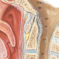
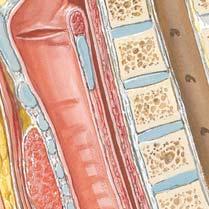
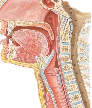
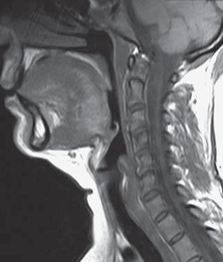
6 Upper Neck, Lower Head Osteology External acoustic meatus Styloid process Mental foramen Stylohyoid ligament Hyoid bone Lateral view of the skeletal elements of the head and neck (Atlas of Human Anatomy, 5th edition, Plate 15) Clinical Note In criminal proceedings, the finding of a fractured hyoid bone is considered to be strong evidence of strangulation. 6 Head and Neck

7 Upper Neck, Lower Head Osteology 1 External acoustic meatus Styloid process Mental foramen Hyoid bone Volume rendered display, maxillofacial CT The lesser horn of the hyoid bone is attached to the stylohyoid ligament, which sometimes ossifies. An elongated styloid process in association with such an ossified ligament (or even without such ossification) can produce neck/ swallowing pain and is known as Eagle s syndrome. In elderly patients who are edentulous, resorption of the alveolar process of the mandible exposes the mental nerve to pressure during chewing as it exits the foramen. Mastication then becomes a painful process for these patients. Head and Neck 7
.")
8 Axis (C2) Dens (odontoid process) Superior articular facet for atlas Anterior arch Inferior articular facet for C3 Anterior view of the axis (C2) (Atlas of Human Anatomy, 5th edition, Plate 19) Clinical Note The dens is susceptible to fracture that is classified by the level of the fracture site. The most common fracture occurs at the base of the dens (type II fracture). 8 Head and Neck
. The articular facet on the dens articulates with the facet on the anterior arch of the atlas.")
9 Axis (C2) 1 Dens (odontoid process) Superior articular facet for atlas Anterior arch Inferior articular facet for C3 Volume rendered CT scan, axis The dens is embryologically the vertebral body of the atlas (C1). The articular facet on the dens articulates with the facet on the anterior arch of the atlas. In rare cases the dens does not appear on radiographs to be fused with the remainder of the vertebra. This condition, known as os odontoideum, may result in atlantoaxial instability. Head and Neck 9
 Clinical Note The hangman s fracture consists of bilateral pedicle or pars interarticularis fractures of the axis.")
10 Cervical Spine, Posterior View Dens Facet on atlas for articulation with occipital condyle Posterior arch of atlas Lamina of axis Zygapophyseal joint Bifid spinous process Posterior view of articulated C1-C4 vertebrae (Atlas of Human Anatomy, 5th edition, Plate 19) Clinical Note The hangman s fracture consists of bilateral pedicle or pars interarticularis fractures of the axis. Associated with this fracture is anterior subluxation or dislocation of the C2 vertebral body. It results from a severe extension injury, such as from an automobile accident in which the face forcibly strikes the dashboard, or from hanging. 10 Head and Neck
")
11 Cervical Spine, Posterior View 1 Dens Posterior arch of atlas Lamina of axis Zygapophyseal joint Bifid spinous process Volume rendered display, cervical spine CT In the cervical region the articular facets of the zygapophyseal joints are oriented superiorly and inferiorly; thus, this is the only region of the vertebral column in which it is possible for adjoining vertebrae to dislocate (rotary) without fracture. The zygapophyseal joints are well innervated by medial branches from dorsal rami associated with both vertebral levels participating in the joint. To denervate a painful arthritic joint, the medial branches from both levels must be ablated. Head and Neck 11
 Clinical Note Degenerative changes of the uncovertebral joints (of Luschka) typically occur with other degenerative changes such as the development of")
12 Cervical Spondylosis Axis (C2) Uncinate processes with loss of joint space in uncovertebral joint Spondylophytes (osteophytes) on uncinate processes Degenerative changes in cervical vertebrae (Atlas of Human Anatomy, 5th edition, Plate 20) Clinical Note Degenerative changes of the uncovertebral joints (of Luschka) typically occur with other degenerative changes such as the development of spondylophytes and the loss of intervertebral disk space. These changes reduce the size of the intervertebral foramina (neuroforamina) resulting in radiculopathy and associated pain, paresthesia, and numbness in the corresponding dermatomes. 12 Head and Neck


13 Cervical Spondylosis 1 Axis Normal uncinate process and uncovertebral joint Uncovertebral joint with loss of joint space Spondylophyte (osteophyte) on body (lipping) Spondylophyte on uncinate process Volume rendered displays, cervical spine CT Surgeons may use an anterior or a posterior approach to address cervical spondylosis. A bone graft is inserted into the disk space to restore vertical spacing between segments and a metal plate is attached along the anterior margin of the spine to provide stability during the process of intervertebral bone fusion. The uncovertebral joints contribute to cervical spine stability and help to limit extension and lateral bending. Head and Neck 13
 Clinical Note Vertebral artery dissection, a subintimal hematoma, may cause cerebellar or brain infarction; occurrence may be idiopathic or secondary to trauma.")
14 Vertebral Artery, Neck Posterior arch of atlas (C1) Vertebral artery C5 transverse process Lateral view of the cervical spine and vertebral artery (Atlas of Human Anatomy, 5th edition, Plate 22) Clinical Note Vertebral artery dissection, a subintimal hematoma, may cause cerebellar or brain infarction; occurrence may be idiopathic or secondary to trauma. 14 Head and Neck

15 Vertebral Artery, Neck 1 Posterior arch of atlas (C1) Vertebral artery C5 transverse process Volume rendered display, CTA of the neck The intimate association of the vertebral artery to the cervical spine makes it susceptible to injury during cervical spine trauma. The vertebral artery is typically the first branch of the subclavian artery, although it can arise directly from the arch of the aorta. Most commonly, the vertebral artery enters the foramina of the transverse processes of the cervical vertebrae at C6. Head and Neck 15
 Clinical Note This is the most tortuous segment of the vertebral artery; increases in tortuosity are associated with atherosclerotic changes.")
16 Vertebral Artery, Atlas Mastoid process Posterior atlantooccipital membrane Transverse process of atlas (C1) Posterior tubercle of atlas Vertebral artery Vertebral artery on the posterior arch of the atlas (Atlas of Human Anatomy, 5th edition, Plate 22) Clinical Note This is the most tortuous segment of the vertebral artery; increases in tortuosity are associated with atherosclerotic changes. 16 Head and Neck

17 Vertebral Artery, Atlas 1 Mastoid process Transverse process of atlas (C1) Posterior tubercle of atlas Vertebral artery Volume rendered display, CTA of the neck The vertebral artery pierces the dura and arachnoid mater and ascends anterior to the medulla to unite with the contralateral vessel to form the basilar artery. The vertebral artery supplies the muscles of the suboccipital triangle before entering the cranial cavity. Head and Neck 17
 Clinical Note Atlanto-occipital dislocation is a rare traumatic injury that is difficult to diagnose and is frequently missed")
18 Craniovertebral Ligaments Clivus portion of occipital bone Alar ligaments Dens covered by cruciate ligament Transverse ligament of atlas Posterior view of the craniovertebral ligaments after removal of the tectorial membrane (Atlas of Human Anatomy, 5th edition, Plate 23) Clinical Note Atlanto-occipital dislocation is a rare traumatic injury that is difficult to diagnose and is frequently missed on initial lateral cervical x-rays. Patients who survive typically have neurologic impairment such as lower cranial neuropathies, unilateral or bilateral weakness, or quadriplegia. Prevertebral soft tissue swelling on a lateral cervical x-ray and craniocervical subarachnoid hemorrhage on an axial CT have been associated with this injury and thus may aid with diagnosis. 18 Head and Neck
 image, cervical spine The alar ligaments are pencil-thick")

19 Craniovertebral Ligaments 1 Alar ligament Dens Epiglottis A Dens Transverse ligament of atlas Superior articular facet of atlas Spinal cord Cerebellum B A, Oblique coronal CT, cervical spine; B, Axial T2 magnetic resonance (MR) image, cervical spine The alar ligaments are pencil-thick ligaments that connect the dens to the rim of the foramen magnum, stabilizing the atlanto-occipital relationship. The transverse ligament holds the dens against the anterior arch of the atlas. Superior and inferior bands arise from the transverse ligament forming with it the cruciate ligament. Head and Neck 19
 Clinical Note Congenital torticollis (wryneck) is typically associated with")
20 Neck Muscles, Lateral View Masseter muscle Mylohyoid muscle Digastric muscle (anterior belly) Hyoid bone Sternocleidomastoid muscle Sternohyoid muscle Posterior Middle Anterior Scalene muscles Pectoralis major muscle Lateral view of the superficial muscles of the neck (Atlas of Human Anatomy, 5th edition, Plate 26) Clinical Note Congenital torticollis (wryneck) is typically associated with a birth injury to the sternocleidomastoid muscle that results in a unilateral shortening of the muscle, and the associated rotated and tilted head position. 20 Head and Neck

21 Neck Muscles, Lateral View 1 Masseter muscle Mylohyoid muscle Digastric muscle (anterior belly) Hyoid bone Sternocleidomastoid muscle Scalene muscles Sternohyoid muscle Pectoralis major muscle Volume rendered display, CT of the neck The sternocleidomastoid is a large and consistent anatomic structure that is easily identifiable and is used to divide the neck into anterior and posterior triangles. The hyoid bone provides an anchor for many neck muscles and is suspended solely by these muscles (it has no bony articulation). Head and Neck 21
Biology 218 Human Anatomy. Adapted from Martini Human Anatomy 7th ed. Chapter 6 The Skeletal System: Axial Division
 Adapted from Martini Human Anatomy 7th ed. Chapter 6 The Skeletal System: Axial Division Introduction The axial skeleton: Composed of bones along the central axis of the body Divided into three regions:
APPENDICULAR SKELETON 126 AXIAL SKELETON SKELETAL SYSTEM. Cranium. Skull. Face. Skull and associated bones. Auditory ossicles. Associated bones.
 SKELETAL SYSTEM 206 AXIAL SKELETON 80 APPENDICULAR SKELETON 26 Skull Skull and associated s 29 Cranium Face Auditory ossicles 8 4 6 Associated s Hyoid Thoracic cage 25 Sternum Ribs 24 Vertebrae 24 column
Human Anatomy and Physiology - Problem Drill 07: The Skeletal System Axial Skeleton
 Human Anatomy and Physiology - Problem Drill 07: The Skeletal System Axial Skeleton Question No. 1 of 10 Which of the following statements about the axial skeleton is correct? Question #01 A. The axial
Anatomy and Physiology. Bones, Sutures, Teeth, Processes and Foramina of the Human Skull
 Anatomy and Physiology Chapter 6 DRO Bones, Sutures, Teeth, Processes and Foramina of the Human Skull Name: Period: Bones of the Human Skull Bones of the Cranium: Frontal bone: forms the forehead and the
Chapter 7. Skeletal System
 Chapter 7 Skeletal System 1 Skull A. The skull is made up of 22 bones: 8 cranial bones, 13 facial bones, and the mandible. B. The Cranium encloses and protects the brain, provides attachments for muscles,
Structure Location Function
 Frontal Bone Cranium forms the forehead and roof of the orbits Occipital Bone Cranium forms posterior and inferior portions of the cranium Temporal Bone Cranium inferior to the parietal bone forms the
Cranium Facial bones. Sternum Rib
 Figure 7.1 The human skeleton. Skull Thoracic cage (ribs and sternum) Cranium Facial bones Sternum Rib Bones of pectoral girdle Vertebral column Sacrum Vertebra Bones of pelvic girdle (a) Anterior view
Dr.Noor Hashem Mohammad Lecture (5)
") Dr.Noor Hashem Mohammad Lecture (5) 2016-2017 If the mandible is discarded, the anterior part of this aspect of the skull is seen to be formed by the hard palate. The palatal processes of the maxillae
Dr.Noor Hashem Mohammad Lecture (5) 2016-2017 If the mandible is discarded, the anterior part of this aspect of the skull is seen to be formed by the hard palate. The palatal processes of the maxillae
THE SKELETAL SYSTEM. Focus on the Skull
 THE SKELETAL SYSTEM Focus on the Skull Review Anatomical Terms Anterior/Posterior Dorsal/Ventral Medial/Lateral Superior/Inferior Bone Markings - Review Projections for attachment of muscles, ligaments
Skull-2. Norma Basalis Interna Norma Basalis Externa. Dr. Heba Kalbouneh Associate Professor of Anatomy and Histology
 Skull-2 Norma Basalis Interna Norma Basalis Externa Dr. Heba Kalbouneh Associate Professor of Anatomy and Histology Norma basalis interna Base of the skull- superior view The interior of the base of the
Anatomy and Physiology II. Spine
 Anatomy and Physiology II Spine Bones and Other Structures Vertibrae Contains Cervical, Thoracic, Lumbar, Sacral and Coccygeal regions We use Capital letters to refer to these (C, T, L, S, and Co) and
ACTIVITY 3: AXIAL SKELETON AND LONG BONE DISSECTION COW BONE DISSECTION
 ACTIVITY 3: AXIAL SKELETON AND LONG BONE DISSECTION Objectives: 1) How to get ready: Read Chapter 7, McKinley et al., Human Anatomy, 4e. All text references are for this textbook. Learning the meanings
University of Palestine. Midterm Exam 2013/2014 Total Grade:
 Course No: DNTS2208 Course Title: Head and Neck Anatomy Date: 09/11/2013 No. of Questions: (50) Time: 1hour Using Calculator (No) University of Palestine Midterm Exam 2013/2014 Total Grade: Instructor
Imaging of Cervical Spine Trauma Tudor H Hughes, M.D.
 Imaging of Cervical Spine Trauma Tudor H Hughes, M.D. General Considerations Most spinal fractures are due to a single episode of major trauma. Fatigue fractures of the spine are unusual except in the
YOU MUST BRING YOUR OWN GLOVES FOR THIS ACTIVITY.
 ACTIVITY 3: AXIAL SKELETON AND LONG BONE DISSECTION Objectives: 1) How to get ready: Read Chapter 7, McKinley et al., Human Anatomy, 5e. All text references are for this textbook. Learning the meanings
Chapter 7 Part A The Skeleton
 Chapter 7 Part A The Skeleton Why This Matters Understanding the anatomy of the skeleton enables you to anticipate problems such as pelvic dimensions that may affect labor and delivery The Skeleton The
Clarification of Terms
 Clarification of Terms The Spine, Spinal Column, and Vertebral Column are synonymous terms referring to the bony components housing the spinal cord Spinal Cord = made of nervous tissue Facet = a small,
Clarification of Terms
 Clarification of Terms The Spine, Spinal Column, and Vertebral Column are synonymous terms referring to the bony components housing the spinal cord Spinal Cord = made of nervous tissue Facet = a small,
Cervical Spine Anatomy and Biomechanics. Typical Cervical Vertebra C3 6. Typical Cervical Vertebra Anterior 10/5/2017
 Cervical Spine Anatomy and Biomechanics Typical Cervical Vertebra C3 6 Small, relatively broad body Bifid SpinousProcess Long and narrow laminae Spinal Canal: large, triangular; remarkably consistent dimensions
Prevertebral Region, Pharynx and Soft Palate
 Unit 20: Prevertebral Region, Pharynx and Soft Palate Dissection Instructions: Step1 Step 2 Step 1: Insert your fingers posterior to the sternocleidomastoid muscle, vagus nerve, internal jugular vein,
Chapter 7: Skeletal System: Gross Anatomy
 Chapter 7: Skeletal System: Gross Anatomy I. General Considerations A. How many bones in an average adult skeleton? B. Anatomic features of bones are based on II. Axial Skeleton A. Skull 1. Functionally
Chapter 7: Head & Neck
 Chapter 7: Head & Neck Osteology I. Overview A. Skull The cranium is composed of irregularly shaped bones that are fused together at unique joints called sutures The skull provides durable protection from
o Diaphysis o Area where red marrow is found o Area where yellow marrow is found o Epiphyseal plate AXIAL SKELETON Skull
 64 Anatomy & Physiology Coloring Workbook 7. Figure 5-2A is a midlevel, cross-sectional view of the diaphysis of the femur. Label the membrane that lines the cavity and the membrane that covers the outside
ANATOMY & PHYSIOLOGY I Laboratory Version B Name Section. REVIEW SHEET Exercise 10 Axial Skeleton
 ANATOMY & PHYSIOLOGY I Laboratory Version B Name Section REVIEW SHEET Exercise 10 Axial Skeleton 1 POINT EACH. THE SKULL MULTIPLE CHOICE 1. The major components of the axial skeleton include the 7. The
Clarification of Terms
 Clarification of Terms The Spine, Spinal Column, and Vertebral Column are synonymous terms referring to the bony components housing the spinal cord Spinal Cord = made of nervous tissue Facet = a small,
Ligaments of the vertebral column:
 In the last lecture we started talking about the joints in the vertebral column, and we said that there are two types of joints between adjacent vertebrae: 1. Between the bodies of the vertebrae; which
Copyright 2010 Pearson Education, Inc.
 E. VERTEBRAL COLUMN 1. The vertebral column extends from the skull to the pelvis and forms the vertical axis of the skeleton. 2. The vertebral column is composed of vertebrae that are separated by intervertebral
University of Palestine. Midterm Exam 2013/2014 Total Grade:
 [ Course No: DNTS2208 Course Title: Head and Neck Anatomy Date: 17/11/1024 No. of Questions: (52) Time: 2hours Using Calculator (No) University of Palestine Midterm Exam 2013/2014 Total Grade: Instructor
INDEPENDENT LEARNING: DISC HERNIATION IN THE NATIONAL FOOTBALL LEAGUE: ANATOMICAL FACTORS TO CONSIDER IN REVIEW
 INDEPENDENT LEARNING: DISC HERNIATION IN THE NATIONAL FOOTBALL LEAGUE: ANATOMICAL FACTORS TO CONSIDER IN REVIEW CDC REPORT - CAUSES OF DISABILITY, 2005 REVIEW QUESTIONS ABOUT DISC HERNIATION IN THE NATIONAL
The craniocervical junction
 Anver Jameel, MD The craniocervical junction A biomechanical and anatomical unit that extends from the skull base to C2 Includes the clivus, foramen magnum and contiguous occipital bone, the occipital
2. The vertebral arch is composed of pedicles (projecting from the body) and laminae (uniting arch posteriorly).
 and laminae (uniting arch posteriorly).") VERTEBRAL COLUMN 2018zillmusom I. VERTEBRAL COLUMN - functions to support weight of body and protect spinal cord while permitting movements of trunk and providing for muscle attachments. A. Typical vertebra
VERTEBRAL COLUMN 2018zillmusom I. VERTEBRAL COLUMN - functions to support weight of body and protect spinal cord while permitting movements of trunk and providing for muscle attachments. A. Typical vertebra
AXIAL SKELETON SKULL
 AXIAL SKELETON SKULL CRANIAL BONES (8 total flat bones w/ 2 paired) 1. Frontal forms forehead & upper portion of eyesocket (orbital) 2. Parietal paired bones; form superior & lateral walls of cranium 3.
Head & Neck Radiology (I) Waseem Jerjes
 Waseem Jerjes") Head & Neck Radiology (I) Waseem Jerjes Lamboid Suture Frontal Sinus Left Orbit (roof) Left Supraorbital Margin Left Frontozygomatic Suture Left Superior Orbital Fissure Rt Petrous Ridge of Temporal Bone
Head & Neck Radiology (I) Waseem Jerjes Lamboid Suture Frontal Sinus Left Orbit (roof) Left Supraorbital Margin Left Frontozygomatic Suture Left Superior Orbital Fissure Rt Petrous Ridge of Temporal Bone
Introduction to Local Anesthesia and Review of Anatomy
 5-Sep Introduction and Anatomy Review 12-Sep Neurophysiology and Pain 19-Sep Physiology and Pharmacology part 1 26-Sep Physiology and Pharmacology part 2 Introduction to Local Anesthesia and Review of
External Acoustic Meatus. Mastoid Process. Zygomatic Process. Temporal Bone
 Bone lab review 1. Frontal Bone 2. Supra-Orbital Foramen 3. Orbit (Orbital Cavity) 4. Superior Orbital Fissure 5. Inferior Orbital Fissure 6. Zygomatic Bone 7. Infra-Orbital Foramen 8. Maxilla 9. Mandible
You have 24 vertebrae in your spinal column. Two are special enough to be individually named.
 You have 24 vertebrae in your spinal column. Two are special enough to be individually named. Your atlas (C01) and axis (C02) are very important vertebrae. Without them, head and neck movement would be
VERTEBRAL COLUMN VERTEBRAL COLUMN
 VERTEBRAL COLUMN FUNCTIONS: 1) Support weight - transmits weight to pelvis and lower limbs 2) Houses and protects spinal cord - spinal nerves leave cord between vertebrae 3) Permits movements - *clinical
Anatomy Skull and Spinal Cord
 1 Anatomy Skull and Spinal Cord 1. Skull The skull is anterior to the spinal column and is the bony structure that encases the brain. Its purpose is to protect the brain and allow attachments for the facial
Infratemporal fossa: Tikrit University college of Dentistry Dr.Ban I.S. head & neck Anatomy 2 nd y.
 Infratemporal fossa: This is a space lying beneath the base of the skull between the lateral wall of the pharynx and the ramus of the mandible. It is also referred to as the parapharyngeal or lateral pharyngeal
Human Anatomy - Problem Drill 06: The Skeletal System Axial Skeleton & Articualtions
 Human Anatomy - Problem Drill 06: The Skeletal System Axial Skeleton & Articualtions Question No. 1 of 10 Instructions: (1) Read the problem and answer choices carefully, (2) Work the problems on paper
Crafton Hills College Human Anatomy & Physiology Axial Skeleton
 A. Major Divisions Crafton Hills College Human Anatomy & Physiology Axial keleton 1. Axial: Part of skeleton lies along long axis of body 2. Appendicular: Bones & features of the appendages B. AXIAL KELETON
Axial Skeleton: Vertebrae and Thorax
 Axial Skeleton: Vertebrae and Thorax Function of the vertebral column (spine or backbone): 1) 2) 3) Composition of Vertebral column The vertebral column is formed by 33 individual vertebrae (some of which
Dr. Sami Zaqout, IUG Medical School
 The skull The skull is composed of several separate bones united at immobile joints called sutures. Exceptions? Frontal bone Occipital bone Vault Cranium Sphenoid bone Zygomatic bones Base Ethmoid bone
Bones of the skull & face
 Bones of the skull & face Cranium= brain case or helmet Copyright The McGraw-Hill Companies, Inc. Permission required for reproduction or display. The cranium is composed of eight bones : frontal Occipital
AXIAL SKELETON FORM THE VERTICAL AXIS OF THE BODY CONSISTS OF 80 BONES INCLUDES BONES OF HEAD, VERTEBRAL COLUMN, RIBS,STERNUM
 AXIAL SKELETON FORM THE VERTICAL AXIS OF THE BODY CONSISTS OF 80 BONES INCLUDES BONES OF HEAD, VERTEBRAL COLUMN, RIBS,STERNUM APPENDICULAR SKELETON BONES OF THE FREE APPENDAGES & THEIR POINTS OF ATTACHMENTS
Vertebral Column. Backbone consists of 26 vertebrae. Five vertebral regions. Cervical
 Vertebral Column Backbone consists of 26 vertebrae. Five vertebral regions Cervical vertebrae (7) in the neck. Thoracic vertebrae (12) in the thorax. Lumbar vertebrae (5) in the lower back. Sacrum (5,
The Back OUTLINE. Vertebral Column (review) Craniovertebral Joints Dorsal Scapular Region(review) Muscles of the Back Suboccipital Region
 Craniovertebral Joints Dorsal Scapular Region(review) Muscles of the Back Suboccipital Region") The Back OUTLINE Vertebral Column (review) Craniovertebral Joints Dorsal Scapular Region(review) Muscles of the Back Suboccipital Region Dept. of Human Anatomy, Si Chuan University Zhou hongying eaglezhyxzy@163.com
The Back OUTLINE Vertebral Column (review) Craniovertebral Joints Dorsal Scapular Region(review) Muscles of the Back Suboccipital Region Dept. of Human Anatomy, Si Chuan University Zhou hongying eaglezhyxzy@163.com
SKULL AS A WHOLE + ANTERIOR CRANIAL FOSSA
 SKULL AS A WHOLE + ANTERIOR CRANIAL FOSSA LEARNING OBJECTIVES At the end of this lecture, the student should be able to know: Parts of skeleton (axial and appendicular) Parts of skull Sutures of skull
Biology 2401 The Skeletal System
 Biology 2401 The Skeletal System Purpose: The lab will describe the microscopic and gross anatomy of bone, identify bones of the body, and identify important bone markings. I. Overview of the Skeleton
Copyright 2010 Pearson Education, Inc. Copyright 2010 Pearson Education, Inc. Figure Sectioned spinous process. Interspinous.
 PowerPoint Lecture Slides prepared by Janice Meeking, Mount Royal College C H A P T E R 7 The Skeleton: Part B Vertebral Column Transmits weight of trunk to lower limbs Surrounds and protects spinal cord
Skeletal System: Skull.
 Skeletal System: Skull www.fisiokinesiterapia.biz Bones of the Skull SPLANCHNOCRANIUM Nasal (2) Maxilla (2) Lacrimal (2) Zygomatic (2) Palatine (2) Inferior concha (2) Vomer Mandible NEUROCRANIUM Frontal
Cervical Cooled RF Training Presentation
 Cervical Cooled RF Training Presentation Agenda Patient Selection Considerations Diagnostic Block General Considerations COOLIEF* Cooled RF Technique Posterior Lateral Precautions Summary Appendix 2 Disclaimer
Overview of the Skeleton: Bone Markings
 Name Overview of the Skeleton: Bone Markings Match the terms in column B with the appropriate description in column A. Column A 1. sharp, slender process* 2. small rounded projection* 3. narrow ridge of
PTERYGOPALATINE FOSSA
 PTERYGOPALATINE FOSSA Outline Anatomical Structure and Boundaries Foramina and Communications with other spaces and cavities Contents Pterygopalatine Ganglion Especial emphasis on certain arteries and
Spinal Cord Injuries: The Basics. Kadre Sneddon POS Rounds October 1, 2003
 Spinal Cord Injuries: The Basics Kadre Sneddon POS Rounds October 1, 2003 Anatomy Dorsal columntouch, vibration Corticospinal tract- UMN Anterior horn-lmn Spinothalamic tractpain, temperature (contralateral)
Chest cavity, vertebral column and back muscles. Respiratory muscles. Sándor Katz M.D., Ph.D.
 Chest cavity, vertebral column and back muscles. Respiratory muscles. Sándor Katz M.D., Ph.D. Chest cavity - bony structures Chest cavity- bony structures Sternum Ribs True ribs: first seven pairs connect
Structure and Function of the Vertebral Column
 Structure and Function of the Vertebral Column Posture Vertebral Alignment Does it really matter? Yes it does! Postural Curves The vertebral column has a series of counterbalancing curves posterior anterior
Cranial Cavity REFERENCES: OBJECTIVES OSTEOLOGY. Stephen A. Gudas, PT, PhD
 Stephen A. Gudas, PT, PhD Cranial Cavity REFERENCES: Moore and Agur, Essential Clinical Anatomy (ECA), 3rd ed., pp. 496 498; 500 507; 512 514 Grant s Atlas 12 th ed., Figs 7.6; 7.19 7.30. Grant s Dissector
Anatomy and Physiology II. Review Spine and Neck
 Anatomy and Physiology II Review Spine and Neck Spine regions How many cervical vertibrae are there? 7 The curvature is the cervical region posterior? Concave posterior How many thoracic? And curvature?
locomotice system Plastinated specimensⅠ: Silicone specimens Regional specimens and organs
 locomotice system Plastinated specimensⅠ: Silicone specimens Regional specimens and organs Art-No. Name Description The locomotor system SL001 Two hundred pieces of plastinated bones (without six The bones
Microsurgical anatomy of the transcondylar, supracondylar, and paracondylar extensions of the far-lateral approach
 J Neurosurg 87:555 585, 1997 Microsurgical anatomy of the transcondylar, supracondylar, and paracondylar extensions of the far-lateral approach HUNG T. WEN, M.D., ALBERT L. RHOTON, JR., M.D., TOSHIRO KATSUTA,
The Axial Skeleton. C h a p t e r. PowerPoint Lecture Slides prepared by Jason LaPres Lone Star College - North Harris
 C h a p t e r 7 The Axial Skeleton PowerPoint Lecture Slides prepared by Jason LaPres Lone Star College - North Harris Copyright 2009 Pearson Education, Inc., publishing as Pearson Benjamin Cummings An
Lec [8]: Mandibular nerve:
![Lec [8]: Mandibular nerve:](/thumbs/94/121295776.jpg "Lec [8]: Mandibular nerve:") Lec [8]: Mandibular nerve: The mandibular branch from the trigeminal ganglion lies in the middle cranial fossa lateral to the cavernous sinus. With the motor root of the trigeminal nerve [motor roots lies
Lec [8]: Mandibular nerve: The mandibular branch from the trigeminal ganglion lies in the middle cranial fossa lateral to the cavernous sinus. With the motor root of the trigeminal nerve [motor roots lies
SUBAXIAL CERVICAL SPINE TRAUMA- DIAGNOSIS AND MANAGEMENT
 SUBAXIAL CERVICAL SPINE TRAUMA- DIAGNOSIS AND MANAGEMENT 1 Anatomy 3 columns- Anterior, middle and Posterior Anterior- ALL, Anterior 2/3 rd body & disc. Middle- Posterior 1/3 rd of body & disc, PLL Posterior-
Skull-2. Norma Basalis Interna. Dr. Heba Kalbouneh Assistant Professor of Anatomy and Histology
 Skull-2 Norma Basalis Interna Dr. Heba Kalbouneh Assistant Professor of Anatomy and Histology Norma basalis interna Base of the skull- superior view The interior of the base of the skull is divided into
Parotid Gland, Temporomandibular Joint and Infratemporal Fossa
 M1 - Anatomy Parotid Gland, Temporomandibular Joint and Infratemporal Fossa Jeff Dupree Sanger 9-057 jldupree@vcu.edu Parotid gland: wraps around the mandible positioned between the mandible and the sphenoid
Craniovertebral Junction Embryology and Anatomy. Presented by: Amandeep Moderators: S.S.Kale G.D.Satyarthi. CVJ-Embryology & Anatomy
 Craniovertebral Junction Embryology and Anatomy Presented by: Amandeep Moderators: S.S.Kale G.D.Satyarthi CVJ-Embryology & Anatomy CVJ-Embryology SOMITE-The building block of vertebrae, skeletal muscle
Temporal region. temporal & infratemporal fossae. Zhou Hong Ying Dept. of Anatomy
 Temporal region temporal & infratemporal fossae Zhou Hong Ying Dept. of Anatomy Temporal region is divided by zygomatic arch into temporal & infratemporal fossae. Temporal Fossa Infratemporal fossa Temporal
Anatomy and Physiology 1 Chapter 7 self quiz Pro, Dima Darwish,MD.
 Anatomy and Physiology 1 Chapter 7 self quiz Pro, Dima Darwish,MD. 1) How many bones make up the axial skeleton? A) 50 B) 60 C) 70 D) 80 E) 90 2) Which of the following is a function of the axial skeleton?
Spinal nerves and cervical plexus Prof. Abdulameer Al Nuaimi. E mail: a.al E. mail:
 Spinal nerves and cervical plexus Prof. Abdulameer Al Nuaimi E mail: a.al nuaimi@sheffield.ac.uk E. mail: abdulameerh@yahoo.com Branches of ophthalmic artery Muscles of face A spinal nerve Spinal
Chapter 7 Part B The Skeleton
 Chapter 7 Part B The Skeleton 7.2 The Vertebral Column General Characteristics Extends from skull to pelvis Also called spine or spinal column Functions to transmit weight of trunk to lower limbs, surround
Bone Flashcards for 10a
 Bone Flashcards for 0a CLAVICLE (collar bone). Sternal extremity (end) flat end. Acromial extremity (end) rounded end. SCAPULA (shoulder blade). Right or left scapula?. Superior border (superior margin).
Temporal fossa Infratemporal fossa Pterygopalatine fossa Terminal branches of external carotid artery Pterygoid venous plexus
 Outline of content Temporal fossa Infratemporal fossa Pterygopalatine fossa Terminal branches of external carotid artery Pterygoid venous plexus Boundary Content Communication Mandibular division of trigeminal
Any of the vertebra in the cervical (neck) region of the spinal column. The cervical vertebra are the smallest vertebra in the spine, reflective of th
 region of the spinal column. The cervical vertebra are the smallest vertebra in the spine, reflective of th") Any of the vertebra in the cervical (neck) region of the spinal column. The cervical vertebra are the smallest vertebra in the spine, reflective of the fact that they support the least load. In humans,
Any of the vertebra in the cervical (neck) region of the spinal column. The cervical vertebra are the smallest vertebra in the spine, reflective of the fact that they support the least load. In humans,
THE THORACIC WALL. Boundaries Posteriorly by the thoracic part of the vertebral column. Anteriorly by the sternum and costal cartilages
 THE THORACIC WALL Boundaries Posteriorly by the thoracic part of the vertebral column Anteriorly by the sternum and costal cartilages Laterally by the ribs and intercostal spaces Superiorly by the suprapleural
The Thoracic Cage ANATOMY 2: THORACIC CAGE AND VERTEBRAL COLUMN
 ANATOMY 2: THORACIC CAGE AND VERTEBRAL COLUMN PSK 4U Mr. S. Kelly North Grenville DHS The Thoracic Cage 7 true ribs 3 false ribs 2 floating ribs Clavicle = collarbone Manubrium Sternum Xiphoid Process
Chapter 7 The Skeletal System:The Axial Skeleton
 Chapter 7 The Skeletal System:The Axial Skeleton Axial Skeleton 80 bones lie along longitudinal axis skull, hyoid, vertebrae, ribs, sternum, ear ossicles Appendicular Skeleton 126 bones upper & lower limbs
Skull basic structures. Neurocranium
 Assoc. Prof. Květuše Lovásová, M.V.D., PhD. Skull basic structures Skull consists of two groups of bones: neurocranium (bones forming the brain box) splanchnocranium (bones forming the facial skeleton)
Anatomy images for MSS practical exam- 2019
 Anatomy images for MSS practical exam- 2019 Ilium Ischium Pubis Acetabulaum Iliac crest Iliac tubercle ASIS (muscle and ligament attached) AIIS (muscle attached) PSIS PIIS Ischial spine Ischial tuberosity
Human Anatomy & Physiology I Dr. Sullivan Unit VIIIa The Axial Skeleton Chapter 8 (Sections )
") Human Anatomy & Physiology I Dr. Sullivan Unit VIIIa The Axial Skeleton Chapter 8 (Sections 8.1-8.3) I. Divisions of the skeletal system a) An adult human skeleton has 206 named bones b) Most are paired
Human Anatomy & Physiology I Dr. Sullivan Unit VIIIa The Axial Skeleton Chapter 8 (Sections 8.1-8.3) I. Divisions of the skeletal system a) An adult human skeleton has 206 named bones b) Most are paired
The Anatomy of the Atlantoaxial Complex
 The Anatomy of the Atlantoaxial Complex T. GLENN PAIT, M.D., KENAN 1. ARNAUTOVIC, M.D., and LUIS A.B. BORBA, M.D. Operating on the atlantoaxial complex has always posed a challenge to the surgeon because
Parotid Gland. Parotid Gland. Largest of 3 paired salivary glands (submandibular; sublingual) Ramus of Mandible. Medial pterygoid.
 Ramus of Mandible. Medial pterygoid.") Parotid region Parotid Gland Largest of 3 paired salivary glands (submandibular; sublingual) Ramus of Mandible Medial pterygoid Cross section of mandible Masseter D S SCM Parotid Gland Mastoid Process
Parotid region Parotid Gland Largest of 3 paired salivary glands (submandibular; sublingual) Ramus of Mandible Medial pterygoid Cross section of mandible Masseter D S SCM Parotid Gland Mastoid Process
Skeletal System -Axial System. Chapter 7 Part A
 Skeletal System -Axial System Chapter 7 Part A Skeleton Learn: Names of the s. Identify specific landmarks that allow: Bones to fit into each other, Organs to fit into the cavities, Muscles to attach,
TEST YOURSELF- Chapter 7
 TEST YOURSELF- Chapter 7 Cranial Bones 1. Give the name of the bone for each of the following markings. Some of the markings are found on more than one bone. List all that apply. Cranium a. Frontal squama:
Exercise 10. The Axial Skeleton
 Exercise 10 The Axial Skeleton The Axial Skeleton Consists of the skeletal structures found along the midline of the body. Includes the skull, hyoid, vertebrae, ribs, sternum, and sacrum. The cartilages
Common fracture & dislocation of the cervical spine. Theerachai Apivatthakakul Department of Orthopaedic Chiangmai University
 Common fracture & dislocation of the cervical spine Theerachai Apivatthakakul Department of Orthopaedic Chiangmai University Objective Anatomy Mechanism and type of injury PE.and radiographic evaluation
3. The Jaw and Related Structures
 Overview and objectives of this dissection 3. The Jaw and Related Structures The goal of this dissection is to observe the muscles of jaw raising. You will also have the opportunity to observe several
MDCT and MRI evaluation of cervical spine trauma
 Insights Imaging (2014) 5:67 75 DOI 10.1007/s13244-013-0304-2 PICTORIAL REVIEW MDCT and MRI evaluation of cervical spine trauma Michael Utz & Shadab Khan & Daniel O Connor & Stephen Meyers Received: 10
Chapter 8A. The Skeletal System: The Axial Skeleton. The Skeletal System: The Axial Skeleton. Types of Bones. Types of Bones
 Chapter 8A The Skeletal System: The Axial Skeleton The Skeletal System: The Axial Skeleton 206 named bones Axial Skeleton 80 bones lie along longitudinal axis skull, hyoid, vertebrae, ribs, sternum, ear
CERVICAL SPINE EVALUATION MARK FIGUEROA PHYSICAL THERAPIST
 CERVICAL SPINE EVALUATION MARK FIGUEROA PHYSICAL THERAPIST OVERVIEW OF CLINICAL REASONING Stage of disorder Pathoanatomical diagnosis Signs and symptoms Consideration of the evidence gathered Common sense
THE VERTEBRAL COLUMN. Average adult length: In male: about 70 cms. In female: about 65 cms.
 THE VERTEBRAL COLUMN Average adult length: In male: about 70 cms. In female: about 65 cms. 1 Vertebral Column (Regions and Curvatures) Curvatures of the vertebral column: A. Primary curvature: C-shaped;
Riverside Community College Anatomy & Physiology 2B SPRING 2012 EXAM #1-ABC (Nervous System)
") Riverside Community College Anatomy & Physiology 2B SPRING 2012 EXAM #1-ABC (Nervous System) Name: 1) This vertebra is an example of a(n). 1) A) thoracic B) axis C) atlas D) lumbar E) sacral 1 2) W hich
Riverside Community College Anatomy & Physiology 2B SPRING 2012 EXAM #1-ABC (Nervous System) Name: 1) This vertebra is an example of a(n). 1) A) thoracic B) axis C) atlas D) lumbar E) sacral 1 2) W hich
Bones of the Skull Lateral View
 Bones of the Skull Lateral View Frontal Bone Parietal Bone Occipital Bone Temporal Bone Sphenoid Bone Pterion Sutures of the Skull Lateral View Coronal Suture Lambdoid Suture Squamous Suture Sutures of
in compact bone, large vertical canals carrying blood vessels and nerves. in compact bone, large horizontal canals carrying blood vessels and nerves.
 Carl Christensen, PhD Skeletal System (Bones`) Bio. 2304 Human Anatomy 1. Identify a term for each of the following: shaft of a long bone ends of a long bone ossified remnant of the "growth plate" connective
Tikrit University collage of dentistry Dr.Ban I.S. head & neck anatomy 2 nd y. Lec [5] / Temporal fossa :
![Tikrit University collage of dentistry Dr.Ban I.S. head & neck anatomy 2 nd y. Lec [5] / Temporal fossa :](/thumbs/88/115294566.jpg "Tikrit University collage of dentistry Dr.Ban I.S. head & neck anatomy 2 nd y. Lec [5] / Temporal fossa :") Lec [5] / Temporal fossa : Borders of the Temporal Fossa: Superior: Superior temporal line. Inferior: gap between zygomatic arch and infratemporal crest of sphenoid bone. Anterior: Frontal process of the
Lec [5] / Temporal fossa : Borders of the Temporal Fossa: Superior: Superior temporal line. Inferior: gap between zygomatic arch and infratemporal crest of sphenoid bone. Anterior: Frontal process of the
Tympanic Bulla Temporal Bone. Digastric Muscle. Masseter Muscle
 Superior view Hyoid Bone The hyoid bone does not articulate with any other bones. It is held in place by ligaments to the styloid process of the temporal bone and the thyroid cartilage of the larynx. It
_CH01redo.qxd 9/24/07 3:07 PM Page 1. [Half-Title to come]
![_CH01redo.qxd 9/24/07 3:07 PM Page 1. [Half-Title to come]](/thumbs/81/84146690.jpg "_CH01redo.qxd 9/24/07 3:07 PM Page 1. [Half-Title to come]") 10752-01_CH01redo.qxd 9/24/07 3:07 PM Page 1 [Half-Title to come] 10752-01_CH01redo.qxd 9/24/07 3:07 PM Page 2 THE BACK Lippincott Williams & Wilkins atlas of ANATOMY CHAPTER 1 Plate 1-01 Palpable Structures
10752-01_CH01redo.qxd 9/24/07 3:07 PM Page 1 [Half-Title to come] 10752-01_CH01redo.qxd 9/24/07 3:07 PM Page 2 THE BACK Lippincott Williams & Wilkins atlas of ANATOMY CHAPTER 1 Plate 1-01 Palpable Structures
Lab Exercise #04 The Skeletal System Student Performance Objectives
 Lab Exercise #04 The Skeletal System Student Performance Objectives The material that you are required to learn in this exercise can be found in either the lecture text or the supplemental materials provided
The Thoracic Cage. Role of the Thoracic Cage 2/13/2019. Anatomy 2: Thoracic Cage and Vertebral Column
 PSK 4U Mr. S. Kelly North Grenville DHS Anatomy 2: Thoracic Cage and Column The Thoracic Cage 7 true ribs 3 false ribs 2 floating ribs Clavicle = collarbone Manubrium Sternum Xiphoid Process 12 thoracic
Why does sideflexion increase ipsilateral vertebral artery occlusion with contralateral atlanto-axial rotation? Thomas Langer
 Why does sideflexion increase ipsilateral vertebral artery occlusion with contralateral atlanto-axial rotation? Thomas Langer 1 Introduction When the head and neck are placed in the premanipulative position