Pediatric Spinal Anomalies
|
|
|
- Noel Stewart
- 6 years ago
- Views:
Transcription


1 Department of Radiology University of California San Diego Pediatric Spinal Anomalies John R. Hesselink, M.D.
2 Spine Embryogenesis 1. Primitive streak 2. Proliferation of cells at primitive pit (Hensen's node) 3. Cells enter pit & migrate cephalad to form notochordal process 4. Notochord induces overlying neural plate Barkovich, Pediatric Neuroimaging Raven Press, New York, 1995
3 Formation of the Spinal Cord Neurulation Canalization & retrogressive differentiation
4 Formation of the Vertebral Column Membrane development Chondrification Ossification


5 Normal Pediatric Spine Stage I: Birth to 1 Month Cartilage Ossified vertebra Disk Vertebra Disk T1-weighted image T2-weighted image


6 Normal Pediatric Spine Stage II: 1 Month to 6 Months Disk Vertebra Vertebra Disk T1-weighted image T2-weighted image
7 Spine Anomalies Embryogenesis Split Notochord from endodermectoderm adhesion Neural plate Neural crest Cutaneous ectoderm Neural ectoderm Notochord Formation of neural folds & groove Premature Dysjunction Neurulation Non-Dysjunction Dysjunction

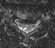
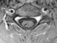
8 Anomalies of Notochord Formation Adhesion between endoderm & ectoderm splits notochord Diastematomyelia Split notochord syndrome Dorsal enteric anomalies (fistula, sinus, cyst)

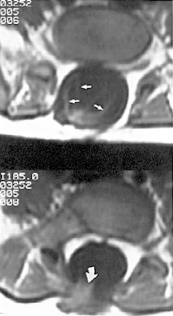
9 Dorsal Enteric Cyst


10 Spine Development Premature Dysjunction Lipomyelo- Lipomyelocele Intradural meningocele lipoma


11 Anomalies of Premature Dysjunction Intradural Lipoma Lipomyelocele
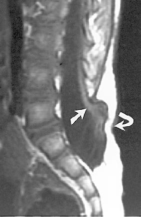
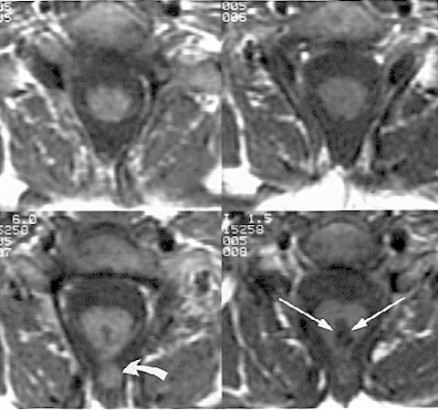
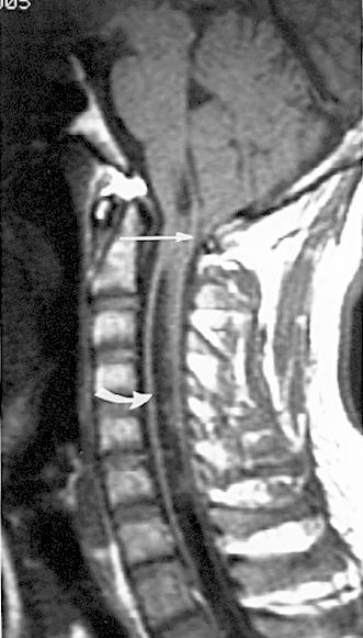
12 Spine Development Non-Dysjunction Myelocele Myelomeningocele Dorsal dermal sinus Hemimyelocele Chiari II Myelocele Myelomeningocele


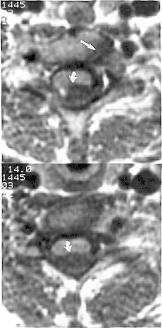
13 624 History: Newborn male with a mass on the lower back
14 Dx: Myelomeningocele {Page 2}
15 1 Myelomeningocele 2 3

16 Postoperative myelomeningocele with clinical deterioration Postop hematoma Postop infection Residual or recurrent tumor Cord ischemia/infarction Myelomalacia Arachnoid cyst Diastematomyelia Syringohydromyelia Cord retethering
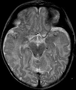
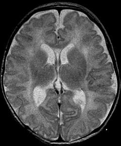
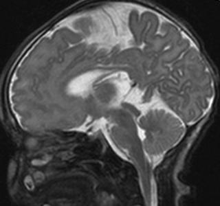
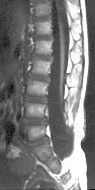
 Associated with epidermoid or dermoid in 50% May have spina bifida History of")
17 Dorsal Dermal Sinus A disorder of nondysjunction Dorsal opening in a hyperpigmented patch or a hairy nevus Terminates in spinal canal (extradural or intradural) Associated with epidermoid or dermoid in 50% May have spina bifida History of meningitis


18 1 Dorsal dermal sinus with dermoid/epidermoid tumor 2 3


19 History: 9 y/o boy with a lumbar nevus & brisk leg reflexes 307


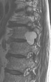
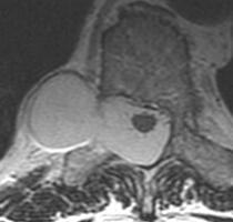
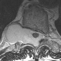
20 Lumbar nevus {Page 2}


21 {Page 3} Dx: Dorsal dermal sinus with an intradural epidermoid

22 Hemimyelocele 1 2


23 {Page 2}


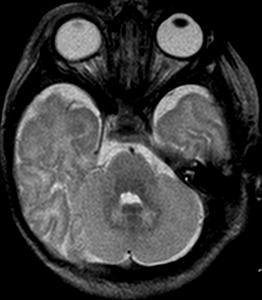
24 History: One day old boy born with an occipital mass 39


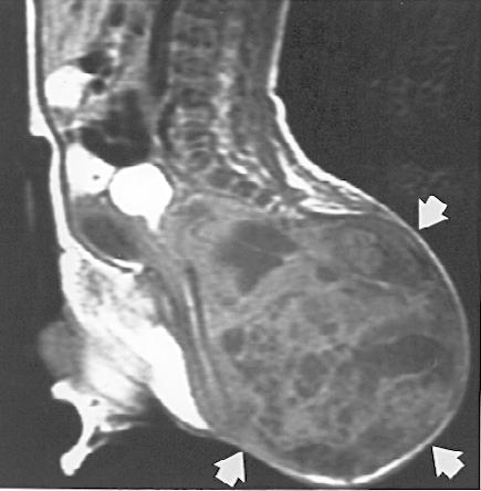
25 {Page 2} Dx: Chiari III - occipital meningoencephalocele - small posterior fossa - low tonsils, culpocephaly - scalloped clivus, beaked tectum - stenogyria
26 Lower Spine Development Canalization Caudal cell mass forms Microcysts coalesce to form neural tube Retrogressive Differentiation Neural tube decreases in size Neural tube forms caudal conus medullaris & filum terminale
27 Spine Development Caudal Cell Mass Anomalies Fibrolipomas of filum terminale Tight filum terminale syndrome Caudal regression Anterior sacral meningoceles Sacrococcygeal teratomas
28 Fibrolipoma of the Filum Terminale


29 Congenital scoliosis secondary to tethered cord
30 Spinal Dysraphism Critical Neural Features Position of conus Location of neural placode Fibromuscular tethering bands Ventral & dorsal nerve roots Hydrosyringomyelia
31 Tethered Spinal Cord Associated Anomalies Myelomeningocele Lipomyeloschisis Diastematomyelia Dorsal dermal sinus Caudal regression

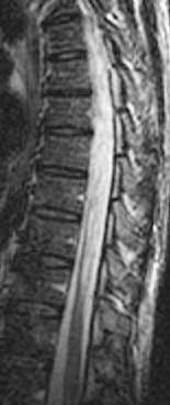
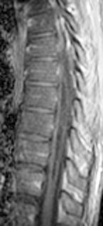
32 Caudal Regression Syndrome

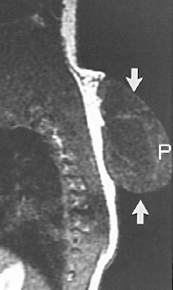
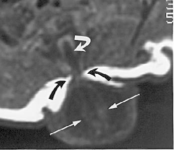
33 Anterior Sacral Meningocele
34 Sacrococcygeal Teratoma
35 Spinal Anomalies of Unknown Origin Myelocystocele Simple meningoceles Lateral meningoceles Syringohydromyelia
36 Myelocystocele


37 History: 67 y/o male with NF1 408

38 Dx: Meningocele {Page 2}
39 Postoperative Myelocystocele
40 Syringohydromyelia Etiology Chiari I 42% 28% 15% Post-Traumatic 15% Idiopathic Neoplastic

41 Chiari I with Syringohydromyelia


42 History: 22 y/o male with brain stem dysfunction Dx: Chiari I malformation with syringohydromyelia 339


43 History: 45 y.o. male with a progressive myelopathy C1 C5 C6 101
44 Myelopathy {Page 2} T5 T6 T9
45 Spinal Cord Cysts Evidence for Benign Syrinx Fluid isointense to CSF Smooth, well-defined internal margins Thinned adjacent parenchyma Cord atrophy No contrast enhancement Presence of Chiari malformation
Sonography of the Neonatal Spine: Part 2, Spinal Disorders
 Neonatal Spine Sonography Pediatric Imaging Pictorial Essay Downloaded from www.ajronline.org by 148.251.232.83 on 04/11/18 from IP address 148.251.232.83. Copyright RRS. For personal use only; all rights
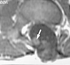
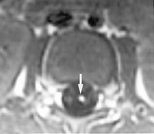
Persistent Terminal Ventricle
 Persistent Terminal Ventricle Ventriculus Terminalis Incomplete regression of TV of 2 neurulation, continuity with central canal small cavity PTV vs terminal myelocystocele (?severe manifestation from
SPLIT NOTOCHORD SYNDROME ASSOCIATION. DR. Hasan Nugud Consultant Paediatric Surgeon
 SPLIT NOTOCHORD SYNDROME ASSOCIATION DR. Hasan Nugud Consultant Paediatric Surgeon CASE PRESENTATION :- New born baby, boy, referred to the paediatric surgical team at the age of 14 hours. Birth History
Congenital Spine and Spinal Cord Malformations Pictorial Review
 JR Integrative Imaging LIFELONG LERNING FOR RDIOLOGY ongenital Spine and Spinal ord Malformations Pictorial Review Stephanie L. Rufener 1,2, Mohannad Ibrahim 2, harles. Raybaud 3, Hemant. Parmar 2 Downloaded
Radiologic and pathologic features of spinal dysraphism. A pictorial review.
 Radiologic and pathologic features of spinal dysraphism. A pictorial review. Poster No.: C-0586 Congress: ECR 2011 Type: Educational Exhibit Authors: N. Arcalis, J. L. Ribó, J. Muchart, L. Riaza, J. Blanch
Imaging the Spinal Cord & Intradural Disease
 Department of Radiology University of California San Diego Imaging the Spinal Cord & Intradural Disease John R. Hesselink, M.D. Spinal Cord Diseases Tumors Syringohydromyelia Trauma Ischemia / Infarction
A Retrospective Analysis of Clinical Profile and Surgical Outcome in Patients with Spinal Dysraphism at Tertiary Care Center
 Original Research Article A Retrospective Analysis of Clinical Profile and Surgical Outcome in Patients with Spinal Dysraphism at Tertiary Care Center Premlal KV * Assistant Professor, Department of Neurosurgery,
Neonatal Spinal Ultrasound Imaging - A Pictorial Review from The Royal Liverpool Children Hospital, Alder Hey, Liverpool
 Neonatal Spinal Ultrasound Imaging - A Pictorial Review from The Royal Liverpool Children Hospital, Alder Hey, Liverpool Poster No.: C-0081 Congress: ECR 2012 Type: Educational Exhibit Authors: K. Chetcuti,
Dorsal dermal sinus in children
 Dorsal dermal sinus in children Poster No.: C-2581 Congress: ECR 2015 Type: Educational Exhibit Authors: J. Marjanovic, A. Paterson, P. C. McSherry, A. Nixon, A. 1 1 2 1 2 1 1 2 TRIPALO BATOS, T. Grmoja
Disclosures None. Spinal Dysraphism
 Spinal Dysraphism Andrew Jea MD MHA FAAP Professor and Chief Section of Pediatric Neurosurgery Riley Hospital for Children Department of Neurosurgery Indiana University School of Medicine Goodman Campbell
Role of helical CT and MRI in the evaluation of spinal dysraphism
 International Journal of Advances in Medicine Kumaran SK et al. Int J Adv Med. 2017 Feb;4(1):124-132 http://www.ijmedicine.com pissn 2349-3925 eissn 2349-3933 Original Research Article DOI: http://dx.doi.org/10.18203/2349-3933.ijam20170095
Development of Spinal Cord & Vertebral Column. Dr. Sanaa Alshaarawi & Prof. Ahmed Fathalla
 Development of Spinal Cord & Vertebral Column Dr. Sanaa Alshaarawi & Prof. Ahmed Fathalla OBJECTIVES At the end of the lecture, students should be able to: q Describe the development of the spinal cord
LUMPS, TUFTS AND DIMPLES IT S THE PITS!! Session Information. Faculty Disclosure Information
 Session Information Session Title: Lumps, Tufts and Dimples Session Number: F3056, F2130 Faculty Name: Mark S. Dias, MD, FAAP Faculty Institution: Penn State Children s Hospital, Penn State University
Spine and spinal cord
 NEURORADIOLOGY Spine and spinal cord Erika Vörös University of Szeged Department of Radiology SZEGED DISEASES OF SPINE AND SPINAL CORD I. Non-tumourous diseases developmental anomalies vascular disorders
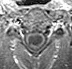
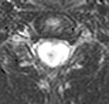
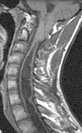
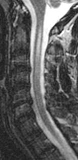
Diastematomyelia: A Case with Familial Aggregation of Neural Tube Defects
 Case Study TheScientificWorldJOURNAL (2004) 4, 847 852 ISSN 1537-744X; DOI 10.1100/tsw.2004.140 Diastematomyelia: A Case with Familial Aggregation of Neural Tube Defects Nuray Öksüz Kanbur 1, *, Pınar
Sonography of the Neonatal Spine: Part 1, Normal Anatomy, Imaging Pitfalls, and Variations That May Simulate Disorders
 Sonography of Neonatal Spine Pediatric Imaging Pictorial Essay Downloaded from www.ajronline.org by 46.3.195.60 on 02/04/18 from IP address 46.3.195.60. Copyright RRS. For personal use only; all rights
Long segment composite split cord malformation with double bony spur
 Long segment composite split cord malformation with double bony spur Anand Sharma, Achal Sharma, R.S. Mittal SMS Medical College, Jaipur, India Abstract: A composite type of SCM is very rare and only a
Spinal dysraphism: genetic relation to
 Journal of Medical Genetics (1976). 13, 343-350. Spinal dysraphism: genetic relation to neural tube malformations C. 0. CARTER, K. A. EVANS, and K. TILL* From MRC Clinical Genetics Unit, Institute of Child
Essentials of Clinical MR, 2 nd edition. 51. Primary Neoplasms
 51. Primary Neoplasms As with spinal central canal neoplasms in other regions, those of the lumbar spine may be classified as extradural, intradural extramedullary, and medullary. If an extradural lesion
Skeletal System. Prof. Dr. Malak A. Al-yawer Department of Anatomy/Embryology Section
 Skeletal System Prof. Dr. Malak A. Al-yawer Department of Anatomy/Embryology Section Learning objectives At the end of this lecture, the medical student will be able to: State the embryonic origin of skeletal
Introduction to Neurosurgical Subspecialties:
 Introduction to Neurosurgical Subspecialties: Pediatric Neurosurgery Brian L. Hoh, MD 1 and Gregory J. Zipfel, MD 2 1 University of Florida, 2 Washington University Pediatric Neurosurgery Pediatric neurosurgeons
University Journal of Surgery and Surgical Specialties
 University Journal of Surgery and Surgical Specialties ISSN 2455-2860 Volume 2 Issue 1 2016 Profile of paediatric patients with split cord malformation MANORANJITHAKUMARI M Department of Neuro Surgery,
Spinal congenital dermal sinus with dual ostia
 J Neurosurg Pediatrics 3:000 000, 3:407 411, 2009 Spinal congenital dermal sinus with dual ostia Clinical article Ch a n g Su b Le e, M.D., 1 Ji Ho o n Ph i, M.D., 2 Se u n g -Ki Kim, M.D., Ph.D., 2 By
Spine. Neuroradiology. Spine. Spine Pathology. Distribution of fractures. Radiological algorithm. Role of radiology 18/11/2015
 Spine Neuroradiology Spine Prof.Dr.Nail Bulakbaşı X Ray: AP/L/Oblique Vertebra & disc spaces CT & CTA Vertebra, discs, vessels MRI & MRA Vertebra, disc, vessels, meninges Spinal cord & nerves Myelography
Prenatal ultrasound evaluation of fetal diastematomyelia: two cases of type I split cord malformation
 Ultrasound Obstet Gynecol 2000; 15: 78 82. Prenatal ultrasound evaluation of fetal diastematomyelia: two cases of type I split cord malformation L.M. ALLEN and R.K. SILVERMAN Perinatal Center, SUNY Health
disclosure Pediatric Tethered cord Syndrome Learning Objectives overview definiton Hoffman 1976 Pediatrics Grand Rounds 26 June 2015
 disclosure Pediatric Tethered cord Syndrome None Izabela, Tarasiewicz, MD,FRCS(C), has no relationships with commercial companies to disclose. Izabela Tarasiewicz MD. FRCS(C) Pediatric Neurosurgery overview
Anatomy of the Nervous System. Brain Components
 Anatomy of the Nervous System Brain Components NERVOUS SYSTEM INTRODUCTION Is the master system of human body, controlling the functions of rest of the body systems Nervous System CLASSIFICATION A. Anatomical
University Journal of Surgery and Surgical Specialties
 University Journal of Surgery and Surgical Specialties ISSN 2455-2860 Volume 2 Issue 1 2016 TWO RARE CASES OF DIASTEMATOMYELIA MUTHURAMAN P Department of Neuro Surgery, THANJAVUR MEDICAL COLLEGE Abstract
Neuroanatomy. Assistant Professor of Anatomy Faculty of Medicine The University of Jordan Dr Maha ELBeltagy
 Neuroanatomy Dr. Maha ELBeltagy Assistant Professor of Anatomy Faculty of Medicine The University of Jordan 2018 Development of the Central Nervous System Development of the nervous system Development
Central Nervous System Congenital Abnormalities
 Central Nervous System Congenital Abnormalities Eva Brichtova, M.D., Ph.D., Department of Pediatric Sugery, Orthopaedics and Traumatology, University Hospital Brno Neural tube defects Dysraphism uncomplete
Moderators Dr A Suri Dr Deepak Gupta. Presented by Dr A Bisht
 Moderators Dr A Suri Dr Deepak Gupta Presented by Dr A Bisht A distinct group of congenital anomalies characterized by a failure of midline structures of ecto- and mesodermal origin to fuse Nicolas Tulp
Tethered spinal cord syndrome: a developmental overview
 International Journal of Sciences & Applied Research www.ijsar.in Tethered spinal cord syndrome: a developmental overview Anushi Singh 1 *, Rekha Kumari 2 1 CHN Department, School of Nursing Science and
Early Development of Neural Tube Development of Medulla Spinalis and Peripheral Nervous System. Assoc.Prof. E.Elif Güzel, M.D.
 Early Development of Neural Tube Development of Medulla Spinalis and Peripheral Nervous System Assoc.Prof. E.Elif Güzel, M.D. Third week of Embryogenesis Primitive streak/pit appears on the epiblast (day
Ligaments of the vertebral column:
 In the last lecture we started talking about the joints in the vertebral column, and we said that there are two types of joints between adjacent vertebrae: 1. Between the bodies of the vertebrae; which
Introduction to Neuroimaging spine. John J. McCormick MD
 Introduction to Neuroimaging spine John J. McCormick MD Neuroanatomy Netter drawings Radiographic Anatomy Cervical Spine Cervical Spine Oblique View Cervical Spine Dens View Thoracic Spine Lumbar Spine
MR Imaging in the Tethered Spinal Cord Syndrome
 27 MR Imaging in the Tethered Spinal Cord Syndrome Narasimhachari Raghavan 1 A. James Barkovich 1 Michael Edwards 2 David Norman 1 MR examinations of the spine were reviewed in 25 patients with a clinical
Asymptomatic posterior cervical myelomeningocele with tethered cord in an adolescent: a rare form of spinal dysraphism with rare presentation
 Romanian Neurosurgery (2016) XXX 1: 113-117 113 Asymptomatic posterior cervical myelomeningocele with tethered cord in an adolescent: a rare form of spinal dysraphism with rare presentation Gangesh Gunjan,
The spinal dermal-sinus-like stalk
 Childs Nerv Syst (2009) 25:191 197 DOI 10.1007/s00381-008-0669-6 ORIGINAL PAPER The spinal dermal-sinus-like stalk J. van Aalst & E. A. M. Beuls & E. M. J. Cornips & H. W. M. van Straaten & A. F. M. Boselie
Wound healing in trophic ulcers in spina bifida patients
 J Neurosurg 82:000 000, 1995 Wound healing in trophic ulcers in spina bifida patients VINOD KUMAR SRIVASTAVA, M.B.B.S, M.CH. Neurosurgical Unit, J. N. Medical College, Aligarh Muslim University, Aligarh,
Chapter 8. Pediatric Surgery
 Chapter 8 Pediatric Surgery 8.1 Hydrocephalus Hydrocephalus is a congenital disorder. There may be difficulties during normal vaginal delivery due large size of the head. In 1970s, when these pictures
Split cord malformation in children undergoing neurosurgical intervetion in India: a descriptive study
 ORIGINAL ARTICLE Journal of Pediatric Neurology 2004; 2(1): 21-27 www.jpneurology.org in children undergoing neurosurgical intervetion in India: a descriptive study Raj Kumar 1, Vinita Singh 2, Satya Narayan
Spinal Neoplasms. First Things First!! Localize the Lesion!! Ependymomas. Common Intramedullary Lesions
 Acta Radiológica Portuguesa, Vol.XXIII, nº 90, pág. 101-114, Abr.-Jun., 2011 Spinal Neoplasms Bruno A Policeni University of Iowa Hospitals and Clinics Assistant Professor of Radiology Disclosure of Commercial
Intraoperative Sonography in Spinal Dysraphism and Syringohydromyelia
 329 Intraoperative Sonography in Spinal Dysraphism and Syringohydromyelia Robert M. Quencerl erta M. Montalvo 2 Thomas P. Naidich 3 M. Judith Donovan Post 1 arth. Green 4 Larry K. Page 4 The use of intraoperative
Contents I MEDICAL RADIOLOGY. Diagnostic Imaging. Editors: A. L. Baert, Leuven M. Knauth, Göttingen K. Sartor, Heidelberg
 Contents I MEDICAL RADIOLOGY Diagnostic Imaging Editors: A. L. Baert, Leuven M. Knauth, Göttingen K. Sartor, Heidelberg Contents III J. W. M. Van Goethem L. van den Hauwe P. M. Parizel (Eds.) Spinal Imaging
NEURORADIOLOGY. Part III. Angela Csomor University of Szeged Department of Radiology
 NEURORADIOLOGY Part III Angela Csomor University of Szeged Department of Radiology DISEASES OF SPINE AND SPINAL CORD I. Non-tumourous diseases developmental anomalies vascular disorders inflammatory processes
Fetal CNS MRI. Daniela Prayer. Division of Neuroradiology And Musculoskeletal Radiology. Medical University of Vienna, AUSTRIA
 Fetal CNS MRI Daniela Prayer Division of Neuroradiology And Musculoskeletal Radiology Medical University of Vienna, AUSTRIA Methods Normal development Malformations Acquired pathology MR- methods for assessment
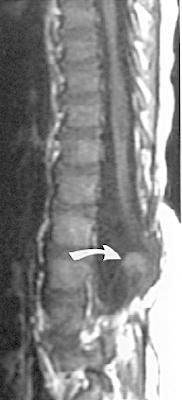
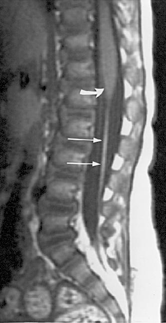
Ventriculus Terminalis of the Conus Medullaris: MR Imaging in Four Patients with Congenital Dilatation
 733 Ventriculus Terminalis of the Conus Medullaris: MR Imaging in Four Patients with Congenital Dilatation Robert Sigal 1 2 Alban Denys 2 Philippe Halimi 2 Lorraine Shapeero 1 3 Dominique Doyon 2 Frank
Congenital Spinal Lipoma: analyzing the perplexed nomenclature and our management
 Congenital Spinal Lipoma: analyzing the perplexed nomenclature and our management Nidal Khasawneh MD *, Rami Alqroom MD *, Firas Sha'ban MD *, Rafeed Al Drous MD *, Rima Nserat **, Amer Al Shurbaji MD
Pediatric Spine Tumors (and other masses)
") Pediatric Spine Tumors (and other masses) Francisco A Perez, MD, PhD Assistant Professor Neuroradiology and Pediatric Radiology Seattle Children s Hospital University of Washington, Seattle Commercial
Pediatric Spine Tumors (and other masses) Francisco A Perez, MD, PhD Assistant Professor Neuroradiology and Pediatric Radiology Seattle Children s Hospital University of Washington, Seattle Commercial
Spinal ultrasound is considered medically necessary for ANY of the following indications:
 Medical Coverage Policy Effective Date...11/15/2017 Next Review Date...11/15/2018 Coverage Policy Number... 0246 Spinal Ultrasound Table of Contents Coverage Policy... 1 Overview... 1 General Background...
Embryology of the Nervous System. Steven McLoon Department of Neuroscience University of Minnesota
 Embryology of the Nervous System Steven McLoon Department of Neuroscience University of Minnesota In the blastula stage embryo, the embryonic disk has two layers. During gastrulation, epiblast cells migrate
Johnson Rogers and colleagues- used Term LMM
 LIPOMENINGOMYELOCELE: CLASSIFICATION, MANAGEMENT AND CONTROVERSIES Definition : Lipomyelomeningocele is a form of OSD in which a subcutaneous fibrofatty mass traverses the lumbodorsal fascia, causes a
Congenital Brain and Spinal Cord Malformations and Their Associated Cutaneous Markers
 CLINICAL REPORT Guidance for the Clinician in Rendering Pediatric Care Congenital Brain and Spinal Cord Malformations and Their Associated Cutaneous Markers Mark Dias, MD, FAANS, FAAP, Michael Partington,
Case Report. Case Report
 34 Case Report Multiple dysraphic anomalies with double thoracic meningocele and lumbosacral myelomeningocele, concurrent Chiari malformation Type I, diastematomyelia, lipomyelomeningocele and hydrocephalus:
Case Report Occult Spinal Dysraphism in the Presence of Rare Cutaneous Stigma in a Neonate: Importance of Ultrasound and Magnetic Resonance Imaging
 Case Reports in Medicine Volume 2013, Article ID 468376, 4 pages http://dx.doi.org/10.1155/2013/468376 Case Report Occult Spinal Dysraphism in the Presence of Rare Cutaneous Stigma in a Neonate: Importance
What Every Spine Surgeon Should Know About Neurosurgical Issues
 What Every Spine Surgeon Should Know About Neurosurgical Issues Amer Samdani, MD Chief of Surgery Shriners Hospitals for Children Philadelphia, PA Objectives Main intraspinal lesions Chiari malformation
Congenital Tethered Spinal Cord Syndrome in Adults
 Page 1 of 7 http://www.medscape.com/ To Print: Click your browser's PRINT button. NOTE: To view the article with Web enhancements, go to: http://www.medscape.com/viewarticle/405679 Congenital Tethered
Neonatal and infantile spinal sonography: A useful investigation often underutilized
 Pediatric Neonatal and infantile spinal sonography: useful investigation often underutilized Nikhil Nair, M Sreenivas 1, run K Gupta, Devasenathipathy Kandasamy, Manisha Jana Departments of Radiodiagnosis,
Congenital tethered spinal cord syndrome in adults
 Neurosurg Focus 10 (1):Article 7, 2001, Click here to return to Table of Contents Congenital tethered spinal cord syndrome in adults BERMANS J. ISKANDAR, M.D., BENJAMIN B. FULMER, M.D., MARK N. HADLEY,
Cutaneous abnormalities of the back may represent
 Coccygeal Pits Bradley E. Weprin, MD*, and W. Jerry Oakes, MD ABSTRACT. Background. Congenital dermal sinuses represent cutaneous depressions or tracts that are lined by stratified squamous epithelium.
Prospective Evaluation of Role of MRI in Suspected Spinal Dysraphism and Its Management
 IOSR Journal of Dental and Medical Sciences (IOSR-JDMS) e-issn: 2279-0853, p-issn: 2279-0861.Volume 17, Issue 5 Ver. 1 (May. 2018), PP 21-28 www.iosrjournals.org Prospective Evaluation of Role of MRI in
Spinal Cord and Properties of Cerebrospinal Fluid: Options for Drug Delivery. SMA Foundation New York
 Spinal Cord and Properties of Cerebrospinal Fluid: Options for Drug Delivery New York Why Do We Need to Know about the Spinal Cord Anatomy and Properties of Cerebrospinal Fluid? SMA therapeutics need to
Disclosures None. Common Neurosurgical Problems Seen in Office Encounters. Macrocephaly Low Back Pain Sacral Dimple Concussion Chiari Malformation
 Common Neurosurgical Problems Seen in Office Encounters When to Manage, When to Refer Andrew Jea MD FAAP Professor and Chief of Pediatric Neurosurgery Riley Hospital for Children Indiana University School
Purpose: To discuss the fatty filum terminale which is incidentally demonstrated on MRI concerning the causes of TCS
 ISPUB.COM The Internet Journal of Spine Surgery Volume 3 Number 1 T Iizuka Citation T Iizuka.. The Internet Journal of Spine Surgery. 2006 Volume 3 Number 1. Abstract Background: Fatty filum terminale
SPINAL CORD AND PROPERTIES OF CEREBROSPINAL FLUID: OPTIONS FOR DRUG DELIVERY
 SPINAL CORD AND PROPERTIES OF CEREBROSPINAL FLUID: OPTIONS FOR DRUG DELIVERY WHY DO WE NEED TO KNOW ABOUT THE SPINAL CORD ANATOMY AND PROPERTIES OF CEREBROSPINAL FLUID? SMA therapeutics need to reach cells
Case Report Surgical Treatment of a Patient with Human Tail and Multiple Abnormalities of the Spinal Cord and Column
 SAGE-Hindawi Access to Research Advances in Orthopedics Volume 2011, Article ID 153797, 4 pages doi:10.4061/2011/153797 Case Report Surgical Treatment of a Patient with Human Tail and Multiple Abnormalities
The Wedge-Shaped Cord Terminus: A Radiographic Sign of
 1223 The Wedge-Shaped Cord Terminus: A Radiographic Sign of Caudal Regression A. James Barkovich 1 2 Narasimhachari Raghavan 1 Sylvester Chuang 3 Wallace W. Peck 4 Imaging studies from 13 patients with
Spinal Cord Workbook. Learning objec&ves
 Spinal Cord Workbook Direc&ons. Watch the following video tutorials and complete this workbook: YouTubeèTheNotedAnatomistèPlaylistsèSpinal cord and nervesèwatch videos with the names of ObjecCves A-E.
Role of MRI in the Evaluation of Spinal Dysraphism
 Role of MRI in the Evaluation of Spinal Dysraphism 1 Dr Ravi N, 2 Dr Ramesh V, 3 Dr Naveen K, G, 4 Dr R Nagaraj, 2 Dr darsh K M, 2 Dr Manjappa H, 2 Dr Lakshmeesha M T 1 ssociate Professor, 2 Resident,
Human Anatomy. Spinal Cord and Spinal Nerves
 Human Anatomy Spinal Cord and Spinal Nerves 1 The Spinal Cord Link between the brain and the body. Exhibits some functional independence from the brain. The spinal cord and spinal nerves serve two functions:
Magnetic resonance imaging in the prenatal diagnosis of neural tube defects
 Insights Imaging (2013) 4:225 237 DOI 10.1007/s13244-013-0223-2 PICTORIAL REVIEW Magnetic resonance imaging in the prenatal diagnosis of neural tube defects A. Zugazaga Cortazar & C. Martín Martinez &
Scoliosis: Orthopaedic Perspectives
 Scoliosis: Orthopaedic Perspectives Scott B. Rosenfeld, MD Division of Pediatric Orthopaedic Surgery Texas Children s Hospital Page 0 xxx00.#####.ppt 9/23/2012 8:26:24 AM I have no disclosures Disclosures
Human Anatomy - Problem Drill 11: The Spinal Cord and Spinal Nerves
 Human Anatomy - Problem Drill 11: The Spinal Cord and Spinal Nerves Question No. 1 of 10 Instructions: (1) Read the problem statement and answer choices carefully, (2) Work the problems on paper as needed,
A Very Unusual Case of a Dorsal Heteropagus Twin
 PRG A Very Unusual Case of a Dorsal Heteropagus Twin Nathan David P. Concepcion, MD 1, Bernard F. Laya, DO 1, Eduardo P. Manrique, MD 2 and Faith Caroline D. Bayabos, MD 1 1 Section of Pediatric Radiology,
Neuroimaging. spine / spinal cord
 Neuroimaging spine / spinal cord Spine & spinal cord imaging methodology Plain x-ray of spine Computed tomography CT - traditional ( normal CT) - reconstructions - myelo-ct Magnetic resonance MR - standard
CNS Embryology 5th Menstrual Week (Dorsal View)
") Imaging of the Fetal Brain; Normal & Abnormal Alfred Abuhamad, M.D. Eastern Virginia Medical School CNS Embryology 5th Menstrual Week (Dorsal View) Day 20 from fertilization Neural plate formed in ectoderm
Imaging of the Fetal Brain; Normal & Abnormal Alfred Abuhamad, M.D. Eastern Virginia Medical School CNS Embryology 5th Menstrual Week (Dorsal View) Day 20 from fertilization Neural plate formed in ectoderm
Week 14. Development of the Musculoskeletal System
 Week 14 Development of the Musculoskeletal System Skeletal System Derived from: paraxial mesoderm somites and somitomeres sclerotome sclerotome differentiation induced by SHH from notochord and floor plate
MD Bones & Joints of the Back. A/Prof Chris Briggs Department of Anatomy & Neuroscience
 MD 2017 Bones & Joints of the Back A/Prof Chris Briggs Department of Anatomy & Neuroscience WARNING This material has been provided to you pursuant to section 49 of the Copyright Act 1968 (the Act) for
8/31/2018 IMPORTANT CONSIDERATIONS. Signalment History Symmetry Progression of signs Painful vs non-painful SURGICAL CONSIDERATIONS
 IMPORTANT CONSIDERATIONS Signalment History Symmetry Progression of signs Painful vs non-painful SURGICAL CONSIDERATIONS Specific region of TL spine Differences in size and shape of articular processes
Imaging in neurofibromatosis type 1: An original research article with focus on spinal lesions
 Original Research Article Imaging in neurofibromatosis type 1: An original research article with focus on spinal lesions Kalpesh Patel 1*, Siddharth Zala 2, C. Raychaudhuri 3 1 Assistant Professor, 2 1
Surgery for Spinal Cord Lipomas
 39 Original Article Surgery for Spinal Cord Lipomas Manish K. Kasliwal and Ashok K. Mahapatra Department of Neurosurgery, Neurosciences Centre, All India Institute of Medical Sciences, New Delhi, India
Lecture 14: The Spinal Cord
 Lecture 14: The Spinal Cord M/O Chapters 16 69. Describe the relationship(s) between the following structures: root, nerve, ramus, plexus, tract, nucleus, and ganglion. 70. Trace the path of information
ISUOG Basic Training. Examining Fetal Anatomy from Longitudinal Sections Titia Cohen-Overbeek, The Netherlands
 ISUOG Basic Training Examining Fetal Anatomy from Longitudinal Sections Titia Cohen-Overbeek, The Netherlands Learning objectives 2 & 3 At the end of the lecture you will be able to: describe how to obtain
MRI of chronic spinal cord injury
 The British Journal of Radiology, 76 (2003), 347 352 DOI: 10.1259/bjr/11881183 E 2003 The British Institute of Radiology Pictorial review MRI of chronic spinal cord injury 1 K POTTER, FRCR and 1 A SAIFUDDIN,
The Spinal Cord. The Nervous System. The Spinal Cord. The Spinal Cord 1/2/2016. Continuation of CNS inferior to foramen magnum.
 The Nervous System Spinal Cord Continuation of CNS inferior to foramen magnum Simpler than the brain Conducts impulses to and from brain Two way conduction pathway Reflex actions Passes through vertebral
MR Imaging Features of Common Variant Spinal Anatomy
 MR Imaging Features of Common Variant Spinal Anatomy Daniel J. Durand, MD a,b, *, Thierry A.G.M. Huisman, MD a,c, John A. Carrino, MD, MPH a,d KEYWORDS MR imaging Spine Embryology Normal variant The spine
Contents I MEDICAL RADIOLOGY. Diagnostic Imaging. Editors: A. L. Baert, Leuven M. Knauth, Göttingen K. Sartor, Heidelberg
 Contents I MEDICAL RADIOLOGY Diagnostic Imaging Editors: A. L. Baert, Leuven M. Knauth, Göttingen K. Sartor, Heidelberg Contents III J. W. M. Van Goethem L. van den Hauwe P. M. Parizel (Eds.) Spinal Imaging
Tethered cord syndrome is a rare intraspinal anomaly, Pediatric tethered cord syndrome: response of scoliosis to untethering procedures
 J Neurosurg Pediatrics 4:000 000, 4:270 274, 2009 Pediatric tethered cord syndrome: response of scoliosis to untethering procedures Clinical article Ma t t h e w J. McGi r t, M.D., 1 Vi v e k Me h ta,
University of Groningen
 University of Groningen Long-term evaluation of intraoperative neurophysiological monitoring-assisted tethered cord surgery Dulfer, S E; Drost, Gerrie; Lange, F; Journee, H L; Wapstra, F H; Hoving, E W
Management of Tethered Cord Syndrome in Adults: Experience of 23 Cases
 DOI: 10.5137/1019-5149.JTN.15892-15.1 Received: 31.08.2015 / Accepted: 02.10.2015 Published Online: 02.06.2016 Original Investigation Management of Tethered Cord Syndrome in Adults: Experience of 23 Cases
Pharyngeal Apparatus. Pouches Endoderm Grooves Ectoderm Arch Neural Crest Somitomeres Aortic Arch - Vessel
 Pharyngeal Apparatus Pouches Endoderm Grooves Ectoderm Arch Neural Crest Somitomeres Aortic Arch - Vessel Segmental Organization Humans: Arch 1-4 prominent Arch 5 absent Arch 6 - transient First Arch Face
Gross Morphology of Spinal Cord
 Gross Morphology of Spinal Cord Lecture Objectives Describe the gross anatomical features of the spinal cord. Describe the level of the different spinal segments compared to the level of their respective
Spinal intramedullary lipoma: report of three cases
 (2003) 41, 310 315 & 2003 International Society All rights reserved 1362-4393/03 $25.00 www.nature.com/sc Case Report Spinal intramedullary lipoma: report of three cases Chi Heon Kim 1, Kyu-Chang Wang
Syrinx location and size according to etiology: identification of Chiari-associated syrinx
 PEDIATRICS clinical article J Neurosurg Pediatr 16:21 29, 2015 Syrinx location and size according to etiology: identification of Chiari-associated syrinx Jennifer Strahle, MD, Karin M. Muraszko, MD, Hugh
Remember from the first year embryology Trilaminar disc has 3 layers: ectoderm, mesoderm, and endoderm
 Development of face Remember from the first year embryology Trilaminar disc has 3 layers: ectoderm, mesoderm, and endoderm The ectoderm forms the neural groove, then tube The neural tube lies in the mesoderm
Chapter 12b. Overview
 Chapter 12b Spinal Cord Overview Spinal cord gross anatomy Spinal meninges Sectional anatomy Sensory pathways Motor pathways Spinal cord pathologies 1 The Adult Spinal Cord About 18 inches (45 cm) long
1/9/2013 EXTRAMEDULLARY TUMORS OF THE PEDIATRIC SPINE. Introduction. Classification for Extramedullary Tumors
 EXTRAMEDULLARY TUMORS OF THE PEDIATRIC SPINE Eugene Wang 1/20/12 Dent Neurologic Institute Introduction 2/3 of all intraspinal tumors of childhood are extramedullary 50% Extradural 10-15% Intradural Back
A Congenital Defect in the Spinal Cord of the Manx Cat
 Vet, Path. 8: 232-238 (1971) A Congenital Defect in the Spinal Cord of the Manx Cat A. H. MARTIN Department of Anatomy, University of Wisconsin, Madison Wisc. Abstract. The lumbar part of the spinal cords
Spine Surgery: Techniques, Complication Avoidance, and Management. 2 Volume Set
 Spine Surgery: Techniques, Complication Avoidance, and Management. 2 Volume Set Benzel, E ISBN-13: 9781437705874 Table of Contents SECTION 1 - HISTORY 1 - History 2 - History of Spine Instrumentation -