Musculoskeletal Imaging What to order? Brian Cole, MD
|
|
|
- Julian McDowell
- 6 years ago
- Views:
Transcription
1 Musculoskeletal Imaging What to order? Brian Cole, MD

2 my background: 1994 University of Illinois 1998 MD University of Illinois College of Medicine Diagnostic Radiology Mayo Clinic 2004 Fellowship Musculoskeletal Radiology, Mayo Clinic 2005-present
3 What is the best imaging test? Plain film radiographs MRI CT US Bone Scan MR Arthrography CT Arthrography
4 What is the cause of musculoskeletal pain?

5 Clinical Evaluation Primary Care Orthopedics
6 Shoulder Pain: Patient Information Form
7

8 Tailored to individual joint Tailored to individual patient
9 Today s Objectives Keep it simple Musculoskeletal MRI basics Utility, terminology A few rules of thumb for what imaging study to order interesting cases


10 You have many imaging options for evaluation of bone/joint complaints.

11 What about radiography (X-rays)?

12 1901 Nobel Prize in Medicine German physicist Wilhelm Roentgen

13 What about radiography (X-rays)?

14 Imaging should be tailored to the joint. In general, plain film evaluation should precede CT, MR, arthrography, and nuclear medicine studies
15 Acute post-traumatic shoulder pain: plain films for fracture or dislocation

16 Acute post-traumatic shoulder pain: plain films for fracture or dislocation

17 Acute post-traumatic shoulder pain: CT for further evaluation of known fracture

18 Acute post-traumatic shoulder pain: CT for further evaluation of known fracture

19 Acute post-traumatic shoulder pain & negative plain films: consider MRI.

20 Acute post-traumatic shoulder pain & negative plain films: consider MRI.

21 Acute post-traumatic shoulder pain & negative plain films: consider MRI.

22 Acute post-traumatic shoulder pain & negative plain films: fracture.

23 Acute post-traumatic shoulder pain & negative plain films: consider MRI.

24 Acute post-traumatic shoulder pain & negative plain films: consider MRI.

25 Acute post-traumatic shoulder pain & negative plain films: rotator cuff tear.
: labral")
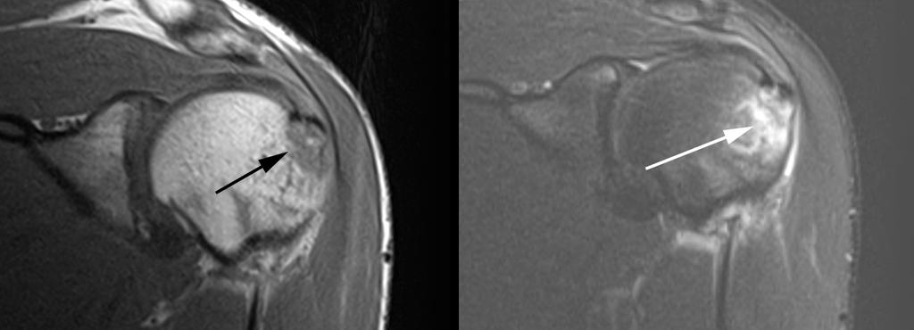
26 Chronic shoulder pain & negative plain films: consider MRI (+/- arthrogram): labral tear.
27 28 year old with shoulder pain

28 28 year old with shoulder pain Labral tear
29 In general radiographs prior to MRI. 58 y/o woman with pain following a fall.

30 Get radiographs prior to MRI.
31 Get radiographs prior to MRI.

32 radiographs prior to MRI. Fibular fracture.
33 radiographs prior to MRI.
34 radiographs prior to MRI.

")
35 radiographs prior to MRI. Cuboid (bone) contusion.
36 Radiographs prior to MRI. Then
37 MRI
38 This is all I know about MR physics. Slide 1 of 2.
39 This is all I know about MR physics. Slide 2 of 2.
40 MRI sequences T1 (short TR, short TE) T2 (long TR, long TE) Proton Density (long TR, short TE) STIR (Inversion Recovery)
41 MRI sequences T1 Fundamental sequence Evaluate fat Soft tissue planes Bone marrow Fat is bright Fluid is dark T2 Fat saturation all fat is saturated out (made dark) Evaluate fluid Soft tissue edema Fluid is bright Everything else is dark
42 MRI sequences Proton Density Intermediate sequence Short TE Fluid sensitive like T2 STIR (inversion recovery IR) Fluid sensitive like turbocharged T2 But less resolution Not susceptible to field inhomogeneity - - pure fat saturation throughout


43 imaging planes axial coronal sagittal


44 T1 sagittal Bone marrow Tissue planes
45 Abnormal marrow

46 64 y/o woman with lung cancer

47 39 y/o man with hip pain

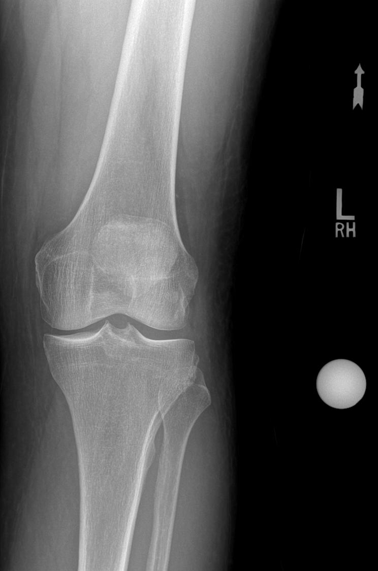
48 Case: 38 y/o left knee blunt injury pain medial and lateral
49


50 MRI Coronal T1 normal Lateral tibial injury


51 Tibial fracture Cor T2 fat sat Cor T1
52


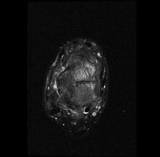
53 MRI Axial T2 fat sat


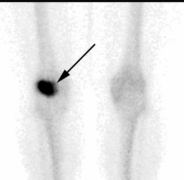
54 38 y/o male injured playing softball, sliding into 2 nd base
55 Chronic persistent pain after injury repeat films? CT? MRI? Bone Scan?
56 Chronic persistent pain after injury repeat films? CT? MRI Bone Scan?
57 Talar Fracture
58 Talar Fracture

59 Talar Fracture
60 What about computed tomography (CT)?
61 What about computed tomography (CT)?

62 What about computed tomography (CT)?
63 CT - characterize complex fractures
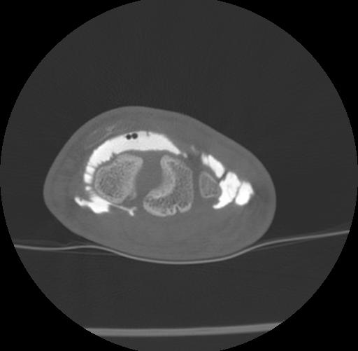
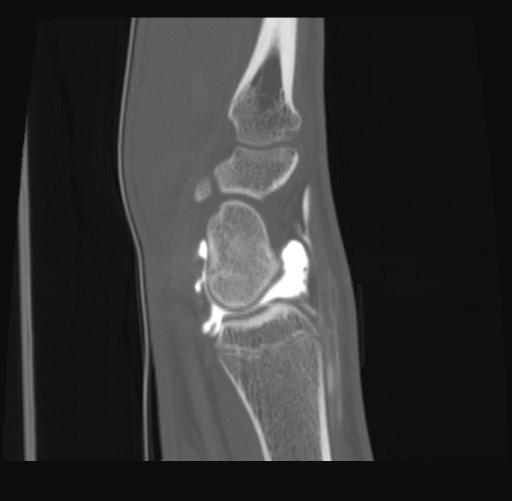
64 CT is great for assessing complex fractures. 12 y/o girl with a trampoline injury.
65 CT is great for assessing complex fractures. 12 y/o girl with a trampoline injury.


66 CT is great for assessing complex fractures. Tri-plane injury.

67 Acute post-traumatic knee pain: plain films and CT for fracture.

68 Acute post-traumatic knee pain: plain films and CT for complex fracture.

69 Acute post-traumatic knee pain: plain films and CT for complex fracture.
70 CT - characterize complex fractures - evaluate fracture healing

71 Scaphoid fracture healing
72 CT scaphoid fractures -Memarsadeghi M, Breitenseher MJ, Schaefer-prokop C et-al. Occult scaphoid fractures: comparison of multidetector CT and MR imaging--initial experience. Radiology. 2006;240 (1):
73 Scaphoid fractures CT High sensitivity/specificity Less for trabecular inj Staging Additional carpal injuries Bone union MRI Can detect completely undisplaced fractures Soft tissues/ligaments AVN
74 CT Arthrogram
75 CT Arthrography WRIST Intrinsic ligaments Extrinsic ligaments Capsule/synovium Cartilage Bone TFCC
76 CT Arthrography technique Fluoroscopic guided injection Radiocarpal compartment 1:2 solution omni/saline 4-6 ml contrast CT bone algorithm multireformatted


77 Fracture healing cartilage ligaments TFCC
78 CT Arthrogram
79 - characterize complex fractures - evaluate fracture healing - negative films with high clinical suspicion for fracture* -scaphoid -midfoot -elbow
80 CT is not good for all fractures. 80 year old with hip pain and inability to walk.


81 CT is not good for all fractures. Isolated greater trochanter fracture.
82 MRI for occult hip fx - Frihagen F, Nordsletten L, Tariq R, et al. MRI diagnosis of occult hip fractures. Acta Orthop. 2005;76: Verbeeten KM, Hermann KL, Hasselqvist M, et al. The advantages of MRI in the detection of occult hip fractures. Eur Radiol. 2005;15:
83 MR is better for some fractures.
84 MR is better for some fractures. Intertrochanteric fracture.

85 MR is better than CT for soft tissue injuries. Intertrochanteric fracture.

86 Acute post-traumatic hip pain and negative plain films: CT or MR?

87 Acute post-traumatic hip pain and negative plain films: fracture.

88 Acute post-traumatic hip pain and negative plain films: muscle tear.
89 CT for known complex fractures, MRI for injury with normal plain films
90 What about ultrasound (US)?
91 What about ultrasound (US)?

92 US offers a less costly method of soft tissue evaluation. ~ technologist experience
93 What about ultrasound (US)? Definite potential for some soft tissue injuries, particularly in the shoulder.

94 The End
This presentation is the intellectual property of the author. Contact them for permission to reprint and/or distribute.
 MRI of the Knee Jennifer Swart, M.D. Musculoskeletal Radiology South Texas Radiology Group Outline Coils, Patient Positioning Acquisition Parameters, Planes and Pulse Sequences Knee Arthrography Normal
This presentation is the intellectual property of the author. Contact them at for permission to reprint and/or distribute.
 MRI of the Knee Jennifer Swart, M.D. Musculoskeletal Radiology South Texas Radiology Group Financial Disclosure Dr. Jennifer Swart has no relevant financial relationships with commercial interests to disclose.
Musculoskeletal MR Protocols
 Musculoskeletal MR Protocols Joint-based protocols MSK 1: Shoulder MRI MSK 1A: Shoulder MR arthrogram MSK 1AB: Shoulder MR arthrogram (instability protocol) MSK 2: Elbow MRI MSK 2A: Elbow MR arthrogram
Radiologic Pitfalls. Objectives: High Risk! Occult Fracture? 2/16/2014
 Objectives: Radiologic Pitfalls Gregory W. Hendey, MD, FACEP Professor of Clinical Emergency Medicine UCSF Fresno, Medical Education Program To discuss plain film and physical findings that suggest an
Imaging Choices in Occult Hip Fracture
 Introduction Imaging Choices in Occult Hip Fracture Jesse Cannon, MD; Salvatore Silvestri, MD; Mark Munro, MD J Emerg Med. 2009;32(3):144-152 Reporter PGY 宋兆家 Supervisor VS 侯勝文 990220 High dependence on
Scapholunate Ligament Lesions Imaging Which and when?
 Scapholunate Ligament Lesions Imaging Which and when? Kolo Frank Lesions to scapholunate ligament(sl) Most frequent cause of carpal instability Traumatic tears of SL ligament = most common ligament injury
Rad Tech 4643 MRI Torso and Extremities
 Rad Tech 4643 MRI Torso and Extremities Prostate Cancer Leiomyoma Retroverted Anteverted Ovarian Cyst Gone Wrong Fibroid (Leiomyoma) IUD Ovary Hysterectomy? What are we to see when imaging a female pelvis
Utility of Dual-Energy CT to Evaluate Patients with Hip and Pelvis Pain in the ER Setting
 Utility of Dual-Energy CT to Evaluate Patients with Hip and Pelvis Pain in the ER Setting Johnson, T., Moran, E., Glazebrook, K., Leng, S., Fletcher, J., and McCollough, C. An educational review ER011
CLINICAL PRESENTATION AND RADIOLOGY QUIZ QUESTION
 Donald L. Renfrew, MD Radiology Associates of the Fox Valley, 333 N. Commercial Street, Suite 100, Neenah, WI 54956 9/22/2012 Radiology Quiz of the Week # 91 Page 1 CLINICAL PRESENTATION AND RADIOLOGY
Basic Radiographic Principles Part II
 Basic Radiographic Principles Part II Kristopher Avant, D.O. October 19 th, 2016 I have no disclosures relevant to the material presented in this discussion. Good Stuff!!! 1 Really? Really! Musculoskeletal
CLINICAL PRESENTATION AND RADIOLOGY QUIZ QUESTION
 Donald L. Renfrew, MD Radiology Associates of the Fox Valley, 333 N. Commercial Street, Suite 100, Neenah, WI 54956 11/24/2012 Radiology Quiz of the Week # 100 Page 1 CLINICAL PRESENTATION AND RADIOLOGY
MRI of the Shoulder What to look for and how to find it? Dr. Eric Handley Musculoskeletal Radiologist Cherry Creek Imaging
 MRI of the Shoulder What to look for and how to find it? Dr. Eric Handley Musculoskeletal Radiologist Cherry Creek Imaging MRI of the Shoulder Benefits of Ultrasound: * Dynamic * Interactive real time
Why Talk About Technique? MRI of the Knee:
 Why Talk About Technique? MRI of the Knee: Part 1 - Imaging Techniques Mark Anderson, M.D. University of Virginia Health Sciences Center Charlottesville, Virginia Always had an interest teach our fellows
IMAGING TECHNIQUES CHAPTER 4. Imaging techniques
 IMAGING TECHNIQUES Imaging techniques 23 4.1. Conventional radiographic findings Conventional radiography, tomography, arthrography and stress views have traditionally been used for imaging the ankle and
CLINICAL PRESENTATION AND RADIOLOGY QUIZ QUESTION
 Donald L. Renfrew, MD Radiology Associates of the Fox Valley, 333 N. Commercial Street, Suite 100, Neenah, WI 54956 10/13/2012 Radiology Quiz of the Week # 94 Page 1 CLINICAL PRESENTATION AND RADIOLOGY
CT ARTHROGRAPHY It s not Always About the
 CT ARTHROGRAPHY It s not Always About the Magnet Kirkland W. Davis, M.D. University of Wisconsin Department of Radiology Disclosures Financial FDA IA Gd! What Is CT Arthrography? (CTR) Arthrogram: imaging
Certificate for Advanced Practice in Hand Therapy
 Certificate for Advanced Practice in Hand Therapy Curriculum Effective: March 2016 EBP 6100 Evidence-based Practice I (15 hours/1 credit) ONLINE SELF-PACED, SELF-STUDY This course is designed to improve
X-ray (Radiography) - Bone
 - Bone") Scan for mobile link. X-ray (Radiography) - Bone Bone x-ray uses a very small dose of ionizing radiation to produce pictures of any bone in the body. It is commonly used to diagnose fractured bones or
Scan for mobile link. X-ray (Radiography) - Bone Bone x-ray uses a very small dose of ionizing radiation to produce pictures of any bone in the body. It is commonly used to diagnose fractured bones or
Viviane Khoury, MD. Assistant Professor Department of Radiology University of Pennsylvania
 U Penn Diagnostic Imaging: On the Cape Chatham, MA July 11-15, 2016 Viviane Khoury, MD Assistant Professor Department of Radiology University of Pennsylvania Hip imaging has changed in recent years: new
CLINICAL PRESENTATION AND RADIOLOGY QUIZ QUESTION
 Donald L. Renfrew, MD Radiology Associates of the Fox Valley, 333 N. Commercial Street, Suite 100, Neenah, WI 54956 12/29/2012 Radiology Quiz of the Week # 105 Page 1 CLINICAL PRESENTATION AND RADIOLOGY
Sensitivity and Specificity in Detection of Labral Tears with 3.0-T MRI of the Shoulder
 Magee and Williams MRI for Detection of Labral Tears Musculoskeletal Imaging Clinical Observations C M E D E N T U R I C L I M G I N G JR 2006; 187:1448 1452 0361 803X/06/1876 1448 merican Roentgen Ray
Orthopedic Hardware Imaging Part II: MRI v. Metal
 Orthopedic Hardware Imaging Trent Roth, MD And Lauren Ladd, MD Indiana University School of Medicine IU Health Physicians-Radiology Recap: Imaging Techniques Radiography Standard for initial and surveillance
CLINICAL PRESENTATION AND RADIOLOGY QUIZ QUESTION
 Donald L. Renfrew, MD Radiology Associates of the Fox Valley, 333 N. Commercial Street, Suite 100, Neenah, WI 54956 12/01/2012 Radiology Quiz of the Week # 101 Page 1 CLINICAL PRESENTATION AND RADIOLOGY
Publication for the Philips MRI Community
 FieldStrength Publication for the Philips MRI Community Issue 38 Summer 2009 Pediatric MSK imaging benefits from tailored scan protocols Vanderbilt University Children s Hospital builds dedicated scans
Musculoskeletal Imaging at 3T with Simultaneous Use of Multipurpose Loop Coils
 Clinical Orthopedic Imaging Musculoskeletal Imaging at 3T with Simultaneous Use of Multipurpose Loop Coils Elena Ferrer 1 ; Rafael Coronado Santos 2 1 Radiology Department, Clínica Creu Blanca, Barcelona,
Magnetic resonance imaging of femoral head development in roentgenographically normal patients
 Skeletal Radiol (1985) 14:159-163 Skeletal Radiology Magnetic resonance imaging of femoral head development in roentgenographically normal patients Peter J. Littrup, M.D. 1, Alex M. Aisen, M.D. 2, Ethan
ORIGINAL ARTICLE. ROLE OF MRI IN EVALUATION OF TRAUMATIC KNEE INJURIES Saurabh Chaudhuri, Priscilla Joshi, Mohit Goel
 ROLE OF MRI IN EVALUATION OF TRAUMATIC KNEE INJURIES Saurabh Chaudhuri, Priscilla Joshi, Mohit Goel 1. Associate Professor, Department of Radiodiagnosis & imaging, Bharati Vidyapeeth Medical College and
MRI PEDIATRIC PROTOCOLS (Updated 6/19/2018)
") MRI PEDIATRIC PROTOCOLS (Updated 6/19/2018) *Please get or let us know where radiologist can review plain films. *For Texas Orthopedics and other Docs requesting only MSK section read for their pediatric
MRI PEDIATRIC PROTOCOLS (Updated 6/19/2018) *Please get or let us know where radiologist can review plain films. *For Texas Orthopedics and other Docs requesting only MSK section read for their pediatric
醫用磁振學 MRM 肌肉骨骼磁振造影簡介 肌肉骨骼磁振造影. 本週課程內容 General Technical Considerations 肌肉骨骼磁振造影簡介 盧家鋒助理教授國立陽明大學生物醫學影像暨放射科學系
 本週課程內容 http://www.ym.edu.tw/~cflu 肌肉骨骼磁振造影簡介 醫用磁振學 MRM 肌肉骨骼磁振造影 盧家鋒助理教授國立陽明大學生物醫學影像暨放射科學系 alvin4016@ym.edu.tw MRI of the musculoskeletal system (5th/6th edition) Editor: Thomas H. Berquist MD 2 General
MR IMAGING OF THE WRIST
 MR IMAGING OF THE WRIST Wrist Instability Dissociative Pattern apparent on routine radiographs Non-dissociative Stress / positional radiographs Dynamic fluoroscopy during stress Arthrography MRI / MR arthrography
Lawrence Gulotta Gillian Lieberman, MD October Gillian Lieberman, MD. Shoulder Imaging. Lawrence V. Gulotta, HMS IV 10/16/02
 October 2002 Shoulder Imaging Lawrence V. Gulotta, HMS IV 10/16/02 Goals Review Anatomy of the Shoulder -Dynamic Stabilizers -> Rotator Cuff -Static Stabilizers -> Labrum and Capsule Systematic Approach
MY PATIENT HAS KNEE PAIN. David Levi, MD Chief, Division of Musculoskeletal l limaging Atlantic Medical Imaging
 MY PATIENT HAS KNEE PAIN David Levi, MD Chief, Division of Musculoskeletal l limaging Atlantic Medical Imaging Causes of knee pain Non traumatic Trauma Osteoarthritis Patellofemoral pain Menisci or ligaments
Original Report. The Reverse Segond Fracture: Association with a Tear of the Posterior Cruciate Ligament and Medial Meniscus
 Eva M. Escobedo 1 William J. Mills 2 John. Hunter 1 Received July 10, 2001; accepted after revision October 1, 2001. 1 Department of Radiology, University of Washington Harborview Medical enter, 325 Ninth
Case Report: Knee MR Imaging of Haemarthrosis in a Case of Haemophilia A
 Clinical > Pediatric Imaging Case Report: Knee MR Imaging of Haemarthrosis in a Case of Haemophilia A M. A. Weber, J. K. Kloth University Hospital Heidelberg, Department of Diagnostic and Interventional
CT Findings of Traumatic Posterior Hip Dislocation after Reduction 1
 CT Findings of Traumatic Posterior Hip Dislocation after Reduction 1 Sung Kyoung Moon, M.D., Ji Seon Park, M.D., Wook Jin, M.D. 2, Kyung Nam Ryu, M.D. Purpose: To evaluate the CT images of reduced hips
Upper Extremity Page Lower Extremity Special Cases
 MSK MRI PROTOCOLS Contents Upper Extremity Page Shoulder Elbow Wrist Finger Thumb Lower Extremity Hip Pelvis Thigh Knee Lower Extremity/Shin Ankle Foot Special Cases Soft Tissue Mass Metal Protocol MSK
Commonly Missed Injuries of the Extremities
 Commonly Missed Injuries of the Extremities Dr. Tudor H. Hughes M.D., FRCR Department of Radiology University of California School of Medicine San Diego, California 1. Base of skull 2. Odontoid process
CLINICAL PRESENTATION AND RADIOLOGY QUIZ QUESTION
 Donald L. Renfrew, MD Radiology Associates of the Fox Valley, 333 N. Commercial Street, Suite 100, Neenah, WI 54956 12/08/2012 Radiology Quiz of the Week # 102 Page 1 CLINICAL PRESENTATION AND RADIOLOGY
Imaging Modalities: Clinical Reasoning and Key Instructional Elements: Radiography
 Imaging Modalities: Clinical Reasoning and Key Instructional Elements: Radiography Michael D. Ross, PT, DHSc, OCS mross@daemen.edu Disclosure No relevant financial relationship exists Objectives Determine
Diagnosis of scaphoid fracture and dedicated extremity MRI
 504 Acta Ofthop Scand 1999; 70 (5): 504-508 Diagnosis of scaphoid fracture and dedicated extremity MRI Thomas Bretlau', Ole Maagaard Christensen*, Per Edstrom', Henrik S Thornsen' and Gunnar Schwarz Lausten2
MRI SHOULDER WHAT TO SEE
 MRI SHOULDER WHAT TO SEE DR SHEKHAR SRIVASTAV Sr. Consultant- Knee & Shoulder Arthroscopy Sant Parmanand Hospital Normal Anatomy Normal Shoulder MRI Coronal Oblique Sagital Oblique Axial Cuts Normal Coronal
The Kienböck disease and scaphoid fractures. Mariusz Bonczar
 The Kienböck disease and scaphoid fractures Mariusz Bonczar The Kienböck disease and scaphoid fractures Mariusz Bonczar Kienböck disease personal experience My special interest for almost 25 years Thesis
Message of the Month for GPs June 2013
 Message of the Month for GPs June 2013 Dr Winn : Consultant Musculoskeletal Radiologist, Manchester Royal Infirmary Imaging of the musculoskeletal system Musculoskeletal pain is a common problem in the
RADIOGRAPHY OF THE ANKLE and LOWER LEG
 RADIOGRAPHY OF THE ANKLE and LOWER LEG Patient Position: ANKLE AP Projection Part Position: True Slight to place foot s long axis Center to Central Ray: to IR Midway Note: Ankle joint is to tips of malleoli
4/28/2010. Fractures. Normal Bone and Normal Ossification Bone Terms. Epiphysis Epiphyseal Plate (physis) Metaphysis
 Metaphysis") Fractures Normal Bone and Normal Ossification Bone Terms Epiphysis Epiphyseal Plate (physis) Metaphysis Diaphysis 1 Fracture Classifications A. Longitudinal B. Transverse C. Oblique D. Spiral E. Incomplete
Fractures Normal Bone and Normal Ossification Bone Terms Epiphysis Epiphyseal Plate (physis) Metaphysis Diaphysis 1 Fracture Classifications A. Longitudinal B. Transverse C. Oblique D. Spiral E. Incomplete
12/13/17. Policy Number: MCR: 627 Revision Date(s): 12/11/18. Review Date: 12/13/17, 12/19/18 DISCLAIMER
: 12/11/18. Review Date: 12/13/17, 12/19/18 DISCLAIMER") Subject: Upper Extremity CT, (73200, 73201, 73202) Policy Number: MCR: 627 Revision Date(s): 12/11/18 Original Effective Date: 12/13/17 Review Date: 12/13/17, 12/19/18 DISCLAIMER This Molina Clinical Review
Subject: Upper Extremity CT, (73200, 73201, 73202) Policy Number: MCR: 627 Revision Date(s): 12/11/18 Original Effective Date: 12/13/17 Review Date: 12/13/17, 12/19/18 DISCLAIMER This Molina Clinical Review
Research Article Investigation of Occult Hip Fractures: The Use of CT and MRI
 The Scientific World Journal Volume 2013, Article ID 830319, 4 pages http://dx.doi.org/10.1155/2013/830319 Research Article Investigation of Occult Hip Fractures: The Use of CT and MRI S. K. Gill, J. Smith,
Staging of Kienböck s disease. F. Hahn
 Staging of Kienböck s disease F. Hahn Introduction Why staging? Standardisation Communication Prognosis Treatment options X-rays 1910 First description by Kienböck 1947 Ståhl(Rx) 1957 Decoulx(modif. Ståhl,
Tendonitis of finger icd 10 code
 Tendonitis of finger icd 10 code Level II Trauma Center verified by the American College of Surgeons. SURGICAL CRITICAL CARE FELLOWSHIP, THOMAS JEFFERSON UNIVERSITY, PHILADELPHIA, PA, 2016-2017. VII. Suspected
Sport Specific MRI. The symptoms of the majority, if not all sports injuries are experienced when upright, and weight-bearing
 Sport Specific MRI The symptoms of the majority, if not all sports injuries are experienced when upright, and weight-bearing A complete, accurate MRI assessment can only be made when in the position of
Mayo Clinic Disorders of the Wrist
 Mayo Clinic Disorders of the Wrist Thursday, May 19, 2016 Pre-Conference Laboratory Workshop Anatomy of the Wrist & Wrist Arthroscopy 6:30 a.m. Registration and Breakfast 7:30 a.m. Welcome and Introduction
Regenexx Procedure: Imaging Case Reports and Medical Provider Information
 TM Regenexx Procedure: Imaging Case Reports and Medical Provider Information www.regenexx.com TM The Regenexx procedure is a patent pending autologous mesenchymal stem cell (MSC) re implantation. This
Complex Fractures and Hip Dislocations
 IMAGING OF HIP PAIN Patients may present with acute (< 2 weeks) or chronic hip pain. Acute pain may be related or not related to an acute traumatic event such as fall or trauma from a motor vehicle accident.
MRI of the Hips and Pelvis
 MRI of the Hips and Pelvis Hips and Pelvis Protocols Vascular abnormalities Fractures Soft tissues Labrum and FAI Hips and Pelvis Protocols Vascular abnormalities Fractures Soft tissues Labrum and FAI
Index. radiologic.theclinics.com. Note: Page numbers of article titles are in boldface type.
 Index Note: Page numbers of article titles are in boldface type. A Acromioclavicular joint injuries in football players, 318, 319 ALPSA. See Anterior labroligamentous periosteal sleeve avulsion. Anterior
Posttraumatic subchondral bone contusions and fractures of the talotibial joint: Occurrence of kissing lesions
 KISSING CONTUSIONS CHAPTER 7 Posttraumatic subchondral bone contusions and fractures of the talotibial joint: Occurrence of kissing lesions Elizabeth S. Sijbrandij 1, Ad P.G. van Gils 1, Jan Willem K.
Upper Extremity Page Lower Extremity Special Cases
 MSK MRI PROTOCOLS Contents Upper Extremity Shoulder Elbow Wrist Finger Thumb Lower Extremity Hip Pelvis Thigh Knee Lower Extremity/Shin Ankle Foot Special Cases Soft Tissue Mass Metal Protocol Page MSK
Laura M. Fayad, MD. Associate Professor of Radiology, Orthopaedic Surgery & Oncology The Johns Hopkins University
 Society of Pediatric Radiology, May 2013 Laura M. Fayad, MD Associate Professor of Radiology, Orthopaedic Surgery & Oncology The Johns Hopkins University Describes surgical techniques that resect and reconstruct
Acute Elbow Trauma in Children: Spectrum of Injury Revealed by MR Imaging Not Apparent on Radiographs
 James F. Griffith 1 Derek J. Roebuck 1,2 Jack C. Y. Cheng 3 Yu Leung Chan 1 Timothy H. Rainer 4 Bobby K. W. Ng 3 Constantine Metreweli 1 Received December 14, 1999; accepted after revision June 8, 2000.
Role of Magnetic Resonance Imaging in Patients with Knee Trauma
 Original Research Article Role of Magnetic Resonance Imaging in Patients with Knee Trauma Bhautik Kapadia 1, Bhumika Suthar 2* 1 Associate Professor, 2 Assistant Professor, Department of Radiodiagnosis,
Comparative study of high resolusion ultrasonography and magnetic resonance imaging in diagnosing traumatic knee injuries & pathologies
 Original article: Comparative study of high resolusion ultrasonography and magnetic resonance imaging in diagnosing traumatic knee injuries & pathologies Dr. Rakesh Gujjar*, Dr. R. P. Bansal, Dr. Sandeep
Digital tomosynthesis in diagnosis of occult hip fractures
 Digital tomosynthesis in diagnosis of occult hip fractures Poster No.: B-0781 Congress: ECR 2013 Type: Authors: Keywords: DOI: Scientific Paper M. Geijer 1, D. Collin 2, J. H. Göthlin 2 ; 1 Lund/SE, 2
SCAHPO-LUNATE DISSOCIATION
 SCAHPO-LUNATE DISSOCIATION Introduction Scapho-lunate dissociation is the most common significant ligamentous injury of the wrist. The condition is also sometimes referred to as rotary subluxation of the
Learning from Discrepancies Meetings - What we've learned from Musculoskeletal Diagnostic Errors in 2014
 Learning from Discrepancies Meetings - What we've learned from Musculoskeletal Diagnostic Errors in 2014 Poster No.: P-0104 Congress: ESSR 2015 Type: Scientific Poster Authors: B. Batohi, R. Chhabra, S.
CLINICAL PRESENTATION AND RADIOLOGY QUIZ QUESTION
 Donald L. Renfrew, MD Radiology Associates of the Fox Valley, 333 N. Commercial Street, Suite 100, Neenah, WI 54956 10/6/2012 Radiology Quiz of the Week # 93 Page 1 CLINICAL PRESENTATION AND RADIOLOGY
RADIOGRAPHY OF THE ELBOW & HUMERUS
 RADIOGRAPHY OF THE ELBOW & HUMERUS Patient Position: ELBOW AP Projection in same plane Part Position: Hand in ; patient Centered to Humeral epicondyles Central Ray: Structures Shown: AP Elbow Criteria
occult ortho injuries Lauren M Westafer, DO, MPH, MS
 hiding in plain sight occult ortho injuries Lauren M Westafer, DO, MPH, MS No financial disclosures No commercial product discussion Photo: Zoltan Horlick Recognize subtle findings on plain films that
FieldStrength. Achieva 3.0T enables cutting-edge applications, best-in-class MSK images
 FieldStrength Publication for the Philips MRI Community Issue 33 December 2007 Achieva 3.0T enables cutting-edge applications, best-in-class MSK images Palo Alto Medical Clinic Sports Medicine Center employs
Imaging the musculoskeletal system. An Introduction
 Imaging the musculoskeletal system An Introduction Objectives Discuss: commonly used imaging modalities in the musculoskeletal system normal imaging anatomy in the extremities fracture description Imaging
Chealon Miller, HMS IV Gillian Lieberman, MD. November Stress Fractures. Chealon Miller, Harvard Medical School Year IV Gillian Lieberman, MD
 November 2005 Stress Fractures Chealon Miller, Harvard Medical School Year IV Our Patient G.F. 29 year old female runner c/o left shin pain and swelling Evaluated at OSH with MRI showing a mass Referred
Knee Contusions and Stress Injuries. Laura W. Bancroft, M.D.
 Knee Contusions and Stress Injuries Laura W. Bancroft, M.D. Objectives Review 5 types of contusion patterns Pivot shift Dashboard Hyperextension Clip Lateral patellar dislocation Demonstrate various stress
Imaging of Articular Cartilage
 Clinical Imaging of Articular Cartilage Imaging of Articular Cartilage Prof. Dr. K. Verstraete Ghent University Introduction : Articular Cartilage Histology and biochemical composition Review of Imaging
Page 1 of 6. Appendix 1
 Page 1 Appendix 1 Rotation Objectives and Schedule 1. Introductory Month 4 weeks 2. Total Joints 4 weeks a. Diagnosis and management of hip and knee arthritis b. Indications for surgery c. Implant selection;
Clinical utility of tomosynthesis in suspected scaphoid fracture: Preliminary results evaluating the VolumeRad technique
 Clinical utility of tomosynthesis in suspected scaphoid fracture: Preliminary results evaluating the VolumeRad technique Poster No.: C-2193 Congress: ECR 2010 Type: Scientific Exhibit Topic: Musculoskeletal
Radiologic Imaging Magnetic Resonance Imaging (MRI)
") Radiologic Imaging X-ray has always been the golden rule in diagnosing and treating podiatric patients. Unfortunately, for some patients the diagnosis is not as evident. That is when we need to utilize
Radiologic Imaging X-ray has always been the golden rule in diagnosing and treating podiatric patients. Unfortunately, for some patients the diagnosis is not as evident. That is when we need to utilize
APPROPRIATE USE GUIDELINES
 APPROPRIATE USE GUIDELINES Appropriateness of Advanced Imaging Procedures (MRI, CT, Bone Scan/PET) in Patients with Shoulder Pain CDI QUALITY INSTITUTE: PROVIDER LED ENTITY (PLE) Compiled by Rob Liddell,
Orthopedics. 1. GOAL: Understand the pediatrician's role in preventing and screening for
 The University of Arizona Pediatric Residency Program Primary Goals for Rotation Orthopedics 1. GOAL: Understand the pediatrician's role in preventing and screening for orthopedic injury, disease and dysfunction.
Imaging the Athlete s Knee. Peter Lowry, MD Musculoskeletal Radiology University of Colorado
 Imaging the Athlete s Knee Peter Lowry, MD Musculoskeletal Radiology University of Colorado None Disclosures Knee Imaging: Radiographs Can be performed weight-bearing or non-weight-bearing View options
Take Pride in Performance
 2017 Take Pride in Performance Knee: Meniscal Tear FSE PD - Sagittal FSE PD - Coronal FSTIR - Coronal Knee: ACL Tibial Avulsion 3D SHARC ISO - Sagittal FSE PD - Sagittal FSTIR - Coronal Knee: Subchondral
Diagnostic Imaging Exams
 Guide for Chiropractors Diagnostic Imaging Exams CREATED FOR OUR CHIROPRACTIC PARTNERS This document has been prepared by the specialized, board-certified radiologists who interpret patient exams for Center
Usefulness of Unenhanced MRI and MR Arthrography of the Shoulder in Detection of Unstable Labral Tears
 Musculoskeletal Imaging Original Research Unenhanced MRI and MR rthrography for Unstable Labral Tears Musculoskeletal Imaging Original Research Thomas 1,2 T Keywords: labral tear, MRI, shoulder DOI:10.2214/JR.14.14262
emoryhealthcare.org/ortho
 COMMON SOCCER INJURIES Oluseun A. Olufade, MD Assistant Professor, Department of Orthopedics and PM&R 1/7/18 GOALS Discuss top soccer injuries and treatment strategies Simplify hip and groin injuries in
Scaphoid Fractures. Mohammed Alasmari. Orthopaedic Surgery Demonstrator Majmaah University
 Scaphoid Fractures Mohammed Alasmari Orthopaedic Surgery Demonstrator Majmaah University 1 2 Scaphoid Fractures Introduction Anatomy History Clinical examination Radiographic evaluation Classification
Persistent ankle pain after inversion lesions: what the radiologist must look for
 Persistent ankle pain after inversion lesions: what the radiologist must look for Poster No.: P-0118 Congress: ESSR 2016 Type: Authors: Keywords: DOI: Educational Poster R. Leao, L. C. Zattar-Ramos, E.
Topics. Musculoskeletal Infection Extremities. Detection of Infection. Role of Imaging in Extremity Infection. Detection of Infection
 Topics Musculoskeletal Infection Extremities Nuttaya Pattamapaspong M.D. Department of Radiology, Faculty of Medicine, Chiang Mai University, Chiang Mai, Thailand Role of imaging in extremity infection
Hand & Wrist Casey G. Batten MD Assistant Clinical Professor UCSF Sports Medicine
 Hand & Wrist Casey G. Batten MD Assistant Clinical Professor UCSF Sports Medicine Topics: Scaphoid Fracture Scapholunate Separation TFCC Injury Thumb Ulnar Collateral Lig (UCL) Injury Extensor Injury /
H: Orthopedic Nursing
 H: Orthopedic Nursing Alberta Licensed Practical Nurses Competency Profile 87 Competency: H-1 Knowledge of H-1-1 H-1-2 H-1-3 H-1-4 H-1-5 Demonstrate knowledge of human anatomy and physiology, specifically
Index. Note: Page numbers of article titles are in boldface type.
 Note: Page numbers of article titles are in boldface type. A Acetabular fractures, 462 464 Achilles tendon rupture, 389 Acromioclavicular dislocations, 302 Acromion fractures, 301 Ankle, anatomy of, 376
Original Effective Date:
 Subject: Upper Extremity Joint MRI 73221, 73222, 73223 Upper Extremity Non-Joint MRI 73218, 73219, 73220 Policy Number: MCR: 629 Revision Date(s): Original Effective Date: Review Date: DISCLAIMER This
Available online at
 Original Research Article Evaluation of knee joint by MRI in 65 patients Gulamus sibtain asad *, Himanshu Singla, Ankit vasoya, P. J. Jhala 2 2 nd year Resident, 2 Professor Radiology Department, SBKS
Chpter 2 Nonoperative Management of Non-displaced Acute Scaphoid Fracture
 Chpter 2 Nonoperative Management of Non-displaced Acute Scaphoid Fracture Megan Tomaino and Thomas B. Hughes Case Presentation The patient is a 15-year-old male with a history of left wrist pain following
The Forearm, Wrist, Hand and Fingers. Contusion Injuries to the Forearm. Forearm Fractures 12/11/2017. Oak Ridge High School Conroe, Texas
 The Forearm, Wrist, Hand and Fingers Oak Ridge High School Conroe, Texas Contusion Injuries to the Forearm The forearm is constantly exposed to bruising and contusions in contact sports. The ulna receives
Index. Note: Page numbers of article titles are in boldface type.
 Note: Page numbers of article titles are in boldface type. A Abscess, epidural, 822 824 Achilles tendon rupture, 894 895, 981 982 Acromioclavicular separations, shoulder pain in, 751 753 Adhesive capsulitis,
RADIAL HEAD FRACTURES. It is far more common in adults than in children, (who more commonly fracture their neck of radius).
.") RADIAL HEAD FRACTURES Introduction Fractures of the head of the radius are relatively common. The injury can be subtle unless specifically looked for. It is far more common in adults than in children,
RADIAL HEAD FRACTURES Introduction Fractures of the head of the radius are relatively common. The injury can be subtle unless specifically looked for. It is far more common in adults than in children,
Evangelia E. Vassalou MD,PhD Radiologist Department of Medical Imaging, Heraklion University Hospital Department of Medical Imaging, Sitia General
 Evangelia E. Vassalou MD,PhD Radiologist Department of Medical Imaging, Heraklion University Hospital Department of Medical Imaging, Sitia General Hospital Osteonecrosis pathophysiology epidemiology imaging
PEM GUIDE CHILDHOOD FRACTURES
 PEM GUIDE CHILDHOOD FRACTURES INTRODUCTION Skeletal injuries account for 10-15% of all injuries in children; 20% of those are fractures, 3 out of 4 fractures affect the physis or growth plate. Always consider
Talus Fractures: When and Why on Screws and Plates
 Talus Fractures: When and Why on Screws and Plates Frank A. Liporace, MD Associate Professor Director of Orthopaedic Research New York University / Hospital for Joint Diseases, NY, NY Director Orthopaedic
Assessment of Recent Occult Scaphoid Fractures by High Resolution Sonography
 Med. J. Cairo Univ., Vol. 77, No. 1, December: 721-727, 2009 www.medicaljournalofcairouniversity.com Assessment of Recent Occult Scaphoid Fractures by High Resolution Sonography MOSTAFA SAYED, M.B.B.Ch.;