Metal Artefact Reduction in CT
|
|
|
- Ilene Carr
- 6 years ago
- Views:
Transcription

1 Metal Artefact Reduction in CT DANIEL MARRINER
2 Metal Artefact Reduction in CT Metal Artefact Clinical Indications for MAR SEMAR and How It Works Technical Considerations Case Studies utilising SEMAR
3 Metal Artefact in CT Polychromatic Beam - Wide spectrum of energies, linear attenuation coefficient very dependent on energy of x-ray beam Beam Hardening Artefact - Attenuation on lower energy photons, increased effective energy of beam and tissue reflected by lower value of µ - Violation of linear superimposition of attenuation values once back projected Photon Starvation - Statistical error of low photon counts - Dark and bright streaks preferably in direction of greatest attenuation


4 Figure 1: Beam hardening artefact from bilateral hip replacements


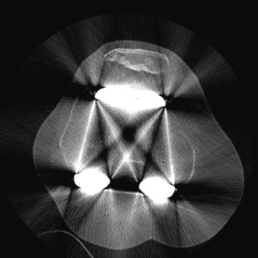
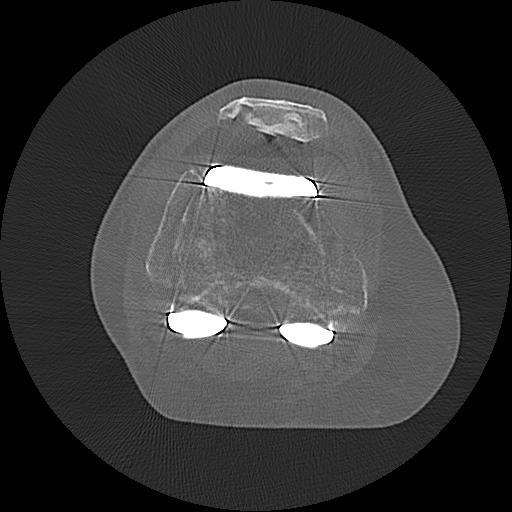
5 Figure 2: SEMAR applied to reveal anatomy normally obscured by beam hardening
6 Clinical Indications for MAR Diagnosis of Post Operative Complications - Prosthetic Loosening - Polyethylene Wearing - Periprosthetic Fractures - Post Operative soft tissue masses/lesions - Aneurysm Clips, Lumbar fusions, Billiary clips, anywhere metal will impede surrounding structures and accurate diagnosis.
7 SEMAR (Single Energy Metal Artefact Reduction) Toshiba based MAR technique Uses Data Segmentation, Forward Projection, and Interpolation Can uncover bone and soft tissue structures normally obscured by beam hardening effects I know what it is but how does it work!?

8 Figure 3: SEMAR algorithm. BPJ; Back Projection. FPJ; Forward Projection.

9 A sinogram of a lemon with a metal rod inserted. The horizontal direction of the sinogram corresponds to the ray data in each projection. The vertical axis represents the projections acquired at different angles through the lemon. The bright region corresponds to the high attenuation of the metal rod and shows as an obvious sine wave. Figure 4: Sinogram of lemon with metal rod and FBJ image


10 Figure 5: A complete 360 degree sonogram of an abdominal cross section is compared with the reconstructed image using FBP.
11 SEMAR Raw data is forward projected to create a sinogram - Same data is reconstructed using filtered back projection (FPB) Metal is segmented in this image and then forward projected creating a metal only sinogram Metal only sinogram subtracted from original sonogram Linear interpolation used to calculate missing data


12 Original Projection Data Metal Only Sinogram Extraction of Metal from Sinogram and Interpolation Original Image Segmentation of Metal
13 SEMAR Interpolated sinogram reconstructed using FBP Image segmented again to further exclude residual metal artefacts This data is again forward projected into a sinogram and linear interpolation used to fill in the gaps Image volume is reconstructed and original metal segmentation is reintroduced
14 AIDR BPJ after first pass interpolation Further elimination of metal objects and corrected sinogram BPJ of corrected sinogram Original Metal Segmentation Reintroduced Final Image
15 Technical Considerations Toshiba 160 Slice 40mm detector width with.5mm slices (ability to do.25mm) 10 volume blocks of 40mm making total scan coverage of 400mm (40cm) Most recent software and scanners have 16cm detector coverage and can be applied retrospectively Trade off scanning helically with increased dose 120kv and AIDR standard protocol - how can we change technical parameters?

16 Figure 6: SEMAR 6 block volume acquisition scout of bone with metal hardware

17 Figure 7: Axial.5mm slice through bone demonstrates beam hardening artefact and associated photon starvation. The acquisition protocol used 120kv, max of 250 ma (modulating), and AIDR standard.


18 Figure 8: Demonstration of how increasing kv decreases metal artefact in addition with SEMAR

19 80kv 100kv 120kv 135kv


20 Figure 9: Demonstration of how increasing AIDR from standard to strong decreases noise associated with metal artefact with SEMAR


21 135kv 250mA 135kv 266mA Figure 10: Demonstration of how lowering the standard deviation and increasing ma values can reduce metal artefact.
22 Case Study 1 Clinical History Periprosthetic fracture right knee post mechanical fall? Assess position and extension. (1st presentation) Right TKR periprosthetic fracture? Please perform fine slice CT with artefact reduction and 3D recons? stable vs unstable prosthesis. May require long stem TKR revision if unstable. (2nd presentation)
23 CONVENTIONAL AXIAL SEMAR AXIAL
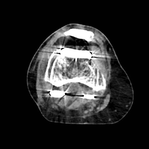
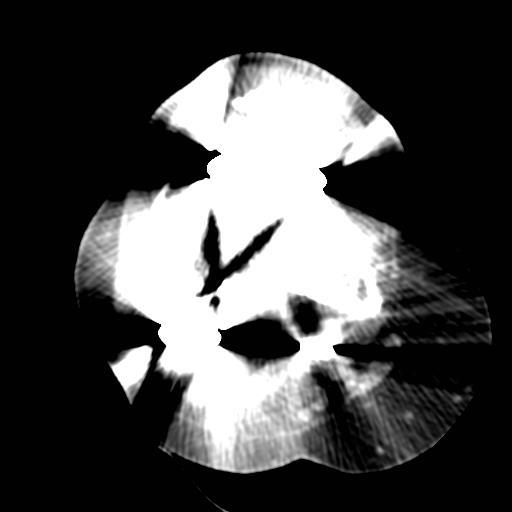
24 CONVENTIONAL AXIAL S E MAR A X I A L


25 CORONAL B O NE S E MAR CORONAL S O F T S E MAR
26 Conclusion Study of better diagnostic quality than CT performed a day prior Periprosthetic fracture of the lateral femoral condyle Vertical fracture line extending from supracondylar region to lateral margin of prosthesis Approximately 5mm of lateral and superior displacement Moderate sized lipohaemarthrosis noted
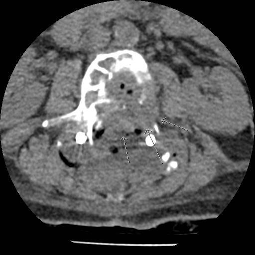
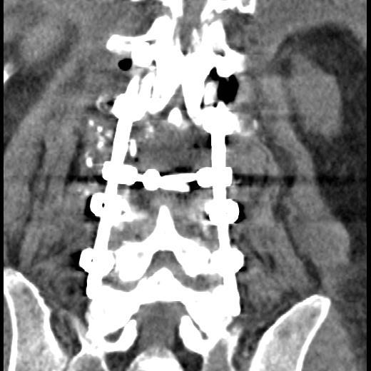
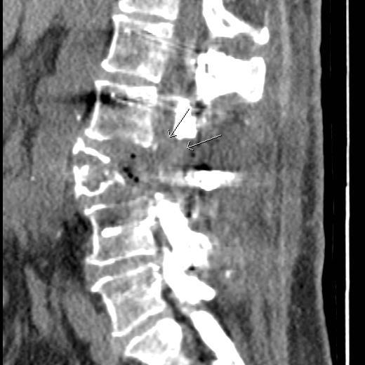
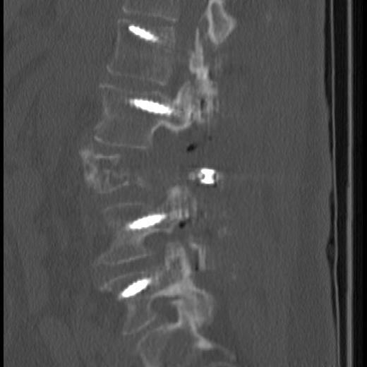
27 Case Study 2 Clinical History 66F L3 pathological fracture D3 post posterior instrumented fusion. For CT lumber spine on 21/4/15 for picture of bony morphology?residual tumour.
28
29
30 Conclusion Pedicle screws been placed at L1-L2 and L4-L5 with posterior rods L3 laminectomy has been performed with resection of most of the posterior aspect of the L3 vertebral body L3 body demonstrates extensive bone destruction extending into both pedicles. Soft tissue density material extending posteriorly in the spinal canal is identified at the L3 level Most likely represents residual soft tissue tumour or haematoma

31 Summary SEMAR plays a critical role in reducing metal artefact to diagnose post operative complications Should be used for a range of anatomy where metal may affect an accurate diagnosis Important to understand how relevant technical parameters can influence a SEMAR image Applying SEMAR can be vital to a patient s diagnosis and future management
32 1. Boas, F.E & Fleischmann, D. (2012). CT artifacts: Causes and reduction techniques. Imaging Med, 4(2), Bushberg, J.T., Seibert, J.A, Leidholdt jr, E.M & Boone, J.M (1994). The Essential Physics of Medical Imaging. Davis, CA: University of California. 3. Teixeira, P., Meyer, J. & Blum, A. (2014). Single energy metal artefact reduction algorithm for CT evaluation of periprosthetic soft tissues: Clinical applications. ViSIONS, 23, Toshiba Medical Systems (2014). CT Clinical Case Study Bilateral Hip Replacement Evaluation with SEMAR. Toshiba Leading Innovation. 5. Sofue, K., Yoshikawa, T., Negi, N., Ohno, Y., Sugihara, N., Murakami, T., Koyama, H., Nishio, M., Sugimura., K, Kobe, JP. & Ohtawara, JP. (2014). Abdominal CT with Single-Energy Metal Artifact Reduction (SEMAR): Initial Experiences, European Society of Radiology, 3-5.
X-Ray & CT Physics / Clinical CT
 Computed Tomography-Basic Principles and Good Practice X-Ray & CT Physics / Clinical CT INSTRUCTORS: Dane Franklin, MBA, RT (R) (CT) Office hours will be Tuesdays from 5pm to 6pm CLASSROOM: TIME: REQUIRED
Uozu city/jp, Minatoku, Tokyo/JP Bones, Extremities, CT, Surgery, Physics, Artifacts, Image verification /ecr2014/C-0462
 Metal Artifact Reduction Algorithm enablesreduce metal artifacts and improvement of diagnosis in the postoperative Pedicle screws implant for Spinal Fusion: A Phantom Study Poster No.: C-0462 Congress:
2
 1 2 3 4 5 6 7 8 9 10 11 12 13 Cine loop of tomosynthesis slice images through the chest. 14 Standard PA chest radiograph (left) and single slice from the tomosynthesis image dataset (right) of a patient
Intraoperative case studies. Portable full body 32-slice CT scanner
 Intraoperative case studies Portable full body 32-slice CT scanner Point-of-care CT imaging Your multi-departmental imaging solution Orthopedic surgery Arthroplasty Musculoskeletal disorders Hip replacement
Emerging Applications in Musculoskeletal CT Imaging
 Emerging pplications in Musculoskeletal CT Imaging y K Murali MD(RD), PDCC, Director of Interventional Radiology, G. Francis DMRD, DN (RD), Consultant Radiologist, and R. Madan, MS, MD, Consultant Radiologist,
Dual-Energy CT: The Technological Approaches
 Dual-Energy CT: The Technological Approaches Dushyant Sahani, M.D Director of CT Associate Professor of Radiology Massachusetts General Hospital Harvard Medical School Email-dsahani@partners.org Disclosure
B. CT protocols for the spine
 B. CT protocols for the spine Poster No.: A-003 Congress: ECR 2010 Type: Invited Speaker Topic: Neuro Authors: B. Tins; Oswestry/UK Keywords: CT, spine, diagnostic imaging protocol DOI: 10.1594/ecr2010/A-003
Metal Artifact Reduction by Dual Energy CT
 Metal Artifact Reduction by Dual Energy CT Poster No.: C-0108 Congress: ECR 2011 Type: Authors: Keywords: DOI: Scientific Paper T. Johnson, F. Bamberg, A. Dierks, H.-C. Becker, M. F. Reiser; Munich/DE
CT SCAN PROTOCOL. Shoulder
 CT SCAN PROTOCOL Shoulder Purpose and Summary CT images made with this protocol are used to provide the orthopedic surgeon with a detailed 3D anatomical reconstruction of the patient s scapula and proximal
Clinical Image Gallery Next Generation Volume 1
 Clinical Image Gallery Next Generation Volume 1 Dr. Russell Bull Royal Bournemouth Hospital, Bournemouth, United Kingdom After long experience with the first generation, a next generation Aquilion ONE
R/F. Clinical Experience Using the SONIALVISION safire II Utility of Tomosynthesis in Orthopedic Surgery
 R/F Clinical Experience Using the SONIALVISION safire II Utility of Tomosynthesis in Orthopedic Surgery Iwate Medical University Hospital, Central Department of Radiology Shouta Miura Mr. Shouta Miura
Functional Orthopedic Imaging Capturing Motion, Flow and Perfusion. Case Study Brochure Centre University Hospital Nancy.
 Capturing Motion, Flow and Perfusion dynamic volume CT Case Study Brochure Centre University Hospital Nancy http://www.toshibamedicalsystems.com Toshiba Medical Systems Corporation 2013. All rights reserved.
Orthopedic Hardware Imaging Part II: MRI v. Metal
 Orthopedic Hardware Imaging Trent Roth, MD And Lauren Ladd, MD Indiana University School of Medicine IU Health Physicians-Radiology Recap: Imaging Techniques Radiography Standard for initial and surveillance
CT NUMBER ACCURACY ANALYSIS FOR RADIOTHERAPY TREATMENT PLANNING IMAGING
 CT NUMBER ACCURACY ANALYSIS FOR RADIOTHERAPY TREATMENT PLANNING IMAGING Julian Liu a, Keisha Robinson a, DhanaJayan Kothandan a and Joshua Luis b (a) Cancer Centre London (b) University College London
Translating Protocols Across Patient Size: Babies to Bariatric
 Translating Protocols Across Patient Size: Babies to Bariatric Cynthia H. McCollough, PhD, FACR, FAAPM Professor of Radiologic Physics Director, CT Clinical Innovation Center Department of Radiology Mayo
SpineFAQs. Lumbar Spondylolisthesis
 SpineFAQs Lumbar Spondylolisthesis Normally, the bones of the spine (the vertebrae) stand neatly stacked on top of one another. The ligaments and joints support the spine. Spondylolisthesis alters the
Clinical details: Details of scan: CONE BEAM CT REPORT: Name: H. B. Gender: Reason for referral: Referred by:
 Name: H. B. Gender: Male DOB: 11/12/1950 Age: 64 Date taken: 16/11/2015 Date reported: 19/11/2015 Clinical details: Reason for referral: Referred by: Investigate symptoms related to left TMJ. Reconstructed
Doses from Cervical Spine Computed Tomography (CT) examinations in the UK. John Holroyd and Sue Edyvean
 examinations in the UK. John Holroyd and Sue Edyvean") Doses from Cervical Spine Computed Tomography (CT) examinations in the UK John Holroyd and Sue Edyvean Why a new dose survey? Number of enquires received concerning the current NDRL Concern that could
Doses from Cervical Spine Computed Tomography (CT) examinations in the UK John Holroyd and Sue Edyvean Why a new dose survey? Number of enquires received concerning the current NDRL Concern that could
Artifact reduction strategies for prosthetic heart valve CT imaging
 Int J Cardiovasc Imaging (2012) 28:2099 2108 DOI 10.1007/s10554-012-0041-5 ORIGINAL PAPER Artifact reduction strategies for prosthetic heart valve CT imaging Jesse Habets Petr Symersky Tim Leiner Bas A.
The Bone Densitometry Examination
 The Bone Densitometry Examination The purpose of The American Registry of Radiologic Technologist (ARRT ) Bone Densitometry Examination is to assess the knowledge and cognitive skills underlying the intelligent
Departement of Neurosurgery A.O.R.N A. Cardarelli- Naples.
 Percutaneous posterior pedicle screw fixation in the treatment of thoracic, lumbar and thoraco-lumbar junction (T12-L1) traumatic and pathological spine fractures. Report of 45 cases. G. Vitale, A. Punzo,
Typical PET Image. Elevated uptake of FDG (related to metabolism) Lung cancer example: But where exactly is it located?
 Lung cancer example: But where exactly is it located?") Typical PET Image Elevated uptake of FDG (related to metabolism) Lung cancer example: But where exactly is it located? PET/CT Oncology Imaging Anatometabolic fusion images are useful in the management
Typical PET Image Elevated uptake of FDG (related to metabolism) Lung cancer example: But where exactly is it located? PET/CT Oncology Imaging Anatometabolic fusion images are useful in the management
Thoracic or lumbar spinal surgery in patients with Parkinson s disease -A two-center experience of 32 cases-
 Thoracic or lumbar spinal surgery in patients with Parkinson s disease -A two-center experience of 32 cases- Department of Orthopedic Surgery, Graduate School of Medicine, Kyoto university Hiroaki Kimura,
HI-Res Extremity Sensation 16
 Page 1 Routine Extremity - (2/14/2013) CTDI: ~20 mgy per acquisition Used for evaluation of: Humerus Forearm Femur Knee Tib/Fib Billing: 1. CT Upper/Lower Extremity of concern without contrast, with contrast,
Fundamentals, Techniques, Pitfalls, and Limitations of MDCT Interpretation and Measurement
 Fundamentals, Techniques, Pitfalls, and Limitations of MDCT Interpretation and Measurement 3 rd Annual Imaging & Physiology Summit November 20-21, 21, 2009 Seoul, Korea Wm. Guy Weigold, MD, FACC Cardiovascular
3D titanium interbody fusion cages sharx. White Paper
 3D titanium interbody fusion cages sharx (SLM selective laser melted) Goal of the study: Does the sharx intervertebral cage due to innovative material, new design, and lordotic shape solve some problems
Bone Densitometry. Total 30 Maximum CE 14. DXA Scanning (10) 7
 7") STRUCTURED SELF ASSESSMENT CONTENT SPECIFICATIONS SSA LAUNCH DATE: JANUARY 1, 2018 Bone Densitometry The purpose of continuing qualifications requirements (CQR) is to assist registered technologists in
STRUCTURED SELF ASSESSMENT CONTENT SPECIFICATIONS SSA LAUNCH DATE: JANUARY 1, 2018 Bone Densitometry The purpose of continuing qualifications requirements (CQR) is to assist registered technologists in
SCALING Radiographic Technique
 SCALING Radiographic Technique SCALING FOR DIGITAL X-RAYS As images become filmless. Current planning practices with acetate sheets become difficult or obsolete. When images are printed to film sometimes
EXAMINATION CONTENT SPECIFICATIONS ARRT BOARD APPROVED: JANUARY 2017 IMPLEMENTATION DATE: JULY 1, 2017
 EXAMINATION CONTENT SPECIFICATIONS Bone Densitometry The purpose of the bone densitometry examination is to assess the knowledge and cognitive skills underlying the intelligent performance of the tasks
STRUCTURED EDUCATION REQUIREMENTS IMPLEMENTATION DATE: JULY 1, 2017
 STRUCTURED EDUCATION REQUIREMENTS Bone Densitometry The purpose of structured education is to provide the opportunity for individuals to develop mastery of discipline-specific knowledge that, when coupled
Quantitative and Qualitative Assessment of Thorax Cone Beam CT Image Quality across Multiple Imaging Systems
 Quantitative and Qualitative Assessment of Thorax Cone Beam CT Image Quality across Multiple Imaging Systems Matthew Williams Pre-registration Clinical Scientist Velindre NHS Trust, Cardiff Computed Tomography
Ask EuroSafe Imaging. Tips & Tricks. CT Working Group. Optimization of scan length to reduce CT radiation dose
 Ask EuroSafe Imaging Tips & Tricks CT Working Group Optimization of scan length to reduce CT radiation dose Alban Gervaise (Centre Hospitalier Universitaire Nancy, FR) Mika Kortesniemi (HUS Medical Imaging
Pedicle Subtraction Osteotomy. Case JB. Antonio Castellvi 5/19/2017
 Pedicle Subtraction Osteotomy John M. Small MD Florida Orthopedic Institute University South Florida Department Orthopedic Surgery Castellvi Spine May 11, 2017 Case JB 66 y/o male 74 235 lbs Retired police
Lauren M. Burke, Warren D. Yu, Anthony Ho, Timothy Wagner, Joseph R. O Brien. Department of Orthopaedic Surgery George Washington University
 Lauren M. Burke, Warren D. Yu, Anthony Ho, Timothy Wagner, Joseph R. O Brien Department of Orthopaedic Surgery George Washington University O Brien: Consultant for Globus, Relivant, Stryker. Royalties:
R/F. Can T-smart Tomosynthesis Improve Diagnostic Accuracy on THA Component Stability? 1. Abstract
 R/F Can T-smart Tomosynthesis Improve Diagnostic Accuracy on THA Component Stability? Professor and Chair Dept. of Adult Reconstructive Surgery Beijing Jishuitan Hospital, the 4th Clinical College of PKU
Practical CT and MRI Anthony J. Fischetti, DVM, MS, DACVR Department Head of Diagnostic Imaging The Animal Medical Center, New York OBJECTIVE:
 Practical CT and MRI Anthony J. Fischetti, DVM, MS, DACVR Department Head of Diagnostic Imaging The Animal Medical Center, New York OBJECTIVE: This lecture describes the most common indications for referred
PROPHECY. Preoperative Navigation Guides ANKLE CT SCAN PROTOCOL
 PROPHECY Preoperative Navigation Guides ANKLE CT SCAN PROTOCOL 90 FIGURE 1 Examples FIGURE 1 Examples of neutral ankle positioning. PROPHECY Ankle CT Scan Protocol PROPHECY INBONE and PROPHECY INFINITY
Quantitative Analysis of Vascular Canals in Vertebral Endplate
 Quantitative Analysis of Vascular Canals in Vertebral Endplate Kristine Tan 1, Won C. Bae, PhD 1, Tomonori Yamaguchi, MS 1,2, Kelli Xu, BS 1, Iris Shieh, BS 1, Jade He, BS 1, Robert L. Sah, MD, ScD 1,
Module: #15 Lumbar Spine Fusion. Author(s): Jenni Buckley, PhD. Date Created: March 27 th, Last Updated:
: Jenni Buckley, PhD. Date Created: March 27 th, Last Updated:") Module: #15 Lumbar Spine Fusion Author(s): Jenni Buckley, PhD Date Created: March 27 th, 2011 Last Updated: Summary: Students will perform a single level lumbar spine fusion to treat lumbar spinal stenosis.
Module: #15 Lumbar Spine Fusion Author(s): Jenni Buckley, PhD Date Created: March 27 th, 2011 Last Updated: Summary: Students will perform a single level lumbar spine fusion to treat lumbar spinal stenosis.
Cardiac Computed Tomography
 Cardiac Computed Tomography Authored and approved by Koen Nieman Stephan Achenbach Francesca Pugliese Bernard Cosyns Patrizio Lancellotti Anastasia Kitsiou Contents CARDIAC COMPUTED TOMOGRAPHY Page 1.
CT Imaging of skeleton in small animals. Massimo Marenzana
 CT Imaging of skeleton in small animals Massimo Marenzana Introduction Osteoporosis is a disease in which bones become fragile and more likely to break. It can be defined as a systemic skeletal disease
To Shield or Not to Shield? Lincoln L. Berland, M.D.
 To Shield or Not to Shield? Lincoln L. Berland, M.D. Disclosures Consultant to: Nuance, Inc. Page 2 Breast Radiation on CT Use of chest CT has increased in women vulnerable to cancer induction by radiation.
Patient Information MIS LLIF. Lateral Lumbar Interbody Fusion Using Minimally Invasive Surgical Techniques
 Patient Information MIS LLIF Lateral Lumbar Interbody Fusion Using Minimally Invasive Surgical Techniques Table of Contents Anatomy of Spine...2 General Conditions of the Spine....4 What is Spondylolisthesis....5
Cardiac CT - Coronary Calcium Basics Workshop II (Basic)
") Cardiac CT - Coronary Calcium Basics Workshop II (Basic) J. Jeffrey Carr, MD, MSCE Dept. of Radiology & Public Health Sciences Wake Forest University School of Medicine Winston-Salem, NC USA No significant
Cardiac CT - Coronary Calcium Basics Workshop II (Basic) J. Jeffrey Carr, MD, MSCE Dept. of Radiology & Public Health Sciences Wake Forest University School of Medicine Winston-Salem, NC USA No significant
STRUCTURED EDUCATION REQUIREMENTS EFFECTIVE: JANUARY 1, 2016
 Computed Tomography The purpose of structured education is to provide the opportunity for individuals to develop mastery of discipline-specific knowledge that, when coupled with selected clinical experiences,
University of Groningen. Thoracolumbar spinal fractures Leferink, Vincentius Johannes Maria
 University of Groningen Thoracolumbar spinal fractures Leferink, Vincentius Johannes Maria IMPORTANT NOTE: You are advised to consult the publisher's version (publisher's PDF) if you wish to cite from
THE TUFFEST STUFF CT REGISTRY REVIEW Live Lecture Seminar SATURDAY CURRICULUM
 1. The CT Imaging Chain-10 major components & their functions a. The x-ray tube b. Generator c. Filter d. Pre-patient collimator e. Pre-detector collimator f. Detector system g. Analog to digital converter
How to interpret computed tomography of the lumbar spine
 REVIEW Ann R Coll Surg Engl 2014; 96: 502 507 doi 10.1308/003588414X13946184902361 How to interpret computed tomography of the lumbar spine Z Ahmad 1, R Mobasheri 2,TDas 3, S Vaidya 4, S Mallik 5, M El-Hussainy
SpineFAQs. Neck Pain Diagnosis and Treatment
 SpineFAQs Neck Pain Diagnosis and Treatment Neck pain is a common reason people visit their doctor. Neck pain typically doesn't start from a single injury. Instead, the problem usually develops over time
A Patient s Guide to Lumbar Spondylolysis. William T. Grant, MD
 A Patient s Guide to Lumbar Spondylolysis Dr. Grant is a talented orthopedic surgeon with more than 30 years of experience helping people return to their quality of life. He and GM Pugh, PA-C pride themselves
SUBAXIAL CERVICAL SPINE TRAUMA- DIAGNOSIS AND MANAGEMENT
 SUBAXIAL CERVICAL SPINE TRAUMA- DIAGNOSIS AND MANAGEMENT 1 Anatomy 3 columns- Anterior, middle and Posterior Anterior- ALL, Anterior 2/3 rd body & disc. Middle- Posterior 1/3 rd of body & disc, PLL Posterior-
General Imaging. Imaging modalities. Incremental CT. Multislice CT Multislice CT [ MDCT ]
![General Imaging. Imaging modalities. Incremental CT. Multislice CT Multislice CT [ MDCT ]](/thumbs/76/74079340.jpg "General Imaging. Imaging modalities. Incremental CT. Multislice CT Multislice CT [ MDCT ]") General Imaging Imaging modalities Conventional X-rays Ultrasonography [ US ] Computed tomography [ CT ] Radionuclide imaging Magnetic resonance imaging [ MRI ] Angiography conventional, CT,MRI Interventional
General Imaging Imaging modalities Conventional X-rays Ultrasonography [ US ] Computed tomography [ CT ] Radionuclide imaging Magnetic resonance imaging [ MRI ] Angiography conventional, CT,MRI Interventional
PACS: ERGONOMIC CONSIDERATIONS 1
 RADIOLOGY RESEARCH Radiographic Tomosynthesis: Acquisition Parameters Michael J. Flynn, PhD Henry Ford Health System Detroit, MI Learning Objectives Learn.. 1. Appreciate the importance of scan direction,
Ligaments of the vertebral column:
 In the last lecture we started talking about the joints in the vertebral column, and we said that there are two types of joints between adjacent vertebrae: 1. Between the bodies of the vertebrae; which
Low Dose Era in Cardiac CT
 Low Dose Era in Cardiac CT DIANA E. LITMANOVICH, MD Department of Radiology Beth Israel Deaconess Medical Center Harvard Medical School Disclosures Neither I nor my immediate family members have a financial
A minimally invasive surgical approach reduces cranial adjacent segment degeneration in patients undergoing posterior lumbar interbody fusion
 A minimally invasive surgical approach reduces cranial adjacent segment degeneration in patients undergoing posterior lumbar interbody fusion T. Tsutsumimoto, M. Yui, S. Ikegami, M. Uehara, H. Kosaku,
Patient Information. Spinal Fusion Using the ST360 or Silhouette Pedicle Screw System
 Patient Information Spinal Fusion Using the ST360 or Silhouette Pedicle Screw System Spinal Fusion Using the ST360 or Silhouette Pedicle Screw System Your doctor has recommended spinal fusion surgery using
ANATOMIC TOTAL SHOULDER REPLACEMENT:
 The Shoulder Replacement A total shoulder arthroplasty (TSA) is a surgery to replace the damaged parts of the ball and socket shoulder joint with an artificial prosthesis. The damage to the shoulder can
CT Scanning Protocol For V2R Guided Surgery Solutions
 CT Scanning Protocol For V2R Guided Surgery Solutions 2 V2R CT Scanning Protocol \\ Contents Contents General requirements... 3 V2R Dual Scan Protocol... 5 V2R Single Scan Protocol... 8 Overview... 10
Clinical Application of Computed Radiography in Orthopedic Surgery
 Clinical Application of Computed Radiography in Orthopedic Surgery Satoru Fujita, Masamichi Tanaka, Sigeaki Hirota, and Takeshi Fuji Since 1988, Fuji Computed Radiography (FCR) system (Fuji Medical Systems,
Bone Densitometry Radiation dose: what you need to know
 Bone Densitometry Radiation dose: what you need to know John Damilakis, PhD Associate Professor and Chairman University of Crete, Iraklion, Crete, GREECE Estimation of bone status using X-rays Assessment
Advantages of MISS. Disclosures. Thoracolumbar Trauma: Minimally Invasive Techniques. Minimal Invasive Spine Surgery 11/8/2013.
 3 rd Annual UCSF Techniques in Complex Spine Surgery Program Thoracolumbar Trauma: Minimally Invasive Techniques Research Support: Stryker Disclosures Murat Pekmezci, MD Assistant Clinical Professor UCSF/SFGH
Study on Mechanical Characteristics of Lumbar Spine for Snatch Action in Weight Lifting Based on Finite Element Method
 ISSN 1750-9823 (print) International Journal of Sports Science and Engineering Vol. 04 (2010) No. 01, pp. 048-052 Study on Mechanical Characteristics of Lumbar Spine for Snatch Action in Weight Lifting
Patient Information MIS LLIF. Lateral Lumbar Interbody Fusion Using Minimally Invasive Surgical Techniques
 Patient Information MIS LLIF Lateral Lumbar Interbody Fusion Using Minimally Invasive Surgical Techniques Table of Contents Anatomy of Spine....2 General Conditions of the Spine....4 What is Spondylolisthesis....5
MEDICAL IMAGING OF THE VERTEBRAE
 MEDICAL IMAGING OF THE VERTEBRAE Vertebrae are your friends Matthew Harper MS-IV LECTURE OBJECTIVES INTRODUCE THE MOST COMMON MODALITIES OF MEDICAL IMAGING AND BASIC TECHNIQUES FOR READING THESE IMAGES
Fractures of the Thoracic and Lumbar Spine
 A spinal fracture is a serious injury. Nader M. Hebela, MD Fellow of the American Academy of Orthopaedic Surgeons http://orthodoc.aaos.org/hebela Cleveland Clinic Abu Dhabi Cleveland Clinic Abu Dhabi Neurological
Neuro CT What s a Good Head Exam?
 Neuro CT What s a Good Head Exam? Rajiv Gupta, PhD, MD Neuroradiology Massachusetts General Hospital Harvard Medical School Outline What we need to see? Routine Head CT protocols Dose optimization strategies
Introduction. Modalities used in imaging guidance. Flat panel detector. X-ray Imaging Dose to Patients in the Era of Image-Guided Radiation Therapy
 X-ray Imaging Dose to Patients in the Era of Image-Guided Radiation Therapy George Ding, Ron Price, Charles Coffey Vanderbilt-Ingram Cancer Center Vanderbilt University Medical Center, Nashville, TN Introduction
Chest X-ray Interpretation
 Chest X-ray Interpretation Introduction Routinely obtained Pulmonary specialist consultation Inherent physical exam limitations Chest x-ray limitations Physical exam and chest x-ray provide compliment
FOR CMS (MEDICARE) MEMBERS ONLY NATIONAL COVERAGE DETERMINATION (NCD) FOR MAGNETIC RESONANCE IMAGING:
 MEMBERS ONLY NATIONAL COVERAGE DETERMINATION (NCD) FOR MAGNETIC RESONANCE IMAGING:") National Imaging Associates, Inc. Clinical guidelines BONE MARROW MRI Original Date: July 2008 Page 1 of 5 CPT Codes: 77084 Last Review Date: September 2014 NCD 220.2 MRI Last Effective Date: July 2011
National Imaging Associates, Inc. Clinical guidelines BONE MARROW MRI Original Date: July 2008 Page 1 of 5 CPT Codes: 77084 Last Review Date: September 2014 NCD 220.2 MRI Last Effective Date: July 2011
Dual Energy CT of the Liver
 34th Annual Course October 2011 Washington, DC Dual Energy CT of the Liver Vassilios Raptopoulos, MD Beth Israel Deaconess Medical Center Harvard Medical School Dual Energy CT (DECT) Different materials
CLINICAL AND OPERATIVE APPROACH FOR TOTAL KNEE REPLACEMENT DR.VINMAIE ORTHOPAEDICS PG 2 ND YEAR
 CLINICAL AND OPERATIVE APPROACH FOR TOTAL KNEE REPLACEMENT DR.VINMAIE ORTHOPAEDICS PG 2 ND YEAR Evolution of TKR In 1860, Verneuil proposed interposition arthroplasty, involving the insertion of soft tissue
Personalized Solutions. MRI Protocol for PSI and Signature Guides
 Personalized Solutions MRI Protocol for PSI and Signature Guides 2 Personalized Solutions MRI Protocol for PSI and Signature Guides Purpose and Summary This protocol is applicable for the Zimmer Biomet
This presentation is the intellectual property of the author. Contact them for permission to reprint and/or distribute.
 MRI of the Knee Jennifer Swart, M.D. Musculoskeletal Radiology South Texas Radiology Group Outline Coils, Patient Positioning Acquisition Parameters, Planes and Pulse Sequences Knee Arthrography Normal
Asymmetric T5 Pedicle Subtraction Osteotomy (PSO) for complex posttraumatic deformity
 for complex posttraumatic deformity") Eur Spine J (2013) 22:2130 2135 DOI 10.1007/s00586-013-2942-y OPEN OPERATING THEATRE (OOT) Asymmetric T5 Pedicle Subtraction Osteotomy (PSO) for complex posttraumatic deformity Ibrahim Obeid Fethi Laouissat
Eur Spine J (2013) 22:2130 2135 DOI 10.1007/s00586-013-2942-y OPEN OPERATING THEATRE (OOT) Asymmetric T5 Pedicle Subtraction Osteotomy (PSO) for complex posttraumatic deformity Ibrahim Obeid Fethi Laouissat
Disclosures: T. Yoshii: None. T. Yamada: None. T. Taniyama: None. S. Sotome: None. T. Kato: None. S. Kawabata: None. A. Okawa: None.
 Dynamic Changes in Spinal Cord Compression by Cervical Ossification of the Posterior Longitudinal Ligament Evaluated by Kinematic Computed Tomography Myelogram Toshitaka Yoshii, Tsuyoshi Yamada, Takashi
Int J Clin Exp Med 2018;11(2): /ISSN: /IJCEM Yi Yang, Hao Liu, Yueming Song, Tao Li
: /ISSN: /IJCEM Yi Yang, Hao Liu, Yueming Song, Tao Li") Int J Clin Exp Med 2018;11(2):1278-1284 www.ijcem.com /ISSN:1940-5901/IJCEM0063093 Case Report Dislocation and screws pull-out after application of an Isobar TTL dynamic stabilisation system at L2/3 in
Int J Clin Exp Med 2018;11(2):1278-1284 www.ijcem.com /ISSN:1940-5901/IJCEM0063093 Case Report Dislocation and screws pull-out after application of an Isobar TTL dynamic stabilisation system at L2/3 in
GE Healthcare. Rad Rx. White Paper
 GE Healthcare Rad Rx White Paper Introduction This publication is part of a series of white papers aimed at communicating the importance of each component in the image chain of a PET/CT study. From data
This presentation is the intellectual property of the author. Contact them at for permission to reprint and/or distribute.
 MRI of the Knee Jennifer Swart, M.D. Musculoskeletal Radiology South Texas Radiology Group Financial Disclosure Dr. Jennifer Swart has no relevant financial relationships with commercial interests to disclose.
Reproducibility of Uptake Estimates in FDG PET: a Monte Carlo study
 Reproducibility of Uptake Estimates in FDG PET: a Monte Carlo study Juliette Feuardent, Marine Soret, Irène Buvat 1 Abstract Tumor glucose metabolism measurements from Fluoro-deoxyglucose (FDG) Positron
Introduction of FIREFLY Technology
 Introduction of FIREFLY Technology FIREFLY Technology is a unique, patent-pending, pre-surgical planning and intra - operative navigation technology that is focused on spinal applications and is derived
HEAD AND NECK IMAGING. James Chen (MS IV)
") HEAD AND NECK IMAGING James Chen (MS IV) Anatomy Course Johns Hopkins School of Medicine Sept. 27, 2011 OBJECTIVES Introduce cross sectional imaging of head and neck Computed tomography (CT) Review head
HEAD AND NECK IMAGING James Chen (MS IV) Anatomy Course Johns Hopkins School of Medicine Sept. 27, 2011 OBJECTIVES Introduce cross sectional imaging of head and neck Computed tomography (CT) Review head
Hi RES Extremity - (04/18/2011) CTDI: ~13 mgy per acquisition Used for evaluation of: Ankle Elbow Hand Wrist Foot /Calcaneous Toes Fingers
 CTDI: ~13 mgy per acquisition Used for evaluation of: Ankle Elbow Hand Wrist Foot /Calcaneous Toes Fingers") P a g e 1 Hi RES Extremity - (04/18/2011) CTDI: ~13 mgy per acquisition Used for evaluation of: Ankle Elbow Hand Wrist Foot /Calcaneous Toes Fingers Billing: 1. CT Upper/Lower Extremity of concern without
P a g e 1 Hi RES Extremity - (04/18/2011) CTDI: ~13 mgy per acquisition Used for evaluation of: Ankle Elbow Hand Wrist Foot /Calcaneous Toes Fingers Billing: 1. CT Upper/Lower Extremity of concern without
Image Diagnostic Technology Ltd IDT Ireland, 15 Market Street, Kinsale, Co. Cork, Ireland P17 XN65 Tel: Mob: IRL:
 Image Diagnostic Technology Ltd IDT Ireland, 15 Market Street, Kinsale, Co. Cork, Ireland P17 XN65 Tel: +44 20 8819 9158 Mob: +44 7767 366596 IRL: +353 21 470 9501 Web: www.simplantscans.com Email: info@ctscan.co.uk
R/F. Use of Tomosynthesis at the Aizawa Hospital. 1. Introduction. 2. Use of Tomosynthesis at the Aizawa Hospital
 R/F Use of Tomosynthesis at the Aizawa Hospital Diagnostic Imaging Center, Aizawa Hospital Masahiro Iwama, Kei Takehara, Kouichi Anraku Mr. Masahiro Iwama 1. Introduction The Aizawa Hospital is located
A more accurate method to estimate patient dose during body CT examinations with tube current modulation
 A more accurate method to estimate patient dose during body CT examinations with tube current modulation Poster No.: C-0738 Congress: ECR 2014 Type: Scientific Exhibit Authors: A. Kawaguchi 1, Y. Matsunaga
.org. Tibia (Shinbone) Shaft Fractures. Anatomy. Types of Tibial Shaft Fractures
 Shaft Fractures. Anatomy. Types of Tibial Shaft Fractures") Tibia (Shinbone) Shaft Fractures Page ( 1 ) The tibia, or shinbone, is the most common fractured long bone in your body. The long bones include the femur, humerus, tibia, and fibula. A tibial shaft fracture
Tibia (Shinbone) Shaft Fractures Page ( 1 ) The tibia, or shinbone, is the most common fractured long bone in your body. The long bones include the femur, humerus, tibia, and fibula. A tibial shaft fracture
Dingjun Hao, Baorong He, Liang Yan. Hong Hui Hospital, Xi an Jiaotong University College. of Medicine, Xi an, Shaanxi , China
 Xi an Hong Hui Hospital Xi an, Shaanxi, China The difference of occurring superior adjacent segment pathology after lumbar posterolateral fusion by using two different pedicle screw insertion techniques
Am I eligible for the TOPS study? Possibly, if you suffer from one or more of the following conditions:
 Am I eligible for the TOPS study? Possibly, if you suffer from one or more of the following conditions: Radiating leg pain Greater leg / buttock pain than back pain Severe pain sets in when walking as
The diagnostic value of Computed Tomography in evaluation of maxillofacial Trauma
 The diagnostic value of Computed Tomography in evaluation of maxillofacial Trauma Qais H. Muassa FICMS College of Dentistry, Babylon University Ibrahim S. Gataa, BDS, FICMS College of Dentistry, Sulaimania
Bringing the power of imaging to your patient. Portable full body 32-slice CT scanner
 Bringing the power of imaging to your patient Portable full body 32-slice CT scanner Point-of-care CT imaging Your multi-departmental imaging solution Orthopedic surgery Arthroplasty Musculoskeletal disorders
Abdomen and Pelvis CT (1) By the end of the lecture students should be able to:
 By the end of the lecture students should be able to:") RAD 451 Abdomen and Pelvis CT (1) By the end of the lecture students should be able to: State the common indications for Abdomen and pelvis CT exams Identify possible contra indications for Abdomen and
RAD 451 Abdomen and Pelvis CT (1) By the end of the lecture students should be able to: State the common indications for Abdomen and pelvis CT exams Identify possible contra indications for Abdomen and
6/23/2009. Inversion Recovery (IR) Techniques and Applications. Variations of IR Technique. STIR, FLAIR, TI and TI Null. Applications of IR
 Techniques and Applications. Variations of IR Technique. STIR, FLAIR, TI and TI Null. Applications of IR") The Anatomy of Basic R Pulse Sequences Inversion Recovery () Techniques and Applications Chen Lin, PhD Indiana University School of edicine & Clarian Health Partners agnetization Preparation Section Chemical
The Anatomy of Basic R Pulse Sequences Inversion Recovery () Techniques and Applications Chen Lin, PhD Indiana University School of edicine & Clarian Health Partners agnetization Preparation Section Chemical
Low Dose Excellent Image Quality Rapid Reconstruction
 Low Dose Excellent Image Quality Rapid Reconstruction Efficient 3 in 1 Dental X-ray System CBCT > Precise 3-D Anatomical structures - Accurate diagnosis for doctors - Safe implant for patients > Significant
UK Biobank. Imaging modality Cardiovascular Magnetic Resonance (CMR) Version th Oct 2015
 Version th Oct 2015") Imaging modality Cardiovascular Magnetic Resonance (CMR) Version 1.0 http://www.ukbiobank.ac.uk/ 30 th Oct 2015 This document details the procedure for the CMR scan performed at an Imaging assessment centre
Imaging modality Cardiovascular Magnetic Resonance (CMR) Version 1.0 http://www.ukbiobank.ac.uk/ 30 th Oct 2015 This document details the procedure for the CMR scan performed at an Imaging assessment centre
MR imaging the post operative spine - What to expect!
 MR imaging the post operative spine - What to expect! Poster No.: C-2334 Congress: ECR 2012 Type: Educational Exhibit Authors: A. Jain, M. Paravasthu, M. Bhojak, K. Das ; Warrington/UK, 1 1 1 2 1 2 Liverpool/UK
FOR CMS (MEDICARE) MEMBERS ONLY NATIONAL COVERAGE DETERMINATION (NCD) FOR MAGNETIC RESONANCE IMAGING:
 MEMBERS ONLY NATIONAL COVERAGE DETERMINATION (NCD) FOR MAGNETIC RESONANCE IMAGING:") National Imaging Associates, Inc. Clinical guidelines TEMPOROMANDIBULAR JOINT (TMJ) MRI Original Date: May 23, 2003 Page 1 of 5 CPT Code: 70336 Last Review Date: May 2016 NCD 220.2 MRI Last Effective Date:
National Imaging Associates, Inc. Clinical guidelines TEMPOROMANDIBULAR JOINT (TMJ) MRI Original Date: May 23, 2003 Page 1 of 5 CPT Code: 70336 Last Review Date: May 2016 NCD 220.2 MRI Last Effective Date:
Managing Radiation Risk in Pediatric CT Imaging
 Managing Radiation Risk in Pediatric CT Imaging Mahadevappa Mahesh, MS, PhD, FAAPM, FACR, FACMP, FSCCT. Professor of Radiology and Cardiology Johns Hopkins University School of Medicine Chief Physicist
Shoulder Position: Supine arm in the neutral position. Collateral arm above head Indication: fracture humerus, fracture scapula
 Shoulder Position: Supine arm in the neutral position. Collateral arm above head Indication: fracture humerus, fracture scapula No instrumentation With metal or cast KV/ Effective mas/rotation time 140/300/1.0